


Abstract
Chronic exposure to ultraviolet A (UVA) and ultraviolet B (UVB) radiation accelerates skin photoaging through oxidative stress and inflammation, emphasizing the importance of antioxidant protection. Astaxanthin (AST), a natural antioxidant, self-assembles into H- and J-aggregates with unique molecular arrangements and optical properties, providing distinct molecular architectures for functional nanomaterial design. However, its high sensitivity to preparation conditions makes the separate preparation and stabilization of H- and J-aggregates astaxanthin difficult. Here, H- and J-aggregates astaxanthin were separately stabilized in bovine serum albumin/chitosan nanoparticles (H-ABC-NPs and J-ABC-NPs) to evaluate their protective effects against UV-induced skin photoaging. H/J-ABC-NPs showed antioxidant activity and protected L929 fibroblasts against H2O2-induced oxidative stress by reducing intracellular ROS and MDA levels and increasing SOD and CAT activities. In a UVB/UVA-induced BALB/c mouse model of skin photoaging, topical H/J-ABC-NPs alleviated wrinkle formation, epidermal thickening and collagen fiber disruption. These effects were accompanied by enhanced SOD, CAT and GSH-Px and reduced MDA and inflammatory responses in the skin and serum. H/J-ABC-NPs reduced MMP-1 levels and collagen loss while increasing HYP and HA levels. Overall, these results indicate that H/J-ABC-NPs effectively alleviate UVB/UVA-induced skin photoaging, accompanied by reduced oxidative stress, reduced inflammation and the preservation of skin structure. The separate stabilization and biological evaluation of H- and J-aggregates astaxanthin provide a basis for further investigating their biological effects and topical applications.
1. Introduction
The skin is the most widely distributed and complex organ in the human body [1,2]. Skin photoaging is a major form of extrinsic skin aging primarily caused by chronic exposure to ultraviolet (UV) radiation [3,4]. Excessive UV exposure promotes the generation of reactive oxygen species (ROS), disrupting cutaneous redox homeostasis and inducing oxidative damage [5]. Persistent oxidative stress is closely associated with inflammatory responses and extracellular matrix (ECM) deterioration, including increased matrix metalloproteinase (MMP) activity and the degradation of dermal collagen [6,7]. These changes progressively impair skin structure and contribute to characteristic photoaging manifestations, such as epidermal thickening, loss of skin elasticity, and wrinkle formation [8]. Therefore, alleviating UV-induced oxidative stress and its associated inflammatory and ECM damage represents an important strategy for protecting the skin against photoaging.
Astaxanthin (AST), a naturally occurring carotenoid found in microalgae, yeast, and marine organisms, exhibits potent antioxidant activity and has attracted considerable attention for the prevention and treatment of UV-induced skin damage [9]. Among these sources, Haematococcus pluvialis can accumulate approximately 3–5% AST of its dry weight under stress conditions [10]. AST is currently obtained through both chemical synthesis and extraction from natural sources [11]. Its conjugated polyene structure enables the efficient quenching of ROS and free radicals, while previous studies have demonstrated that AST can alleviate UV-induced oxidative stress, inflammatory responses, and collagen degradation in experimental models of skin photoaging [12,13]. However, AST exhibits poor aqueous solubility and is sensitive to light, heat, oxygen, and pH changes, which limit its practical application [14,15]. Nanotechnology-based strategies to improve AST stability and bioavailability have attracted increasing research interest [16,17].
In our previous work, we prepared H- and J-aggregates astaxanthin/Bovine serum albumin/chitosan nanoparticles (H/J-ABC-NPs) through bottom-up molecular self-assembly. Here, we evaluated the protective effects of H/J-ABC-NPs against H2O2-induced oxidative injury in L929 cells and UV-induced skin photoaging in a BALB/c mouse model. This study aimed to evaluate the potential of H/J-ABC-NPs for topical protection against UV-induced skin photoaging and provide experimental evidence to support their further development for this application.
2. Materials and Methods
2.1. Materials
Astaxanthin (AST, 596.84 Da, purity 99%) and bovine serum albumin (BSA, 66.4 kDa, purity 97%) were purchased from Solarbio Life Sciences (Beijing, China). Chitosan (CS, average molecular weight of 80 kDa, degree of deacetylation 90.25%) was acquired from the Aoxin Biological Product Co., Ltd. (Taizhou, China). Vaseline, stearic acid, liquid paraffin, lanolin, glycerin monostearate, glycerol, triethanolamine, and ethylparaben were purchased from Chemicell Co., Ltd. (Berlin, Germany). RPMI-1640 culture medium was purchased from Thermo Fisher Scientific (Waltham, MA, USA). Fetal bovine serum (FBS) was purchased from Zhejiang Tianhang Biotechnology Co., Ltd. (Hangzhou, China). Paraformaldehyde fixative (4%) was purchased from Beyotime Biotechnology Co., Ltd. (Shanghai, China). A CCK-8 assay kit and penicillin-streptomycin solution (100×, P1400) were obtained from Solarbio Life Sciences (Beijing, China). Reactive oxygen species (ROS) assay kits, Calcein-AM/PI, and superoxide dismutase (SOD) assay kits were obtained from Beyotime Biotechnology Co., Ltd. (Shanghai, China). Malondialdehyde (MDA), catalase (CAT), hydroxyproline (Hyp), glutathione peroxidase (GSH-Px), hyaluronic acid (HA), type I collagen, type III collagen, IL-6, IL-1α, and IL-1β assay kits were obtained from Nanjing Jiancheng Bioengineering Institute (Nanjing, China). The matrix metalloproteinase-1 (MMP-1) ELISA kit was obtained from Sangon Biotech (Shanghai, China).
2.2. Preparation and Characterization of H/J-ABC-NPs
2.2.1. Preparation of H/J-ABC-NPs
H-/J-aggregates astaxanthin/bovine serum albumin/chitosan nanoparticles (H-ABC-NPs and J-ABC-NPs, H/J-ABC-NPs) were prepared by a bottom-up molecular self-assembly method, as detailed in our previous study [18]. The aggregation of AST was regulated by adjusting the ethanol volume fraction in the aqueous–organic solvent system. Previous studies demonstrated that H-aggregates and J-aggregates astaxanthin predominantly formed at ethanol volume fractions of 10–20% and 25–50%, respectively. Following the previously reported preparation process [19], an AST ethanolic solution was added to a CS aqueous solution, followed by the addition of BSA solution. Ethanol was subsequently removed by a rotary evaporator.
2.2.2. X-Ray Diffraction Analysis
Freeze-dried AST, CS, BSA, their physical mixture (AST/BSA/CS), and H/J-ABC-NPs were analyzed using Cu Kα radiation (λ = 1.54056 Å) at a tube current of 15 mA and voltage of 40 kV. Samples were scanned over a 2θ range of 5–90° at 10°/min.
2.2.3. Differential Scanning Calorimetry Analysis
The thermal properties of H/J-ABC-NPs were analyzed by differential scanning calorimetry (DSC, Q20, TA Instruments, New Castle, DE, USA) according to Liu et al. [20], with slight modifications. AST, BSA, CS, and lyophilized H/J-ABC-NPs (1.3 mg each) were placed in aluminum crucibles, with an empty crucible used as a reference. Measurements were conducted under nitrogen at a flow rate of 40.0 mL/min over 20–400 °C at a heating rate of 5 °C/min.
2.3. W/O Cream of H/J-ABC-NPs
H/J-ABC-NPs were lyophilized according to our previous study [19] and subsequently incorporated into a W/O cream following the method described by Yang J et al. [21], with minor adjustments. Different amounts of H/J-ABC-NPs lyophilized powder were dispersed in water to 5 g. The amount of lyophilized powder was calculated according to the AST content in 100 g of cream, giving final AST concentrations of 0, 1.875, 7.5, and 30 μg/g. The formulation contained H/J-ABC-NPs, white vaseline (1 g), stearic acid (12 g), liquid paraffin (6 g), lanolin (5 g), glycerin monostearate (5.0 g), glycerol (7.5 g), triethanolamine (0.4 g), and ethylparaben (0.3 g), with water added to a final weight of 100 g. The oil and aqueous phases were separately heated to 75 °C in a water bath. The aqueous phase was then gradually added to the oil phase to form the cream base. During cooling, the pre-prepared H/J-ABC-NPs dispersion was incorporated, and the mixture was continuously stirred for 10 min and allowed to cool completely.
2.4. Antioxidant Activity of H/J-ABC-NPs
The antioxidant activities of H/J-ABC-NPs were evaluated by DPPH and ABTS radical-scavenging assays, hydroxyl (·OH) and superoxide anion (O2·−) radical-scavenging assays, and the ferric reducing antioxidant power (FRAP) assay. The prepared H/J-ABC-NPs suspensions were used directly for the antioxidant assays. Free AST was dissolved in ethanol and diluted to match the theoretical AST concentration of H/J-ABC-NPs, calculated from the initial AST amount and final suspension volume.
2.4.1. DPPH Radical-Scavenging Activity Assay
The DPPH radical-scavenging rate of H/J-AST was determined by referring to the method of Wang et al. [22] with slight modifications. A DPPH solution (7.9 mg in 200 mL of anhydrous ethanol) was prepared by stirring at 25 °C and 500 rpm for 1 h in the dark, followed by storage at 4 °C until use. The absorbance at 517 nm was measured after adding 1 mL of different concentrations of the samples to be tested into 1 mL of DPPH solution and reacting for 30 min away from light.
where Asample, Ablank and Acontrol represent the absorbance of the sample mixed with DPPH solution, the sample mixed with anhydrous ethanol, and the corresponding sample solvent mixed with DPPH solution, respectively.
2.4.2. ABTS Radical-Scavenging Activity Assay
The ABTS radical-scavenging activity of the samples was determined according to the method of Betancur et al. [23] with slight modifications. The ABTS stock solution was obtained by mixing 7 mmol/L ABTS solution with 4.9 mmol/L potassium persulfate in an equal volume at room temperature for 14 h. The stock solution was diluted with anhydrous ethanol so that the absorbance at 734 nm was 0.7 ± 0.2 to obtain the ABTS working solution. For the sample group, 1 mL of the test sample was mixed with 4 mL of ABTS working solution. For the control group, 1 mL of the corresponding sample solvent was mixed with 4 mL of ABTS working solution. The mixtures were incubated at room temperature and protected from light for 30 min.
where Asample and Acontrol represent the absorbance values of the sample mixed with ABTS working solution and the corresponding sample solvent mixed with ABTS working solution, respectively.
2.4.3. Hydroxyl (•OH) Radical Scavenging Assay
The method reported by Zhang et al. [24] was applied to determine the hydroxyl radical-scavenging activity of H/J-ABC-NPs with slight modifications. For the sample group, 0.4 mL of phosphate buffer (0.1 M pH 7.5) was mixed well with 0.6 mL of 5 mmol/L 1, 10-phenanthroline solution, 0.6 mL of different concentrations of the samples to be tested and 0.6 mL of 15 mmol/L EDTA solution, mix well, then add 0.6 mL of 5 mmol/L ferrous sulfate solution, mix well then add 0.8 mL of 0.1% H2O2 solution, at 37 °C for 1 h. The absorbance was measured at 536 nm.
where Asample represents the absorbance of the reaction mixture containing the test sample; A0 represents the absorbance of the reaction mixture in which the test sample was replaced with the corresponding sample solvent; and A1 represents the absorbance of the reaction mixture in which H2O2 was replaced with distilled water.
2.4.4. Superoxide Anion-Scavenging Activity Assay
The superoxide anion scavenging rates of the seven antioxidants were determined with reference to the method of Wang et al. [25] with slight modifications. Briefly, 4.5 mL of Tris-HCl buffer at pH 8.2, 3 mL of distilled water and 2 mL of the samples to be tested were mixed well away from light. Then, 0.5 mL of a 3 mmol/L solution of phthalimide was added, and the absorbance at 325 nm was recorded every 30 s for 5 min for the sample group. In the same way, the samples to be tested were replaced with Tris-HCl buffer, as the control group.
where Acontrol and Asample represent the average rate of change in absorption per minute in the control and sample groups, respectively.
2.4.5. Ferric Reducing Antioxidant Power (FRAP) Assay
The ferric ion reduction capacity of the antioxidants was determined by the FRAP method with reference to the method used by Qie et al. [26]. The FRAP reagent was obtained by mixing 0.3 M pH 3.6 acetate buffer solution, a 10 mM TPTZ solution and a 20 mM ferric chloride solution at 10:1:1 (v/v/v). Then, 4.9 mL of the FRAP solution and 0.1 mL of the sample solution to be tested were mixed, and the absorbance was measured at 593 nm after the reaction at 37 °C for 30 min. The FRAP values were calculated using a FeSO4 standard curve (A = 0.0275 C + 0.0116, R2 = 0.9944, Figure S1), where A represents the absorbance and C represents the Fe2+ equivalent concentration (mmol/L).
2.5. Cytotoxicity of H/J-ABC-NPs in L929 Cells
2.5.1. CCK-8 Cell Viability
Cell viability was evaluated using the CCK-8 assay according to our previous work [27]. The L929 (NCTC clone 929) mouse fibroblast cell line was purchased from Procell Life Science and Technology Co., Ltd. (Wuhan, China), catalog number CL-0137. L929 cells were cultured in RPMI-1640 medium supplemented with 10% fetal bovine serum, 100 µg/mL penicillin, and 100 µg/mL streptomycin at 37 °C under 5% CO2. Cells were treated with H/J-ABC-NPs at AST-equivalent concentrations of 0, 5, 10, 15, 20, 25, and 30 µg/mL for 24 h, with a final volume of 100 µL/well. The medium was removed and replaced with 10 µL of CCK-8 solution and 90 µL of culture medium, followed by incubation for 4 h in the dark. Absorbance was measured at 450 nm and cell viability was calculated according to Equation (5).
where ODx, OD1, and OD0 represent the absorbance of H/J-ABC-NP-treated cells, untreated cells, and cell-free blank wells, respectively.
2.5.2. Calcein-AM/PI Cell Viability
L929 cells were treated with H/J-ABC-NPs at AST-equivalent concentrations of 0, 3.75, 7.5, and 15 µg/mL for 12 h, with a final volume of 400 µL/well. After removal of the medium, 250 µL Calcein-AM/PI staining solution was added, and the cells were incubated for 30 min. Cells were observed using an inverted fluorescence microscope (Axio Observer 3, Carl Zeiss IMT Co., Ltd., Shanghai, China).
2.6. H2O2-Induced Oxidative Damage in L929 Cells
2.6.1. Screening of H2O2 Concentration
To establish an oxidative damage model, L929 cells were exposed to H2O2 at concentrations of 0, 300, 400, 500, 600, 700, 800, 900, and 1000 μmol/L for 24 h, followed by assessment of cell viability using the CCK-8 assay. The H2O2 concentration resulting in approximately 50% cell viability was selected for subsequent experiments.
2.6.2. Cell Viability After H2O2-Induced Oxidative Damage in L929 Cells
L929 cells were treated with H/J-ABC-NPs at ASTequivalent concentrations of 0, 1.875, 3.75, 7.5, 15, and 30 μg/mL for 24 h, followed by exposure to 700 μmol/L H2O2 for 2 h.
2.7. Protective Effects of H/J-ABC-NPs on L929 Cells of Oxidative Damage
2.7.1. Determination of Intracellular ROS Levels
L929 cells were treated with H/J-ABC-NPs at an AST-equivalent concentration of 7.5 μg/mL for 6 h, followed by treatment with 1.5 mL of 700 μmol/L H2O2 for 2 h. DCFH-DA diluted with RPMI-1640 medium was then added, and intracellular ROS levels were observed using an inverted fluorescence microscope.
2.7.2. Determination of SOD, CAT, MDA Levels
Based on the preliminary CCK-8 assay results, 7.5 μg/mL (AST equivalent) was selected for subsequent experiments, as both H-ABC-NPs and J-ABC-NPs showed improved cell viability at this concentration compared with the respective H2O2-treated groups. L929 cells were treated with H/J-ABC-NPs at the selected concentration for 6 h, followed by exposure to 1.5 mL of 700 μmol/L H2O2 for 2 h. Cells were collected by centrifugation. MDA levels and SOD and CAT activities were determined using the corresponding commercial assay kits according to the manufacturers’ instructions.
2.8. Animals and Treatments
The female BALB/c mice (6–8 weeks, 18–22 g) were provided by Changsheng Biotechnology Co. (Benxi, China) and housed at 25 °C and 60% relative humidity under a 12 h light/dark cycle with food and water. All procedures were conducted in accordance with the Guiding Principles in the Care and Use of Animals (China) and were approved by the Institutional Animal Care and Use Committee of Henan University of Technology (approval No. Haut 202110-5). A dorsal area of 2.5 cm × 3 cm was depilated using Veet depilatory cream (Reckitt Benckiser Household Products Co., Ltd., Jingzhou, China). Mice were randomly divided into ten groups (n = 6): Normal, Model group, AST-oil, empty carrier (BC-NPs), H-ABC-NPs (L, M, H) and J-ABC-NPs (L, M, H) groups. All groups except the Normal group received UVB/UVA irradiation. Before irradiation, the treatment groups received topical applications of AST in soybean oil (AST-oil), BC-NPs cream, or the corresponding H-ABC-NPs- or J-ABC-NPs-containing W/O creams. The low-dose, medium-dose and high-dose creams contained AST at 1.875, 7.5, and 30 μg/g, respectively.
The skin photoaging model was established according to previous research [28], with minor modifications. UV irradiation was performed in a custom-built chamber equipped with two UVB-313 lamps (40 W each; nominal wavelength: 313 nm) and four UVA-340 lamps (40 W each; nominal wavelength: 340 nm), supplied by Shenzhen Guanhongya Optoelectronic Technology Co., Ltd. (Shenzhen, China). The lamps were arranged alternately in parallel, approximately 20 cm above the mice. The UVA and UVB irradiances were 0.245 and 0.068 mW/cm2, respectively, as measured using a dual-channel UV irradiometer (UV313/UV340, Beijing Shida Optoelectronics Co., Ltd., Beijing, China). After 1 week of acclimation, the corresponding formulations were evenly applied to the dorsal skin 30 min before irradiation in all treatment groups. Mice were irradiated five times per week at 600, 1200, 1800, and 2400 mJ/cm2 during weeks 1, 2, 3, and 4, respectively, followed by 2400 mJ/cm2 until week 8. Serum and dorsal skin tissues were collected after 8 weeks.
2.9. Apparent Morphology of Mice Skin
The dorsal skin of the mice was photographed during the experiment, and macroscopic changes, including wrinkles, erythema, laxity and roughness were evaluated.
2.10. Histological Skin Analysis
Skin tissues from the depilated dorsal area of the mice were fixed in 4% paraformaldehyde solution, embedded in paraffin, and sectioned. Hematoxylin and eosin (H&E) staining was performed to evaluate histological changes and epidermal thickness, while Masson staining was used to evaluate the deposited collagen [29].
2.11. Effects of H/J-ABC-NPs Cream on BALB/c Mice of Skin Photoaging
2.11.1. Determination of MDA, HYP, HA Levels and SOD, CAT and GSH-Px Activities
Skin tissues from the irradiated dorsal area of the mice were homogenized in 0.9% saline (1:9, m/v) and centrifuged at 8000 g for 10 min at 4 °C. Blood collected from the orbital venous plexus was allowed to stand at room temperature for 2 h and at 4 °C for 4 h, followed by centrifugation at 4000 rpm for 10 min to obtain serum. MDA, HYP and HA levels and SOD, CAT, and GSH-Px activities were determined using the corresponding commercial assay kits. Measurements were performed in triplicate.
2.11.2. Determination Inflammatory Factor Expression Levels
The levels of IL-1α, IL-1β, and IL-6 were measured by utilizing the appropriate test kits and doing all measurements in accordance with the guidelines provided by the manufacturer. Each group was measured three times in parallel.
2.11.3. Determination of MMP-1, Type I Collagen, and Type III Collagen Quantity
The quantities of MMP-1, type I collagen, and type III collagen were measured using the appropriate test kits in accordance with the manufacturers’ instructions. Each group was measured three times in parallel.
2.12. Statistical Analysis
In vitro antioxidant assays were performed in triplicate, and replicate wells in the cell experiments were considered technical replicates. For the animal experiments, each group comprised six mice, with each mouse representing an independent biological replicate (n = 6). Data were presented as the mean ± standard deviation (SD). Statistical comparisons were performed using one-way analysis of variance (ANOVA), followed by Dunnett’s multiple comparison test for comparisons with the designated control group. A p-value < 0.05 was considered statistically significant.
3. Results and Discussion
3.1. XRD and DSC Analysis of H/J-ABC-NPs
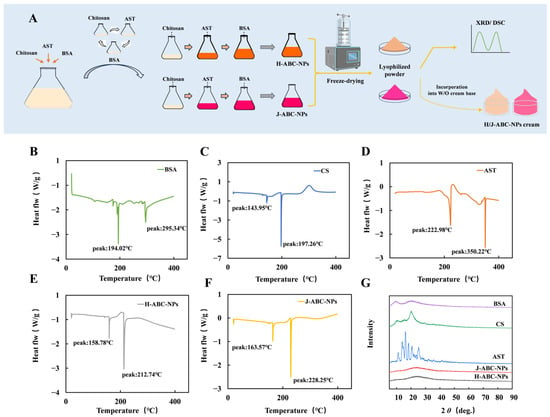
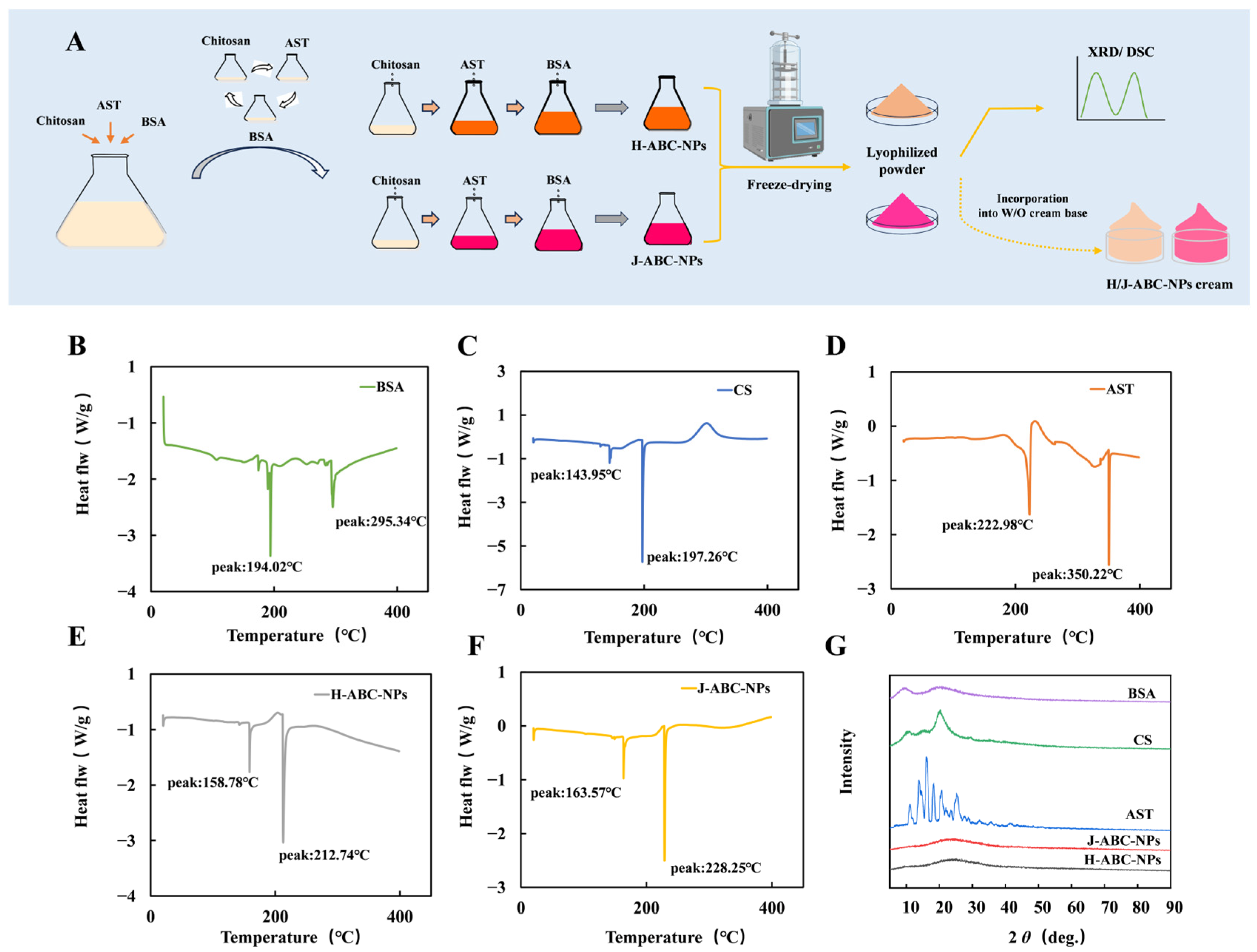
H/J-ABC-NPs were prepared via a bottom-up molecular self-assembly method by sequentially adding an AST ethanolic solution to a CS aqueous solution, followed by a BSA solution, with AST aggregation regulated by the ethanol volume fraction in the aqueous–organic solvent system. The physicochemical characterization of H/J-ABC-NPs has been systematically reported in our previous study [18]. The average hydrodynamic diameters of H-ABC-NPs and J-ABC-NPs were 281 ± 9 and 368 ± 5 nm, respectively, with corresponding PDI values of 0.27 ± 0.16 and 0.28 ± 0.04 and zeta potentials of +26.0 ± 0.8 and +30.2 ± 0.6 mV. Therefore, the present study focused on the DSC and XRD analyses of the H/J-ABC-NPs. The preparation process of H/J-ABC-NPs was illustrated in Figure 1A and the corresponding DSC and XRD results are shown in Figure 1B–G. AST exhibited a sharp melting endotherm at 222.98 °C [30], while CS and BSA showed endothermic peaks at 197.26 °C and 194.02 °C, respectively [31]. In H/J-ABC-NPs, the BSA melting peak disappeared and a new endothermic peak appeared at 150–170 °C, suggesting interactions between AST and BSA. Meanwhile, the AST-related endothermic peak shifted to 212.74 °C in H-ABC-NPs and 228.25 °C in J-ABC-NPs, indicating that the thermal behavior of AST was altered following nanoparticle formation.
Figure 1.
Preparation and physicochemical characterization of H-/J-aggregates astaxanthin/bovine serum albumin/chitosan nanoparticles (H-ABC-NPs and J-ABC-NPs, H/J-ABC-NPs). (A) Schematic illustration of the preparation of H/J-ABC-NPs and W/O creams; (B–F) DSC thermograms of individual components and H/J-ABC-NPs; (G) XRD patterns.
Generally, the number and intensity of XRD diffraction peaks serve as indicators of crystallinity, with stronger peaks representing a higher degree of crystallinity [32]. The XRD patterns are shown in Figure 1G. Crystalline AST exhibited several characteristic diffraction peaks between 10° and 30°, with the strongest peak at 2θ = 17.84°. In contrast, these characteristic peaks disappeared in H/J-ABC-NPs, indicating that AST was encapsulated within the carrier matrix and predominantly existed in an amorphous state.
3.2. Antioxidant Activity Analysis of H/J-ABC-NPs
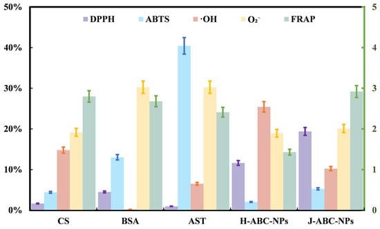
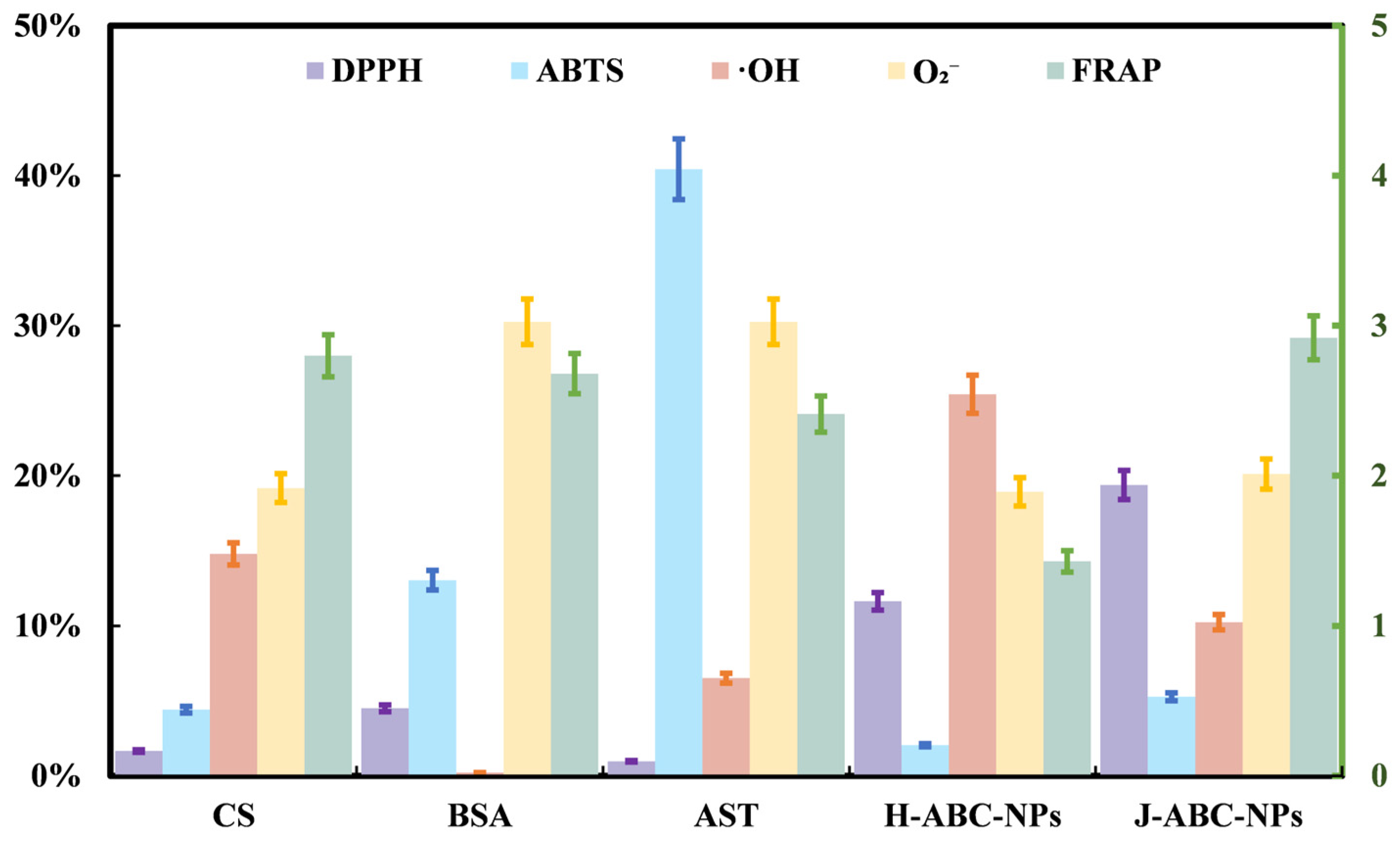
The antioxidant activities of H/J-ABC-NPs are shown in Figure 2. Compared with free AST, H/J-ABC-NPs showed higher DPPH and •OH radical-scavenging activities, as well as relatively high ferric reducing activity. H-ABC-NPs showed higher DPPH radical-scavenging activity, whereas J-ABC-NPs showed higher •OH radical-scavenging activity. In contrast, the ABTS and superoxide anion (O2•−) radical-scavenging activities of H/J-ABC-NPs were lower than those of free AST. These results indicated that encapsulation in H/J-ABC-NPs altered the antioxidant properties of AST. The difference among these assays may be related to their distinct reaction mechanisms [33] and changes in the accessibility of AST to radicals following nanoparticle formation. In addition, the antioxidant activities of CS and BSA suggest that the carrier components may also contribute to the observed responses.
Figure 2.
Antioxidant activity of H/J-ABC-NPs, orange for DPPH, yellow for ABTS, blue for FRAP, green for •OH-, purple for superoxide anion.
3.3. Cytotoxicity Against H2O2-Induced Oxidative Damage in L929 Cells
3.3.1. Cytotoxicity of H/J-ABC-NPs on L929 Cells
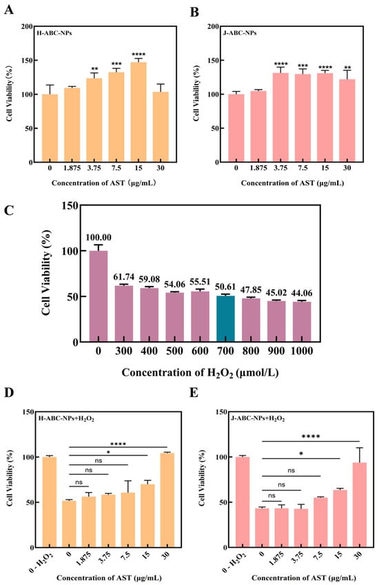
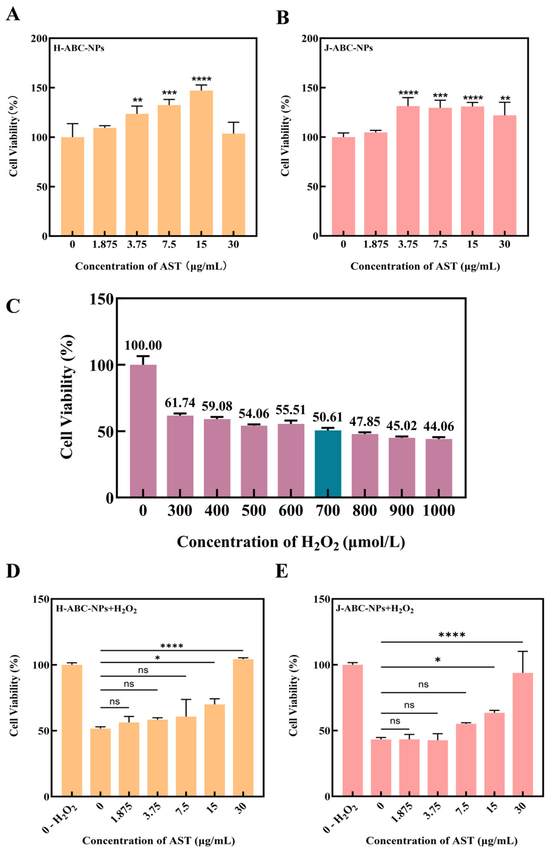
As shown in Figure 3A,B, L929 cell viability remained above 95% at all tested AST concentrations, indicating the good cytocompatibility of H/J-ABC-NPs. In our previous study [27], L929 cell viability decreased with increasing lutein concentrations above 5 μg/mL in H-LBC-NPs, suggesting the lower cytotoxicity of H/J-ABC-NPs under the tested conditions. Consistently, Calcein-AM/PI staining showed predominantly green fluorescence with only a few PI-positive cells (Figure S2). No obvious differences in cell morphology or live/dead staining were observed between the H/J-ABC-NPs and control groups, further confirming the good cytocompatibility of H/J-ABC-NPs.
Figure 3.
Cytotoxicity and cytoprotective effects of H/J-ABC-NPs in L929 cells. (A) H-ABC-NPs; (B) J-ABC-NPs; (C) Damaging ability of different concentrations of H2O2 on L929 cells; (D,E) Effect of H/J-ABC-NPs on L929 cell viability after H2O2-induced oxidative damage. (ns represents no significant difference, * p < 0.05, ** p < 0.01. *** p < 0.001, **** p < 0.0001).
3.3.2. Screening for H2O2 Oxidative Damage Concentration
Skin photoaging is closely associated with oxidative damage [34],and H2O2 is commonly used to induce oxidative damage in cell culture [35]. As shown in Figure 3C, L929 cell viability decreased in a dose-dependent manner with increasing H2O2 concentrations. At 700 μmol/L H2O2, cell viability was approximately 50%. Therefore, 700 μmol/L H2O2 was selected to establish the oxidative damage model for subsequent experiments.
3.3.3. Protective Effects of H/J-ABC-NPs Against H2O2-Induced Oxidative Damage
The viability of H2O2-treated L929 cells increased with increasing concentrations of AST in H/J-ABC-NPs (Figure 3D,E). At 7.5 μg/mL, both H-ABC-NPs and J-ABC-NPs showed an increase in cell viability compared with the Model group, although the differences were not statistically significant. At higher concentrations of 15 and 30 μg/mL H/J-ABC-NPs significantly improved cell viability (p < 0.05 and p < 0.0001, respectively). Calcein-AM/PI staining showed a large number of PI-positive cells after treatment with 700 μmol/L H2O2, whereas the number of PI-positive cells decreased with increasing concentrations of H-ABC-NPs and J-ABC-NPs (Figure S3). This trend was consistent with the CCK-8 results, indicating that H/J-ABC-NPs protected L929 cells against H2O2-induced oxidative damage in a concentration-dependent manner.
3.4. Effect of H/J-ABC-NPs on L929 Cells with Oxidative Damage
3.4.1. ROS Level in L929 Cells
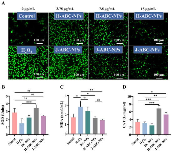
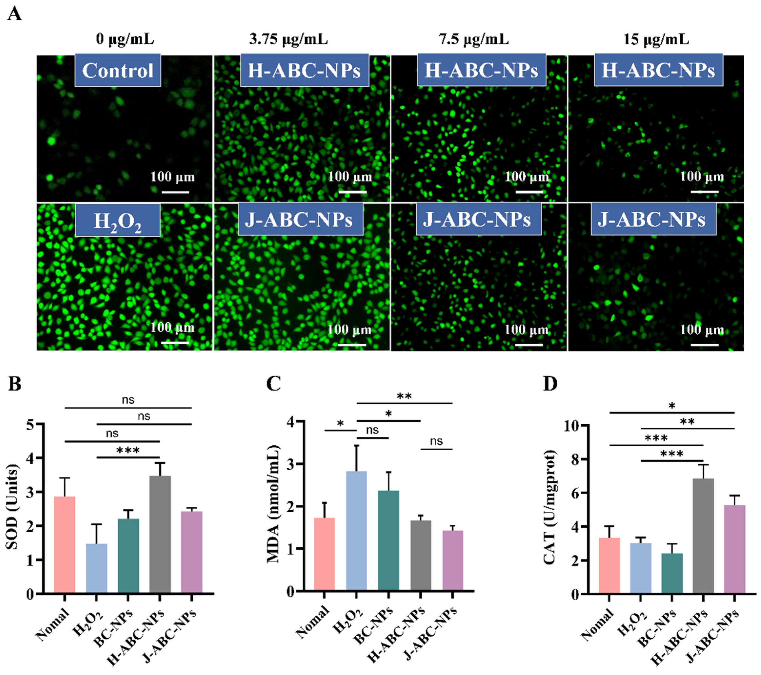
ROS serve as important signaling molecules, whereas the over-accumulation of ROS in pathological conditions results in oxidative damage [36]. As shown in Figure 4A, a low level of intracellular ROS was observed in the Control group, whereas H2O2 treatment resulted in a substantial increase in ROS levels. Consistent with previous findings on H2O2-induced oxidative stress [37], H2O2 treatment resulted in a substantial increase in ROS levels. Pretreatment with H/J-ABC-NPs reduced intracellular ROS levels. These results indicated that H/J-ABC-NPs alleviated H2O2-induced oxidative stress in L929 cells and played a protective role.
Figure 4.
Antioxidant marker content in L929 cells. (A) ROS distribution in L929 cells after protection by H/J-ABC-NPs and H2O2 damage; changes in SOD (B), MDA (C) and CAT (D) levels in L929 cells. (ns represents no significant difference, * p < 0.05, ** p < 0.01, *** p < 0.001).
3.4.2. SOD, CAT, MDA Levels in L929 Cells
SOD and CAT are important antioxidant enzymes involved in cellular defense against oxidative stress, whereas MDA is commonly used as an indicator of lipid peroxidation [38,39,40]. As shown in Figure 4B, SOD activity was significantly increased in the H-ABC-NPs group compared with the H2O2 and BC-NPs groups. J-ABC-NPs also increased SOD activity, which was comparable to that of the Control group. H2O2 treatment increased the MDA level, whereas both H-ABC-NPs and J-ABC-NPs significantly reduced MDA accumulation (Figure 4C). Similarly, CAT activity was increased following H/J-ABC-NPs treatment compared with the H2O2 group (Figure 4D). These results, together with the changes in intracellular ROS, indicated that H/J-ABC-NPs alleviated H2O2-induced oxidative stress by enhancing cellular antioxidant capacity and reducing lipid peroxidation.
3.5. Histological Skin Analysis in BALB/c Mice
3.5.1. Appearance of BALB/c Mice
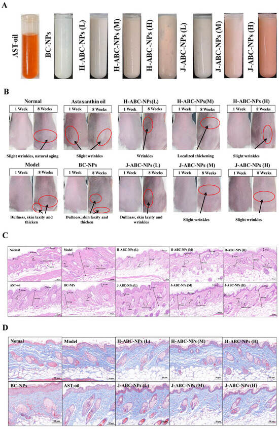
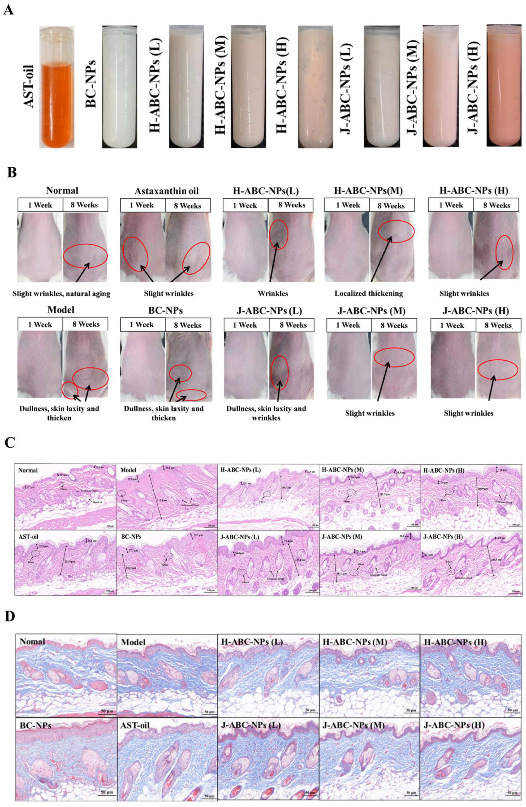
All mice survived throughout the 8-week experiment. The H/J-ABC-NPs creams were smooth, homogeneous, and readily spread on the dorsal skin (Figure 5A). Consistent with previous observations in HR-1 hairless mice [41], BALB/c mice developed visible signs of skin photoaging after 8 weeks of UVB/UVA irradiation (Figure 5B). During the first week, the Normal group showed no visible wrinkles, whereas slight wrinkles were observed in the irradiated groups. After 8 weeks, mice in the Model and BC-NPs groups exhibited skin dullness, wrinkles, laxity, and thickening, confirming the establishment of the photoaging model [42]. In contrast, these changes were alleviated in the H-ABC-NPs and J-ABC-NPs groups. Mice treated with high-dose H-ABC-NPs or J-ABC-NPs showed only slight wrinkles and skin laxity, with an appearance comparable to that of the AST-oil group. These observations indicated that H/J-ABC-NPs treatment alleviated the visible signs of UVB/UVA-induced skin photoaging.
Figure 5.
Skin condition analysis. (A): W/O cream of H/J-ABC-NPs; (B): Skin appearance; (C): H&E staining; (D): Masson staining.
3.5.2. H&E Staining
Epidermal thickening is a characteristic histological feature of skin photoaging [43]. As shown in Figure 5C, the Model group exhibited marked epidermal and dermal thickening, accompanied by disrupted and aggregated dermal fibers compared with the Normal group. Similar histological changes were observed in the BC-NPs group, confirming the establishment of UVB/UVA-induced skin photoaging [44]. The low-dose H-ABC-NPs and J-ABC-NPs groups also showed more pronounced fiber breakage and aggregation. With increasing AST concentrations, epidermal thickening appeared less pronounced, while dermal fibers showed improved organization and continuity. These results indicated that H/J-ABC-NPs treatment alleviated UVB/UVA-induced histological changes in the skin.
3.5.3. Masson Staining
Collagen fibers are the major structural components of the dermis and are important for maintaining skin integrity [45]. Masson staining was used to evaluate collagen fiber distribution (Figure 5D). In the Normal group, collagen fibers were continuous and loosely arranged, whereas the Model and BC-NPs groups showed disrupted and aggregated collagen fibers with reduced inter-fiber spaces. No obvious improvement was observed in the low-dose H/J-ABC-NPs groups. With increasing AST concentrations, collagen fiber disruption and aggregation were reduced, and the fiber arrangement became more regular. These observations suggest that H/J-ABC-NPs alleviated UVB/UVA-induced alterations in dermal collagen fiber organization.
3.6. Antioxidant Enzyme Levels in BALB/c Mice
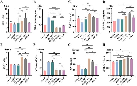
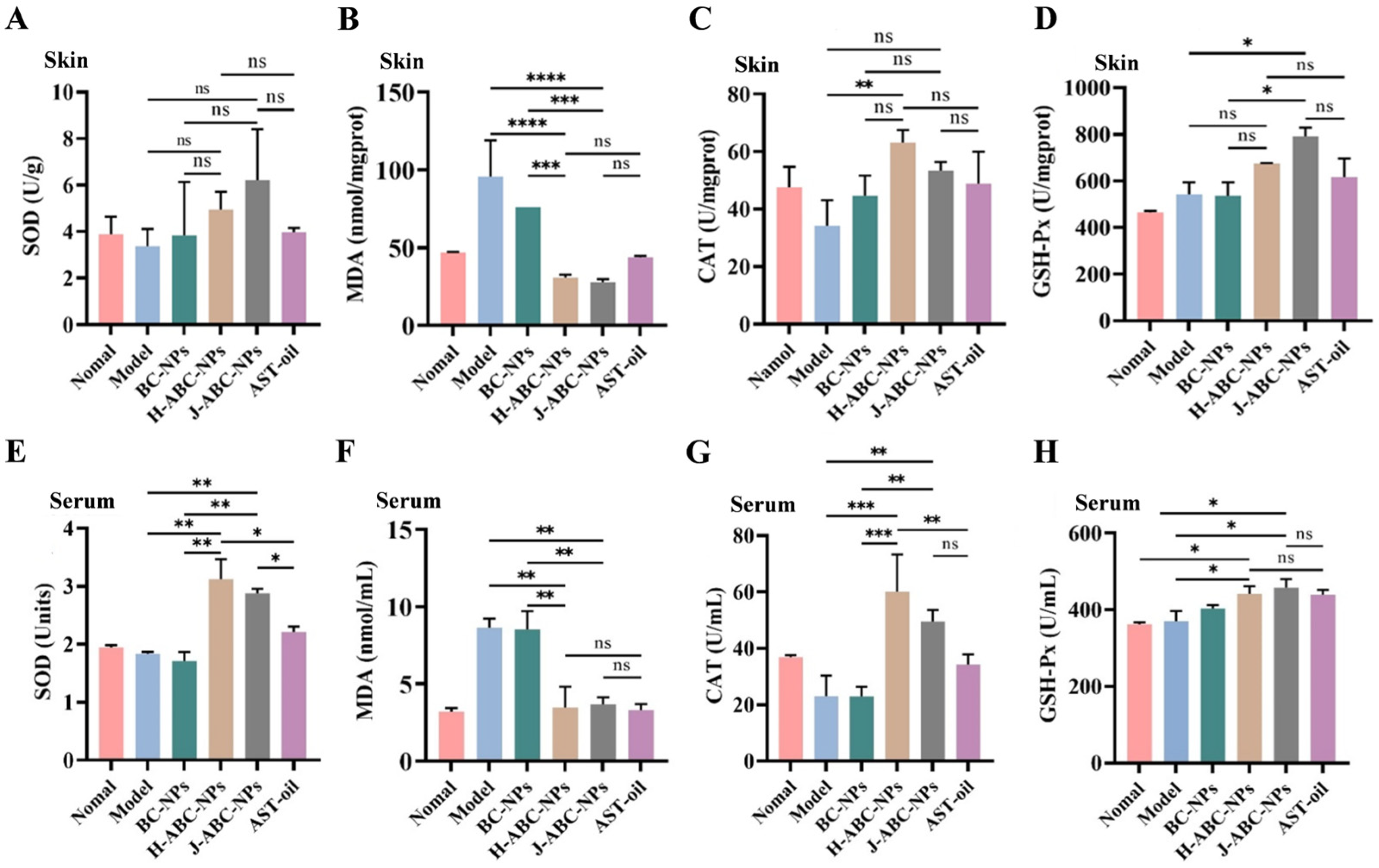
Oxidative stress plays an important role in UV-induced skin photoaging [46]. Excessive ROS production can impair antioxidant defenses and promote lipid peroxidation, resulting in changes in antioxidant enzyme activities and MDA levels [1,47]. As shown in Figure 6A,E, H/J-ABC-NPs treatment increased SOD activity in the skin and serum compared with the Model and BC-NPs groups. In contrast, MDA levels were elevated in the Model group and decreased following H/J-ABC-NPs treatment (Figure 6B,F), indicating reduced lipid peroxidation.
Figure 6.
Levels of antioxidant enzymes in mice serum and skin: (A) SOD activity in skin; (B) MDA levels in skin; (C) CAT activity in skin; (D) GSH-Px activity in skin; (E) SOD activity in serum; (F) MDA levels in serum; (G) CAT activity in serum; (H) GSH-Px activity in serum. (ns represents no significant difference, * p < 0.05, ** p < 0.01. *** p < 0.001, **** p < 0.0001).
CAT activity was also increased after H/J-ABC-NPs treatment (Figure 6C,G). In skin tissues, a significant difference was observed between the high-dose H-ABC-NPs and Model groups, while serum CAT activity in the H-ABC-NPs and J-ABC-NPs groups was higher than that in the AST-oil group. Similarly, GSH-Px activity was increased following H/J-ABC-NPs treatment in both skin and serum (Figure 6D,H). The highest GSH-Px activity was observed in the J-ABC-NPs group, although no significant difference was found among the J-ABC-NPs, H-ABC-NPs, and AST-oil groups. Overall, the increased activities of SOD, CAT, and GSH-Px, together with the decreased MDA levels, indicated that H/J-ABC-NPs enhanced antioxidant defense and reduced lipid peroxidation in UVB/UVA-irradiated mice.
3.7. Inflammatory Factor Levels in BALB/c Mice
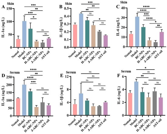
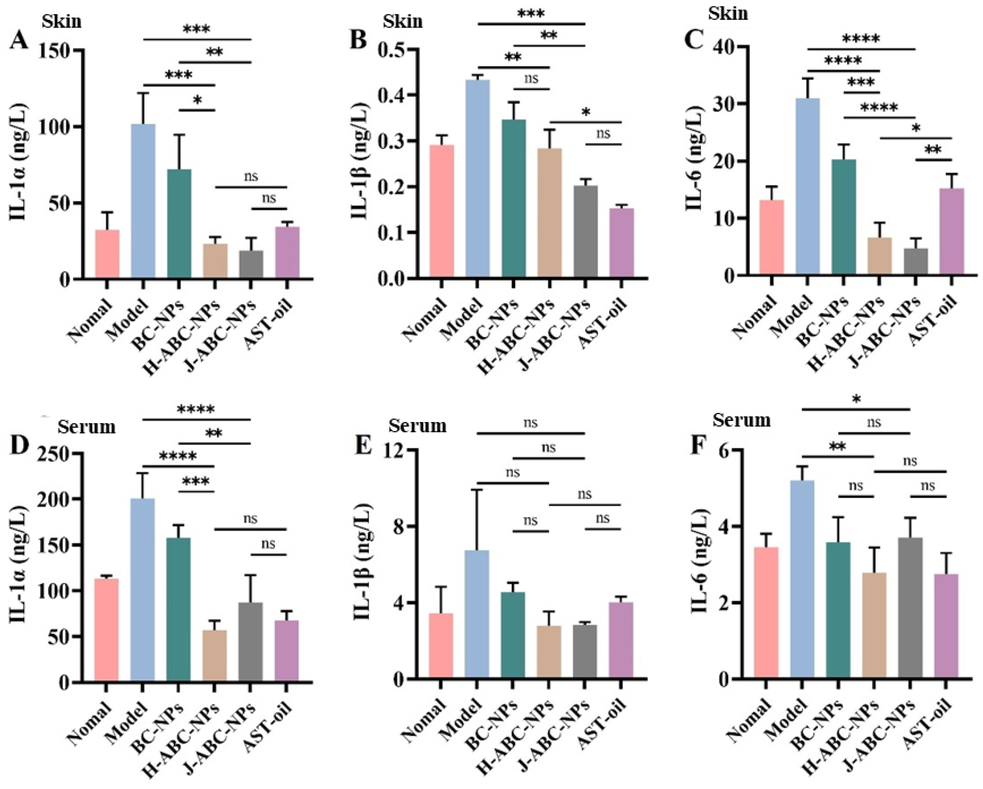
Inflammatory responses are closely associated with UV-induced skin photoaging [3]. As shown in Figure 7, the levels of IL-1α, IL-1β, and IL-6 were elevated in the serum and skin tissues of the Model and BC-NPs groups following UVB/UVA irradiation. Compared with the Model group, IL-1α levels were significantly reduced in the high-dose H-ABC-NPs and J-ABC-NPs groups. H/J-ABC-NPs treatment also reduced IL-1β levels in both skin and serum. Similarly, IL-6 levels were significantly decreased in the skin following H/J-ABC-NPs treatment, while serum IL-6 levels were comparable to those of the Normal group. These results indicated that H/J-ABC-NPs alleviated the inflammatory response induced by UVB/UVA irradiation.
Figure 7.
Inflammatory factor levels in serum and skin. (A): IL-1α level in skin; (B): IL-1β level in skin; (C): IL-6 level in skin; (D): IL-1α level in serum; (E): IL-1β level in serum; (F): IL-6 level in serum. (ns represents no significant difference, * p < 0.05, ** p < 0.01. *** p < 0.001, **** p < 0.0001).
3.8. Collagen Content in BALB/c Mice
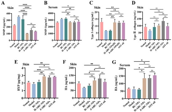
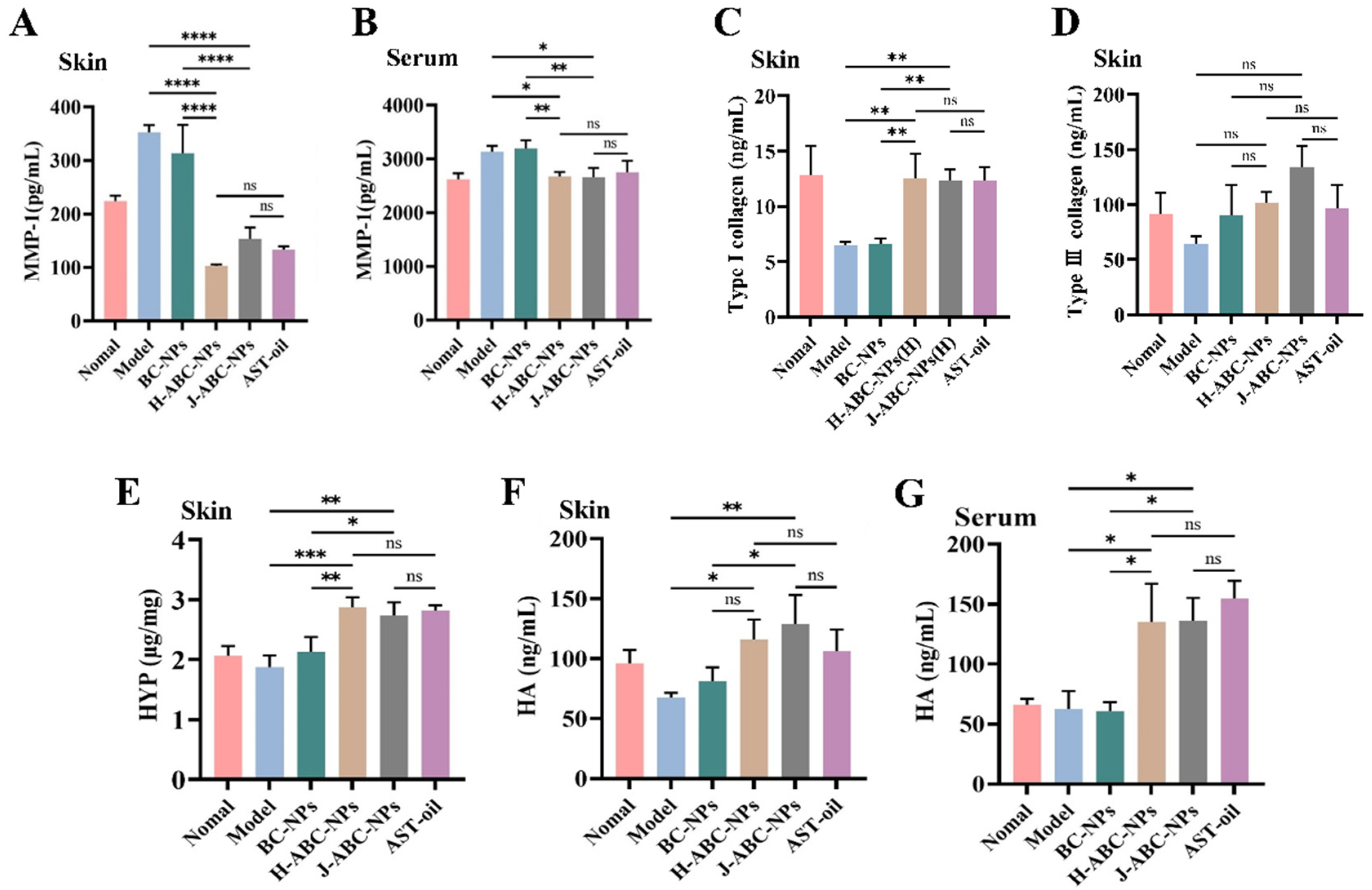
MMP-1 plays an important role in collagen degradation during skin photoaging, and its expression can be induced by UV irradiation [1]. As shown in Figure 8A,B, MMP-1 levels were significantly increased in the Model and BC-NPs groups compared with the H-ABC-NPs, J-ABC-NPs, and AST-oil groups. Previous studies have shown that excessive ROS and inflammatory cytokines, including IL-1α, IL-1β, and IL-6, are associated with increased MMP-1 levels [48,49,50,51]. Together with the reduced ROS and inflammatory cytokine levels observed in this study, these results suggested that H/J-ABC-NPs suppressed UVB/UVA-induced MMP-1 production.
Figure 8.
MMP-1, HYP, HA levels and collagen content in mice. (A,B): MMP-1 levels in serum and skin; (C,D): collagen content in skin; (E): HYP levels in skin; (F,G): HA levels in serum and skin. (ns represents no significant difference, * p < 0.05, ** p < 0.01. *** p < 0.001, **** p < 0.0001).
Type I and type III collagen are major components of the dermal extracellular matrix and are closely associated with skin structure and integrity [52]. As shown in Figure 8C,D, the levels of type I and type III collagen were decreased in the Model and BC-NPs groups following UVB/UVA irradiation. In contrast, H-ABC-NPs and J-ABC-NPs treatment reduced collagen loss, with type I collagen levels comparable to those of the Normal group. Together with the decreased MMP-1 levels (Figure 8A,B), these results suggest that H/J-ABC-NPs reduced collagen degradation in UVB/UVA-irradiated skin, which was consistent with the improvements in skin morphology and histological structure.
3.9. HYP and HA Levels in BALB/c Mice
HYP is a major component of collagen and is commonly used as an indicator of collagen content [53]. As shown in Figure 8E, HYP levels were significantly higher in the H/J-ABC-NPs and AST-oil groups than in the Model and BC-NPs groups, consistent with the reduced collagen loss observed after H/J-ABC-NPs treatment.
HA is a major component of the extracellular matrix (ECM) and is responsible for maintaining skin hydration, elasticity, and volume [54]. Compared with the Model and BC-NPs groups, H/J-ABC-NPs and AST-oil treatment increased HA levels in the skin (Figure 8F,G). These results indicated that H/J-ABC-NPs reduced the UVB/UVA-induced loss of collagen-related components and HA in photoaged skin.
Although H-ABC-NPs and J-ABC-NPs exhibited some differences in individual indicators, neither formulation demonstrated a consistent advantage in improving skin-related outcomes. Thus, the present findings mainly support their shared protective effects against UVB/UVA-induced skin photoaging.
4. Conclusions
H/J-ABC-NPs exhibited antioxidant activity and showed no obvious cytotoxicity toward L929 cells at the tested concentrations. In the H2O2-induced oxidative stress model, H/J-ABC-NPs reduced intracellular ROS and MDA levels and increased SOD and CAT activities. Topical treatment with H/J-ABC-NPs alleviated wrinkles, skin laxity, and epidermal thickening in UVB/UVA-irradiated mice. H/J-ABC-NPs treatment also improved antioxidant enzyme activities, reduced MDA and inflammatory cytokine levels, and was associated with lower MMP-1 levels and reduced collagen loss. In addition, HYP and HA levels were increased after H/J-ABC-NPs treatment. These results show that H/J-ABC-NPs can alleviate UVB/UVA-induced skin photoaging, with the observed effects mainly associated with reduced oxidative stress and inflammation and the improved maintenance of skin structure.
Supplementary Materials
The following supporting information can be downloaded at: https://www.mdpi.com/article/10.3390/antiox15111473/s1, Figure S1: Standard curve of FeSO4 for the FRAP assay; Figure S2: Cell safety of H/J-ABC-NPs in L929 cells by Calcein/PI staining; Figure S3: Cell safety of H/J-ABC-NPs in L929 cells after H2O2 induced oxidative stress by Calcein/PI staining.
Author Contributions
Conceptualization, Y.Z.; methodology, S.X. and Y.L.; software, Y.G. and Y.X.; validation, Y.S., W.Q. and Y.Z.; formal analysis, Y.C.; investigation, Y.S. and Y.L.; resources, Y.Z.; data curation, W.Q.; writing—original draft preparation, W.Q.; writing—review and editing, Y.Z.; visualization, W.Q. and X.F.; supervision, Y.Z.; project administration, Y.Z.; funding acquisition, Y.Z. All authors have read and agreed to the published version of the manuscript.
Funding
This research was funded by the Food Engineering Technology Research Center/Key Laboratory of Henan Province, Henan University of Technology, grant number GO202520; Training Program for Youth Backbone Teachers in Colleges and Universities of Henan Province, grant number 2025GGJS057; Natural Science Foundation of Henan Province, grant number 252300420172; the Key Scientific Research Projects of Colleges and Universities in Henan Province, grant number 26A180007; Henan Province Deputy General Manager for Technology Program, grant number 31210649; Key R&D Projects in Henan Province, grant number 231111113300; the Science and Technology Department of Henan Province, grant number 242102310382; Cultivation Program for Young Backbone Teachers in Henan University of Technology, grant number 21420167; China Scholarship Council (scholarship for Yingyuan Zhao), grant number 202008410088. The APC was funded by the Food Engineering Technology Research Center/Key Laboratory of Henan Province, grant number GO202520.
Institutional Review Board Statement
The animal study protocol was approved by the Ethics Committee of Henan University of Technology (protocol code Haut 202110-5).
Informed Consent Statement
Not applicable.
Data Availability Statement
The original contributions presented in this study are included in the article/Supplementary Materials. Further inquiries can be directed to the corresponding author.
Conflicts of Interest
The authors declare no conflicts of interest.
References
- Yang, M.; Tao, L.; Wang, Z.; Li, L.; Luo, J.; Pai, K.; Li, W.; Zhao, C.; Sheng, J.; Tian, Y. The Mechanism of Peach Gum Polysaccharide Preventing UVB-Induced Skin Photoaging by Regulating Matrix Metalloproteinanse and Oxidative Factors. Molecules 2023, 28, 4104. [Google Scholar] [CrossRef] [Scilit]
- Lee, H.; Hong, Y.; Kim, M. Structural and Functional Changes and Possible Molecular Mechanisms in Aged Skin. Int. J. Mol. Sci. 2021, 22, 12489. [Google Scholar] [CrossRef] [Scilit]
- Zhong, X.; Deng, Y.; Yang, H.; Du, X.; Liu, P.; Du, Y. Role of autophagy in skin photoaging: A narrative review. Medicine 2024, 103, e37178. [Google Scholar] [CrossRef] [Scilit]
- Sun, X.; Zhang, F.; Liu, Y.; Wang, K.; Huang, X.; Han, Y.; Wang, L.; Hu, H.; Zhou, Z.; Yang, Z.; et al. Eco-friendly extraction of polysaccharides from Polygonatum cyrtonema Hua using ultrasound-assisted NADES: Optimization, characterization, and efficacy against skin photoaging. Microchem. J. 2026, 224, 117699. [Google Scholar] [CrossRef] [Scilit]
- Singh, K.N.; Patil, S.; Barkate, H. Protective effects of astaxanthin on skin: Recent scientific evidence, possible mechanisms, and potential indications. J. Cosmet. Dermatol. 2020, 19, 22–27. [Google Scholar] [CrossRef] [Scilit]
- Fitsiou, E.; Pulido, T.; Campisi, J.; Alimirah, F.; Demaria, M. Cellular Senescence and the Senescence-Associated Secretory Phenotype as Drivers of Skin Photoaging. J. Investig. Dermatol. 2021, 141, 1119–1126. [Google Scholar] [CrossRef] [Scilit]
- Liu, C.; Guo, X.; Chen, Y.; Zhao, M.; Shi, S.; Luo, Z.; Song, J.; Zhang, Z.; Yang, W.; Liu, K. Anti-photoaging effect and mechanism of flexible liposomes co-loaded with apigenin and doxycycline. Biomed. Pharmacother. 2023, 164, 114998. [Google Scholar] [CrossRef] [Scilit]
- Zhou, J.; Guo, M.; Peng, Y.; Xu, W.; Hao, X.; Tang, Y.; Tang, T.; Wu, J.; Xiang, D. Beta vulgaris-derived exosome-like nanovesicles mitigate photoaging by attenuating oxidative stress and promoting collagen biosynthesis. Colloids Surf. B Biointerfaces 2026, 261, 115412. [Google Scholar] [CrossRef] [Scilit]
- Catanzaro, E.; Bishayee, A.; Fimognari, C. On a Beam of Light: Photoprotective Activities of the Marine Carotenoids Astaxanthin and Fucoxanthin in Suppression of Inflammation and Cancer. Mar. Drugs 2020, 18, 544. [Google Scholar] [CrossRef] [Scilit]
- Yang, S.; Lu, X.; Wang, J.; Liu, Y.; Nie, M.; Liu, J.; Sun, H. Astaxanthin from Haematococcus pluvialis and Chromochloris zofingiensis: Biosynthetic Pathways, Engineering Strategies, and Industrial Prospects. Mar. Drugs 2025, 23, 485. [Google Scholar] [CrossRef] [Scilit]
- Jannel, S.; Caro, Y.; Bermudes, M.; Petit, T. Novel Insights into the Biotechnological Production of Haematococcus pluvialis-Derived Astaxanthin: Advances and Key Challenges to Allow Its Industrial Use as Novel Food Ingredient. J. Mar. Sci. Eng. 2020, 8, 789. [Google Scholar] [CrossRef] [Scilit]
- Komatsu, T.; Sasaki, S.; Manabe, Y.; Hirata, T.; Sugawara, T. Preventive effect of dietary astaxanthin on UVA-induced skin photoaging in hairless mice. PLoS ONE 2017, 12, e0171178. [Google Scholar] [CrossRef] [Scilit]
- Song, B.; Liu, D.; Liu, T.C.; Li, K.; Wang, S.; Liu, J.; Regenstein, J.M.; Wu, Y.; Zhou, P. The combined effect of commercial tilapia collagen peptides and antioxidants against UV-induced skin photoaging in mice. Food Funct. 2023, 14, 5936–5948. [Google Scholar] [CrossRef] [Scilit]
- Yang, L.; Qiao, X.; Gu, J.; Li, X.; Cao, Y.; Xu, J.; Xue, C. Influence of molecular structure of astaxanthin esters on their stability and bioavailability. Food Chem. 2021, 343, 128497. [Google Scholar] [CrossRef] [Scilit]
- Brotosudarmo, T.H.P.; Limantara, L.; Setiyono, E.; Heriyanto. Structures of Astaxanthin and Their Consequences for Therapeutic Application. Int. J. Food Sci. 2020, 2020, 2156582. [Google Scholar] [CrossRef] [Scilit]
- Jia, L.; Wang, W.; Zhao, H.; Ding, X.; Zheng, M.; Cai, D.; Wang, Y.; Wang, Z.; Liu, H. Innovative Nano Delivery Systems for Astaxanthin: Enhancing Stability, Bioavailability, and Targeted Therapeutic Applications. J. Agric. Food Chem. 2025, 73, 3286–3304. [Google Scholar] [CrossRef] [Scilit]
- Ge, Y.; Jiang, X.; Peng, W.; Liu, Y.; Ge, Y.; Wang, Z.; Wang, Y.; Wu, W.; Zhang, C. Preparation, characterization, and stability of chitosan-tremella polysaccharide layer-by-layer encapsulated astaxanthin nanoemulsion delivery system. Int. J. Biol. Macromol. 2024, 282, 136741. [Google Scholar] [CrossRef] [Scilit]
- Zhao, Y.; Liu, J.; Zhang, S.; Wang, Z.; Jia, H.; Oda, H.; Li, R. Fabrication and characterization of the H/J-type aggregates astaxanthin/bovine serum albumin/chitosan nanoparticles. Int. J. Biol. Macromol. 2022, 223, 1186–1195. [Google Scholar] [CrossRef] [Scilit]
- Zhao, Y.; Wang, Z.; Liu, S.; Xie, S.; Xie, Y.; Li, R.; Oda, H. Lyoprotectant Formulation and Optimization of the J-Aggregates Astaxanthin/BSA/Chitosan Nanosuspension. Biomolecules 2023, 13, 496. [Google Scholar] [CrossRef] [Scilit]
- Liu, Y.; Li, X.; Sun, H.; Zhang, J.; Cai, C.; Xu, N.; Feng, J.; Nan, B.; Wang, Y.; Liu, J. Whey protein concentrate/pullulan gel as a novel microencapsulated wall material for astaxanthin with improving stability and bioaccessibility. Food Hydrocoll. 2023, 138, 108467. [Google Scholar] [CrossRef] [Scilit]
- Yang, J.; Xiong, Y.-A.; Xiao, S.-J.; Xu, Y.-S. Anti-inflammatory Effects of Saxifrage Cream in Hapten-induced Chronic Dermatitis-eczema in Mice. Chin. Herb. Med. 2017, 9, 176–182. [Google Scholar] [CrossRef] [Scilit]
- Wang, Z.; Wang, L.; Huang, H.; Li, Q.; Wang, X.; Sun, Q.; Wang, Q.; Li, N. In vitro antioxidant analysis of flavonoids extracted from Artemisia argyi stem and their anti-inflammatory activity in lipopolysaccharide-stimulated RAW 264.7 macrophages. Food Chem. 2023, 407, 135198. [Google Scholar] [CrossRef] [Scilit]
- Betancur, M.C.; Salazar, F.; Araya, M.; Behn, A.; López, J.; Quesille-Villalobos, A.; Villatoro, J.M.; Poblete, J.; Zambrano, A. Comprehensive Characterization of Bioactive Properties in Extracts from Different Chilean Hop Ecotypes (Humulus lupulus L.). Antioxidant, Antimicrobial and Antitumor Activities. Antioxidants 2025, 14, 1224. [Google Scholar] [CrossRef] [Scilit]
- Zhang, X.; Ren, X.; Zhao, X.; Liu, H.; Wang, M.; Zhu, Y. Stability, structure, and antioxidant activity of astaxanthin crystal from Haematococcus pluvialis. J. Am. Oil Chem.’ Soc. 2022, 99, 367–377. [Google Scholar] [CrossRef] [Scilit]
- Chen, Y.; Lin, Q.; Wang, J.; Mu, J.; Liang, Y. Proteins, polysaccharides and their derivatives as macromolecular antioxidant supplements: A review of in vitro screening methods and strategies. Int. J. Biol. Macromol. 2023, 224, 958–971. [Google Scholar] [CrossRef] [Scilit]
- Qie, X.; Chen, W.; Zeng, M.; Wang, Z.; Chen, J.; Goff, H.D.; He, Z. Interaction between β-lactoglobulin and chlorogenic acid and its effect on antioxidant activity and thermal stability. Food Hydrocoll. 2021, 121, 107059. [Google Scholar] [CrossRef] [Scilit]
- Sun, Y.; Zhao, Y.; Qiu, M.; Zhang, Y.; Liang, J.; Xie, S.; Li, R.; Wang, X. Preparation, characterization, stability and application of the H-type aggregates lutein/whey protein/chitosan nanoparticles. Int. J. Biol. Macromol. 2024, 282, 136739, Correction in Int. J. Biol. Macromol. 2024, 288, 138563. [Google Scholar] [CrossRef] [Scilit]
- Liu, S.; You, L.; Zhao, Y.; Chang, X. Hawthorn Polyphenol Extract Inhibits UVB-Induced Skin Photoaging by Regulating MMP Expression and Type I Procollagen Production in Mice. J. Agric. Food Chem. 2018, 66, 8537–8546. [Google Scholar] [CrossRef] [Scilit]
- Huynh, C.B.; Vu, N.B.; Van, T.T.; Van Pham, P. Establishment of a BALB/c Mouse Model for Photoaged Skin: Insights into UV-Induced Dermatological Changes. Ski. Res. Technol. 2026, 32, e70353. [Google Scholar] [CrossRef] [Scilit]
- Zarandona, I.; Correia, D.M.; Moreira, J.; Costa, C.M.; Lanceros-Mendez, S.; Guerrero, P.; de la Caba, K. Magnetically responsive chitosan-pectin films incorporating Fe3O4 nanoparticles with enhanced antimicrobial activity. Int. J. Biol. Macromol. 2023, 227, 1070–1077. [Google Scholar] [CrossRef] [Scilit]
- Benamer Oudih, S.; Tahtat, D.; Nacer Khodja, A.; Mahlous, M.; Hammache, Y.; Guittoum, A.-E.; Kebbouche Gana, S. Chitosan nanoparticles with controlled size and zeta potential. Polym. Eng. Sci. 2023, 63, 1011–1021. [Google Scholar] [CrossRef] [Scilit]
- Uzun, İ. Methods of determining the degree of crystallinity of polymers with X-ray diffraction: A review. J. Polym. Res. 2023, 30, 394. [Google Scholar] [CrossRef] [Scilit]
- Munteanu, I.G.; Apetrei, C. Analytical Methods Used in Determining Antioxidant Activity: A Review. Int. J. Mol. Sci. 2021, 22, 3380. [Google Scholar] [CrossRef] [Scilit]
- Teng, Y.; Huang, Y.; Danfeng, X.; Tao, X.; Fan, Y. The Role of Probiotics in Skin Photoaging and Related Mechanisms: A Review. Clin. Cosmet. Investig. Dermatol. 2022, 15, 2455–2464. [Google Scholar] [CrossRef] [Scilit]
- Quadri, Z.; Elsherbini, A.; Crivelli, S.M.; El-Amouri, S.S.; Tripathi, P.; Zhu, Z.; Ren, X.; Zhang, L.; Spassieva, S.D.; Nikolova-Karakashian, M.; et al. Ceramide-mediated orchestration of oxidative stress response through filopodia-derived small extracellular vesicles. J. Extracell. Vesicles 2024, 13, e12477. [Google Scholar] [CrossRef] [Scilit]
- Catanesi, M.; D’Angelo, M.; Tupone, M.G.; Benedetti, E.; Giordano, A.; Castelli, V.; Cimini, A. MicroRNAs Dysregulation and Mitochondrial Dysfunction in Neurodegenerative Diseases. Int. J. Mol. Sci. 2020, 21, 5986. [Google Scholar] [CrossRef] [Scilit]
- Xin, X.; Gong, T.; Hong, Y. Hydrogen peroxide initiates oxidative stress and proteomic alterations in meningothelial cells. Sci. Rep. 2022, 12, 14519. [Google Scholar] [CrossRef] [Scilit]
- Ighodaro, O.M.; Akinloye, O.A. First line defence antioxidants-superoxide dismutase (SOD), catalase (CAT) and glutathione peroxidase (GPX): Their fundamental role in the entire antioxidant defence grid. Alex. J. Med. 2018, 54, 287–293. [Google Scholar] [CrossRef] [Scilit]
- Zheng, M.; Liu, Y.; Zhang, G.; Yang, Z.; Xu, W.; Chen, Q. The Applications and Mechanisms of Superoxide Dismutase in Medicine, Food, and Cosmetics. Antioxidants 2023, 12, 1675. [Google Scholar] [CrossRef] [Scilit]
- Tsikas, D. GC–MS and GC–MS/MS measurement of malondialdehyde (MDA) in clinical studies: Pre-analytical and clinical considerations. J. Mass Spectrom. Adv. Clin. Lab 2023, 30, 10–24. [Google Scholar] [CrossRef] [Scilit]
- Li, X.; Matsumoto, T.; Takuwa, M.; Saeed Ebrahim Shaiku Ali, M.; Hirabashi, T.; Kondo, H.; Fujino, H. Protective Effects of Astaxanthin Supplementation against Ultraviolet-Induced Photoaging in Hairless Mice. Biomedicines 2020, 8, 18. [Google Scholar] [CrossRef] [Scilit]
- Stoykova, I.D.; Koycheva, I.K.; Binev, B.K.; Mihaylova, L.V.; Benina, M.Y.; Alipieva, K.I.; Georgiev, M.I. Myconoside and Calceolarioside E Restrain UV-Induced Skin Photoaging by Activating NRF2-Mediated Defense Mechanisms. Int. J. Mol. Sci. 2024, 25, 2441. [Google Scholar] [CrossRef] [Scilit]
- Wang, J.; Qiu, H.; Xu, Y.; Gao, Y.; Tan, P.; Zhao, R.; Liu, Z.; Tang, Y.; Zhu, X.; Bao, C.; et al. The biological effect of recombinant humanized collagen on damaged skin induced by UV-photoaging: An in vivo study. Bioact. Mater. 2022, 11, 154–165. [Google Scholar] [CrossRef] [Scilit]
- Zhou, Y.; Bai, R.; Huang, Y.; Li, W.; Chen, J.; Cheng, Z.; Wu, X.; Diao, Y. The anti-photoaging effect of C-phycocyanin on ultraviolet B-irradiated BALB/c-nu mouse skin. Front. Bioeng. Biotechnol. 2023, 11, 1229387. [Google Scholar] [CrossRef] [Scilit]
- Reilly, D.M.; Lozano, J. Skin collagen through the lifestages: Importance for skin health and beauty. Plast. Aesthetic Res. 2021, 8, 2. [Google Scholar] [CrossRef] [Scilit]
- Moon, K.M.; Lee, B.; Kim, D.H.; Chung, H.Y. FoxO6 inhibits melanogenesis partly by elevating intracellular antioxidant capacity. Redox Biol. 2020, 36, 101624. [Google Scholar] [CrossRef] [Scilit]
- Zhang, J.; Hu, J.; Chen, B.; Zhao, T.; Gu, Z. Superabsorbent poly(acrylic acid) and antioxidant poly(ester amide) hybrid hydrogel for enhanced wound healing. Regen. Biomater. 2021, 8, rbaa059. [Google Scholar] [CrossRef] [Scilit]
- Bang, E.; Kim, D.H.; Chung, H.Y. Protease-activated receptor 2 induces ROS-mediated inflammation through Akt-mediated NF-κB and FoxO6 modulation during skin photoaging. Redox Biol. 2021, 44, 102022. [Google Scholar] [CrossRef] [Scilit]
- Hong, J.A.; Bae, D.; Oh, K.N.; Oh, D.R.; Kim, Y.; Kim, Y.; Im, S.J.; Choi, E.J.; Lee, S.G.; Kim, M.; et al. Protective effects of Quercus acuta Thunb. fruit extract against UVB-induced photoaging through ERK/AP-1 signaling modulation in human keratinocytes. BMC Complement. Med. Ther. 2022, 22, 6. [Google Scholar] [CrossRef] [Scilit]
- Pittayapruek, P.; Meephansan, J.; Prapapan, O.; Komine, M.; Ohtsuki, M. Role of Matrix Metalloproteinases in Photoaging and Photocarcinogenesis. Int. J. Mol. Sci. 2016, 17, 868. [Google Scholar] [CrossRef] [Scilit]
- Yan, X.A.; Shen, E.; Cui, A.; Zhou, F.; Zhuang, Y. Assessing the causal relationship between CRP, IL-1α, IL-1β, and IL-6 levels and intervertebral disc degeneration: A two-sample Mendelian randomization study. Sci. Rep. 2024, 14, 23716. [Google Scholar] [CrossRef] [Scilit]
- Chattopadhyay, S.; Raines, R.T. Review collagen-based biomaterials for wound healing. Biopolymers 2014, 101, 821–833. [Google Scholar] [CrossRef] [Scilit]
- Šínová, R.; Pavlík, V.; Ondrej, M.; Velebný, V.; Nešporová, K. Hyaluronan: A key player or just a bystander in skin photoaging? Exp. Dermatol. 2022, 31, 442–458. [Google Scholar] [CrossRef] [Scilit]
- Kobayashi, T.; Chanmee, T.; Itano, N. Hyaluronan: Metabolism and Function. Biomolecules 2020, 10, 1525. [Google Scholar] [CrossRef] [Scilit]
Disclaimer/Publisher’s Note: The statements, opinions and data contained in all publications are solely those of the individual author(s) and contributor(s) and not of MDPI and/or the editor(s). MDPI and/or the editor(s) disclaim responsibility for any injury to people or property resulting from any ideas, methods, instructions or products referred to in the content. |
© 2026 by the authors. Licensee MDPI, Basel, Switzerland. This article is an open access article distributed under the terms and conditions of the Creative Commons Attribution (CC BY) license.