


Abstract
Radiotherapy is the standard radical therapy for locally advanced cervical cancer, with approximately 80% of patients receiving radiation treatment. Nevertheless, intrinsic or acquired radioresistance reduces therapeutic efficacy. Previous studies have confirmed that mRNA methylation modification, metabolic reprogramming and redox homeostasis are involved in the regulation of radioresistance; however, the specific molecular mechanism underlying radioresistance in cervical cancer remains unclear. We integrated transcriptomic–metabolomic profiling, cell-biological experiments, radiobiological assays, RIP, molecular rescue experiments, xenograft tumor models and clinical specimens to dissect this regulatory axis. We identified IGF2BP2 as a key molecule governing radioresistance in cervical cancer. IGF2BP2 binds to the m6A-modified sites on PHGDH mRNA to maintain its stability and subsequently potentiates serine metabolism. Excessive serine metabolism elevates intracellular GSH and SAM levels, sustains cellular antioxidant homeostasis and confers cellular radioresistance. Data from animal models and clinical specimens further validated this regulatory axis. This study uncovers the m6A-IGF2BP2-PHGDH–serine metabolism–antioxidant regulatory axis, providing promising prognostic biomarkers and a radiosensitizing strategy combining a serine-restricted diet with radiotherapy for cervical cancer.
1. Introduction
Cervical cancer is the most common gynecological malignancy and was the fourth leading cause of cancer death among women worldwide in 2018 [1]. Radiation therapy is an effective treatment for cervical cancer and is widely used for advanced cervical cancer and to eliminate residual tumor cells following surgical resection. Approximately 80% of cervical cancer patients undergo radiotherapy during clinical management. However, inherent or acquired radiation resistance can lead to tumor recurrence, metastasis, and poor prognosis in cancer patients [2,3]. Therefore, a thorough elucidation of the molecular mechanisms underlying radiation resistance in cervical cancer is of significant clinical urgency and practical importance.
Current research has found that radiation resistance is associated with various biological mechanisms, such as DNA damage repair, X-ray repair cross-complementing 1 (XRCC1), cell cycle redistribution, epithelial–mesenchymal transition, and high expression of stemness genes [4]. Recent studies have elucidated that the N6-methyladenosine (m6A) modification can modulate the tumor’s resistance to radiation [5]. However, although existing studies have gradually demonstrated that abnormalities in m6A modification are associated with radiation resistance, the specific molecular mechanisms by which m6A modification regulates radiation resistance in cervical cancer cells remain unclear.
m6A is the most abundant reversible epigenetic modification on eukaryotic mRNA [6]. Under the coordinated regulation of methyltransferases, demethylases, and modification-recognition proteins, it participates in multiple biological processes, including the maintenance of target gene mRNA stability, splicing, export from the nucleus, and translation, and plays a broad regulatory role in cell proliferation, metabolism, DNA damage repair, and tumor progression [7]. Insulin-like growth factor 2 messenger RNA (mRNA)-binding protein 2 (IGF2BP2) is a well-characterized reader of m6A modification. As a canonical m6A reader, IGF2BP2 specifically recognizes and binds m6A-modified transcripts to enhance target mRNA stability [8], thereby regulating multiple malignant processes including cancer cell proliferation, apoptosis, metabolic reprogramming, and immune evasion [9]. Accordingly, targeting IGF2BP2 to modulate the mRNA stability of its downstream targets may effectively alter the radiosensitivity of cervical cancer cells.
Serine is a non-essential amino acid in the human body; it can be obtained through dietary intake and synthesized in vivo via the de novo serine synthesis pathway (SSP) [10], with 70% or more of serine derived from the SSP [11]. As a versatile m6A regulatory factor, IGF2BP2 likely regulates key enzymes in the SSP, thereby modulating serine synthesis. Specifically, the SSP consists of three enzymatic reactions in which 3-PG is converted to serine via three rate-limiting enzymes: phosphoglycerate dehydrogenase (PHGDH), phosphoserine phosphatase (PSPH), and phosphoserine aminotransferase 1 (PSAT1) [12]. Whether derived from the SSP or from external sources, serine can provide a one-carbon unit for the one-carbon cycle during its conversion to glycine [13]. One-carbon metabolism covers folate, methionine and trans-sulfuration pathways, yielding precursors for glutathione (GSH), S-adenosylmethionine (SAM) and nucleotide synthesis [14]. Therefore, serine metabolism can regulate radiation resistance by influencing cellular antioxidant levels and nucleotide biosynthesis. Furthermore, IGF2BP2 acts as an RNA-binding protein with widespread metabolic regulatory functions, yet existing studies have not thoroughly clarified whether it modulates serine metabolism, and the underlying regulatory mechanism remains uncharacterized.
To date, no systematic research has clarified IGF2BP2’s function in cervical cancer radioresistance. Given IGF2BP2 stabilizes target m6A-modified transcripts and serine metabolism drives tumor radioresistance, we integrate clinical samples and cellular assays to explore whether IGF2BP2 elevates PHGDH via m6A modification to activate serine metabolism. We hypothesize IGF2BP2 confers radioresistance through the m6A-PHGDH–serine metabolism axis. Unraveling this cascade will lay a theoretical foundation, and combined inhibition of IGF2BP2 and serine metabolism is highly likely to reverse cervical cancer radiotherapy resistance.
2. Materials and Methods
2.1. Cell Culture
Human cervical cancer SiHa (#JY096, Jinyuanbio) and HeLa cells (#JY161, Jinyuanbio) were cultured in Dulbecco’s modified Eagle’s medium (DMEM) (BOSTER, Wuhan, China) supplemented with 10% FBS (Gibco, NY, USA). Customized serine- and glycine-free DMEM, also purchased from BOSTER, was used for related metabolic assays. Cells were incubated at 37 °C in a humidified 5% CO2 atmosphere. Both cell lines were mycoplasma-negative and verified via short tandem repeat (STR) genotyping prior to experiments.
2.2. RNA Interference and Generation of Stable Cell Lines
Small interfering RNAs (siRNAs) against YTH domain-containing protein 2 (YTHDC2), heterogeneous nuclear ribonucleoprotein A2/B1 (HNRNPA2B), IGF2BP2, eukaryotic translation initiation factor 3 subunit A (EIF3A), as well as PHGDH overexpression plasmids, were commercially obtained from Tsingke Biotechnology (Shanghai, China). Cells were transfected with target siRNAs and negative control (NC) siRNA using Lipofectamine 3000 transfection reagent (Invitrogen, Carlsbad, CA, USA), followed by a 48 h incubation period. Quantitative real-time PCR and Western blot analysis were adopted to confirm the transfection efficiency. For stable cell line construction, the IGF2BP2-targeting shRNA was cloned into the Plvx-u6-luc-puro lentiviral vector (Tsingke, Shanghai, China). The constructed lentivirus was then transduced into Siha cells, and positive cell clones were selected via continuous culture in DMEM supplemented with 5 μg/mL polybrene. All sequences are listed in Supplementary Table S1.
2.3. Quantitative Real-Time Reverse Transcription Quantitative PCR
Total RNA was isolated from HeLa and SiHa cells using an RNA extraction kit (TSINKGE, Shanghai, China), followed by reverse transcription into cDNA via one-step RT-PCR. SYBR Green PCR Master Mix (Vazyme Biotech, Nanjing, China) was used for real-time qPCR amplification. All primer sequences applied in this study are listed in Supplementary Table S2.
2.4. Western Blot
Protein expression was measured by Western blot as previously described [15]. The general process is as follows: total cellular protein was extracted with RIPA lysis buffer supplemented with a protease inhibitor cocktail. Equal amounts of protein were separated by SDS-PAGE and transferred onto PVDF membranes. After blocking with 5% skim milk, membranes were incubated with primary antibodies overnight at 4 °C, followed by corresponding HRP-conjugated secondary antibodies. Protein bands were visualized using ECL chemiluminescence reagent, and band intensities were quantified via ImageJ software (v1.4.1). Specific information about the antibodies used is listed in Supplementary Table S3.
2.5. Transcriptomic and Untargeted Metabolomic Analysis
Transcriptomic and untargeted metabolomic analyses of cellular specimens were outsourced to Majorbio Bio-Pharm Technology Co., Ltd. (Shanghai, China). Professional technicians from the corporation finished successive experimental procedures including sample quantification, sequencing library construction, data clustering and high-throughput sequencing. Subsequent bioinformatics interpretation and all related analytical figures were fully generated via the online Majorbio cloud analysis platform. To reduce the bias inherent to a single omics layer, transcriptomic and metabolomic datasets were analyzed in an integrated manner: differentially expressed genes (DEGs) and differentially expressed metabolites (DEMs) were identified, and KEGG pathway enrichment was performed separately for the two datasets. Pathways showing consistent enrichment signals in both transcriptomic and metabolomic data were selected as candidate pathways for subsequent experimental validation.
2.6. Cell and Tumor Irradiation
Cultured cells and subcutaneous tumors from tumor-bearing mice were locally irradiated using the RS2000 Plus biological irradiator (Rad Source Technologies, Buford, GA, USA).
2.7. Single-Hit Multi-Target Model
Equal numbers of differentially treated cells were seeded into 6-well plates and irradiated at doses of 0, 2, 4, 6 and 8 Gy. After continuous incubation for 10–14 days, cell colonies were fixed and stained with crystal violet. Colonies containing more than 50 cells were defined as surviving colonies and counted via ImageJ software. Cell survival curves were plotted with radiation dose on the x-axis and survival fraction (SF) on the y-axis. D0 (mean lethal dose), Dq (quasi-threshold dose reflecting cellular sublethal damage repair capacity) and extrapolation number (N) were derived from fitted survival curves. Calculations were based on the single-hit multi-target model formulas: (Y = 1 − (1 − exp(−X/D0))N), Dq = D0 × ln(N) [16,17].
2.8. CCK-8 Cell Proliferation Assay
Cell viability was determined via the CCK-8 kit (InCellGene LLC, San Antonio, TX, USA). siRNA-transfected cells and matched negative controls were seeded into 96-well plates at a defined cell density. After a 2 h incubation with culture medium containing CCK-8 working solution, absorbance was quantified at 450 nm.
2.9. Colony Formation Assays
Cells were plated into 6-well plates or 60 mm dishes at a defined cell quantity. After a two-week incubation, cell colonies were fixed with 4% paraformaldehyde and stained in 1% Solarbio crystal violet for 15 min. Following PBS rinsing, colony counts were analyzed with ImageJ.
2.10. 5-Ethynyl-2′-Deoxyuridine (EdU) Incorporation Assay
Cell proliferation EdU staining was implemented with the BeyoClick EdU Cell Proliferation Kit (Beyotime Biotechnology, Shanghai, China). Cells were treated with 10 μM EdU working solution for a 2 h labeling incubation. Afterwards, samples were fixed with 4% paraformaldehyde (PFA) over 15 min, then permeabilized in PBS buffer containing 0.3% Triton X-100. The Click staining mixture was added and incubated with cells at 37 °C for 30 min. DAPI (Beyotime) was used for nuclear counterstaining. Fluorescence images of stained cells were acquired under an EVOS M5000 fluorescence microscope (Thermo Fisher Scientific, Waltham, MA, USA).
2.11. RNA Immunoprecipitation (RIP) and m6A-RNA Immunoprecipitation (Me-RIP)
RNA immunoprecipitation (RIP) and m6A-modified RNA immunoprecipitation (MeRIP) experiments were carried out with a commercial RIP-Assay Kit (MBL Life Science, Nagoya, Japan). HeLa and SiHa cells were seeded in 15 cm culture dishes and transfected for 48 h before collection. Cell scrapers were applied to harvest adherent cells, which were then resuspended in nuclease-free PBS. For immunoprecipitation reactions, fresh nuclease-free centrifuge tubes were prepared with 50% bead suspension and 15 μg of corresponding antibodies. Specifically, an anti-IGF2BP2 antibody was utilized to capture bound RNA in conventional RIP assays, while an anti-m6A antibody was adopted for MeRIP detection; normal IgG was set as the negative control in both assays. Pre-cleared cell lysates were incubated with Protein A/G PLUS-Agarose beads conjugated with target antibodies, followed by one wash with lysis buffer and 3 h rotating incubation at 4 °C. Centrifugation was conducted to collect bead–RNP complexes crosslinked with antibodies. After three rounds of washing using wash buffer, total RNA was extracted from the precipitated complexes for subsequent quantitative real-time PCR detection. Specific information about the antibodies used is listed in Supplementary Table S3.
2.12. Dual Luciferase Reporter Assay
Cells transfected with wild-type/mutant Slug CDS plasmids were seeded in 96-well plates 12 h after transfection and cultured for 24 h. Luciferase activity was measured using the Luciferase Reporter Assay Kit (Vazyme, Nanjing, China). Cells were lysed with 1× lysis buffer, and supernatants were sequentially incubated with firefly and Renilla substrates for luminescence detection on a luminometer. Relative luciferase activity was normalized to Renilla luminescence signals. Specific information about the antibodies used is listed in Supplementary Table S3.
2.13. Immunohistochemistry (IHC)
Immunohistochemical staining of human clinical tissues and xenograft tumor tissues harvested from nude mice was commissioned to Seville Biotechnology. The full IHC staining workflow was completed by the corporation following standard laboratory protocols. Stained sections were scanned using a 3DHISTECH imaging system (Hungary) and analyzed by Indica Labs software (Version 4.2). Use ImageJ to measure the percentage of positive area in tissue sections.
2.14. Flow Cytometry
Flow cytometry was applied to detect cell cycle distribution, cellular apoptosis and intracellular reactive oxygen species levels. Cell apoptosis was evaluated using an Annexin V-FITC/PI apoptosis detection kit. Cell cycle distribution was analyzed with PI/RNase staining buffer (BD Biosciences, Milpitas, CA, USA). Intracellular reactive oxygen species (ROS) levels were quantified with Reactive Oxygen Species Assay Kit (Beyotime). All procedures were performed in accordance with the manufacturer’s protocols. Data collection and quantitative analysis were conducted using NovoExpress software (v1.4.1).
2.15. Metabolic Assay
Intracellular extraction of serine, glycine, SAM and GSH was performed according to previously reported methods [18]. Briefly, harvested cells were counted for subsequent normalization of metabolite abundance. Cell pellets were immediately supplemented with pre-cooled 80% methanol containing 10 μg/mL L-norvaline. After centrifugation, the supernatants were collected, stored at −80 °C and subjected to LC-MS analysis within 24 h. The quantitative detection of serine, glycine, GSH and SAM was accomplished by professional technicians at the University Military Medical Innovation Center using triple quadrupole ion trap liquid chromatography-tandem mass spectrometry (LC-MS/MS) (Agilent 1290 + AB SCIEX 3200 QTRAP). Prior to analysis, the LC-MS system was calibrated using appropriate standards to quantify the four metabolites. Authentic standards of L-serine, L-glycine, L-norvaline, GSH, and SAM were purchased from Shanghai yuanye Bio-Technology Co., Ltd. (Shanghai, China).
2.16. Comet Assay
The comet assay was performed using the Comet Assay Kit (Beyotime). In short, mix the cells with low-melting-point agarose gel in an appropriate ratio and spread the mixture onto a non-adhesive slide. Next, immerse the slide in lysis buffer for 1 h, then perform electrophoresis at 30 V for 40 min. Finally, stain the slide with Propidium Iodide Solution. Images were captured using an EVOS M5000 Fluorescence Microscope (Thermo Fisher Scientific, USA). The quantitation of tail moments was analyzed by CASP Software (Version 1.2.3beta2), and at least 10 cells were measured in every group.
2.17. Immunofluorescence Assays
Cells were seeded in 15 mm glass-bottom culture dishes when the cell density reached 30%. Prior to fixation with 4% formaldehyde, the cells were rinsed three times with phosphate-buffered saline (PBS). Subsequently, cells were permeabilized with 0.3% Triton X-100 for 10 min. After permeabilization, cells were blocked with 4% BSA solution at 37 °C for 30 min, followed by three washes with PBS. The cells were then incubated overnight at 4 °C with a rabbit polyclonal antibody against γ-H2AX. After three washes with PBS, the cells were stained with CL488 anti-rabbit secondary antibody at room temperature for 1.5 h. Following three rounds of PBS rinsing, cell nuclei were stained with 4′,6-diamidino-2-phenylindole (DAPI). The FV3000 confocal microscope (Olympus, Tokyo, Japan) was employed to analyze all our samples. Use ImageJ to count the number of foci in each cell, and at least 10 cells were measured in every group.
2.18. Animal Experiments
A total of 30 female BALB/c nude mice aged 4–6 weeks were randomly divided into six groups of five mice each. Each mouse was subcutaneously inoculated with 5 × 10⁶ stably transfected cells, including the NC group, shIGF2BP2 group, radiotherapy (RT) + NC group, RT + shIGF2BP2 + oeNC group, RT + shIGF2BP2 + oePHGDH group, and RT + NC + serine/glycine restriction diet(-SG) group. Starting from day 13 after tumor inoculation, the tumors of the latter four groups received local radiation treatment at a single dose of 2 Gy per day for five consecutive days. Meanwhile, the last group was subjected to serine and glycine dietary restriction throughout the intervention period. Tumor length and width were measured every two days from day 13 after tumor inoculation, and tumor volume was calculated according to the standard formula: volume = length × width2/2. In vivo luminescence images of tumor-bearing mice were captured via the PerkinElmer IVIS preclinical imaging platform. Meanwhile, the survival status of all experimental mice was tracked and documented. All animal experiments in this work were reviewed and ethically approved by the Ethics Committee of the Fourth Military Medical University (Approval No.: 251250). The maximum allowed tumor burden was tumor volume ≤ 1500 mm3 and diameter ≤ 15 mm. No animals in this study exceeded these limits. All animal work complied with approved protocol and relevant regulations.
2.19. Statistical Analysis
GraphPad Prism 10 was used for statistical analysis and graphing. For intergroup difference analysis, the unpaired Student’s t-test was applied for two-group comparisons, while one-way analysis of variance (ANOVA) was adopted to evaluate differences across multiple groups.
3. Results
3.1. Identification of IGF2BP2 as a Candidate Mediator of Radioresistance in Cervical Cancer
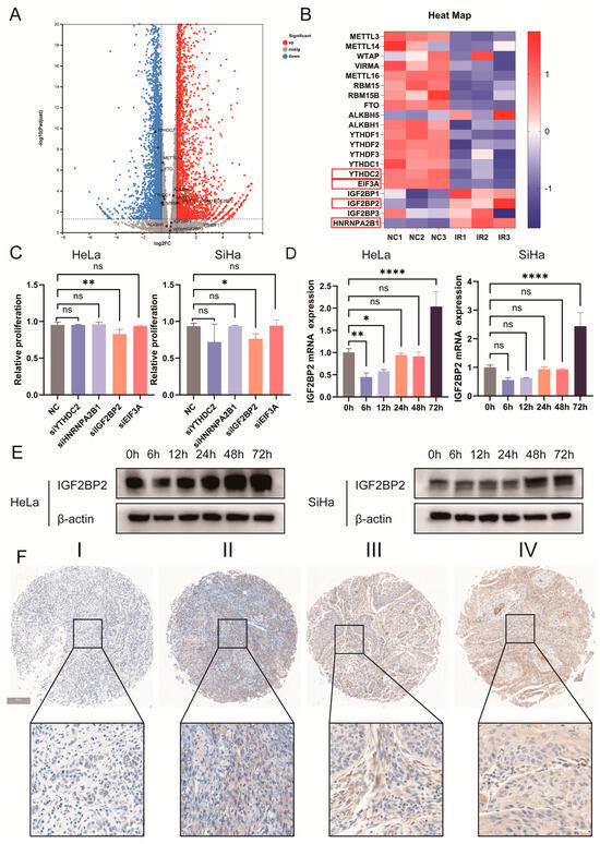
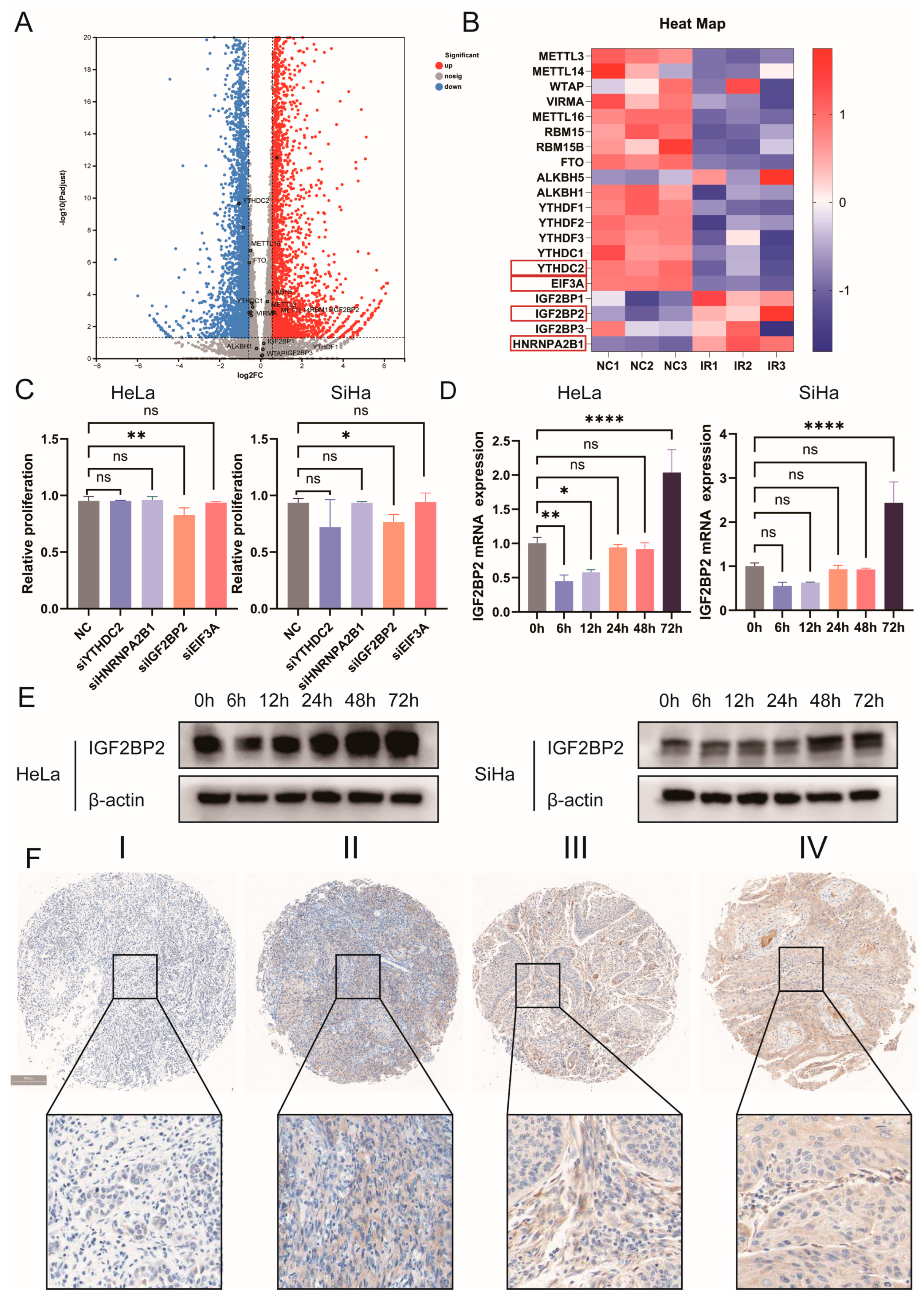
To investigate the relationship between m6A methylation and radiation resistance, we used RNA sequencing to analyze differentially expressed genes (DEGs) in cervical cancer HeLa cells before and after irradiation. By setting a threshold of |log2 fold change| ≥ 1.5 and FDR ≤ 0.05, four genes—YTHDC2, HNRNPA2B, IGF2BP2 and EIF3A—were identified from the regulated m6A gene panel (Figure 1A,B). We then knocked down each of the four genes individually, exposed the cells to radiation, and measured the survival rates of each group. Among the four m6A-related factors—YTHDC2, HNRNPA2B, IGF2BP2 and EIF3A—detected in our study, only knockdown of IGF2BP2 significantly lowered cell survival after irradiation, which suggests that IGF2BP2 contributes to radioresistance in cervical cancer cells (Figure 1C). We then used Western blotting and qPCR to examine IGF2BP2 expression at different time points following irradiation and found that IGF2BP2 expression initially decreased and then increased after irradiation (Figure 1D,E). Finally, based on IHC staining of clinical samples, we found that IGF2BP2 expression was positively correlated with clinical tumor analysis and with recurrence following postoperative radiotherapy for cervical cancer (Figure 1F, Table 1). Taken together, we screened out IGF2BP2 from multiple candidate genes and identified it as a critical regulator of radioresistance in cervical cancer, which was selected as the core target for subsequent mechanistic investigations.
Figure 1.
Identification of IGF2BP2 as a candidate mediator of radioresistance in cervical cancer. (A) Volcano plot of differentially expressed genes in HeLa cells before and after irradiation. Four candidate genes including YTHDC2, HNRNPA2B, IGF2BP2 and EIF3A were screened from m6A-related regulatory factors with thresholds of |log2 fold change| ≥ 1.5 and FDR ≤ 0.05. (B) Heatmap of m6A-regulated gene expression in HeLa cells before and after irradiation. Red indicates high gene expression, and blue indicates low gene expression. The red boxes mark four candidate m6A-reader genes: YTHDC2, HNRNPA2B, IGF2BP2, and EIF3A. (C) The CCK-8 cell viability assay showed that only the IGF2BP2 knockout group exhibited a decrease in cell survival rate following irradiation in both HeLa and SiHa cells. (D,E) RT-PCR and Western blotting were conducted to detect IGF2BP2 expression in SiHa and HeLa cells at different time points following irradiation. (F) IHC staining was used to assess IGF2BP2 expression in tumor samples at different stages. I–IV correspond to cervical cancer samples of stages I–IV, respectively. Each value represents the mean ± SD of triplicate samples (Student’s t-test). * p < 0.05, ** p < 0.01, and **** p < 0.0001, ns, not significant.
Table 1.
Correlation between IGF2BP2 expression and clinicopathological parameters in cervical cancer.
3.2. Silencing of the IGF2BP2 Gene Impairs the Proliferative Capacity of Cervical Cancer Cells
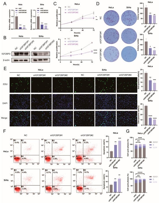
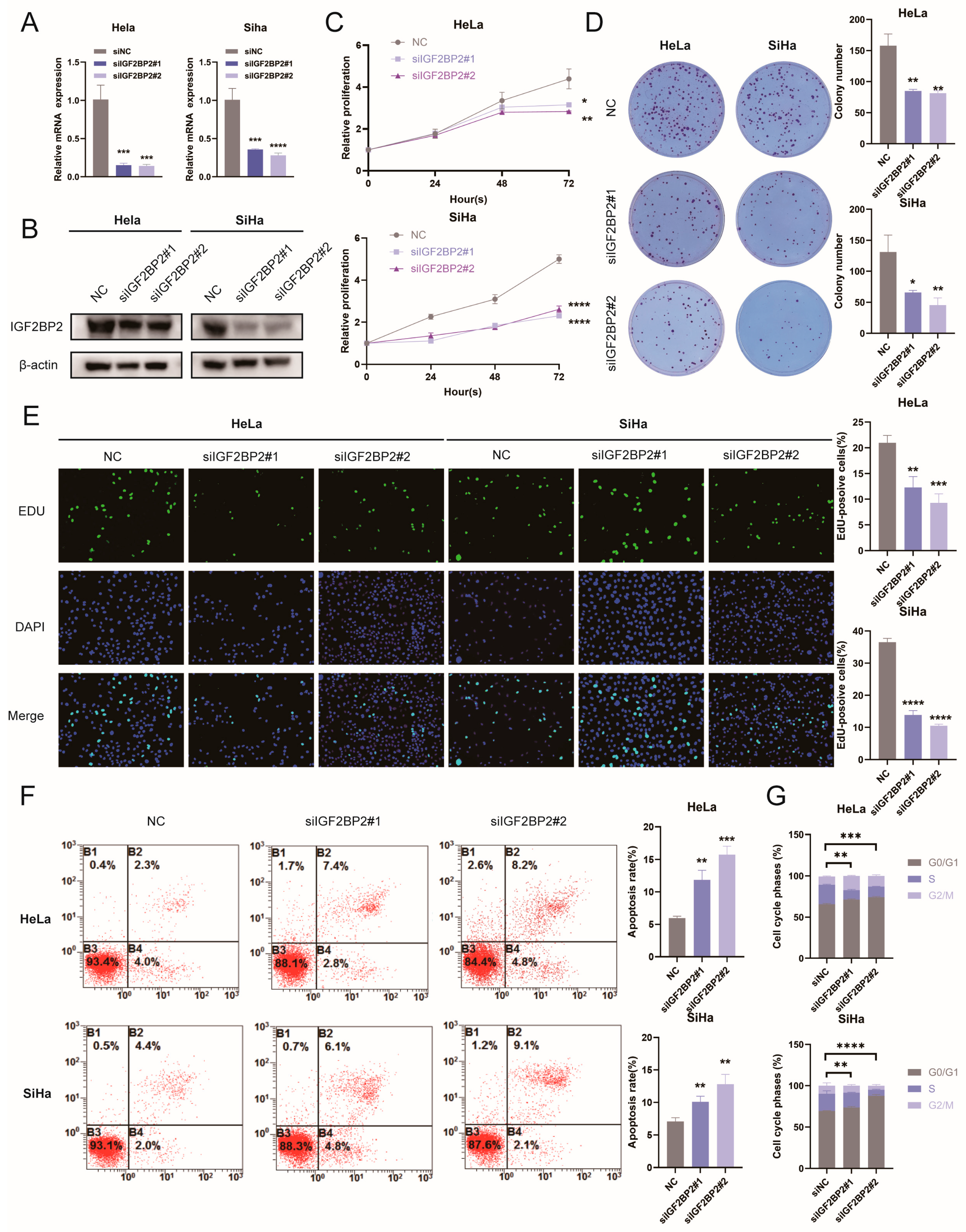
To systematically explore the biological function of IGF2BP2 in cervical cancer progression, we successfully constructed IGF2BP2 knockdown cell models in two typical cervical cancer cell lines, HeLa and SiHa, via siRNA transfection. Western blotting and qRT-PCR were performed to verify the knockdown efficiency at both protein and mRNA levels, respectively (Figure 2A,B). On the basis of the validated cell models, a series of in vitro functional experiments were conducted to evaluate the regulatory effects of IGF2BP2 on the malignant phenotypes of cervical cancer cells. Cell viability assays demonstrated that IGF2BP2 silencing markedly reduced the survival ability of cervical cancer cells (Figure 2C). Consistent with these findings, colony formation and cell proliferation assays further confirmed that depletion of IGF2BP2 significantly impaired the colony-forming potential and proliferative capacity of HeLa and SiHa cells (Figure 2D,E). To further elucidate the mechanism by which IGF2BP2 modulates cervical cancer cell growth, flow cytometry analysis was utilized to detect alterations in cell apoptosis and cell cycle distribution. The results revealed that IGF2BP2 knockdown prominently increased the apoptotic rate of cervical cancer cells and induced obvious G1-phase cell cycle arrest compared with the control group (Figure 2F,G). Collectively, these phenotypic results indicate that downregulation of IGF2BP2 suppresses cell proliferation, facilitates cell apoptosis, and blocks cell cycle progression, thereby inhibiting the malignant progression of cervical cancer. Considering the critical regulatory role of IGF2BP2 in the fundamental malignant behaviors of cervical cancer cells, we hypothesized that IGF2BP2 may also participate in modulating cellular radiotherapy responses. Subsequent radiobiological experiments were therefore performed to further clarify the functional implication of IGF2BP2 in regulating cervical cancer radioresistance.
Figure 2.
Silencing of the IGF2BP2 gene impairs the proliferative capacity of cervical cancer cells. (A,B) qRT-PCR and Western blotting were used to detect protein and mRNA expression of IGF2BP2 in HeLa and SiHa cells following siRNA silencing. (C) The CCK-8 cell proliferation assay was used to assess cell survival rates of HeLa and SiHa cells following IGF2BP2 knockout. (D) A colony formation assay was used to assess the ability of HeLa and SiHa cells to form colonies following IGF2BP2 knockout. (E) The EdU assay is used to assess HeLa and SiHa cell proliferation capacity. (F,G) Flow cytometry was performed to detect cell cycle distribution and apoptosis in IGF2BP2-silenced HeLa and SiHa cells. Each value represents the mean ± SD of triplicate samples (Student’s t-test). * p < 0.05, ** p < 0.01, *** p < 0.001, and **** p < 0.0001.
3.3. Knockdown of IGF2BP2 Impairs Radiation Resistance in Cervical Cancer Cells
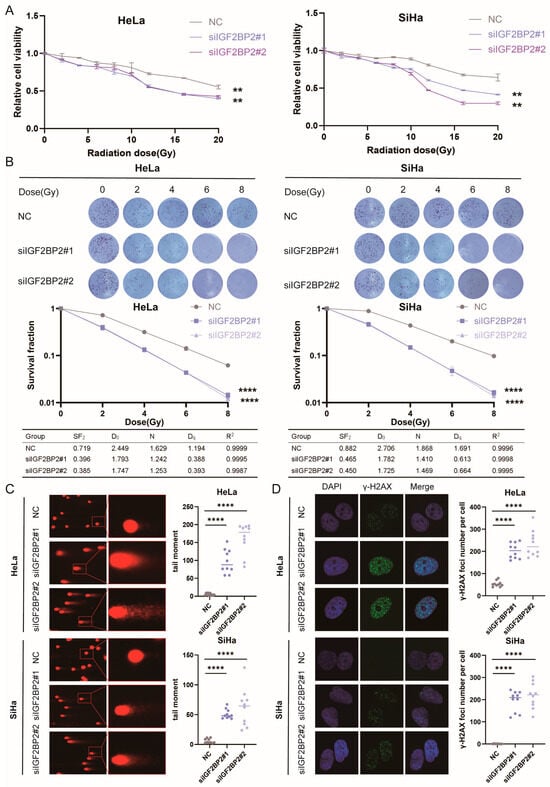
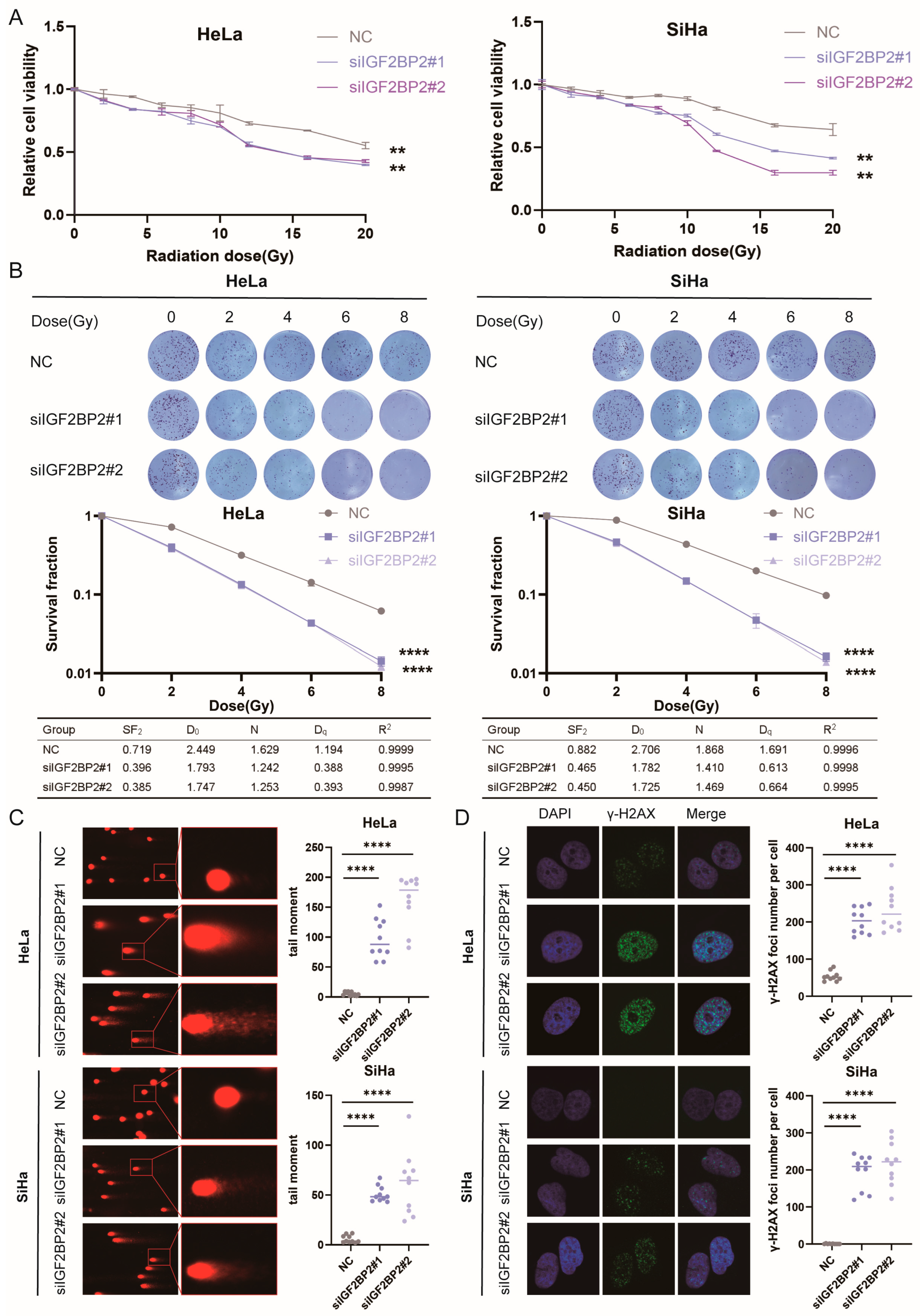
As shown above, IGF2BP2 knockdown markedly suppresses cell proliferation. To eliminate the confounding effects of basal growth defects, a series of radiobiological experiments were conducted to explore the function of IGF2BP2 in the radiation response of cervical cancer cells. First, cell viability was measured 24 h after irradiation with different doses. Normalized analysis revealed that the survival level of the IGF2BP2 knockdown group was significantly lower than that of the NC group (Figure 3A). Subsequently, cells were seeded in six-well plates and irradiated with gradient doses. The clonogenic survival data were normalized, and the single-hit multi-target model (Y = 1 − (1 − exp(−X/D0))N) was used to fit survival curves and calculate corresponding model parameters. Collectively, these results demonstrate that IGF2BP2 depletion greatly attenuates radioresistance in cervical cancer cells (Figure 3B). Since radiation-induced damage primarily results from DNA damage, we used the comet assay to assess the level of DNA damage in cells from different groups following irradiation. The results showed that the IGF2BP2 knockdown group exhibited higher levels of DNA damage, indicating lower radiation resistance (Figure 3C). Since γ-H2AX is a marker protein for double-strand breaks (DSBs) in DNA, we assessed the level of DNA damage by counting the number of γ-H2AX foci within the cells. Overall, the number of γ-H2AX foci in the cell nuclei of the IGF2BP2 knockdown group was significantly higher than that in control cells, which is consistent with the findings of the comet assay (Figure 3D). Collectively, these findings demonstrate that IGF2BP2 knockdown significantly enhances the radiosensitivity of cervical cancer cells, as evidenced by reduced normalized clonogenic survival and increased radiation-induced DNA damage, further supporting the important role of IGF2BP2 in regulating cervical cancer radioresistance.
Figure 3.
Knockdown of IGF2BP2 impairs radiation resistance in cervical cancer cells. (A) The survival rate of IGF2BP2-silenced HeLa and SiHa cells at different radiation doses was determined using the CCK-8 cell proliferation assay. (B) Colony formation assays, survival fractions, and radiological parameters (D0, N, Dq) of IGF2BP2-silenced HeLa and SiHa cells following irradiation with 0, 2, 4, 6, and 8 Gy. (C) Comet assays were performed on IGF2BP2-silenced HeLa and SiHa cells treated with IR. (D) Immunofluorescence staining of IGF2BP2-silenced HeLa and SiHa cells treated with different protocols following IR (γ-H2AX foci are green, nuclear counterstain is blue). Each value represents the mean ± SD of triplicate samples (Student’s t-test). ** p < 0.01, **** p < 0.0001.
3.4. IGF2BP2 Regulates Serine Metabolism Reprogramming via Its Downstream Target PHGDH
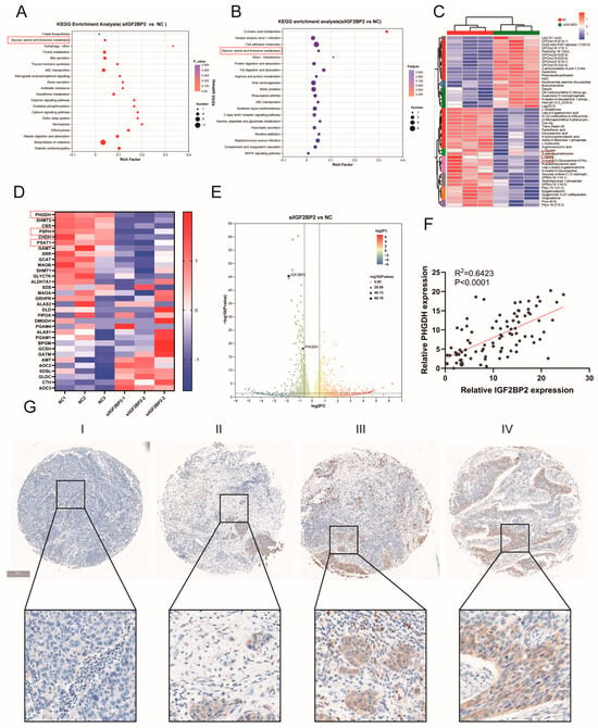
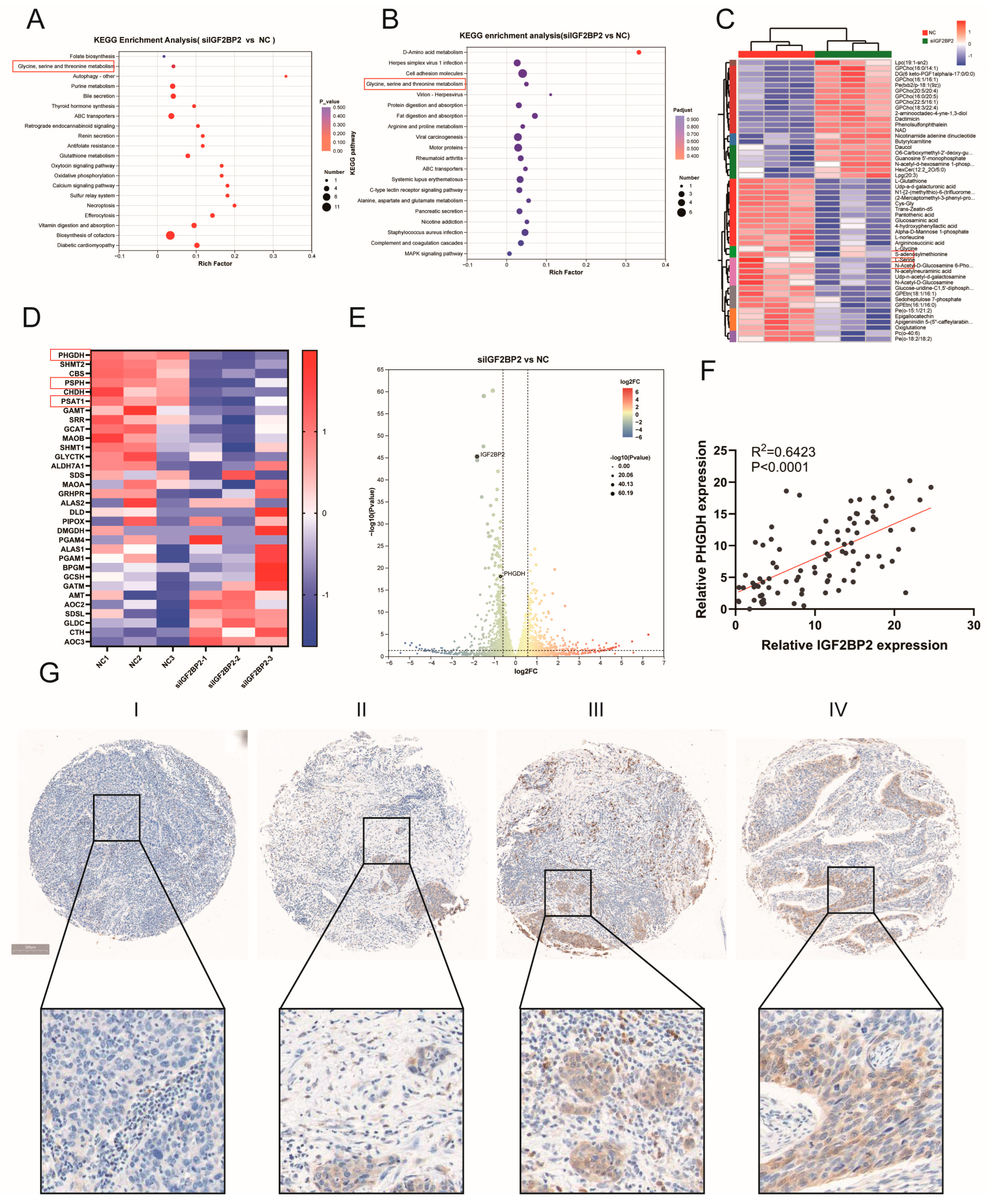
To uncover the molecular mechanisms underlying IGF2BP2-mediated radioresistance in cervical cancer, we integrated transcriptomic and metabolomic profiling by conducting KEGG enrichment analysis on differentially expressed genes (DEGs) and metabolites between IGF2BP2-knockdown and control cells. The results showed that the serine metabolism pathway was commonly enriched in both differentially expressed metabolites and DEGs (Figure 4A,B). We then found that L-Serine and L-Glycine, key products of serine metabolism, were both downregulated in the IGF2BP2 knockdown group, confirming the pathway enrichment results at the metabolite level (Figure 4C). Meanwhile, genes encoding the rate-limiting enzymes of de novo serine synthesis, including PHGDH, PSPH and PSAT1, were synchronously downregulated after IGF2BP2 knockdown. As the first and primary rate-limiting enzyme in this pathway, PHGDH exhibited the most dramatic expression alteration, further supporting the overall suppression of this pathway at the transcriptional level (Figure 4D). Among these three genes, only PHGDH satisfied the screening thresholds of |log2 fold change| ≥ 1.5 and an FDR ≤ 0.05, which designated PHGDH as the primary downstream effector of IGF2BP2 governing cervical cancer radioresistance (Figure 4E). Clinical tissue data further corroborated our multi-omics observations: IGF2BP2 expression was positively correlated with PHGDH levels (Figure 4F), and PHGDH expression was also associated with cervical cancer stage (Figure 4G). Combined with the integrated enrichment analysis of transcriptomic and metabolomic data, cellular molecular validation and correlation analysis of clinical tissue specimens, we screened PHGDH as a core downstream candidate target gene for IGF2BP2-dependent radioresistance.
Figure 4.
IGF2BP2 regulates serine metabolism reprogramming via its downstream target PHGDH. (A) KEGG pathway enrichment analysis of differentially expressed metabolites. The glycine, serine and threonine metabolism pathway is marked. (B) Enrichment results of KEGG pathways for differentially expressed genes (DEGs). The glycine, serine and threonine metabolism pathway is marked. (C) Clustering heatmap of differential metabolites. Glycine and serine are highlighted in the plot. (D) Heatmap of target genes. PHGDH, PSPH and PSAT1 are highlighted in the plot. (E) Volcano plot of differentially expressed genes, with the key candidate gene PHGDH highlighted. (F) Correlation between IGF2BP2 and PHGDH expression in TMA. (G) IHC staining demonstrates the expression of PHGDH across different tumor stages. I–IV correspond to cervical cancer samples of stages I–IV, respectively. Each value represents the mean ± SD of triplicate samples (Student’s t-test).
3.5. IGF2BP2 Regulates PHGDH mRNA Stability Through m6A Modification
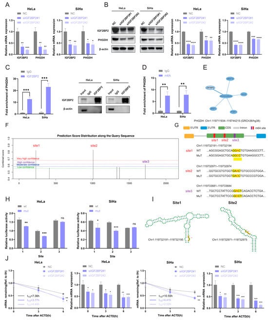
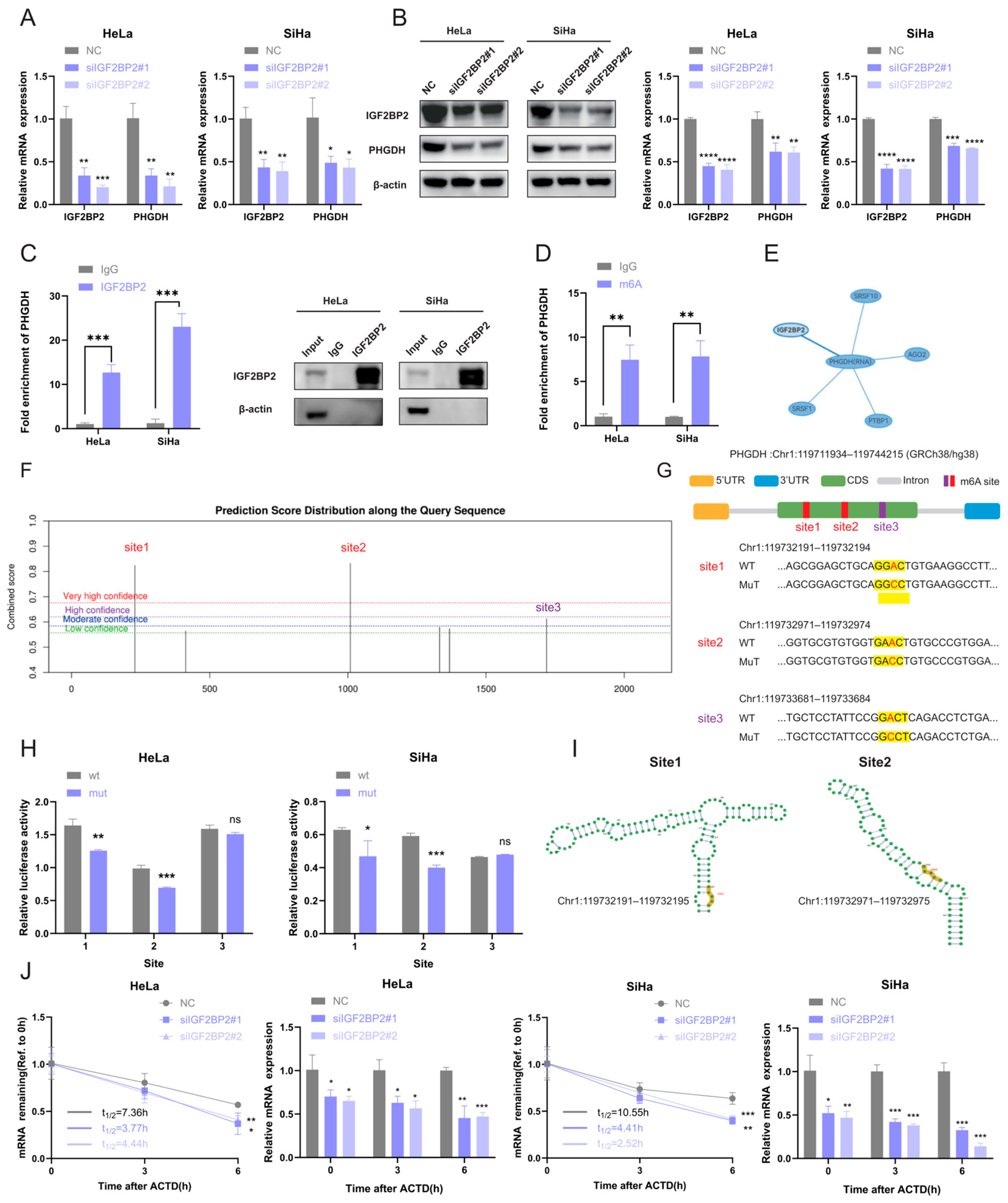
Since IGF2BP2 is widely recognized as an m6A reader and is known to promote mRNA stability and translation, we investigated whether IGF2BP2 interacts with PHGDH mRNA via m6A modification [19]. First, we found that both the mRNA (Figure 5A) and protein (Figure 5B) levels of PHGDH were reduced in IGF2BP2-silenced cells. After confirming that IGF2BP2 downregulates PHGDH expression, we performed RIP-qPCR assays on HeLa and SiHa cells using an IGF2BP2 antibody. The results showed that the enrichment of PHGDH mRNA was significantly higher than that of the IgG control, confirming that the IGF2BP2 protein directly binds to PHGDH mRNA (Figure 5C). We then employed MeRIP-qPCR to validate m6A modification of PHGDH. The PHGDH mRNA enrichment in the m6A antibody group was significantly higher than that in the IgG control, confirming the presence of m6A modification in its mRNA (Figure 5D). Next, the POSTAR3 website (http://111.198.139.65/RBS.html, accessed on 5 December 2025) was used to predict the RBP binding sites of PHGDH, with IGF2BP2 identified as one of the candidate binding proteins (Figure 5E). To identify the binding sites of IGF2BP2 on PHGDH mRNA, we used the DeepSRAMP website (http://www.cuilab.cn/deepsramp/, accessed on 5 December 2025) to predict m6A candidate sites in PHGDH, identifying two high-confidence sites and one medium-confidence site (Figure 5F). We subsequently constructed wild-type (WT) and site-mutant (MUT) plasmids (Figure 5G) and performed dual-luciferase assays. The results showed significant differences in luciferase activity between the Site 1 and Site 2 groups, while Site 3 showed no significant changes (Figure 5H), confirming that Sites 1 and 2 are functional m6A modification sites in PHGDH. To verify the accuracy of sites 1 and 2, this study predicted their three-dimensional structures using the DeepSRAMP website, confirming that the structures of both sites are correct (Figure 5I). In addition, mRNA stability analysis experiments showed that the half-life of PHGDH mRNA was reduced following IGF2BP2 silencing (Figure 5J). In summary, these findings indicate that IGF2BP2 directly binds to PHGDH mRNA via m6A modification, thereby promoting its stability and expression. Taken together, these results verify that IGF2BP2 modulates PHGDH mRNA stability in an m6A-dependent manner.
Figure 5.
IGF2BP2 regulates PHGDH mRNA stability through m6A modification. (A,B) qRT-PCR and Western blotting were used to detect the protein and mRNA expression of PHGDH in HeLa and SiHa cells following IGF2BP2 silencing. (C) RIP-qPCR analysis showing the enrichment of PHGDH mRNA in anti-IGF2BP2 and anti-IgG precipitates in HeLa and SiHa cells (left panel). Western blot detected the IGF2BP2 immunoprecipitation efficiency of the IGF2BP2 RIP assay (right panel). (D) MeRIP-qPCR analysis showing the enrichment of PHGDH mRNA in anti-m6A and anti-IgG precipitates in HeLa and SiHa cells. (E) Prediction of PHGDH mRNA RBP binding sites based on POSTAR3. (F) Predicting m6A binding sites on PHGDH mRNA using DeepSRAMP. (G) Schematic representation of PHGDH- WT (wild-type) or PHGDH- MUT (mutated-type) sequence. (H) Luciferase reporter assay was used to measure the luciferase activities of PHGDH- WT or PHGDH- MUT in HeLa and SiHa cells. (I) Prediction of the conformations of sites 1 and 2 based on the DeepSRAM. (J) Following IGF2BP2 knockdown in HeLa and SiHa cells, the mRNA half-life and expression levels of PHGDH were measured at predetermined time points following treatment with actinomycin D (5 μg/ml). Each value represents the mean ± SD of triplicate samples (Student’s t-test). * p < 0.05, ** p < 0.01, *** p < 0.001, and **** p < 0.0001, ns, not significant.
3.6. PHGDH Overexpression Partially Rescued the Decrease in Radiation Resistance Caused by IGF2BP2 Knockdown
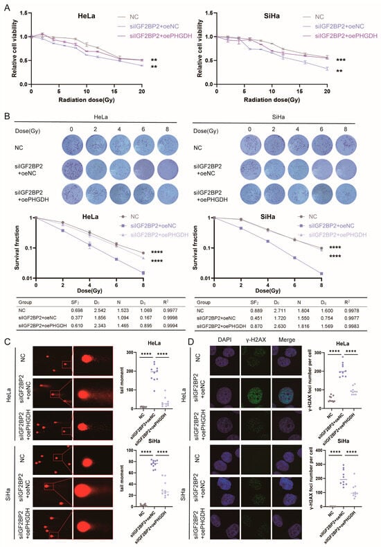
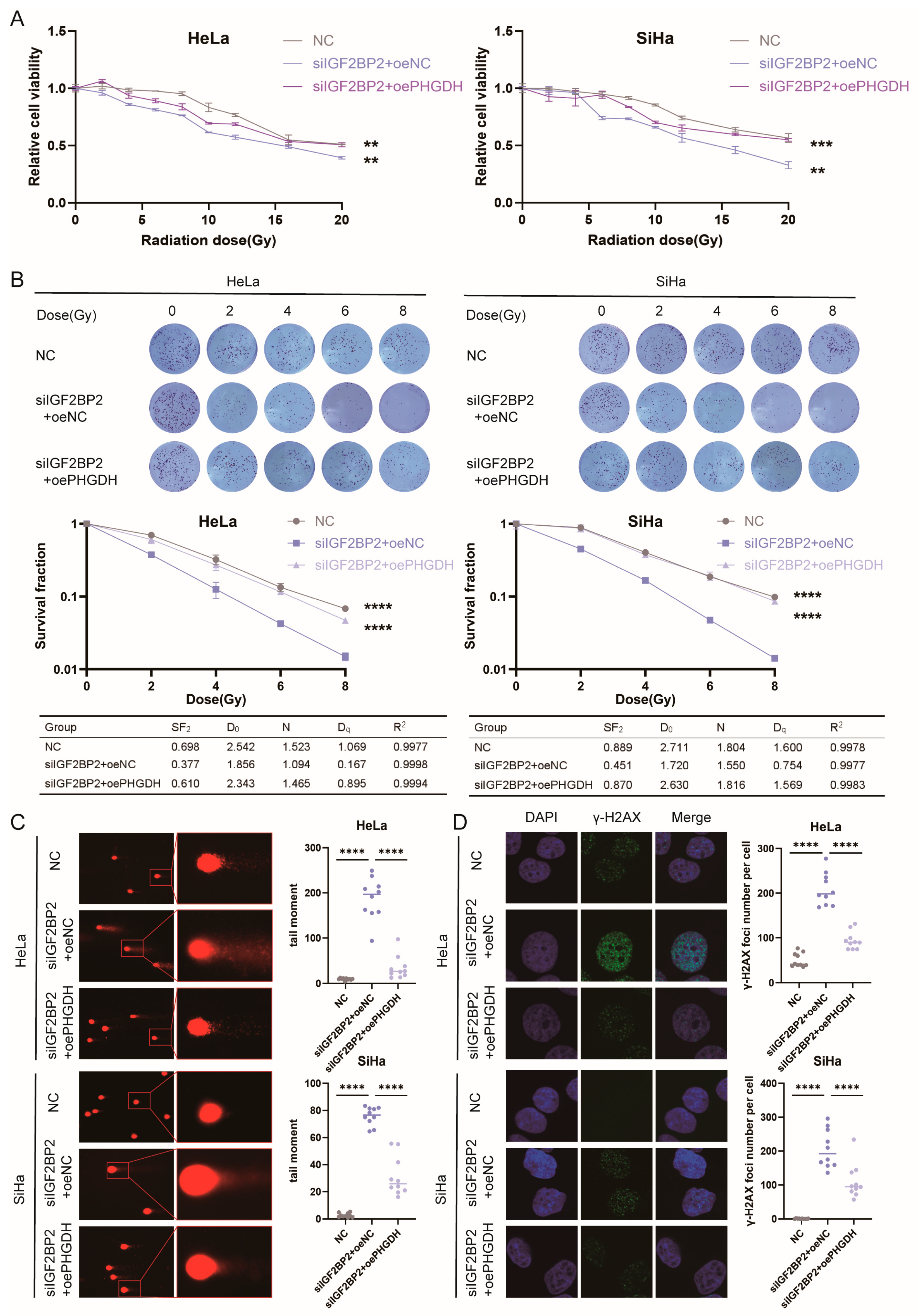
As demonstrated above, IGF2BP2 knockdown markedly attenuates radioresistance in cervical cancer cells. To verify that PHGDH acts as a functional downstream effector of IGF2BP2 in regulating radiation response, we performed rescue experiments by overexpressing PHGDH in IGF2BP2-silenced cells and confirmed that PHGDH overexpression could partially rescue the radiosensitive phenotype induced by IGF2BP2 depletion. First, through CCK-8 cell proliferation assays, we found that in cervical cancer cells with IGF2BP2 knockdown, overexpression of PHGDH can partially restore cell survival following irradiation (Figure 6A). We further used a single-hit multi-target model to fit cell survival fractions and estimate characteristic parameters. The results suggest that upregulation of PHGDH can partially restore the impaired radiation resistance caused by IGF2BP2 knockdown (Figure 6B). Consistently, comet assay-based DNA tail moment measurement (Figure 6C) and γ-H2AX foci immunofluorescence analysis (Figure 6D) confirmed that PHGDH overexpression effectively mitigated radiation-triggered DNA damage in cervical cancer cells. Consistent with our expectations, these results confirm that exogenous PHGDH restores compromised radioresistance in IGF2BP2-silenced cells. To further dissect the mechanisms by which the IGF2BP2-PHGDH axis regulates radioresistance, we performed three sets of rescue and pharmacological intervention experiments to examine how distinct downstream components of this axis modulate radiation survival. We found that supplementation with bioactive folate 5-MTHF reversed the reduction in radioresistance induced by IGF2BP2 knockdown (Figure S1A). Blocking de novo serine synthesis with the selective PHGDH inhibitor NCT-503 partially abrogated the elevated radioresistance triggered by IGF2BP2 overexpression (Figure S1B). In addition, the ROS scavenger NAC specifically restored radioresistance in IGF2BP2-knockdown cells. Collectively, the IGF2BP2-PHGDH axis governs cellular radioresistance through multiple parallel routes, including de novo serine synthesis, one-carbon metabolism and redox homeostasis.
Figure 6.
PHGDH overexpression partially rescued the decrease in radiation resistance caused by IGF2BP2 knockdown. (A) The survival rate of IGF2BP2-knockdown HeLa and SiHa cells with or without PHGDH overexpression at different radiation doses was determined using the CCK-8 cell proliferation assay. (B) Colony formation assays, survival fractions, and radiological parameters (D0, N, Dq) of IGF2BP2-knockdown HeLa and SiHa cells with or without PHGDH overexpression following irradiation with 0, 2, 4, 6, and 8 Gy. (C) Comet assays were performed on IGF2BP2-knockdown HeLa and SiHa cells with or without PHGDH overexpression treated with IR. (D) Immunofluorescence staining of IGF2BP2-knockdown HeLa and SiHa cells with or without PHGDH overexpression treated with different protocols following IR (γ-H2AX foci are green, nuclear counterstain is blue). Each value represents the mean ± SD of triplicate samples (Student’s t-test). ** p < 0.01, *** p < 0.001, and **** p < 0.0001.
3.7. IGF2BP2 Regulates Cellular Antioxidant Defense and Proliferation via the PHGDH–Serine Metabolic Axis
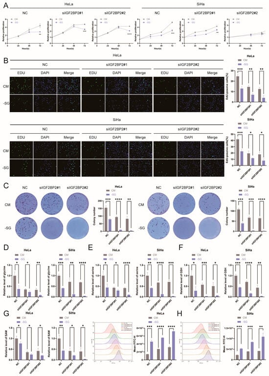
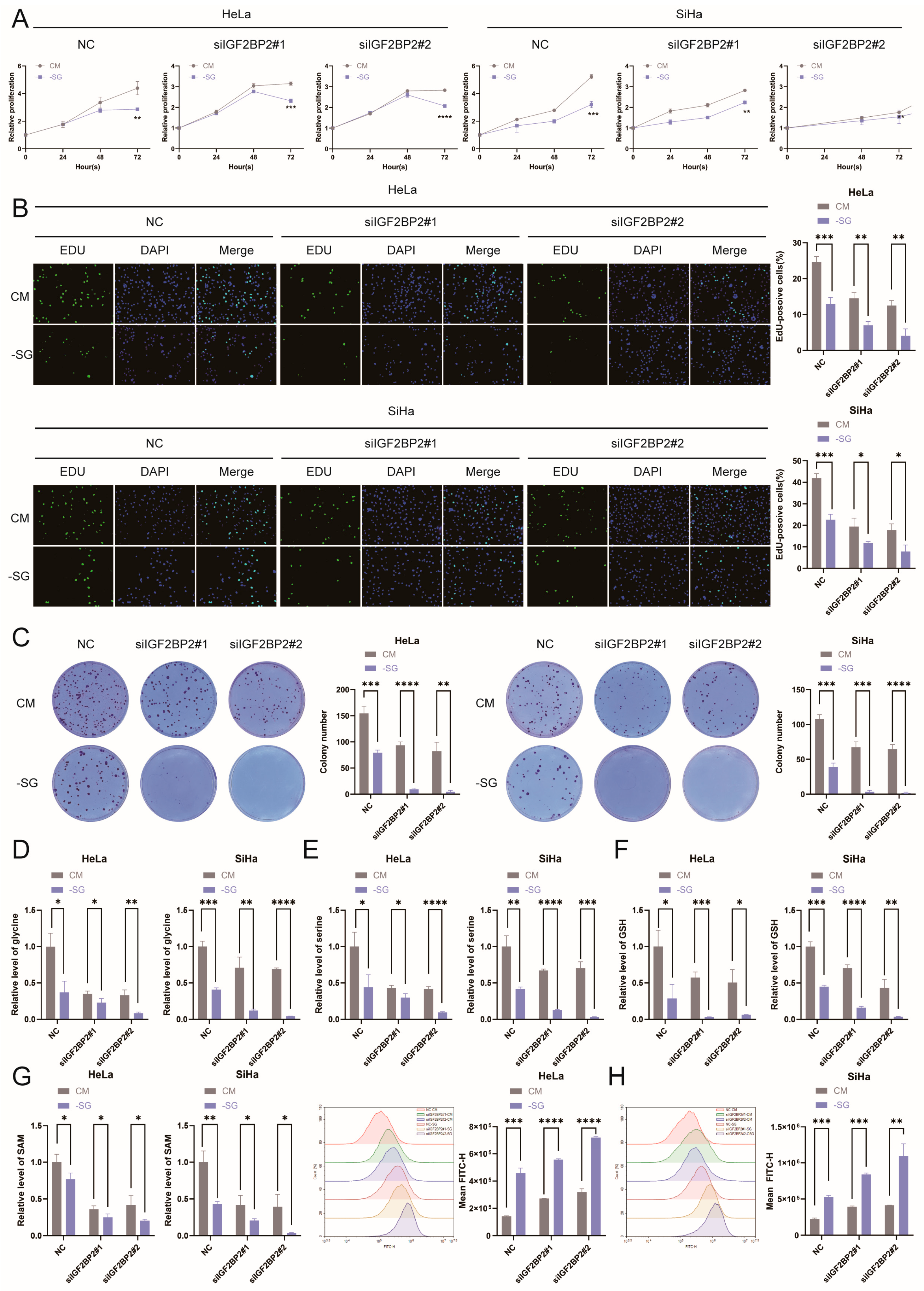
To investigate the regulatory role of serine metabolism in cervical cancer cell proliferation, we simultaneously restricted intracellular serine supply through both endogenous and exogenous pathways. On the one hand, we used IGF2BP2 knockdown to inhibit endogenous serine synthesis within cells; on the other hand, we employed DMEM supplemented without serine and glycine (-SG) conditions to block the cells’ uptake of exogenous serine. Combined results from CCK-8 (Figure 7A), EdU (Figure 7B), and colony formation (Figure 7C) assays confirmed that, regardless of whether endogenous serine was blocked, exogenous serine deprivation significantly inhibited cervical cancer cell proliferation. This finding is also consistent with current research on developing tumor intervention strategies based on limiting serine availability [20]. Serine metabolism generates one-carbon units and antioxidant metabolites, and governs tumor proliferation and genome stability [21]. Based on this, we hypothesize that depletion of both endogenous and exogenous serine can downregulate cellular antioxidant capacity and reduce levels of methyl donors. To test this hypothesis, this study employed mass spectrometry to quantitatively measure intracellular levels of serine, glycine, GSH, and SAM. The results were consistent with our expectations: following exogenous serine deprivation, intracellular levels of serine (Figure 7D) and glycine (Figure 7E) decreased significantly, accompanied by a concurrent decline in GSH (Figure 7F) and SAM spectrometry (Figure 7G) levels. Furthermore, under conditions of combined endogenous and exogenous serine depletion, changes in intracellular reactive oxygen species levels were consistent with the above findings, further supporting the notion that serine deficiency inhibits cellular antioxidant capacity (Figure 7H). In summary, IGF2BP2 can upregulate PHGDH expression; as the key rate-limiting enzyme in serine de novo synthesis, PHGDH drives intracellular serine production, thereby enhancing cellular antioxidant reserves.
Figure 7.
IGF2BP2 regulates cellular antioxidant defense and proliferation via the PHGDH–serine metabolic axis. (A) The CCK-8 cell proliferation assay was used to assess cell survival rates of HeLa and SiHa cells. CM: complete medium; -SG: serine- and glycine-deprived medium, consistent with the descriptions below. (B) The EdU assay is used to assess HeLa and SiHa cell proliferation capacity. (C) A colony formation assay was used to assess the ability of HeLa and SiHa cells to form colonies. (D–G) Determination of serine, glycine, GSH, and SAM levels using mass spectrometry. (H) The mean fluorescence intensity (MFI) of intracellular ROS was quantified by flow cytometry. Each value represents the mean ± SD of triplicate samples (Student’s t-test). * p < 0.05, ** p < 0.01, *** p < 0.001, and **** p < 0.0001.
3.8. In Vivo Validation of IGF2BP2 Regulation of PHGDH-Mediated Radioresistance
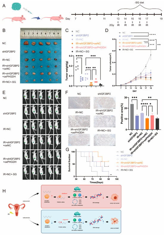
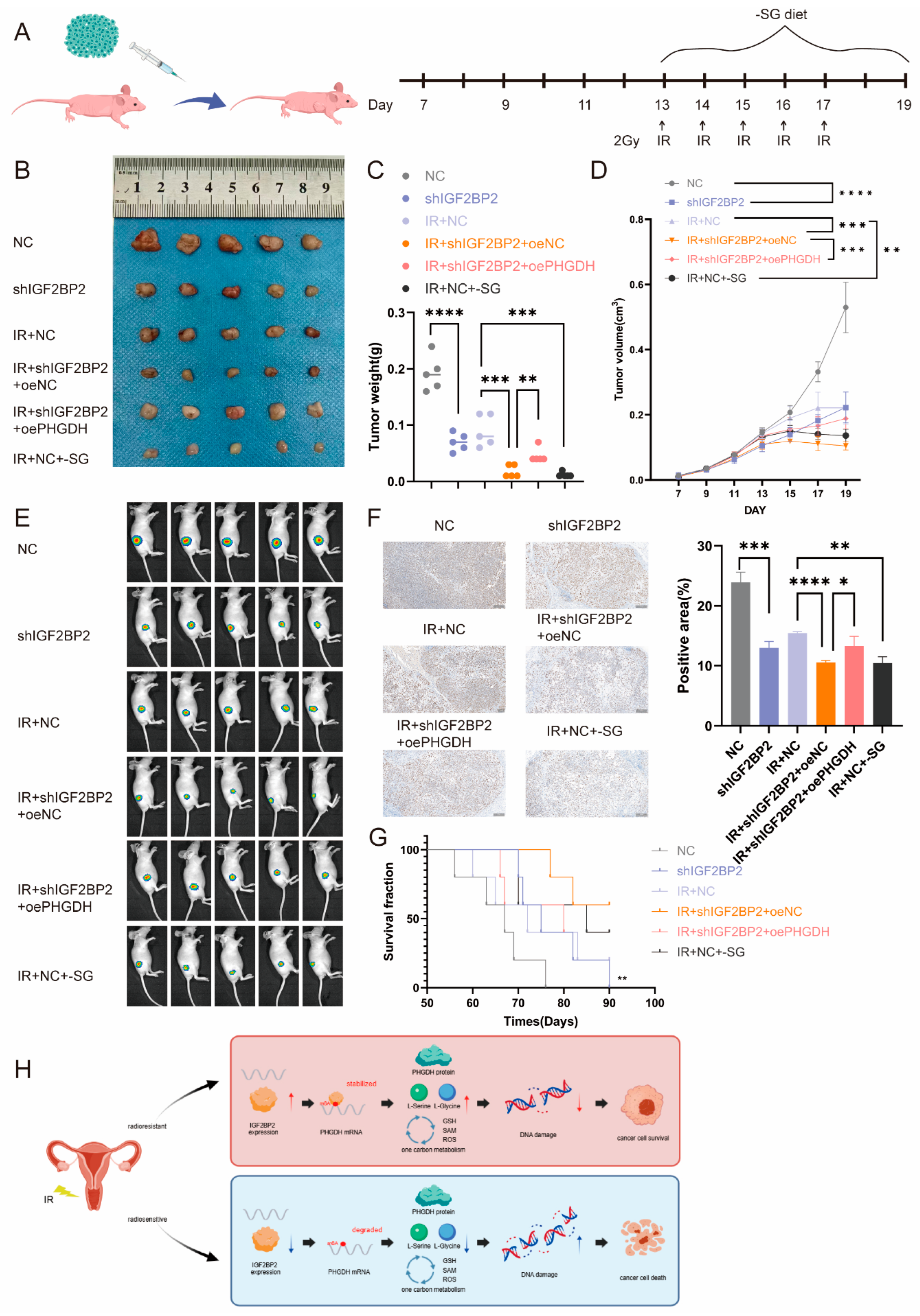
To investigate the in vivo function of the IGF2BP2-PHGDH–serine axis in cervical cancer radioresistance, six groups of subcutaneous xenograft models were established in nude mice using genetically modified SiHa cells. Lentiviral transfection was applied to construct shNC and shIGF2BP2 cell lines, and empty oe-NC or oe-PHGDH plasmids were transfected into shIGF2BP2 cells for rescue experiments. Group 1 mice were implanted with shNC cells and maintained on standard diet without radiation treatment, while Group 2 bore shIGF2BP2 cells under the same non-irradiation and normal diet conditions. Starting at day 13 after cell injection, mice in Groups 3 to 6 received daily 2 Gy irradiation for five consecutive days. Among these irradiated groups, Group 3 carried shNC cells with standard diet, Group 4 was injected with shIGF2BP2 + oeNC cells with standard diet, Group 5 was implanted with shIGF2BP2 + oePHGDH rescue cells with standard diet, and Group 6 bore shNC cells but received a serine/glycine-deficient diet from day 13 onwards (Figure 8A). Analysis of final tumor volume revealed that tumor volumes in the IR group were consistently smaller than those in the non-IR group. Within the non-IR group, the IGF2BP2 knockdown group exhibited significantly smaller tumor volumes than the control group. In the IR group, both IGF2BP2 knockdown and a serine-restricted diet led to further reductions in tumor volume. However, in the IGF2BP2 knockdown xenograft model, restoring PHGDH expression reversed the antitumor effect, causing tumor volume to increase again (Figure 8B). This confirms that inhibiting the IGF2BP2-PHGDH pathway or adopting a serine-restricted diet can overcome radiation resistance in cervical cancer. This conclusion was consistently validated by the results of multiple assays, including tumor weight measurements (Figure 8C), tumor growth curves (Figure 8D), in vivo fluorescence imaging (Figure 8E), Ki-67 proliferation staining (Figure 8F), and survival analysis (Figure 8G). We propose a regulatory model for IGF2BP2-mediated cervical cancer radioresistance (Figure 8H). The in vivo results were consistent with the conclusions drawn from our preceding in vitro cell experiments.
Figure 8.
In vivo validation of IGF2BP2 regulation of PHGDH-mediated radioresistance. (A) Schematic diagram of the construction of a nude mouse tumor model and the experimental workflow. IR: Local irradiation at 2 Gy per day for 5 successive days starting at day 13. -SG: Mice were fed a serine/glycine-free diet from day 13 until the end of the experiment. (B) General view of tumor mass in each indicated group at 19 days. (C) The weight of the xenografted tumors was measured. (D) From day 7 after injection, the tumor volume was measured every two days. (E) Tumor images were acquired using the PerkinElmer IVIS preclinical in vivo imaging system for 19 days. (F) Tumor sections were stained by immunohistochemistry using antibodies against Ki-67. (G) Kaplan–Meier survival curve in each group. (H) Working model showing how the IGF2BP2-PHGDH–serine metabolic axis drives cervical cancer radioresistance. The red panel indicates the radioresistant state, and the blue panel indicates the radiosensitive state. Arrows represent promoting effects, while blunt-ended arrows indicate inhibitory effects. Each value represents the mean ± SD of triplicate samples (Student’s t-test). (n = 5) * p < 0.05, ** p < 0.01, *** p < 0.001, and **** p < 0.0001.
4. Discussion
Radiotherapy serves as the standard first-line therapeutic regimen for locally advanced cervical cancer, yet radioresistance markedly impairs therapeutic efficacy and constitutes a primary cause of treatment failure and patient mortality [2]. Approximately 30% of patients with advanced cervical cancer develop intrinsic or acquired radioresistance following radiotherapy [22]. Accordingly, identifying core molecular drivers responsible for cervical cancer radioresistance and developing targeted intervention strategies are urgently required to enhance radiotherapy response and reduce postoperative tumor recurrence.
Numerous previous studies have utilized high-throughput screening to identify that epigenetic modifications modulate cervical cancer radioresistance via regulating tumor metabolic reprogramming, miRNA-mediated signaling and DNA damage repair [23,24,25]. Nevertheless, few studies have combined m6A reader proteins with the de novo serine synthesis axis to fully delineate the upstream and downstream molecular cascade through which they cooperatively regulate radioresistance in cervical cancer. To fill this research gap, the present study integrated in vitro cellular experiments and in vivo xenograft models to systematically dissect the IGF2BP2-PHGDH–serine metabolic regulatory network and explore intervention strategies to reverse cervical cancer radioresistance.
As a canonical m6A reader, IGF2BP2 remodels multiple malignant phenotypes including metabolic reprogramming, immune escape, metastasis and cell cycle progression by stabilizing target mRNAs [26,27,28,29]. Accumulating evidence indicates that IGF2BP2 modulates both chemoresistance and radioresistance in malignancies. For instance, IGF2BP2 induces docetaxel resistance in prostate cancer through boosting glycolytic flux, and it also confers radioresistance to lung cancer, glioblastoma, gastric cancer, esophageal cancer and hepatocellular carcinoma [30,31,32,33,34,35]. Consistent with previous findings, our work extends current knowledge of IGF2BP2-driven tumor radioresistance to cervical cancer and confirms its oncogenic and radioresistance-promoting roles in cervical cancer cells. Notably, the IGF2BP2–PHGDH serine-synthesis axis was previously described in thyroid carcinoma [36]. Consistent with our data, IGF2BP2 stabilizes PHGDH mRNA in an m6A-dependent fashion to boost serine metabolism. Unlike that report focusing on tumor proliferation, our study links this metabolic cascade to radioresistance in cervical cancer. This highlights a context-dependent function of the IGF2BP2–PHGDH axis in radiation response.
We next focused on serine metabolism, a core metabolic hub bridging glycolysis, the tricarboxylic acid cycle, and one-carbon metabolism. Serine supports rapid tumor proliferation by providing precursors for nucleic acid, lipid, and protein biosynthesis. In addition, it sustains intracellular redox homeostasis via glutathione production and supplies methyl donors for epigenetic modification [10]. The de novo serine synthesis pathway (SSP) is well established to maintain tumor cell viability, preserve cancer stem cell stemness, and stabilize redox balance in hypoxic and nutrient-deficient microenvironments, thereby promoting tumor radioresistance [37]. PHGDH acts as the rate-limiting enzyme initiating the SSP and directly determines intracellular serine abundance [12]. Although the correlation between SSP activation and tumor radioresistance has been documented, the specific role of PHGDH in regulating radiosensitivity remains poorly investigated. To date, only one glioma study has reported the pro-radioresistant function of PHGDH [38], while its biological role and upstream regulatory mechanism in cervical cancer radioresistance remain completely uncharacterized, which constitutes a critical unresolved research gap. Through rescue experiments, our study thoroughly fills this unaddressed research gap and provides the first evidence that PHGDH overexpression reverses the radiosensitizing effect induced by IGF2BP2 knockdown in cervical cancer.
This study still has several limitations. First, the subcutaneous tumor-bearing model used only the cervical cancer cell line SiHa, which cannot fully replicate the complex native microenvironment of in situ cervical cancer. Second, the in vivo intervention involved only short-term treatment with a serine-glycine-restricted diet; the absence of long-term dietary restriction models and corresponding toxicological assessments means the findings are insufficient to directly support clinical dietary adjunctive therapy regimens. Third, it remains necessary to incorporate clinical cohorts with complete radiotherapy follow-up data to further validate the predictive value of IGF2BP2 and PHGDH expression in patient samples. Future work could involve establishing in situ tumor models, expanding clinical sample cohorts, and developing targeted small-molecule inhibitors to facilitate the translation of these basic research findings into clinical practice.
5. Conclusions
Our study fully delineates the complete regulatory cascade: IGF2BP2 binds to m6A-modified PHGDH mRNA and enhances its transcript stability, thereby aberrantly activating de novo serine synthesis and ultimately driving radioresistance in cervical cancer. The IGF2BP2-PHGDH–serine metabolic axis identified in this work possesses prominent translational value. Clinically, the expression levels of IGF2BP2 and core SSP enzymes in tumor tissues can be detected to establish a stratified evaluation system, serving as novel molecular biomarkers for predicting radiotherapy response, long-term recurrence risk and survival prognosis of cervical cancer patients, and providing evidence for individualized stratified clinical management. More importantly, targeted inhibition of IGF2BP2 or dietary restriction of serine and glycine can effectively eliminate the metabolic dependency of tumor cells; these approaches can be adopted as adjuvant combinatorial strategies alongside radiotherapy to overcome intrinsic and acquired radioresistance in locally advanced cervical cancer. For cervical cancer patients with a high risk of radiotherapy failure, this combined metabolic intervention is expected to reduce the rate of treatment failure, slow tumor progression and maximize the clinical benefits of comprehensive therapy.
Supplementary Materials
The following supporting information can be downloaded at: https://www.mdpi.com/article/10.3390/antiox15111468/s1, Table S1: The sequences of siRNAs, shRNAs and plasmids. Table S2: Primer sequences. Table S3: antibodies information. Figure S1: Dissection of downstream mechanisms by which the IGF2BP2-PHGDH axis regulates radioresistance.
Author Contributions
Conceptualization, J.R., S.Y. and C.H.; methodology, J.R.; software, J.R.; validation, J.R., L.G.; formal analysis, J.R.; investigation, J.R., L.G., Y.S., R.Z., Y.H., F.H. and X.D.; resources, S.Y.; data curation, J.R.; writing—original draft preparation, J.R.; writing—review and editing, C.H. and S.Y.; visualization, J.R.; supervision, J.R.; project administration, Y.S.; funding acquisition, S.Y. and K.Y. All authors have read and agreed to the published version of the manuscript.
Funding
This research was funded by The National Natural Science Foundation of China, grant number 8237282 and 82573107; The Young Talent Fund of Association for Science and Technology in Shaanxi, China, grant number 20240324; Key Laboratory Projects of the Shaanxi Provincial Natural Science Basic Research Program, grant number 2025SYS-SYSZD-028.
Institutional Review Board Statement
This study was performed in accordance with the ethical principles of the Declaration of Helsinki and approved by the Ethics Committee of Liaocheng Second People’s Hospital (Approval No. KB2025-Ethics-48-01). The animal experimental protocol was approved by the Institutional Ethics Committee of Fourth Military Medical University (Approval No. 251250).
Informed Consent Statement
Informed consent was obtained from all subjects involved in the study.
Data Availability Statement
The original contributions presented in this study are included in the article/Supplementary Material. Further inquiries can be directed to the corresponding author(s).
Conflicts of Interest
The authors declare no conflicts of interest.
References
- Bray, F.; Ferlay, J.; Soerjomataram, I.; Siegel, R.L.; Torre, L.A.; Jemal, A. Global cancer statistics 2018: GLOBOCAN estimates of incidence and mortality worldwide for 36 cancers in 185 countries. CA Cancer J. Clin. 2018, 68, 394–424. [Google Scholar] [CrossRef] [Scilit] [PubMed]
- Jiang, W.; Huang, G.; Pan, S.; Chen, X.; Liu, T.; Yang, Z.; Chen, T.; Zhu, X. TRAIL-driven targeting and reversing cervical cancer radioresistance by seleno-nanotherapeutics through regulating cell metabolism. Drug Resist. Update 2024, 72, 101033. [Google Scholar] [CrossRef] [Scilit] [PubMed]
- Moreno-Acosta, P.; Vallard, A.; Carrillo, S.; Gamboa, O.; Romero-Rojas, A.; Molano, M.; Acosta, J.; Mayorga, D.; Rancoule, C.; Garcia, M.A.; et al. Biomarkers of resistance to radiation therapy: A prospective study in cervical carcinoma. Radiat. Oncol. 2017, 12, 120. [Google Scholar] [CrossRef] [Scilit] [PubMed]
- Liu, Y.P.; Zheng, C.C.; Huang, Y.N.; He, M.L.; Xu, W.W.; Li, B. Molecular mechanisms of chemo- and radiotherapy resistance and the potential implications for cancer treatment. MedComm 2021, 2, 315–340. [Google Scholar] [CrossRef] [Scilit] [PubMed]
- Shen, R.; Jiang, Z.; Wang, H.; Zheng, Z.; Jiang, X. Molecular mechanisms of m6A modifications regulating tumor radioresistance. Mol. Med. 2025, 31, 64. [Google Scholar] [CrossRef] [Scilit] [PubMed]
- Yue, Y.; Liu, J.; He, C. RNA N6-methyladenosine methylation in post-transcriptional gene expression regulation. Genes. Dev. 2015, 29, 1343–1355. [Google Scholar] [CrossRef] [Scilit] [PubMed]
- Sun, T.; Wu, R.; Ming, L. The role of m6A RNA methylation in cancer. Biomed. Pharmacother. 2019, 112, 108613. [Google Scholar] [CrossRef] [Scilit] [PubMed]
- Liu, S.; Liao, S.; He, J.; Zhou, Y.; He, Q. IGF2BP2: An m(6)A reader that affects cellular function and disease progression. Cell Mol. Biol. Lett. 2025, 30, 43. [Google Scholar] [CrossRef] [Scilit] [PubMed]
- Wang, J.; Chen, L.; Qiang, P. The role of IGF2BP2, an m6A reader gene, in human metabolic diseases and cancers. Cancer Cell Int. 2021, 21, 99. [Google Scholar] [CrossRef] [Scilit] [PubMed]
- Amelio, I.; Cutruzzolá, F.; Antonov, A.; Agostini, M.; Melino, G. Serine and glycine metabolism in cancer. Trends Biochem. Sci. 2014, 39, 191–198. [Google Scholar] [CrossRef] [Scilit] [PubMed]
- Kalhan, S.C.; Uppal, S.O.; Moorman, J.L.; Bennett, C.; Gruca, L.L.; Parimi, P.S.; Dasarathy, S.; Serre, D.; Hanson, R.W. Metabolic and genomic response to dietary isocaloric protein restriction in the rat. J. Biol. Chem. 2011, 286, 5266–5277. [Google Scholar] [CrossRef] [Scilit] [PubMed]
- Pan, S.; Fan, M.; Liu, Z.; Li, X.; Wang, H. Serine, glycine and one-carbon metabolism in cancer. Int. J. Oncol. 2021, 58, 158–170. [Google Scholar] [CrossRef] [Scilit] [PubMed]
- Sowers, M.L.; Herring, J.; Zhang, W.; Tang, H.; Ou, Y.; Gu, W.; Zhang, K. Analysis of glucose-derived amino acids involved in one-carbon and cancer metabolism by stable-isotope tracing gas chromatography mass spectrometry. Anal. Biochem. 2019, 566, 1–9. [Google Scholar] [CrossRef] [Scilit] [PubMed]
- Newman, A.C.; Maddocks, O.D.K. One-carbon metabolism in cancer. Br. J. Cancer 2017, 116, 1499–1504. [Google Scholar] [CrossRef] [Scilit] [PubMed]
- Zhang, X.; Jiang, D.; Yang, S.; Sun, Y.; Liu, Y.; Shi, J.; Hu, C.; Pan, J.; Liu, T.; Jin, B.; et al. BAP31 Promotes Tumor Cell Proliferation by Stabilizing SERPINE2 in Hepatocellular Carcinoma. Front. Cell Dev. Biol. 2020, 8, 607906. [Google Scholar] [CrossRef] [Scilit] [PubMed]
- Puck, T.T.; Marcus, P.I. Action of x-rays on mammalian cells. J. Exp. Med. 1956, 103, 653–666. [Google Scholar] [CrossRef] [Scilit] [PubMed]
- Atwood, K.C.; Norman, A. On the Interpretation of Multi-Hit Survival Curves. Proc. Natl. Acad. Sci. USA 1949, 35, 696–709. [Google Scholar] [CrossRef] [Scilit] [PubMed]
- Xu, F.; Song, C.; Liu, W.; Chen, G. Protocol for intracellular and extracellular metabolite detection in human embryonic stem cells. STAR Protoc. 2021, 2, 100740. [Google Scholar] [CrossRef] [Scilit] [PubMed]
- An, Y.; Duan, H. The role of m6A RNA methylation in cancer metabolism. Mol. Cancer 2022, 21, 14. [Google Scholar] [CrossRef] [Scilit] [PubMed]
- Maddocks, O.D.; Berkers, C.R.; Mason, S.M.; Zheng, L.; Blyth, K.; Gottlieb, E.; Vousden, K.H. Serine starvation induces stress and p53-dependent metabolic remodelling in cancer cells. Nature 2013, 493, 542–546. [Google Scholar] [CrossRef] [Scilit] [PubMed]
- Locasale, J.W. Serine, glycine and one-carbon units: Cancer metabolism in full circle. Nat. Rev. Cancer 2013, 13, 572–583. [Google Scholar] [CrossRef] [Scilit] [PubMed]
- Schwarz, J.K.; Siegel, B.A.; Dehdashti, F.; Grigsby, P.W. Association of posttherapy positron emission tomography with tumor response and survival in cervical carcinoma. JAMA 2007, 298, 2289–2295. [Google Scholar] [CrossRef] [Scilit] [PubMed]
- Hu, S.; Zhan, N.; Li, J.; Wang, L.; Liu, Y.; Jin, K.; Wang, Y.; Zhang, J.; Chen, Y.; Bai, Y.; et al. RBM15 recruits myeloid-derived suppressor cells via the m6A-IGF2BP3/CBR3-AS1/miR-409-3p/CXCL1 axis, facilitating radioresistance in non-small-cell lung cancer. J. Transl. Med. 2025, 23, 191. [Google Scholar] [CrossRef] [Scilit] [PubMed]
- Dai, Z.; Lin, B.; Qin, M.; Lin, Y.; Wang, L.; Liao, K.; Xie, G.; Wang, F.; Zhang, J. METTL3-mediated m6A modification of SLC7A11 enhances nasopharyngeal carcinoma radioresistance by inhibiting ferroptosis. Int. J. Biol. Sci. 2025, 21, 1837–1851. [Google Scholar] [CrossRef] [Scilit] [PubMed]
- Yu, M.; Ni, M.; Xu, F.; Liu, C.; Chen, L.; Li, J.; Xia, S.; Diao, Y.; Chen, J.; Zhu, J.; et al. NSUN6-mediated 5-methylcytosine modification of NDRG1 mRNA promotes radioresistance in cervical cancer. Mol. Cancer 2024, 23, 139. [Google Scholar] [CrossRef] [Scilit] [PubMed]
- Weng, H.; Huang, F.; Yu, Z.; Chen, Z.; Prince, E.; Kang, Y.; Zhou, K.; Li, W.; Hu, J.; Fu, C.; et al. The m(6)A reader IGF2BP2 regulates glutamine metabolism and represents a therapeutic target in acute myeloid leukemia. Cancer Cell 2022, 40, 1566–1582.e1510. [Google Scholar] [CrossRef] [Scilit] [PubMed]
- Tang, R.; Zhang, Z.; Liu, X.; Liao, Y.; Chen, Y.; Xiao, M.; Li, Y.; Zhou, C.; Tan, Z.; Zhang, C.; et al. Stromal Stiffness-Regulated IGF2BP2 in Pancreatic Cancer Drives Immune Evasion via Sphingomyelin Metabolism. Gastroenterology 2025, 169, 615–631.e632. [Google Scholar] [CrossRef] [Scilit] [PubMed]
- Fang, H.; Sun, Q.; Zhou, J.; Zhang, H.; Song, Q.; Zhang, H.; Yu, G.; Guo, Y.; Huang, C.; Mou, Y.; et al. m(6)A methylation reader IGF2BP2 activates endothelial cells to promote angiogenesis and metastasis of lung adenocarcinoma. Mol. Cancer 2023, 22, 99. [Google Scholar] [CrossRef] [Scilit] [PubMed]
- Xia, T.; Dai, X.Y.; Sang, M.Y.; Zhang, X.; Xu, F.; Wu, J.; Shi, L.; Wei, J.F.; Ding, Q. IGF2BP2 Drives Cell Cycle Progression in Triple-Negative Breast Cancer by Recruiting EIF4A1 to Promote the m6A-Modified CDK6 Translation Initiation Process. Adv. Sci. 2024, 11, e2305142. [Google Scholar] [CrossRef] [Scilit] [PubMed]
- Jiang, X.; Guo, S.; Wang, S.; Zhang, Y.; Chen, H.; Wang, Y.; Liu, R.; Niu, Y.; Xu, Y. EIF4A3-Induced circARHGAP29 Promotes Aerobic Glycolysis in Docetaxel-Resistant Prostate Cancer through IGF2BP2/c-Myc/LDHA Signaling. Cancer Res. 2022, 82, 831–845. [Google Scholar] [CrossRef] [Scilit] [PubMed]
- Zhou, Z.; Zhang, B.; Deng, Y.; Deng, S.; Li, J.; Wei, W.; Wang, Y.; Wang, J.; Feng, Z.; Che, M.; et al. FBW7/GSK3β mediated degradation of IGF2BP2 inhibits IGF2BP2-SLC7A5 positive feedback loop and radioresistance in lung cancer. J. Exp. Clin. Cancer Res. 2024, 43, 34. [Google Scholar] [CrossRef] [Scilit] [PubMed]
- Dong, Q.; Huo, J.; Yan, Z.; Li, J.; Chen, X.; Li, L.; Zhen, H. POT1-AS1/IGF2BP2/GPX4 promotes temozolomide resistance and radioresistance in glioblastoma by inhibiting ferroptosis. Int. J. Radiat. Biol. 2026, 102, 49–60. [Google Scholar] [CrossRef] [Scilit] [PubMed]
- Zhang, Q.; Liu, X.; Chen, Y.; Wang, R.; Zhao, S.; Tian, Y.; Li, S.; Liu, X. IGF2BP2 regulates gastric cancer radiotherapy resistance through HIF1α-mediated glycolysis. Front. Oncol. 2025, 15, 1512177. [Google Scholar] [CrossRef] [Scilit] [PubMed]
- Yao, X.; Feng, C.; Huang, X.; Wu, S.; Lu, S.; Gao, Y.; Sun, T.; Bai, X.; Li, C.; Jia, K.; et al. P4HA2 interacted with ATAD3A to modulate PINK1/parkin-dependent mitophagy and (125)I brachytherapy sensitization in esophageal carcinoma. Cell Death Dis. 2025, 16, 685. [Google Scholar] [CrossRef] [Scilit] [PubMed]
- Hu, P.; Lin, L.; Huang, T.; Li, Z.; Xiao, M.; Guo, H.; Chen, G.; Liu, D.; Ke, M.; Shan, H.; et al. Circular RNA circEYA3 promotes the radiation resistance of hepatocellular carcinoma via the IGF2BP2/DTX3L axis. Cancer Cell Int. 2023, 23, 308. [Google Scholar] [CrossRef] [Scilit] [PubMed]
- Zhang, Y.; Xiao, Z.; Dong, X.; Wang, Q. IGF2BP2-driven serine metabolism promotes the progression of thyroid carcinoma via m(6)A-PHGDH. J. Transl. Med. 2026, 24, 796. [Google Scholar] [CrossRef] [Scilit] [PubMed]
- Sánchez-Castillo, A.; Vooijs, M.; Kampen, K.R. Linking Serine/Glycine Metabolism to Radiotherapy Resistance. Cancers 2021, 13, 1191. [Google Scholar] [CrossRef] [Scilit] [PubMed]
- Cheng, Z.; Ding, F.; Yin, J.; Wang, Y.; Gao, Y.; Ge, X.; Zhang, J.; Wang, X.; Zhao, M.; Zhao, N.; et al. PGK1/PHGDH axis drives radioresistance in glioblastoma stem cells. Cancer Lett. 2026, 650, 218517. [Google Scholar] [CrossRef] [Scilit] [PubMed]

Disclaimer/Publisher’s Note: The statements, opinions and data contained in all publications are solely those of the individual author(s) and contributor(s) and not of MDPI and/or the editor(s). MDPI and/or the editor(s) disclaim responsibility for any injury to people or property resulting from any ideas, methods, instructions or products referred to in the content. |
© 2026 by the authors. Licensee MDPI, Basel, Switzerland. This article is an open access article distributed under the terms and conditions of the Creative Commons Attribution (CC BY) license.