


Abstract
Background and Aim: Sepsis-associated skeletal muscle injury involves interactions among inflammation, oxidative stress, and disrupted trace element homeostasis. This study investigated the effects of magnesium malate (MgM) on these processes in a rat model of LPS-induced acute endotoxemia. Materials and Methods: Twenty-one male Sprague–Dawley rats were assigned to three groups (n = 7): Control, LPS (8 mg/kg, i.p.), and LPS + MgM (8 mg/kg LPS, followed by 200 mg/kg MgM, i.p., 1 h later). Six hours after LPS administration, serum TNF-α, IL-1β, IL-10, procalcitonin, and presepsin, as well as muscle IL-6, were measured by ELISA. Muscle Cu, Fe, and Zn levels were determined by ICP-OES. Histopathological and electrophysiological assessments were performed, and tissue CAT, GSH, and MDA levels were measured. Results: LPS significantly increased serum TNF-α, IL-1β, IL-10, and presepsin, as well as muscle IL-6 and Fe levels (p < 0.05–p < 0.01). It also caused severe muscle fiber degeneration and interstitial edema, with moderate inflammation (p < 0.001). MgM significantly reduced histopathological scores for degeneration, edema, and inflammation (p < 0.05), attenuated the reduction in motor unit potential (MUP) amplitude (p < 0.05), and decreased muscle CAT and MDA levels (p < 0.05). Conclusions: MgM attenuated skeletal muscle damage and inflammatory cell infiltration while partially preserving electrophysiological function in acute endotoxemia. It also affected some oxidative stress-related parameters. However, its effects on inflammatory markers and trace element levels were limited, with most parameters remaining unchanged. Further studies involving different doses, treatment protocols, and longer follow-up periods are needed to clarify the therapeutic potential of MgM in acute endotoxemia-associated muscle injury.
1. Introduction
Sepsis is a life-threatening clinical condition characterized by organ dysfunction resulting from a dysregulated host response to infection. Despite significant advances in treatment, it remains a major global health concern associated with high morbidity and mortality rates [1]. Its pathogenesis involves complex interactions among inflammation, oxidative stress, and immune dysregulation, all of which contribute to tissue damage [2,3]. One of the major complications of sepsis is sepsis-associated muscle wasting (SAMW), which is characterized by a substantial loss of muscle mass and strength [4]. Affecting approximately 40–70% of patients, this condition can adversely affect clinical outcomes and has been observed in both humans and experimental animal models [5,6,7,8].
Lipopolysaccharide (LPS), a component of the outer membrane of Gram-negative bacteria, is widely used to induce experimental endotoxemia and investigate the mechanisms underlying inflammatory responses. LPS interacts with CD14 and activates Toll-like receptor 4 (TLR4), leading to the activation of adaptor proteins such as myeloid differentiation primary response 88 (MyD88). Subsequent activation of the nuclear factor kappa B (NF-κB) signaling pathway increases the production of pro-inflammatory cytokines, including tumor necrosis factor-alpha (TNF-α), interleukin-1 beta (IL-1β), and interleukin-6 (IL-6) [7,9,10]. The release of these cytokines varies over time, with their levels reaching peak concentrations at different time points following the onset of inflammation [11,12]. In contrast, interleukin-10 (IL-10), an anti-inflammatory cytokine, is released as part of a regulatory feedback mechanism aimed at limiting excessive inflammatory responses, particularly those mediated by TNF-α and IL-1β [11].
In skeletal muscle, inflammatory cytokines can activate protein degradation pathways, particularly the ubiquitin–proteasome system, and increase the expression of muscle atrophy-related genes, such as Atrogin-1 and MuRF1, thereby contributing to acute muscle loss [13,14,15]. LPS has also been shown to increase TNF-α and IL-6 expression in myoblasts in a dose-dependent manner [16]. While IL-6 may directly affect myofibrillar proteins, IL-1β can contribute to muscle fiber atrophy [17,18].
Oxidative stress, inflammation, and apoptosis are closely interconnected processes involved in muscle wasting. Interactions among these processes may exacerbate muscle injury [19]. Under pathological conditions, oxidative balance is disrupted by increased intracellular production of reactive oxygen species (ROS) or impaired antioxidant defense mechanisms. Excessive ROS accumulation can cause cellular damage, including disruption of cell membrane integrity, impairment of the structural and functional properties of intracellular proteins, and DNA damage. Therefore, oxidative stress markers such as malondialdehyde (MDA), glutathione (GSH), and catalase (CAT) are widely investigated to evaluate oxidative damage and antioxidant capacity in sepsis [20,21].
Trace elements contribute to essential physiological processes, including metabolism, tissue repair, growth, and development. Some trace elements also play important roles in antioxidant defense and the regulation of inflammatory responses [22]. Copper (Cu), iron (Fe), and zinc (Zn) are involved in numerous cellular processes and can directly or indirectly influence cellular redox balance [23]. In sepsis, alterations in trace element homeostasis may interact with inflammatory and oxidative stress responses, thereby contributing to tissue damage. Although systemic changes in trace element levels and redox status during critical illness are well recognized [22], the precise relationship between sepsis-associated systemic inflammation, skeletal muscle trace element alterations, and local oxidative injury remains insufficiently understood.
Given the complex inflammatory response associated with sepsis, various serum biomarkers have been investigated for diagnostic and monitoring purposes. Among these, procalcitonin and presepsin are widely used to help distinguish sepsis from non-infectious causes of systemic inflammation [24]. However, meta-analytic evidence regarding their diagnostic performance remains limited [25].
Magnesium (Mg) is the second most abundant intracellular cation in the human body and is essential for maintaining cellular functions, including those of skeletal muscle myocytes. It acts as a cofactor in numerous enzymatic reactions and adenosine triphosphate (ATP)-related processes [26]. ROS also play an important role in the regulation of myogenesis. While moderate ROS production is necessary for myogenic differentiation, excessive ROS levels may adversely affect this process [27,28]. Magnesium malate (MgM) is an organic Mg salt containing malate, an intermediate of the tricarboxylic acid (TCA) cycle [29]. Therefore, MgM may have potential relevance to cellular energy metabolism and muscle function [29,30].
Some studies suggest that organic magnesium (Mg) compounds may exhibit higher bioavailability than certain inorganic forms. However, this advantage has not been consistently demonstrated, and the superiority of magnesium malate (MgM) over other magnesium salts has not yet been established. Therefore, the selection of MgM in the present study was based on the consideration of the complementary potential roles of magnesium in cellular enzymatic processes and malate in energy metabolism [31,32].
Previous experimental studies have investigated the effects of Mg supplementation, particularly magnesium sulfate (MgSO4), in sepsis models and reported potential protective effects on inflammatory responses, oxidative stress, mitochondrial dysfunction, and organ injury [33,34]. However, these studies have primarily focused on specific organs or individual pathophysiological mechanisms. Therefore, their findings cannot be directly extrapolated to MgM or sepsis-associated skeletal muscle injury. Furthermore, although antioxidant and anti-inflammatory interventions have been investigated in experimental sepsis, the effects of MgM on concurrent changes in skeletal muscle redox status, inflammatory cytokine responses, and trace element levels remain insufficiently characterized.
Thus, an important knowledge gap remains regarding the potential effects of MgM on the interplay among oxidative stress, inflammation, and trace element homeostasis in acute endotoxemia-associated skeletal muscle injury. Unlike approaches focusing on a single inflammatory or oxidative stress pathway, the present study evaluated these interconnected biological processes together with systemic sepsis biomarkers using an integrated approach. Accordingly, the novelty of this study lies in the combined assessment of changes in redox status, inflammatory responses, and trace element levels following MgM administration.
The primary hypothesis of this study was that MgM administration could attenuate acute endotoxemia-induced skeletal muscle injury by modulating oxidative stress, inflammatory responses, and trace element balance, thereby preserving muscle structure and electrophysiological function. To investigate this hypothesis, MDA, GSH, and CAT levels were measured to assess oxidative stress and antioxidant defense, while IL-6, IL-1β, TNF-α, and IL-10 levels were evaluated to characterize inflammatory responses. In addition, Cu, Fe, and Zn levels were measured to investigate alterations in trace element balance, and procalcitonin and presepsin levels were assessed as markers of the systemic response to endotoxemia. Electromyographic (EMG) recordings and histopathological examinations were also performed to evaluate skeletal muscle electrophysiological function and tissue integrity, respectively. By integrating biochemical, electrophysiological, and histopathological findings, this study aimed to provide a comprehensive assessment of the potential effects of MgM on acute endotoxemia-induced skeletal muscle injury.
2. Materials and Methods
2.1. Experimental Animals and Ethical Approval
The study was approved by the Sakarya University Local Ethics Committee for Animal Experiments (approval no. 04/10/2024-41). Twenty-one male Sprague–Dawley rats, aged 10–12 weeks and weighing 250 ± 30 g, were obtained from the Sakarya University Faculty of Medicine Experimental Animal Center (SUDETAM). The animals were housed in transparent cages under controlled environmental conditions, including a constant temperature of 22 ± 2 °C, a 12-h light/dark cycle, and relative humidity of 45–50%. They were provided with standard pellet chow throughout the study.
2.2. Chemicals and Reagents
LPS from Escherichia coli O111 (Sigma-Aldrich, catalog No. L2630, St. Louis, MO, USA) was used to induce endotoxemia.
MgM (CAS No. 869-06-7, purity ≥ 97%) was purchased from Shanghai Macklin Biochemical Co., Ltd. (Shanghai, China) and used in this study. According to the product specifications, the anhydrous form has a molecular formula of C4H4MgO5 and a molecular weight of 156.38 g/mol, corresponding to a theoretical elemental magnesium content of approximately 15.54% by mass. Accordingly, the administered dose of 200 mg/kg MgM corresponds to approximately 31.1 mg/kg of elemental magnesium, calculated on an anhydrous basis.
All reagents, unless otherwise noted, were obtained from Merck (Darmstadt, Germany) and Sigma-Aldrich Chemicals (St. Louis, MO, USA).
2.3. Experimental Design
The rats were randomly allocated into three experimental groups (n = 7 per group):
Control group: Rats received a single intraperitoneal (i.p.) injection of sterile saline.
LPS group: Rats received a single i.p. injection of LPS (8 mg/kg) to induce endotoxemia [35].
LPS + MgM group: Rats received a single i.p. injection of LPS (8 mg/kg) [35], followed 1 h later by an i.p. injection of MgM (200 mg/kg) [32,36].
2.4. Sample Collection
Six hours after LPS administration, electromyographic (EMG) recordings were obtained from the triceps brachii muscle of each rat under general anesthesia induced with xylazine (10 mg/kg) and ketamine (50 mg/kg). Each recording lasted 2 min. Following EMG recordings, the animals were euthanized by exsanguination via high-volume intracardiac blood collection. Blood samples were allowed to clot and then centrifuged at 1500× g for 15 min to obtain serum. The serum samples were stored at −20 °C until biochemical analyses.
The triceps brachii muscles were subsequently dissected. A portion of each muscle sample was stored at −20 °C for biochemical analyses, while the remaining tissue was fixed for histopathological examination.
2.5. Measurement of Cytokines and Sepsis Biomarkers
Serum levels of TNF-α, IL-1β, IL-10, procalcitonin, and presepsin, as well as IL-6 levels in triceps brachii muscle tissue, were measured using commercially available rat-specific ELISA kits (BT LAB, Shanghai, China). All biochemical assays were performed in strict accordance with the manufacturer’s instructions and protocols.
2.6. Measurement of Trace Element Levels
Muscle tissue samples (0.5 g) were mixed with 2.5 mL of 65% nitric acid (HNO3) and left at room temperature for 1 h. The samples were then placed in an oven at 100–120 °C for 2 h. After cooling to room temperature, 1 mL of 65% perchloric acid (HClO4) was added, and wet digestion was performed by heating the samples. The materials were then completely digested at 120 °C until the materials diminished to half of the original total volume. Digested materials were diluted with deionized water to 10 mL and vortex-mixed [37].
Cu, Fe, and Zn concentrations were determined using inductively coupled plasma optical emission spectroscopy (ICP-OES; iCAP 6000 Series, Thermo Fisher Scientific, Waltham, MA, USA). Calibration standards were prepared from a 1,000 mg/L stock solution (Chem-Lab, Zedelgem, Belgium) at concentrations of 0.0025, 0.005, 0.010, 0.025, 0.05, 0.25, and 0.50 ppm using a solution containing 0.3% HNO3 and distilled water. Cu, Fe, and Zn were measured at wavelengths of 324.754, 259.940, and 213.800 nm, respectively. Trace element concentrations were expressed as μg/g of wet tissue [38,39].
2.7. Electromyographic (EMG) Recording
EMG recordings were performed using a PowerLab 8/35 data acquisition system and LabChart 7 software (ADInstruments, Australia). All bioelectrical signals were acquired at a sampling rate of 1 kHz. Spontaneous muscle activity was recorded for 2 min in each rat. Recording needle electrodes were inserted into the triceps brachii muscle, while the ground electrode was placed on a distant muscle. Peak-to-peak (P-P) amplitudes of motor unit potentials (MUPs) were measured from the EMG recordings.
2.8. Measurement of Oxidative Stress Parameters
Tissue samples were weighed, washed with 0.9% sodium chloride (NaCl) solution, and centrifuged at 1500× g for 10 min at +4 °C. The samples were then homogenized in cold 1.15% potassium chloride (KCl) containing 0.01 M sodium phosphate buffer (pH 7.4) to obtain a 10% tissue homogenate. The homogenates were subsequently centrifuged at 10,000× g for 20 min at +4 °C. MDA, GSH, and CAT levels were measured in the resulting supernatants. Protein concentrations were determined using the Lowry method [40]. MDA, GSH, and CAT levels were measured according to the methods described by Buege and Aust [41], Beutler et al. [42], and Beers and Sizer [43], respectively. MDA and GSH levels were expressed as nM/mg protein, whereas CAT activity was expressed as U/mg protein.
2.9. Histopathological Examination
Triceps brachii muscle samples were fixed in 10% buffered formalin for 48 h, embedded in paraffin, and sectioned at a thickness of 4 μm. The sections were stained with hematoxylin and eosin (H&E) and examined under a light microscope (SOIF, BK5000).
Histopathological changes, including degeneration, edema, and inflammation, were evaluated using a 0–4 scoring system based on the severity and extent of the lesions: 0, no lesions (0%); 1, very mild (1–10%); 2, mild (11–25%); 3, moderate (26–50%); and 4, severe (51–100%). For each animal, five non-consecutive sections, separated by at least 100 μm, were evaluated. Five randomly selected, non-overlapping microscopic fields per section were examined at 400× magnification. To ensure blinded assessment, tissue sections were coded before evaluation, and group identities were revealed only after all assessments had been completed. The mean histopathological score across the evaluated microscopic fields was calculated for each animal and used for statistical analysis. To prevent pseudoreplication, field-level measurements were averaged for each animal, which was considered the independent statistical unit in all analyses.
2.10. Sample Size Calculation
The sample size was determined using an a priori power analysis performed with G*Power software (version 3.1.9.7), based on mean and standard deviation values reported in the relevant literature [35]. The effect size (f) was set at 1.63, with a significance level (α) of 0.05 and a target statistical power of 80%. The analysis indicated that a total of nine animals (three per group) would be sufficient for the three-group comparison, with an estimated statistical power of 92.37%. Nevertheless, to increase the sample size and support the reliability of between-group comparisons, 21 animals were included in the study, with seven animals allocated to each group.
2.11. Statistical Analysis
Statistical analyses were performed using SPSS software (version 24.0; SPSS, Inc., Chicago, IL, USA) and GraphPad Prism (version 10.5.0; GraphPad Software, San Diego, CA, USA). Data distribution was assessed using the Shapiro–Wilk test, while homogeneity of variances was evaluated using Levene’s test. Normally distributed continuous data are presented as mean ± standard deviation (SD), whereas histopathological scores are presented as median and interquartile range (IQR) and illustrated using violin plots.
For normally distributed data that met the assumptions of parametric testing, differences among groups were analyzed using one-way analysis of variance (ANOVA), followed by Tukey’s honestly significant difference (HSD) post hoc test for pairwise comparisons. When the assumptions of parametric testing were not met, the Kruskal–Wallis test followed by Dunn’s post hoc test was used for multiple-group comparisons. A 95% confidence level was adopted for statistical analyses, and p < 0.05 was considered the threshold for statistical significance.
3. Results
3.1. Inflammatory and Sepsis Biomarkers
Evaluation of inflammatory and sepsis biomarkers revealed that serum levels of TNF-α, IL-1β, IL-10, and presepsin were significantly higher in the LPS group compared to the control group (p = 0.038, p = 0.010, p = 0.042, and p = 0.014, respectively). Similarly, serum IL-10 levels were significantly elevated in the LPS + MgM group compared to controls (p = 0.004). Furthermore, muscle tissue IL-6 levels were found to be higher in the LPS group than in the control group (p = 0.031). Serum TNF-α, IL-1β, procalcitonin, and presepsin, and muscle IL-6 levels were decreased in the LPS + MgM Group compared to the LPS Group. However, these differences were not statistically significant (Table 1).
Table 1.
Comparison of Serum and Muscle Inflammatory Markers and Sepsis Biomarkers.
3.2. Trace Element Levels
Fe levels were significantly increased in the LPS Group compared to the Control Group (Table 2). However, no significant difference was found in Cu and Zn levels between the groups.
Table 2.
Comparison of Trace Element Levels in Triceps Brachii Muscle Tissue.
3.3. EMG Findings
The LPS Group MUP amplitude was significantly lower compared to the Control Group (p = 0.003) and the LPS + MgM Group (p = 0.041) (Table 3).
Table 3.
Comparison of Motor Unit Potential (MUP) Amplitudes in Triceps Brachii Muscle.
3.4. Oxidative Stress Levels
CAT and GSH levels were significantly higher in the LPS group than in the Control group (p = 0.015 and p = 0.042, respectively). In the LPS + MgM group, CAT and MDA levels were significantly lower than in the LPS group (p = 0.037 and p = 0.013, respectively) (Table 4).
Table 4.
Comparison of Oxidative Stress Markers in Triceps Brachii Muscle Tissue.
3.5. Histopathological Findings
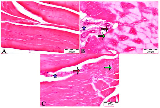
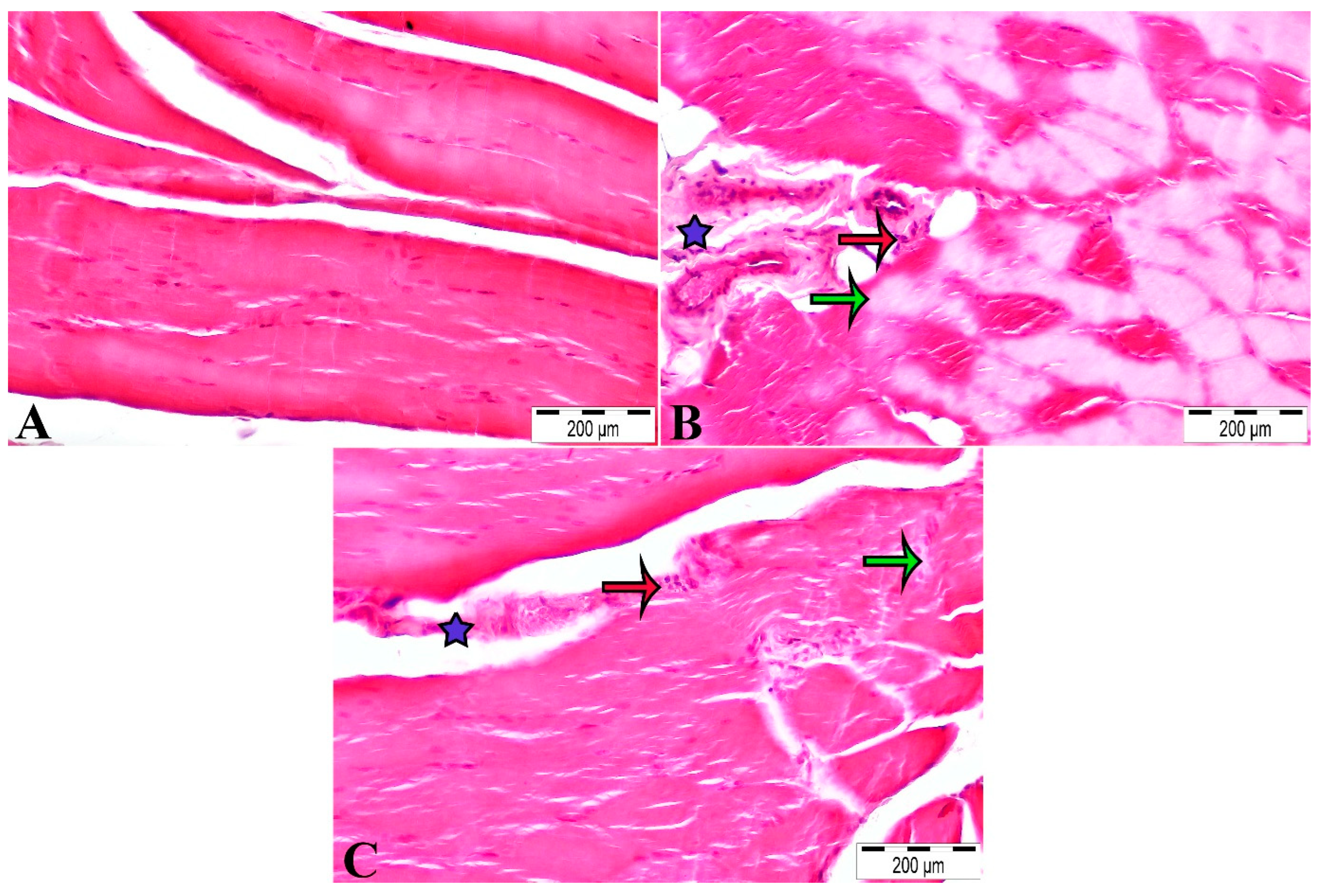
In the Control group, the muscle tissue exhibited normal histological architecture (Figure 1A). In contrast, the LPS group showed severe muscle fiber degeneration, severe edema, and moderate inflammation (Figure 1B). These histopathological alterations were significantly attenuated in the LPS + MgM group compared with the LPS group (Figure 1C).
Figure 1.
H&E-stained images and histopathological findings of the triceps brachii muscle. (A) Control group, (B) LPS group, and (C) LPS + MgM group. Muscle fiber degeneration is indicated by green arrows, inflammatory cell infiltration by red arrows, and interstitial edema by blue asterisks. Scale bar: 200 µm.
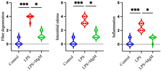
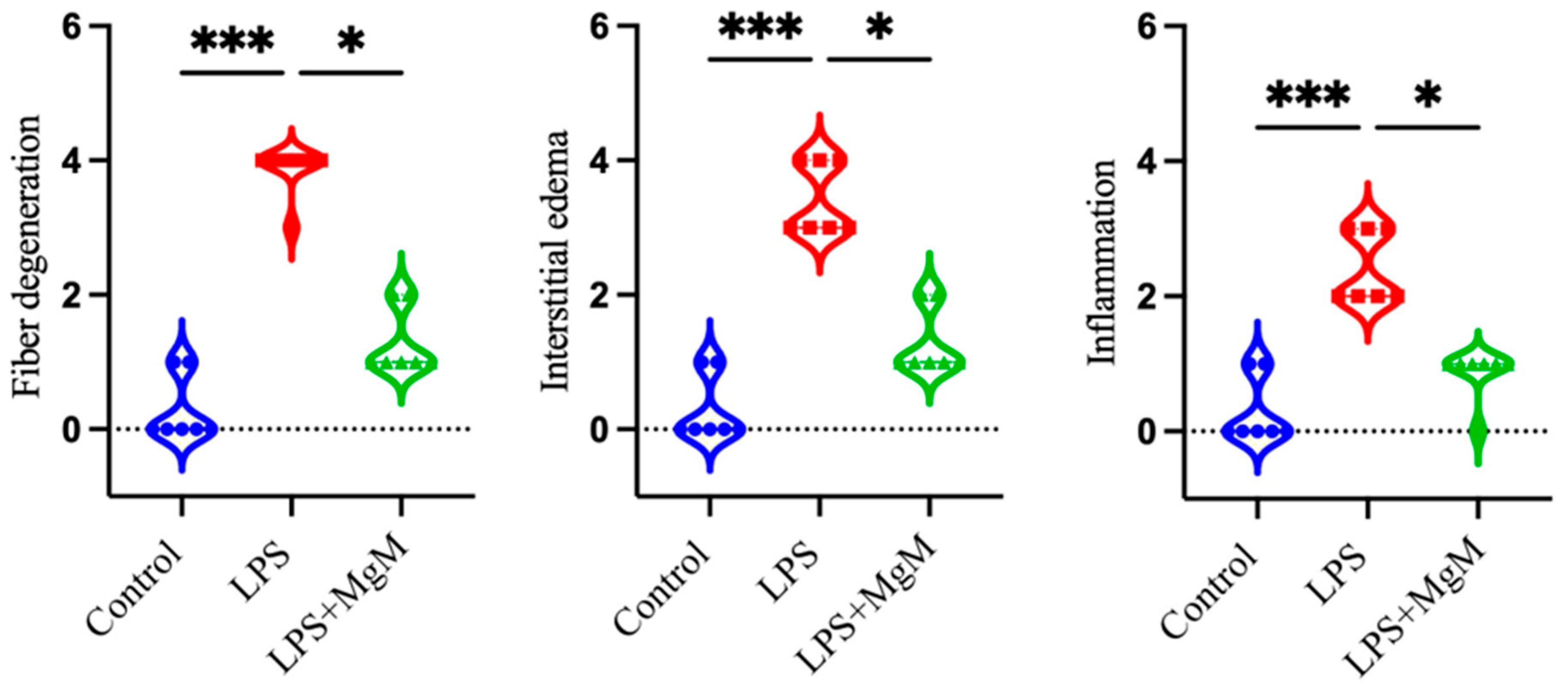
The histopathological scores for the triceps brachii muscle and the corresponding statistical analyses are presented in Figure 2. LPS administration induced significant histopathological alterations, as evidenced by increased muscle fiber degeneration, interstitial edema, and inflammation scores compared with the Control group (all p < 0.001). MgM treatment (LPS + MgM) significantly attenuated these alterations, resulting in lower scores for all three parameters compared with the LPS group (all p < 0.05).
Figure 2.
Results of the histomorphological evaluation of the triceps brachii muscle. Results are presented as median and interquartile range (IQR). * p < 0.05, *** p < 0.001.
4. Discussion
In this study, the effects of MgM on skeletal muscle injury, inflammation, oxidative stress, trace element levels, and electrophysiological findings were evaluated in an LPS-induced acute endotoxemia model. Our findings demonstrated that LPS administration caused histopathological alterations in skeletal muscle, a decrease in MUP amplitude, and changes in certain oxidative stress parameters. MgM administration 1 h after LPS exposure reduced inflammatory cell infiltration, edema, and muscle fiber degeneration in skeletal muscle tissue. It also significantly improved MUP amplitude. However, most of the observed changes in inflammatory markers and trace elements did not reach statistical significance.
One of the major findings of our study was the beneficial effect of MgM on skeletal muscle integrity and electrophysiological function following LPS administration. The attenuation of histopathological alterations, together with the improvement in MUP amplitude, suggests that MgM may help preserve both the structural integrity and functional properties of skeletal muscle under acute endotoxemic conditions. These findings indicate that the potential protective effects of MgM may extend beyond tissue morphology to include neuromuscular function.
Regarding the inflammatory response, LPS administration induced changes associated with both systemic and local inflammation. Serum TNF-α and IL-1β levels, as well as IL-6 levels in muscle tissue, showed decreasing trends following MgM administration; however, these changes were not statistically significant. In contrast, IL-10, an anti-inflammatory cytokine, was elevated in both the LPS and LPS + MgM groups compared with the control group. This finding may reflect the activation of a regulatory anti-inflammatory response alongside the pro-inflammatory response during acute endotoxemia. Furthermore, the significant increase in serum presepsin levels in the LPS group supports the induction of a systemic inflammatory response by the experimental model. However, systemic biomarker levels and local histopathological findings provide different aspects of the inflammatory response, highlighting the importance of evaluating serum markers together with tissue-specific changes. Overall, the lack of statistically significant reductions in pro-inflammatory cytokines prevents a definitive conclusion regarding the anti-inflammatory effects of MgM.
Previous experimental studies have suggested that Mg supplementation may exert protective effects in sepsis and endotoxemia through different mechanisms. However, these findings should be interpreted in the context of the magnesium formulations and experimental protocols used. Wang et al. [44] investigated the protective effects of different Mg salts, including MgSO4, magnesium chloride (MgCl2), and magnesium gluconate (MgGlu), against LPS-induced septic shock. In their two-hit LPS model in male mice, an initial LPS injection (0.4 mg/kg) was followed by a second challenge (10 mg/kg) 6 h later, with magnesium salts administered together with the second LPS injection. Mg supplementation was reported to partly inhibit gasdermin D N-terminal-mediated pyroptosis by regulating P2X7-dependent Ca2+ influx. The main outcomes included survival, circulating inflammatory cytokines, immune cell populations, and lung injury [44]. Jiang et al. investigated the effects of MgSO4 on sepsis-induced diaphragmatic dysfunction in rats subjected to cecal ligation and puncture (CLP) and 18 h of controlled mechanical ventilation. MgSO4 was administered as a loading dose of 270 mg/kg, followed by continuous infusion at 27 mg/kg/h. The authors reported partial preservation of diaphragmatic contractility and attenuation of systemic and local inflammatory responses, potentially through modulation of the HMGB1/TLR4/NF-κB signaling pathway. Although this study provides relevant evidence regarding sepsis-associated muscle dysfunction, its protocol differed from ours. Jiang et al. used continuous MgSO4 infusion in a polymicrobial sepsis model, whereas we administered a single intraperitoneal dose of MgM (200 mg/kg) 1 h after LPS injection (8 mg/kg). Moreover, their study focused on diaphragmatic contractility, whereas we evaluated skeletal muscle histopathology, motor unit potential amplitude, oxidative stress, and trace element levels [34].
Gu et al. [33] investigated the protective effects of MgSO4 against sepsis-associated acute kidney injury in LPS-challenged mice. Three MgSO4 doses (50, 100, and 150 mg/kg) were evaluated, and treatment was associated with improvements in renal function and histopathological findings, together with reductions in inflammation, oxidative stress, mitochondrial dysfunction, and apoptosis. These effects were partly associated with the inhibition of NF-κB signaling. However, Gu et al. examined renal injury in mice following LPS administration (10 mg/kg), whereas our study focused on skeletal muscle injury in rats following an 8 mg/kg LPS challenge. In addition, their study evaluated multiple MgSO4 doses and various renal and molecular parameters, while we investigated a single dose of MgM [33].
Esen et al. investigated the effects of MgSO4 on blood–brain barrier (BBB) integrity in a rat model of intraperitoneal sepsis induced by an infected fibrin–thrombin clot. MgSO4 was administered intramuscularly at a dose of 750 µmol/kg immediately after surgery, and outcomes were assessed 24 h later. The authors reported that magnesium treatment reduced sepsis-induced BBB permeability and brain edema. They also observed that MgSO4 helped maintain plasma magnesium levels, which decreased in untreated septic rats. Unlike our LPS-induced endotoxemia model, this study used a polymicrobial sepsis model and focused on neurological injury and plasma magnesium levels [45]. Zhong et al. investigated the effects of MgSO4 on histone-induced coagulation dysfunction and lung injury in mice. Using intravenous histone administration and a CLP-based sepsis model, they reported that intravenous MgSO4 (50 mg/kg) improved survival and attenuated endothelial injury, coagulation abnormalities, thrombosis, and pulmonary damage. These effects were associated with improvements in coagulation and fibrinolytic balance. Their study primarily examined histone-mediated endothelial and coagulation abnormalities, whereas our study focused on skeletal muscle injury following LPS-induced endotoxemia. Furthermore, the intravenous administration of MgSO4 in their study differed from the intraperitoneal administration of MgM 1 h after LPS injection in ours [46].
Overall, most previous experimental studies have investigated the effects of MgSO4 and other inorganic magnesium salts in different models of sepsis. The present study differs from these investigations by evaluating the effects of a single MgM dose on skeletal muscle under conditions of LPS-induced acute endotoxemia, using histopathological, electrophysiological, oxidative stress, and trace element parameters. Thus, previous findings provide a rationale for investigating magnesium-based interventions but do not establish the efficacy or superiority of MgM over other magnesium salts.
In the present study, skeletal muscle Fe levels were significantly increased following LPS administration compared with the control group, whereas no significant changes were observed in Cu and Zn levels. During the inflammatory response, iron metabolism may be altered through the hepcidin–ferroportin regulatory axis, potentially increasing intracellular iron retention. Intracellular iron accumulation may contribute to oxidative tissue injury by facilitating ROS generation through the Fenton reaction. In contrast, Cu and Zn homeostasis is regulated by different mechanisms involving metal transport, storage, and redistribution. Therefore, tissue levels of these elements may respond differently to inflammation and may not necessarily show significant changes during the early stages of endotoxemia. Although Fe and Cu levels tended to decrease and Zn levels tended to increase in the LPS + MgM group compared with the LPS group, these differences were not statistically significant. The relatively short observation period (6 h after LPS administration) may also have limited the detection of time-dependent changes in trace element homeostasis. Further studies involving different time points are needed to clarify the effects of MgM on skeletal muscle trace element balance during endotoxemia.
Regarding oxidative stress, MDA levels showed an increasing trend following LPS administration, whereas MgM administration significantly reduced these levels. Considering that MDA is an important marker of lipid peroxidation, this finding suggests that MgM may contribute to the attenuation of oxidative damage. On the other hand, the increases in CAT and GSH levels observed in the LPS group may reflect a compensatory antioxidant response to oxidative stress. The fact that CAT levels approached those of the control group following MgM administration may be associated with an attenuation of oxidative stress.
Previous experimental studies have demonstrated that various antioxidant and anti-inflammatory compounds can alleviate LPS-induced tissue injury. Shareef et al. reported that pretreatment with L-carvone attenuated kidney injury by modulating inflammatory signaling pathways and oxidative stress-related processes [47]. Similarly, Okan et al. demonstrated that resveratrol alleviated liver and kidney injury by suppressing the TLR4/NF-κB signaling pathway [48]. Mansoor et al. reported that carnosol significantly reduced LPS-induced pulmonary inflammation and IL-1β, IL-6, and TNF-α levels, while its administration in combination with methylprednisolone acetate further enhanced these effects [49]. In addition, Koc et al. demonstrated that chrysin administration in an experimental LPS-induced sepsis model reduced serum inflammatory cytokine levels and MDA levels in liver, lung, and kidney tissues while increasing the activities of antioxidant enzymes, including SOD and CAT [50]. Gao et al. also showed that aloe-emodin suppressed the inflammatory response and supported antioxidant defense [51]. Collectively, these findings suggest that targeting inflammation and oxidative stress may represent an important approach to reducing LPS-induced tissue injury. Nevertheless, direct comparisons with MgM are limited by differences in the compounds and experimental models investigated.
The role of Mg in muscle physiology is associated with its functions in energy metabolism and cellular enzymatic reactions. Malate, on the other hand, is an intermediate of the tricarboxylic acid cycle and contributes to cellular energy metabolism. Therefore, the selection of MgM in the present study was based on the potential complementary biological roles of Mg and malate. Although some studies have reported that organic Mg compounds may exhibit higher bioavailability than certain inorganic forms, this advantage has not been consistently demonstrated for all preparations, and the superiority of MgM over other Mg salts has not been established [31]. Liguori et al. also evaluated the relationship between Mg, muscle homeostasis, oxidative stress, and muscle function, highlighting the potential benefits of Mg supplementation for muscle mass, respiratory muscle strength, and recovery [52]. However, these findings cannot be directly extrapolated to sepsis-associated muscle wasting.
The MgM dose used in our study was determined by considering doses employed in previous experimental studies investigating organic Mg compounds [32,36]. Koç et al. investigated the effects of MgM supplementation in rats using approximately 35.4 mg/kg/day of elemental Mg, focusing on tissue Mg levels and neuromuscular performance [32]. The dose of 200 mg/kg MgM used in our study corresponds to approximately 31.1 mg/kg of elemental Mg. This similarity provides indirect support for the dose selection; however, the present study was not designed to determine the optimal dose. Furthermore, administering MgM 1 h after LPS exposure represents an early intervention during acute endotoxemia rather than treatment of established or advanced-stage sepsis.
Our study has several limitations. First, the administration of a single LPS dose (8 mg/kg) and the assessment of outcomes at 6 h limit the interpretation of our findings to the acute phase of endotoxemia. Although this model allows the investigation of early inflammatory responses, it does not reflect the later stages of sepsis or long-term tissue injury.
Second, the absence of a healthy control group receiving MgM alone prevented the assessment of the independent effects of MgM under physiological conditions. Furthermore, serum and skeletal muscle Mg levels were not measured; therefore, tissue Mg availability could not be evaluated, and the potential contributions of Mg and malate to the observed effects could not be distinguished.
Third, only a single MgM dose (200 mg/kg), administered 1 h after LPS, and one observation time (6 h after LPS administration) were investigated. Consequently, dose–response relationships, the effects of different treatment schedules, and long-term outcomes could not be assessed. Although the sample size was supported by an a priori power analysis (power = 92.37%), the relatively small number of animals per group may have limited the detection of smaller biological differences. In addition, the molecular and cellular mechanisms underlying the observed effects were not directly investigated.
Finally, only male rats were included in the study. Although this approach may have reduced biological variability associated with sex-related differences, it prevented the assessment of potential differences in inflammatory responses, Mg metabolism, and muscle physiology between sexes, thereby limiting the generalizability of the findings to female animals.
5. Conclusions
In conclusion, our findings suggest that MgM may exert beneficial effects on skeletal muscle histopathology, electrophysiological function, and certain oxidative stress parameters under conditions of acute endotoxemia. However, most inflammatory markers and trace elements did not show statistically significant changes. Future studies should investigate different doses and administration protocols, longer follow-up periods, and experimental models that more closely reflect clinical sepsis. Furthermore, direct measurement of serum and tissue Mg levels would contribute to the evaluation of MgM bioavailability and tissue distribution. Investigation of the TLR4/NF-κB, Nrf2/HO-1, and NLRP3 signaling pathways, together with mitochondrial function and apoptosis-related mechanisms, may also help elucidate the potential mechanisms underlying the effects of MgM.
Author Contributions
Conceptualization, K.K., B.A., C.B.T. and S.D.; methodology, K.K., B.A., C.B.T. and S.D.; investigation, K.K., S.D. and B.A.; formal analysis, K.K., N.B., S.D., I.A., A.D., İ.B. and K.Y.I.; data curation, K.K., C.B.T. and S.D.; validation, B.A.; writing—original draft preparation, M.E.; writing—review and editing, M.E. and B.A.; visualization, M.E. and K.K.; supervision, B.A.; project administration, C.B.T. and B.A.; funding acquisition, C.B.T. All authors have read and agreed to the published version of the manuscript.
Funding
This study was supported by the Sakarya University Scientific Research Projects Coordination Unit (project numbers: 2024-25-60-184 and 2024-25-60-183) and the Scientific and Technological Research Council of Türkiye (1919B012420455 and 1919B012423688). Parts of this study were presented at the 6th Congress on the Future of Medicine in Sakarya, Türkiye, 8–10 May 2026, and at the 8th International and 37th National Biophysics Congress in Ankara, Türkiye, 26–29 August 2026.
Institutional Review Board Statement
The animal study protocol was approved by the Sakarya University Local Ethics Committee for Animal Experiments (approval number: 04/10/2024-41).
Informed Consent Statement
Not applicable.
Data Availability Statement
The original contributions presented in this study are included in the article. Further inquiries can be directed to the corresponding author.
Acknowledgments
The authors would like to thank the institutions that supported this study through the funding sources listed below.
Conflicts of Interest
The authors declare no conflicts of interest.
Abbreviations
The following abbreviations are used in this manuscript:
| H&E | Hematoxylin and eosin |
| HSD | Honestly significant difference |
| ICP-OES | Inductively coupled plasma optical emission spectroscopy |
| IL-1β | Interleukin-1 beta |
| IL-6 | Interleukin-6 |
| IL-10 | Interleukin-10 |
| i.p. | Intraperitoneal |
| IQR | Interquartile range |
| LPS | Lipopolysaccharide |
| MDA | Malondialdehyde |
| Mg | Magnesium |
| MgM | Magnesium malate |
| MUP | Motor unit potential |
| MyD88 | Myeloid differentiation primary response 88 |
| NF-κB | Nuclear factor kappa B |
| P–P | Peak-to-peak |
| ROS | Reactive oxygen species |
| SAMW | Sepsis-associated muscle wasting |
| SD | Standard deviation |
| SPSS | Statistical Package for the Social Sciences |
| TBARS | Thiobarbituric acid reactive substances |
| TLR4 | Toll-like receptor 4 |
| TNF-α | Tumor necrosis factor alpha |
| Cu | Copper |
| Zn | Zinc |
References
- Remick, D.G. Pathophysiology of sepsis. Am. J. Pathol. 2007, 170, 1435–1444. [Google Scholar] [CrossRef] [Scilit] [PubMed]
- He, C.; Zhang, W.; Li, S.; Ruan, W.; Xu, J.; Xiao, F. Edaravone improves septic cardiac function by inducing an HIF-1α/HO-1 pathway. Oxid. Med. Cell. Longev. 2018, 2018, 5216383. [Google Scholar] [CrossRef] [Scilit] [PubMed]
- Lei, S.; Zhang, Y.; Su, W.; Zhou, L.; Xu, J.; Xia, Z.Y. Remifentanil attenuates lipopolysaccharide-induced oxidative injury by downregulating PKCβ2 activation and inhibiting autophagy in H9C2 cardiomyocytes. Life Sci. 2018, 213, 109–115. [Google Scholar] [CrossRef] [Scilit] [PubMed]
- Ono, Y.; Saito, M.; Yoshihara, I.; Kondo, Y.; Sakamoto, K.; Sugiyama, J.; Nakanishi, N.; Inoue, S.; Kotani, J. Sepsis-associated skeletal muscle wasting is ameliorated by pharmacological inhibition of the STAT3 signaling pathway in mice. Sci. Rep. 2026, 16, 5008. [Google Scholar] [CrossRef] [Scilit] [PubMed]
- Perner, A.; Gordon, A.C.; De Backer, D.; Dimopoulos, G.; Russell, J.A.; Lipman, J.; Jensen, J.U.; Myburgh, J.; Singer, M.; Bellomo, R.; et al. Sepsis: Frontiers in diagnosis, resuscitation and antibiotic therapy. Intensive Care Med. 2016, 42, 1958–1969. [Google Scholar] [CrossRef] [Scilit] [PubMed]
- Zhang, J.; Huang, Y.; Chen, Y.; Shen, X.; Pan, H.; Yu, W. Impact of muscle mass on survival in patients with sepsis: A systematic review and meta-analysis. Ann. Nutr. Metab. 2021, 77, 330–336. [Google Scholar] [CrossRef] [Scilit] [PubMed]
- Ono, Y.; Maejima, Y.; Saito, M.; Sakamoto, K.; Horita, S.; Shimomura, K.; Inoue, S.; Kotani, J. TAK-242, a specific inhibitor of Toll-like receptor 4 signalling, prevents endotoxemia-induced skeletal muscle wasting in mice. Sci. Rep. 2020, 10, 694. [Google Scholar] [CrossRef] [Scilit] [PubMed]
- Wu, Q.; Huang, Q.; Xu, Z.; Hou, C.; Wang, C. Comprehensive review of sepsis-related skeletal muscle atrophy: Mechanisms, diagnosis and therapeutic strategies. J. Inflamm. Res. 2025, 18, 17879–17894. [Google Scholar] [CrossRef] [Scilit] [PubMed]
- Virzì, G.M.; Mattiotti, M.; De Cal, M.; Ronco, C.; Zanella, M.; De Rosa, S. Endotoxin in sepsis: Methods for LPS detection and the use of omics techniques. Diagnostics 2022, 13, 79. [Google Scholar] [CrossRef] [Scilit] [PubMed]
- Lee, K.H.; Yeh, M.H.; Kao, S.T.; Hung, C.M.; Chen, B.C.; Liu, C.J.; Yeh, C.C. Xia-bai-san inhibits lipopolysaccharide-induced activation of intercellular adhesion molecule-1 and nuclear factor-kappa B in human lung cells. J. Ethnopharmacol. 2009, 124, 530–538. [Google Scholar] [CrossRef] [Scilit] [PubMed]
- Cavaillon, J.M.; Adib-Conquy, M.; Fitting, C.; Adrie, C.; Payen, D. Cytokine cascade in sepsis. Scand. J. Infect. Dis. 2003, 35, 535–544. [Google Scholar] [CrossRef] [Scilit] [PubMed]
- Dinarello, C.A. Interleukin-1 and interleukin-1 antagonism. Blood 1991, 77, 1627–1652. [Google Scholar] [CrossRef] [Scilit]
- Li, Y.P.; Reid, M.B. NF-κB mediates the protein loss induced by TNF-α in differentiated skeletal muscle myotubes. Am. J. Physiol. Regul. Integr. Comp. Physiol. 2000, 279, R1165–R1170. [Google Scholar] [CrossRef] [Scilit] [PubMed]
- Cohen, S.; Nathan, J.A.; Goldberg, A.L. Muscle wasting in disease: Molecular mechanisms and promising therapies. Nat. Rev. Drug Discov. 2015, 14, 58–74. [Google Scholar] [CrossRef] [Scilit] [PubMed]
- Mankowski, R.T.; Laitano, O.; Clanton, T.L.; Brakenridge, S.C. Pathophysiology and treatment strategies of acute myopathy and muscle wasting after sepsis. J. Clin. Med. 2021, 10, 1874. [Google Scholar] [CrossRef] [Scilit] [PubMed]
- Frost, R.A.; Nystrom, G.J.; Lang, C.H. Lipopolysaccharide regulates proinflammatory cytokine expression in mouse myoblasts and skeletal muscle. Am. J. Physiol. Regul. Integr. Comp. Physiol. 2002, 283, R698–R709. [Google Scholar] [CrossRef] [Scilit] [PubMed]
- Zanders, L.; Kny, M.; Hahn, A.; Schmidt, S.; Wundersitz, S.; Todiras, M.; Lahmann, I.; Bandyopadhyay, A.; Wollersheim, T.; Kaderali, L.; et al. Sepsis induces interleukin 6, gp130/JAK2/STAT3, and muscle wasting. J. Cachexia Sarcopenia Muscle 2022, 13, 713–727. [Google Scholar] [CrossRef] [Scilit] [PubMed]
- Eggelbusch, M.; Shi, A.; Broeksma, B.C.; Vázquez-Cruz, M.; Soares, M.N.; de Wit, G.M.; Everts, B.; Jaspers, R.T.; Wüst, R.C. The NLRP3 inflammasome contributes to inflammation-induced morphological and metabolic alterations in skeletal muscle. J. Cachexia Sarcopenia Muscle 2022, 13, 3048–3061. [Google Scholar] [CrossRef] [Scilit] [PubMed]
- Kubat, G.B.; Turkel, I.; Akat, F. Sepsis-induced skeletal muscle atrophy and mitochondrial dysfunction: The beneficial effects of exercise. Spor Hekim. Derg. 2025, 60, 25–31. [Google Scholar] [CrossRef] [Scilit]
- Prauchner, C.A. Oxidative stress in sepsis: Pathophysiological implications justifying antioxidant co-therapy. Burns 2017, 43, 471–485. [Google Scholar] [CrossRef] [Scilit] [PubMed]
- Lin, Y.; Alhaskawi, A.; Chen, L.; Moqbel, S.A.A. Recent advances in understanding oxidative stress in sepsis: Pathogenic roles and antioxidant therapeutic prospects—A narrative review. Front. Pharmacol. 2025, 16, 1695992. [Google Scholar] [CrossRef] [Scilit] [PubMed]
- Silveira, T.T.; Stefenon, D.O.; Júnior, E.L.; Konstantyner, T.; Leite, H.P.; Moreno, Y.M.F. Assessment of trace elements in critically ill patients with systemic inflammatory response syndrome: A systematic review. J. Trace Elem. Med. Biol. 2023, 78, 127155. [Google Scholar] [CrossRef] [Scilit] [PubMed]
- Shi, Y.; Zou, Y.; Shen, Z.; Xiong, Y.; Zhang, W.; Liu, C.; Chen, S. Trace elements, PPARs, and metabolic syndrome. Int. J. Mol. Sci. 2020, 21, 2612. [Google Scholar] [CrossRef] [Scilit] [PubMed]
- Hoeboer, S.H.; van der Geest, P.J.; Nieboer, D.; Groeneveld, A.J. The diagnostic accuracy of procalcitonin for bacteraemia: A systematic review and meta-analysis. Clin. Microbiol. Infect. 2015, 21, 474–481. [Google Scholar] [CrossRef] [Scilit] [PubMed]
- Kondo, Y.; Umemura, Y.; Hayashida, K.; Hara, Y.; Aihara, M.; Yamakawa, K. Diagnostic value of procalcitonin and presepsin for sepsis in critically ill adult patients: A systematic review and meta-analysis. J. Intensive Care 2019, 7, 22. [Google Scholar] [CrossRef] [Scilit] [PubMed]
- Barbagallo, M.; Veronese, N.; Dominguez, L. Magnesium in aging, health and diseases. Nutrients 2021, 13, 463. [Google Scholar] [CrossRef] [Scilit] [PubMed]
- Barbieri, E.; Sestili, P. Reactive oxygen species in skeletal muscle signaling. J. Signal Transduct. 2012, 2012, 982794. [Google Scholar] [CrossRef] [Scilit] [PubMed]
- Malinska, D.; Kudin, A.P.; Bejtka, M.; Kunz, W.S. Changes in mitochondrial reactive oxygen species synthesis during differentiation of skeletal muscle cells. Mitochondrion 2012, 12, 144–148. [Google Scholar] [CrossRef] [Scilit] [PubMed]
- Huypens, P.; Pillai, R.; Sheinin, T.; Schaefer, S.; Huang, M.; Odegaard, M.L.; Ronnebaum, S.M.; Wettig, S.D.; Joseph, J.W. The dicarboxylate carrier plays a role in mitochondrial malate transport and in the regulation of glucose-stimulated insulin secretion from rat pancreatic beta cells. Diabetologia 2011, 54, 135–145. [Google Scholar] [CrossRef] [Scilit] [PubMed]
- Ates, M.; Kizildag, S.; Yuksel, O.; Hosgorler, F.; Yuce, Z.; Guvendi, G.; Kandis, S.; Karakilic, A.; Koc, B.; Uysal, N. Dose-dependent absorption profile of different magnesium compounds. Biol. Trace Elem. Res. 2019, 192, 244–251. [Google Scholar] [CrossRef] [Scilit] [PubMed]
- Schuchardt, J.P.; Hahn, A. Intestinal absorption and factors influencing bioavailability of magnesium-An update. Curr. Nutr. Food Sci. 2017, 13, 260–278. [Google Scholar] [CrossRef] [Scilit] [PubMed]
- Koc, B.; Hosgorler, F.; Kandis, S.; Acikgoz, B.; Kizildag, S.; Guner, O.; Durmus, N.; Ates, M.; Uysal, N. Chronic organic magnesium supplementation enhances tissue-specific bioavailability and functional capacity in rats: A focus on brain, muscle, and vascular health. Biol. Trace Elem. Res. 2026, 204, 253–267. [Google Scholar] [CrossRef] [Scilit] [PubMed]
- Gu, W.J.; Chen, J.L.; Duan, X.J.; Zhang, C.Y.; Huang, Y.; Zhu, M.G.; Yin, H. Magnesium sulfate alleviates sepsis-associated acute kidney injury by reducing inflammation, oxidative stress, and apoptosis via inhibiting the NF-κB pathway. FASEB J. 2026, 40, e71456. [Google Scholar] [CrossRef] [Scilit] [PubMed]
- Jiang, J.; Chen, Q.; Chen, X.; Li, J.; Li, S.; Yang, B. Magnesium sulfate ameliorates sepsis-induced diaphragm dysfunction in rats via inhibiting HMGB1/TLR4/NF-κB pathway. Neuroreport 2020, 31, 902–908. [Google Scholar] [CrossRef] [Scilit] [PubMed]
- Dumbuya, J.S.; Li, S.; Liang, L.; Chen, Y.; Du, J.; Zeng, Q. Effects of hydrogen-rich saline in neuroinflammation and mitochondrial dysfunction in rat model of sepsis-associated encephalopathy. J. Transl. Med. 2022, 20, 546. [Google Scholar] [CrossRef] [Scilit] [PubMed]
- Virág, V.; May, Z.; Kocsis, I.; Blázovics, A.; Szentmihályi, K. Effects of magnesium supplementation on calcium and magnesium levels, and redox homeostasis in normolipidemic and food-induced hyperlipidemic rats. Orv. Hetil. 2011, 152, 1075–1081. [Google Scholar] [CrossRef] [Scilit] [PubMed]
- Doğanay, S.; Trabzon, Ş.; Ozdemir, A.E.; Bahtiyar, N.; Erdoğan, D.G. Effect of melatonin on trace element distribution in lung, kidney, brain, and testicular tissues in experimental type 1 diabetes. Trace Elem. Electrolytes 2021, 38, 77–84. [Google Scholar] [CrossRef] [Scilit]
- Öğüt, S.; Bahtiyar, N.; Mordeniz, C.; Cinemre, F.B.; Aydemir, B.; Karaçetin, D.; Değirmencioğlu, S.; Hacıosmanoğlu, E.; Kural, A.; Kızıler, A.R.; et al. Effect of breast cancer and breast cancer treatment on the blood serum concentrations of trace elements and selenoproteins. J. Elem. 2022, 27, 289–302. [Google Scholar] [CrossRef] [Scilit]
- Serinkan Cinemre, F.B.; Cinemre, H.; Bahtiyar, N.; Kahyaoğlu, B.; Ağaç, M.T.; Shundo, H.; Sevinç, L.; Aydemir, B. Apelin, omentin-1, and vaspin in patients with essential hypertension: Association of adipokines with trace elements, inflammatory cytokines, and oxidative damage markers. Ir. J. Med. Sci. 2021, 190, 97–106. [Google Scholar] [CrossRef] [Scilit] [PubMed]
- Lowry, O.H.; Rosebrough, N.J.; Farr, A.L.; Randall, R.J. Protein measurement with the Folin phenol reagent. J. Biol. Chem. 1951, 193, 265–275. [Google Scholar] [CrossRef] [Scilit]
- Buege, J.A.; Aust, S.D. Microsomal lipid peroxidation. Methods Enzymol. 1978, 52, 302–310. [Google Scholar] [CrossRef] [Scilit] [PubMed]
- Beutler, E.; Duron, O.; Kelly, B.M. Improved method for the determination of blood glutathione. J. Lab. Clin. Med. 1963, 61, 882–888. [Google Scholar] [PubMed]
- Beers, R.F., Jr.; Sizer, I.W. A spectrophotometric method for measuring the breakdown of hydrogen peroxide by catalase. J. Biol. Chem. 1952, 195, 133–140. [Google Scholar] [CrossRef] [Scilit]
- Wang, D.; Zheng, J.; Hu, Q.; Zhao, C.; Chen, Q.; Shi, P.; Chen, Q.; Zou, Y.; Zou, D.; Liu, Q.; et al. Magnesium protects against sepsis by blocking gasdermin D N-terminal-induced pyroptosis. Cell Death Differ. 2020, 27, 466–481. [Google Scholar] [CrossRef] [Scilit] [PubMed]
- Esen, F.; Erdem, T.; Aktan, D.; Orhan, M.; Kaya, M.; Eraksoy, H.; Cakar, N.; Telci, L. Effect of magnesium sulfate administration on blood-brain barrier in a rat model of intraperitoneal sepsis: A randomized controlled experimental study. Crit. Care 2005, 9, R18–R23. [Google Scholar] [CrossRef] [Scilit] [PubMed]
- Zhong, T.; Zhang, J.; Chen, S.; Chen, S.; Deng, K.; Guan, J.; Yang, J.; Lv, R.; Liu, Z.; Liu, Y.; et al. Magnesium sulfate ameliorates histone-induced coagulation dysfunction and lung damage in mice. Shock 2024, 61, 132–141. [Google Scholar] [CrossRef] [Scilit] [PubMed]
- Shareef, S.M.; Kathem, S.H.; Ridha-Salman, H. Nephroprotective effect of L-carvone on a mouse model of LPS-induced sepsis-associated renal injury via regulation of TLR4/NF-κB/AP-1/IRF-3 and Nrf2/iNOS molecular signaling cascades. J. Taibah Univ. Med. Sci. 2026, 21, 49–66. [Google Scholar] [CrossRef] [Scilit] [PubMed]
- Okan, A.; Doğanyiğit, Z.; Yilmaz, S.; Uçar, S.; Arikan Söylemez, E.S.; Attar, R. Evaluation of the protective role of resveratrol against sepsis caused by LPS via TLR4/NF-κB/TNF-α signaling pathways: Experimental study. Cell Biochem. Funct. 2023, 41, 423–433. [Google Scholar] [CrossRef] [Scilit] [PubMed]
- Abed Mansoor, A.F.; Abu-Raghif, A.R.; Ridha-Salman, H.; Al-Asady, F.M.; Shleghm, M.R.; Hussein, M.N. Pulmonoprotective effect of carnosol on LPS-induced cytokine storm model in mice. Eur. J. Clin. Exp. Med. 2026, 24, 138–149. [Google Scholar] [CrossRef] [Scilit]
- Koc, F.; Tekeli, M.Y.; Kanbur, M.; Karayigit, M.; Liman, B.C. The effects of chrysin on lipopolysaccharide-induced sepsis in rats. J. Food Biochem. 2020, 44, e13359. [Google Scholar] [CrossRef] [Scilit] [PubMed]
- Gao, H.; Ren, Y.; Liu, C. Aloe-emodin suppresses oxidative stress and inflammation via a PI3K-dependent mechanism in a murine model of sepsis. Evid. Based Complement. Altern. Med. 2022, 2022, 9697887. [Google Scholar] [CrossRef] [Scilit] [PubMed]
- Liguori, S.; Moretti, A.; Paoletta, M.; Gimigliano, F.; Iolascon, G. Role of magnesium in skeletal muscle health and neuromuscular diseases: A scoping review. Int. J. Mol. Sci. 2024, 25, 11220. [Google Scholar] [CrossRef] [Scilit] [PubMed]

Disclaimer/Publisher’s Note: The statements, opinions and data contained in all publications are solely those of the individual author(s) and contributor(s) and not of MDPI and/or the editor(s). MDPI and/or the editor(s) disclaim responsibility for any injury to people or property resulting from any ideas, methods, instructions or products referred to in the content. |
© 2026 by the authors. Licensee MDPI, Basel, Switzerland. This article is an open access article distributed under the terms and conditions of the Creative Commons Attribution (CC BY) license.