


Abstract
Lycium ruthenicum Murr. polysaccharides (LRP), the major constituents of black goji berry, exhibit diverse biological activities. Pulmonary fibrosis (PF) frequently arises as a long-term consequence of acute lung injury and is marked by progressive scarring of lung tissue and an irreversible decline in pulmonary function. Persistent inflammatory responses, oxidative stress, and disturbance of the gut–lung axis are closely related to the development of PF. This study aimed to extract LRP from black goji berry and systematically evaluate its protective effects and underlying mechanisms in a bleomycin-induced murine model of PF. The results show that LRP treatment markedly attenuated alveolar structural disruption, inflammatory cell infiltration, and fibrotic lesions. These protective effects were associated with the modulation of inflammatory cytokines and the enhancement of antioxidant capacity through increased superoxide dismutase activity and glutathione levels. Furthermore, LRP reshaped the gut microbiotal community structure. Collectively, these findings provide new insights into the pulmonary protective properties of LRP and support its potential as a promising natural therapeutic agent for PF.
1. Introduction
Pulmonary fibrosis (PF) is a fatal interstitial lung disease characterized by chronic and progressive scarring of lung parenchyma [1]. Clinically, a substantial proportion of PF cases arise as a long-term consequence of unresolved acute lung injury (ALI). When ALI-induced inflammation persists, or the regulatory mechanisms governing tissue repair become dysregulated, physiological lung regeneration is replaced by pathological wound healing, ultimately leading to the development of PF [2]. A hallmark of PF is the excessive deposition and aberrant remodeling of the extracellular matrix (ECM), which progressively disrupts normal alveolar architecture, reduces lung compliance, and impairs gas exchange [3,4]. The formation of fibroblastic foci, mediated by activated myofibroblasts, represents one of the defining pathological features of PF. Resident and infiltrating fibroblasts undergo aberrant differentiation into myofibroblasts, which show increased α-smooth muscle actin (α-SMA) expression and enhanced ECM synthesis and secretion. Consequently, the excessive accumulation of fibrotic connective tissue within the injured lung results in irreversible scar formation and progressive deterioration of pulmonary function [5].
Accumulating evidence also suggests that gut microbiota dysbiosis is a key driver of PF progression. Disruption of the gut microbial ecosystem reduces the production of beneficial microbial metabolites, resulting in sustained activation of the transforming growth factor-β (TGF-β) signaling pathway. Moreover, gut dysbiosis may promote fungal β-glucan-induced epithelial–mesenchymal transition (EMT), thereby accelerating fibroblast activation, excessive collagen deposition, and the progression of PF [6].
Currently, first-line pharmacological therapies for PF primarily include the antifibrotic agents pirfenidone and nintedanib. However, their clinical efficacy remains limited and is frequently complicated by considerable adverse reactions and organ toxicity [7]. Long-term administration is associated with complications such as hepatotoxicity and gastrointestinal disorders. Moreover, these agents exhibit limited therapeutic efficacy against the complex pathological network underlying PF, which involves TGF-β/Smad3-mediated fibroblast activation, excessive ECM deposition, and EMT. In contrast, naturally derived plant polysaccharides have gained growing interest owing to their favorable safety profile, low toxicity, and ability to exert therapeutic effects through multitarget mechanisms. Emerging evidence suggests that plant polysaccharides can simultaneously promote macrophage polarization toward the anti-inflammatory M2 phenotype, suppress EMT, and restore the integrity of the alveolar epithelial barrier, thereby conferring both antifibrotic and pulmonary protective effects [8,9,10,11,12]. These multifunctional properties highlight the potential of plant polysaccharides as promising candidates for the comprehensive management and adjunctive treatment of PF.
Lycium ruthenicum is rich in a wide range of bioactive constituents, including polysaccharides, anthocyanins, flavonoids, and trace elements. Many of these compounds are present at substantially higher levels than those found in the commonly consumed Lycium barbarum [13]. Lycium ruthenicum Murr. polysaccharide (LRP) is a bioactive polysaccharide extracted from the fruits of Lycium ruthenicum. LRP has been reported to effectively scavenge excessive reactive free radicals, inhibit tumor cell proliferation, and suppress pro-inflammatory cytokine expression, thereby exhibiting potent antioxidant and anti-inflammatory activities [14]. LRP has exhibited considerable therapeutic promise for managing inflammation-related disorders. Its complex monosaccharide composition contributes to its potent anti-inflammatory activity [15]. Mechanistically, LRP exerts its anti-inflammatory effects primarily by inhibiting the Toll-like receptor 4/nuclear factor-κB signaling pathway and suppressing the production of pro-inflammatory cytokines. Pharmacological studies have also demonstrated that LRP is capable of modulating the composition and function of the gut microbiota [16]. In addition, LRP exhibits prebiotic properties by modulating the gut microbiota, promoting the proliferation of beneficial bacteria such as Lactobacillus and Bifidobacterium, while suppressing the intestinal colonization of pathogenic members of the Enterobacteriaceae family [17]. Although the therapeutic potential of LRP has been extensively investigated in metabolic syndrome, liver injury, and other disease models [18,19,20], its protective effects and underlying mechanisms in PF are still largely unknown. Therefore, this study established a bleomycin (BLM)-induced mouse model of PF to assess the therapeutic effects of LRP against PF and to investigate its underlying mechanisms.
2. Materials and Methods
2.1. Materials
Bleomycin was obtained from Shanghai Aladdin Biochemical Technology Co., Ltd. (Shanghai, China). ELISA kits for interleukin (IL)-1β, IL-6, tumor necrosis factor-α (TNF-α), and IL-10 were supplied by Xiamen Huijia Biotechnology Co., Ltd. (Xiamen, China). Primary antibodies against collagen type I (Col-I), α-SMA, and cluster of differentiation 73 (CD73) were purchased from Proteintech Group, Inc. (Wuhan, China). The DAB staining kit and Polymer-HRP anti-rabbit secondary antibody kit were acquired from Hunan Aifang Biotechnology Co., Ltd. (Changsha, China). The hydroxyproline (Hyp) assay kit, Masson’s trichrome staining kit, and Sirius Red staining kit were purchased from Beijing Solarbio Science & Technology Co., Ltd. (Beijing, China). Hematoxylin and eosin (H&E) staining kit, 4% paraformaldehyde, goat serum, and phosphate-buffered saline (PBS) were provided by Beyotime Biotech Inc. (Shanghai, China).
2.2. LRP Preparation and Characterization
The LRP used in this study was prepared and characterized as described in our previous study [21]. Briefly, air-dried Lycium ruthenicum fruits were ground into powder and dispersed in ultrapure water at a 1:10 (g/mL) ratio. Ultrasonic treatment was carried out at 100 °C for 60 min, after which the mixture was centrifuged at 8000 rpm for 15 min to collect the filtrate. The supernatant was concentrated by rotary evaporation under reduced pressure at 50 °C, followed by the addition of 10% trichloroacetic acid and allowed to stand for 4 h. Centrifugation at 8000 rpm for 15 min was performed, and the supernatant was retained. Anhydrous ethanol was then introduced into the supernatant, and the mixture was left standing at 4 °C overnight. The precipitate was recovered by centrifugation at 8000 rpm for 10 min at 4 °C and then lyophilized.
The phenol-sulfuric acid method was used to quantify the total sugar content of LRP [22]. For FT-IR analysis, LRP (5 mg) and dry KBr (500 mg) were ground together and pressed into a pellet. The spectrum was recorded in the range of 4000–400 cm−1. High-performance liquid chromatography (HPLC) was used to profile the monosaccharide composition of LRP. High-performance gel permeation chromatography yielded the molecular weight of LRP. UV-Vis spectra were recorded for LRP in the 190–400 nm wavelength range.
2.3. Animals
A total of 48 male C57BL/6J mice, specific pathogen-free (SPF), aged 8 weeks and weighing 20–25 g, were used in this study [7]. Mice were purchased from Zhejiang Vital River Laboratory Animal Technology Co., Ltd. (Jiaxing, China). They were maintained under SPF conditions on a 12 h light/dark cycle at 22 ± 2 °C and 50 ± 5% relative humidity, with free access to standard laboratory chow and sterile ultrapure water. All animal experimental procedures were approved by the Animal Ethics Committee of Jiangnan University (Approval number: JN. No20250515c0600706[273]) and conducted in compliance with the National Institutes of Health guidelines.
2.4. Experimental Design
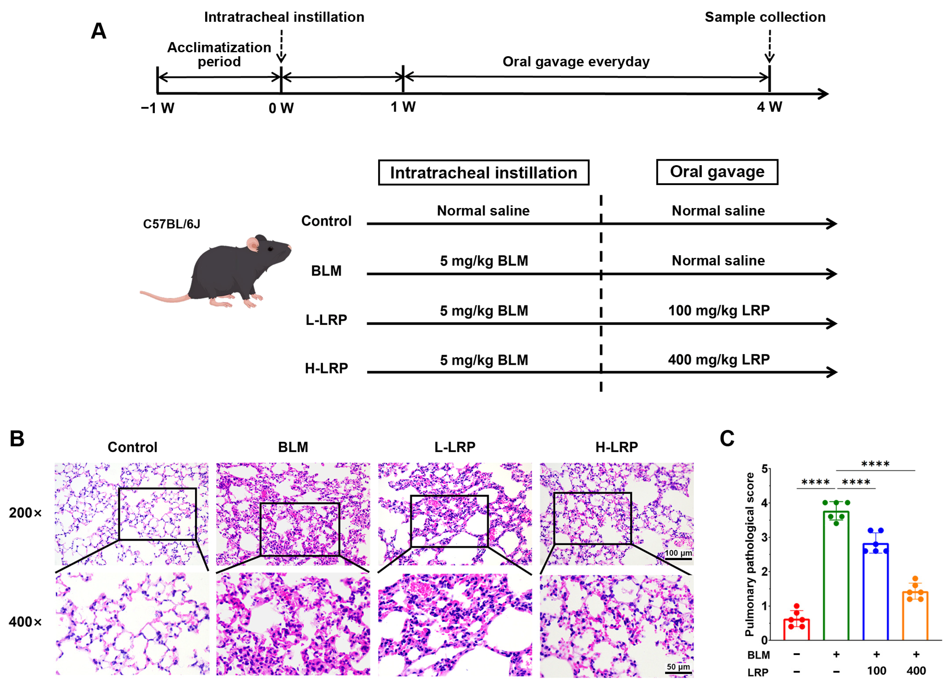
Following a 1-week acclimatization period, mice were allocated randomly to four groups (n = 12 per group) using a random number table: Control, BLM (bleomycin-treated), L-LRP (low-dose LRP), and H-LRP (high-dose LRP). The investigators who performed the animal experiments were aware of the group allocation, whereas all subsequent outcome assessments were performed by researchers blinded to group allocation. Mice in the BLM, L-LRP, and H-LRP groups received a single intratracheal instillation of bleomycin (5 mg/kg) under isoflurane anesthesia, whereas control mice received the same volume of sterile saline. Seven days after bleomycin administration, LRP was administered to the L-LRP and H-LRP groups by oral gavage at 100 mg/kg and 400 mg/kg, respectively [23,24]. The Control and BLM groups received an equal volume of normal saline by oral gavage. All treatments were administered once daily for 21 successive days [25].
At the end of the treatment period, all mice were anesthetized. For the six mice per group randomly selected for bronchoalveolar lavage fluid (BALF) analysis, blood samples were collected via the retro-orbital venous plexus before euthanasia. Blood samples were allowed to clot at room temperature for 1 h and then centrifuged to obtain serum, which was stored at −80 °C until analysis. All mice were then euthanized by cervical dislocation. BALF was collected from these six mice per group for inflammatory cell counting and cytokine analysis. The left lungs of the remaining six mice in each group were used for determination of the lung wet-to-dry weight ratio (W/D). The lower lobe of the right lung was immersed in 4% paraformaldehyde for histopathology. The remaining lung tissue was snap-frozen in liquid nitrogen and kept at −80 °C for later biochemical assays. Therefore, each endpoint was based on n = 6 biological replicates per group.
2.5. Lung W/D Ratio
Following euthanasia by cervical dislocation, the intact left lung was carefully excised, and residual surface moisture was blotted away using filter paper. The wet weight of the lung was immediately measured and recorded. The lung tissue was then placed in a constant-temperature drying oven and dried for 72 h, until no further weight change was detected. Tissue weight was measured every 24 h, and complete dehydration was confirmed when the difference between three consecutive measurements was <0.2%. The dry weight was then recorded to calculate the W/D ratio. To ensure data comparability, all measurements were performed under identical temperature and humidity conditions.
2.6. Collection of BALF and Inflammatory Cell Counting
BALF was collected using a three-step lavage procedure with 1 mL of phosphate-buffered saline (PBS) administered for each lavage. The collected BALF samples were centrifuged at 3000 rpm for 10 min at 4 °C. The supernatants were collected and aliquoted into microcentrifuge tubes for total protein and cytokine analysis, and the resulting cell pellets were resuspended in 1 mL PBS for total and differential cell counting. Total and differential cell counts were determined using an automated hematology analyzer (BC-5000 Vet, Mindray, Shenzhen, China) according to the manufacturer’s instructions. BALF total protein concentration was assessed using a bicinchoninic acid (BCA) protein assay kit as directed by the manufacturer.
2.7. H&E Staining
Following euthanasia, lung tissues were harvested and kept overnight in 4% paraformaldehyde for fixation. The fixed tissues were subsequently dehydrated, embedded in paraffin, sectioned, and subjected to H&E staining according to routine histological procedures. Histopathological changes were assessed by light microscopy. Semi-quantitative assessment of lung injury was performed using the Smith scoring method [26]. Alveolar and interstitial inflammation, hemorrhage, edema, atelectasis and hyaline membrane formation were graded on a 0–4 scale. (0: no injury; 1: mild, <25%; 2: moderate, 25–50%; 3: strong, 50–75%; 4: intense, >75%).
2.8. Masson’s Trichrome and Sirius Red Staining
Paraffin sections of lung tissue were subjected to Masson’s trichrome and Sirius Red staining following the manufacturers’ protocols. Masson’s trichrome was employed to examine histopathological alterations and collagen accumulation, while Sirius Red was applied to evaluate collagen remodeling. Representative images were captured under a light microscope. For quantification, images were analyzed using ImageJ software (version 1.54g) with a fixed, standardized threshold applied uniformly to all images.
2.9. Determination of Hyp Content
Lung tissue Hyp content was determined with a commercially available assay kit following the manufacturer’s protocol. Hyp content was expressed as μg/mg lung tissue.
2.10. Determination of Inflammatory Cytokine Levels
The concentrations of IL-1β, IL-6, TNF-α, and IL-10 in BALF and serum were quantified by commercial ELISA kits as instructed by the manufacturer.
2.11. Determination of Oxidative Stress Biomarkers
Glutathione (GSH) levels and superoxide dismutase (SOD) activity in lung tissue were quantified with commercial assay kits as directed by the manufacturer.
2.12. Immunohistochemistry (IHC)
The expression of Col-I, α-SMA, and CD73 in lung tissues was determined by IHC. Paraffin-embedded lung tissue sections were deparaffinized, rehydrated, and incubated with 3% H2O2 to block endogenous peroxidase activity, followed by antigen retrieval using citrate buffer. After blocking with 10% goat serum, the sections were incubated overnight at 4 °C with primary antibodies and subsequently incubated with the corresponding secondary antibody at room temperature. DAB substrate was applied to reveal immunoreactivity, followed by hematoxylin counterstaining. After dehydration, clearing, and mounting, the sections were viewed by light microscopy and photographed. For quantification, images were analyzed using ImageJ software with a fixed, standardized threshold applied uniformly to all images.
2.13. 16S rRNA Gene Sequencing
Fresh cecal contents were collected aseptically from mice immediately after euthanasia. Three biological replicates per group (n = 3) were analyzed. The V3–V4 hypervariable regions of the 16S rRNA gene were amplified by PCR using specific primers. After purification and quantification, the PCR products were used for library preparation, and high-throughput sequencing was performed on an Illumina NovaSeq platform by Shanghai Biozeron Biotechnology Co., Ltd. (Shanghai, China) according to the company’s standard protocols. The resulting sequences were clustered into operational taxonomic units (OTUs) at 97% sequence similarity and taxonomically annotated. The composition, relative abundance, and α- and β-diversity of the gut microbiota were subsequently analyzed.
2.14. Statistical Analysis
Statistical analyses were performed with GraphPad Prism 9.0. Data are presented as the mean ± standard deviation (SD). Normality was assessed using the Shapiro–Wilk test, and homogeneity of variance was assessed using the Brown–Forsythe test. When the data were normally distributed with homogeneous variances, comparisons among multiple groups were performed using one-way analysis of variance (ANOVA) followed by Tukey’s multiple comparisons test. When the data were normally distributed but variances were heterogeneous, the Brown–Forsythe ANOVA was used for group comparisons. Significance levels are denoted as follows: * p < 0.05, ** p < 0.01, *** p < 0.001, **** p < 0.0001, and “ns” means not significant. QIIME 2 was used for OTU clustering and for α- and β-diversity analysis of the 16S rRNA gene sequencing data. Differences in the relative abundance of microbial taxa at the phylum and genus levels were analyzed using the Kruskal–Wallis test, followed by LSD post hoc multiple comparisons. Statistically significant differences among groups were denoted by different lowercase letters (a, b, and c; p < 0.05).
3. Results
3.1. Effects of LRP on Lung Histopathology in BLM-Induced PF Mice
The characterization data of LRP have been reported in our previous study [21] and are briefly summarized here. The total sugar content of the extracted LRP was determined to be 91.02 ± 1.76%. In addition, no absorption peaks corresponding to proteins or nucleic acids were observed in the UV-Vis spectrum, indicating the effective removal of impurities during the extraction process (Figure S1). HPLC analysis revealed LRP consisted predominantly of D-glucose, L-arabinose, and D-galactose (Figure S2). The FT-IR spectrum of LRP showed a broad absorption band around 3200 cm−1 (O-H stretching) and an absorption peak at 1740 cm−1 (C=O stretching), consistent with the presence of uronic acid residues in the polysaccharide (Figure S3). Molecular weight analysis indicated that the weight-average molecular weight of LRP was 59,432 Da, with a polydispersity index of approximately 2.65.
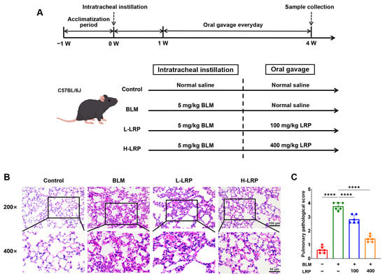
Figure 1A presents the overall experimental scheme. The protective effect of LRP against pulmonary injury was evaluated by assessing histopathological changes in lung tissue with H&E staining. As shown in Figure 1B,C, the Control group exhibited intact bronchial and alveolar architecture with normal alveolar epithelial morphology and no apparent inflammatory cell infiltration. By comparison, the BLM group showed severe disruption of lung architecture, characterized by alveolar collapse, thickening of the alveolar septa, marked vascular congestion, diffuse inflammatory cell infiltration, pulmonary consolidation, and fibrosis. In addition, prominent pulmonary bullae were observed in mice exposed to BLM. Relative to the BLM group, LRP treatment ameliorated these histopathological abnormalities in a dose-related manner, with the high-dose LRP group exhibiting the most pronounced improvement. Specifically, LRP treatment attenuated alveolar septal thickening, reduced intra-alveolar exudation, and markedly decreased inflammatory cell infiltration within the submucosal and interstitial regions.
Figure 1.
Experimental design and the protective effect of LRP against BLM-induced PF in mice. (A) Schematic diagram of the animal experimental protocol. (B) Images of H&E staining of lung tissues. (C) Semi-quantitative scoring results of pathological lung injury. **** p < 0.0001.
3.2. Effects of LRP on PF in BLM-Induced PF Mice
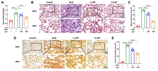
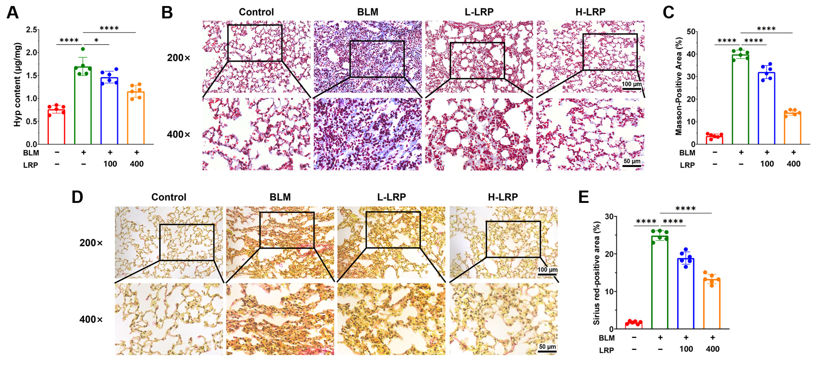
Hyp content in lung tissue was measured to evaluate the antifibrotic effect of LRP in BLM-induced PF mice. Figure 2A shows that Hyp content was significantly higher in the BLM group than in the Control group. In contrast, Hyp content was reduced to varying degrees following LRP treatment, with the H-LRP group showing the greatest reduction.
Figure 2.
LRP reduced Hyp content and alleviated collagen deposition and remodeling in lung tissues of BLM-induced PF mice. (A) Pulmonary Hyp content. (B) Images of Masson’s trichrome staining in lung tissues. (C) Quantitative analysis of Masson-positive area. (D) Images of Sirius red staining in lung tissues. (E) Quantitative analysis of Sirius red-positive area. * p < 0.05,**** p < 0.0001.
To further evaluate collagen deposition, Masson’s trichrome staining was performed. Collagen fibers in the Control group were sparsely distributed, primarily around the peribronchial and perivascular basement membranes, with no obvious thickening of the alveolar septa (Figure 2B,C). In contrast, the BLM group exhibited extensive collagen deposition throughout the lung parenchyma, characterized by diffuse collagen accumulation, marked thickening of the alveolar septa, dense collagen fiber networks, and pronounced peribronchial collagen deposition. LRP treatment markedly alleviated these fibrotic changes, with the H-LRP group exhibiting collagen deposition similar to that in the Control group.
Sirius Red staining was further performed to assess collagen maturation and spatial remodeling. Figure 2D,E shows extensive areas of intense orange-red staining in the BLM group, forming continuous reticular structures throughout the lung tissue. Confluent collagen deposition was also observed around the bronchi. Following LRP treatment, the orange-red staining became more focal and fragmented.
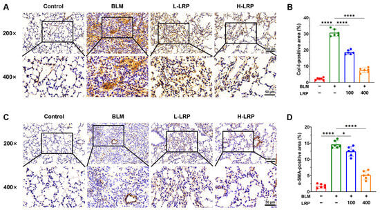
Immunohistochemical analysis was further performed to evaluate the levels of Col-I and α-SMA in lung tissues. As shown in Figure 3A,B, only weak and linear Col-I staining was observed around the bronchi in the Control group. In contrast, the BLM group exhibited extensive and confluent brown staining in areas of disrupted alveolar septa. LRP treatment markedly alleviated this abnormal collagen deposition, with the H-LRP group showing a more delicate and reticular staining pattern. Similar changes were observed in α-SMA immunostaining (Figure 3C,D). In the BLM group, α-SMA-positive myofibroblasts were densely accumulated within fibrotic lesions, whereas LRP treatment reduced the abundance of α-SMA-positive cells in a dose-dependent manner.
Figure 3.
LRP reduced Col-I and α-SMA expression in lung tissues of BLM-induced PF mice. (A) Images of immunohistochemical staining for Col-I in lung tissues. (B) Quantitative analysis of Col-I-positive area. (C) Images of immunohistochemical staining for α-SMA. (D) Quantitative analysis of α-SMA-positive area. * p < 0.05, **** p < 0.0001.
3.3. Effects of LRP on BALF Protein Content and Pulmonary Inflammation in BLM-Induced PF Mice
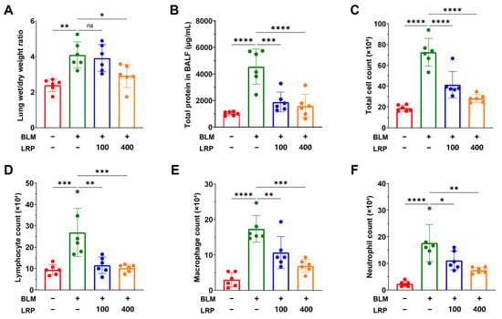
The lung W/D ratio was measured to evaluate pulmonary edema. The W/D ratio in the BLM group was 1.71-fold that of the Control group. In contrast, LRP treatment significantly reduced the W/D ratio in the H-LRP group to 0.71 times that of the BLM group (Figure 4A).
Figure 4.
LRP ameliorated pulmonary edema, attenuated protein leakage, and reduced the infiltration of inflammatory cells in BALF of BLM-induced PF mice. (A) Lung W/D ratio. (B) Total protein levels. (C) Total inflammatory cell count. (D) Lymphocyte count. (E) Macrophage count. (F) Neutrophil count. * p < 0.05, ** p < 0.01, *** p < 0.001, **** p < 0.0001.
BALF analysis showed that BLM exposure significantly increased total protein levels. LRP treatment attenuated this elevation in a dose-dependent manner (Figure 4B). In Figure 4C, persistent pulmonary inflammation caused by BLM markedly raised total inflammatory cell counts in BALF, and these counts decreased significantly following LRP treatment. Relative to the BLM group, the numbers of lymphocytes, macrophages, and neutrophils in the L-LRP group showed significant decreases of 57%, 38%, and 37%, respectively, while those in the H-LRP group showed significant decreases of 62%, 60%, and 58%, respectively (Figure 4D–F).
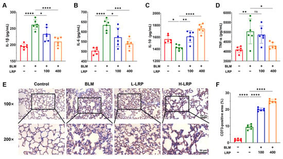
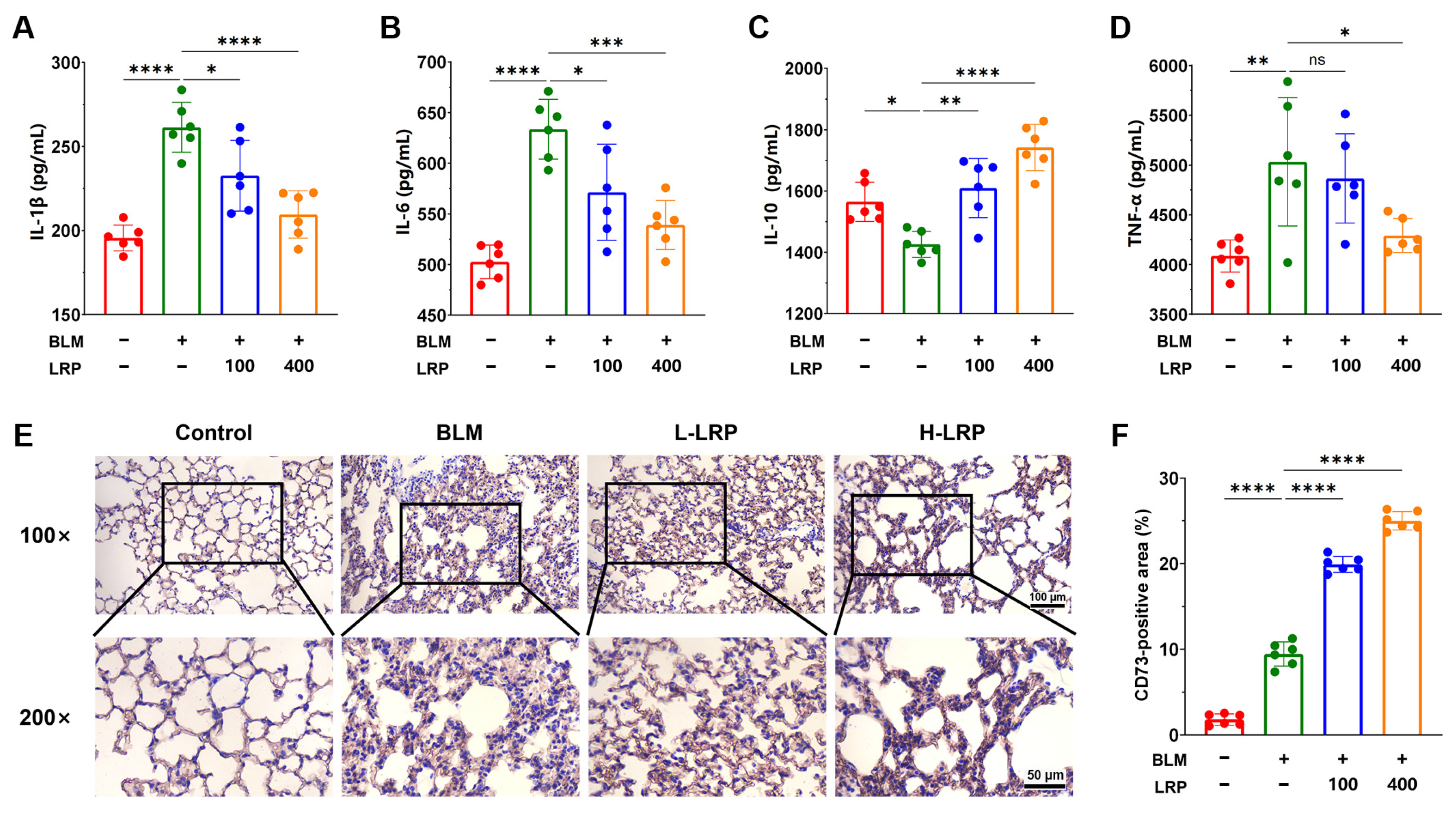
Inflammatory cytokine levels in BALF were measured by ELISA. LRP treatment reduced the levels of the pro-inflammatory cytokines TNF-α, IL-1β, and IL-6, while increasing the level of the anti-inflammatory cytokine IL-10 (Figure 5A–D). Specifically, the H-LRP group showed significant reductions in all three pro-inflammatory cytokines, whereas the L-LRP group showed significant reductions in IL-1β and IL-6, with the reduction in TNF-α not exhibiting statistical significance. CD73 expression in lung tissues was assessed by immunohistochemistry. LRP treatment significantly increased the area of CD73-positive staining in lung tissues (Figure 5E,F).
Figure 5.
LRP regulated inflammatory cytokines in BALF and CD73 expression in lung tissues of BLM-induced PF mice. (A) IL-1β level. (B) IL-6 level. (C) IL-10 level. (D) TNF-α level. (E) Images of immunohistochemical staining for CD73 in lung tissues. (F) Quantitative analysis of CD73-positive area. Note: The y-axes in (A–D) do not start at zero. * p < 0.05, ** p < 0.01, *** p < 0.001, **** p < 0.0001.
3.4. Effects of LRP on Systemic Inflammatory Responses and Antioxidant Defense in BLM-Induced PF Mice
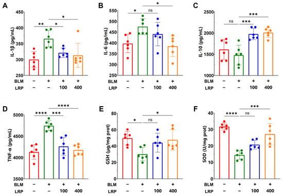
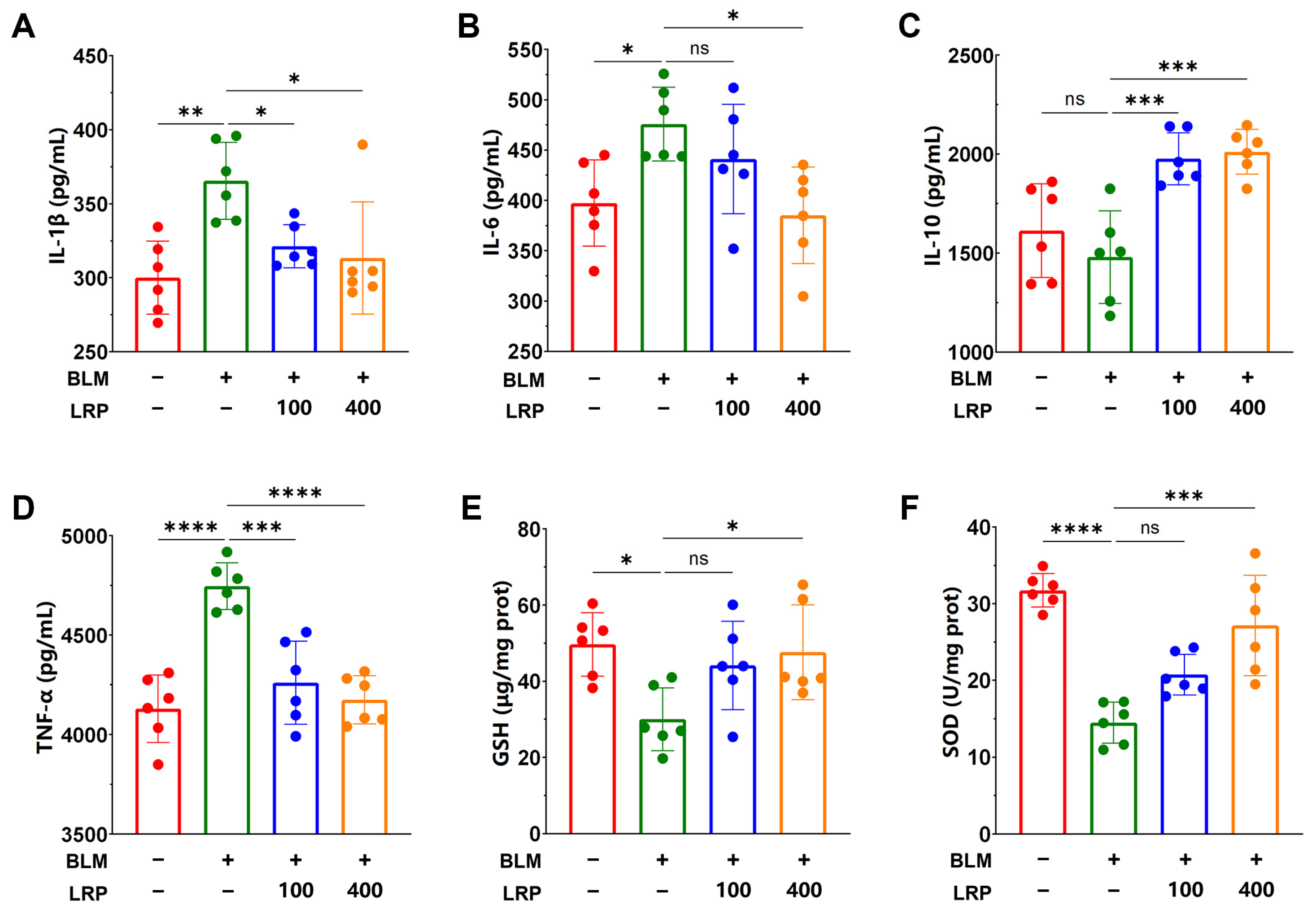
As presented in Figure 6A–D, BLM-treated mice showed significant elevations in the pro-inflammatory cytokines TNF-α, IL-6, and IL-1β relative to the Control group, together with a reduction in the anti-inflammatory cytokine IL-10. Following LRP treatment, these changes in inflammatory cytokine levels were ameliorated. In the L-LRP group, TNF-α and IL-1β were significantly reduced, whereas the reduction in IL-6 did not reach statistical significance. In the H-LRP group, all three pro-inflammatory cytokines were significantly reduced.
Figure 6.
LRP regulated inflammatory cytokines in serum and antioxidant capacity in lung tissues of PF mice. (A) IL-1β level in serum. (B) IL-6 level in serum. (C) IL-10 level in serum. (D) TNF-α level in serum. (E) GSH level. (F) SOD level. Note: The y-axes in (A–D) do not start at zero. * p < 0.05, ** p < 0.01, *** p < 0.001, **** p < 0.0001.
The effects on antioxidant capacity are shown in Figure 6E,F. Compared with the Control group, the BLM group exhibited significantly decreased GSH levels and SOD activity in lung tissue, with a 40% decrease in GSH and a 54% decrease in SOD activity. In contrast to the BLM group, the L-LRP group showed numerical increases of 47% in GSH and 43% in SOD activity, neither of which reached statistical significance, whereas the H-LRP group showed significant increases of 58% in GSH and 87% in SOD activity.
3.5. Effects of LRP on Gut Microbial Diversity in BLM-Induced PF Mice
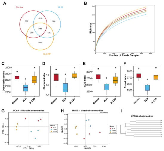
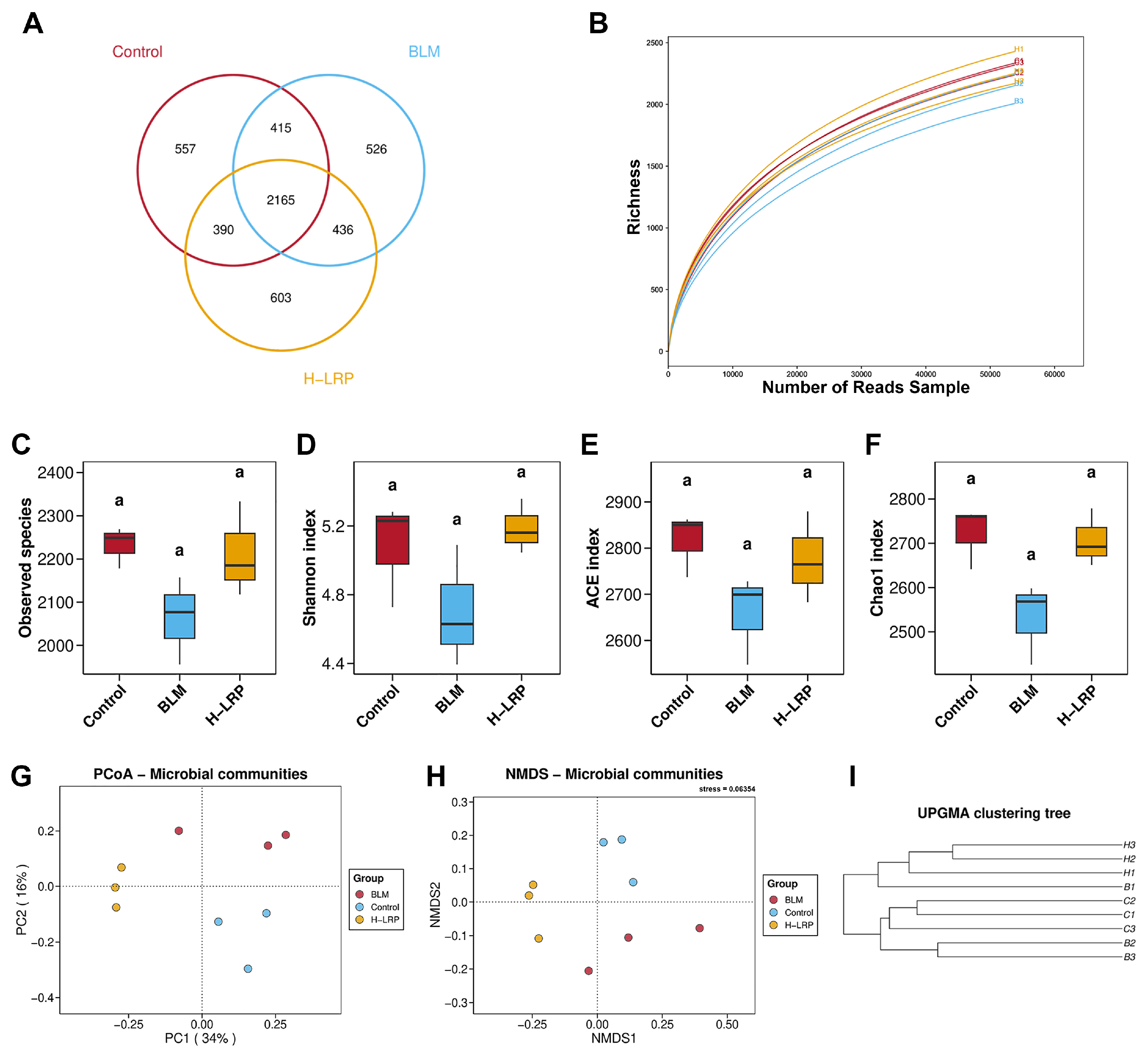
16S rRNA gene sequencing was performed to systematically characterize the composition and structural features of the cecal microbiota in the Control, BLM, and H-LRP groups (the H-LRP group is hereafter referred to as the LRP group). A total of 5092 OTUs were identified across all samples, of which 2165 were shared among the three groups. The numbers of group-specific OTUs were 557, 526, and 603 for the Control, BLM, and LRP groups, respectively. In addition, 415, 390, and 436 OTUs were shared between the Control and BLM, Control and LRP, and BLM and LRP groups, respectively (Figure 7A). As sequencing reads increased, the rarefaction curves for all samples leveled off (Figure 7B).
Figure 7.
Analysis of gut microbiota composition and diversity. (A) Venn diagram of gut microbiota OTUs among three mouse groups. (B) Sample sequencing dilution curve. (C) Observed Species. (D) Shannon index. (E) ACE index. (F) Chao1 index. In (C–F), different lowercase letters indicate significant differences (p < 0.05), whereas groups sharing the same letter are not significantly different. (G) PCoA analysis of gut microbiota. (H) NMDS analysis of gut microbiota. (I) UPGMA clustering tree analysis of gut microbiota.
The distribution of the curves further indicated that the LRP group exhibited slightly greater microbial richness than the Control and BLM groups. Compared with the Control group, the BLM group showed a trend toward decreased α-diversity indices, while LRP treatment showed a trend toward increased α-diversity indices. Nevertheless, no statistically significant differences were detected among these indices (Figure 7C–F). β-diversity was assessed using the Bray–Curtis distance matrix. Principal coordinate analysis (PCoA) revealed a clear separation between the BLM and Control groups. The LRP samples were distributed along PC2 between the Control group and the BLM group, and were separated from the Control group along PC1 (Figure 7G). Non-metric multidimensional scaling (NMDS) analysis showed a similar pattern (Figure 7H). The UPGMA clustering tree showed that samples tended to cluster according to their respective groups, with the LRP group forming a unique branch (Figure 7I).
3.6. Effects of LRP on Gut Microbial Composition in BLM-Induced PF Mice
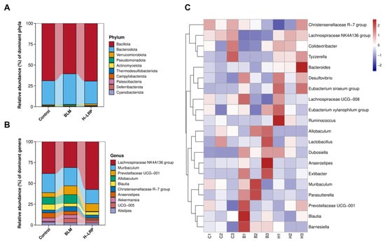
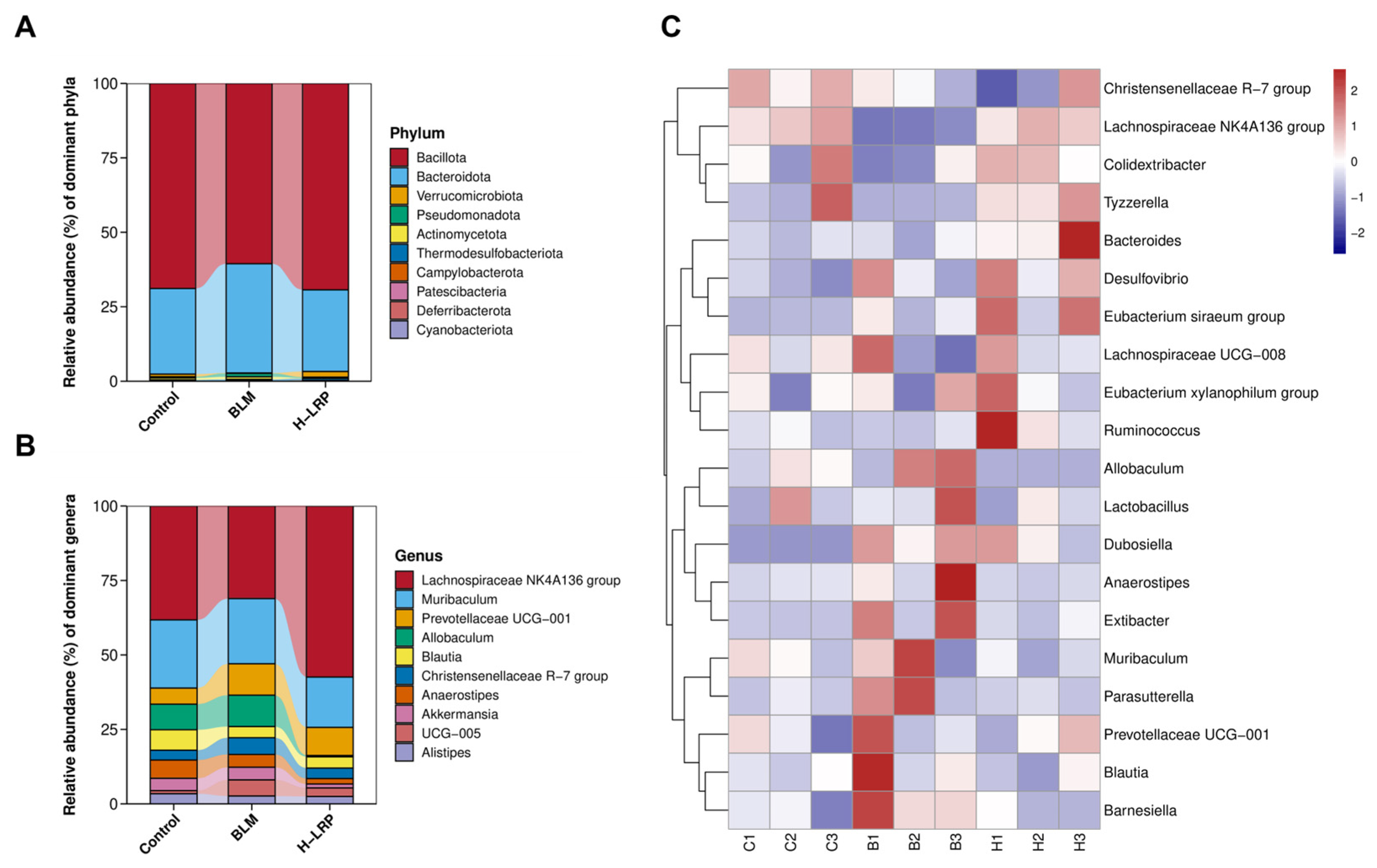
The relative abundances of key bacterial taxa were compared at the phylum and genus levels (Figure 8). Bacillota, Bacteroidota, and Pseudomonadota were the most abundant phyla in the cecal microbiota of all three groups. Compared with the Control group, BLM induction markedly altered the gut microbial composition, as evidenced by a considerable decline in the relative abundance of Bacillota and a significant increase in Bacteroidota, resulting in a marked decrease in the Bacillota/Bacteroidota (F/B) ratio. Following high-dose LRP treatment, these alterations were partially reversed. The relative abundance of Bacillota increased, that of Bacteroidota decreased and the F/B ratio largely recovered toward control levels (Figure 8A). Analysis at the genus level further revealed the regulatory effects of LRP on key gut microbial taxa in PF mice (Figure 8B). BLM induction significantly lowered the proportion of the Lachnospiraceae NK4A136 group relative to the Control group, whereas LRP treatment markedly increased its relative abundance.
Figure 8.
Composition and differential analysis of gut microbiota species. (A) Relative abundance of gut microbiota at phylum level. (B) Relative abundance of gut microbiota at genus level. (C) Heatmap of the top 20 species composition in samples.
A clustering heatmap based on the 20 most abundant bacterial taxa at the genus level further visualized the differences and clustering patterns in the gut microbiota among the three groups (Figure 8C). The heatmap showed distinct within-group clustering of microbial profiles in the Control, BLM, and LRP groups, with marked differences in microbial abundance between groups. In particular, the BLM group exhibited increased abundances of several potentially pro-inflammatory taxa, accompanied by a marked reduction in short-chain fatty acid (SCFA)-associated beneficial taxa. Following LRP treatment, the abundances of these differentially represented taxa showed a clear trend toward restoration.
4. Discussion
During the progression of PF, structural remodeling of the lung follows a centripetal pattern. Pathological injury initially occurs around the terminal bronchioles and is accompanied by infiltration of macrophages and T lymphocytes. The injury subsequently extends to the alveolar septa, leading to disruption of the alveolar basement membrane and abnormal hyperplasia of type II alveolar epithelial cells, ultimately progressing to extensive pulmonary consolidation [27]. In the present study, we established a BLM-induced PF mouse model and demonstrated that LRP treatment significantly attenuated pulmonary fibrosis, as evidenced by improved histopathological architecture, reduced collagen deposition, and decreased myofibroblast activation. Furthermore, LRP treatment was associated with the suppression of systemic inflammation, enhancement of antioxidant defenses, and remodeling of the gut microbial community, suggesting that LRP exerts its protective effects against PF through multiple mechanisms.
Hyp is a collagen-specific amino acid and a well-established quantitative marker of collagen accumulation and fibrosis in lung tissue [28]. The elevated Hyp content and collagen staining in BLM-treated mice confirm excessive collagen accumulation [29,30,31,32], and their reduction by LRP supports an antifibrotic effect. These findings are consistent with previous studies demonstrating that BLM promotes pathological accumulation of type I collagen and extensive ECM remodeling [33]. As the principal ECM protein, Col-I is routinely used to indicate collagen deposition [34], while α-SMA serves as a common marker of myofibroblast activation, and its increased expression is associated with the progression of PF [35]. The reduced myofibroblast activation and collagen accumulation observed following LRP treatment suggest that LRP exerts its antifibrotic effects by inhibiting myofibroblast activation and pathological type I collagen deposition while promoting collagen remodeling.
During lung injury, disruption of the alveolar-capillary barrier leads to increased pulmonary microvascular permeability, resulting in the continuous leakage of fluid and macromolecules into the pulmonary interstitium and alveolar spaces, and consequently promoting pulmonary edema. Persistent pulmonary edema can further exacerbate alveolar epithelial injury, fibroblast activation, and ECM deposition, thereby contributing to the progression of PF [36]. The elevated W/D ratio and BALF protein content in BLM-treated mice indicate increased pulmonary permeability, and their reduction by LRP suggests attenuation of vascular leakage and edema. Neutrophils, macrophages, and lymphocytes represent three major inflammation-associated cell populations in BALF [37]. Neutrophils can release neutrophil extracellular traps, which activate the TGF-β1 signaling pathway and promote fibroblast activation [38]. Macrophages are also key regulators of PF progression, and their phenotypic polarization plays an important role in modulating fibrotic responses. Aberrant macrophage activation can promote the release of profibrotic factors, including TGF-β and platelet-derived growth factor (PDGF), thereby directly stimulating fibroblast activation [39]. The reduction in inflammatory cell infiltration by LRP suggests that it may alleviate BLM-induced pulmonary inflammation and potentially limit subsequent fibrotic progression by suppressing the early inflammatory response. Previous studies have reported anti-inflammatory effects of LRP in hepatic injury models [24,40]. The present findings support the extension of this anti-inflammatory effect to BLM-induced pulmonary inflammation.
Persistent inflammation and the subsequent fibrotic cascade are central features of PF pathogenesis, with activated immune cells releasing a variety of profibrotic mediators [41]. In this study, LRP treatment modulated the inflammatory cytokine profile by suppressing the release of the pro-inflammatory cytokines TNF-α, IL-1β, and IL-6, while increasing the level of the anti-inflammatory cytokine IL-10. These findings suggest that LRP protects against PF by regulating the inflammatory cytokine balance.
Dysregulated macrophage polarization is considered a key feature of PF progression [42,43]. CD73, a key enzyme involved in adenosine generation, has been implicated in the regulation of macrophage polarization and may promote tissue repair when highly expressed. In the present study, CD73-positive area was already elevated in the BLM group, likely reflecting a compensatory anti-inflammatory response to injury. However, this endogenous upregulation was insufficient to prevent fibrotic progression. LRP treatment further increased the area of CD73-positive staining in lung tissues. This increase was accompanied by elevated IL-10 levels and reduced collagen deposition, suggesting a potential link between CD73-related pathways, macrophage responses, and the protective effects of LRP. Notably, the roles of CD73 and macrophages in pulmonary fibrosis are context-dependent. CD73 has been reported to exert both protective and profibrotic effects depending on the disease stage and microenvironment, and macrophages can similarly display anti-inflammatory or profibrotic phenotypes [44,45,46,47]. Therefore, the involvement of CD73-related pathways and macrophage responses in protective effects requires further systematic validation.
In BLM-induced PF, dysregulated local pulmonary inflammation may extend into the systemic circulation, contributing to systemic inflammatory imbalance. Persistently elevated circulating pro-inflammatory cytokines may, in turn, promote the recruitment of inflammatory cells to pulmonary lesions and further amplify local inflammatory responses. In this study, the systemic inflammatory imbalance observed in BLM-treated mice, characterized by elevated pro-inflammatory cytokines and reduced IL-10, is consistent with the inflammatory characteristics of PF. Following LRP treatment, these abnormal changes in inflammatory cytokine levels were ameliorated, with the H-LRP group showing the most pronounced effects. Collectively, these findings suggest that the protective effects of LRP against PF may extend beyond the modulation of the local pulmonary inflammatory microenvironment to include broader systemic immunomodulatory effects.
Excessive accumulation of reactive oxygen species (ROS) induced by BLM can damage alveolar epithelial cells and trigger EMT, thereby promoting fibroblast activation. In addition, excessive ROS production can activate the NLRP3 inflammasome and further exacerbate inflammatory responses [48]. GSH is an important endogenous antioxidant, and its depletion may increase the susceptibility of lung tissue to oxidative stress-induced injury [49]. SOD is a key indicator of antioxidant capacity, and reduced SOD activity reflects impaired antioxidant defenses against oxidative stress in the lung [50]. The increased GSH levels and SOD activity observed following LRP treatment in this study indicate an enhancement of endogenous antioxidant defense capacity. This finding is consistent with previous reports on plant-derived polysaccharides. In previous studies, Ganoderma lucidum polysaccharide increased pulmonary SOD and GSH levels in BLM-treated rats [51]; polysaccharide from Sarcodon aspratus enhanced antioxidant enzyme activities and reduced MDA and ROS levels, attenuating bleomycin-induced pulmonary fibrosis [52]; and Achyranthes bidentata polysaccharide alleviated oxidative damage by increasing SOD and GSH-Px activity [53].
The composition and functional homeostasis of the gut microbiota play important roles in the development and progression of PF [54]. Gut microbiota alterations during PF are characterized by sustained ecological remodeling and selective shifts in functional microbial populations. These changes may contribute to pulmonary fibrotic progression through mechanisms involving the persistent production of pro-inflammatory metabolites and modulation of immune cell differentiation [55]. In this study, 16S rRNA gene sequencing revealed that none of the α-diversity indices differed significantly among the groups, consistent with previous studies suggesting that α-diversity may not be the most sensitive indicator for evaluating the effects of LRP intervention [56,57]. The regulatory effects of LRP on the gut microbiota may be primarily reflected in the enrichment of specific microbial taxa with potential functional roles [58]. β-diversity analysis showed a clear separation between the BLM and Control groups, whereas the LRP group exhibited a distinct community structure rather than a simple return to the Control profile. Compared with the BLM group, the LRP group showed markedly greater intra-group aggregation, indicating reduced inter-individual variability. These findings suggest that LRP may reshape microbial communities into new structures.
At the phylum level, BLM induction markedly altered the gut microbial composition and reduced the F/B ratio. Previous studies have suggested that the F/B ratio may serve as an indicator of gut microbial homeostasis, and its reduction has been associated with gut dysbiosis, impaired intestinal barrier integrity, systemic inflammation, and the progression of PF in mice. Consistent with our findings, previous studies have reported that plant-derived polysaccharides can improve gut microbial dysbiosis by modulating the F/B ratio [10]. Following high-dose LRP treatment, these alterations were partially reversed, with the F/B ratio largely recovering toward control levels. At the genus level, the Lachnospiraceae NK4A136 group is considered an important SCFA-associated bacterial taxon in the gut [59], and its depletion may contribute to reduced SCFA production, impaired intestinal barrier integrity, and increased pro-inflammatory responses, thereby potentially exacerbating pulmonary inflammation and fibrotic progression. Previous studies have shown that various plant-derived polysaccharides can enrich SCFA-producing beneficial taxa, including the Lachnospiraceae NK4A136 group, and increase intestinal SCFA production, thereby exerting anti-inflammatory effects [60,61,62]. These results provide further support for the potential role of LRP in modulating the gut microbiota. In addition, dysbiosis of the gut microbiota may contribute to the development and progression of PF by impairing intestinal barrier integrity, increasing the translocation of pro-inflammatory microbial metabolites into the circulation, and modulating systemic immune and inflammatory responses [63,64]. The regulatory effects of LRP on the gut microbiota may therefore be associated with the attenuation of pulmonary inflammation and fibrotic progression. In summary, these findings suggest that gut microbiota remodeling may contribute to the protective effects of LRP.
5. Conclusions
In this study, a murine model of PF was established by intratracheal instillation of BLM, and the protective effects of LRP and its potential regulatory mechanisms during PF progression were investigated. Histopathological analysis demonstrated that LRP treatment markedly alleviated the characteristic pathological features of PF, including alveolar wall thickening, alveolar collapse, hemorrhage, and edema, thereby preserving alveolar structural integrity and reducing BLM-induced pulmonary tissue disruption and inflammatory cell infiltration. At the molecular level, LRP exhibited potent anti-inflammatory effects by modulating the balance between pro-inflammatory cytokines (TNF-α, IL-1β and IL-6) and the anti-inflammatory cytokine IL-10. In addition, LRP enhanced antioxidant defenses, as evidenced by increased SOD activity and GSH levels. Furthermore, this study suggests that LRP treatment is associated with remodeling of the gut microbial community. These findings provide new evidence for the protective effects of LRP against PF, suggesting the potential of LRP as a natural therapeutic agent.
Supplementary Materials
The following supporting information can be downloaded at https://www.mdpi.com/article/10.3390/antiox15111433/s1. Figure S1: UV-Vis absorption spectrum of LRP; Figure S2: HPLC profile of standard and LRP for monosaccharide composition analysis; Figure S3: FT-IR spectrum of LRP revealing characteristic polysaccharide absorption peaks.
Author Contributions
Conceptualization, N.L.; methodology, W.X., N.L. and X.W.; formal analysis, Z.C.; investigation, Z.C. and S.X.; data curation, S.X.; writing—original draft preparation, Z.C. and S.X.; writing—review and editing, S.Y.G. and Q.L.; supervision, J.C. and X.W.; project administration, N.L. and X.W.; funding acquisition, W.X. All authors have read and agreed to the published version of the manuscript.
Funding
This research was funded by the Youth Research Project of Wuxi Municipal Health Commission, grant number Q202416.
Institutional Review Board Statement
The use of animals and experimental design were approved by the Jiangnan University Animal Care and Ethics Committee (Approval number: JN. No20250515c0600706[273], approval date: 15 May 2025).
Informed Consent Statement
Not applicable.
Data Availability Statement
The original 16S rRNA gene sequencing data presented in the study are openly available in the NCBI Sequence Read Archive under accession number PRJNA1550545.
Conflicts of Interest
The authors declare no conflicts of interest.
References
- Mathai, S.K.; Schwartz, D.A. Translational research in pulmonary fibrosis. Transl. Res. 2019, 209, 1–13. [Google Scholar] [CrossRef] [Scilit] [PubMed]
- Yu, D.K.; Xiang, Y.; Gou, T.T.; Tong, R.S.; Xu, C.; Chen, L.; Zhong, L.; Shi, J.Y. New therapeutic approaches against pulmonary fibrosis. Bioorg. Chem. 2023, 138, 106592. [Google Scholar] [CrossRef] [Scilit] [PubMed]
- Kolahian, S.; Fernandez, I.E.; Eickelberg, O.; Hartl, D. Immune Mechanisms in Pulmonary Fibrosis. Am. J. Respir. Cell Mol. 2016, 55, 309–322. [Google Scholar] [CrossRef] [Scilit] [PubMed]
- Ortiz-Zapater, E.; Signes-Costa, J.; Montero, P.; Roger, I. Lung Fibrosis and Fibrosis in the Lungs: Is It All about Myofibroblasts? Biomedicines 2022, 10, 1423. [Google Scholar] [CrossRef] [Scilit] [PubMed]
- Pan, L.; Cheng, Y.J.; Yang, W.T.; Wu, X.; Zhu, H.L.; Hu, M.G.; Zhang, Y.Q.; Zhang, M.L. Nintedanib Ameliorates Bleomycin-Induced Pulmonary Fibrosis, Inflammation, Apoptosis, and Oxidative Stress by Modulating PI3K/Akt/mTOR Pathway in Mice. Inflammation 2023, 46, 1531–1542. [Google Scholar] [CrossRef] [Scilit] [PubMed]
- Wu, Y.L.; Li, Y.H.; Luo, Y.B.; Zhou, Y.; Wen, J.; Chen, L.; Liang, X.P.; Wu, T.; Tan, C.Y.; Liu, Y. Gut microbiome and metabolites: The potential key roles in pulmonary fibrosis. Front. Microbiol. 2022, 13, 943791. [Google Scholar] [CrossRef] [Scilit] [PubMed]
- Huang, C.W.; Lee, S.Y.; Du, C.X.; Wu, S.T.; Kuo, Y.H.; Ku, H.C. Caffeic acid ethanolamide induces antifibrosis, anti-inflammatory, and antioxidant effects protects against bleomycin-induced pulmonary fibrosis. Biomed. Pharmacother. 2024, 173, 116298. [Google Scholar] [CrossRef] [Scilit] [PubMed]
- Wu, X.B.; Huang, J.L.; Wang, J.; Xu, Y.H.; Yang, X.W.; Sun, M.H.; Shi, J.Y. Multi-Pharmaceutical Activities of Chinese Herbal Polysaccharides in the Treatment of Pulmonary Fibrosis: Concept and Future Prospects. Front. Pharmacol. 2021, 12, 707491. [Google Scholar] [CrossRef] [Scilit] [PubMed]
- Ming, K.; Zhuang, S.; Ma, N.; Nan, S.; Li, Q.H.; Ding, M.X.; Ding, Y. polysaccharides alleviates lipopolysaccharides-induced inflammatory lung injury by altering intestinal microbiota in mice. Front. Microbiol. 2022, 13, 1033875. [Google Scholar] [CrossRef] [Scilit] [PubMed]
- Wei, Y.; Qi, M.; Liu, C.; Li, L.J. Astragalus polysaccharide attenuates bleomycin-induced pulmonary fibrosis by inhibiting TLR4/NF-κB signaling pathway and regulating gut microbiota. Eur. J. Pharmacol. 2023, 944, 175594. [Google Scholar] [CrossRef] [Scilit] [PubMed]
- Liu, R.J.; Wu, T.; Zhou, W.Q.; Zhu, A.M.; Liao, W.F.; Ding, K. A Novel Polysaccharide from the Flowers of Lilium lancifolium Alleviates Pulmonary Fibrosis In Vivo and In Vitro. J. Agric. Food Chem. 2025, 73, 7774–7787. [Google Scholar] [CrossRef] [Scilit] [PubMed]
- Abai, J.; Wang, Y.D.; Jin, R.H.; Abduwali, H.; Wan, J.Y.; Li, Y.Z.; Wei, W.; Lv, W.J.; Abdureyim, S.; Maimaijiang, M.; et al. A novel polysaccharide from Qamgur (Brassica rapa L.) mitigates pulmonary fibrosis by regulating inflammatory, epithelial-mesenchymal transition, and fibrotic pathways. J. Ethnopharmacol. 2026, 360, 121174. [Google Scholar] [CrossRef] [Scilit] [PubMed]
- Xiong, L.; Deng, N.; Zheng, B.S.; Li, T.; Liu, R.H. Goji berry (Lycium spp.) extracts exhibit antiproliferative activity via modulating cell cycle arrest, cell apoptosis, and the p53 signaling pathway. Food Funct. 2021, 12, 6513–6525. [Google Scholar] [CrossRef] [Scilit] [PubMed]
- Lu, J.X.; Xiang, X.Q.; Zhang, Z.F.; Ren, J.N.; Li, M.Z.; Yang, J.C.; Gao, Y.; Fan, G.; Pan, S.Y. Nutritional compositions, phytochemical components, functional activities, and food applications of Lycium ruthenicum Murr.: A comprehensive review. J. Food Compos. Anal. 2025, 140, 107301. [Google Scholar] [CrossRef] [Scilit]
- Dong, D.; Wang, H.L.; Bi, H.T.; Li, Y.; Gao, T.T.; Feng, J.Y.; Li, G.Q.; Guo, S.Q.; Yuan, H.Y.; Ni, W.H. A pectic polysaccharide from Lycium ruthenicum Murray alleviates dextran sulfate sodium-induced colitis in mice. Curr. Res. Food Sci. 2025, 10, 100955. [Google Scholar] [CrossRef] [Scilit] [PubMed]
- Liu, B.; Ma, J.Y.; Li, T.; Li, P.; Yan, D.H.; Zhu, J.; Zhang, X.G. Advances in the Preparation, Structure and Bioactivity of Polysaccharides from Lycium ruthenicum Murr.: A Review. Foods 2024, 13, 1995. [Google Scholar] [CrossRef] [Scilit] [PubMed]
- Zhu, W.; Zhou, S.X.; Liu, J.H.; McLean, R.J.C.; Chu, W.H. Prebiotic, immuno-stimulating and gut microbiota-modulating effects of Lycium barbarum polysaccharide. Biomed. Pharmacother. 2020, 121, 109591. [Google Scholar] [CrossRef] [Scilit] [PubMed]
- Xia, N.T.; Ding, Z.M.; Dong, M.R.; Li, S.Y.; Liu, J.; Xue, H.W.; Wang, Z.G.; Lu, J.; Chen, X. Protective Effects of Lycium ruthenicum Murray against Acute Alcoholic Liver Disease in Mice via the Nrf2/HO-1/NF-κB Signaling Pathway. Pharmaceuticals 2024, 17, 497. [Google Scholar] [CrossRef] [Scilit] [PubMed]
- Qin, X.S.; Wang, X.Y.; Xu, K.; Yang, X.B.; Wang, Q.; Liu, C.; Wang, X.K.; Guo, X.; Sun, J.Y.; Li, L.; et al. Synergistic antitumor effects of polysaccharides and anthocyanins from Lycium ruthenicum Murr. on human colorectal carcinoma LoVo cells and the molecular mechanism. Food Sci. Nutr. 2022, 10, 2956–2968. [Google Scholar] [CrossRef] [Scilit] [PubMed]
- Zheng, G.Z.; Ben, H.J.; Li, H.Q.; Li, X.H.; Dong, T.C.; Xu, S.M.; Yan, Y.L.; Sun, B.K.; Bai, J.W.; Li, Y.S. polysaccharide reduces hyperoxic acute lung injury in mice through Nrf2 pathway. Biomed. Pharmacother. 2019, 111, 733–739. [Google Scholar] [CrossRef] [Scilit] [PubMed]
- Lu, N.Y.; Xu, S.H.; Xiang, W.; Mei, X.; Hu, H.W.; Tang, X.; Gong, X.L.; Wang, X. Lycium ruthenicum Murr. Polysaccharide Attenuated Inflammatory Response and Intestinal Flora Dysbiosis in LPS-Induced Acute Lung Injury in Mice. Nutrients 2025, 17, 2944. [Google Scholar] [CrossRef] [Scilit] [PubMed]
- Dubois, M.; Gilles, K.A.; Hamilton, J.K.; Rebers, P.A.; Smith, F. Colorimetric Method for Determination of Sugars and Related Substances. Anal. Chem. 1956, 28, 350–356. [Google Scholar] [CrossRef] [Scilit]
- Cui, Y.M.; Zhao, J.; Chen, J.; Kong, Y.W.; Wang, M.Y.; Ma, Y.; Meng, X.J. Cyanidin-3-galactoside from Aronia melanocarpa ameliorates silica-induced pulmonary fibrosis by modulating the TGF-β/mTOR and NRF2/HO-1 pathways. Food Sci. Nutr. 2022, 10, 2558–2567. [Google Scholar] [CrossRef] [Scilit] [PubMed]
- Wang, W.J.; Wu, D.S.; Liu, J.G.; Yang, D.A. Potential protective role of Lycium ruthenicum Murray polysaccharides against lipopolysaccharide-induced liver injury via mitochondrial biogenesis. Int. J. Biol. Macromol. 2025, 306, 141365. [Google Scholar] [CrossRef] [Scilit] [PubMed]
- Wang, T.; Xu, L.T.; Li, P.P.; Zhang, C.H.; Han, Q.T.; Wang, X.N.; Xiang, L.; Xu, Z.P.; Shen, T. Physalis Calyx seu Fructus inhibited pulmonary fibrosis through regulating Wnt/fi-catenin signaling pathway. Phytomedicine 2024, 131, 155797. [Google Scholar] [CrossRef] [Scilit] [PubMed]
- Smith, K.M.; Mrozek, J.D.; Simonton, S.C.; Bing, D.R.; Meyers, P.A.; Connett, J.E.; Mammel, M.C. Prolonged partial liquid ventilation using conventional and high-frequency ventilatory techniques: Gas exchange and lung pathology in an animal model of respiratory distress syndrome. Crit. Care Med. 1997, 25, 1888–1897. [Google Scholar] [PubMed]
- Ogawa, T.; Shichino, S.; Ueha, S.; Matsushima, K. Macrophages in lung fibrosis. Int. Immunol. 2021, 33, 665–671. [Google Scholar] [CrossRef] [Scilit] [PubMed]
- Yin, Y.J.; Jia, N.; Luo, H.; Feng, X.L.; He, X.; Huang, J.; Wang, F. Protective effects and mechanism of resveratrol in animal models of pulmonary fibrosis: A preclinical systematic review and meta-analysis. Front. Pharmacol. 2025, 16, 1666698. [Google Scholar] [CrossRef] [Scilit] [PubMed]
- Zhou, P.; Yang, C.; Zhang, S.; Ke, Z.X.; Chen, D.X.; Li, Y.Q.; Li, Q. The Imbalance of MMP-2/TIMP-2 and MMP-9/TIMP-1 Contributes to Collagen Deposition Disorder in Diabetic Non-Injured Skin. Front. Endocrinol. 2021, 12, 734485. [Google Scholar] [CrossRef] [Scilit] [PubMed]
- Zhou, S.B.; Zhou, Y.C.; Yu, J.J.; Du, Y.X.; Tan, Y.; Ke, Y.M.; Wang, J.; Han, B.Y.; Ge, F. polysaccharides attenuate pulmonary fibrosis in mice. Biomed. Pharmacother. 2020, 126, 110058. [Google Scholar] [CrossRef] [Scilit] [PubMed]
- Park, S.J.; Hahn, H.J.; Oh, S.R.; Lee, H.J. Theophylline Attenuates BLM-Induced Pulmonary Fibrosis by Inhibiting Th17 Differentiation. Int. J. Mol. Sci. 2023, 24, 1019. [Google Scholar] [CrossRef] [Scilit] [PubMed]
- Soundararajan, R.; Varanasi, S.M.; Patil, S.S.; Srinivas, S.; Hernández-Cuervo, H.; Czachor, A.; Bulkhi, A.; Fukumoto, J.; Galam, L.; Lockey, R.F.; et al. Lung fibrosis is induced in ADAR2 overexpressing mice via HuR-induced CTGF signaling. FASEB J. 2022, 36, e22143. [Google Scholar] [CrossRef] [Scilit] [PubMed]
- Strandjord, T.P.; Madtes, D.K.; Weiss, D.J.; Sage, E.H. Collagen accumulation is decreased in SPARC-null mice with bleomycin-induced pulmonary fibrosis. Am. J. Physiol.-Lung Cell. Mol. Physiol. 1999, 277, L628–L635. [Google Scholar] [CrossRef] [Scilit] [PubMed]
- Li, W.; Liu, J.R.; Wu, T.B.; Qiang, X.; Peng, Y.J.; Zhao, Q.Y.; He, D. Design and synthesis of glycyrrhetinic acid glycosides against acute lung injury and pulmonary fibrosis. Mol. Divers. 2025, 29, 4555–4577. [Google Scholar] [CrossRef] [Scilit] [PubMed]
- Zhao, J.L.; Ma, X.; Li, S.Q.; Liu, C.; Liu, Y.M.; Tan, J.F.; Yu, L.; Li, X.H.; Li, W.W. Berberine hydrochloride ameliorates PM2.5-induced pulmonary fibrosis in mice through inhibiting oxidative stress and inflammatory. Chem.-Biol. Interact. 2023, 386, 110731. [Google Scholar] [CrossRef] [Scilit] [PubMed]
- Miserocchi, G.; Beretta, E. A century of exercise physiology: Lung fluid balance during and following exercise. Eur. J. Appl. Physiol. 2023, 123, 1–24. [Google Scholar] [CrossRef] [Scilit] [PubMed]
- Wang, F.; Fu, X.Z.; Wu, X.W.; Zhang, J.H.; Zhu, J.L.; Zou, Y.; Li, J.B. Bone marrow derived M2 macrophages protected against lipopolysaccharide-induced acute lung injury through inhibiting oxidative stress and inflammation by modulating neutrophils and T lymphocytes responses. Int. Immunopharmacol. 2018, 61, 162–168. [Google Scholar] [CrossRef] [Scilit] [PubMed]
- Crowley, L.E.; Stockley, R.A.; Thickett, D.R.; Dosanjh, D.; Scott, A.; Parekh, D. Neutrophil dynamics in pulmonary fibrosis: Pathophysiological and therapeutic perspectives. Eur. Respir. Rev. 2024, 33, 240139. [Google Scholar] [CrossRef] [Scilit] [PubMed]
- Li, R.S.; Hanna, A.; Huang, S.B.; Hernandez, S.C.; Tuleta, I.; Kubota, A.; Humeres, C.; Chen, B.J.; Liu, Y.; Zheng, D.Y.; et al. Macrophages in the infarcted heart acquire a fibrogenic phenotype, expressing matricellular proteins, but do not undergo fibroblast conversion. J. Mol. Cell. Cardiol. 2024, 196, 152–167. [Google Scholar] [CrossRef] [Scilit] [PubMed]
- Xiao, J.; Li, C.P.; Pei, Y.X.; Xu, S.H.; Zhao, H.T.; Xiang, W.; Wei, J.Y. Polysaccharides Alleviate CCl-Induced Acute Liver Injury Through Antioxidant and Anti-Inflammatory Effects. Nutrients 2025, 17, 3359. [Google Scholar] [CrossRef] [Scilit] [PubMed]
- Kleinman, H.K.; Kulik, V.; Goldstein, A.L. Prepared for special issue on thymosins Thymosin β4 and the anti-fibrotic switch. Int. Immunopharmacol. 2023, 115, 109628. [Google Scholar] [PubMed]
- Zhou, B.W.; Liu, H.M.; Xu, F.; Jia, X.H. The role of macrophage polarization and cellular crosstalk in the pulmonary fibrotic microenvironment: A review. Cell Commun. Signal. 2024, 22, 172. [Google Scholar] [CrossRef] [Scilit] [PubMed]
- Kishore, A.; Petrek, M. Roles of Macrophage Polarization and Macrophage-Derived miRNAs in Pulmonary Fibrosis. Front. Immunol. 2021, 12, 678457. [Google Scholar] [CrossRef] [Scilit] [PubMed]
- Volmer, J.B.; Thompson, L.F.; Blackburn, M.R. Ecto-5′-nucleotidase (CD73)-mediated adenosine production is tissue protective in a model of bleomycin-induced lung injury. J. Immunol. 2006, 176, 4449–4458. [Google Scholar] [CrossRef] [Scilit] [PubMed]
- de Leve, S.; Wirsdörfer, F.; Cappuccini, F.; Schütze, A.; Meyer, A.V.; Röck, K.; Thompson, L.F.; Fischer, J.W.; Stuschke, M.; Jendrossek, V. Loss of CD73 prevents accumulation of alternatively activated macrophages and the formation of prefibrotic macrophage clusters in irradiated lungs. FASEB J. 2017, 31, 2869–2880. [Google Scholar] [CrossRef] [Scilit] [PubMed]
- Kamiya, M.; Carter, H.; Espindola, M.S.; Doyle, T.J.; Lee, J.S.; Merriam, L.T.; Zhang, F.; Kawano-Dourado, L.; Sparks, J.A.; Hogaboam, C.M.; et al. Immune mechanisms in fibrotic interstitial lung disease. Cell 2024, 187, 3506–3530. [Google Scholar] [CrossRef] [Scilit] [PubMed]
- Wynn, T.A.; Vannella, K.M. Macrophages in Tissue Repair, Regeneration, and Fibrosis. Immunity 2016, 44, 450–462. [Google Scholar] [CrossRef] [Scilit] [PubMed]
- Karamalakova, Y.; Stefanov, I.; Georgieva, E.; Nikolova, G. Pulmonary Protein Oxidation and Oxidative Stress Modulation by lemna minor L. in Progressive Bleomycin-Induced Idiopathic Pulmonary Fibrosis. Antioxidants 2022, 11, 523. [Google Scholar] [CrossRef] [Scilit] [PubMed]
- Zheng, H.Y.; Zhang, L.; Wang, C.J.; Wang, Y.; Zeng, C.X. Metabolic dysregulation in pulmonary fibrosis: Insights into amino acid contributions and therapeutic potential. Cell Death Discov. 2025, 11, 411. [Google Scholar] [CrossRef] [Scilit] [PubMed]
- Jomova, K.; Alomar, S.Y.; Alwasel, S.H.; Nepovimova, E.; Kuca, K.; Valko, M. Several lines of antioxidant defense against oxidative stress: Antioxidant enzymes, nanomaterials with multiple enzyme-mimicking activities, and low-molecular-weight antioxidants. Arch. Toxicol. 2024, 98, 1323–1367. [Google Scholar] [CrossRef] [Scilit] [PubMed]
- Chen, J.H.; Shi, Y.Y.; He, L.; Hao, H.R.; Wang, B.L.; Zheng, Y.L.; Hu, C.P. Protective roles of polysaccharides from Ganoderma lucidum on bleomycin-induced pulmonary fibrosis in rats. Int. J. Biol. Macromol. 2016, 92, 278–281. [Google Scholar] [CrossRef] [Scilit] [PubMed]
- Dong, H.B.; Yang, J.L.; Wang, Y.; Jiang, Y.; Chen, J.; Zhang, W.N.; Lu, Y.M.; Chen, L.; Chen, Y. Polysaccharide SAFP from Sarcodon aspratus attenuates oxidative stress-induced cell damage and bleomycin-induced pulmonary fibrosis. Int. J. Biol. Macromol. 2020, 164, 1215–1236. [Google Scholar] [CrossRef] [Scilit] [PubMed]
- Zhang, A.Y.; Qiu, Y.H.; Wang, Z.X.; Wang, J.; Lv, S.Y.; Chen, Y.Y.; Liu, Y.H.; Hu, Y.L.; Jiang, S.Q.; Dong, C.H. Novel pectic polysaccharide from Achyranthes bidentata regulates the TGF-β pathway and intestinal microbiota and displays anti-pulmonary fibrosis effects. Int. J. Biol. Macromol. 2025, 319, 145417. [Google Scholar] [CrossRef] [Scilit] [PubMed]
- Zhang, D.P.; Li, S.; Wang, N.; Tan, H.Y.; Zhang, Z.M.; Feng, Y.B. The Cross-Talk Between Gut Microbiota and Lungs in Common Lung Diseases. Front. Microbiol. 2020, 11, 301. [Google Scholar] [CrossRef] [Scilit] [PubMed]
- Qu, L.K.; Zhang, R.N.; Chu, Z.Y.; Cai, J.P.; He, Y.H.; Zhang, X.Y.; Liu, J.X.; Xie, X.F.; Cao, Y.G. Gut microbiota: A key player for soluble dietary fiber in regulating inflammatory disease. J. Adv. Res. 2026, 84, 287–303. [Google Scholar] [CrossRef] [Scilit] [PubMed]
- Yang, J.J.; Chi, Y.L.; Zhu, Q.Y.; Chang, J.M.; Ding, S.Q.; Min, L.L.; Ji, S.P. Effects of tea polysaccharides on gut microbiota and physiological health indicators in mice. Sci. Rep. 2025, 15, 24879. [Google Scholar] [CrossRef] [Scilit] [PubMed]
- Malcomson, F.C.; Louca, P.; Nelson, A.; Willis, N.D.; Mccallum, I.; Xie, L.; Ouwehand, A.C.; Stowell, J.D.; Preston, T.; Morrison, D.J.; et al. Effects of non-digestible carbohydrates on gut microbiota and microbial metabolites: A randomised, controlled dietary intervention in healthy individuals. Br. J. Nutr. 2024, 132, 1433–1445. [Google Scholar] [CrossRef] [Scilit] [PubMed]
- Shen, Y.; Song, M.M.; Wu, S.H.; Zhao, H.B.; Zhang, Y. Plant-Based Dietary Fibers and Polysaccharides as Modulators of Gut Microbiota in Intestinal and Lung Inflammation: Current State and Challenges. Nutrients 2023, 15, 3321. [Google Scholar] [CrossRef] [Scilit] [PubMed]
- Ma, L.Y.; Ni, Y.H.; Wang, Z.; Tu, W.Q.; Ni, L.Y.; Zhuge, F.; Zheng, A.Q.; Hu, L.T.; Zhao, Y.F.; Zheng, L.J.; et al. Spermidine improves gut barrier integrity and gut microbiota function in diet-induced obese mice. Gut Microbes 2020, 12, 1832857. [Google Scholar] [CrossRef] [Scilit] [PubMed]
- Mou, J.J.; Yang, J.; Sun, Y.Y.; Liu, J.; Zhao, Y.X.; Lin, H.; Yang, J.J. An arabinogalactan from Lycium barbarum mitigated DSS caused intestinal injury via inhibiting mucosal damage and regulating the gut microbiota disorder. Carbohydr. Polym. 2025, 352, 123155. [Google Scholar] [CrossRef] [Scilit] [PubMed]
- Moorthy, M.; Wie, C.C.; Mariño, E.; Palanisamy, U.D. The Prebiotic Potential of Geraniin and Geraniin-Enriched Extract against High-Fat-Diet-Induced Metabolic Syndrome in Sprague Dawley Rats. Antioxidants 2022, 11, 632. [Google Scholar] [CrossRef] [Scilit] [PubMed]
- Li, M.Y.; Yu, L.L.; Zhai, Q.X.; Chu, C.Q.; Wang, S.H.; Zhao, J.X.; Zhang, H.; Tian, F.W.; Chen, W. Combined Ganoderma lucidum polysaccharide and ciprofloxacin therapy alleviates Salmonella enterica infection, protects the intestinal barrier, and regulates gut microbiota. Food Funct. 2023, 14, 6896–6913. [Google Scholar] [CrossRef] [Scilit] [PubMed]
- Bai, B.; Li, F.Z.; Zheng, P.Y.; Hua, C.F.; Li, X.C.; Guo, D.; Zhao, W.F. Gut microbiota dysbiosis and metabolic abnormalities promote oxidative stress and fibrosis in idiopathic pulmonary fibrosis. Sci. Rep. 2026, 16, 12305. [Google Scholar] [CrossRef] [Scilit]
- Sun, M.D.; Lu, F.; Yu, D.H.; Wang, Y.; Chen, P.P.; Liu, S.M. Respiratory diseases and gut microbiota: Relevance, pathogenesis, and treatment. Front. Microbiol. 2024, 15, 1358597. [Google Scholar] [CrossRef] [Scilit] [PubMed]
Disclaimer/Publisher’s Note: The statements, opinions and data contained in all publications are solely those of the individual author(s) and contributor(s) and not of MDPI and/or the editor(s). MDPI and/or the editor(s) disclaim responsibility for any injury to people or property resulting from any ideas, methods, instructions or products referred to in the content. |
© 2026 by the authors. Licensee MDPI, Basel, Switzerland. This article is an open access article distributed under the terms and conditions of the Creative Commons Attribution (CC BY) license.