


Abstract
Redox signaling is a key regulator of lymphocyte activation and adaptation, in which reactive oxygen species (ROS) function not only as mediators of oxidative stress but also as context-dependent signaling molecules. G protein-coupled estrogen receptor (GPER1) is an estrogen-responsive receptor with emerging roles in immune regulation, yet how GPER1 signaling intersects with cellular redox networks in lymphocytes remains incompletely understood. Evidence from different biological systems supports a relationship between GPER1 signaling and redox regulation, whereas direct evidence connecting GPER1 to mitochondrial ROS (mtROS) regulation in lymphocytes remains to be established. This review examines the potential intersection between GPER1 signaling, redox regulation, and lymphocyte immunity. We first summarize current evidence regarding GPER1 expression and immunomodulatory functions, followed by a discussion of signaling mechanisms through which GPER1 may influence ROS generation, antioxidant responses, and mitochondrial function. Particular emphasis is placed on mitochondrial redox signaling and its context-dependent roles in lymphocyte activation, metabolic adaptation, and functional states. We propose that GPER1-associated redox regulation may influence lymphocyte adaptation to changing signaling and metabolic demands, although this potential relationship has not been directly established in normal lymphocytes. Further investigation of this potential relationship may provide insights into the role of GPER1-associated redox signaling in lymphocyte function and chronic inflammatory diseases.
1. Introduction
The G protein-coupled estrogen receptor (GPER1), also known as GPR30, is a seven-transmembrane G protein-coupled receptor that mediates rapid non-genomic estrogen signaling [1]. Since it was identified as a functional estrogen receptor in 1997 [2], GPER1 has attracted increasing attention due to its involvement in a wide range of physiological and pathological processes, including cell proliferation, metabolism, cardiovascular regulation, inflammation, and immune responses. Unlike the classical nuclear estrogen receptors ERα and ERβ, GPER1 primarily mediates rapid non-genomic signaling through multiple intracellular pathways, thereby exerting diverse biological effects across different cell types [3,4,5].
Recent studies have increasingly recognized GPER1 as an important regulator of immune responses. GPER1 expression has been reported in several immune-cell populations, including monocytes/macrophages and lymphocytes, suggesting its potential involvement in immune regulation [6,7,8]. However, mechanistic understanding of GPER1 signaling has been derived largely from non-immune cellular and disease models, whereas its downstream signaling mechanisms in immune cells remain comparatively less well defined [9]. Whether these mechanisms are conserved in lymphocytes, which possess distinct metabolic requirements and tightly controlled redox signaling networks, remains poorly understood.
Among immune cells, lymphocytes represent a particularly important context for investigating GPER1-mediated regulation because their activation, differentiation, and functional adaptation require precise coordination between intracellular signaling networks and metabolic states. Recent advances in redox biology have fundamentally changed the understanding of reactive oxygen species (ROS), demonstrating that ROS function not only as mediators of oxidative damage but also as signaling molecules involved in immune-cell activation, differentiation, and metabolic regulation [10,11]. In lymphocytes, tightly controlled ROS production contributes to antigen receptor signaling and cellular adaptation, whereas dysregulated ROS dynamics can impair mitochondrial function and promote immune dysfunction. GPER1 signaling has itself been linked to redox regulation in several cellular contexts, where receptor activation can modulate both ROS-generating and antioxidant pathways. Evidence from several cellular models suggests that GPER1 signaling can influence redox regulation through distinct pathways. In vascular endothelial cells, GPER1 has been linked to PI3K/Akt–Nrf2-mediated antioxidant responses, whereas in mantle cell lymphoma cells, GPER1 agonism has been associated with NOX1-dependent ROS production and mitochondrial dysfunction, highlighting the context-dependent nature of GPER1-mediated redox regulation [12,13]. These findings raise the possibility that ROS may serve as a mechanistic link connecting GPER1 activation with lymphocyte function.
Although GPER1-mediated ROS regulation has been investigated in diverse biological contexts, particularly in tumor biology where oxidative stress and metabolic remodeling are central determinants of cellular fate, the potential role of this signaling axis in lymphocyte regulation remains insufficiently explored [14,15]. Unlike tumor cells, lymphocytes undergo highly dynamic transitions between quiescence, activation, differentiation, and memory formation, processes that are closely coupled to changes in mitochondrial metabolism and redox adaptation [16,17,18,19]. Furthermore, studies in other cellular models have implicated GPER1 in processes such as proliferation and cell-cycle regulation, suggesting that redox signaling may represent only one of several potential mechanisms relevant to lymphocyte function [20]. This review analyses the previous research results concerning GPER1 signaling and redox regulation, and discusses the potential role of mitochondrial ROS (mtROS) in lymphocyte metabolic adaptation and functional responses.
2. GPER1 as an Upstream Signaling Hub in Immune and Redox Regulation
2.1. GPER1 Expression in Immune Contexts and Its Potential Immunomodulatory Roles
GPER1, initially characterized as a membrane-associated estrogen-responsive receptor, has expanded the classical concept of estrogen signaling beyond nuclear estrogen receptors. Unlike conventional estrogen receptors that primarily regulate gene transcription through nuclear mechanisms, GPER1 mediates rapid intracellular signaling events and participates in diverse physiological processes, including cardiovascular regulation, metabolism, inflammation, and immune modulation [21,22,23]. The broad biological functions of GPER1 are consistent with its widespread expression across multiple tissues and cell types [22], suggesting that GPER1 acts as a context-dependent signaling regulator rather than a receptor restricted to classical reproductive organs.
The expression pattern and functional activity of GPER1 are tightly regulated by cellular and pathological contexts. In addition to transcriptional and post-transcriptional mechanisms, epigenetic regulation may contribute to the tissue-specific and disease-associated expression of estrogen signaling components. Previous studies investigating estrogen receptor genes in normal and malignant human tissues demonstrated correlations between RNA expression levels and DNA methylation patterns, suggesting that epigenetic modifications may influence estrogen receptor signaling landscapes under different biological conditions [24]. Consistently, differential expression patterns of GPER1 have been observed between esophageal squamous cell carcinoma and adenocarcinoma [25]. Recent studies further support this context dependence across malignancies. Pan-cancer analyses have revealed substantial variation in GPER1 expression and prognostic associations among different tumor types, while reduced GPER1 expression has been reported during tumorigenesis in non-small-cell lung cancer [26]. The effects of GPER1 on tumor-cell behavior may differ according to histological subtype, as recently demonstrated in cervical carcinoma [27]. Moreover, GPER1 exhibits a complex and dynamic subcellular distribution. Substantial intracellular localization, particularly within the endoplasmic reticulum, has been reported; a fraction of GPER1 can also localize to the plasma membrane and undergo constitutive internalization and intracellular trafficking [28,29]. These findings highlight the importance of considering cellular context when interpreting GPER1-mediated functions.
Beyond tissue-specific expression and intracellular localization, the biological context of GPER1 signaling is also shaped by the hormonal environment. As an estrogen-responsive receptor, GPER1 participates in signaling processes that are relevant to both females and males, and its physiological functions extend beyond classical reproductive tissues. Estrogen availability varies across the lifespan, particularly during puberty and following menopause, while peripheral estrogen production provides an additional source of hormonal regulation [30]. Sex-related differences in immune responses are well documented, with many autoimmune diseases and certain allergic conditions exhibiting a female predominance, particularly during the reproductive years. These observations suggest that sex hormones may contribute to immune regulation, although such differences arise from complex interactions among hormonal, genetic, and environmental factors [31,32]. Adipose tissue contributes to local and systemic estrogen availability through aromatase-mediated conversion of androgens, a process that may be influenced by adiposity and metabolic status [33]. Experimental studies have also implicated GPER1 in the regulation of metabolic homeostasis in both sexes [34], suggesting that its biological relevance cannot be inferred from circulating estrogen levels or sex alone.
GPER1 expression has also been documented in immune-related tissues. Publicly available data from the Human Protein Atlas (HPA) demonstrate GPER1 expression across multiple human lymphoid tissues [35] (Figure 1). GPER1 transcripts were detected in the spleen, lymph node, appendix, tonsil, and bone marrow, with the highest transcript abundance among these tissues observed in the spleen. At the protein level, immunohistochemical annotation indicated detectable GPER1 expression across these lymphoid tissues, and representative staining was observed in the lymph node, spleen, and tonsil. Together, these tissue-level expression patterns provide an anatomical basis for further considering the immunological functions of GPER1.
Figure 1.
GPER1 expression in human lymphoid tissues. (A) RNA expression levels and protein expression categories of GPER1 across human bone marrow and lymphoid tissues. RNA expression is presented as normalized transcripts per million (nTPM), whereas protein expression represents HPA immunohistochemical annotation. (B–D) Representative immunohistochemical images of GPER1 in human lymph node (B), spleen (C), and tonsil (D). The images were obtained from female donors aged 22 years ((B), lymph node), 74 years ((C), spleen), and 40 years ((D), tonsil). These tissue-level data indicate GPER1 expression in lymphoid tissues but do not establish its expression in specific lymphocyte populations. Data and images were obtained from the HPA [35]. Image credit: HPA. HPA content is used under the Creative Commons Attribution 4.0 International License (CC BY 4.0). GPER1 tissue expression data are available at https://v25.proteinatlas.org/ENSG00000164850-GPER1/tissue (accessed on 18 August 2026), and the corresponding primary immunohistochemistry data are available at https://v25.proteinatlas.org/ENSG00000164850-GPER1/tissue/primary+data (accessed on 18 August 2026).
Accumulating evidence indicates that GPER1 is expressed in multiple immune-cell populations and contributes to both innate and adaptive immune regulation. In innate immune cells, including neutrophils, eosinophils, monocytes, and macrophages, GPER1 signaling has been implicated in inflammatory regulation, cytokine responses, and cellular adaptation to environmental stimuli [7,36,37,38]. These findings support a potential immunomodulatory role for GPER1, although the cellular mechanisms underlying these effects may differ across experimental contexts.
Compared with innate immunity, the role of GPER1 in adaptive immune regulation remains less extensively characterized. GPER1 expression has been demonstrated at both the mRNA and protein levels in FACS-sorted human CD4+ and CD8+ T lymphocytes [39], while GPER1 protein expression has also been reported in human regulatory T cells [40]. In experimental autoimmune encephalomyelitis (EAE), GPER1 agonist G-1 treatment reduced disease severity, with evidence from GPER1-deficient mice supporting receptor dependence at the organismal level [41]. Similarly, G-1 treatment improved neurological outcomes in experimental models of multiple sclerosis [40]. Studies have implicated GPER1-associated signaling in lymphocyte development, activation, and functional differentiation [8,42,43], providing the foundation for exploring how GPER1-associated signaling networks influence redox regulation and immune adaptation. However, pharmacological findings should be interpreted cautiously, as G-1 has been reported to exert GPER1-independent effects, including antiproliferative and pro-apoptotic activities, in T cell leukemia-derived and vascular smooth muscle cell models [44,45]. Therefore, pharmacological approaches using GPER1 antagonists, such as G-15 or G-36, should ideally be complemented by genetic validation through GPER1 knockdown or knockout to establish receptor dependence. This is particularly important when investigating lymphocyte-intrinsic mechanisms, as the reported immunomodulatory effects may also involve indirect regulation through other cell populations. These observations provide a basis for exploring potential links between GPER1 signaling, redox regulation, and lymphocyte adaptation (Figure 2).
Figure 2.
Expanding roles of GPER1 from endocrine signaling to immune regulation. GPER1 was initially characterized primarily in the context of estrogen-related signaling and has been implicated in diverse biological processes, including cell proliferation, metabolic regulation, and cardiovascular regulation. Emerging evidence indicates that GPER1 is also expressed and functionally involved in multiple immune-cell populations, including T cells, B cells, macrophages, and dendritic cells. Its immune effects appear to be context dependent and may involve modulation of immune-cell activation and inflammatory responses. This expanding view provides a basis for investigating the potential roles of GPER1 signaling in immune regulation.
2.2. GPER1-Associated Signaling Pathways and Their Potential Links to Redox Regulation
Upon ligand activation, GPER1 initiates rapid intracellular signaling through heterotrimeric G proteins and activates multiple interconnected signaling modules involved in cellular metabolism, inflammatory responses, and stress adaptation. These signaling networks converge on the regulation of intracellular redox homeostasis, providing potential mechanisms through which GPER1 may influence ROS-dependent cellular responses.
Among the best-characterized pathways downstream of GPER1 is epidermal growth factor receptor (EGFR) transactivation and downstream ERK1/2 activation, as demonstrated primarily in breast cancer, with additional evidence of GPER1-associated ERK signaling in Jurkat T cells [3,46,47]. Beyond its established roles in proliferation, migration, and survival, MAPK/ERK signaling represents an important interface between receptor activation and redox regulation [46]. GPER1 can transactivate EGFR and subsequently activate the MAPK/ERK signaling cascade [3,4]. In Jurkat T cells, H2O2-mediated inhibition of protein tyrosine phosphatases has been shown to regulate MAPK signaling, suggesting that redox-sensitive pathways may provide a potential interface between GPER1 signaling and cellular redox responses [47,48]. However, whether such crosstalk operates in lymphocytes remains to be established.
In parallel, GPER1 activates the phosphatidylinositol 3-kinase (PI3K)/Akt pathway in breast cancer cell models [4]. This pathway is a central regulator of cell survival, growth, proliferation, and metabolism [49]. PI3K/Akt signaling has been implicated in the regulation of antioxidant defenses through downstream effectors, including glycogen synthase kinase-3β and nuclear factor erythroid 2-related factor 2 (Nrf2). Mechanistic studies have demonstrated that glycogen synthase kinase-3β can directly phosphorylate Nrf2 and promote its nuclear exclusion, thereby suppressing Nrf2-dependent antioxidant responses [50]. Therefore, GPER1-dependent PI3K/Akt activation provides a plausible mechanism through which receptor signaling may promote redox adaptation.
In addition to kinase-dependent pathways, GPER1 signaling can also engage second-messenger pathways involving cyclic AMP (cAMP) production and intracellular Ca2+ mobilization. GPER1-dependent Ca2+ release has been linked to PLC/IP3 signaling in neuronal cells [51,52]. These signaling events regulate downstream kinase activity while also influencing mitochondrial function, cellular metabolism, and immune-cell activation. Given the close coupling between intracellular Ca2+ dynamics and mitochondrial Ca2+ homeostasis observed in non-immune cell models [53,54], Ca2+ signaling represents a potential interface between GPER1 activation and mitochondrial regulation. Supporting a direct link between GPER1 and mitochondrial function, estrogen increased mitochondrial resistance to Ca2+ overload and inhibited mitochondrial permeability transition pore opening in cardiac tissue, effects that were lost in GPER1-deficient mice [55]. Whether comparable GPER1-dependent mitochondrial regulation occurs in lymphocytes remains unknown.
Collectively, studies across different cellular models have implicated GPER1 in the regulation of kinase activation and second-messenger signaling, with potential links to mitochondrial function and antioxidant responses. These findings provide a basis for proposing that GPER1 may influence cellular redox homeostasis through multiple signaling pathways. However, whether these mechanisms converge to regulate ROS production, antioxidant defense, and lymphocyte function remains to be experimentally established (Figure 3).
Figure 3.
Potential intersections between GPER1 signaling and redox regulation in lymphocytes. GPER1 can engage intracellular signaling pathways including PI3K/Akt, MAPK/ERK1/2, and PLC/Ca2+ signaling, which may intersect with mechanisms regulating ROS generation and antioxidant responses. The balance between ROS production and antioxidant capacity may subsequently influence lymphocyte activation, differentiation, and functional adaptation. Solid arrows indicate experimentally demonstrated signaling relationships, and arrows represent proposed mechanistic connections.
3. Redox Signaling as a Regulator of Lymphocyte Activation, Metabolism, and Cell Fate
Reactive oxygen species were initially considered harmful by-products of cellular metabolism that contribute to oxidative damage [56]. However, accumulating evidence has established redox signaling as a fundamental regulatory mechanism in immune cells. Redox signaling refers to the regulated generation and action of reactive species on redox-sensitive molecular targets. Its biological effects depend not simply on the overall abundance of ROS, but also on the chemical species involved, their subcellular source and localization, and the magnitude and duration of ROS production [57]. Unlike nonspecific oxidative stress, redox signaling refers to the controlled generation, sensing, and elimination of reactive molecules that regulate intracellular signaling pathways through reversible modifications of redox-sensitive proteins [10,11,58,59,60].
In lymphocytes, redox signaling is determined not only by the amount of ROS produced but also by their molecular species, subcellular localization, and temporal dynamics. Mitochondria and NADPH oxidases (NOXs) represent major sources of signaling ROS, which regulate key immune processes including antigen receptor activation, metabolic adaptation, cytokine production, and cell fate determination [16,61,62]. Therefore, disruption of redox homeostasis does not simply reflect excessive oxidation but represents a failure of coordinated signaling regulation.
The importance of redox regulation in lymphocyte biology has shifted the traditional view of ROS from harmful metabolic by-products to dynamic signaling mediators [63,64]. Through precise control of ROS production and elimination, lymphocytes utilize redox signaling networks to integrate extracellular stimuli with intracellular metabolic states and functional outcomes, thereby influencing activation thresholds and downstream functional responses [16,62,65,66]. This concept provides the foundation for understanding how upstream signaling pathways may regulate lymphocyte immunity through redox-dependent mechanisms.
3.1. ROS as Signaling Molecules During Lymphocyte Activation
ROS-mediated regulation was first recognized in the context of antigen receptor signaling. In lymphocytes, H2O2 can function as a redox signaling molecule that modulates antigen receptor–initiated signal transduction through the reversible oxidation of redox-sensitive proteins [67,68].
Following antigen receptor engagement, ROS are rapidly generated through both mitochondrial pathways and NADPH oxidases. Devadas et al. demonstrated that T cell receptor stimulation induces discrete production of superoxide and hydrogen peroxide, suggesting that different ROS are selectively generated during early T cell activation events [48]. These findings indicate that ROS production during immune activation is spatially and temporally regulated rather than a nonspecific oxidative response.
Sena et al. demonstrated that mitochondrial ROS are indispensable for antigen-specific T cell activation, as disruption of mitochondrial ROS generation markedly impaired T cell activation despite preserved mitochondrial ATP production [16]. Consistent with this observation, recent advances in immunometabolism have further highlighted mitochondria-derived ROS as signaling intermediates that link mitochondrial function to immune-cell signaling and functional adaptation [10].
Together, these studies highlight the signaling roles of specific ROS and mitochondrial ROS during lymphocyte activation, while NOX-derived ROS represent another potentially relevant source of redox signals. The functional outcome of ROS production depends on the specific ROS, cellular origin, and signaling context.
3.2. Mitochondrial Redox Regulation Links Metabolism to Lymphocyte Functional Adaptation
Among the intracellular sources of ROS, mitochondria are particularly relevant to lymphocyte biology because mitochondrial activity is tightly coupled to changes in cellular metabolic demand during activation and differentiation. Mitochondria serve not only as energy-producing organelles but also as signaling platforms that integrate metabolic activity with ROS generation. Mitochondria-derived ROS can therefore act as signaling intermediates linking mitochondrial function to antigen-receptor signaling and downstream lymphocyte responses [16].
During lymphocyte activation and differentiation, mitochondrial metabolism undergo extensive remodeling. Naive lymphocytes, activated effector cells, and memory lymphocytes exhibit distinct metabolic characteristics, reflecting differences in mitochondrial activity, nutrient utilization, and redox requirements [17,18,19,69,70,71]. These metabolic transitions can influence both mitochondrial ROS production and the cellular capacity to buffer and interpret redox signals. Thus, mitochondrial redox regulation provides a potential mechanistic link between metabolic state and lymphocyte functional adaptation.
Mitochondria-derived ROS represent an important component of this process. Controlled mitochondrial ROS production can support immune signaling and cellular adaptation. Impaired mitochondrial ROS production has been shown to reduce antigen-specific T cell activation and expansion. For example, impaired mitochondrial ROS production has been shown to reduce antigen-specific T cell activation and expansion [15]. Separately, excessive lipid ROS accumulation has been associated with aberrant T cell activation under inflammatory conditions [16,72]. Excessive ROS can also damage cellular macromolecules and disrupt mitochondrial and cellular homeostasis [56]. Antioxidant systems also actively participate in lymphocyte functional regulation rather than simply preventing oxidative damage. For example, glutaredoxin-dependent redox regulation has been reported to influence B cell differentiation, highlighting the importance of intracellular redox control in shaping immune-cell fate decisions [73]. Together, these observations indicate that mitochondrial redox signaling exerts context-dependent effects on lymphocyte function, with physiological ROS supporting immune activation and dysregulated ROS contributing to pathological immune responses.
3.3. Context-Dependent Effects of Redox Signaling on Lymphocyte Function and Immune Homeostasis
Although physiological ROS signaling is essential for immune-cell function, excessive or persistent ROS production can disrupt redox homeostasis and contribute to immune dysfunction. The biological consequences of ROS are determined not simply by ROS abundance but by their cellular source, localization, duration, and the capacity of antioxidant systems to maintain balance [11,56,57].
During aging, cumulative oxidative stress, impaired mitochondrial quality control, and reduced antioxidant capacity contribute to immunosenescence and inflammaging, thereby altering lymphocyte function and increasing susceptibility to immune-related disorders [74,75]. Similarly, pathological environments such as the tumor microenvironment can reshape immune-cell function through persistent redox imbalance, influencing immune adaptation and therapeutic responses [76]. Oxidative-stress-induced telomere instability has recently been identified as one mechanism contributing to T cell dysfunction in cancer [77].
Beyond directly regulating intracellular signaling pathways, dysregulated redox states can also promote immune activation through damage-associated molecular patterns. Excessive mitochondrial ROS production may impair mitochondrial integrity and induce oxidative modification of mitochondrial DNA (mtDNA). Oxidized mtDNA released into the cytoplasm can be recognized by cyclic GMP–AMP synthase (cGAS), leading to activation of the cGAS–STING pathway and subsequent induction of type I interferon responses and inflammatory programs [78,79,80]. Beyond innate inflammatory signaling, cGAS–STING activation may also influence adaptive immunity by enhancing immunoproteasome activity and CD8+ T cell activation [81]. This redox–inflammatory coupling is particularly relevant to aging. Mitochondrial dysfunction and mtDNA instability can sustain inflammatory circuits and reinforce cellular senescence, providing a mechanistic bridge between organelle stress and chronic low-grade inflammation [82]. In cancer, the same principle may be co-opted by the tissue microenvironment: age-associated hormonal changes and chemokine-rich inflammation can jointly favor immune dysfunction and tumor permissiveness [83]. Thus, mitochondrial redox dysregulation may contribute to long-term immune remodeling during aging and cancer, although the specific contribution of lymphocyte-intrinsic mitochondrial ROS signaling remains to be clarified.
Although excessive ROS accumulation can disrupt immune-cell function, the consequences of redox signaling depend on the balance between ROS generation and antioxidant capacity, as well as the specific ROS, cellular source, localization, and duration of ROS production [11,57]. Collectively, these observations highlight redox homeostasis as a context-dependent regulatory framework linking antigen-receptor signaling, mitochondrial metabolism, and lymphocyte functional states [10,16,61]. Identifying upstream signaling pathways capable of modulating these processes is therefore important for understanding how lymphocyte redox states are established and remodeled. This framework provides the basis for examining whether and how GPER1-associated signaling intersects with redox regulation in lymphocytes.
4. Potential Links Between GPER1 Signaling, Redox Regulation, and Lymphocyte Immunity
Accumulating studies support an immunomodulatory role for GPER1 in lymphocytes, including the regulation of T cell cytokine production and functional differentiation [42,84,85]. Although GPER1 expression has been detected in multiple lymphocyte populations, including T and B cells, the molecular mechanisms linking GPER1 activation to lymphocyte functional outcomes remain incompletely understood [6,41]. Current studies suggest that GPER1 influences immune-cell activation, differentiation, and cytokine production through multiple interconnected signaling pathways rather than through a single regulatory mechanism [86].
4.1. GPER1-Mediated Regulation of Lymphocyte Responses
The extent and nature of current evidence for GPER1-mediated regulation differ substantially among lymphocyte subsets.
4.1.1. GPER1 Regulation of CD4+ T Cell Responses
Among lymphocyte populations, CD4+ T cells represent the most extensively investigated context for GPER1-mediated immune regulation. In EAE, selective activation of GPER1 with G-1 reduced disease severity and was accompanied by decreased production of the pro-inflammatory cytokines IFN-γ and IL-17 [40]. Consistently, the therapeutic effect of ethinyl estradiol on established EAE was retained in ERα-deficient mice but lost in GPER1-deficient mice, together with differential regulation of the anti-inflammatory cytokine IL-10 [87]. These findings suggest that GPER1 signaling can modulate pathogenic immune responses in autoimmune inflammation.
Mechanistically, GPER1 activation has been reported to enhance programmed death-1 expression on CD4+ T cells, thereby suppressing autoreactive T cell activation and limiting excessive immune responses [41]. Consistently, Brunsing et al. demonstrated that GPER1 activation promoted IL-10 production in Th17 effector cells, shifting inflammatory T cell responses toward a more immunoregulatory phenotype [43,88]. These findings suggest that GPER1 may regulate CD4+ T cell responses by modulating immune activation thresholds and promoting mechanisms associated with immune tolerance. Although pharmacological studies using enriched CD4+ T cell populations provide evidence of GPER1-associated functional responses, they do not fully exclude contributions from residual antigen-presenting cells or establish T cell-intrinsic receptor dependence.
4.1.2. GPER1 in CD8+ T Cell Responses
Whether GPER1 intrinsically regulates CD8+ T cell activation, cytotoxic differentiation, or memory formation remains to be established. In an acute myeloid leukemia model, activation of GPER1 enhanced the therapeutic efficacy of venetoclax by promoting leukemic pyroptosis and improving CD8+ T cell immune function [89]. More recently, GPER1 expression in non-small-cell lung cancer has been associated with distinct patterns of immune-cell infiltration and immune-checkpoint expression; in lung squamous cell carcinoma, GPER1 expression was also negatively correlated with CD8+ T cell infiltration [26]. These observations suggest that GPER1 may influence CD8+ T cell-mediated antitumor immunity primarily through tumor-cell and microenvironmental remodeling, whereas its cell-intrinsic roles in CD8+ T cell differentiation and memory remain largely unresolved.
4.1.3. GPER1 in Regulatory T Cells and B Cells
Regulatory T cells (Tregs) are essential for maintaining immune tolerance and preventing excessive immune activation. A clinical study demonstrated that the miR-103/GPER1 axis contributes to Treg differentiation and postoperative immune recovery in patients with papillary thyroid carcinoma [90], suggesting that GPER1 may participate in regulating immune tolerance-associated pathways.
In a female mouse model of Parkinson’s disease, estrogen deficiency reduced the frequency of peripheral Treg cells and aggravated neuroinflammation and neurodegeneration, whereas GPER1 activation restored Treg frequency and alleviated these pathological changes. Notably, depletion of Treg cells diminished the protective effects of GPER1 activation, supporting the involvement of Treg-mediated immune regulation in this response [91]. Similarly, in a mouse model of allergic airway inflammation, G-1 treatment increased the frequencies of Foxp3+CD4+ Treg cells and IL-10-producing CD4+ T cells, while its anti-inflammatory effect was abolished in IL-10-deficient mice [92]. Together, these studies indicate that GPER1 signaling can promote Treg-associated immunoregulatory responses in distinct inflammatory settings, although its direct effects on Treg lineage stability and suppressive function remain incompletely defined.
Compared with T cells, GPER1-mediated regulation of B cell biology remains largely unexplored. The original identification of GPER1 in lymphoid tissues provided early evidence suggesting a potential role of GPER1 in lymphocyte biology [93]. Subsequent studies demonstrated that estrogen signaling contributes to B-cell activation, survival, and differentiation, establishing a physiological basis for investigating GPER1 involvement in humoral immunity [94]. Some evidence from malignant plasma-cell models [95], suggests that GPER1 signaling may influence B cell lineage-associated processes through regulation of oxidative stress and cell survival pathways.
Together, the strength and nature of evidence for GPER1-associated regulation vary considerably among lymphocyte subsets. CD4+ T cells have been more extensively investigated, with studies providing direct evidence of GPER1-associated functional responses, whereas evidence concerning CD8+ T cells is largely derived from tumor-associated immune-infiltration analyses. Evidence for GPER1-specific functions in normal B cells remains particularly scarce, with existing observations largely originating from broader estrogen-signaling studies or malignant B-lineage models.
4.2. Context-Dependent Regulation of ROS Homeostasis by GPER1
The relationship between GPER1 signaling and intracellular ROS regulation has emerged as an important mechanism underlying its diverse biological functions. However, GPER1 does not act as a universal antioxidant or pro-oxidant regulator. Instead, evidence indicates that GPER1 dynamically regulates redox homeostasis according to cellular context, metabolic state, and pathological conditions [96].
In a cisplatin-induced CKD model, pharmacological activation of GPER1 with G-1 attenuated oxidative stress and restored antioxidant capacity, accompanied by increased Nrf2 signaling and antioxidant defense [97]. A related study further showed that G-1 treatment enhanced Nrf2/HO-1 signaling and improved biochemical indices of oxidative injury in cisplatin-treated mice [98]. Beyond renal injury, GPER1 has also been linked to antioxidant regulation in angiotensin II-induced vascular oxidative stress, where loss of GPER1 was associated with reduced catalase activity and increased oxidative stress [99]. In cardiovascular and inflammatory disease models, GPER1 activation reduced ROS accumulation and enhanced endogenous antioxidant defenses, thereby limiting oxidative damage [100,101]. Together, these studies suggest that GPER1 can modulate endogenous antioxidant defenses across different oxidative-stress conditions, with Nrf2-associated signaling and ROS-scavenging systems representing potential components of this regulation.
Conversely, GPER1 activation may also induce ROS generation under specific pathological conditions [12,102]. In mantle cell lymphoma, GPER1 stimulation by G-1 promoted NOX1-dependent ROS production, resulting in mitochondrial dysfunction and inhibition of tumor-cell proliferation [12].
More broadly, GPER1 signaling exhibits substantial functional heterogeneity across tumor types and cellular compartments [103], while oxidative stress can exert divergent effects on tumor-cell behavior and microenvironmental remodeling depending on ROS source, magnitude, and cellular context [104]. Rather than simply increasing or decreasing ROS levels, GPER1 appears to regulate the balance between ROS generation, antioxidant defense, and cellular adaptation. Such flexibility may be particularly relevant in immune cells.
4.3. Potential Mechanisms Linking GPER1 Signaling to Redox Regulation
Evidence from different experimental models suggests that GPER1 signaling may in-fluence intracellular redox homeostasis through pathways associated with antioxidant defense, ROS-generating systems, and mitochondrial regulation [3,12].
In an atherosclerosis model, GPER1 upregulation was associated with activation of the PI3K/Akt/Nrf2 pathway and enhanced antioxidant responses [12]. GPER1-associated Nrf2 activation has also been reported through other signaling routes, including AMPK–Nrf2 signaling in hepatocytes [105]. Through this mechanism, GPER1 activation may promote redox adaptation by maintaining antioxidant capacity rather than simply suppressing ROS production.
In parallel, MAPK signaling, intracellular calcium mobilization, and NADPH oxidase activity represent potential mechanisms through which GPER1-associated signaling could intersect with cellular ROS regulation. PLC–IP3-dependent calcium signaling increases intracellular Ca2+ levels, which may influence mitochondrial calcium uptake and mitochondrial ROS production [52,106]. GPER1 has been associated with NOX1-related redox regulation, although direct mechanistic evidence is limited [107]. NOX1 itself is a major source of ROS and can participate in EGFR-linked redox signaling in non-immune models [108,109], suggesting a potential point of crosstalk with GPER1 signaling. Under pathological conditions, sustained NOX activation can drive excessive ROS production and oxidative stress. In neurodegenerative disorders, NOX hyperactivation has been implicated in early oxidative stress and neuroinflammation, which can subsequently disrupt glucose metabolism, promote mitochondrial dysfunction, and reinforce neurodegenerative processes [110]. Together, these mechanisms illustrate how GPER1-associated signaling can couple receptor activation to intracellular redox regulation, with controlled ROS production supporting signaling responses and sustained ROS generation favoring oxidative and inflammatory stress.
Mitochondria represent another important regulatory platform through which GPER1 may influence redox balance. Mitochondria integrate metabolic demands, ROS production, and immune signaling, while mitochondria-derived ROS function as important regulators of immune-cell activation and metabolic adaptation [10]. These observations raise the possibility that GPER1-associated signaling could influence mitochondrial redox regulation, although direct evidence for this relationship in lymphocytes remains to be established.
More broadly, mitochondrial regulation extends beyond ROS production itself. Mitochondrial metabolic remodeling and quality-control pathways, including mitophagy, mitochondrial dynamics, and biogenesis, are essential determinants of lymphocyte fate decisions [10,111,112,113] (Figure 4). Taken together, these findings provide a rationale for investigating whether GPER1 signaling may influence lymphocyte function through mitochondrial redox regulation and associated immunometabolic pathways. However, this proposed relationship has not yet been directly established in lymphocytes. To clarify the experimental basis of these potential relationships, Table 1 summarizes the available evidence across different cellular models and distinguishes experimentally supported findings from proposed mechanistic connections.
Figure 4.
Mitochondrial redox homeostasis in lymphocyte adaptation. Mitochondria integrate ROS signaling, redox balance, and metabolic regulation in lymphocytes. Controlled mitochondrial ROS production functions as a signaling mechanism, whereas antioxidant capacity maintains redox homeostasis. In addition, mitochondrial adaptation processes, including metabolic remodeling, quality control, and biogenesis.
Table 1.
Summary of Experimental Evidence Relevant to the Proposed GPER1–Redox Axis in Lymphocyte Immunity.
4.4. GPER1–ROS Axis as a Potential Regulator of Lymphocyte Fate Through Mitochondrial Redox Adaptation
Mitochondria represent a critical signaling platform integrating receptor-derived signals, intracellular redox states, and lymphocyte functional programs [69,70,71,116]. Weinberg et al. described mitochondria as important signaling platforms that integrate cellular metabolism and ROS production with immune-cell activation and function [71]. Metabolic studies further demonstrated that T cell activation and differentiation are accompanied by substantial remodeling of mitochondrial metabolism, with distinct metabolic programs supporting effector and memory states [17,18,19]. Collectively, these observations position mitochondrial function as an integral component of lymphocyte adaptation, linking ROS signaling, metabolic flexibility, and transitions between functional states.
Studies in different experimental models, including malignant lymphoid cells, suggest that GPER1 signaling can influence mitochondrial function and ROS-associated responses. GPER1-mediated activation of the p44/42 MAPK–Drp1 pathway has been implicated in mitochondrial remodeling [117], indicating that GPER1 signaling can influence mitochondrial dynamics beyond its classical kinase-associated effects. In addition, GPER1 activation by G-1 induces NOX1-dependent ROS production and mitochondrial dysfunction in mantle cell lymphoma models, demonstrating that receptor-mediated ROS regulation can be coupled with mitochondrial responses [12]. Although these findings are mainly derived from tumor, they suggest potential links between GPER1 signaling, ROS regulation, and mitochondrial responses.
Mitochondrial ROS should not be interpreted solely as indicators of oxidative damage. In lymphocytes, controlled mtROS production functions as a signaling mechanism that regulates antigen receptor signaling, metabolic adaptation, and cellular fate determination through multiple redox-sensitive pathways, including SATB1-dependent regulation of TCR signaling [16,118]. Distinct lymphocyte subsets rely on different mitochondrial metabolic states to maintain their specialized functions: naive and memory T cells depend predominantly on oxidative phosphorylation to support long-term survival, whereas activated effector T cells undergo metabolic remodeling to sustain proliferation and cytokine production [17,18]. Mitochondrial remodeling is closely associated with the metabolic and functional adaptation of T cells. Ron-Harel et al. demonstrated that T cell activation induces mitochondrial biogenesis and extensive proteome remodeling, thereby supporting metabolic pathways required for proliferative responses [114]. Mitochondrial dynamics contribute to T cell fate determination, with distinct mitochondrial organization and metabolic programs associated with effector and memory T cell development [115]. Mitochondrial ROS and mitochondrial dynamics have also been implicated in the maintenance of T cell redox and metabolic homeostasis [119]. Continuous stimulation under hypoxic conditions induces mitochondrial stress and rapidly promotes T cell exhaustion [120]. Collectively, these findings indicate that effective mitochondrial adaptation supports T cell metabolic flexibility and functional persistence, whereas its disruption can contribute to the transition from adaptive redox signaling toward immune dysfunction.
Given the reported associations between GPER1 signaling and pathways involved in ROS regulation, antioxidant defense, calcium signaling, and mitochondrial function across different experimental models, we propose that GPER1 may influence lymphocyte fate through mitochondrial redox adaptation. For example, mitochondrial metabolites and quality-control pathways can regulate immune-cell plasticity, as demonstrated by the role of succinate-driven mitophagy in promoting stem-like CD8+ T cell properties [121]. These findings suggest that modulation of mitochondrial redox states represents a potential mechanism through which GPER1 signaling may affect lymphocyte activation, differentiation, and long-term functional adaptation.
However, direct evidence demonstrating a GPER1–mtROS axis in lymphocytes remains limited. Future studies integrating conditional GPER1 models, mitochondrial redox sensors, and single-cell multi-omics approaches will be required to determine whether GPER1-mediated mitochondrial redox remodeling directly regulates lymphocyte fate decisions. Taken together, the available evidence provides a rationale for proposing a conceptual model in which GPER1 may act as an upstream modulator of redox-associated signaling, while mtROS may serve as a potential mediator linking mitochondrial adaptation to lymphocyte responses.
5. Future Perspectives
Despite accumulating evidence supporting the involvement of GPER1 in immune regulation and redox-associated signaling, whether these processes are mechanistically linked through a GPER1–ROS axis in lymphocytes needs further study to verify. Future studies should first establish whether GPER1 activation or inhibition directly alters intracellular redox states in distinct lymphocyte populations and determine whether and how such redox changes causally influence lymphocyte function. This will require simultaneous assessment of defined ROS and sources, particularly mitochondrial versus NOX-derived ROS, together with antioxidant responses, mitochondrial membrane potential, respiration, and lymphocyte functional readouts. Genetic manipulation of GPER1, combined with pharmacological approaches, will be particularly important for distinguishing receptor-dependent effects from compound-specific responses.
Advanced technologies integrating single-cell transcriptomics, spatial profiling, metabolomics, and redox proteomics may provide new opportunities to identify GPER1-associated redox programs at cellular resolution. Such approaches may reveal whether GPER1 regulates specific lymphocyte states, including activated, exhausted, memory, or regulatory populations, under physiological and pathological conditions. A particularly important question is whether the same GPER1 stimulus produces distinct redox responses across Th1, Th17, Treg, effector CD8+, memory CD8+, and exhausted T-cell states, given their different metabolic and mitochondrial requirements. Integrating these approaches with functional perturbation experiments will be necessary to distinguish correlative molecular signatures from causal GPER1-dependent redox programs.
Furthermore, future studies should determine how disease context and the tissue microenvironment reshape GPER1–redox interactions. Chronic inflammation, persistent antigen exposure, metabolic stress, and aging can alter mitochondrial fitness and redox buffering capacity, potentially changing the functional consequences of GPER1 signaling. Longitudinal and disease-specific studies will therefore be required to determine whether GPER1-associated redox regulation contributes to immune adaptation, dysfunction, or exhaustion under different pathological conditions.
Finally, the pharmacological tractability of GPER1 provides an additional direction for future investigation. The context-dependent nature of GPER1 signaling argues against assuming that either receptor activation or inhibition will be uniformly beneficial. Future studies should therefore define the cell type, ROS source, disease stage, hormonal environment, and direction of GPER1 modulation required to achieve specific biological effects. Equally important will be the development and validation of more selective pharmacological tools and the use of genetic models to confirm GPER1 dependence. Only after these context-specific determinants are resolved will it be possible to evaluate whether targeting GPER1–redox interactions offer a viable strategy for therapeutic immune modulation.
6. Conclusions
This review proposes GPER1-associated redox regulation as a potential mechanistic interface linking estrogen-responsive signaling with lymphocyte immunity. GPER1 may influence cellular redox states through coordinated regulation of ROS generation, antioxidant defenses and mitochondrial function. In lymphocytes, these processes may shape activation, metabolic adaptation, and functional fate, although direct evidence establishing a GPER1–mitochondrial ROS axis remains limited.
The significance of this framework may extend beyond acute lymphocyte regulation. Persistent mitochondrial redox disturbance can promote mitochondrial damage and inflammatory signaling, providing a mechanistic link between redox dysregulation, chronic inflammation, and cellular aging. Previous studies have also indicated that age-associated changes in hormonal and inflammatory environments can contribute to tumor-permissive conditions. These observations raise the possibility that GPER1-associated redox regulation may have broader relevance to chronic inflammatory states and cancer.
Thus, defining this context dependence across immune-cell states and disease environments will be essential for understanding the broader biological significance of the GPER1–redox axis.
Author Contributions
Conceptualization: Z.H. and Z.S.; Writing—original draft: Z.H.; Investigation: Z.H., X.T., Z.J. and Z.S.; Writing—review and editing: Z.H., X.T., Z.J., X.W. and Z.S.; Supervision: Z.S.; Project administration: Z.S.; Funding acquisition: Z.S. All authors have read and agreed to the published version of the manuscript.
Funding
This research was funded by National Natural Science Foundation of China, No. 31870745.
Institutional Review Board Statement
Not applicable.
Informed Consent Statement
Not applicable.
Data Availability Statement
Data sharing is not applicable to this article as no datasets were generated or analyzed during the current study.
Acknowledgments
During the preparation of this manuscript, the authors used ChatGPT (GPT-6, OpenAI) for the purposes of language polishing. The authors have reviewed and edited the output and take full responsibility for the content of this publication.
Conflicts of Interest
The authors declare no conflicts of interest.
Abbreviations
The following abbreviations are used in this manuscript:
| ROS | reactive oxygen species |
| GPER1 | G protein-coupled estrogen receptor |
| mtROS | mitochondrial ROS |
| HPA | Human Protein Atlas |
| EAE | experimental autoimmune encephalomyelitis |
| EGFR | epidermal growth factor receptor |
| PI3K | phosphatidylinositol 3-kinase |
| Nrf2 | nuclear factor erythroid 2-related factor 2 |
| cAMP | cyclic AMP |
| PLC | phospholipase C |
| NOXs | NADPH oxidases |
| mtDNA | mitochondrial DNA |
| cGAS | cyclic GMP–AMP synthase |
| Tregs | Regulatory T cells |
References
- Talia, M.; De Francesco, E.M.; Rigiracciolo, D.C.; Muoio, M.G.; Muglia, L.; Belfiore, A.; Maggiolini, M.; Sims, A.H.; Lappano, R. The G Protein-Coupled Estrogen Receptor (GPER) Expression Correlates with Pro-Metastatic Pathways in ER-Negative Breast Cancer: A Bioinformatics Analysis. Cells 2020, 9, 622. [Google Scholar] [CrossRef] [Scilit] [PubMed]
- Carmeci, C.; Thompson, D.A.; Ring, H.Z.; Francke, U.; Weigel, R.J. Identification of a gene (GPR30) with homology to the G-protein-coupled receptor superfamily associated with estrogen receptor expression in breast cancer. Genomics 1997, 45, 607–617. [Google Scholar] [CrossRef] [Scilit] [PubMed]
- Filardo, E.J.; Quinn, J.A.; Bland, K.I.; Frackelton, A.R., Jr. Estrogen-induced activation of Erk-1 and Erk-2 requires the G protein-coupled receptor homolog, GPR30, and occurs via trans-activation of the epidermal growth factor receptor through release of HB-EGF. Mol. Endocrinol. 2000, 14, 1649–1660. [Google Scholar] [CrossRef] [PubMed]
- Filardo, E.J.; Quinn, J.A.; Frackelton, A.R., Jr.; Bland, K.I. Estrogen action via the G protein-coupled receptor, GPR30: Stimulation of adenylyl cyclase and cAMP-mediated attenuation of the epidermal growth factor receptor-to-MAPK signaling axis. Mol. Endocrinol. 2002, 16, 70–84. [Google Scholar] [CrossRef] [PubMed]
- Sharma, G.; Prossnitz, E.R. Mechanisms of estradiol-induced insulin secretion by the G protein-coupled estrogen receptor GPR30/GPER in pancreatic beta-cells. Endocrinology 2011, 152, 3030–3039. [Google Scholar] [CrossRef] [Scilit] [PubMed]
- Notas, G.; Kampa, M.; Castanas, E. G Protein-Coupled Estrogen Receptor in Immune Cells and Its Role in Immune-Related Diseases. Front. Endocrinol. 2020, 11, 579420. [Google Scholar] [CrossRef] [Scilit] [PubMed]
- Pelekanou, V.; Kampa, M.; Kiagiadaki, F.; Deli, A.; Theodoropoulos, P.; Agrogiannis, G.; Patsouris, E.; Tsapis, A.; Castanas, E.; Notas, G. Estrogen anti-inflammatory activity on human monocytes is mediated through cross-talk between estrogen receptor ERα36 and GPR30/GPER1. J. Leukoc. Biol. 2016, 99, 333–347. [Google Scholar] [CrossRef] [Scilit] [PubMed]
- Wang, C.; Dehghani, B.; Magrisso, I.J.; Rick, E.A.; Bonhomme, E.; Cody, D.B.; Elenich, L.A.; Subramanian, S.; Murphy, S.J.; Kelly, M.J.; et al. GPR30 contributes to estrogen-induced thymic atrophy. Mol. Endocrinol. 2008, 22, 636–648. [Google Scholar] [CrossRef] [Scilit] [PubMed]
- Prossnitz, E.R.; Barton, M. The G protein-coupled oestrogen receptor GPER in health and disease: An update. Nat. Rev. Endocrinol. 2023, 19, 407–424. [Google Scholar] [CrossRef] [Scilit] [PubMed]
- Weinberg, S.E.; Chandel, N.S. Mitochondria reactive oxygen species signaling-dependent immune responses in macrophages and T cells. Immunity 2025, 58, 1904–1921. [Google Scholar] [CrossRef] [Scilit] [PubMed]
- Bassoy, E.Y.; Walch, M.; Martinvalet, D. Reactive Oxygen Species: Do They Play a Role in Adaptive Immunity? Front. Immunol. 2021, 12, 755856. [Google Scholar] [CrossRef] [Scilit] [PubMed]
- Zhou, L.; Yu, T.; Yang, F.; Han, J.; Zuo, B.; Huang, L.; Bai, X.; Jiang, M.; Wu, D.; Chen, S.; et al. G Protein-Coupled Estrogen Receptor Agonist G-1 Inhibits Mantle Cell Lymphoma Growth in Preclinical Models. Front. Oncol. 2021, 11, 668617. [Google Scholar] [CrossRef] [Scilit] [PubMed]
- Feng, Z.; Wang, C.; Jin, Y.; Meng, Q.; Wu, J.; Sun, H. Kaempferol-induced GPER upregulation attenuates atherosclerosis via the PI3K/AKT/Nrf2 pathway. Pharm. Biol. 2021, 59, 1106–1116. [Google Scholar] [CrossRef] [Scilit] [PubMed]
- Liu, Q.; Chen, Z.; Jiang, G.; Zhou, Y.; Yang, X.; Huang, H.; Liu, H.; Du, J.; Wang, H. Epigenetic down regulation of G protein-coupled estrogen receptor (GPER) functions as a tumor suppressor in colorectal cancer. Mol. Cancer 2017, 16, 87. [Google Scholar] [CrossRef] [Scilit] [PubMed]
- Wu, T.; Ding, K.; Wang, C.; Lin, G.; Xie, C.; Chen, X.; Li, Q.; Yu, F.; Mao, Y.; Hong, W.; et al. G-protein-coupled estrogen receptor 1 promotes peritoneal metastasis of gastric cancer through nicotinamide adenine dinucleotide kinase 1-mediated redox modulation. FASEB J. 2024, 38, e23449. [Google Scholar] [CrossRef] [Scilit] [PubMed]
- Sena, L.A.; Li, S.; Jairaman, A.; Prakriya, M.; Ezponda, T.; Hildeman, D.A.; Wang, C.R.; Schumacker, P.T.; Licht, J.D.; Perlman, H.; et al. Mitochondria are required for antigen-specific T cell activation through reactive oxygen species signaling. Immunity 2013, 38, 225–236. [Google Scholar] [CrossRef] [Scilit] [PubMed]
- Geltink, R.I.K.; Kyle, R.L.; Pearce, E.L. Unraveling the Complex Interplay Between T Cell Metabolism and Function. Annu. Rev. Immunol. 2018, 36, 461–488. [Google Scholar] [CrossRef] [Scilit] [PubMed]
- Pearce, E.L.; Pearce, E.J. Metabolic pathways in immune cell activation and quiescence. Immunity 2013, 38, 633–643. [Google Scholar] [CrossRef] [Scilit] [PubMed]
- van der Windt, G.J.; Pearce, E.L. Metabolic switching and fuel choice during T-cell differentiation and memory development. Immunol. Rev. 2012, 249, 27–42. [Google Scholar] [CrossRef] [Scilit] [PubMed]
- Scaling, A.L.; Prossnitz, E.R.; Hathaway, H.J. GPER mediates estrogen-induced signaling and proliferation in human breast epithelial cells and normal and malignant breast. Horm. Cancer 2014, 5, 146–160. [Google Scholar] [CrossRef] [Scilit] [PubMed]
- Prossnitz, E.R.; Barton, M. The G-protein-coupled estrogen receptor GPER in health and disease. Nat. Rev. Endocrinol. 2011, 7, 715–726. [Google Scholar] [CrossRef] [Scilit] [PubMed]
- Olde, B.; Leeb-Lundberg, L.M. GPR30/GPER1: Searching for a role in estrogen physiology. Trends Endocrinol. Metab. 2009, 20, 409–416. [Google Scholar] [CrossRef] [Scilit] [PubMed]
- Prossnitz, E.R.; Hathaway, H.J. What have we learned about GPER function in physiology and disease from knockout mice? J. Steroid Biochem. Mol. Biol. 2015, 153, 114–126. [Google Scholar] [CrossRef] [Scilit] [PubMed]
- Rong, J.; Xie, X.; Niu, Y.; Su, Z. Correlation between the RNA Expression and the DNA Methylation of Estrogen Receptor Genes in Normal and Malignant Human Tissues. Curr. Issues Mol. Biol. 2024, 46, 3610–3625. [Google Scholar] [CrossRef] [Scilit] [PubMed]
- Liu, J.; Niu, Y.; Zhang, B.; Sun, Q.; Li, H.; Bai, L.; Su, Z. Different Expression Pattern of G Protein-Coupled Estrogen Receptor GPER1 in Esophageal Squamous Cell Carcinoma and Adenocarcinoma. Int. J. Mol. Sci. 2023, 24, 14055. [Google Scholar] [CrossRef] [Scilit] [PubMed]
- Li, Z.; Xie, C.; Cui, J.; Xu, H.; Li, D. GPER1 is involved in shaping tumor immune microenvironment and its expression is decreased in NSCLC tumorigenesis. Sci. Rep. 2025, 15, 29488. [Google Scholar] [CrossRef] [Scilit] [PubMed]
- Rörig, L.; Ruckriegl, S.; Gallwas, J.; Gründker, C. G Protein-coupled Estrogen Receptor 1 (GPER1) Regulates Expression of SERPINE1/PAI-1 and Inhibits Tumorigenic Potential of Cervical Squamous Cell Carcinoma Cells In Vitro. Cancer Genom. Proteom. 2025, 22, 13–23. [Google Scholar] [CrossRef] [Scilit] [PubMed]
- Revankar, C.M.; Cimino, D.F.; Sklar, L.A.; Arterburn, J.B.; Prossnitz, E.R. A transmembrane intracellular estrogen receptor mediates rapid cell signaling. Science 2005, 307, 1625–1630. [Google Scholar] [CrossRef] [Scilit] [PubMed]
- Cheng, S.B.; Quinn, J.A.; Graeber, C.T.; Filardo, E.J. Down-modulation of the G-protein-coupled estrogen receptor, GPER, from the cell surface occurs via a trans-Golgi-proteasome pathway. J. Biol. Chem. 2011, 286, 22441–22455. [Google Scholar] [CrossRef] [Scilit] [PubMed]
- Lee, A.A.; Den Hartigh, L.J. Metabolic impact of endogenously produced estrogens by adipose tissue in females and males across the lifespan. Front. Endocrinol. 2025, 16, 1682231. [Google Scholar] [CrossRef] [Scilit] [PubMed]
- Yang, Q.; Kennicott, K.; Zhu, R.; Kim, J.; Wakefield, H.; Studener, K.; Liang, Y. Sex hormone influence on female-biased autoimmune diseases hints at puberty as an important factor in pathogenesis. Front. Pediatr. 2023, 11, 1051624. [Google Scholar] [CrossRef] [Scilit] [PubMed]
- Forsyth, K.S.; Jiwrajka, N.; Lovell, C.D.; Toothacre, N.E.; Anguera, M.C. The conneXion between sex and immune responses. Nat. Rev. Immunol. 2024, 24, 487–502. [Google Scholar] [CrossRef] [Scilit] [PubMed]
- Bernasochi, G.B.; Bell, J.R.; Simpson, E.R.; Delbridge, L.M.D.; Boon, W.C. Impact of Estrogens on the Regulation of White, Beige, and Brown Adipose Tissue Depots. Compr. Physiol. 2019, 9, 457–475. [Google Scholar] [CrossRef] [Scilit] [PubMed]
- Sharma, G.; Prossnitz, E.R. G-Protein-Coupled Estrogen Receptor (GPER) and Sex-Specific Metabolic Homeostasis. In Sex and Gender Factors Affecting Metabolic Homeostasis, Diabetes and Obesity; Advances in Experimental Medicine and Biology; Springer: Cham, Switzerland, 2017; Volume 1043, pp. 427–453. [Google Scholar] [CrossRef] [Scilit] [PubMed]
- Uhlén, M.; Fagerberg, L.; Hallström, B.M.; Lindskog, C.; Oksvold, P.; Mardinoglu, A.; Sivertsson, Å.; Kampf, C.; Sjöstedt, E.; Asplund, A.; et al. Proteomics. Tissue-based map of the human proteome. Science 2015, 347, 1260419. [Google Scholar] [CrossRef] [Scilit] [PubMed]
- Blesson, C.S.; Sahlin, L. Expression pattern and signalling pathways in neutrophil like HL-60 cells after treatment with estrogen receptor selective ligands. Mol. Cell. Endocrinol. 2012, 361, 179–190. [Google Scholar] [CrossRef] [Scilit] [PubMed]
- Tamaki, M.; Konno, Y.; Kobayashi, Y.; Takeda, M.; Itoga, M.; Moritoki, Y.; Oyamada, H.; Kayaba, H.; Chihara, J.; Ueki, S. Expression and functional roles of G-protein-coupled estrogen receptor (GPER) in human eosinophils. Immunol. Lett. 2014, 160, 72–78. [Google Scholar] [CrossRef] [Scilit] [PubMed]
- Xie, L.; Cheng, Y.; Du, W.; Fu, L.; Wei, Z.; Guan, Y.; Wang, Y.; Mei, C.; Hao, C.; Chen, M.; et al. Activation of GPER1 in macrophages ameliorates UUO-induced renal fibrosis. Cell Death Dis. 2023, 14, 818. [Google Scholar] [CrossRef] [Scilit] [PubMed]
- Piazza, M.; Caroccia, B.; Carraro, S.; Rossi, G.P. Expression of functional mineralocorticoid receptor (MR) and G-protein coupled estrogen receptor (GPER) in human T lymphocytes. Steroids 2023, 200, 109327. [Google Scholar] [CrossRef] [Scilit] [PubMed]
- Blasko, E.; Haskell, C.A.; Leung, S.; Gualtieri, G.; Halks-Miller, M.; Mahmoudi, M.; Dennis, M.K.; Prossnitz, E.R.; Karpus, W.J.; Horuk, R. Beneficial role of the GPR30 agonist G-1 in an animal model of multiple sclerosis. J. Neuroimmunol. 2009, 214, 67–77. [Google Scholar] [CrossRef] [Scilit] [PubMed]
- Wang, C.; Dehghani, B.; Li, Y.; Kaler, L.J.; Proctor, T.; Vandenbark, A.A.; Offner, H. Membrane estrogen receptor regulates experimental autoimmune encephalomyelitis through up-regulation of programmed death 1. J. Immunol. 2009, 182, 3294–3303. [Google Scholar] [CrossRef] [Scilit] [PubMed]
- Zang, X.; Chen, W.; Liu, R.; Lin, Y.; Liu, X.; Geng, H.; Liang, J. Association Between Serum G Protein-Coupled Estrogen Receptor and the Peripheral Blood Balance of T-Helper/New Effector T-Cells in Patients With Hashimoto’s Thyroiditis. Int. J. Endocrinol. 2025, 2025, 1918396. [Google Scholar] [CrossRef] [Scilit] [PubMed]
- Brunsing, R.L.; Prossnitz, E.R. Induction of interleukin-10 in the T helper type 17 effector population by the G protein coupled estrogen receptor (GPER) agonist G-1. Immunology 2011, 134, 93–106. [Google Scholar] [CrossRef] [Scilit] [PubMed]
- Torres-López, L.; Olivas-Aguirre, M.; Villatoro-Gómez, K.; Dobrovinskaya, O. The G-Protein-Coupled Estrogen Receptor Agonist G-1 Inhibits Proliferation and Causes Apoptosis in Leukemia Cell Lines of T Lineage. Front. Cell Dev. Biol. 2022, 10, 811479. [Google Scholar] [CrossRef] [Scilit] [PubMed]
- Gui, Y.; Shi, Z.; Wang, Z.; Li, J.J.; Xu, C.; Tian, R.; Song, X.; Walsh, M.P.; Li, D.; Gao, J.; et al. The GPER agonist G-1 induces mitotic arrest and apoptosis in human vascular smooth muscle cells independent of GPER. J. Cell. Physiol. 2015, 230, 885–895. [Google Scholar] [CrossRef] [Scilit] [PubMed]
- Zekas, E.; Prossnitz, E.R. Estrogen-mediated inactivation of FOXO3a by the G protein-coupled estrogen receptor GPER. BMC Cancer 2015, 15, 702. [Google Scholar] [CrossRef] [Scilit] [PubMed]
- Lee, K.; Esselman, W.J. Inhibition of PTPs by H(2)O(2) regulates the activation of distinct MAPK pathways. Free Radic. Biol. Med. 2002, 33, 1121–1132. [Google Scholar] [CrossRef] [Scilit] [PubMed]
- Devadas, S.; Zaritskaya, L.; Rhee, S.G.; Oberley, L.; Williams, M.S. Discrete generation of superoxide and hydrogen peroxide by T cell receptor stimulation: Selective regulation of mitogen-activated protein kinase activation and fas ligand expression. J. Exp. Med. 2002, 195, 59–70. [Google Scholar] [CrossRef] [Scilit] [PubMed]
- Manning, B.D.; Cantley, L.C. AKT/PKB signaling: Navigating downstream. Cell 2007, 129, 1261–1274. [Google Scholar] [CrossRef] [Scilit] [PubMed]
- Salazar, M.; Rojo, A.I.; Velasco, D.; de Sagarra, R.M.; Cuadrado, A. Glycogen synthase kinase-3beta inhibits the xenobiotic and antioxidant cell response by direct phosphorylation and nuclear exclusion of the transcription factor Nrf2. J. Biol. Chem. 2006, 281, 14841–14851. [Google Scholar] [CrossRef] [Scilit] [PubMed]
- Urban, N.; Leonhardt, M.; Schaefer, M. Multiplex G Protein-Coupled Receptor Screen Reveals Reliably Acting Agonists and a Gq-Phospholipase C Coupling Mode of GPR30/GPER1. Mol. Pharmacol. 2023, 103, 48–62. [Google Scholar] [CrossRef] [Scilit] [PubMed]
- Ding, X.; Gao, T.; Gao, P.; Meng, Y.; Zheng, Y.; Dong, L.; Luo, P.; Zhang, G.; Shi, X.; Rong, W. Activation of the G Protein-Coupled Estrogen Receptor Elicits Store Calcium Release and Phosphorylation of the Mu-Opioid Receptors in the Human Neuroblastoma SH-SY5Y Cells. Front. Neurosci. 2019, 13, 1351. [Google Scholar] [CrossRef] [Scilit] [PubMed]
- Koren, S.A.; Ahmed Selim, N.; De la Rosa, L.; Horn, J.; Farooqi, M.A.; Wei, A.Y.; Müller-Eigner, A.; Emerson, J.; Johnson, G.V.W.; Wojtovich, A.P. All-optical spatiotemporal mapping of ROS dynamics across mitochondrial microdomains in situ. Nat. Commun. 2023, 14, 6036. [Google Scholar] [CrossRef] [Scilit] [PubMed]
- Vetralla, M.; Wischhof, L.; Kahsay, A.; Cadenelli, V.; Scifo, E.; Xie, B.; Sbrissa, M.; Habert, M.S.; Ehninger, D.; Rizzuto, R.; et al. TMEM65-dependent Ca2+ extrusion safeguards mitochondrial homeostasis. Nat. Commun. 2025, 17, 923. [Google Scholar] [CrossRef] [Scilit] [PubMed]
- Kabir, M.E.; Singh, H.; Lu, R.; Olde, B.; Leeb-Lundberg, L.M.; Bopassa, J.C. G Protein-Coupled Estrogen Receptor 1 Mediates Acute Estrogen-Induced Cardioprotection via MEK/ERK/GSK-3β Pathway after Ischemia/Reperfusion. PLoS ONE 2015, 10, e0135988. [Google Scholar] [CrossRef] [Scilit] [PubMed]
- Juan, C.A.; Pérez de la Lastra, J.M.; Plou, F.J.; Pérez-Lebeña, E. The Chemistry of Reactive Oxygen Species (ROS) Revisited: Outlining Their Role in Biological Macromolecules (DNA, Lipids and Proteins) and Induced Pathologies. Int. J. Mol. Sci. 2021, 22, 4642. [Google Scholar] [CrossRef] [Scilit] [PubMed]
- Sies, H.; Belousov, V.V.; Chandel, N.S.; Davies, M.J.; Jones, D.P.; Mann, G.E.; Murphy, M.P.; Yamamoto, M.; Winterbourn, C. Defining roles of specific reactive oxygen species (ROS) in cell biology and physiology. Nat. Rev. Mol. Cell Biol. 2022, 23, 499–515. [Google Scholar] [CrossRef] [Scilit] [PubMed]
- Hoffmann, M.H.; Griffiths, H.R. The dual role of Reactive Oxygen Species in autoimmune and inflammatory diseases: Evidence from preclinical models. Free Radic. Biol. Med. 2018, 125, 62–71. [Google Scholar] [CrossRef] [Scilit] [PubMed]
- Hong, Y.; Boiti, A.; Vallone, D.; Foulkes, N.S. Reactive Oxygen Species Signaling and Oxidative Stress: Transcriptional Regulation and Evolution. Antioxidants 2024, 13, 312. [Google Scholar] [CrossRef] [Scilit] [PubMed]
- Kesarwani, P.; Murali, A.K.; Al-Khami, A.A.; Mehrotra, S. Redox regulation of T-cell function: From molecular mechanisms to significance in human health and disease. Antioxid. Redox Signal. 2013, 18, 1497–1534. [Google Scholar] [CrossRef] [Scilit] [PubMed]
- Shu, P.; Liang, H.; Zhang, J.; Lin, Y.; Chen, W.; Zhang, D. Reactive oxygen species formation and its effect on CD4(+) T cell-mediated inflammation. Front. Immunol. 2023, 14, 1199233. [Google Scholar] [CrossRef] [Scilit] [PubMed]
- Gülow, K.; Tümen, D.; Heumann, P.; Schmid, S.; Kandulski, A.; Müller, M.; Kunst, C. Unraveling the Role of Reactive Oxygen Species in T Lymphocyte Signaling. Int. J. Mol. Sci. 2024, 25, 6114. [Google Scholar] [CrossRef] [Scilit] [PubMed]
- Belikov, A.V.; Schraven, B.; Simeoni, L. T cells and reactive oxygen species. J. Biomed. Sci. 2015, 22, 85. [Google Scholar] [CrossRef] [Scilit] [PubMed]
- Simeoni, L.; Bogeski, I. Redox regulation of T-cell receptor signaling. Biol. Chem. 2015, 396, 555–568. [Google Scholar] [CrossRef] [Scilit] [PubMed]
- Peng, H.Y.; Lucavs, J.; Ballard, D.; Das, J.K.; Kumar, A.; Wang, L.; Ren, Y.; Xiong, X.; Song, J. Metabolic Reprogramming and Reactive Oxygen Species in T Cell Immunity. Front. Immunol. 2021, 12, 652687. [Google Scholar] [CrossRef] [Scilit] [PubMed]
- Mak, T.W.; Grusdat, M.; Duncan, G.S.; Dostert, C.; Nonnenmacher, Y.; Cox, M.; Binsfeld, C.; Hao, Z.; Brüstle, A.; Itsumi, M.; et al. Glutathione Primes T Cell Metabolism for Inflammation. Immunity 2017, 46, 675–689. [Google Scholar] [CrossRef] [Scilit] [PubMed]
- Gill, T.; Levine, A.D. Mitochondria-derived hydrogen peroxide selectively enhances T cell receptor-initiated signal transduction. J. Biol. Chem. 2013, 288, 26246–26255. [Google Scholar] [CrossRef] [Scilit] [PubMed]
- Chen, J.; Liu, C.; Chernatynskaya, A.V.; Newby, B.; Brusko, T.M.; Xu, Y.; Barra, J.M.; Morgan, N.; Santarlas, C.; Reeves, W.H.; et al. NADPH Oxidase 2-Derived Reactive Oxygen Species Promote CD8+ T Cell Effector Function. J. Immunol. 2024, 212, 258–270. [Google Scholar] [CrossRef] [Scilit] [PubMed]
- Ahmed Selim, N.; Wojtovich, A.P. Mitochondrial membrane potential and compartmentalized signaling: Calcium, ROS, and beyond. Redox Biol. 2025, 86, 103859. [Google Scholar] [CrossRef] [Scilit] [PubMed]
- Pham, L.; Arroum, T.; Wan, J.; Pavelich, L.; Bell, J.; Morse, P.T.; Lee, I.; Grossman, L.I.; Sanderson, T.H.; Malek, M.H.; et al. Regulation of mitochondrial oxidative phosphorylation through tight control of cytochrome c oxidase in health and disease—Implications for ischemia/reperfusion injury, inflammatory diseases, diabetes, and cancer. Redox Biol. 2024, 78, 103426. [Google Scholar] [CrossRef] [Scilit] [PubMed]
- Weinberg, S.E.; Sena, L.A.; Chandel, N.S. Mitochondria in the regulation of innate and adaptive immunity. Immunity 2015, 42, 406–417. [Google Scholar] [CrossRef] [Scilit] [PubMed]
- Luoqian, J.; Yang, W.; Ding, X.; Tuo, Q.Z.; Xiang, Z.; Zheng, Z.; Guo, Y.J.; Li, L.; Guan, P.; Ayton, S.; et al. Ferroptosis promotes T-cell activation-induced neurodegeneration in multiple sclerosis. Cell. Mol. Immunol. 2022, 19, 913–924. [Google Scholar] [CrossRef] [Scilit] [PubMed]
- Jiang, Y.; Sun, C.; He, Q.; Liu, S.; Tian, S.; Zhang, Y.; Du, Y.; Li, F.; Zhang, H. Glutaredoxin2 reduces age-associated B cell differentiation through maintaining redox homeostasis. Front. Pharmacol. 2025, 16, 1593816. [Google Scholar] [CrossRef] [Scilit] [PubMed]
- Santoro, A.; Bientinesi, E.; Monti, D. Immunosenescence and inflammaging in the aging process: Age-related diseases or longevity? Ageing Res. Rev. 2021, 71, 101422. [Google Scholar] [CrossRef] [Scilit] [PubMed]
- Wang, L.; Lu, Z.; Zhao, J.; Schank, M.; Cao, D.; Dang, X.; Nguyen, L.N.; Nguyen, L.N.T.; Khanal, S.; Zhang, J.; et al. Selective oxidative stress induces dual damage to telomeres and mitochondria in human T cells. Aging Cell 2021, 20, e13513. [Google Scholar] [CrossRef] [Scilit] [PubMed]
- Akter, S.; Madhuvilakku, R.; Kar, A.K.; Nila, I.S.; Liu, P.; Inuzuka, H.; Wei, W.; Hong, Y. Reactive oxygen species (ROS) in cancer: From mechanism to therapeutic implications. Signal Transduct. Target. Ther. 2026, 11, 111. [Google Scholar] [CrossRef] [Scilit] [PubMed]
- Rivadeneira, D.B.; Thosar, S.; Quann, K.; Gunn, W.G.; Dean, V.G.; Xie, B.; Parise, A.; McGovern, A.C.; Spahr, K.; Lontos, K.; et al. Oxidative-stress-induced telomere instability drives T cell dysfunction in cancer. Immunity 2025, 58, 2524–2540.e5. [Google Scholar] [CrossRef] [Scilit] [PubMed]
- West, A.P.; Khoury-Hanold, W.; Staron, M.; Tal, M.C.; Pineda, C.M.; Lang, S.M.; Bestwick, M.; Duguay, B.A.; Raimundo, N.; MacDuff, D.A.; et al. Mitochondrial DNA stress primes the antiviral innate immune response. Nature 2015, 520, 553–557. [Google Scholar] [CrossRef] [Scilit] [PubMed]
- Kim, J.; Kim, H.S.; Chung, J.H. Molecular mechanisms of mitochondrial DNA release and activation of the cGAS-STING pathway. Exp. Mol. Med. 2023, 55, 510–519. [Google Scholar] [CrossRef] [Scilit] [PubMed]
- West, A.P.; Shadel, G.S. Mitochondrial DNA in innate immune responses and inflammatory pathology. Nat. Rev. Immunol. 2017, 17, 363–375. [Google Scholar] [CrossRef] [Scilit] [PubMed]
- Wang, X.; Zhang, H.; Wang, Y.; Bramasole, L.; Guo, K.; Mourtada, F.; Meul, T.; Hu, Q.; Viteri, V.; Kammerl, I.; et al. DNA sensing via the cGAS/STING pathway activates the immunoproteasome and adaptive T-cell immunity. EMBO J. 2023, 42, e110597. [Google Scholar] [CrossRef] [Scilit] [PubMed]
- Wang, S.; Wu, F.; Zhou, H. Mitochondrial DNA: Bridging cellular senescence and chronic inflammation in aging and beyond. Mutat. Res. Rev. Mutat. Res. 2026, 797, 108587. [Google Scholar] [CrossRef] [Scilit] [PubMed]
- Carleton, N.; Chang, A.C.; Lee, S.; Li, R.; Zou, J.; Brown, D.D.; Hooda, J.; Chen, J.; Kumar, R.; Klei, L.R.; et al. Systemic and local chronic inflammation and hormone disposition promote a tumor-permissive environment for breast cancer in older women. Nat. Aging 2026, 6, 1695–1716. [Google Scholar] [CrossRef] [Scilit] [PubMed]
- Dong, H.; Zeng, X.; Xu, J.; He, C.; Sun, Z.; Liu, L.; Huang, Y.; Sun, Z.; Cao, Y.; Peng, Z.; et al. Advances in immune regulation of the G protein-coupled estrogen receptor. Int. Immunopharmacol. 2024, 136, 112369. [Google Scholar] [CrossRef] [Scilit] [PubMed]
- Itoga, M.; Ishioka, Y.; Makiguchi, T.; Tanaka, H.; Taima, K.; Saito, N.; Tomita, H.; Tasaka, S. Role of G-protein-coupled estrogen receptor in the pathogenesis of chronic asthma. Immunol. Lett. 2024, 265, 16–22. [Google Scholar] [CrossRef] [Scilit] [PubMed]
- Hu, C.; Liu, H.; Pang, B.; Wu, H.; Lin, X.; Zhen, Y.; Yi, H. Supraphysiological estradiol promotes human T follicular helper cell differentiation and favours humoural immunity during in vitro fertilization. J. Cell. Mol. Med. 2021, 25, 6524–6534. [Google Scholar] [CrossRef] [Scilit] [PubMed]
- Yates, M.A.; Li, Y.; Chlebeck, P.J.; Offner, H. GPR30, but not estrogen receptor-alpha, is crucial in the treatment of experimental autoimmune encephalomyelitis by oral ethinyl estradiol. BMC Immunol. 2010, 11, 20. [Google Scholar] [CrossRef] [Scilit] [PubMed]
- Brunsing, R.L.; Owens, K.S.; Prossnitz, E.R. The G protein-coupled estrogen receptor (GPER) agonist G-1 expands the regulatory T-cell population under TH17-polarizing conditions. J. Immunother. 2013, 36, 190–196. [Google Scholar] [CrossRef] [Scilit] [PubMed]
- Ren, J.; Tao, Y.; Peng, M.; Xiao, Q.; Jing, Y.; Huang, J.; Yang, J.; Lin, C.; Sun, M.; Lei, L.; et al. Targeted activation of GPER enhances the efficacy of venetoclax by boosting leukemic pyroptosis and CD8+ T cell immune function in acute myeloid leukemia. Cell Death Dis. 2022, 13, 915. [Google Scholar] [CrossRef] [Scilit] [PubMed]
- Fang, T.; Li, J.; Wu, X. Shenmai injection improves the postoperative immune function of papillary thyroid carcinoma patients by inhibiting differentiation into Treg cells via miR-103/GPER1 axis. Drug Dev. Res. 2018, 79, 324–331. [Google Scholar] [CrossRef] [Scilit] [PubMed]
- Xie, Y.; Chen, J.; Xu, S.; Cheng, O. Estrogen deficiency promotes neurodegeneration in female hemi-parkinsonian mice: The role of regulatory T cells. Int. Immunopharmacol. 2025, 148, 114104. [Google Scholar] [CrossRef] [Scilit] [PubMed]
- Itoga, M.; Konno, Y.; Moritoki, Y.; Saito, Y.; Ito, W.; Tamaki, M.; Kobayashi, Y.; Kayaba, H.; Kikuchi, Y.; Chihara, J.; et al. G-protein-coupled estrogen receptor agonist suppresses airway inflammation in a mouse model of asthma through IL-10. PLoS ONE 2015, 10, e0123210. [Google Scholar] [CrossRef] [Scilit] [PubMed]
- Kvingedal, A.M.; Smeland, E.B. A novel putative G-protein-coupled receptor expressed in lung, heart and lymphoid tissue. FEBS Lett. 1997, 407, 59–62. [Google Scholar] [CrossRef] [Scilit] [PubMed]
- Grimaldi, C.M.; Cleary, J.; Dagtas, A.S.; Moussai, D.; Diamond, B. Estrogen alters thresholds for B cell apoptosis and activation. J. Clin. Investig. 2002, 109, 1625–1633. [Google Scholar] [CrossRef] [Scilit] [PubMed]
- Gallo Cantafio, M.E.; Torcasio, R.; Scionti, F.; Mesuraca, M.; Ronchetti, D.; Pistoni, M.; Bellizzi, D.; Passarino, G.; Morelli, E.; Neri, A.; et al. GPER1 Activation Exerts Anti-Tumor Activity in Multiple Myeloma. Cells 2023, 12, 2226. [Google Scholar] [CrossRef] [Scilit] [PubMed]
- Jacenik, D.; Zielińska, M.; Mokrowiecka, A.; Michlewska, S.; Małecka-Panas, E.; Kordek, R.; Fichna, J.; Krajewska, W.M. G protein-coupled estrogen receptor mediates anti-inflammatory action in Crohn’s disease. Sci. Rep. 2019, 9, 6749. [Google Scholar] [CrossRef] [Scilit] [PubMed]
- Ahmed, H.A.; Shaaban, A.A.; Ibrahim, T.M.; Makled, M.N. G protein-coupled estrogen receptor activation attenuates cisplatin-induced CKD in C57BL/6 mice: An insight into sex-related differences. Food Chem. Toxicol. 2024, 194, 115079. [Google Scholar] [CrossRef] [Scilit] [PubMed]
- Ahmed, H.A.; Shaaban, A.A.; Makled, M.N.; Ibrahim, T.M. G protein-coupled estrogen receptor selective agonist, G1, improves the molecular and biochemical markers in a cisplatin mouse model of CKD. Chem. Biol. Interact. 2024, 398, 111065. [Google Scholar] [CrossRef] [Scilit] [PubMed]
- Ogola, B.O.; Zimmerman, M.A.; Sure, V.N.; Gentry, K.M.; Duong, J.L.; Clark, G.L.; Miller, K.S.; Katakam, P.V.G.; Lindsey, S.H. G Protein-Coupled Estrogen Receptor Protects From Angiotensin II-Induced Increases in Pulse Pressure and Oxidative Stress. Front. Endocrinol. 2019, 10, 586. [Google Scholar] [CrossRef] [Scilit] [PubMed]
- Wang, H.; Sun, X.; Lin, M.S.; Ferrario, C.M.; Van Remmen, H.; Groban, L. G protein-coupled estrogen receptor (GPER) deficiency induces cardiac remodeling through oxidative stress. Transl. Res. 2018, 199, 39–51. [Google Scholar] [CrossRef] [Scilit] [PubMed]
- Imam Aliagan, A.; Madungwe, N.B.; Tombo, N.; Feng, Y.; Bopassa, J.C. Chronic GPER1 Activation Protects Against Oxidative Stress-Induced Cardiomyoblast Death via Preservation of Mitochondrial Integrity and Deactivation of Mammalian Sterile-20-Like Kinase/Yes-Associated Protein Pathway. Front. Endocrinol. 2020, 11, 579161. [Google Scholar] [CrossRef] [Scilit] [PubMed]
- Yang, X.; Zhang, J.; Ma, J.; Huang, J.; Wang, Y.; Wang, P.; Wang, F.; Tang, X. GPER governs the immune infiltration of gastric cancer and activates the NF-κB/ROS/Apoptosis pathway in gastric mucosal epithelium. Int. Immunopharmacol. 2023, 122, 110641. [Google Scholar] [CrossRef] [Scilit] [PubMed]
- Pepermans, R.A.; Sharma, G.; Prossnitz, E.R. G Protein-Coupled Estrogen Receptor in Cancer and Stromal Cells: Functions and Novel Therapeutic Perspectives. Cells 2021, 10, 672. [Google Scholar] [CrossRef] [Scilit] [PubMed]
- Liang, X.; Weng, J.; You, Z.; Wang, Y.; Wen, J.; Xia, Z.; Huang, S.; Luo, P.; Cheng, Q. Oxidative stress in cancer: From tumor and microenvironment remodeling to therapeutic frontiers. Mol. Cancer 2025, 24, 219. [Google Scholar] [CrossRef] [Scilit] [PubMed]
- Yao, Y.; Wang, H.; Yang, Y.; Jiang, Z.; Ma, H. Dehydroepiandrosterone protects against oleic acid-triggered mitochondrial dysfunction to relieve oxidative stress and inflammation via activation of the AMPK-Nrf2 axis by targeting GPR30 in hepatocytes. Mol. Immunol. 2023, 155, 110–123. [Google Scholar] [CrossRef] [Scilit] [PubMed]
- Zhang, Y.; Xue, Z.; Hu, S.; Bai, H.; Wang, J.; Wang, N. Chrysin Inhibits Pseudo-allergic Reaction by Suppressing Mitochondrial STAT3 Activation via MAS-Related GPR Family Member X2. J. Agric. Food Chem. 2021, 69, 6569–6577. [Google Scholar] [CrossRef] [Scilit] [PubMed]
- Hurşitoğlu, O.; Kurutaş, E.B.; Uygur, Ö.F.; Ozturk, N.; Uygur, H.; Saglam, F.; Strawbridge, R. Serum Nox1 and Gper Levels in Patients With Generalized Anxiety Disorder: A Cross-Sectional Study in Turkiye. Brain Behav. 2025, 15, e70645. [Google Scholar] [CrossRef] [Scilit] [PubMed]
- Quilaqueo-Millaqueo, N.; Brown-Brown, D.A.; Vidal-Vidal, J.A.; Niechi, I. NOX proteins and ROS generation: Role in invadopodia formation and cancer cell invasion. Biol. Res. 2024, 57, 98. [Google Scholar] [CrossRef] [Scilit] [PubMed]
- van der Post, S.; Birchenough, G.M.H.; Held, J.M. NOX1-dependent redox signaling potentiates colonic stem cell proliferation to adapt to the intestinal microbiota by linking EGFR and TLR activation. Cell Rep. 2021, 35, 108949. [Google Scholar] [CrossRef] [Scilit] [PubMed]
- Zilberter, Y.; Tabuena, D.R.; Zilberter, M. NOX-induced oxidative stress is a primary trigger of major neurodegenerative disorders. Prog. Neurobiol. 2023, 231, 102539. [Google Scholar] [CrossRef] [Scilit] [PubMed]
- Norton, E.G.; Chapman, N.M.; Chi, H. Mitochondria and lysosomes in T cell immunometabolism. Trends Immunol. 2025, 46, 635–651. [Google Scholar] [CrossRef] [Scilit] [PubMed]
- Baixauli, F.; Acín-Pérez, R.; Villarroya-Beltrí, C.; Mazzeo, C.; Nuñez-Andrade, N.; Gabandé-Rodriguez, E.; Ledesma, M.D.; Blázquez, A.; Martin, M.A.; Falcón-Pérez, J.M.; et al. Mitochondrial Respiration Controls Lysosomal Function during Inflammatory T Cell Responses. Cell Metab. 2015, 22, 485–498. [Google Scholar] [CrossRef] [Scilit] [PubMed]
- Li, W.; Zhang, L. Rewiring Mitochondrial Metabolism for CD8(+) T Cell Memory Formation and Effective Cancer Immunotherapy. Front. Immunol. 2020, 11, 1834. [Google Scholar] [CrossRef] [Scilit] [PubMed]
- Ron-Harel, N.; Santos, D.; Ghergurovich, J.M.; Sage, P.T.; Reddy, A.; Lovitch, S.B.; Dephoure, N.; Satterstrom, F.K.; Sheffer, M.; Spinelli, J.B.; et al. Mitochondrial Biogenesis and Proteome Remodeling Promote One-Carbon Metabolism for T Cell Activation. Cell Metab. 2016, 24, 104–117. [Google Scholar] [CrossRef] [Scilit] [PubMed]
- Buck, M.D.; O’Sullivan, D.; Klein Geltink, R.I.; Curtis, J.D.; Chang, C.H.; Sanin, D.E.; Qiu, J.; Kretz, O.; Braas, D.; van der Windt, G.J.; et al. Mitochondrial Dynamics Controls T Cell Fate through Metabolic Programming. Cell 2016, 166, 63–76. [Google Scholar] [CrossRef] [Scilit] [PubMed]
- Wang, D.K.; Zheng, H.L.; Zhou, W.S.; Duan, Z.W.; Jiang, S.D.; Li, B.; Zheng, X.F.; Jiang, L.S. Mitochondrial Dysfunction in Oxidative Stress-Mediated Intervertebral Disc Degeneration. Orthop. Surg. 2022, 14, 1569–1582. [Google Scholar] [CrossRef] [Scilit] [PubMed]
- Rekha, P.; Gupta, A.; Goud, K.S.; Biswas, B.; Bhattar, S.; Vijayakumar, G.; Selvaraju, S. GPER induces mitochondrial fission through p44/42 MAPK—Drp1 pathway in breast cancer cells. Biochem. Biophys. Res. Commun. 2023, 643, 16–23. [Google Scholar] [CrossRef] [Scilit] [PubMed]
- Kuwabara, T.; Ishikawa, F.; Ikeda, M.; Ide, T.; Kohwi-Shigematsu, T.; Tanaka, Y.; Kondo, M. SATB1-dependent mitochondrial ROS production controls TCR signaling in CD4 T cells. Life Sci. Alliance 2021, 4, e202101093. [Google Scholar] [CrossRef] [Scilit] [PubMed]
- Chávez, M.D.; Tse, H.M. Targeting Mitochondrial-Derived Reactive Oxygen Species in T Cell-Mediated Autoimmune Diseases. Front. Immunol. 2021, 12, 703972. [Google Scholar] [CrossRef] [Scilit] [PubMed]
- Scharping, N.E.; Rivadeneira, D.B.; Menk, A.V.; Vignali, P.D.A.; Ford, B.R.; Rittenhouse, N.L.; Peralta, R.; Wang, Y.; Wang, Y.; DePeaux, K.; et al. Mitochondrial stress induced by continuous stimulation under hypoxia rapidly drives T cell exhaustion. Nat. Immunol. 2021, 22, 205–215. [Google Scholar] [CrossRef] [Scilit] [PubMed]
- Ma, K.; Cheng, H.; Wang, L.; Xiao, H.; Liu, F.; Yang, Y.; Xiao, Z.; Tang, K.; Li, S.; Wang, G.; et al. Succinate preserves CD8(+) T cell fitness to augment antitumor immunity. Immunity 2025, 58, 2505–2523.e8. [Google Scholar] [CrossRef] [Scilit] [PubMed]
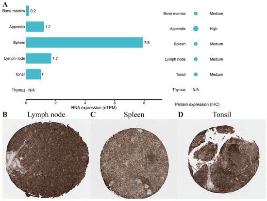
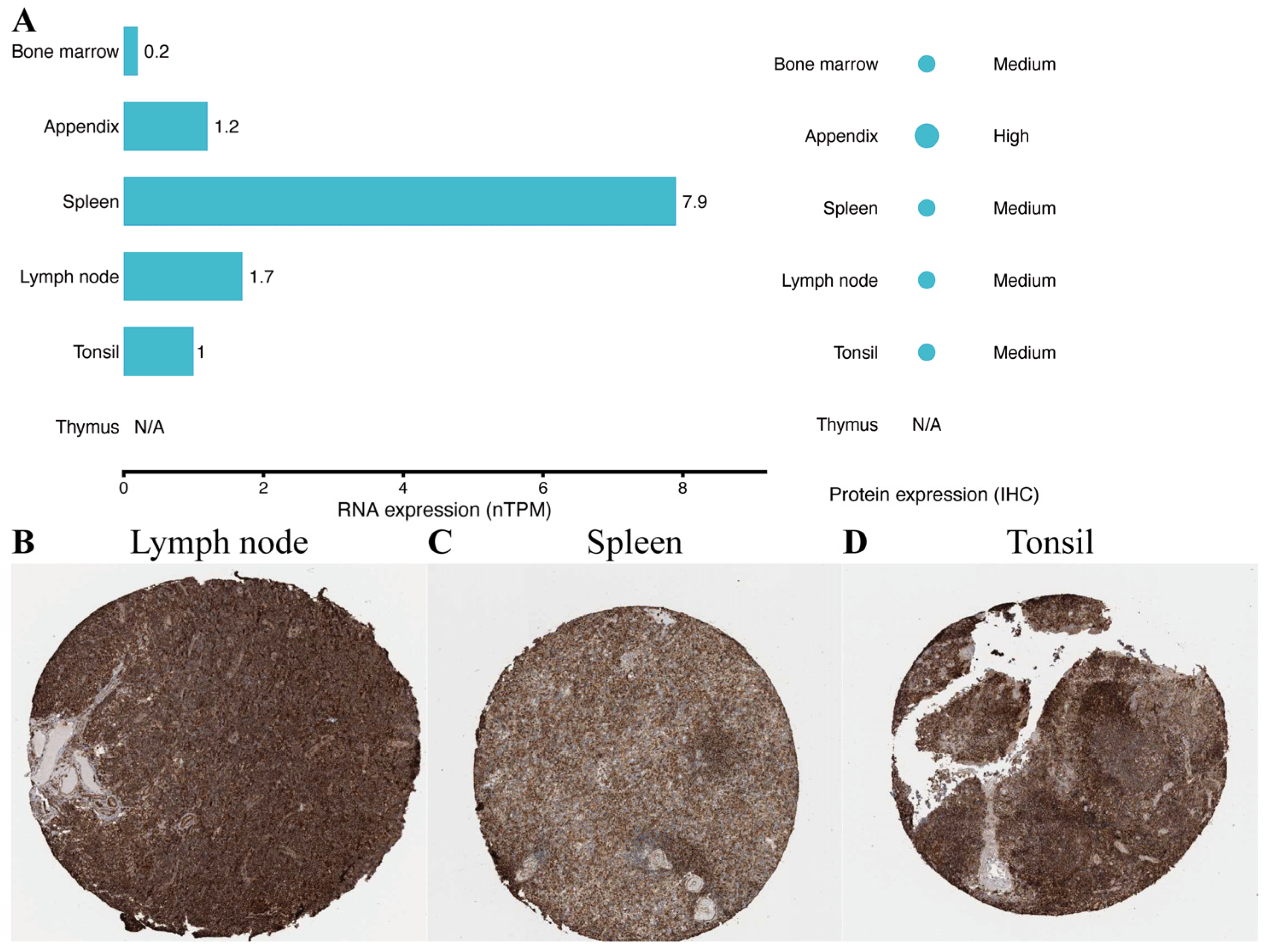
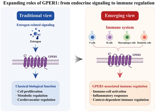
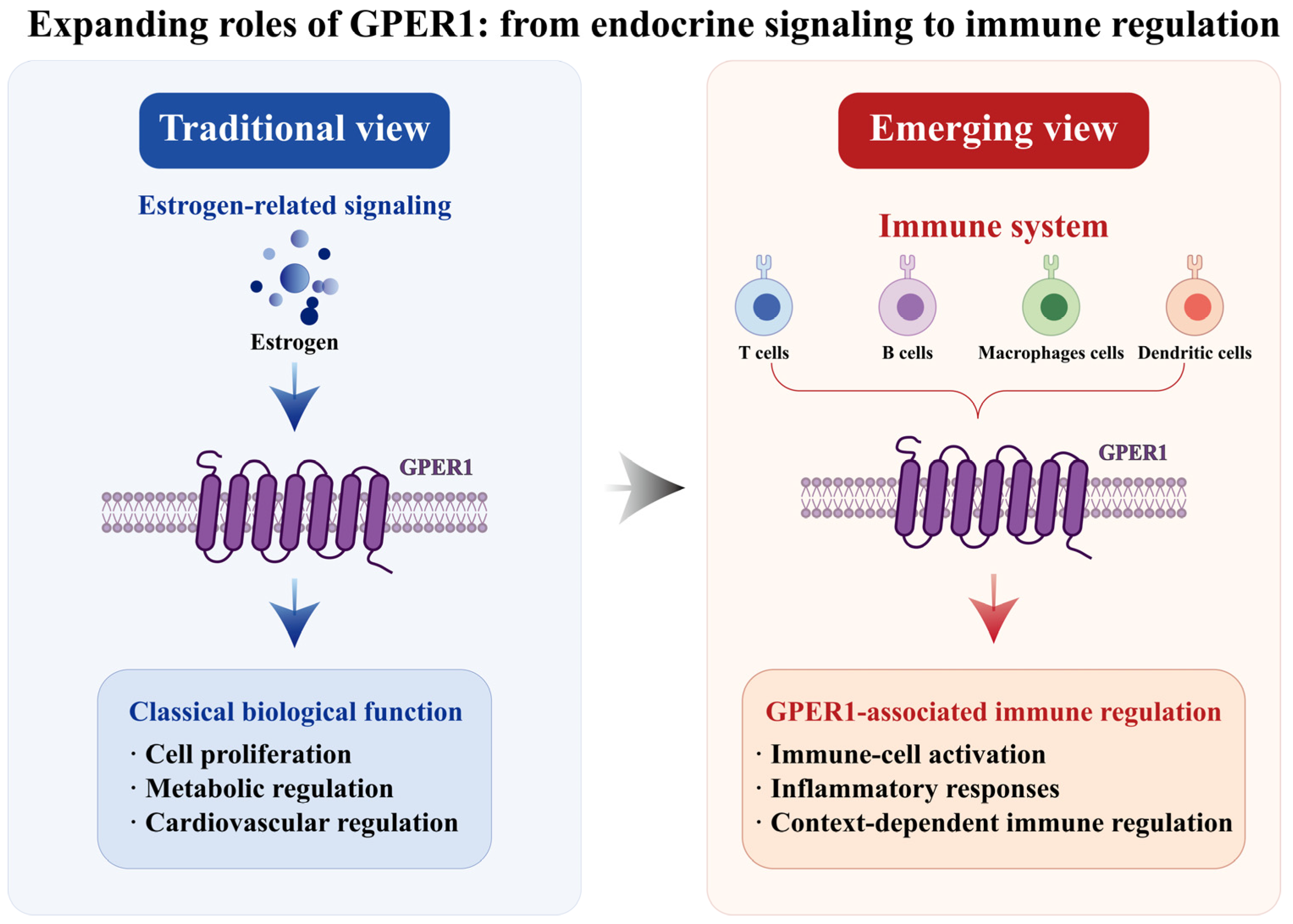
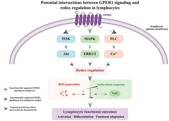
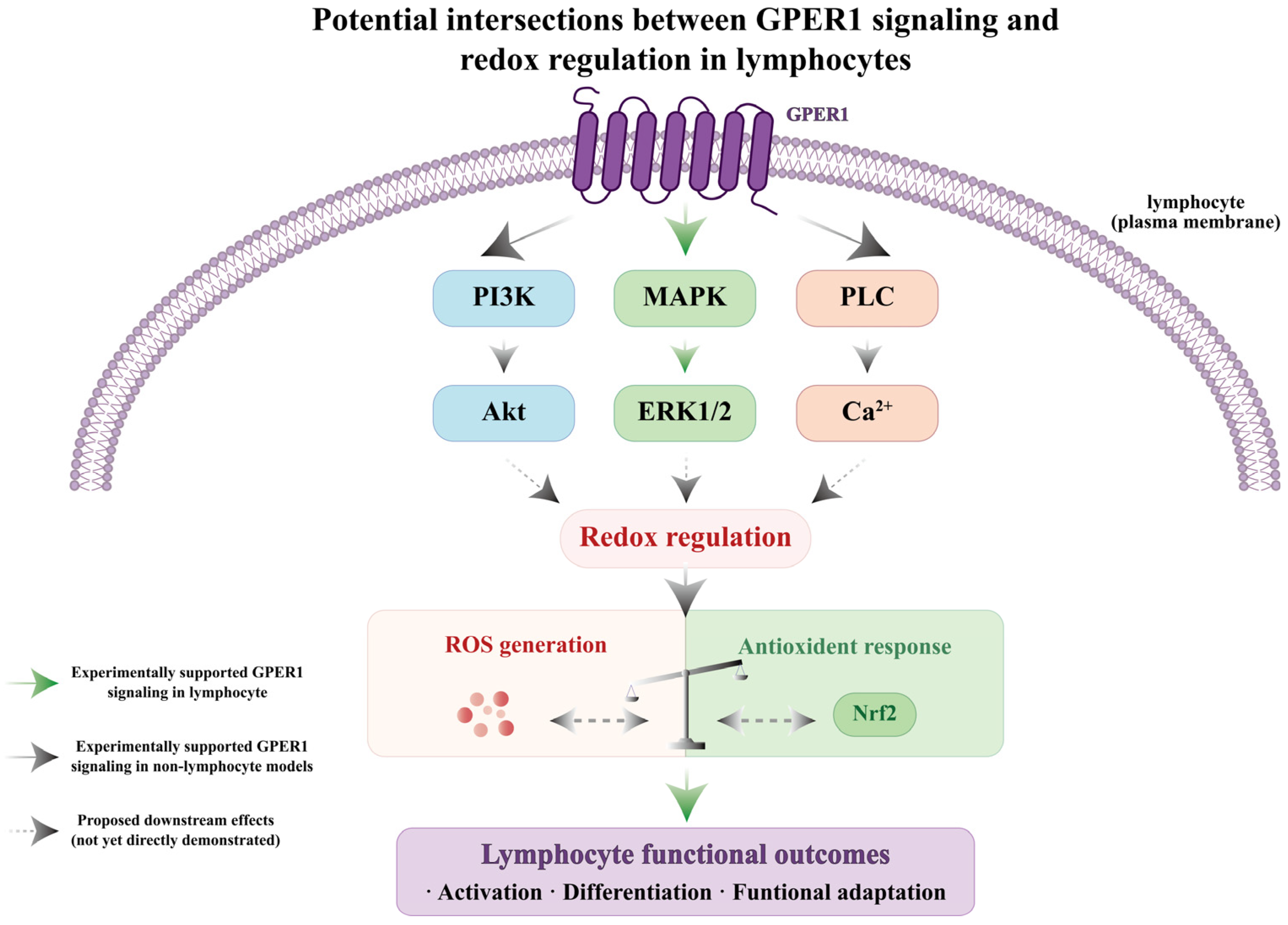
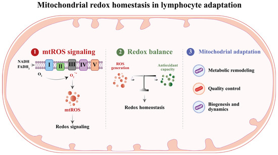
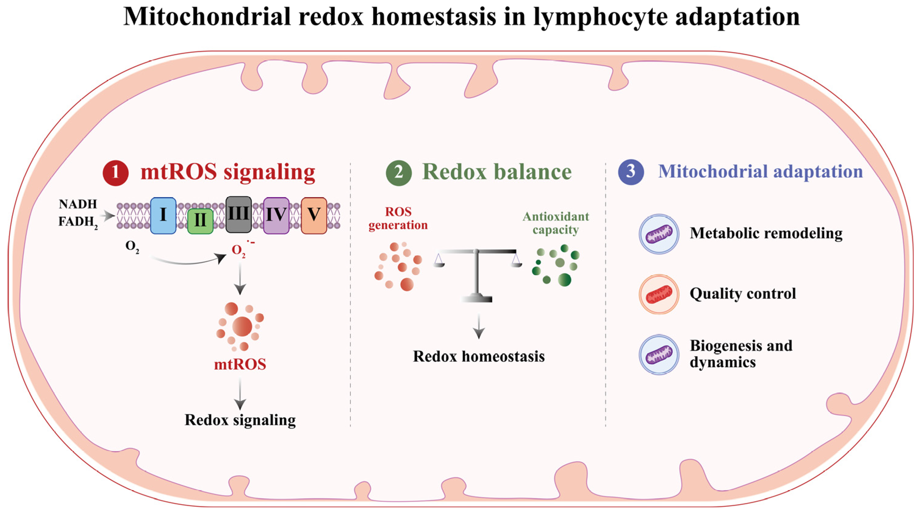

Disclaimer/Publisher’s Note: The statements, opinions and data contained in all publications are solely those of the individual author(s) and contributor(s) and not of MDPI and/or the editor(s). MDPI and/or the editor(s) disclaim responsibility for any injury to people or property resulting from any ideas, methods, instructions or products referred to in the content. |
© 2026 by the authors. Licensee MDPI, Basel, Switzerland. This article is an open access article distributed under the terms and conditions of the Creative Commons Attribution (CC BY) license.