


Abstract
Nitric oxide (NO) is an important regulator of motile cilia. Coordinated ciliary beating moves mucus, extracellular fluids, or cells across tissue surfaces and, during embryonic development, drives the establishment of correct organ left–right asymmetry. The connection between NO and cilia function is particularly evident in primary ciliary dyskinesia (PCD), where reduced nasal NO is a diagnostic hallmark, although whether it contributes to or results from ciliary dysfunction remains unclear. Studies of PCD and other airway diseases also show that NO levels do not necessarily correlate with nitric oxide synthase (NOS) expression or activity, indicating that NO bioavailability in ciliated tissues is regulated by additional mechanisms. Despite decades of evidence linking NO to ciliary function, fundamental questions remain regarding how NO is produced and regulated within ciliated cells, how NO signaling is spatially organized within the ciliary microenvironment, and how it reaches the axoneme to regulate ciliary structure and motility. In this review, we examine these questions focusing on soluble guanylate cyclase (sGC)–cyclic guanosine monophosphate (cGMP) signaling, S-nitrosylation, and emerging redox-dependent mechanisms regulating NO bioavailability in motile cilia.
1. Introduction
The origins of nitric oxide (NO) production and its role as a signaling molecule can be traced to the common ancestor of modern eukaryotes, where NO was likely generated initially as a metabolic byproduct. As atmospheric oxygen increased and aerobic metabolism evolved, NO gradually acquired regulatory functions and became one of the most prevalent signaling molecules in biology. Today, NO is known to regulate numerous physiological processes, including vascular tone, neurotransmission, immune responses and mitochondrial function [1]. In the context of cilia function, evidence that NO positively regulates cilia motility dates back more than three decades. Independent studies in the early 1990s showed that pharmacological inhibition of nitric oxide synthase (NOS) reduced cilia beat frequency in mammalian airway epithelial cells and that this effect was rescued by L-arginine or an NO donor, providing direct evidence that endogenous NO is a key physiological regulator of airway cilia function [2,3,4]. Subsequently, Jain et al. showed that the pro-inflammatory cytokines TNF-α and IL-1β stimulate airway cilia beat frequency by inducing endogenous NO production [5], consistent with the ability of inflammatory cytokines to induce NOS2/iNOS expression in airway epithelial cells [6]. However, in diseases characterized by low NO, more recent studies have failed to establish a consistent relationship between reduced NO levels and the expression of any specific NOS isoform. This is particularly evident in primary ciliary dyskinesia (PCD), where nasal NO remains markedly low despite airway inflammation and preserved iNOS expression, suggesting that reduced NO bioavailability cannot be explained simply by decreased NOS expression [7,8,9,10]. Additional evidence supporting a functional role for NO in regulating cilia came from studies showing that clinically relevant concentrations of ethanol stimulate NO release by the airway epithelium, resulting in increased cilia beat frequency [4]. Soon after the finding that NO affects cilia motility, the involvement of the canonical NO receptor soluble guanylate cyclase (sGC) in transducing NO signaling in ciliated cells was established as a major mechanism mediating NO-dependent stimulation of cilia beat frequency in rat airway epithelium [11,12,13].
During the following decade, the association between NO and airway health became increasingly apparent, and measurement of NO emerged as an important clinical tool in respiratory disease. Following the initial observation that patients with PCD exhibit markedly reduced nasal NO levels [14], subsequent studies established nasal NO as a sensitive screening marker for PCD [15]. Low nasal NO was also associated with reduced cilia function and a higher proportion of immotile cilia, further linking NO bioavailability to ciliary dysfunction [16]. Standardization of nasal NO measurement by the American Thoracic Society (ATS) and European Respiratory Society (ERS) established uniform sampling procedures and facilitated its incorporation into international guidelines for PCD diagnosis, alongside genetic testing and ciliary structural and functional analyses [17,18,19]. Altered NO bioavailability is not restricted to PCD but is also observed in other airway diseases associated with impaired mucociliary clearance. Nasal NO is also reduced, although generally to a lesser extent, in cystic fibrosis and chronic rhinosinusitis [15,18,20,21], while altered exhaled NO levels are also observed in chronic obstructive pulmonary disease (COPD), although these changes vary with disease severity, inflammatory phenotype, and smoking status [22]. Importantly, nasal NO and exhaled NO represent distinct measurements: nasal NO predominantly reflects NO reaching the gas phase in the nasal cavity and paranasal sinuses, whereas exhaled NO primarily reflects NO originating from the lower respiratory tract. Neither measurement directly reflects cellular NO production or local NO bioavailability. Despite the strong association between altered measurable NO levels and ciliary dysfunction, the mechanisms responsible for altered NO bioavailability remain poorly understood and likely extend beyond changes in NOS-dependent NO production.
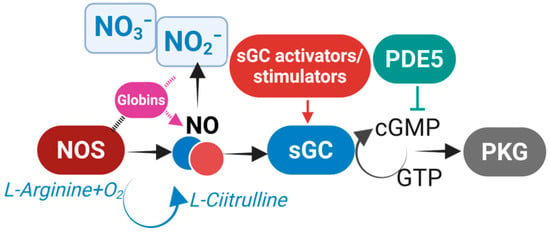
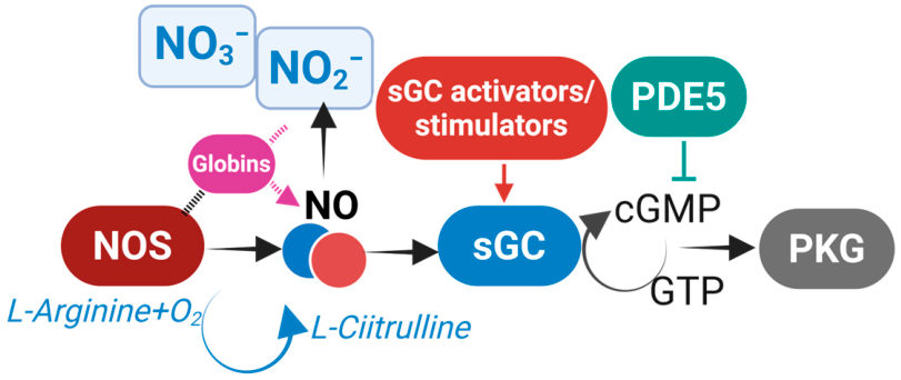
There are different possible mechanisms through which NO regulates cilia function. The best established is the canonical NO–sGC–cGMP pathway, in which NO activates soluble guanylate cyclase (sGC), increasing cGMP and activating downstream effectors that ultimately regulate axonemal activity and cilia beat frequency [23] (Figure 1). NO can also regulate protein function through reversible S-nitrosylation of cysteine residues, a signaling mechanism increasingly recognized as a major mediator of NO biology [24,25]. S-nitrosothiols (SNOs) are important endogenous carriers of NO bioactivity that regulate protein function through S-nitrosylation, and accumulating evidence indicates that S-nitrosylation and sGC activation represent complementary, rather than mutually exclusive, NO signaling pathways [25,26]. An additional mechanism through which NO can influence cellular signaling is through its interaction with ROS. Under conditions of oxidative stress and redox imbalance, NO readily reacts with reactive oxygen species, particularly superoxide, generating reactive nitrogen species such as peroxynitrite that can modify proteins and alter cellular function [27]. ROS are themselves well-established regulators of motile cilia function, influencing cilia beat frequency and mucociliary clearance under both physiological and pathological conditions [27], but how ROS influence NO availability and signaling in motile cilia remains uncharacterized. Despite more than three decades of investigation demonstrating that NO regulates cilia function, the molecular mechanisms linking NO production to changes in axonemal activity remain surprisingly poorly defined. Fundamental questions include which NOS isoforms generate the relevant NO, where within the ciliated cell NO is produced, how newly identified regulators such as globins control NO bioavailability, how NO signaling is spatially organized relative to the axoneme, and how sGC–cGMP signaling, S-nitrosylation, and redox-dependent mechanisms converge on the ciliary machinery. In this review, we discuss the progress made in understanding NO signaling in motile cilia and highlight these major mechanistic gaps and emerging directions in the field.
Figure 1.
Sources of NO and canonical NO signaling. Nitric oxide (NO) is produced by nitric oxide synthase (NOS) from L-arginine or through alternative pathways involving nitrite reduction. Globins regulate NO bioavailability through interactions with NOS and nitrite metabolism. NO activates soluble guanylate cyclase (sGC), increasing cGMP production and activating protein kinase G (PKG). This pathway is regulated by phosphodiesterase 5 (PDE5) and can be pharmacologically enhanced by sGC stimulators and activators. Solid arrows indicate established pathways, whereas dashed arrows indicate proposed or incompletely characterized mechanisms. Created in BioRender. Corti, P. (2026) https://BioRender.com/qtoe10y.
2. Conservation of Calcium–NO Signaling in Motile Cilia
Motile cilia are specialized cellular projections consisting of a 9 + 2 microtubule-based axoneme anchored to a basal body, which serves as the structural template for ciliary assembly, orientation, and protein entry. Cilia may occur as a single organelle or in hundreds per cell, with each axoneme associated with its own basal body. The basal body derives from the centriole and, together with the cilium, represents an evolutionarily ancient structure already present in the last eukaryotic common ancestor [28]. Indeed, the 9+2 axonemal architecture is remarkably conserved across eukaryotes, maintaining fundamentally conserved functions across diverse organisms. Motile cilia and flagella are present in unicellular eukaryotes, including algae, ciliates, and choanoflagellates, where they support locomotion, feeding, and environmental sensing. They are also widespread throughout the animal kingdom, from marine invertebrates, where they drive larval swimming, feeding, and gamete transport, to vertebrates, where they propel extracellular fluids in multiple organs. In the embryonic node, motile cilia generate the leftward fluid flow required for the establishment of left–right asymmetry, while in multiciliated epithelia of the respiratory tract, reproductive tract, and brain ventricles, coordinated ciliary beating drives mucus transport, gamete movement, and cerebrospinal fluid circulation, respectively [29,30,31,32,33,34,35].
One of the best-established intracellular regulators of cilia motility is calcium, which modulates both cilia beat frequency and waveform through calcium-sensitive proteins distributed throughout the axoneme. In the unicellular green alga Chlamydomonas reinhardtii, changes in intracellular Ca2+ regulate flagellar waveform, phototaxis, and axonemal dynein activity through conserved calcium sensors such as calmodulin, LC4, centrin, and calaxin, demonstrating that calcium-dependent regulation of motility predates the emergence of multicellular animals [36,37,38]. Similarly, the ciliate Paramecium tetraurelia provides one of the most elegant examples of calcium-mediated regulation of motility, where membrane depolarization triggers Ca2+ influx into the cilia, producing a rapid calmodulin-dependent switch in axonemal dynein activity that reverses the ciliary waveform and causes the organism to swim backward [39,40,41]. Indeed, calmodulin and calmodulin-associated complexes localize to the radial spokes, where they regulate communication between the central apparatus and axonemal dyneins, while several inner dynein arm components are directly regulated by calcium-dependent mechanisms controlling cilia bend amplitude and waveform [29,41]. Although the underlying calcium-dependent regulation of axonemal dyneins is evolutionarily conserved, its physiological output has diversified across organisms. In vertebrates, calcium does not induce reversal of cilia beating but instead modulates cilia beat frequency, waveform, and beat coordination, contributing to mucociliary clearance, oocyte transport, sperm motility, cerebrospinal fluid circulation, and embryonic left–right patterning [42].
Given that calmodulin is a highly conserved calcium sensor within the axoneme, that both neuronal nNOS (NOS1) and endothelial eNOS (NOS3) are activated by Ca2+/calmodulin [1,43], and that nNOS and eNOS have been localized to motile cilia and shown to regulate cilia beat frequency [11,44,45], it is tempting to speculate that calcium-dependent activation of NOS represents an evolutionarily conserved mechanism linking calcium signaling to NO production to regulate cilia motility. This hypothesis is supported by physiological studies in mammalian airway epithelium demonstrating that ATP-induced increases in intracellular Ca2+ require activation of the NO–sGC–cGMP pathway to stimulate cilia beat frequency, as pharmacological inhibition of NOS, sGC, or PKG abolishes the calcium-dependent response [11,12]. It was further demonstrated that calcium-dependent NO signaling regulates the phosphorylation and dephosphorylation of axonemal proteins, providing direct evidence that NO links calcium signaling to post-translational regulation of the ciliary machinery [13].
Subsequent studies demonstrated that diverse physiological stimuli converge on this calcium–NO signaling axis. In airway epithelial cells, mechanical stimulation associated with sneezing triggers ATP release, intracellular Ca2+ elevation, and NOS-dependent NO production, resulting in increased cilia beat frequency and enhanced mucociliary clearance [46]. Similarly, bacterial lipopolysaccharide activates the mechanosensitive TRPV4 channel, producing Ca2+-dependent NO generation that increases cilia beat frequency while simultaneously enhancing innate immune defense [47]. Activation of bitter taste receptors by bacterial products likewise stimulates Ca2+-dependent NO production to accelerate mucociliary clearance. This response is impaired in cystic fibrosis because of reduced CFTR-dependent NO signaling [48,49]. Importantly, the interplay between Ca2+ and NO signaling in the regulation of motile cilia and flagella is strongly dependent on the cellular and physiological context. The reproductive tract provides a particularly striking example. Near the time of fertilization, increased NO production in the oviduct promotes sperm flagellar motility associated with hyperactivation, characterized by increased flagellar bending and a more asymmetric waveform that involves NO-dependent modulation of intracellular Ca2+ signaling [49,50]. In contrast, in ciliated epithelial cells of the oviduct, ATP induces a Ca2+-dependent increase in ciliary beat frequency that is attenuated by progesterone-induced, NOS-dependent NO signaling [49]. Functionally, NO restrains epithelial ciliary transport while promoting sperm activity, potentially favoring sperm-oocyte encounter and fertilization.
3. Sources of NO in Ciliated Cells
The discovery that NO is the long-sought endothelium-derived relaxing factor (EDRF) revolutionized the field of cell signaling and was recognized by the 1998 Nobel Prize in Physiology or Medicine [51,52,53]. In the endothelium, NO is synthesized from L-arginine by endothelial NOS (eNOS/NOS3) in response to fluid shear stress, diffuses to the underlying smooth muscle, and activates soluble guanylyl cyclase (sGC), leading to cyclic GMP (cGMP)-dependent vasorelaxation [1]. Here, NO production is itself regulated by the primary cilium, where the polycystin-1/polycystin-2 (PC1/PC2) mechanosensory complex detects fluid flow, triggers intracellular Ca2+ signaling, and promotes Ca2+/calmodulin-dependent NO synthesis [54]. These findings established an early direct link between ciliary mechanosensation and NO production and raised the possibility that analogous mechanisms operate in motile cilia.
The primary enzymatic source of NO in motile ciliated cells is nitric oxide synthase (NOS). The three mammalian NOS isoforms, neuronal (nNOS/NOS1), inducible (iNOS/NOS2), and endothelial (eNOS/NOS3), have all been reported in motile ciliated cells, although their relative contribution to local NO production remains unclear [1,43,55]. nNOS and eNOS are constitutively expressed and activated by Ca2+/calmodulin, whereas iNOS binds calmodulin constitutively and is primarily regulated at the transcriptional level [56]. eNOS has been detected in the apical portion of the ciliary compartment in airway epithelial cells, ciliated epithelia of the oviduct, ependyma and testis, with localization also reported at basal bodies [44]. Neuronal NOS has been localized to human and mouse airway cilia [45,57]. In mouse tracheal multiciliated cells, nNOS localizes to basal bodies, basal feet, and the apical actin and microtubule networks. Loss of nNOS disrupts planar cell polarity, cilia organization and directional fluid flow [57]. Consistent with a broader role in cell polarity determination, an nNOS ortholog in Xenopus embryos is found to regulate cell polarization during embryonic axis elongation [58]. The contribution of iNOS to motile cilia is less clearly defined. iNOS is predominantly regulated by inflammatory stimuli and represents an important source of airway NO during inflammation [59]. Asthma provides a well-established example, in which iNOS is induced in the bronchial epithelium [60], with increased iNOS expression compared with healthy controls and elevated fractional exhaled NO (FeNO), now widely used as a clinical biomarker of airway inflammation [61]. In diseases characterized by impaired mucociliary clearance, however, this relationship largely breaks down, and the available data on NOS expression and NO levels are often contradictory.
PCD provides the clearest illustration of the disconnection between NOS expression and measurable airway NO. Some evidence from transcriptional analysis of nasal epithelial cells indicates that iNOS expression correlates with nasal NO and is reduced in PCD compared with secondary ciliary dyskinesia [16]. However, other studies indicate that iNOS expression is largely preserved despite the profound reduction in nasal NO characteristic of PCD. Studies in upper airway tissue from patients with PCD showed that iNOS immunoreactivity remained prominent at the apical pole of ciliated epithelial cells and was comparable to controls, despite markedly reduced nasal NO [7]. Similarly, airway epithelial cultures from PCD patients express iNOS, eNOS, and nNOS and retain the capacity to produce NO, although their ability to increase NO production in response to inflammatory stimuli appears to be dependent on the nature and duration of the stimulus [8,62]. Specifically, the response of iNOS to inflammatory stimulation is variable. During early Streptococcus pneumoniae infection, healthy ciliated epithelial cells increased iNOS expression and NO production, whereas PCD cells failed to induce either response [8]. In contrast, PCD cultures exposed to Haemophilus influenzae or pro-inflammatory cytokines showed increased NO production comparable to non-PCD cultures [62]. These differences may reflect the pathogen, duration of exposure, and inflammatory context rather than a constitutive loss of iNOS in PCD. Thus, while low nasal NO is a remarkably consistent feature of PCD, iNOS expression and inducibility are not consistently reduced in the disease. Importantly, nasal NO and exhaled NO are measurements of NO reaching the gas phase in different regions of the respiratory tract and should not be considered equivalent to cellular NO production or local NO bioavailability.
Several observations indicate that NO is synthesized in the PCD airway but does not reach the gas phase, suggesting that low nasal NO does not necessarily reflect a corresponding reduction in cellular NO production. NO produced in the airways can undergo oxidation and other redox reactions to form more stable metabolites, including nitrite and nitrate, and S-nitrosothiols. These metabolites are not reduced in children with PCD, suggesting that NOS activity is not diminished to the degree that the low nasal NO would imply [63]. PCD airway epithelial cells accumulate 3-nitrotyrosine and nitrate, while PCD airways exhibit higher concentrations of H2O2, and consume gas-phase NO more rapidly than control cells, an effect reversed by superoxide dismutase and by NADPH oxidase inhibition, indicating that in some cases NO is oxidized locally rather than never formed [9]. Airway anatomy contributes independently: aplasia or hypoplasia of the frontal and sphenoidal sinuses is considerably more frequent in PCD than in secondary ciliary dyskinesia and correlates inversely with nasal NO [64], so a reduced NO-producing and NO-storing surface lowers nasal NO without any change in iNOS expression or activity within the airway cells. Flow-partitioned measurements localize the deficit further still, with bronchial NO flux reduced in PCD while alveolar NO is indistinguishable from that of healthy subjects [65].
Cystic fibrosis (CF) and COPD further illustrate the dissociation between NOS expression and airway NO. In CF, iNOS is markedly reduced in the airway epithelium, despite the inflammatory environment characteristic of the disease. iNOS was absent from CF bronchial epithelial cells and could not be induced by cytokine stimulation, while remaining readily detectable in infiltrating inflammatory cells [66]. Reduced epithelial iNOS expression was subsequently demonstrated in both murine and human CF airway cells [67] and linked to CFTR function [68]. Yet CF airways contain abundant NO oxidation products, with increased nitrite and nitrate detected in sputum or exhaled breath condensate even when exhaled NO is not elevated [69,70,71]. Increased arginase activity may further limit NOS-dependent NO production by competing with NOS for L-arginine [72]. In COPD, the relationship is reversed: iNOS-positive cells and NOS expression are increased in lung tissue [73,74], while exhaled NO is not elevated to the extent observed in asthma [75], with increases largely confined to the alveolar compartment and associated with disease severity [76]. Together, these observations show that NOS expression alone is a poor predictor of airway NO availability and that the relationship between NOS expression, NO production, and its downstream metabolites varies considerably across airway diseases.
4. Globins Emerge as Regulators of NO Signaling in Motile Cilia
One possible explanation for the apparent disconnection between NOS expression and NO levels is the existence of additional regulatory mechanisms that control NOS activity and local NO bioavailability. Recent studies have identified members of the globin family as important modulators of NO signaling beyond their classical role in oxygen transport. Cytoglobin (Cygb) was recently identified as an upstream regulator of NOS-dependent NO production in motile cilia through molecular interaction with NOS [43]. In the zebrafish left–right organizer, cytoglobin colocalizes with Nos2b, the zebrafish eNOS homolog, in ciliated cells and promotes local NO production. Loss of cytoglobin reduces NO bioavailability, shortens cilia, alters axonemal ultrastructure, and disrupts left–right patterning. Similarly, cytoglobin-deficient mice exhibit reduced tracheal NO levels and shorter airway cilia, establishing Cygb as a conserved regulator of NO signaling and motile cilia function [43].
Beyond regulating NOS activity, globins have emerged as physiologically relevant regulators of NO bioavailability through nitrite metabolism. Under hypoxic conditions, deoxygenated ferrous globins (Fe2+) reduce nitrite (NO2−) to NO, providing an alternative source of NO when oxygen-dependent NOS activity becomes limited [77,78,79,80]. Nitrite reductase activity has been demonstrated for several globins, including hemoglobin, cytoglobin, and the fish-specific Globin X [79]. The physiological relevance of globin-dependent nitrite reduction has been demonstrated in several biological systems, and in fish a potential role for nitrite-derived NO is illustrated by the finding that nitrite supplementation promotes cardiac regeneration in zebrafish [81]. Furthermore, globin expression itself is dynamically regulated by environmental oxygen availability and thermal stress in both fish and mammals, linking globin function to physiological adaptation [82,83].
The emerging role of globins as local regulators of NO signaling is further supported by studies of other globin family members. Endothelial hemoglobin α was shown to regulate eNOS-derived NO signaling in vascular endothelial cells, establishing the concept that globins function as tissue-specific modulators of NO bioavailability beyond erythrocytes [84]. This concept was subsequently extended to the airway epithelium, where hemoglobin is expressed in differentiated ciliated epithelial cells. In these cells, Hbβ colocalizes with eNOS at the apical surface and influences nitrogen oxide metabolism, supporting a role for globins in the local regulation of NO signaling in ciliated tissues [85].
Another emerging member of this regulatory network is androglobin (Adgb), a large chimeric globin whose expression is tightly linked to motile ciliogenesis. Adgb is transcriptionally regulated by FOXJ1 and is highly enriched in multiciliated tissues, including the airway, fallopian tube, brain ependyma, and testis [86,87], with loss of Adgb causing male infertility [87]. Biochemical studies of the isolated Adgb heme domain have demonstrated NO binding and nitrite reductase activity in vitro [88]. In Tetrahymena thermophila, Adgb is enriched in motile cilia and associates with components of the C1b/C1f central apparatus complex, although loss of Adgb causes only minor alterations in ciliary motility [89]. Recent studies further demonstrated that Adgb undergoes Ca2+-dependent proteolytic processing, generating stable cleavage products, and that the isolated globin domain exhibits enhanced interaction with calmodulin and preferential localization to centrosomal structures [86]. These findings suggest that Ca2+-dependent processing may regulate the accessibility and subcellular localization of the Adgb globin domain [86]. Its localization and biochemical findings raise the intriguing possibility that Adgb may regulate motile cilia signaling, potentially through modulation of NO bioavailability or other mechanisms that remain to be defined. Although these functions remain to be demonstrated in vivo, the combined evidence from cytoglobin, hemoglobin, and androglobin supports an emerging paradigm in which globins act as tissue- and compartment-specific regulators of NO signaling, contributing to the spatial and temporal control of NO bioavailability in motile cilia.
5. sGC-cGMP Signaling Pathway in Cilia
Soluble guanylate cyclase (sGC) is the principal receptor for NO and mediates the canonical NO–cGMP signaling pathway. sGC is an α/β heterodimer containing a heme prosthetic group in the β subunit that binds NO and activates the catalytic domain, resulting in conversion of GTP to cGMP [90,91]. cGMP can subsequently activate cGMP-dependent protein kinase (PKG), a major downstream effector of NO–sGC signaling (Figure 1). Both sGC and PKG have been detected in respiratory ciliated epithelium [92,93]. Mechanistically, NO-dependent increase in ciliary beat frequency is associated with increased cGMP and PKG activity, and inhibition of PKG prevents this response [11,12]. Gertsberg et al. demonstrated that Ca2+-dependent changes in the phosphorylation state of axonemal proteins require NOS, sGC, and PKG activity, providing a mechanistic connection between NO production and modification of the axonemal machinery [13]. Using phosphoserine immunoblotting of porcine isolated airway cilia, they identified several ciliary polypeptides that underwent phosphorylation or dephosphorylation in an NOS/sGC/PKG-dependent manner. However, these proteins were not molecularly identified, and none was demonstrated to be a direct PKG substrate [13]. More recent phosphoproteomic studies have substantially expanded the identification of phosphorylation sites in axonemal dyneins, including sites in outer- and inner-arm dynein heavy chains and associated regulatory components across multiple motile-cilia systems [94,95]. In Paramecium, direct phosphorylation of ciliary protein substrates by purified PKG has been demonstrated in vitro [23,96]. However, the molecular identity of direct PKG substrates in motile cilia remains unresolved.
cGMP can also regulate airway ciliary activity through PKG-independent mechanisms [97], indicating that the downstream organization of NO–cGMP signaling in motile cilia is more complex than a simple linear pathway. Despite extensive evidence supporting the role of NO–cGMP signaling in regulating ciliary motility, several fundamental questions remain. First, does NO–cGMP signaling act directly on the axoneme, or does it primarily target cytoplasmic or membrane-associated proteins that influence beating indirectly? Second, where are the individual signaling components positioned relative to the axoneme? Third, because epithelial cells contain hundreds of motile cilia, each capable of generating and responding to NO, is cGMP signaling spatially compartmentalized to achieve local and coordinated regulation of ciliary activity? The molecular properties of sGC, PKG, and phosphodiesterases have been reviewed extensively elsewhere and are discussed here only insofar as they address these questions.
cGMP signaling in other cellular systems is highly compartmentalized, with spatially restricted cGMP pools generated by guanylyl cyclases and shaped by local phosphodiesterase (PDE) activity [98]. Such organization extends to sensory cilia, where cyclases, PDEs, and downstream effectors can be localized to distinct subciliary domains [99,100,101]. PDEs such as the cGMP-hydrolyzing PDE5 (Figure 1) have also been identified in airway ciliated epithelium, and here they are shown to modulate ciliary activity [102]. Whether cGMP signaling is similarly compartmentalized within motile cilia, however, has not been directly demonstrated. Such organization could provide a mechanism for coupling local NO production to specific axonemal targets without requiring large changes in whole-cell cGMP. This possibility is particularly intriguing given emerging evidence that NO production itself may be spatially organized within motile cilia. NOS isoforms have been reported at apical, basal-body, and ciliary locations; Cygb regulates ciliary NO production [43]; and Adgb has been identified along the axoneme [89]. A locally organized NO–sGC–cGMP signaling hub could therefore couple NO generated at or near the axoneme to PKG-dependent regulation of ciliary motors and associated proteins. However, while the localization of NO-generating and -regulating proteins is becoming increasingly defined, the localization of sGC relative to the motile axoneme, the spatial organization of cGMP–PKG signaling, and the identity of its direct axonemal targets remain poorly understood and direct axonemal PKG substrates in mammalian motile cilia remain unidentified. The NO sources of motile cilia have been mapped with increasing precision, whereas the receptor that transduces their output has not yet been placed relative to the axoneme.
An additional level of regulation may arise from the redox state of sGC itself. Oxidation of its ferrous heme or heme loss decreases responsiveness to NO and can generate NO-insensitive apo-sGC [90,91]. Whether this occurs in ciliated cells has received little attention but may be particularly important under the oxidative and inflammatory conditions associated with chronic airway diseases such as COPD. In this setting, NO–cGMP signaling could become impaired even when NOS expression and NO production are preserved, providing an additional mechanism for the dissociation between NOS expression and NO bioavailability described above. A growing pharmacological repertoire has been developed to overcome impaired NO–sGC signaling, particularly in cardiovascular and pulmonary vascular diseases: sGC stimulators have been introduced into clinical practice to enhance signaling through the reduced, heme-containing enzyme, whereas sGC activators can stimulate oxidized or heme-deficient sGC independently of NO [103,104]. The possibility of restoring cilia function by directly targeting sGC has been demonstrated in the zebrafish left–right organizer, where embryos lacking Cygb and consequently deficient in NO signaling exhibit laterality defects that can be rescued by pharmacological activation of sGC [43]. This finding demonstrates that the ciliary sGC–cGMP pathway remains pharmacologically accessible even when upstream NO availability is impaired and raises the possibility that direct targeting of sGC could restore ciliary signaling in conditions characterized by defective NO bioavailability. Together with the unresolved spatial organization of the pathway, these observations raise important questions as to whether disease disrupts NO production, its local transmission through sGC–cGMP, or both.
6. S-Nitrosylation (SNO): A Novel Avenue for Motile Cilia Regulation
Nitric oxide is a broad regulator of biological function, best characterized through activation of the sGC–cGMP signaling pathway. However, NO can also regulate protein function directly through S-nitrosylation, a reversible post-translational modification involving the addition of an NO group to a cysteine thiol to form an S-nitrosothiol (SNO) [25]. S-nitrosylation provides a mechanism through which local NO availability and the cellular redox environment can rapidly and reversibly modify protein activity. NO available for SNO formation can originate from NOS-dependent synthesis [105], transfer of NO groups between protein thiols through transnitrosylation [106], or alternative sources of NO, including globin-mediated nitrite reduction [77,79,80,107].
The best evidence for SNO-mediated regulation of the motile ciliary axoneme comes from the sperm flagellum. A proteomic study of human spermatozoa treated with S-nitrosoglutathione (GSNO), a physiological NO donor and SNO reservoir, identified 240 S-nitrosylated proteins in human spermatozoa, with S-nitrosylated targets localized throughout the flagellum [50], establishing a large candidate pool of SNO-regulated targets in this system. Beyond target identification, S-nitrosylation also has a demonstrated functional role in this system, particularly in Ca2+ signaling. Ca2+ mobilization drives flagellar excursion and motility [108,109], and in human spermatozoa, exposure to an NO donor or GSNO triggers Ca2+ mobilization that is reversible upon SNO-protein reduction and independent of sGC [110]. NO also potentiates the Ca2+ response to progesterone and enhances flagellar excursion. Additional findings in human female reproductive tract explants showing induced S-nitrosylation in spermatozoa suggest that NO and S-nitrosylation together regulate axonemal behavior in a physiologically relevant context [110].
Direct evidence for S-nitrosylation in airway motile cilia has also been reported. Proteomic analysis of isolated bovine tracheal axonemes identified 1,001 putative S-nitrosylation sites across 622 proteins under control and alcohol-exposed conditions, and identified protein phosphatase 1 (PP1), a regulator of ciliary motility, as an S-nitrosylated ciliary protein [111]. Subsequent studies extended these findings to mouse and human airway models, demonstrating that S-nitrosylation is localized to cilia and identifying Cys155 of PP1 as a functionally important S-nitrosylation site. Mutation of PP1 Cys155 prevented alcohol-induced PP1 S-nitrosylation and restored cAMP-dependent ciliary beat frequency responsiveness in isolated human airway axonemes [112]. GSNO is also present in airway lining fluid and has been reported to enhance ciliary motility [113]. More recently, GSNO treatment of differentiated human airway epithelial cultures was shown to increase ciliary beat frequency in cystic fibrosis and chronic rhinosinusitis cultures exposed to bacterial biofilms [114]. However, these studies did not establish that the effects of GSNO on ciliary motility were mediated by S-nitrosylation of the axonemal apparatus. This relationship is especially relevant to primary ciliary dyskinesia (PCD) pathology. Despite markedly reduced nasal NO in PCD airways, recent work points to substantial alterations in nitrosative and oxidative signaling [9], and transcriptional profiling of PCD airway cells has identified a cilia-associated glutathione pathway, further linking the ciliary compartment to thiol/redox regulation [115].
Together, these findings position S-nitrosylation as a potential second arm of NO signaling in motile cilia, operating alongside the canonical NO–sGC–cGMP–PKG pathway. Although proteomic studies have identified numerous candidate S-nitrosylated proteins in airway axonemes, the functional significance of most candidate targets remains unknown. In particular, whether core components of the axonemal motor machinery, including dyneins and their regulatory complexes, are directly regulated by S-nitrosylation remains poorly understood. Mapping the S-nitrosoproteome of motile cilia under both physiological and disease conditions could reveal an additional layer through which local NO availability and redox state control the ciliary machinery.
7. Are Cilia Only Effectors or Also Regulators of Cell Signaling?
In previous sections, we discussed how cilia formation and motility are major regulatory outcomes of the NO–sGC–PKG axis. Whether the inverse is also true remains an open question, and is particularly intriguing in motile cilia, which both generate and respond to mechanical forces. Unlike primary cilia, which sample a flow generated elsewhere, motile cilia generate the flow that they would be sensing. Any mechanosensory capacity in a motile cilium is therefore embedded in a potential feedback loop: beating produces shear, shear is transduced, and transduction alters beating. Whether this feedback engages NOS and NO signaling in motile cilia has not been established. If NO participates in that loop, ciliary motility would not simply be an output of NO signaling but also one of its determinants.
The precedent comes from primary cilia, where mechanosensation is well established. NOS isoforms have been reported within the ciliary compartment of primary cilia across a range of cell types [116]. Endothelial cilia act as fluid-shear sensors, transducing flow into Ca2+ influx and NO production through polycystin-1, and blocking antibodies directed against PC2 abolish the flow response in wild-type cells [117]. In the macula densa, disruption of primary cilia through IFT88 knockdown markedly reduces flow-induced, NOS1-mediated NO production [118,119]. In osteocytes, NO production scales with ciliary length [120], further suggesting that ciliary structure can determine the magnitude of the mechanosensory response. In these systems, therefore, the cilium functions upstream of NO production, converting mechanical stimuli into NOS activation and NO signaling. Thus, a mechanosensory cilium–Ca2+–NOS–NO pathway is supported in primary cilia, but this is not shown to operate in motile cilia.
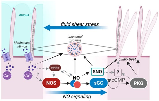
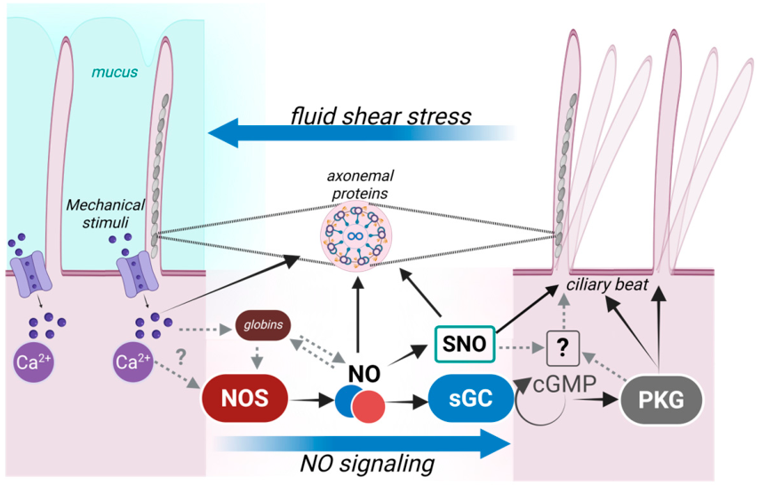
Whether motile cilia share this capacity is less clear, although they possess machinery capable of mechanosensation. TRPV4 localizes to airway motile cilia and transduces physical and chemical stimuli into Ca2+ signals that regulate ciliary beat frequency [121]. Functional evidence that motile cilia can respond to mechanical cues also comes from Xenopus ciliated epithelium, where cilia-generated fluid flow feeds back to refine ciliary polarity [122]. Whether these mechanosensory responses are coupled to NO production, however, remains unknown. Neither of these observations demonstrates that mechanical stimulation of motile cilia activates NOS or increases NO production. Thus, the proposed coupling of cilia-generated flow or mechanical stimulation to Ca2+-dependent NOS activation and NO production in motile cilia remains hypothetical. The clearest evidence for the opposite direction of regulation comes from Cygb, which colocalizes with NOS in motile cilia and whose deletion impairs NOS activity, reduces NO availability, and disrupts ciliary motility and fluid flow, phenotypes rescued by NO donors or sGC stimulation [43]. Thus, adequate NO availability is required for normal motile cilia function, establishing one arm of a potential bidirectional relationship (Figure 2); whether ciliary mechanosensation or motility reciprocally regulates NO production remains to be determined.
Figure 2.
Proposed bidirectional relationship between NO signaling and motile cilia function. Mechanical stimuli, including cilia-generated fluid shear stress, can activate Ca2+ signaling in ciliated cells. Ca2+/calmodulin-dependent activation of nNOS/eNOS can promote NO production, although coupling of cilia-generated mechanical stimuli and Ca2+ signaling to NOS activation has not been demonstrated in motile cilia. Globins may further regulate local NO bioavailability through modulation of NOS activity, NO binding, and nitrite-dependent NO production. NO regulates ciliary function through the canonical sGC–cGMP–PKG pathway and potentially through S-nitrosylation (SNO) of ciliary proteins. In turn, NO-dependent signaling regulates ciliary beating and fluid flow, raising the possibility of a feedback mechanism in which cilia-generated shear stress reciprocally regulates NO production. Solid arrows indicate established pathways, whereas dashed grey arrows indicate proposed or incompletely characterized mechanisms in motile cilia. It should be noted that the feedback from cilia-generated mechanical stimuli and Ca2+ signaling to NOS activation and NO production remains hypothetical. NO, nitric oxide; NOS, nitric oxide synthase; sGC, soluble guanylate cyclase; cGMP, cyclic guanosine monophosphate; PKG, protein kinase G; SNO, S-nitrosylation. Solid arrows indicate established pathways, whereas dashed arrows indicate proposed or incompletely characterized mechanisms. Created in BioRender. Corti, P. (2026) https://BioRender.com/qtoe10y.
This is the gap. Human diseases of impaired ciliary motility, PCD foremost among them, are strongly associated with reduced airway NO [123,124], yet the direction of causation has never been resolved: low NO may impair ciliary function, immotility may reduce shear-dependent NO production, or, as the discordant expression data discussed above would allow, both may follow from a shared upstream change in the airway environment. Distinguishing these possibilities requires experiments that directly manipulate ciliary beating and the resulting flow through pharmacological or genetic arrest of motility or controlled application of shear to ciliated cultures, while measuring NO output, NOS expression, and flux through the NOS–sGC–cGMP axis. Such studies are, to our knowledge, absent, and they represent one of the more approachable open problems in the field. One practical barrier has recently eased: protocols for quantifying NO production directly in mouse tracheal epithelial cells differentiated at the air–liquid interface have now been described.
8. Conclusions
Together, these studies establish NO signaling as an important regulator of motile cilia across development, reproduction, and airway physiology, while its dysregulation is increasingly linked to human ciliary disease. PCD provides the clearest clinical connection, with markedly reduced nasal NO representing a defining biochemical feature of the disease [14,18], while altered NO signaling and impaired ciliary function are also observed in chronic airway diseases such as COPD [74,76]. An important future direction will be to determine whether these changes are a consequence of ciliary dysfunction or actively contribute to disease progression. The identification of Cygb as a regulator of ciliary NO signaling, together with the emerging potential role of Adgb, further expands this pathway beyond NOS and suggests that additional regulators of local NO homeostasis remain to be discovered. Defining how these mechanisms differ across ciliated tissues and disease states will ultimately be essential for translating the biology of NO signaling into strategies to restore ciliary function [125].
Author Contributions
Conceptualization, P.C.; original draft preparation, P.C., T.T.W. and A.C.; writing, T.T.W., A.C. and P.C.; review and editing, P.C.; visualization, P.C.; supervision, P.C. All authors have read and agreed to the published version of the manuscript.
Funding
This work was supported by the National Heart, Lung, and Blood Institute (NHLBI), National Institutes of Health (NIH), grant R01HL168775 and by the University of Maryland, Baltimore Institute for Clinical and Translational Research (ICTR).
Institutional Review Board Statement
Not applicable.
Informed Consent Statement
Not applicable.
Data Availability Statement
No new data were created or analyzed in this study. Data sharing is not applicable to this article.
Acknowledgments
During the preparation of this manuscript, the authors used ChatGPT (OpenAI; GPT-5) for the purpose of language editing and refinement of the text. All AI-assisted suggestions were critically reviewed and revised by the authors, who take full responsibility for the final content of the publication.
Conflicts of Interest
The authors declare no conflicts of interest.
References
- Förstermann, U.; Sessa, W.C. Nitric oxide synthases: Regulation and function. Eur. Heart J. 2012, 33, 829–837. [Google Scholar] [CrossRef] [Scilit]
- Jain, B.; Rubinstein, I.; Robbins, R.A.; Leise, K.L.; Sisson, J.H. Modulation of airway epithelial cell ciliary beat frequency by nitric oxide. Biochem. Biophys. Res. Commun. 1993, 191, 83–88. [Google Scholar] [CrossRef] [Scilit]
- Tamaoki, J.; Chiyotani, A.; Kondo, M.; Konno, K. Role of NO generation in beta-adrenoceptor-mediated stimulation of rabbit airway ciliary motility. Am. J. Physiol. 1995, 268, C1342–C1347. [Google Scholar] [CrossRef] [Scilit]
- Sisson, J.H. Ethanol stimulates apparent nitric oxide-dependent ciliary beat frequency in bovine airway epithelial cells. Am. J. Physiol. 1995, 268, L596–L600. [Google Scholar] [CrossRef] [Scilit]
- Jain, B.; Rubinstein, I.; Robbins, R.A.; Sisson, J.H. TNF-alpha and IL-1 beta upregulate nitric oxide-dependent ciliary motility in bovine airway epithelium. Am. J. Physiol. 1995, 268, L911–L917. [Google Scholar] [CrossRef] [Scilit]
- Asano, K.; Chee, C.B.; Gaston, B.; Lilly, C.M.; Gerard, C.; Drazen, J.M.; Stamler, J.S. Constitutive and inducible nitric oxide synthase gene expression, regulation, and activity in human lung epithelial cells. Proc. Natl. Acad. Sci. USA 1994, 91, 10089–10093. [Google Scholar] [CrossRef] [Scilit]
- Degano, B.; Valmary, S.; Serrano, E.; Brousset, P.; Arnal, J.F. Expression of nitric oxide synthases in primary ciliary dyskinesia. Hum. Pathol. 2011, 42, 1855–1861. [Google Scholar] [CrossRef] [Scilit]
- Smith, C.M.; Fadaee-Shohada, M.J.; Sawhney, R.; Baker, N.; Williams, G.; Hirst, R.A.; Andrew, P.W.; O’Callaghan, C. Ciliated cultures from patients with primary ciliary dyskinesia do not produce nitric oxide or inducible nitric oxide synthase during early infection. Chest 2013, 144, 1671–1676. [Google Scholar] [CrossRef] [Scilit]
- Gaston, B.; Smith, L.A.; Davis, M.D.; Saunders, J.; Daniels, I.; Horani, A.; Brody, S.L.; Giddings, O.; Zhao, Y.; Marozkina, N. Antigen stasis and airway nitrosative stress in human primary ciliary dyskinesia. Am. J. Physiol. Lung Cell Mol. Physiol. 2024, 326, L468–l476. [Google Scholar] [CrossRef] [Scilit]
- Wallmeier, J.; Nielsen, K.G.; Kuehni, C.E.; Lucas, J.S.; Leigh, M.W.; Zariwala, M.A.; Omran, H. Motile ciliopathies. Nat. Rev. Dis. Primers 2020, 6, 77. [Google Scholar] [CrossRef] [Scilit]
- Li, D.; Shirakami, G.; Zhan, X.; Johns, R.A. Regulation of ciliary beat frequency by the nitric oxide-cyclic guanosine monophosphate signaling pathway in rat airway epithelial cells. Am. J. Respir. Cell Mol. Biol. 2000, 23, 175–181. [Google Scholar] [CrossRef] [Scilit]
- Braiman, A.; Uzlaner, N.; Priel, Z. The Role of Cyclic Nucleotide Pathways and Calmodulin in Ciliary Stimulation; New York, NY, USA, 2001; pp. 53–64. [Google Scholar]
- Gertsberg, I.; Hellman, V.; Fainshtein, M.; Weil, S.; Silberberg, S.D.; Danilenko, M.; Priel, Z. Intracellular Ca2+ Regulates the Phosphorylation and the Dephosphorylation of Ciliary Proteins Via the NO Pathway. J. Gen. Physiol. 2004, 124, 527–540. [Google Scholar] [CrossRef] [Scilit]
- Lundberg, J.O.; Weitzberg, E.; Nordvall, S.L.; Kuylenstierna, R.; Lundberg, J.M.; Alving, K. Primarily nasal origin of exhaled nitric oxide and absence in Kartagener’s syndrome. Eur. Respir. J. 1994, 7, 1501–1504. [Google Scholar] [CrossRef] [Scilit]
- Wodehouse, T.; Kharitonov, S.A.; Mackay, I.S.; Barnes, P.J.; Wilson, R.; Cole, P.J. Nasal nitric oxide measurements for the screening of primary ciliary dyskinesia. Eur. Respir. J. 2003, 21, 43–47. [Google Scholar] [CrossRef] [Scilit]
- Pifferi, M.; Bush, A.; Maggi, F.; Michelucci, A.; Ricci, V.; Conidi, M.E.; Cangiotti, A.M.; Bodini, A.; Simi, P.; Macchia, P.; et al. Nasal nitric oxide and nitric oxide synthase expression in primary ciliary dyskinesia. Eur. Respir. J. 2011, 37, 572–577. [Google Scholar] [CrossRef] [Scilit]
- American Thoracic Society; European Respiratory Society. ATS/ERS recommendations for standardized procedures for the online and offline measurement of exhaled lower respiratory nitric oxide and nasal nitric oxide, 2005. Am. J. Respir. Crit. Care Med. 2005, 171, 912–930. [Google Scholar] [CrossRef] [Scilit]
- Leigh, M.W.; Hazucha, M.J.; Chawla, K.K.; Baker, B.R.; Shapiro, A.J.; Brown, D.E.; Lavange, L.M.; Horton, B.J.; Qaqish, B.; Carson, J.L.; et al. Standardizing nasal nitric oxide measurement as a test for primary ciliary dyskinesia. Ann. Am. Thorac. Soc. 2013, 10, 574–581. [Google Scholar] [CrossRef] [Scilit]
- Lucas, J.S.; Barbato, A.; Collins, S.A.; Goutaki, M.; Behan, L.; Caudri, D.; Dell, S.; Eber, E.; Escudier, E.; Hirst, R.A.; et al. European Respiratory Society guidelines for the diagnosis of primary ciliary dyskinesia. Eur. Respir. J. 2017, 49, 1601090. [Google Scholar] [CrossRef] [Scilit]
- Collins, S.A.; Gove, K.; Walker, W.; Lucas, J.S. Nasal nitric oxide screening for primary ciliary dyskinesia: Systematic review and meta-analysis. Eur. Respir. J. 2014, 44, 1589–1599. [Google Scholar] [CrossRef] [Scilit]
- Raidt, J.; Loges, N.T.; Olbrich, H.; Wallmeier, J.; Pennekamp, P.; Omran, H. Primary ciliary dyskinesia. Presse Med. 2023, 52, 104171. [Google Scholar] [CrossRef] [Scilit]
- Högman, M.; Pham-Ngoc, H.; Nguyen-Duy, B.; Ellingsen, J.; Hua-Huy, T.; Van Nguyen, D.; Dinh-Xuan, A.T. Measuring exhaled nitric oxide in COPD: From theoretical consideration to practical views. Expert. Rev. Respir. Med. 2024, 18, 1013–1024. [Google Scholar] [CrossRef] [Scilit]
- Wyatt, T.A. Cyclic GMP and Cilia Motility. Cells 2015, 4, 315–330. [Google Scholar] [CrossRef] [Scilit]
- Stamler, J.S.; Singel, D.J.; Loscalzo, J. Biochemistry of nitric oxide and its redox-activated forms. Science 1992, 258, 1898–1902. [Google Scholar] [CrossRef] [Scilit]
- Hess, D.T.; Matsumoto, A.; Kim, S.O.; Marshall, H.E.; Stamler, J.S. Protein S-nitrosylation: Purview and parameters. Nat. Rev. Mol. Cell Biol. 2005, 6, 150–166. [Google Scholar] [CrossRef] [Scilit]
- Pacher, P.; Beckman, J.S.; Liaudet, L. Nitric oxide and peroxynitrite in health and disease. Physiol. Rev. 2007, 87, 315–424. [Google Scholar] [CrossRef] [Scilit]
- Price, M.E.; Sisson, J.H. Redox regulation of motile cilia in airway disease. Redox Biol. 2019, 27, 101146. [Google Scholar] [CrossRef] [Scilit]
- Allen, R.D. The morphogenesis of basal bodies and accessory structures of the cortex of the ciliated protozoan Tetrahymena pyriformis. J. Cell Biol. 1969, 40, 716–733. [Google Scholar] [CrossRef] [Scilit]
- Pinskey, J.M.; Lagisetty, A.; Gui, L.; Phan, N.; Reetz, E.; Tavakoli, A.; Fu, G.; Nicastro, D. Three-dimensional flagella structures from animals’ closest unicellular relatives, the Choanoflagellates. Elife 2022, 11, e78133. [Google Scholar] [CrossRef] [Scilit]
- McGrath, J.; Somlo, S.; Makova, S.; Tian, X.; Brueckner, M. Two populations of node monocilia initiate left-right asymmetry in the mouse. Cell 2003, 114, 61–73. [Google Scholar] [CrossRef] [Scilit]
- Nonami, Y.; Narita, K.; Nakamura, H.; Inoue, T.; Takeda, S. Developmental changes in ciliary motility on choroid plexus epithelial cells during the perinatal period. Cytoskeleton 2013, 70, 797–803. [Google Scholar] [CrossRef] [Scilit]
- Nonaka, S.; Shiratori, H.; Saijoh, Y.; Hamada, H. Determination of left-right patterning of the mouse embryo by artificial nodal flow. Nature 2002, 418, 96–99. [Google Scholar] [CrossRef] [Scilit]
- Okada, Y.; Takeda, S.; Tanaka, Y.; Belmonte, J.I.; Hirokawa, N. Mechanism of nodal flow: A conserved symmetry breaking event in left-right axis determination. Cell 2005, 121, 633–644. [Google Scholar] [CrossRef] [Scilit]
- Norekian, T.P.; Moroz, L.L. Recording cilia activity in ctenophores: Effects of nitric oxide and low molecular weight transmitters. Front. Neurosci. 2023, 17, 1125476. [Google Scholar] [CrossRef] [Scilit]
- Rompolas, P.; Patel-King, R.S.; King, S.M. An outer arm Dynein conformational switch is required for metachronal synchrony of motile cilia in planaria. Mol. Biol. Cell 2010, 21, 3669–3679. [Google Scholar] [CrossRef] [Scilit]
- Inaba, K. Calcium sensors of ciliary outer arm dynein: Functions and phylogenetic considerations for eukaryotic evolution. Cilia 2015, 4, 6. [Google Scholar] [CrossRef] [Scilit]
- King, S.M. Sensing the mechanical state of the axoneme and integration of Ca2+ signaling by outer arm dynein. Cytoskeleton 2010, 67, 207–213. [Google Scholar] [CrossRef] [Scilit]
- Kung, C.; Chang, S.Y.; Satow, Y.; Houten, J.V.; Hansma, H. Genetic dissection of behavior in paramecium. Science 1975, 188, 898–904. [Google Scholar] [CrossRef] [Scilit]
- Preston, R.R.; Kink, J.A.; Hinrichsen, R.D.; Saimi, Y.; Kung, C. Calmodulin mutants and Ca2+-dependent channels in Paramecium. Annu. Rev. Physiol. 1991, 53, 309–319. [Google Scholar] [CrossRef]
- Plattner, H. Membrane traffic and Ca2+ signals in ciliates. J. Eukaryot. Microbiol. 2022, 69, e12895. [Google Scholar] [CrossRef] [Scilit]
- Rao, Q.; Han, L.; Wang, Y.; Chai, P.; Kuo, Y.W.; Yang, R.; Hu, F.; Yang, Y.; Howard, J.; Zhang, K. Structures of outer-arm dynein array on microtubule doublet reveal a motor coordination mechanism. Nat. Struct. Mol. Biol. 2021, 28, 799–810. [Google Scholar] [CrossRef] [Scilit]
- Schmid, A.; Salathe, M. Ciliary beat co-ordination by calcium. Biol. Cell 2011, 103, 159–169. [Google Scholar] [CrossRef] [Scilit]
- Rochon, E.R.; Xue, J.; Mohammed, M.S.; Smith, C.; Hay-Schmidt, A.; DeMartino, A.W.; Clark, A.; Xu, Q.; Lo, C.W.; Tsang, M.; et al. Cytoglobin regulates NO-dependent cilia motility and organ laterality during development. Nat. Commun. 2023, 14, 8333. [Google Scholar] [CrossRef] [Scilit]
- Zhan, X.; Li, D.; Johns, R.A. Expression of endothelial nitric oxide synthase in ciliated epithelia of rats. J. Histochem. Cytochem. 2003, 51, 81–87. [Google Scholar] [CrossRef] [Scilit]
- Jackson, C.L.; Lucas, J.S.; Walker, W.T.; Owen, H.; Premadeva, I.; Lackie, P.M. Neuronal NOS localises to human airway cilia. Nitric Oxide 2015, 44, 3–7. [Google Scholar] [CrossRef] [Scilit]
- Zhao, K.Q.; Cowan, A.T.; Lee, R.J.; Goldstein, N.; Droguett, K.; Chen, B.; Zheng, C.; Villalon, M.; Palmer, J.N.; Kreindler, J.L.; et al. Molecular modulation of airway epithelial ciliary response to sneezing. FASEB J. 2012, 26, 3178–3187. [Google Scholar] [CrossRef] [Scilit]
- Alpizar, Y.A.; Boonen, B.; Sanchez, A.; Jung, C.; López-Requena, A.; Naert, R.; Steelant, B.; Luyts, K.; Plata, C.; De Vooght, V.; et al. TRPV4 activation triggers protective responses to bacterial lipopolysaccharides in airway epithelial cells. Nat. Commun. 2017, 8, 1059. [Google Scholar] [CrossRef] [Scilit]
- Carey, R.M.; Palmer, J.N.; Adappa, N.D.; Lee, R.J. Loss of CFTR function is associated with reduced bitter taste receptor-stimulated nitric oxide innate immune responses in nasal epithelial cells and macrophages. Front. Immunol. 2023, 14, 1096242. [Google Scholar] [CrossRef] [Scilit]
- Kerr, B.; Ríos, M.; Droguett, K.; Villalón, M. Nitric oxide activation by progesterone suppresses ATP-induced ciliary activity in oviductal ciliated cells. Reprod. Fertil. Dev. 2018, 30, 1666–1674. [Google Scholar] [CrossRef] [Scilit]
- Lefièvre, L.; Chen, Y.; Conner, S.J.; Scott, J.L.; Publicover, S.J.; Ford, W.C.; Barratt, C.L. Human spermatozoa contain multiple targets for protein S-nitrosylation: An alternative mechanism of the modulation of sperm function by nitric oxide? Proteomics 2007, 7, 3066–3084. [Google Scholar] [CrossRef] [Scilit]
- Furchgott, R.F.; Zawadzki, J.V. The obligatory role of endothelial cells in the relaxation of arterial smooth muscle by acetylcholine. Nature 1980, 288, 373–376. [Google Scholar] [CrossRef] [Scilit]
- Ignarro, L.J.; Buga, G.M.; Wood, K.S.; Byrns, R.E.; Chaudhuri, G. Endothelium-derived relaxing factor produced and released from artery and vein is nitric oxide. Proc. Natl. Acad. Sci. USA 1987, 84, 9265–9269. [Google Scholar] [CrossRef] [Scilit]
- Palmer, R.M.; Ferrige, A.G.; Moncada, S. Nitric oxide release accounts for the biological activity of endothelium-derived relaxing factor. Nature 1987, 327, 524–526. [Google Scholar] [CrossRef] [Scilit]
- AbouAlaiwi, W.A.; Takahashi, M.; Mell, B.R.; Jones, T.J.; Ratnam, S.; Kolb, R.J.; Nauli, S.M. Ciliary polycystin-2 is a mechanosensitive calcium channel involved in nitric oxide signaling cascades. Circ. Res. 2009, 104, 860–869. [Google Scholar] [CrossRef] [Scilit]
- Xie, Q.-w.; Nathan, C. The high-output nitric oxide pathway: Role and regulation. J. Leukoc. Biol. 1994, 56, 576–582. [Google Scholar] [CrossRef] [Scilit]
- Alderton, W.K.; Cooper, C.E.; Knowles, R.G. Nitric oxide synthases: Structure, function and inhibition. Biochem. J. 2001, 357, 593–615. [Google Scholar] [CrossRef] [Scilit]
- Mikhailik, A.; Michurina, T.V.; Dikranian, K.; Hearn, S.; Maxakov, V.I.; Siller, S.S.; Takemaru, K.I.; Enikolopov, G.; Peunova, N. nNOS regulates ciliated cell polarity, ciliary beat frequency, and directional flow in mouse trachea. Life Sci. Alliance 2021, 4, e202000981. [Google Scholar] [CrossRef] [Scilit]
- Peunova, N.; Scheinker, V.; Ravi, K.; Enikolopov, G. Nitric Oxide Coordinates Cell Proliferation and Cell Movements During Early Development of Xenopus. Cell Cycle 2007, 6, 3132–3144. [Google Scholar] [CrossRef] [Scilit]
- Cinelli, M.A.; Do, H.T.; Miley, G.P.; Silverman, R.B. Inducible nitric oxide synthase: Regulation, structure, and inhibition. Med. Res. Rev. 2020, 40, 158–189. [Google Scholar] [CrossRef] [Scilit]
- Hamid, Q.; Springall, D.R.; Riveros-Moreno, V.; Chanez, P.; Howarth, P.; Redington, A.; Bousquet, J.; Godard, P.; Holgate, S.; Polak, J.M. Induction of nitric oxide synthase in asthma. Lancet 1993, 342, 1510–1513. [Google Scholar] [CrossRef] [Scilit]
- Guo, F.H.; Comhair, S.A.; Zheng, S.; Dweik, R.A.; Eissa, N.T.; Thomassen, M.J.; Calhoun, W.; Erzurum, S.C. Molecular mechanisms of increased nitric oxide (NO) in asthma: Evidence for transcriptional and post-translational regulation of NO synthesis. J. Immunol. 2000, 164, 5970–5980. [Google Scholar] [CrossRef] [Scilit]
- Walker, W.T.; Jackson, C.L.; Coles, J.; Lackie, P.M.; Faust, S.N.; Hall-Stoodley, L.; Lucas, J.S. Ciliated cultures from patients with primary ciliary dyskinesia produce nitric oxide in response to Haemophilus influenzae infection and proinflammatory cytokines. Chest 2014, 145, 668–669. [Google Scholar] [CrossRef] [Scilit]
- Csoma, Z.; Bush, A.; Wilson, N.M.; Donnelly, L.; Balint, B.; Barnes, P.J.; Kharitonov, S.A. Nitric oxide metabolites are not reduced in exhaled breath condensate of patients with primary ciliary dyskinesia. Chest 2003, 124, 633–638. [Google Scholar] [CrossRef] [Scilit]
- Pifferi, M.; Bush, A.; Caramella, D.; Di Cicco, M.; Zangani, M.; Chinellato, I.; Macchia, P.; Boner, A.L. Agenesis of paranasal sinuses and nasal nitric oxide in primary ciliary dyskinesia. Eur. Respir. J. 2011, 37, 566–571. [Google Scholar] [CrossRef] [Scilit]
- Paraskakis, E.; Zihlif, N.; Bush, A. Nitric oxide production in PCD: Possible evidence for differential nitric oxide synthase function. Pediatr. Pulmonol. 2007, 42, 876–880. [Google Scholar] [CrossRef] [Scilit]
- Meng, Q.H.; Springall, D.R.; Bishop, A.E.; Morgan, K.; Evans, T.J.; Habib, S.; Gruenert, D.C.; Gyi, K.M.; Hodson, M.E.; Yacoub, M.H.; et al. Lack of inducible nitric oxide synthase in bronchial epithelium: A possible mechanism of susceptibility to infection in cystic fibrosis. J. Pathol. 1998, 184, 323–331. [Google Scholar] [CrossRef] [Scilit]
- Kelley, T.J.; Drumm, M.L. Inducible nitric oxide synthase expression is reduced in cystic fibrosis murine and human airway epithelial cells. J. Clin. Investig. 1998, 102, 1200–1207. [Google Scholar] [CrossRef] [Scilit]
- Steagall, W.K.; Elmer, H.L.; Brady, K.G.; Kelley, T.J. Cystic fibrosis transmembrane conductance regulator-dependent regulation of epithelial inducible nitric oxide synthase expression. Am. J. Respir. Cell Mol. Biol. 2000, 22, 45–50. [Google Scholar] [CrossRef] [Scilit]
- Linnane, S.J.; Keatings, V.M.; Costello, C.M.; Moynihan, J.B.; O’Connor, C.M.; Fitzgerald, M.X.; McLoughlin, P. Total sputum nitrate plus nitrite is raised during acute pulmonary infection in cystic fibrosis. Am. J. Respir. Crit. Care Med. 1998, 158, 207–212. [Google Scholar] [CrossRef] [Scilit]
- Ho, L.P.; Innes, J.A.; Greening, A.P. Nitrite levels in breath condensate of patients with cystic fibrosis is elevated in contrast to exhaled nitric oxide. Thorax 1998, 53, 680–684. [Google Scholar] [CrossRef] [Scilit][Green Version]
- Jones, K.L.; Hegab, A.H.; Hillman, B.C.; Simpson, K.L.; Jinkins, P.A.; Grisham, M.B.; Owens, M.W.; Sato, E.; Robbins, R.A. Elevation of nitrotyrosine and nitrate concentrations in cystic fibrosis sputum. Pediatr. Pulmonol. 2000, 30, 79–85. [Google Scholar] [CrossRef] [Scilit]
- Grasemann, H.; Schwiertz, R.; Matthiesen, S.; Racké, K.; Ratjen, F. Increased arginase activity in cystic fibrosis airways. Am. J. Respir. Crit. Care Med. 2005, 172, 1523–1528. [Google Scholar] [CrossRef] [Scilit]
- Maestrelli, P.; Páska, C.; Saetta, M.; Turato, G.; Nowicki, Y.; Monti, S.; Formichi, B.; Miniati, M.; Fabbri, L.M. Decreased haem oxygenase-1 and increased inducible nitric oxide synthase in the lung of severe COPD patients. Eur. Respir. J. 2003, 21, 971–976. [Google Scholar] [CrossRef] [Scilit]
- Brindicci, C.; Kharitonov, S.A.; Ito, M.; Elliott, M.W.; Hogg, J.C.; Barnes, P.J.; Ito, K. Nitric oxide synthase isoenzyme expression and activity in peripheral lung tissue of patients with chronic obstructive pulmonary disease. Am. J. Respir. Crit. Care Med. 2010, 181, 21–30. [Google Scholar] [CrossRef] [Scilit]
- Maziak, W.; Loukides, S.; Culpitt, S.; Sullivan, P.; Kharitonov, S.A.; Barnes, P.J. Exhaled nitric oxide in chronic obstructive pulmonary disease. Am. J. Respir. Crit. Care Med. 1998, 157, 998–1002. [Google Scholar] [CrossRef] [Scilit]
- Brindicci, C.; Ito, K.; Resta, O.; Pride, N.B.; Barnes, P.J.; Kharitonov, S.A. Exhaled nitric oxide from lung periphery is increased in COPD. Eur. Respir. J. 2005, 26, 52–59. [Google Scholar] [CrossRef] [Scilit]
- Amdahl, M.B.; Petersen, E.E.; Bocian, K.; Kaliszuk, S.J.; DeMartino, A.W.; Tiwari, S.; Sparacino-Watkins, C.E.; Corti, P.; Rose, J.J.; Gladwin, M.T.; et al. The Zebrafish Cytochrome b(5)/Cytochrome b(5) Reductase/NADH System Efficiently Reduces Cytoglobins 1 and 2: Conserved Activity of Cytochrome b(5)/Cytochrome b(5) Reductases during Vertebrate Evolution. Biochemistry 2019, 58, 3212–3223. [Google Scholar] [CrossRef] [Scilit]
- Rochon, E.R.; Corti, P. Globins and nitric oxide homeostasis in fish embryonic development. Mar. Genom. 2020, 49, 100721. [Google Scholar] [CrossRef] [Scilit]
- Corti, P.; Xue, J.; Tejero, J.; Wajih, N.; Sun, M.; Stolz, D.B.; Tsang, M.; Kim-Shapiro, D.B.; Gladwin, M.T. Globin X is a six-coordinate globin that reduces nitrite to nitric oxide in fish red blood cells. Proc. Natl. Acad. Sci. USA 2016, 113, 8538–8543. [Google Scholar] [CrossRef] [Scilit]
- Corti, P.; Tejero, J.; Gladwin, M.T. Evidence mounts that red cells and deoxyhemoglobin can reduce nitrite to bioactive NO to mediate intravascular endocrine NO signaling: Commentary on “Anti-platelet effects of dietary nitrate in healthy volunteers: Involvement of cGMP and influence of sex”. Free Radic. Biol. Med. 2013, 65, 1518–1520. [Google Scholar] [CrossRef] [Scilit]
- Rochon, E.R.; Missinato, M.A.; Xue, J.; Tejero, J.; Tsang, M.; Gladwin, M.T.; Corti, P. Nitrite Improves Heart Regeneration in Zebrafish. Antioxid. Redox Signal 2020, 32, 363–377. [Google Scholar] [CrossRef] [Scilit]
- Giordano, D.; Corti, P.; Coppola, D.; Altomonte, G.; Xue, J.; Russo, R.; di Prisco, G.; Verde, C. Regulation of globin expression in Antarctic fish under thermal and hypoxic stress. Mar. Genom. 2021, 57, 100831. [Google Scholar] [CrossRef] [Scilit]
- Giordano, D.; Verde, C.; Corti, P. Nitric Oxide Production and Regulation in the Teleost Cardiovascular System. Antioxidants 2022, 11, 957. [Google Scholar] [CrossRef] [Scilit]
- Straub, A.C.; Lohman, A.W.; Billaud, M.; Johnstone, S.R.; Dwyer, S.T.; Lee, M.Y.; Bortz, P.S.; Best, A.K.; Columbus, L.; Gaston, B.; et al. Endothelial cell expression of haemoglobin α regulates nitric oxide signalling. Nature 2012, 491, 473–477. [Google Scholar] [CrossRef] [Scilit]
- Marozkina, N.; Smith, L.; Zhao, Y.; Zein, J.; Chmiel, J.F.; Kim, J.; Kiselar, J.; Davis, M.D.; Cunningham, R.S.; Randell, S.H.; et al. Somatic cell hemoglobin modulates nitrogen oxide metabolism in the human airway epithelium. Sci. Rep. 2021, 11, 15498. [Google Scholar] [CrossRef] [Scilit]
- Koay, T.W.; Osterhof, C.; Orlando, I.M.C.; Keppner, A.; Andre, D.; Yousefian, S.; Suárez Alonso, M.; Correia, M.; Markworth, R.; Schödel, J.; et al. Androglobin gene expression patterns and FOXJ1-dependent regulation indicate its functional association with ciliogenesis. J. Biol. Chem. 2021, 296, 100291. [Google Scholar] [CrossRef] [Scilit]
- Keppner, A.; Correia, M.; Santambrogio, S.; Koay, T.W.; Maric, D.; Osterhof, C.; Winter, D.V.; Clerc, A.; Stumpe, M.; Chalmel, F.; et al. Androglobin, a chimeric mammalian globin, is required for male fertility. Elife 2022, 11, e72374. [Google Scholar] [CrossRef] [Scilit]
- Reeder, B.J.; Deganutti, G.; Ukeri, J.; Atanasio, S.; Svistunenko, D.A.; Ronchetti, C.; Mobarec, J.C.; Welbourn, E.; Asaju, J.; Vos, M.H.; et al. The circularly permuted globin domain of androglobin exhibits atypical heme stabilization and nitric oxide interaction. Chem. Sci. 2024, 15, 6738–6751. [Google Scholar] [CrossRef] [Scilit]
- Joachimiak, E.; Osinka, A.; Farahat, H.; Świderska, B.; Sitkiewicz, E.; Poprzeczko, M.; Fabczak, H.; Wloga, D. Composition and function of the C1b/C1f region in the ciliary central apparatus. Sci. Rep. 2021, 11, 11760. [Google Scholar] [CrossRef] [Scilit]
- Olea, C., Jr.; Herzik, M.A., Jr.; Kuriyan, J.; Marletta, M.A. Structural insights into the molecular mechanism of H-NOX activation. Protein Sci. 2010, 19, 881–887. [Google Scholar] [CrossRef] [Scilit]
- Russwurm, M.; Koesling, D. NO activation of guanylyl cyclase. EMBO J. 2004, 23, 4443–4450. [Google Scholar] [CrossRef] [Scilit]
- Zhan, X.; Li, D.; Johns, R.A. Immunohistochemical evidence for the NO cGMP signaling pathway in respiratory ciliated epithelia of rat. J. Histochem. Cytochem. 1999, 47, 1369–1374. [Google Scholar] [CrossRef] [Scilit]
- Stout, S.L.; Wyatt, T.A.; Adams, J.J.; Sisson, J.H. Nitric oxide-dependent cilia regulatory enzyme localization in bovine bronchial epithelial cells. J. Histochem. Cytochem. 2007, 55, 433–442. [Google Scholar] [CrossRef] [Scilit]
- Sakato-Antoku, M.; Balsbaugh, J.L.; King, S.M. N-Terminal Processing and Modification of Ciliary Dyneins. Cells 2023, 12, 2492. [Google Scholar] [CrossRef] [Scilit]
- Sakato-Antoku, M.; Patel-King, R.S.; Inaba, K.; Balsbaugh, J.L.; King, S.M. Isoform-specific phosphorylation of axonemal dynein heavy chains. Mol. Biol. Cell 2025, 36, ar67. [Google Scholar] [CrossRef] [Scilit]
- Ann, K.S.; Nelson, D.L. Protein substrates for cGMP-dependent protein phosphorylation in cilia of wild type and atalanta mutants of Paramecium. Cell Motil. Cytoskelet. 1995, 30, 252–260. [Google Scholar] [CrossRef] [Scilit]
- Zhang, L.; Sanderson, M.J. The role of cGMP in the regulation of rabbit airway ciliary beat frequency. J. Physiol. 2003, 551, 765–776. [Google Scholar] [CrossRef]
- Bork, N.I.; Nikolaev, V.O. cGMP Signaling in the Cardiovascular System-The Role of Compartmentation and Its Live Cell Imaging. Int. J. Mol. Sci. 2018, 19, 801. [Google Scholar] [CrossRef] [Scilit]
- Nguyen, P.A.; Liou, W.; Hall, D.H.; Leroux, M.R. Ciliopathy proteins establish a bipartite signaling compartment in a C. elegans thermosensory neuron. J. Cell Sci. 2014, 127, 5317–5330. [Google Scholar] [CrossRef] [Scilit]
- Muthaiyan Shanmugam, M.; Bhan, P.; Huang, H.Y.; Hsieh, J.; Hua, T.E.; Wu, G.H.; Punjabi, H.; Lee Aplícano, V.D.; Chen, C.W.; Wagner, O.I. Cilium Length and Intraflagellar Transport Regulation by Kinases PKG-1 and GCK-2 in Caenorhabditis elegans Sensory Neurons. Mol. Cell Biol. 2018, 38, e00612-17. [Google Scholar] [CrossRef] [Scilit]
- O’Halloran, D.M.; Hamilton, O.S.; Lee, J.I.; Gallegos, M.; L’Etoile, N.D. Changes in cGMP levels affect the localization of EGL-4 in AWC in Caenorhabditis elegans. PLoS ONE 2012, 7, e31614. [Google Scholar] [CrossRef] [Scilit]
- Fuhrmann, M.; Jahn, H.U.; Seybold, J.; Neurohr, C.; Barnes, P.J.; Hippenstiel, S.; Kraemer, H.J.; Suttorp, N. Identification and function of cyclic nucleotide phosphodiesterase isoenzymes in airway epithelial cells. Am. J. Respir. Cell Mol. Biol. 1999, 20, 292–302. [Google Scholar] [CrossRef] [Scilit]
- Stasch, J.P.; Pacher, P.; Evgenov, O.V. Soluble guanylate cyclase as an emerging therapeutic target in cardiopulmonary disease. Circulation 2011, 123, 2263–2273. [Google Scholar] [CrossRef] [Scilit]
- Liu, R.; Kang, Y.; Chen, L. Activation mechanism of human soluble guanylate cyclase by stimulators and activators. Nat. Commun. 2021, 12, 5492. [Google Scholar] [CrossRef] [Scilit]
- Tejero, J.; Shiva, S.; Gladwin, M.T. Sources of Vascular Nitric Oxide and Reactive Oxygen Species and Their Regulation. Physiol. Rev. 2019, 99, 311–379. [Google Scholar] [CrossRef] [Scilit]
- Nakamura, T.; Lipton, S.A. Emerging role of protein-protein transnitrosylation in cell signaling pathways. Antioxid. Redox Signal 2013, 18, 239–249. [Google Scholar] [CrossRef] [Scilit]
- Amdahl, M.B.; Sparacino-Watkins, C.E.; Corti, P.; Gladwin, M.T.; Tejero, J. Efficient Reduction of Vertebrate Cytoglobins by the Cytochrome b(5)/Cytochrome b(5) Reductase/NADH System. Biochemistry 2017, 56, 3993–4004. [Google Scholar] [CrossRef] [Scilit]
- Darszon, A.; Treviño, C.L.; Wood, C.; Galindo, B.; Rodríguez-Miranda, E.; Acevedo, J.J.; Hernandez-González, E.O.; Beltrán, C.; Martínez-López, P.; Nishigaki, T. Ion channels in sperm motility and capacitation. Soc. Reprod. Fertil. Suppl. 2007, 65, 229–244. [Google Scholar]
- Publicover, S.; Harper, C.V.; Barratt, C. [Ca2+]i signalling in sperm--making the most of what you’ve got. Nat. Cell Biol. 2007, 9, 235–242. [Google Scholar] [CrossRef] [Scilit]
- Machado-Oliveira, G.; Lefièvre, L.; Ford, C.; Herrero, M.B.; Barratt, C.; Connolly, T.J.; Nash, K.; Morales-Garcia, A.; Kirkman-Brown, J.; Publicover, S. Mobilisation of Ca2+ stores and flagellar regulation in human sperm by S-nitrosylation: A role for NO synthesised in the female reproductive tract. Development 2008, 135, 3677–3686. [Google Scholar] [CrossRef] [Scilit]
- Price, M.E.; Pavlik, J.A.; Liu, M.; Ding, S.J.; Wyatt, T.A.; Sisson, J.H. Alcohol drives S-nitrosylation and redox activation of protein phosphatase 1, causing bovine airway cilia dysfunction. Am. J. Physiol. Lung Cell Mol. Physiol. 2017, 312, L432–L439. [Google Scholar] [CrossRef] [Scilit]
- Price, M.E.; Case, A.J.; Pavlik, J.A.; DeVasure, J.M.; Wyatt, T.A.; Zimmerman, M.C.; Sisson, J.H. S-nitrosation of protein phosphatase 1 mediates alcohol-induced ciliary dysfunction. Sci. Rep. 2018, 8, 9701. [Google Scholar] [CrossRef] [Scilit]
- Snyder, A.H.; McPherson, M.E.; Hunt, J.F.; Johnson, M.; Stamler, J.S.; Gaston, B. Acute effects of aerosolized S-nitrosoglutathione in cystic fibrosis. Am. J. Respir. Crit. Care Med. 2002, 165, 922–926. [Google Scholar] [CrossRef] [Scilit][Green Version]
- Wolf, A.; Tabasi, M.; Zacharek, M.; Martin, G.; Hershenson, M.B.; Meyerhoff, M.E.; Sajjan, U. S-Nitrosoglutathione Reduces the Density of Staphylococcus aureus Biofilms Established on Human Airway Epithelial Cells. ACS Omega 2023, 8, 846–856. [Google Scholar] [CrossRef] [Scilit]
- Koenitzer, J.R.; Gupta, D.K.; Twan, W.K.; Xu, H.; Hadas, N.; Hawkins, F.J.; Beermann, M.L.; Penny, G.M.; Wamsley, N.T.; Berical, A.; et al. Transcriptional analysis of primary ciliary dyskinesia airway cells reveals a dedicated cilia glutathione pathway. JCI Insight 2024, 9, e180198. [Google Scholar] [CrossRef] [Scilit]
- Saternos, H.C.; AbouAlaiwi, W.A. Signaling interplay between primary cilia and nitric oxide: A mini review. Nitric Oxide 2018, 80, 108–112. [Google Scholar] [CrossRef] [Scilit]
- Nauli, S.M.; Kawanabe, Y.; Kaminski, J.J.; Pearce, W.J.; Ingber, D.E.; Zhou, J. Endothelial Cilia Are Fluid Shear Sensors That Regulate Calcium Signaling and Nitric Oxide Production Through Polycystin-1. Circulation 2008, 117, 1161–1171. [Google Scholar] [CrossRef] [Scilit]
- Wang, L.; Shen, C.; Liu, H.; Wang, S.; Chen, X.; Roman, R.J.; Juncos, L.A.; Lu, Y.; Wei, J.; Zhang, J.; et al. Shear stress blunts tubuloglomerular feedback partially mediated by primary cilia and nitric oxide at the macula densa. Am. J. Physiol. Regul. Integr. Comp. Physiol. 2015, 309, R757–766. [Google Scholar] [CrossRef] [Scilit]
- Wang, X.; Yin, G.; Yang, Y.; Tian, X. Ciliary and Non-Ciliary Roles of IFT88 in Development and Diseases. Int. J. Mol. Sci. 2025, 26, 2110. [Google Scholar] [CrossRef] [Scilit]
- Ding, D.; Tian, R.; Yang, X.; Ren, Z.; Jing, Z.C.; Wu, X.T.; Sun, L.W. The impact of ciliary length on the mechanical response of osteocytes to fluid shear stress. Nitric Oxide 2025, 155, 1–11. [Google Scholar] [CrossRef] [Scilit]
- Lorenzo, I.M.; Liedtke, W.; Sanderson, M.J.; Valverde, M.A. TRPV4 channel participates in receptor-operated calcium entry and ciliary beat frequency regulation in mouse airway epithelial cells. Proc. Natl. Acad. Sci. USA 2008, 105, 12611–12616. [Google Scholar] [CrossRef] [Scilit]
- Mitchell, B.; Jacobs, R.; Li, J.; Chien, S.; Kintner, C. A positive feedback mechanism governs the polarity and motion of motile cilia. Nature 2007, 447, 97–101. [Google Scholar] [CrossRef] [Scilit]
- Shapiro, A.J.; Dell, S.D.; Gaston, B.; O’Connor, M.; Marozkina, N.; Manion, M.; Hazucha, M.J.; Leigh, M.W. Nasal Nitric Oxide Measurement in Primary Ciliary Dyskinesia. A Technical Paper on Standardized Testing Protocols. Ann. Am. Thorac. Soc. 2020, 17, e1–e12. [Google Scholar] [CrossRef] [Scilit]
- Walker, W.T.; Liew, A.; Harris, A.; Cole, J.; Lucas, J.S. Upper and lower airway nitric oxide levels in primary ciliary dyskinesia, cystic fibrosis and asthma. Respir. Med. 2013, 107, 380–386. [Google Scholar] [CrossRef] [Scilit]
- Iqbal, M.; Mahan, D.; Clark, A.; Bruce, A.; DeMartino, A.W.; Corti, P. Measurement of Nitric Oxide Production in Mouse Tracheal Epithelial Cell Cultures Differentiated at the Air Liquid Interface. J. Vis. Exp. 2026, 233, e70311. [Google Scholar] [CrossRef] [Scilit]
Disclaimer/Publisher’s Note: The statements, opinions and data contained in all publications are solely those of the individual author(s) and contributor(s) and not of MDPI and/or the editor(s). MDPI and/or the editor(s) disclaim responsibility for any injury to people or property resulting from any ideas, methods, instructions or products referred to in the content. |
© 2026 by the authors. Licensee MDPI, Basel, Switzerland. This article is an open access article distributed under the terms and conditions of the Creative Commons Attribution (CC BY) license.