


Abstract
Fatty acid synthase (FASN) is the rate-limiting core enzyme for de novo fatty acid biosynthesis in aquatic animals, yet its regulatory role in teleost cellular antioxidant defense and redox balance remains poorly characterized. In this study, we identified and bioinformatically characterized the complete coding sequence (CDS) of Jian carp FASN, which encodes a 2514-amino acid polypeptide containing five conserved polyketide synthase (PKS) catalytic domains and 188 phosphorylation sites. qRT-PCR tissue profiling revealed ubiquitous FASN transcription across all tested tissues, with the highest abundance in liver tissue. To decipher the physiological functions of FASN, siRNA-mediated functional depletion was performed in isolated primary Jian carp hepatocytes. Suppression of endogenous FASN expression induced markers of oxidative damage, evidenced by significantly reduced activities of superoxide dismutase (SOD), glutathione peroxidase (GPx), and catalase (CAT) (p < 0.05), alongside elevated malondialdehyde (MDA) levels (p < 0.05). GC-MS detected global declines in cellular saturated, monounsaturated and polyunsaturated fatty acids, especially C14:0, C16:0 and C18:0. Transcriptionally, the 6-PGD was downregulated (p < 0.05), whereas the fatty acid oxidation gene PPARα was upregulated (p < 0.05). RNA-seq identified 13,546 differentially expressed genes, with enrichment analyses indicating transcriptional shifts associated with oxidative damage and NF-κB-related inflammatory signaling. A total of 23 hub genes related to lipid turnover and arachidonic acid metabolism were screened. Collectively, our results demonstrate that FASN acts as an essential upstream regulator of hepatic redox balance in Jian carp. FASN disrupts fatty acid pools and triggers metabolic–inflammatory reprogramming, offering new insights into lipid–antioxidant crosstalk in teleost hepatocytes.
1. Introduction
De novo fatty acid synthesis is an indispensable metabolic process governing energy storage, membrane composition, and cellular signaling across vertebrates. Fatty acid synthase (FASN) is the multifunctional catalytic enzyme responsible for the complete synthesis of short- and medium-chain saturated fatty acids from acetyl-CoA and malonyl-CoA precursors [1]. Mammalian research has firmly established that FASN activity is tightly coupled to cellular redox status. Excessive inhibition of FASN depletes cellular antioxidant capacity, accumulates reactive oxygen species (ROS), and exacerbates lipid peroxidation, ultimately inducing oxidative stress and inflammatory activation in mammalian hepatocytes and tumor cells [2,3,4].
In farmed teleosts, hepatic lipid metabolism directly determines flesh quality, growth performance, and disease resistance, and balanced antioxidant capacity is critical to mitigate oxidative damage induced by nutritional and environmental stressors [5,6,7]. Jian carp (Cyprinus carpio var. Jian) is the most economically vital freshwater aquaculture species in China, with annual production exceeding one million tons [8,9]; elucidating the molecular regulators of its hepatic redox balance carries high practical value for improving cultivation stress resistance. To date, existing teleost FASN studies (including golden pompano [5], mandarin fish [10] and Nile tilapia [11]) have merely described tissue expression patterns and simple lipid phenotypes, while mechanistic investigations linking FASN to cellular redox homeostasis and antioxidant defense are extremely limited. The teleost liver serves as the dual central organ for endogenous lipogenesis and phase I/II antioxidant detoxification, equipped with complete pentose phosphate pathway (PPP) machinery for both fatty acid synthesis and antioxidant recycling [12]. Our preliminary transcriptomic screening successfully obtained the complete coding sequence (CDS) of Jian carp FASN, laying a foundation for functional verification of its redox regulatory roles.
In the present study, we integrated bioinformatic CDS characterization, tissue expression profiling, siRNA-mediated gene knockdown in primary hepatocytes, fatty acid metabolomics, antioxidant biochemical detection, and RNA-seq transcriptomics to address three core redox-focused questions: (1) How does FASN deficiency alter the activity of key antioxidant enzymes and lipid peroxidation levels in carp hepatocytes? (2) What changes occur in cellular fatty acid pools upon disrupted FASN expression, and how do these lipids mediate oxidative damage? (3) What transcriptional signaling cascades connect FASN loss to oxidative stress and inflammatory immune activation? Our findings reveal a novel regulatory axis linking lipogenic FASN to teleost hepatic redox balance, offering new mechanistic insights into antioxidant regulation in freshwater fish.
2. Materials and Methods
2.1. Bioinformatics Analysis
The ORF Finder (https://www.ncbi.nlm.nih.gov/orffinder/) (accessed on 20 May 2026), BLASTX and BLASTN (http://www.ncbi.nlm.nih.gov/BLAST/) (accessed on 20 May 2026) from NCBI were used to infer amino acid sequences. BLAST searches were carried out on the non-redundant protein database with an E-value of 0.05. The online server TMHMM 2.0 (https://services.healthtech.dtu.dk/services/TMHMM-2.0/) (accessed on 20 May 2026) was adopted for the prediction of protein transmembrane regions. InterProScan (http://www.ebi.ac.uk/Tools/pfa/iprscan/) (accessed on 20 May 2026) was employed to identify conserved domains of the protein. The molecular weight and isoelectric point (pI) were calculated via the ProtParam tool (https://web.expasy.org/protparam/) (accessed on 20 May 2026). Signal peptide sequences and their cleavage sites were predicted using SignalP 5.0 Server (https://services.healthtech.dtu.dk/service.php?SignalP-5.0) (accessed on 20 May 2026) [13]. In addition, ProtScale (http://web.expasy.org/protscale/) (accessed on 20 May 2026) was applied to analyze the hydrophilicity and hydrophobicity profiles of the protein [14]. NetPhos 3.1 (https://services.healthtech.dtu.dk/services/NetPhos-3.1/) (accessed on 20 May 2026) was applied to predict phosphorylation sites of the protein. The secondary and tertiary structures were separately modeled via the Phyre2 Server (http://www.sbg.bio.ic.ac.uk/phyre2/) (accessed on 20 May 2026) and SWISS-MODEL [15]. Multiple sequence alignment among homologous sequences from different species was carried out using DNAMAN 8.0. Meanwhile, sequence alignment was also performed with ClustalW under default parameter settings [16]. Phylogenetic analysis was conducted in MEGA 4.0 using the neighbor-joining algorithm with 500 bootstrap replicates, and all gap-containing and incomplete sites were removed prior to tree construction [17].
2.2. Fish Preparation
The Jian carp used in the current study were procured from the Freshwater Fisheries Research Center (FFRC) farm, Wuxi city, China. The juveniles were maintained in a recirculating aquaculture system (RAS) and fed a commercial diet for 14 days to allow for acclimatization to the experimental conditions. During this period, Jian carp were fed the commercial diet twice a day (8: 30 and 16:00), with the amount of feed provided at 2% of their body weight.
2.3. Tissue Expression Analysis by Quantitative Real-Time PCR (qRT-PCR)
The mRNA expression levels of FASN were measured in various tissues of five Jian carp (13.74 ± 1.54 g, 7.84 ± 0.37 cm) by qPCR analysis. Fish were collected and anesthetized in eugenol solution (Aladdin, Shanghai, China) for 5 min before being sacrificed by dissection. The tissues tested, including brain, kidney, spleen, gill, liver, muscle and intestine, were sampled using the sharp tweezers and scissors, respectively. The samples were quickly placed into 1.5 mL centrifuge tubes, and immediately immersed in liquid nitrogen until use for RNA extraction.
Total RNA was extracted from specimens with TRIzol reagent (Sangon Biotech, Shanghai, China), and first-strand complementary DNA (cDNA) was reverse-transcribed utilizing the HiScript IV All-in-One Ultra RT SuperMix Kit (Vazyme, Nanjing, China). SYBR Green-based quantitative real-time PCR (qRT-PCR) amplification was performed on a CFX Opus 96 Real-Time PCR Detection System (Bio-Rad, Shanghai, China). All reaction protocols and subsequent data analytical pipelines complied with the official protocols supplied by the reagent manufacturers. All oligonucleotide primers applied for qRT-PCR quantification are summarized in Table 1. The β-actin transcript was chosen as the internal control gene, and relative gene expression levels were determined through the comparative 2(−ΔΔCt) algorithm [18].
Table 1.
Sequences of primers used.
2.4. Primary Hepatocytes Isolation and Cultivation
Primary hepatocytes were isolated from the Jian carp as previously described by Le et al. (2017) [19] with minor modifications. Briefly, three healthy fish were anesthetized with eugenol (Aladdin, China), and then the body surface was wiped with 75% ethanol. Livers were aseptically excised and immediately placed in ice-cold phosphate-buffered saline (PBS) (Sangon Biotech, Shanghai, China) containing antibiotics (penicillin, 100 U/mL; streptomycin, 100 μg/mL). The tissue was rinsed once with 75% ethanol for 30 s, followed by three rinses with PBS. The liver was then minced into small pieces (1–3 mm3) using sterilized surgical scissors. Tissue pieces were placed into 25 cm 2 culture flasks (Corning, New York, NY, USA) with 1 mL of Dulbecco’s modified Eagle’s Medium (DMEM; Gibco, Miami, FL, USA) supplemented with 15% fetal bovine serum (FBS; Gibco, USA), 20 mM HEPES, 100 U/mL penicillin, and 100 μg/mL streptomycin, pH 7.4. Cultures were maintained at 26 °C in a standard atmosphere. After 24 h, an additional 3 mL of DMEM was added. Thereafter, half of the medium (2 mL) was replaced with fresh medium every two days. All siRNA transfection and downstream functional experiments were performed on non-passaged primary hepatocytes at 48 h after cell isolation. MitoTracker staining verified intact mitochondrial activity and good cellular condition of these primary hepatocytes.
2.5. siRNA Transfection
Small interfering RNA (siRNA) duplexes, specifically targeting distinct coding regions of the fatty acid synthase (FASN) gene, were designed and synthesized through chemical means by Sangon Biotech based in Shanghai, China. In this experiment, a non-targeting scrambled siRNA, referred to as siRNA-NC, served as a negative control to evaluate the efficacy of the targeted siRNA. The siRNA-NC represents scrambled non-targeting siRNA, which was transfected into hepatocytes following exactly the same transfection protocol as the FASN-targeting siRNA, serving as a negative control to rule out the off-target effects caused by siRNA transfection reagents. The sequences of the siRNA used in this study can be found in Table 1. For the transfection process, hepatocytes were cultured in six-well plates, with a seeding density of 1 × 10⁶ cells per well in Dulbecco’s Modified Eagle Medium (DMEM) supplemented with 10% fetal bovine serum (FBS). The cells were allowed to adhere for a duration of 12 h, achieving approximately 70% confluence at the time of transfection, which is considered optimal for effective uptake of the siRNA. To facilitate transfection, 100 pmol of the siRNA was combined with Lipofectamine™ 3000 (from Invitrogen, Carlsbad, CA, USA) in Opti-MEM medium, following the manufacturer’s specified guidelines. The resulting siRNA–lipid complex from Beyotime Biotechnology, Shanghai, China, was then added dropwise to the cells in a careful manner. Six hours after transfection, the culture medium was replaced with fresh complete medium to reduce potential cytotoxicity associated with the transfection reagents. Each experimental condition, including both the targeting siRNA and the siRNA-NC, was executed in triplicate to ensure reliable results. The efficiency of the transfection was routinely assessed by conducting a parallel transfection utilizing a fluorescently labeled control siRNA, specifically a Cy3-labeled variant, to monitor uptake and confirm successful transfection.
2.6. Mitochondrial Activity Assessment
To ensure cellular health and monitor potential non-specific cytotoxic effects of the transfection procedure, mitochondrial activity was assessed 48 h post-transfection. Cells were incubated with 100 nM MitoTracker Red CMXRos (Beyotime Biotechnology, China) in complete medium for 30 min at 28 °C in the dark. This cationic dye selectively accumulates in active mitochondria. After staining, cells were washed twice with pre-warmed PBS and immediately visualized using a fluorescence microscope (Nikon Eclipse Ti2, Tokyo, Japan) equipped with a TRITC filter cube (excitation/emission: 579/599 nm). Cells displaying a bright, punctate red fluorescence pattern throughout the cytoplasm were considered to have functionally active mitochondria.
2.7. Knockdown Efficiency Validation and Real-Time PCR
Cells were harvested 48 h post-transfection for downstream analyses. The procedures for total RNA extraction and cDNA synthesis were identical to those described in Section 2.3 above. The knockdown efficiency of each siRNA was quantified by qPCR analysis of FASN mRNA expression, normalized to a β-actin, using the 2(−ΔΔCt) method. Furthermore, genes related to fatty acid synthesis in cells (6-PGD and PPARa,) were also analyzed from these two groups, and the sequences of qRT-PCR primers were provided in Table 1.
2.8. Fatty Acid Analysis
Cellular samples obtained from both the siRNA treatment group (n = 4) and the siRNA-NC group (n = 4) underwent two rinses with phosphate-buffered saline (PBS). Following this, the samples were gently shaken for 5 min and then centrifuged at 1200× g for an additional 5 min. Around 100 mg of the cell pellets were used for total fatty acid extraction in clean centrifuge tubes, utilizing a mixture of sulfuric acid and methanol (2 mL at a ratio of 2.5:1, v/v), adhering to the traditional extraction method established by Folch et al. (1957) [20]. Transesterified lipid derivatives were subsequently characterized by gas chromatography–mass spectrometry (GC-MS, Agilent, Santa Clara, CA, USA) fitted with an HP-5 capillary column (60 m × 0.25 mm i.d., film thickness = 0.25 μm), with minor adjustments made to a previously described analytical workflow [21]. A 2 μL aliquot of each sample was injected at a split ratio of 1:10, with helium carrier gas delivered at a constant flow rate of 0.8 mL/min. The initial oven temperature was held at 40 °C for 2 min, then ramped up to 240 °C at 8 °C/min and maintained at this final temperature for 15 min. The temperatures for the inlet and ion source were adjusted to 250 °C and 280 °C, respectively. Ionization via electron impact was conducted at 70 eV, and mass spectra were obtained over the mass range from 35 to 500 amu.
2.9. Transcriptome Sequencing and Analysis
Additional cell pellets harvested from the siRNA treatment group (n = 3) and siRNA-NC (n = 3) were submitted to Majorbio Bio-Pharm Technology Co., Ltd. (Shanghai, China) for RNA-seq transcriptome profiling. Total RNA was isolated from these specimens with TRIzol reagent (Invitrogen, USA). RNA purity was verified by measuring the OD260/OD280 absorbance ratio, where acceptable values ranged from 1.8 to 2.2. RNA integrity was further examined on a NanoDrop 2000 spectrophotometer (Thermo Fisher, Shanghai, China), with all retained samples required to exhibit an RQN value no less than 6.5. Only samples containing a minimum of 1 μg total RNA at a concentration ≥30 ng/μL were selected for subsequent cDNA library construction. Procedures for library preparation included the random fragmentation of RNA into segments of approximately 300 bp, the synthesis of double-stranded cDNA, polishing of terminal ends, and the ligation of sequencing adapters. The final libraries underwent paired-end sequencing of 150 bp on the Illumina HiSeq 2500 platform, conducted by Majorbio Bio-Pharm Technology Co., Ltd. based in Shanghai, China.
High-quality clean reads were mapped to the reference genome of Jian carp (accession: GCF_019924925.1) via the HiSat2 alignment algorithm [22], and gene expression abundances were quantified according to mapped read counts. The DESeq2 and DEGseq2 packages were adopted for differential expression screening, with differentially expressed genes (DEGs) filtered under the criteria of FDR < 0.05 and |log2FC| ≥ 1. Transcript expression abundances were normalized to transcripts per million (TPM) values to facilitate cross-sample comparison of DEG expression patterns.
Furthermore, Gene Ontology (GO) and Kyoto Encyclopedia of Genes and Genomes (KEGG) functional enrichment analyses were performed to detect GO terms and metabolic pathways significantly enriched among the DEGs. Genes from the entire transcriptome were used as the background population, and statistical significance was defined as a Bonferroni-adjusted p-value < 0.05. GO enrichment and KEGG pathway analyses were performed separately using Goatools and the SciPy module in Python (version 3.13).
2.10. Antioxidant Enzyme Activity and MDA Content Detection
After siRNA transfection, primary hepatocytes were harvested from the siRNA treatment and the siRNA-NC group, and lysed with cell lysis buffer for subsequent biochemical detection. Cell lysates were centrifuged at 12,000 rpm for 10 min at 4 °C to collect supernatants. A BCA protein assay kit (Cat. No. A045-4-2, Nanjing Jiancheng Bioengineering) was used to quantify the total protein concentration of each sample, and all antioxidant indicators were normalized to total protein content to eliminate differences in sample loading. All antioxidant enzyme activities and lipid peroxidation levels were detected using commercial assay kits (Nanjing Jiancheng Bioengineering Institute, Nanjing, China) following the manufacturer’s standard protocols, with three biological replicates set for each group.
2.11. Quantitative Real-Time PCR (qRT-PCR) Validation
Quantitative real-time PCR (qRT-PCR) was carried out on six randomly selected differentially expressed genes (DEGs) to verify the reliability of transcriptomic sequencing data. The identical RNA specimens subjected to Illumina RNA-seq were reused for this validation assay. First-strand cDNA was reverse-transcribed with a commercial cDNA synthesis kit (Cat. No. RR047A, Tokyo, Takara). Subsequently, 2 μL of diluted cDNA template was mixed with gene-specific primers and amplified via qPCR using a dedicated fluorescence quantification kit (Cat. No. RR820A, Takara). Sequences of primers corresponding to target genes and the internal reference β-actin are listed in Table 1. Relative mRNA expression abundances of all candidate DEGs were calculated in accordance with the analytical workflow outlined in Section 2.7.
2.12. Statistical Analysis
All experimental results were presented as mean ± standard error (SE). For comparisons between the two groups (siRNA-NC control versus FASN-silenced groups, including qPCR gene expression, antioxidant enzyme activities (SOD, GPx, CAT), MDA content, and cellular fatty-acid composition), an independent-samples Student’s t-test was used. One-way analysis of variance (ANOVA) followed by Duncan’s multiple range test was applied only to datasets with three or more experimental groups. Normality and homogeneity-of-variance assumptions were checked for all datasets prior to statistical analysis using SPSS version 27.0. A p-value < 0.05 was considered statistically significant for all tests.
3. Results
3.1. Bioinformatic and Phylogenetic Characterization of Jian Carp FASN Protein
The full-length transcript containing the complete 7545 bp FASN open reading frame (ORF) was retrieved from Jian carp transcriptomic data. The total transcript length reached 9175 bp, including 184 bp 5′-UTR, 1446 bp 3′-UTR and the 7545 bp ORF encoding a 2514-amino acid polypeptide (Figure S1).
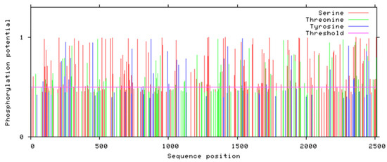
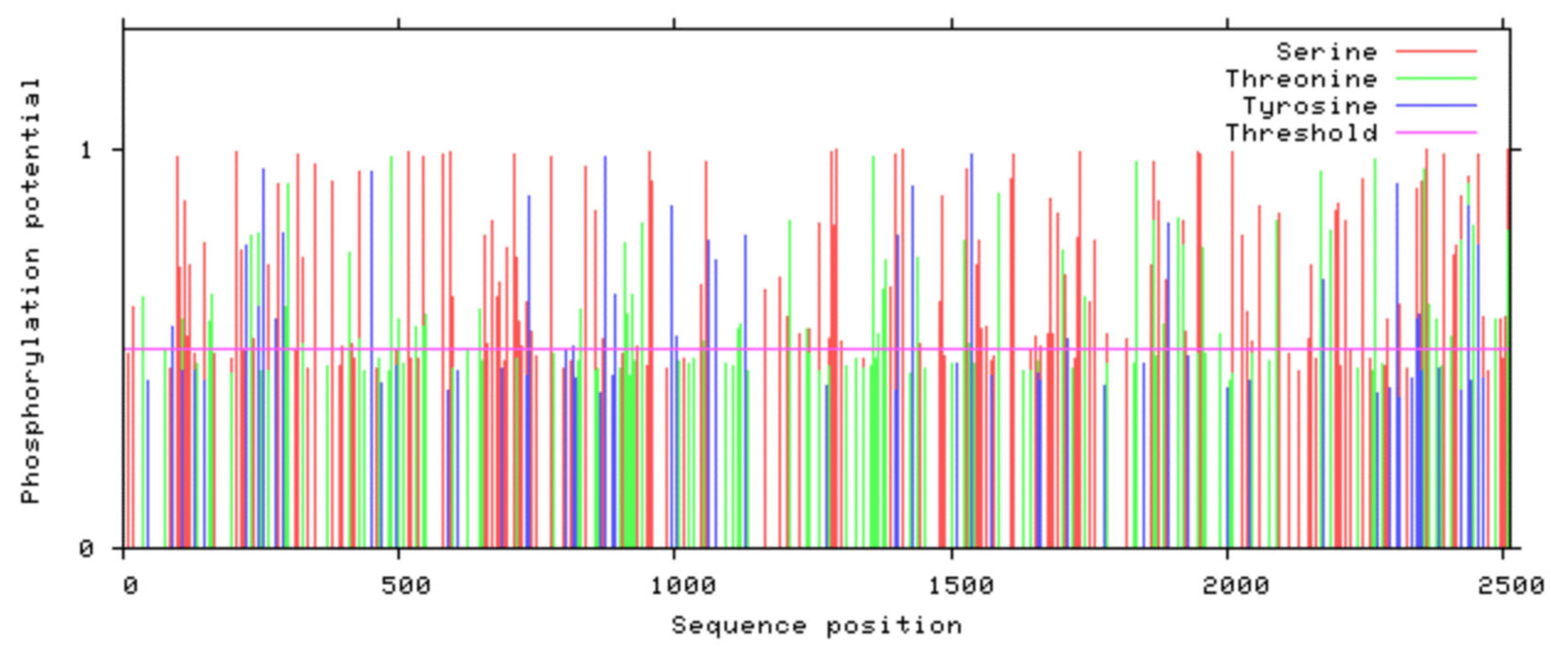
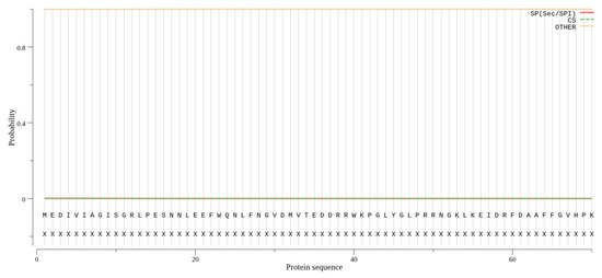
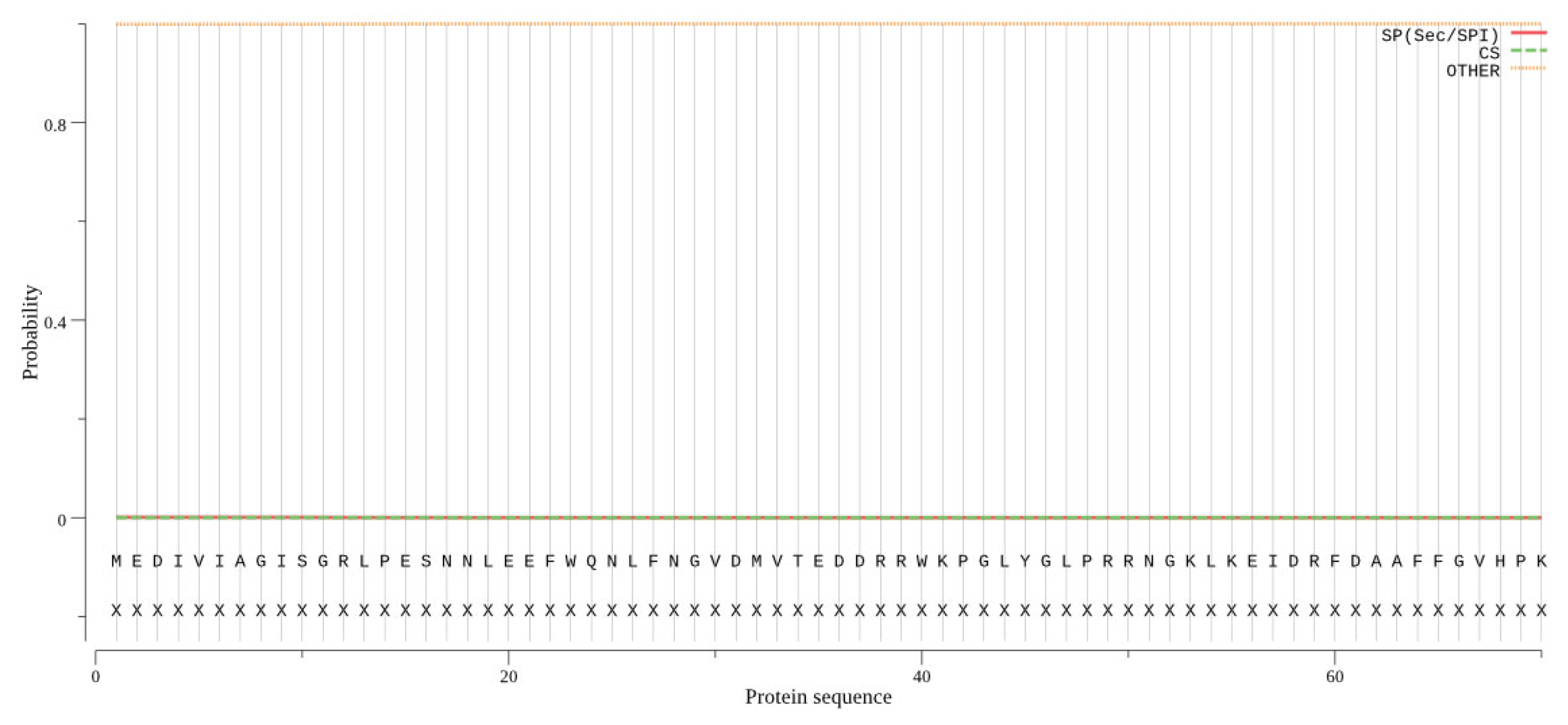
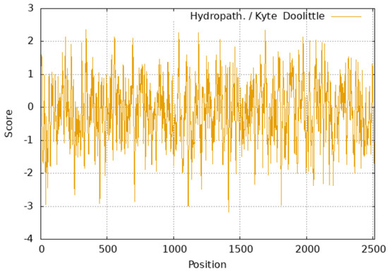
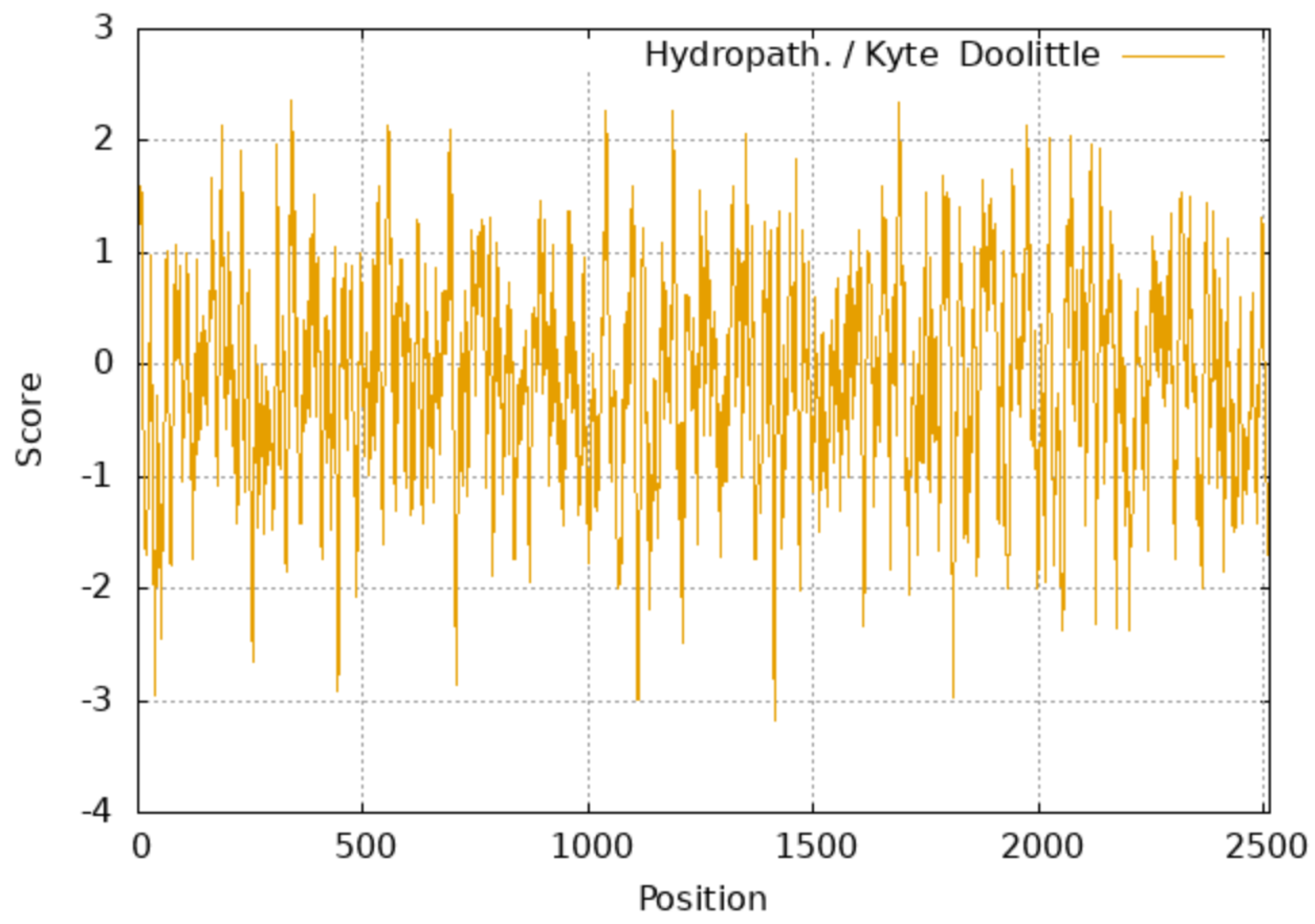
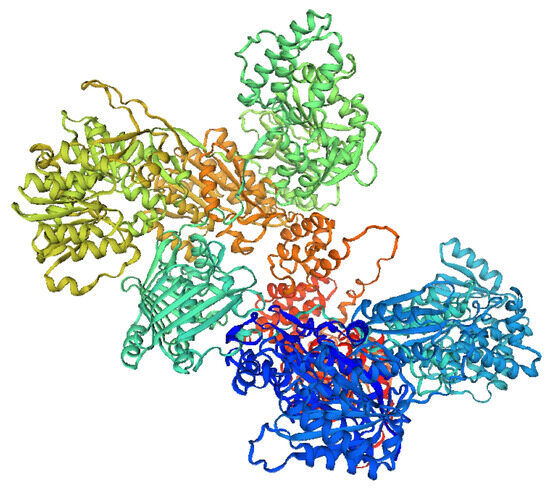
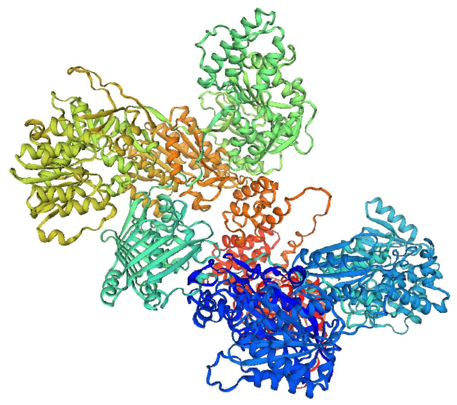
According to ProtParam analysis, the putative molecular weight of the protein encoded by the FASN gene was 274,727.61 Da (≈274.7 kDa), and its isoelectric point was 6.07. A total of five domains were predicted by InterProScan, and the domains spanning amino acids 1–409, 495–789, 827–1012, 1545–1860 and 1892–2070 corresponded to the PKS-KS, PKS-AT, PKS-DH, PKS-ER, and PKS-KR domains, respectively (Figure S1). The amino acid sequence of FASN contains 188 phosphorylation sites (Figure 1); however, no signal peptide was found by SignalP 5.0 Server (Figure 2). Hydrophilicity analysis showed that FASN is a hydrophilic protein, and the peak (positive) and the negative regions represent the hydrophobic and hydrophilic regions, respectively (Figure 3). TMHMM revealed that FASN is not a transmembrane protein. The secondary structure of FASN was found to contain 27.0% α-helix, 17.0% β-strand, and 8.0%, random coil, respectively, with the remaining 48.0% comprising other structural elements such as turns and loops. Moreover, the tertiary structure prediction of FASN protein was consistent with that of the secondary structure prediction, which mainly consisted of α-helix and β-strand (Figure 4).
Figure 1.
Phosphorylation site prediction of FASN protein.
Figure 2.
Signal peptide prediction of FASN protein.
Figure 3.
The hydrophilicity prediction of FASN protein.
Figure 4.
Tertiary structure prediction of FASN protein.
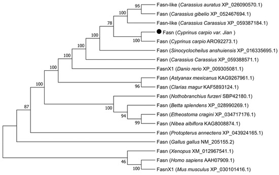
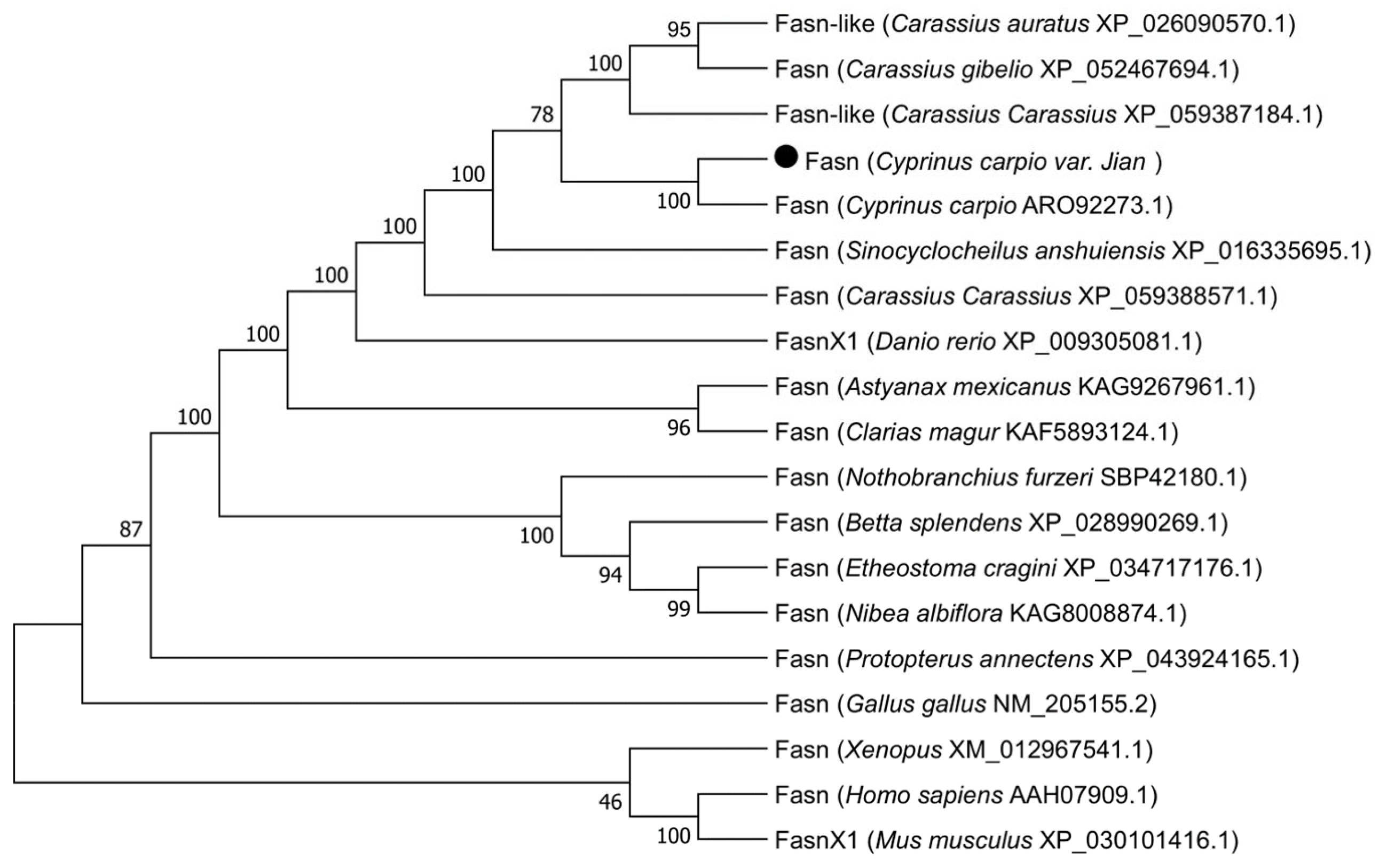
Blast searches showed that the similarity between the FASN protein sequence of Jian carp and Cyprinus carpio (ARO92273.1) was the highest (98.17%), followed by Carassius auratus (XP_026090570.1) (97.37%), Carassius gibelio (XP_052467694.1) (97.22%), Carassius carassius (XP_059387184.1) (97.18%), and Sinocyclocheilus anshuiensis (XP_016335695.1) (96.98%). The phylogenetic tree constructed on the basis of FASN amino acid sequence similarity (Figure 5) also showed that Jian carp first clustered with C. carpio, and then with C. Carassius, C. auratus and C. gibelio in one branch. The mammals are grouped into individual branch. These results revealed that the FASN protein is conserved in the evolutionary process of fish.
Figure 5.
Phylogenetic analysis based on FASN amino acid sequence similarity.
3.2. Tissue Expression Distribution of FASN
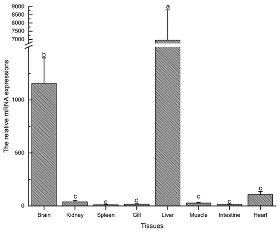
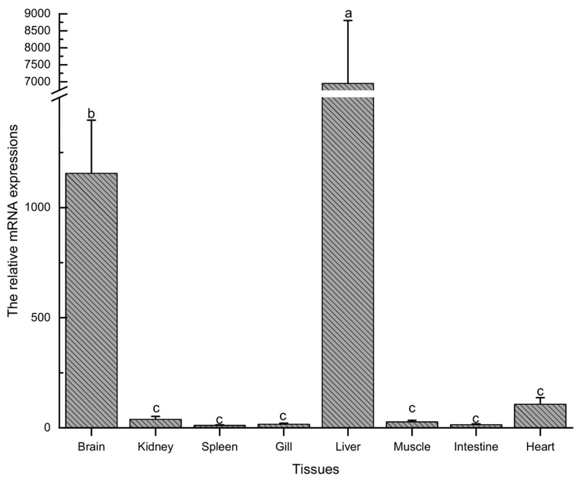
The expression levels of the FASN gene in the various tissues of Jian carp were examined by qRT-PCR. The results showed that the FASN mRNA was expressed in every tissue examined, including brain, kidney, spleen, gill, liver, muscle, intestine and heart (Figure 6). However, the FASN gene was mainly expressed in the liver and brain tissues. The liver had the highest expression, followed by the brain, and these levels were significantly different from those in other tested tissues (p < 0.05). No significant differences were observed among the kidney, spleen, gill, muscle, intestine and heart tissues (p > 0.05).
Figure 6.
Tissue distribution profiles of FASN mRNA associated with hepatic lipid and redox metabolism in Jian carp. Note that the Y-axis is broken (marked by//) to accommodate the large dynamic range of mRNA expression values. Different lowercase letters indicate significant differences among tissues (p < 0.05).
3.3. Altered Antioxidant Enzyme Activities and Elevated Lipid Peroxidation Following FASN Knockdown
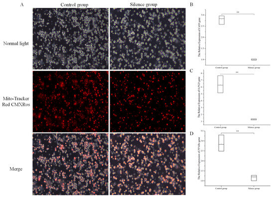
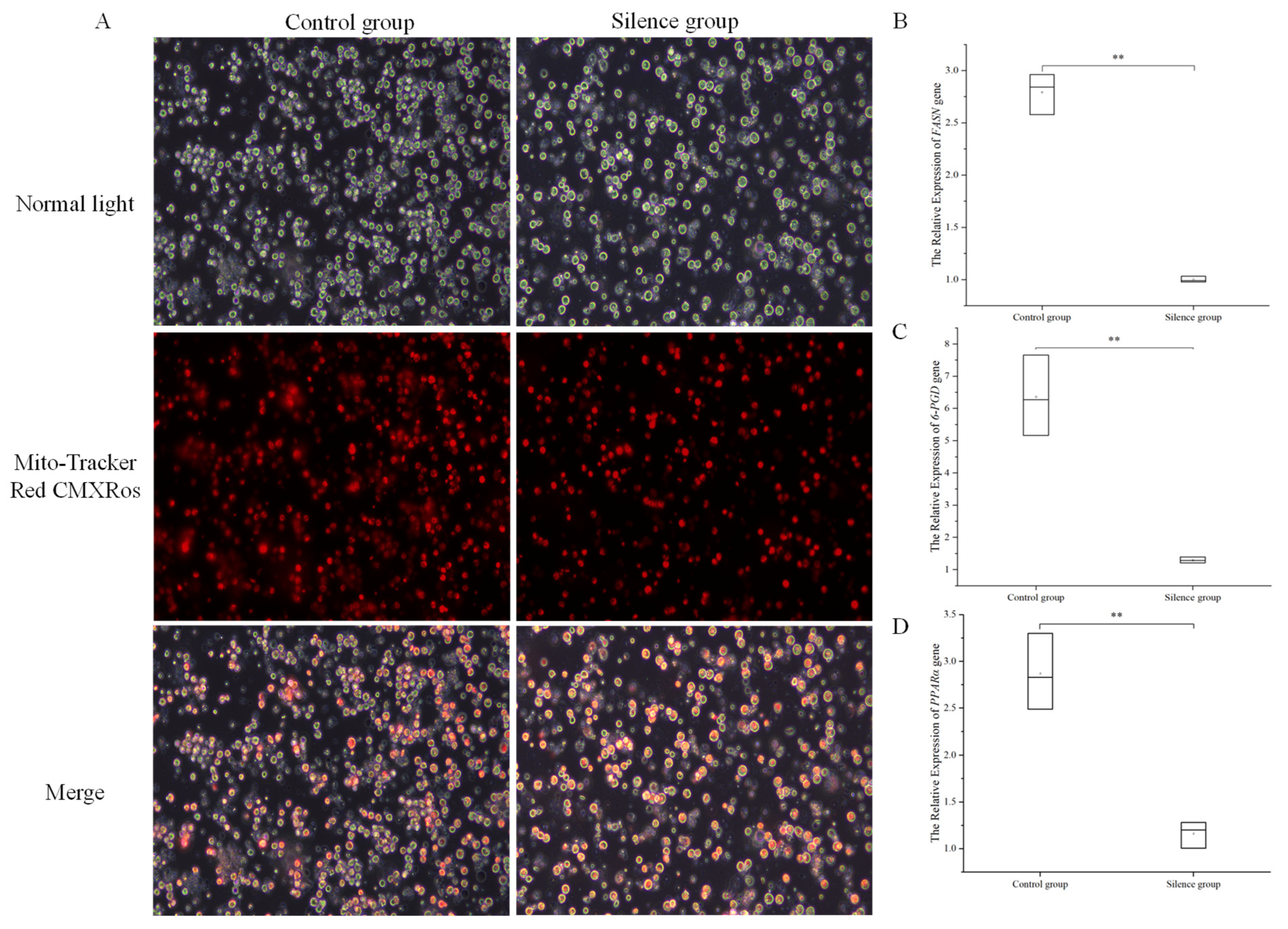
MitoTracker fluorescence staining demonstrated detectable mitochondrial membrane potential in both the control and FASN knockdown groups (Figure 7A). This observation makes severe mitochondrial dysfunction originating from transfection toxicity less likely, yet subtle transfection-associated cytotoxicity cannot be fully excluded based solely on this qualitative staining. FASN mRNA abundance was significantly reduced in si-FASN hepatocytes (p < 0.01, Figure 7B), confirming successful gene suppression. Transcripts of the pentose phosphate pathway gene 6-PGD were markedly downregulated (p < 0.01), while PPARα (the transcription factor driving fatty acid catabolism and compensatory stress response) was strongly upregulated (p < 0.01, Figure 7D). This reciprocal transcriptional reprogramming disrupts the cellular supply of reducing equivalents required for antioxidant enzyme function.
Figure 7.
Effects of FASN knockdown on mitochondrial activity (A) and expression of lipid metabolism-related genes (FASN, 6-PGD, PPARα) (B–D) in primary carp hepatocytes. siRNA-NC was used as the negative control group (control group). ** p < 0.01 compared with the control group.
3.4. Changes in Antioxidant Enzyme Activities and Elevated Lipid Peroxidation upon FASN Silencing
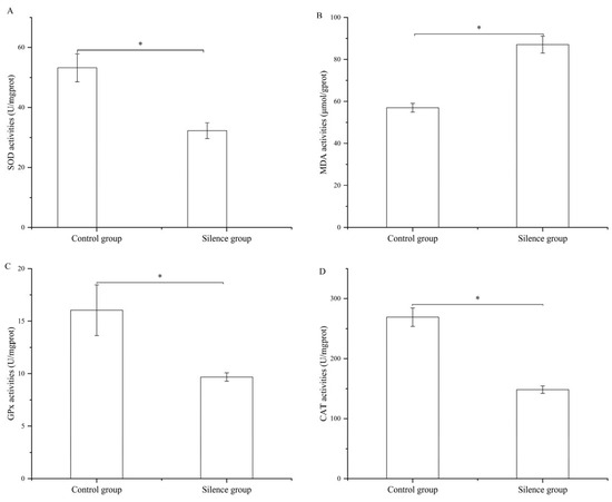
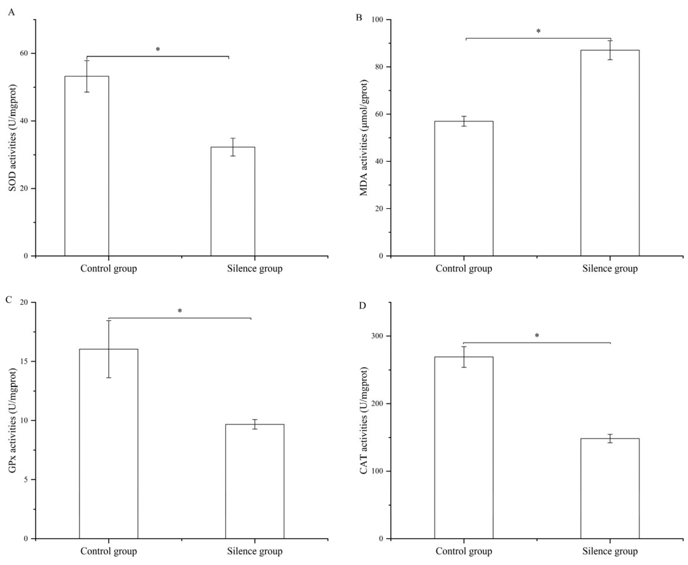
Compared with the negative control group, silencing FASN significantly reduced the activities of three core antioxidant enzymes (Figure 8): SOD activity decreased significantly (p < 0.05), weakening the superoxide anion scavenging capacity; GPx activity was markedly inhibited (p < 0.05), impairing glutathione-dependent hydroperoxide elimination; CAT activity declined sharply (p < 0.05), reducing cellular hydrogen peroxide clearance efficiency. Concomitantly, MDA content—the direct marker of lipid peroxidation and oxidative damage—was significantly elevated in FASN-deficient hepatocytes (p < 0.05). These data suggest that endogenous FASN expression is essential for maintaining basal antioxidant capacity and preventing ROS-mediated lipid oxidative injury in carp hepatocytes.
Figure 8.
Effects of FASN knockdown on core antioxidant enzyme activities and lipid peroxidation marker MDA in primary Jian carp hepatocytes. siRNA-NC was used as the negative control group (control group). The activities of SOD (A), MDA (B), GPx (C), and CAT content (D) were measured in control and FASN-siRNA groups. Data are presented as mean ± SE (n = 3). * p < 0.05, compared with the control group.
3.5. FASN Depletion Causes Global Reduction in Cellular Fatty Acid Substrates
GC-MS fatty acid profiling revealed a universal decline in all fatty acid classes upon FASN suppression (Table 2). The primary FASN catalytic products C14:0, C16:0, and C18:0 exhibited highly significant reductions (p < 0.01). Total saturated fatty acids (∑SFA), monounsaturated fatty acids (∑MUFA), and polyunsaturated fatty acids (∑PUFA) all decreased significantly. Long-chain n-3 and n-6 polyunsaturated fatty acids (including DHA) were also depleted; PUFA depletion further exacerbates oxidative susceptibility, as polyunsaturated lipids are highly susceptible to ROS-initiated peroxidation cascades.
Table 2.
Fatty acid composition and content in between control and silence group (g/100 g).
3.6. Transcriptome Sequencing Analysis
3.6.1. Sequencing and Assembly
To elucidate the regulatory role of the FASN gene in lipid metabolism of Jian carp, transcriptome sequencing was performed on primary hepatocytes following siRNA-mediated FASN knockdown (siRNA group) alongside the siRNA-NC negative-control group, with three biological replicates per group. After filtering, transcriptome sequencing generated 240,618,302 bp clean reads, with Q 20 and Q 30 values higher than 99.11% and 95.1%, respectively (Table 3). The GC content ranged from 44.62% to 47.41%. These results suggested that the transcriptome data obtained from the carp hepatocytes were reliable.
Table 3.
Summary of RNA-seq data.
3.6.2. Analysis of DEGs in the Silence and Control Group
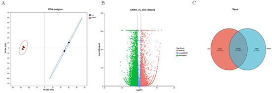
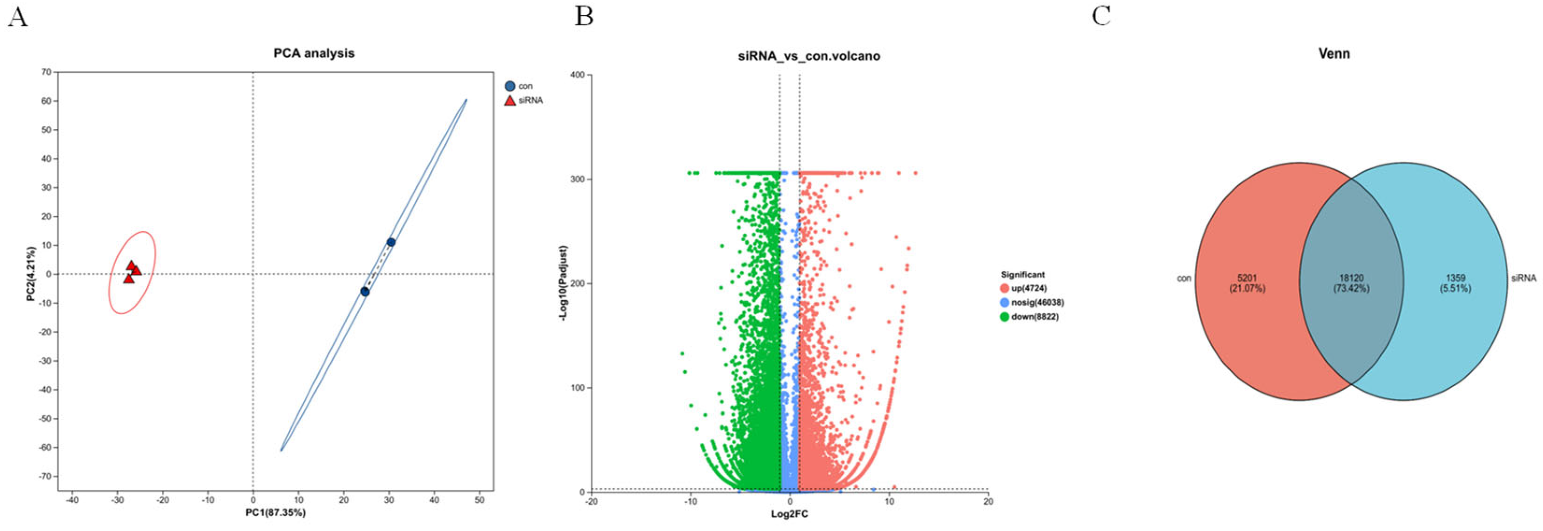
Principal component analysis (PCA) revealed a clear separation between the control and siRNA-treated groups along the PC1 axis, indicating substantial transcriptomic alterations induced by FASN silencing, with good clustering of replicates within each group (Figure 9A). Differential expression analysis identified a total of 13,546 significantly differentially expressed genes (DEGs) based on the criteria of |log2FC| ≥ 1 and FDR < 0.05, of which 4724 were upregulated and 8822 were downregulated in response to FASN knockdown (Figure 9B). Further analysis using a Venn diagram showed that 18,120 genes (73.42%) were co-expressed in both groups, while 1359 genes (5.51%) were uniquely expressed in the control group and 5201 genes (21.07%) were exclusively detected in the siRNA-treated group (Figure 9C). Collectively, these results demonstrate that FASN silencing profoundly reshapes the transcriptional landscape of carp hepatocytes.
Figure 9.
Transcriptomic analysis of primary hepatocytes following FASN knockdown. siRNA-NC was used as the negative control group (control group). (A) Principal component analysis (PCA) showing the separation between control (blue) and FASN-silenced (red) groups. (B) Volcano plot illustrating differentially expressed genes (DEGs) between the two groups. Red and blue dots represent significantly upregulated and downregulated genes, respectively (|log2FC| ≥ 1, FDR < 0.05). (C) Venn diagram displaying the number of uniquely and co-expressed genes in control and FASN-silenced groups.
3.6.3. Functional Enrichment Analysis
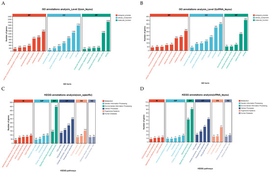
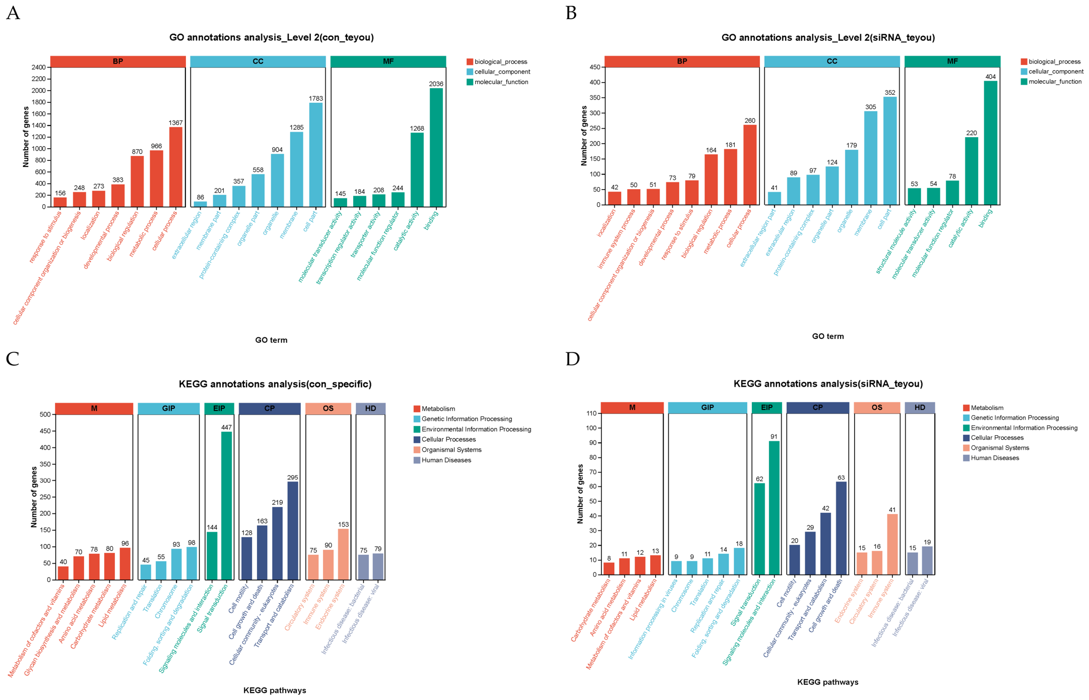
To further explore the functional implications of the uniquely expressed genes identified in the control group (5201 genes) and the FASN-silenced group (1359 genes), Gene Ontology (GO) and KEGG pathway enrichment analyses were performed. GO analysis at Level 2 revealed that genes uniquely expressed in the control group were primarily enriched in metabolic processes, cellular processes, cell part, organelle, binding, and catalytic activities, reflecting the active metabolic state of normal hepatocytes (Figure 10A). In contrast, genes specifically expressed in the FASN-silenced group were predominantly enriched in oxidative stress response, membrane lipid remodeling, and inflammatory biological processes (Figure 10B). KEGG pathway analysis further demonstrated distinct enrichment patterns: control-specific genes were most significantly enriched in Signal transduction, Transport and catabolism, and Cellular community—eukaryotes, with Lipid metabolism being the primary metabolism-related pathway (Figure 10C); siRNA-specific genes were most significantly enriched in Signaling molecules and interaction, Signal transduction, and Cell growth and death, with Lipid metabolism also emerging as the main metabolism-related pathway (Figure 10D).
Figure 10.
Functional enrichment analysis of group-specific genes in control and FASN-silenced hepatocytes. (A) GO enrichment analysis (Level 2) of genes uniquely expressed in the control group. (B) GO enrichment analysis (Level 2) of genes uniquely expressed in the FASN-silenced group. (C) KEGG pathway enrichment analysis of control-specific genes. (D) KEGG pathway enrichment analysis of siRNA-specific genes.
3.6.4. Identification of Oxidative Stress-Related Pathways and Genes Regulated by FASN Gene Silencing
Transcriptome analysis revealed that FASN silencing in primary carp hepatocytes induced significant transcriptional reprogramming, marked by the downregulation of key lipogenic gene ACACA, gluconeogenic gene PCK, and glyoxylate/dicarboxylate metabolism gene GRHPR. Concurrently, alterations were observed in genes governing fatty acid (LIPC), glycerophospholipid (LPCAT1), sphingolipid (ENPP6, GM2A, SGMS), and arachidonic acid (PTGES, PLA2G, ALOX12) metabolism, as well as protein palmitoylation (PPT). Moreover, robust activation of immune-related pathways was evident, including chemokine signaling (CCL25, CCR9, CCR7, CCL22), cytokine–cytokine receptor interaction (IL12A, IL1B, TNF, IL6), and the NF-kappa B pathway (TRAF2), alongside perturbation of cell cycle (SGOL1) (Table 4). These findings suggest that FASN knockdown not only disrupts multiple facets of hepatic lipid homeostasis but also triggers a coordinated inflammatory response in Jian carp.
Table 4.
Key candidate genes specifically expressed in the silencing group.
3.6.5. mRNA Expression Profiling of Lipid- and Inflammation-Associated Candidate Genes
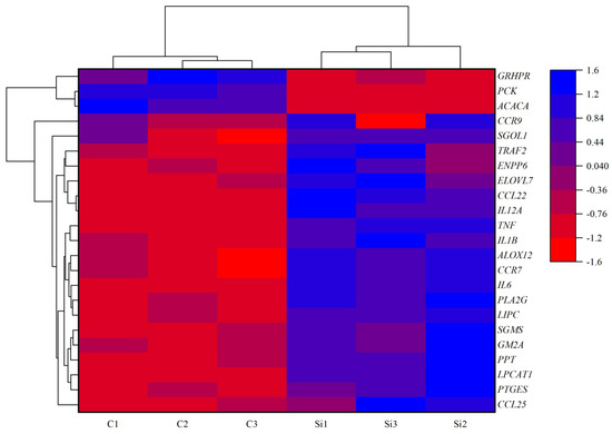
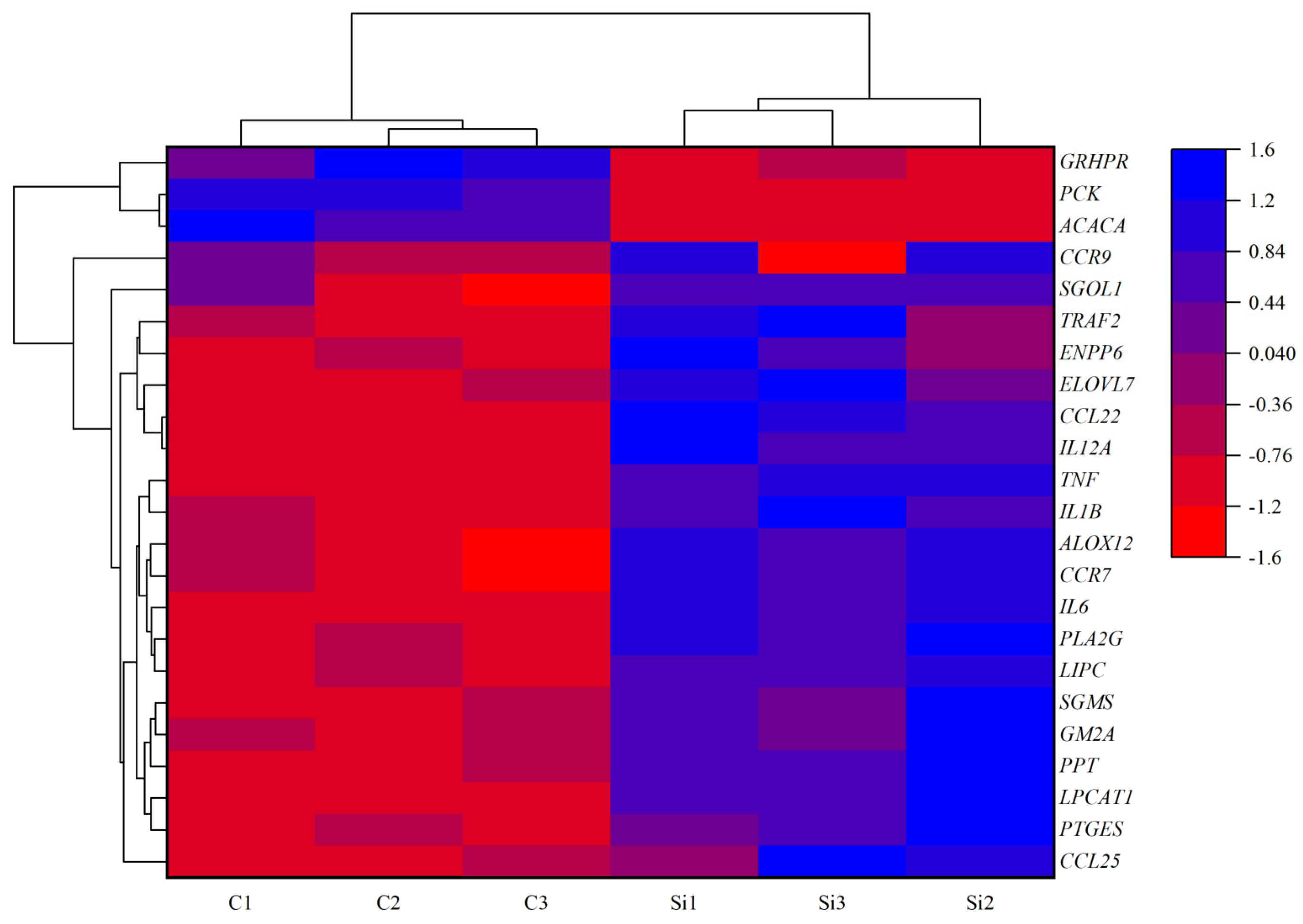
Based on the clustering analysis shown in Figure 11, the heatmap clearly segregates the control and FASN-silenced groups into two distinct clusters, with the three biological replicates of each condition displaying high intra-group consistency. In the knockdown samples (Si1–Si3), a sharp reduction in transcript abundance is observed for ACACA, PCK, and GRHPR relative to the controls (C1–C3). Conversely, the majority of the remaining genes, including those involved in inflammatory responses (CCL22, CCR7, CCR9, IL12A, IL1B, TNF, IL6), lipid-related pathways (ELOVL7, LIPC, ENPP6, SGMS, GM2A, ALOX12, PLA2G), as well as TRAF2 and SGOL1, show a pronounced increase in expression following FASN interference. This reciprocal expression pattern between metabolic repressors and immune-related activators further supports a functional shift from lipid/glucose metabolism toward inflammatory signaling upon FASN knockdown in carp hepatocytes.
Figure 11.
Heatmap clustering of differentially expressed genes in primary Jian carp hepatocytes following FASN silencing.
3.6.6. qRT-PCR Validation
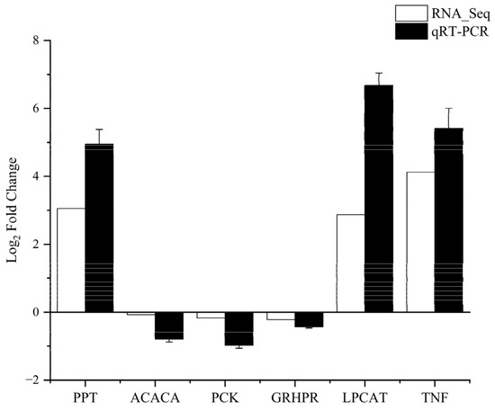
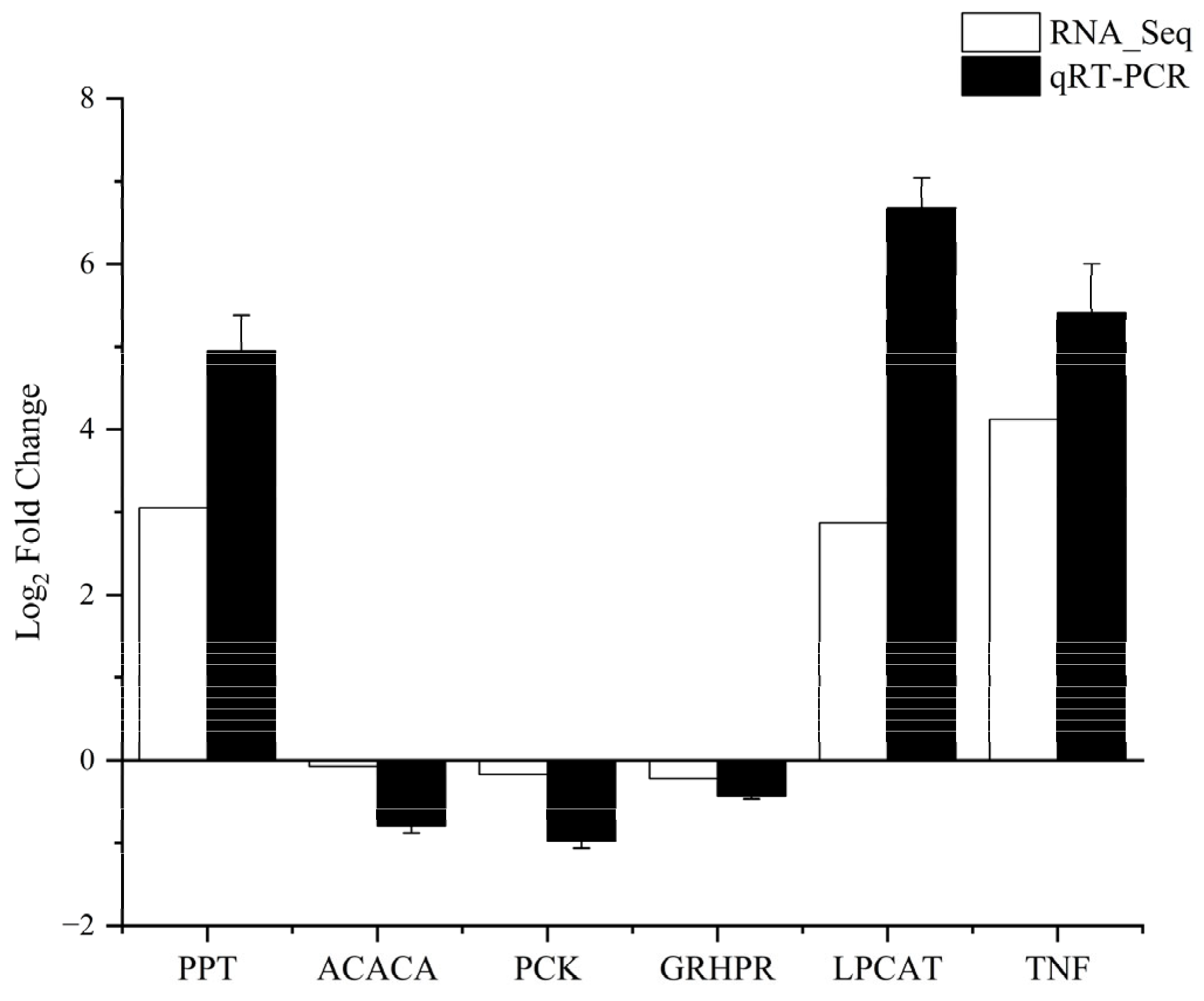
To validate the reliability of the expression of DEGs identified via RNA-seq, six genes were randomly selected for validation using RT-qPCR (Figure 12). The qPCR results exhibited consistent expression patterns with those obtained from RNA-seq analysis. This confirmed the accuracy and reliability of the RNA-seq experiments.
Figure 12.
Validation of transcriptome expression profiles by qRT-PCR in FASN silencing hepatocytes of primary Jian carp hepatocytes.
4. Discussion
4.1. Structural Conservation and Tissue Expression of FASN Lay a Molecular Basis for Its Hepatic Redox Regulatory Function
In the present study, we retrieved the complete coding sequence (CDS) of Jian carp FASN from transcriptomic resources and performed systematic bioinformatic characterization of its encoded protein, providing structural evidence supporting the intrinsic linkage to FASN catalytic function. The 9175 bp FASN transcript encodes a 2514-amino-acid hydrophilic polypeptide without transmembrane or secretory signal sequences, confirming that FASN functions as an intracellular cytoplasmic enzyme responsible for cytosolic de novo lipogenesis, consistent with the subcellular localization of mammalian FASN. NetPhos prediction identified a total of 188 serine, threonine and tyrosine phosphorylation sites distributed across the full-length sequence. Post-translational phosphorylation is well-documented to modulate the catalytic efficiency of vertebrate FASN in mammalian models [23]: altered phosphorylation status changes the substrate binding affinity of PKS domains, and could indirectly shape cellular antioxidant capacity [24]. This abundant phosphorylation modification profile suggests that Jian carp FASN activity can be dynamically tuned under oxidative or nutritional stress, serving as a reversible molecular switch balancing lipogenesis and redox homeostasis.
Phylogenetic analysis revealed that Jian carp FASN shares the highest sequence identity (98.17%) with common carp and clusters tightly with other cyprinid species, while forming independent clades with mammals, amphibians and birds [25]. This evolutionary conservation indicates that the function of FASN is maintained across bony fish lineages, implying universal relevance of the FASN–redox axis in teleost hepatic physiology [26]. Tissue expression profiling via qRT-PCR further reinforced the liver as the core functional organ of FASN: FASN mRNA was detected in all tested tissues, but exhibited significantly higher abundance in the liver, followed by the brain [27]. The teleost liver is a dual-functional hub coordinating both de novo fatty acid synthesis and phase I/II antioxidant detoxification [28]. Brain FASN expression may support myelin lipid synthesis for neuronal membranes [27], where balanced redox status is critical to prevent lipid peroxidation of polyunsaturated neuronal lipids [29]. By contrast, peripheral tissues including muscle, kidney and gill displayed negligible FASN transcription, implying that these tissues rely largely on circulating exogenous lipids rather than endogenous FASN-mediated synthesis [25]. Prior fish studies only described tissue expression patterns of FASN without linking transcriptional distribution to antioxidant regulation; our expression data fills this gap and provides tissue-specific evidence that hepatic FASN is the primary mediator of lipid-redox balance in Jian carp.
4.2. FASN Depletion Suppresses Core Antioxidant Enzyme Activity
The most definitive finding of this work, aligned with the core scope of antioxidants, is that FASN silencing resulted in altered antioxidant enzyme profiles and elevated lipid peroxidation markers in primary Jian carp hepatocytes. Our biochemical measurements demonstrated significant declines in three rate-limiting antioxidant enzymes (SOD, GPx, CAT) alongside a prominent elevation of MDA, the canonical biomarker of lipid peroxidation and oxidative damage. This phenotype can be fully explained by the reciprocal transcriptional changes of 6-PGD and PPARα observed upon FASN silencing.
6-PGD is the rate-limiting enzyme of the oxidative branch of the pentose phosphate pathway [30]. in vertebrate hepatocytes. In our FASN-knockdown model, transcriptional repression of 6-PGD was observed. Importantly, transcript down-regulation does not necessarily correspond to reduced protein abundance or reduced catalytic activity, because we have not performed a biochemical measurement of 6-PGD enzyme activity in the present work. Even so, the reduced 6-PGD transcript, together with decreased SOD, GPx and CAT enzyme activities and increased MDA content, raises the hypothesis that transcriptional perturbation of the pentose phosphate pathway may be associated with impaired hepatic antioxidant capacity following FASN silencing. Further biochemical work will be needed to confirm functional changes in the pentose phosphate pathway in carp hepatocytes under FASN deficiency.
Consistent with this metabolic feedback loop, we observed compensatory upregulation of PPARα, a master transcription factor governing fatty acid β-oxidation. In mammalian hepatocytes, PPARα activation accelerates lipid catabolism to compensate for blocked de novo lipogenesis [31], yet enhanced fatty acid oxidation concurrently elevates mitochondrial ROS generation [32], exacerbating oxidative burden when antioxidant systems are already compromised [33]. MitoTracker staining showed preserved mitochondrial membrane potential in both groups, which argues against severe mitochondrial damage induced by transfection. Nevertheless, we cannot completely rule out mild transfection-related cytotoxicity, as no quantitative cell-viability assay was performed in this study.
4.3. FASN Suppression Reshapes Cellular Fatty Acid Pools and Increases Lipid Peroxidation Susceptibility
GC-MS fatty acid profiling revealed global depletion of saturated (SFA), monounsaturated (MUFA) and polyunsaturated (PUFA) fatty acids following FASN silencing, with the greatest reduction detected in C14:0, C16:0 and C18:0—the primary saturated fatty acid products catalyzed by FASN. This result aligns with the well-established catalytic function of FASN, which exclusively synthesizes short-to-medium chain saturated acyl chains as the foundational precursors for all downstream long-chain and unsaturated lipid species via elongase and desaturase enzymes [34].
The universal reduction of all fatty acid classes creates two interconnected risks to cellular redox stability, both relevant to lipid peroxidation [35]. First, depleted SFA substrates limit the de novo synthesis of membrane phospholipids, altering the structural integrity of hepatocyte plasma and organelle membranes [36]. Membranes deficient in saturated acyl chains exhibit increased fluidity, facilitating the diffusion of reactive oxygen species (ROS) across lipid bilayers and amplifying oxidative damage to intracellular compartments [37]. Second, residual polyunsaturated fatty acids (n-3 and n-6 PUFAs including DHA and ARA) within FASN-deficient cells become more prone to ROS-initiated peroxidation [38,39]. PUFAs contain multiple double bonds that are highly susceptible to free radical attack, generating cytotoxic lipid hydroperoxides that further raise MDA concentrations [40], matching our biochemical measurement of elevated MDA content in the knockdown group.
Additionally, transcriptome enrichment identified significant upregulation of arachidonic acid (ARA) metabolic pathways upon FASN loss. ARA, a key n-6 PUFA, acts as the precursor for pro-inflammatory eicosanoids (prostaglandins, leukotrienes) synthesized under oxidative stress conditions [41]. These eicosanoids amplify inflammatory signaling cascades, creating a positive feedback loop between lipid peroxidation, ROS accumulation and innate immune activation—an immunometabolic crosstalk thoroughly characterized in mammalian oxidative stress models but rarely reported in teleost hepatocytes [42]. Collectively, our fatty acid and transcriptome results indicate that FASN participates in the maintenance of hepatocyte redox homeostasis largely through sustaining balanced cellular fatty-acid pools, which constrains spontaneous lipid peroxidation and the generation of pro-oxidant eicosanoids.
4.4. Transcriptomic Reprogramming Links FASN Deficiency to Oxidative Stress-Mediated Inflammatory Signaling
Whole-transcriptome RNA-seq captured profound transcriptional remodeling triggered by FASN suppression, with 13,546 total differentially expressed genes (DEGs) clearly segregated by PCA between control and silencing groups. GO and KEGG functional enrichment revealed a dramatic transcriptional shift away from basal lipogenic and anabolic metabolism toward oxidative stress response, membrane lipid remodeling, cytokine signaling and cell cycle pathways—consistent with the physiological phenotype of impaired antioxidant defense and inflammatory activation suggested by our biochemical measurements.
Key lipogenic genes such as ACACA (acetyl-CoA carboxylase α), the upstream rate-limiting enzyme of FASN-mediated lipogenesis [43], were significantly downregulated alongside FASN itself, forming a coordinated suppression of the entire de novo fatty acid synthesis module. Concurrently, genes governing gluconeogenesis and glyoxylate metabolism (PCK, GRHPR) were also repressed, indicating global attenuation of carbon reductive biosynthesis when FASN-dependent lipid production is blocked. In contrast, a large suite of genes associated with oxidative stress and inflammation was transcriptionally activated, including chemokine ligands/receptors (CCL22, CCR7, CCR9), pro-inflammatory cytokines (TNF, IL1B, IL6), and the core NF-κB signaling mediator TRAF2. The NF-κB pathway is the master transcriptional switch for oxidative stress-triggered innate immunity in mammals. In the current study, we only detected transcriptional upregulation of NF-κB-related signaling genes; NF-κB protein translocation and post-translational activation were not experimentally assayed. Based on mammalian literature, ROS accumulation and lipid peroxidation products promote IκB phosphorylation and NF-κB nuclear translocation, driving the transcription of multiple pro-inflammatory mediators [44,45]. Our heatmap clustering results visually confirmed this reciprocal expression pattern between lipogenic repressors and inflammatory activators, which was independently validated via qRT-PCR on six randomly selected DEGs, verifying the reliability of transcriptomic profiling.
We further screened 23 hub genes bridging FASN function, lipid turnover, arachidonic acid metabolism, sphingolipid signaling and inflammatory cascades. Sphingolipid metabolic genes (ENPP6, SGMS, GM2A) were also enriched in DEGs; sphingolipids are critical lipid second messengers that transduce oxidative stress signals to initiate apoptosis and inflammatory responses [46], representing another layer linking disrupted FASN lipid synthesis to redox imbalance [47]. Collectively, the transcriptomic landscape provides genome-wide molecular evidence that FASN loss initiates a multi-layered stress response combining lipid pool dysregulation, markers of oxidative damage and transcriptional activation of NF-κB-associated inflammatory pathways, pointing toward a potential lipid–redox–immune regulatory axis in teleost hepatocytes.
Several key limitations of this study require acknowledgment. First, FASN knockdown was only verified at the mRNA level; species-specific antibodies and standardized activity assays for Jian carp were unavailable to confirm reduced FASN protein and catalytic activity. Even so, consistent downstream changes in antioxidant markers, cellular fatty acids and transcriptomes support the functional suppression of FASN. Second, our conclusions on oxidative stress and NF-κB-mediated inflammation rely solely on biochemical indicators and transcriptomics, without direct measurements of ROS, GSH redox state, or NF-κB/inflammatory protein levels. Third, mitochondrial health was evaluated only via qualitative MitoTracker staining, lacking quantitative viability assays; mild transfection-related cytotoxicity cannot be fully ruled out despite no obvious severe mitochondrial damage. Fourth, GC-MS fatty acid data were normalized to cell pellet wet weight rather than cell count or total protein, leaving minor cell mass differences unaccounted for. Nonetheless, dramatic declines in FASN-direct products (C14:0, C16:0, C18:0) confirm impaired de novo lipogenesis rather than non-specific biomass artifacts. Fifth, only one siRNA duplex was applied for FASN silencing with a single scrambled negative control; we did not test multiple independent siRNAs or rescue overexpression experiments to exclude off-target transcriptional effects.
Additional experimental constraints exist. All assays were conducted on isolated primary hepatocytes, which fail to mimic the intact in vivo hepatic microenvironment and systemic signals of Jian carp. Sample replicates for cell and omics tests were limited, and our findings remain unvalidated in live fish. Future work will validate FASN suppression at the protein/enzymatic level, quantify cell viability and redox/inflammatory protein signals, and replicate these phenotypes using in vivo carp models.
5. Conclusions
In summary, this study systematically clarifies the redox regulatory functions of FASN in primary Jian carp hepatocytes via CDS characterization, tissue expression screening, siRNA knockdown, antioxidant biochemical assays, fatty acid profiling and transcriptomic sequencing. Loss of FASN function triggers three interconnected oxidative disorders: (1) repression of pentose phosphate pathway-related gene 6-PGD occurs alongside reduced antioxidant-enzyme activities and elevated lipid-peroxidation markers, indicative of oxidative damage; (2) global depletion of saturated and polyunsaturated fatty acids disrupts membrane lipid homeostasis and increases cellular vulnerability to ROS damage; (3) and genome-wide transcriptional reprogramming triggers metabolic remodeling together with up-regulation of gene sets associated with NF-κB—linked inflammatory signaling. This work identifies FASN as a vital upstream modulator of teleost hepatic redox balance and suggests the existence of a novel lipid–antioxidant–immune crosstalk axis in cyprinid fish. Our results provide theoretical support for developing nutritional strategies to improve intrinsic antioxidant capacity and stress tolerance in freshwater farmed carp.
Supplementary Materials
The following supporting information can be downloaded at: https://www.mdpi.com/article/10.3390/antiox15101237/s1.
Author Contributions
G.J. and Y.Z.: Writing—original draft, Methodology, Data curation. Q.W.: Writing—original draft, Methodology. J.S.: Writing—original draft, Methodology. W.F.: Writing—review and editing, Resources. Y.X.: Writing—review and editing, Resources. J.L.: Writing—review and editing, Resources. Y.T.: Writing—review and editing, Validation, Supervision, Resources, Funding acquisition. All authors have read and agreed to the published version of the manuscript.
Funding
This research was funded by the earmarked fund for Central Public-interest Scientific Institution Basal Research Fund, Freshwater Fisheries Research Center, CAFS (NO. 2025JBFR05), the earmarked fund for CARS (CARS-45), Central Public-interest Scientific Institution Basal Research Fund, CAFS [2023TD40].
Institutional Review Board Statement
This study was conducted in accordance with the institutional guidelines for animal care and use for scientific purposes, following review and approval by the Animal Ethics Committee of the Freshwater Fisheries Research Center, Chinese Academy of Fishery Sciences (Wuxi, China) (Protocol LAECFFRC-2023-06-12). The study was conducted in accordance with the local legislation and institutional requirements.
Informed Consent Statement
Not applicable.
Data Availability Statement
The original contributions presented in this study are included in the article and Supplementary Materials. The raw data supporting the conclusions of this article will be made available by the authors on request.
Conflicts of Interest
The authors declare no conflicts of interest.
References
- Raab, S.; Lefebvre, T. Fatty acid synthase, a “multi-FASette” enzyme. Med. Sci. 2022, 38, 445–452. [Google Scholar] [CrossRef] [Scilit]
- Weng, L.; Tang, W.S.; Wang, X.; Gong, Y.; Liu, C.; Hong, N.N.; Tao, Y.; Li, K.Z.; Liu, S.N.; Jiang, W. Surplus fatty acid synthesis increases oxidative stress in adipocytes and induces lipodystrophy. Nat. Commun. 2024, 15, 16. [Google Scholar] [CrossRef] [Scilit]
- Wei, H.; Weaver, Y.M.; Yang, C.D. Proteolytic activation of fatty acid synthase signals pan-stress resolution. Nat. Metab. 2024, 6, 113–126. [Google Scholar] [CrossRef] [Scilit]
- Huang, W.C.; Li, X.; Liu, J.; Lin, J.; Chung, L.W.K. Activation of androgen receptor, lipogenesis, and oxidative stress converged by SREBP-1 is responsible for regulating growth and progression of prostate cancer cells. Mol. Cancer Res. 2012, 10, 133–142. [Google Scholar] [CrossRef] [Scilit]
- Zhang, Y.; Hu, C.; Liu, J.; Jiang, S.; Liu, Q.; Zhou, X.; Liu, Y.; Song, F.; Luo, J. Tissue-specific metabolic remodeling to opposite fatty acid oxidation regulation in golden pompano identified by dual-tissue multi-omics. Aquaculture 2026, 623, 744174. [Google Scholar] [CrossRef] [Scilit]
- Li, C.; Han, B.; Sun, G.; Tian, H.; Wu, Z.; Li, C.; Meng, Y.; Ma, R. High lipid diet can keep liver health but cause muscle oxidative stress of triploid rainbow trout treated with acute hypoxic challenge. Aquac. Rep. 2025, 40, 102575. [Google Scholar] [CrossRef] [Scilit]
- Song, L.L.; Jia, Y.; Hai, T.Z.; Zi, K.L.; Nai, S.C. Effects of dietary carbohydrate and lipid levels on growth performance, feed utilization, body composition and non-specific immunity of large yellow croaker (Larimichthys crocea). Aquac. Nutr. 2019, 25, 995–1005. [Google Scholar] [CrossRef] [Scilit]
- Yuan, X.; Liu, W.; Jiang, G.; Dai, Y.; Desouky, H.E. Effect of replacing fish meal by yeast hydrolysate on growth and intestinal function of juvenile Jian carp (Cyprinus carpio var. jian). Indian J. Fish. 2021, 68, 95–104. [Google Scholar] [CrossRef] [Scilit]
- Zhao, Y.; Li, J.Y.; Yin, L.; Feng, L.; Liu, Y.; Jiang, W.D.; Wu, P.; Zhao, J.; Chen, D.F.; Zhou, X.Q. Effects of dietary glutamate supplementation on flesh quality, antioxidant defense and gene expression related to lipid metabolism and myogenic regulation in Jian carp (Cyprinus carpio var. jian). Aquaculture 2019, 502, 212–222. [Google Scholar] [CrossRef] [Scilit]
- Fang, W.; Leng, X.; Yun, B.; Wang, L.; Qian, X. Artificial diets affect glucose and lipid metabolism, antioxidant capacity, and inflammatory response in the muscle of mandarin fish (Siniperca chuatsi). Front. Mar. Sci. 2024, 11, 1445902. [Google Scholar] [CrossRef] [Scilit]
- Alotaibi, B.S.; Ahmed, A.A.A.; Khamis, T.; Osman, A.; Metwally, M.M.M.; Mohamed, A.A.R.; Alsubaie, N.; Bawahab, A.A.; Eskandrani, A.A.; Abd-Elhakim, Y.M. Restoration potential of a protein-hydrolysate fortified diet against neuro-behavioral alterations, neurotransmitter dysfunction, and FASN/FABP-2 expression in Oreochromis niloticus stocked under alkaline conditions. Aquac. Int. 2025, 33, 427. [Google Scholar] [CrossRef] [Scilit]
- Wang, W.; Su, S.; Dong, P.; Feng, W.; Li, J.; Zhang, C.; Tang, Y. Effects of Seasonal Photoperiod on Growth, Lipid Metabolism, and Antioxidant Response in the Huanghe Carp (Cyprinus carpio haematopterus). Fishes 2023, 8, 595. [Google Scholar] [CrossRef] [Scilit]
- Petersen, T.N.; Brunak, S.; Heijne, G.V.; Nielsen, H.H. SIGNALP 4.0: Discriminating signal peptides from transmembrane regions. Nat. Methods 2011, 8, 785–786. [Google Scholar] [CrossRef] [Scilit]
- Wilkins, M.R.; Gasteiger, E.; Bairoch, A.; Sanchez, J.C.; Hochstrasser, D.F. Protein Identification and Analysis Tools in the ExPASy Server. Methods Mol. Biol. 1999, 112, 531–552. [Google Scholar] [CrossRef] [Scilit]
- Kelley, L.A.; Mezulis, S.; Yates, C.M.; Wass, M.N.; Sternberg, M.J.E. The Phyre2 web portal for protein modeling, prediction and analysis. Nat. Protoc. 2015, 10, 845–858. [Google Scholar] [CrossRef] [Scilit]
- Higgins, D.G.; Jeanmougin, F.; Gibson, T.J.; Plewniak, F.; Thompson, J.D. The CLUSTAL_X Windows Interface: Flexible Strategies for Multiple Sequence Alignment Aided by Quality Analysis Tools; Oxford University Press: Oxford, UK, 1997; Volume 24. [Google Scholar] [CrossRef] [Scilit]
- Koichiro, T.; Joel, D.; Masatoshi, N.; Sudhir, K. MEGA4: Molecular Evolutionary Genetics Analysis (MEGA) Software Version 4.0. Mol. Biol. Evol. 2007, 24, 1596–1599. [Google Scholar] [CrossRef] [Scilit]
- Livak, K.J.; Schmittgen, T.D. Analysis of relative gene expression data using real-time quantitative PCR and the 2−ΔΔCT method. Methods 2001, 25, 402–408. [Google Scholar] [CrossRef] [Scilit]
- Le, Y.; Li, Y.; Jin, Y.; Jia, P.; Jia, K.; Yi, M. Establishment and characterization of a brain cell line from sea perch, Lateolabrax japonicus. In Vitro Cell. Dev. Biol.-Anim. 2017, 53, 834–840. [Google Scholar] [CrossRef] [Scilit]
- Jordi, F.M.; Lees, G.H.; Sloane, S. A simple method for the isolation and purification of total lipides from animal tissues. J. Biol. Chem. 1957, 226, 497–509. [Google Scholar] [CrossRef] [Scilit]
- Wang, W.; Luo, J.; Zhong, Y.; Lin, X.Z.; Shi, H.B.; Zhu, J.J.; Li, J.; Sun, Y.T.; Zhao, W.S. Goat liver X receptor α, molecular cloning, functional characterization and regulating fatty acid synthesis in epithelial cells of goat mammary glands. Gene 2012, 505, 114–120. [Google Scholar] [CrossRef] [Scilit]
- Kim, D.; Langmead, B.; Salzberg, S.L. HISAT: A fast spliced aligner with low memory requirements. Nat. Methods 2015, 12, 357–360. [Google Scholar] [CrossRef] [Scilit]
- Jin, Q.; Yuan, L.X.; Boulbes, D.; Baek, J.M.; Wang, Y.N.; Gomez-Cabello, D.; Hawke, D.H.; Yeung, S.C.; Lee, M.H.; Hortobagyi, G.N. Fatty acid synthase phosphorylation: A novel therapeutic target in HER2-overexpressing breast cancer cells. Breast Cancer Res. 2010, 12, R96. [Google Scholar] [CrossRef] [Scilit]
- Liu, H.; Liu, J.Y.; Wu, X.; Zhang, J.T. Biochemistry, molecular biology, and pharmacology of fatty acid synthase, an emerging therapeutic target and diagnosis/prognosis marker. Int. J. Biochem. Mol. Biol. 2010, 1, 69–89. [Google Scholar]
- Zhang, W.; Tan, B.; Liu, K.; Dong, X.; Yang, Q.; Chi, S.; Liu, H.; Zhang, S.; Wang, H. Effects of different dietary lipids on growth, body composition and lipid metabolism-related enzymes and genes in juvenile largemouth bass, Micropterus salmoides. Aquac. Nutr. 2019, 25, 1318–1326. [Google Scholar] [CrossRef] [Scilit]
- Bernardo, A.; Greco, A.; Levi, G.; Minghetti, L. Differential lipid peroxidation, mn superoxide, and bcl-2 expression contribute to the maturation-dependent vulnerability of oligodendrocytes to oxidative stress. J. Neuropathol. Exp. Neurol. 2003, 62, 509–519. [Google Scholar] [CrossRef] [Scilit]
- Dimas, P.; Montani, L.; Pereira, J.A.; Moreno, D.; Troetzmueller, M.; Gerber, J.; Semenkowich, C.F.; Koefeler, H.C.; Suter, U. CNS myelination and remyelination depend on fatty acid synthesis by oligodendrocytes. eLife 2019, 8, 44702. [Google Scholar] [CrossRef] [Scilit]
- Panserat, S.; Kolditz, C.; Richard, N.; Plagnes-Juan, E.; Piumi, F.; Esquerre, D.; Medale, F.; Corraze, G.; Kaushik, S. Hepatic gene expression profiles in juvenile rainbow trout (Oncorhynchus mykiss) fed fishmeal or fish oil-free diets. Br. J. Nutr. 2008, 100, 953–967. [Google Scholar] [CrossRef] [Scilit]
- Dong, X.; Jiang, D.; Chen, S.; Tan, J.; Zhao, J.; Liu, X.; Lu, Z.; Yeung, K.W.K.; Liao, Y.; Liu, X. KNC nanozyme repairs hypoxia ischemia brain damage through ALOX12 mediated lipid peroxidation inhibition. Bioact. Mater. 2025, 54, 531–548. [Google Scholar] [CrossRef] [Scilit]
- Juan, B.B.; Juan, P.; García-Salguero, L.; Manuel, D.L.H.; José, A.L. Kinetic behavior and protein expression of hepatic NADPH-production systems during development of rainbow trout (Oncorhynchus mykiss) 1. Aquaculture 1999, 179, 375–385. [Google Scholar] [CrossRef] [Scilit]
- Chakravarthy, M.V.; Pan, Z.J.; Zhu, Y.M.; Tordjman, K.; Schneider, J.G.; Coleman, T.; Turk, J.; Semenkovich, C.F. New” hepatic fat activates PPARα to maintain glucose, lipid, and cholesterol homeostasis. Cell Metab. 2005, 1, 309–322. [Google Scholar] [CrossRef] [Scilit]
- Todisco, S.; Santarsiero, A.; Convertini, P.; De Stefano, G.; Gilio, M.; Iacobazzi, V.; Infantino, V. PPAR Alpha as a Metabolic Modulator of the Liver: Role in the Pathogenesis of Nonalcoholic Steatohepatitis (NASH). Biology 2022, 11, 792. [Google Scholar] [CrossRef] [Scilit]
- Matsukawa, T.; Yagi, T.; Uchida, T.; Sakai, M.; Mitsushima, M.; Naganuma, T.; Yano, H.; Inaba, Y.; Inoue, H.; Yanagida, K. Hepatic FASN deficiency differentially affects nonalcoholic fatty liver disease and diabetes in mouse obesity models. JCI Insight 2023, 8, e161282. [Google Scholar] [CrossRef] [Scilit]
- Chavin, K.D.; Fiorini, R.N.; Shafizadeh, S.; Cheng, G.; Wan, C.; Evans, Z.; Rodwell, D.; Polito, C.; Haines, J.K.; Baillie, G.M. Fatty Acid Synthase Blockade Protects Steatotic Livers from Warm Ischemia Reperfusion Injury and Transplantation. Orig. Artic. Basic Sci. 2004, 4, 1440–1447. [Google Scholar] [CrossRef] [Scilit]
- Jiang, X.; Fang, G.; Li, W.; Liu, Y.; Chen, G.; Perea, S.E.; Perera, Y.; Ma, R.; Hu, X.; Long, X. FASN at the crossroads of tumor metabolism, immune evasion, and therapy resistance: Mechanistic insights and therapeutic opportunities. Crit. Rev. Oncol. Hematol. 2026, 220, 105155. [Google Scholar] [CrossRef] [Scilit]
- Almaida-Pagan, P.F.; de Costa, J.; Mendiola, P.; Tocher, D.R. Changes in tissue and mitochondrial membrane composition during rapid growth, maturation and aging in rainbow trout, Oncorhynchus mykiss. Comp. Biochem. Physiol. B Biochem. Mol. Biol. 2012, 161, 404–412. [Google Scholar] [CrossRef] [Scilit]
- Grim, J.M.; Hyndman, K.A.; Kriska, T.; Girotti, A.W.; Crockett, E.L. Relationship between oxidizable fatty acid content and level of antioxidant glutathione peroxidases in marine fish. J. Exp. Biol. 2011, 214, 3751–3759. [Google Scholar] [CrossRef] [Scilit]
- Cheng, Q.; Chen, M.; Chen, X.; Xie, Y.; Du, Y.; Wu, H. Fatty Acid Synthase Is a Critical Repressor of Ferroptosis in Rheumatoid Arthritis. Arthritis Rheumatol. 2024, 75, 3479–3481. [Google Scholar]
- Watkins, P.A. Fatty acid activation. Prog. Lipid Res. 1997, 36, 55–83. [Google Scholar] [CrossRef] [Scilit]
- Li, Y.; Yang, W.; Zheng, Y.; Dai, W.; Ji, J.; Wu, L.; Cheng, Z.; Zhang, J.; Li, J.; Xu, X. Targeting fatty acid synthase modulates sensitivity of hepatocellular carcinoma to sorafenib via ferroptosis. J. Exp. Clin. Cancer Res. 2023, 42, 6. [Google Scholar] [CrossRef] [Scilit]
- O’Donnell, V.B.; Murphy, R.C. New families of bioactive oxidized phospholipids generated by immune cells: Identification and signaling actions. Blood 2012, 120, 1985–1992. [Google Scholar] [CrossRef] [Scilit]
- Higdon, A.; Diers, A.R.; Oh, J.Y.; Landar, A.; Darley-Usmar, V.M. Cell signalling by reactive lipid species: New concepts and molecular mechanisms. Biochem. J. 2012, 442, 453–464. [Google Scholar] [CrossRef] [Scilit]
- Hudson, N.J.; Reverter, A.; Griffiths, W.J.; Yutuc, E.; Greenwood, P.L. Gene expression identifies metabolic and functional differences between intramuscular and subcutaneous adipocytes in cattle. BMC Genom. 2020, 21, 77. [Google Scholar] [CrossRef] [Scilit]
- Markopoulos, G.S.; Roupakia, E.; Tokamani, M.; Alabasi, G.; Sandaltzopoulos, R.; Marcu, K.B.; Kolettas, E. Roles of NF-κB Signaling in the Regulation of miRNAs Impacting on Inflammation in Cancer. Biomedicines 2018, 6, 40. [Google Scholar] [CrossRef] [Scilit]
- Ji, L.L. Nuclear factor κB signaling revisited: Its role in skeletal muscle and exercise. Free Radic. Biol. Med. 2025, 232, 158–170. [Google Scholar] [CrossRef] [Scilit]
- Won, J.S.; Singh, I. Sphingolipid signaling and redox regulation. Free Radic. Biol. Med. 2006, 40, 1875–1888. [Google Scholar] [CrossRef] [Scilit]
- Bartolacci, C.; Andreani, C.; Vale, G.; Berto, S.; Melegari, M.; Crouch, A.C.; Baluya, D.L.; Kemble, G.; Hodges, K.; Starrett, J. Targeting de novo lipogenesis and the Lands cycle induces ferroptosis in KRAS-mutant lung cancer. Nat. Commun. 2022, 13, 4327. [Google Scholar] [CrossRef] [Scilit]

Disclaimer/Publisher’s Note: The statements, opinions and data contained in all publications are solely those of the individual author(s) and contributor(s) and not of MDPI and/or the editor(s). MDPI and/or the editor(s) disclaim responsibility for any injury to people or property resulting from any ideas, methods, instructions or products referred to in the content. |
© 2026 by the authors. Licensee MDPI, Basel, Switzerland. This article is an open access article distributed under the terms and conditions of the Creative Commons Attribution (CC BY) license.