


Abstract
This study evaluated the effects of blueberry extract and resistance training on lung and right ventricle (RV) function and structure in severe experimental pulmonary arterial hypertension (PAH). PAH was induced in male Wistar rats by a single intraperitoneal injection of monocrotaline (60 mg/kg). One day later, the animals started receiving blueberry extract (100 mg/kg daily by gavage) and underwent a three-week moderate-intensity resistance exercise protocol (ladder climbing—15 climbs, a 60 s interval, a 60% load, one session/day, 5 days/wk). After these treatments and echocardiographic assessment, the animals were euthanized, and lung and RV samples were collected for mechanical, morphological, biochemical, and molecular analyses. Both blueberry extract and resistance training attenuated RV dysfunction and pulmonary artery resistance observed over the development of severe PAH. In addition, single RV myocyte mechanical function was maintained in treated rats. Moreover, the applied treatments mitigated oxidative imbalance and adverse structural remodeling in both the lung and RV. Furthermore, the interventions promoted partial or complete preservation of calcium regulatory protein expression. In conclusion, blueberry extract and resistance training, either alone or in combination, alleviate cardiopulmonary oxidative imbalance and adverse structural remodeling, sustain RV myocyte contractility and the expression of calcium regulatory proteins, and thus conserve pulmonary and RV function in severe PAH induced by monocrotaline.
1. Introduction
Pulmonary arterial hypertension (PAH) is a rare and progressive disease characterized by sustained increases in pulmonary vascular pressure and pulmonary artery resistance [1,2,3]. This chronic process imposes a progressive overload on the right ventricle (RV), ultimately leading to hypertrophy, dysfunction, failure, and death [1,4,5]. Oxidative stress and inflammation are closely involved in the progression of PAH [6,7,8,9]. In this context, excessive ROS production associated with inadequate antioxidant defenses may promote cellular and tissue injury, contributing to cardiopulmonary dysfunction [10,11,12,13,14]. Within the pulmonary vasculature, oxidative stress in PAH is driven by increased ROS production from multiple sources, including NADPH oxidases, mitochondrial dysfunction, and uncoupled endothelial nitric oxide synthase (eNOS), alongside a reduction in endogenous antioxidant capacity. Elevated ROS levels can decrease nitric oxide availability and favor pulmonary vasoconstriction. At the same time, redox-sensitive signaling pathways may stimulate vascular smooth muscle cell proliferation, inflammatory responses, and extracellular matrix accumulation, thereby promoting pulmonary vascular remodeling [13,14,15,16]. In addition, sustained RV pressure overload may further promote oxidative stress in cardiomyocytes, favoring structural remodeling and oxidative alterations in Ca2+-handling proteins that contribute to impaired RV contractile function [10,11,12]. The concomitant inflammatory response can further amplify these alterations, worsening pulmonary vascular and RV dysfunction [15,16]. These complications contribute to the chronic fatigue and impaired quality of life commonly observed in patients with PAH [17].
Despite advancements in conventional treatments for PAH, there are limitations in preventing disease progression, and high mortality and morbidity persist [18,19]. Therefore, non-pharmacological and combined strategies are required to lessen adverse PAH outcomes, like pulmonary artery pressure and RV overload. In this sense, physical exercise [20,21,22,23] and natural antioxidants [24,25,26] have potential as adjuvant components in PAH management. Recent evidence provided by our research group [27,28,29,30,31,32] and elsewhere [9,33,34] indicates that aerobic, resistance, and combined exercise training can preserve cardiac and pulmonary function and structure by counteracting oxidative stress and inflammation, resulting in improved exercise capacity and survival in rats with MCT-induced PAH. Likewise, blueberry extract, which is rich in polyphenols and anthocyanins, can protect cardiac and pulmonary tissues by preventing oxidative imbalance and attenuating functional and structural damage in this model [35,36,37].
Given the therapeutic potential of combining non-pharmacological strategies for severe PAH, previous studies have investigated the effects of exercise training or natural antioxidant interventions separately. However, evidence regarding resistance training and blueberry-derived antioxidants, particularly when assessed alone and in combination through an integrated evaluation of pulmonary and right-ventricular adaptations, remains limited. Therefore, the present study evaluated the effects of blueberry extract and resistance training, alone or combined, on lung and RV structure and function, cardiomyocyte contractility, oxidative stress, and the expression of Ca2+-handling proteins in a model of severe MCT-induced PAH.
2. Materials and Methods
2.1. Experimental Design
Male Wistar rats (~200 g, 6 weeks of age) were randomly distributed into five experimental groups: sedentary control (SC, n = 7), sedentary hypertensive (SH, n = 7), sedentary hypertensive blueberry (SHB, n = 7), exercise hypertensive (EH, n = 7), and exercise hypertensive blueberry (EHB, n = 7). Animals were housed in transparent polycarbonate cages (4 per cage) under controlled environmental conditions (23 ± 1 °C, ~60% relative humidity, 12/12 h light/dark cycle), with free access to water and standard rodent chow. All experimental procedures were conducted in accordance with the Ethical Principles in Animal Experimentation and were approved by the Ethics Committee on the Use of Animals of the Federal University of Viçosa (protocol no. 11/2022).
2.2. PAH Induction
PAH was induced in the hypertensive groups by a single intraperitoneal administration of monocrotaline (MCT; 60 mg/kg; Sigma-Aldrich, St. Louis, MO, USA), diluted in saline solution (140 mM NaCl, pH 7.4) [38]. Animals assigned to the control group received an equivalent volume of the same saline solution without MCT.
2.3. Administration and Phenolic Profile of the Blueberry Extract
Throughout the experimental period, animals in the SHB and EHB groups received a daily oral dose of blueberry extract (100 mg/kg body weight) by gavage at 7:00 a.m. The dose was established based on our previous experimental protocols, in which 100 mg/kg exerted beneficial effects in MCT-induced severe PAH [35,36,39]. In the original protocol, this dose was selected considering the total phenolic content of the extract and the average dietary intake of polyphenols reported for populations in North and South America [39].
The blueberry extract was prepared as previously described [39]. Briefly, fresh blueberries were extracted in 50% ethanol, subjected to mechanical extraction followed by sonication in an ice bath to minimize compound degradation, vacuum filtered, and concentrated using a rotary evaporator. Following preparation, the extract was divided into aliquots and stored protected from light at −80 °C until use. A fresh working solution was prepared daily immediately before oral administration.
The blueberry extract used in the present study was previously characterized in detail [39]. The extract contained 38.89 mg gallic acid equivalents (GAE) of total phenolic compounds per gram. Among the phenolic compounds analyzed, the anthocyanidins cyanidin (219.21 µg/mL) and malvidin (128.68 µg/mL) were detected as the predominant compounds, whereas rutin, gallic acid, catechin, and epicatechin were not detected. Further details regarding the extraction procedure and chromatographic characterization are available in our previous study [39].
2.4. Resistance Training Protocol
All animals were adapted to a resistance training protocol (i.e., Ladder climb), as described by Hornberger and Farrar [40]. During a one-week familiarization period, the rats were exposed to the protocol, which consisted of climbing an 80–inclined grid with devices attached to their tails, but without any load. To encourage climbing, a gentle tap was applied to the animals’ tails, and after each climb, they rested for 60 s in a cage (20 cm × 20 cm × 20 cm). This familiarization procedure was performed five days per week. Following the adaptation period, maximum load capacity was assessed before PAH induction and again on days 14 and 21 after induction. The test started with a load corresponding to 75% of body weight, with 30 g increments added on successive climbs. Each attempt was separated by a 120 s recovery interval, and the test continued until the animal was unable to complete the climb.
Resistance training began 24 h after MCT administration and was performed on a vertical ladder adapted for rats. Training loads were individualized according to the maximum load tests, and sessions were conducted five days per week (Monday to Friday) for approximately three weeks. Each session comprised 15 climbs separated by 60 s recovery intervals [41]. Exercise intensity was set at 60% of the maximum load determined during testing, corresponding to a moderate-intensity protocol previously shown by our group to produce beneficial effects in rats with severe PAH [29,31].
2.5. Echocardiography
Echocardiographic assessments were conducted on day 23 following MCT administration. Animals were anesthetized with isoflurane (3% induction, 1.5% maintenance; oxygen flow: 1 L/min) and positioned in lateral decubitus. Images were acquired in M-mode (120 frames/s) using a MyLab™30 ultrasound system (Esaote, Genoa, Italy) equipped with a 10 MHz transducers, with the scan speed adjusted to 200 mm/s based on heart rate. Pulmonary artery flow was assessed by pulsed Doppler to calculate the RV acceleration time (AT) and ejection time (ET), with the AT/ET ratio serving as an indirect marker of pulmonary vascular resistance. RV systolic function was evaluated through tricuspid annular plane systolic excursion (TAPSE), obtained by placing the M-mode cursor at the lateral tricuspid annulus and measuring the systolic displacement from end-diastole to end-systole. All images were acquired in accordance with the American Society of Echocardiography guidelines and stored for subsequent analysis [42].
2.6. Sample Collection
Twenty-four days after MCT administration, animals from all experimental groups (SC, SH, SHB, EH, and EHB) were euthanized by decapitation. This time point was selected according to previous reports [27,30,32], as it represents the average survival period of rats developing PAH after receiving a 60 mg/kg dose of MCT.
2.7. Lung and RV Oxidative Stress Biomarkers
2.7.1. Tissue Preparation
Frozen lung and right ventricle (RV) samples (100 mg each) were homogenized in 1 mL of phosphate buffer (pH 7.4) using a Tissue Master 125 homogenizer (OMNI International, Kennesaw, GA, USA) and centrifuged at 10,000× g (12,000 rpm) for 10 min at 4 °C. Supernatants were collected to assess the enzymatic activities of superoxide dismutase (SOD), catalase (CAT), and glutathione S-transferase (GST), as well as the levels of malondialdehyde (MDA), nitric oxide (NO), and total protein (TP). The pellets were reserved for carbonyl protein (CP) quantification. Measurements were performed using either an ELISA microplate reader (Multiskan GO, Thermo Fisher Scientific, Waltham, MA, USA) or a UV-visible spectrophotometer (UVmini-1240, Shimadzu Corporation, Kyoto, Japan).
2.7.2. Superoxide Dismutase Activity
SOD activity was quantified following the method of Dieterich et al. [43], based on the inhibition of pyrogallol autoxidation. For the assay, 30 μL of sample was combined with 99 μL of potassium phosphate buffer (5 mmol/L, pH 8.0) and 15 μL of pyrogallol (100 μmol/L). Absorbance was recorded at 570 nm. Enzymatic activity was normalized to protein content and expressed as U/mg of protein, with one unit defined as the activity required to inhibit pyrogallol autoxidation by 50%. Blank and standard preparations were analyzed in duplicate without and with pyrogallol, respectively.
2.7.3. Catalase Activity
The CAT activity was measured based on an adaptation of the Hadwan method [44], using hydrogen peroxide (H2O2) as a substrate. For this, 100 μL of H2O2 (20 mmol/L) was pipetted into 5 μL of the sample. After 3 min, 150 μL of ammonium molybdate (32.4 mmol/L) was added to stop the reaction. Blank samples were prepared by replacing H2O2 with sodium and potassium phosphate buffer (50 mmol/L, pH 7.0). The reading was taken at 374 nm using a spectrophotometer. To calculate the CAT value, a standard curve was prepared with serial dilutions of H2O2. Test values were corrected by subtracting control values to eliminate interference from other serum compounds that could react with ammonium molybdate. CAT activity was expressed as CAT KU/mg of protein.
2.7.4. Glutathione S-Transferase Activity
GST activity was assessed according to the method described by Habig et al. [45]. The reaction mixture contained 970 μL of 50 mmol/L buffer (pH 7.0), 10 μL of reduced glutathione (1 mmol/L), 10 μL of CDNB (1 mmol/L), and 10 μL of sample. Following the addition of CDNB, changes in absorbance were recorded at 340 nm for 60 s. Enzyme activity was calculated using a molar extinction coefficient of ε340 = 9.6 mmol/L/cm. One unit of GST activity corresponded to the amount of enzyme catalyzing the formation of 1 μmol of product per minute per milliliter, and the results were expressed as μmol/min/g.
2.7.5. Nitric Oxide Production
NO production was assessed using the Griess reaction according to Tsikas [46]. Samples (50 μL) were mixed with an equal volume of Griess reagent containing 1% sulfanilamide, 0.1% N-(1-naphthyl)ethylenediamine, and 2.5% phosphoric acid, followed by incubation for 10 min at room temperature. Absorbance was then recorded at 570 nm using a microplate reader. NO levels were calculated from a sodium nitrite standard curve ranging from 0 to 100 μmol/L and expressed as μmol/L.
2.7.6. Malondialdehyde Determination
Lipid peroxidation was evaluated by measuring MDA according to Buege and Aust [47]. Tissue supernatant (0.2 mL) was mixed with 0.4 mL of a solution containing 15% trichloroacetic acid, 0.375% thiobarbituric acid, and 0.6% hydrochloric acid. The reaction mixture was incubated in a water bath at 90 °C for 40 min and subsequently cooled on ice. Afterward, 0.6 mL of butanol was added, and the samples were vortexed for 2 min and centrifuged at 9000× g for 10 min. MDA content in the resulting supernatant was determined by absorbance at 540 nm using a microplate spectrophotometer (Multiskan GO, Thermo Fisher Scientific, Waltham, MA, USA). Concentrations were calculated from a standard curve prepared with 1,1,3,3-tetramethoxypropane and expressed as μmol/L per mg of protein.
2.7.7. Carbonylated Protein Determination
Protein carbonyl (PC) content was determined using the 2,4-dinitrophenylhydrazine (DNPH) method [48]. Pellets obtained from the tissue homogenates were resuspended in 0.5 mL of 10 mmol/L DNPH prepared in 7% hydrochloric acid and incubated for 30 min at room temperature in the dark, with intermittent mixing. Subsequently, 0.5 mL of ice-cold 10% trichloroacetic acid was added, and the samples were centrifuged at 5000× g for 10 min at 4 °C. The resulting supernatant was discarded, and the pellet was washed three times with 1 mL of an ethyl acetate/ethanol mixture (1:1, v/v). Thereafter, 1 mL of 6% sodium dodecyl sulfate was added, and the samples were vortexed until the pellet was completely dissolved. Absorbance was measured at 370 nm, and PC levels were calculated using the molar extinction coefficient ε370 = 22 mmol/L/cm and expressed as nmol/mg of protein.
2.7.8. Total Protein
Total protein determination was performed according to the method described by Lowry et al., [49], using bovine serum albumin as a standard. The obtained total protein concentrations were used to standardize the results of CAT, SOD, MDA, and PC.
2.8. Isolation of Right Ventricular Myocytes
RV myocytes were isolated using a protocol previously described [38]. After euthanasia, the heart was quickly dissected, dried, and weighed. The heart was then connected to a Langendorff retrograde perfusion system via the aorta, using Tyrode solution with the following components in millimoles (mM): 130 NaCl, 1.43 MgCl2, 5.4 KCl, 0.75 CaCl2, 5.0 HEPES, 10.0 glucose, 20.0 taurine, and 10.0 creatine, adjusted to pH 7.4, for about 5 min. Following this step, the solution was replaced with Tyrode containing EGTA (0.1 mM) for another 5 min. Enzymatic digestion was then performed by perfusing the heart for approximately 12 min with Tyrode solution containing type II collagenase (1 mg/mL; Worthington Biochemical Corporation, Lakewood, NJ, USA) and protease (0.1 mg/mL; Sigma-Aldrich, St. Louis, MO, USA). The RV was subsequently isolated, minced into small fragments, and transferred to a conical flask containing the same enzymatic solution. Mechanical agitation was applied for 5 min to promote cell dissociation, after which dispersed cells were separated from the remaining tissue by filtration. Following centrifugation, the isolated cells were resuspended in Tyrode solution, while the residual tissue underwent an additional cycle of mechanical dispersion. All solutions were continuously oxygenated with 100% O2 (White Martins Gases Industriais Ltd., Rio de Janeiro, RJ, Brazil) and maintained at 37 °C throughout the procedure. Isolated cardiomyocytes were stored at 5 °C and used within 2–3 h after isolation.
2.9. Measurement of Cellular Contractility
Cardiomyocyte mechanical function was evaluated by monitoring changes in cell length using an edge-detection system (IonWizard, version 6.3.4.69, IonOptix LLC, Milton, MA, USA) coupled to an inverted microscope (Nikon Eclipse TS100, Nikon Corporation, Tokyo, Japan) equipped with a 40× oil-immersion objective (S Fluor, Nikon Corporation, Tokyo, Japan), as previously described. Isolated myocytes were transferred to an experimental chamber positioned on the microscope and superfused with HEPES-buffered solution maintained at 37 °C. Cells were field-stimulated at 5 Hz using 10 V pulses with a duration of 5 ms (MyoPacer Field Stimulator, IonOptix LLC, Milton, MA, USA). Images were acquired with a MyoCam camera (IonOptix LLC, Milton, MA, USA) at a sampling frequency of 240 Hz and analyzed using IonWizard software (version 6.3.4.69, IonOptix, Milton, MA, USA). Changes in cell length during contraction and relaxation were recorded and subsequently used to determine mechanical parameters. Only viable myocytes displaying clearly defined cell borders and sarcomeric striations, remaining quiescent at rest and free of spontaneous contractions, were included in the analysis. The variables assessed were contraction amplitude, contraction velocity, and relaxation velocity.
2.10. Protein Expression
The expression of type 2 ryanodine receptors (RyR2), Phospholamban (PLB), Phospho-Phospholamban, sarcoplasmic/endoplasmic reticulum Ca2+-ATPase (SERCA2) and sodium/calcium exchanger (NCX1) proteins was determined by Western blotting, as previously described [50]. Frozen RV fragments (100 mg) were homogenized in RIPA buffer (150 mM NaCl, 50 mM Tris-HCl pH 8.0, 0.1% SDS, 1% NP-40) supplemented with protease and phosphatase inhibitors (Sigma-Aldrich, St. Louis, MO, USA; PhosSTOP, Roche Diagnostics GmbH, Mannheim, Germany). After centrifugation (3000× g, 4 °C, 10 min), supernatants were subjected to SDS-PAGE (8–10% gels), and proteins were transferred to nitrocellulose membranes. Equal protein loading (40 μg) was confirmed with 0.5% Ponceau S staining. Membranes were blocked (5% milk in TBS-T) for 2 h at room temperature, then incubated overnight at 4 °C with primary antibodies. Primary antibodies included α-Tubulin (#2144) and GAPDH (#97166, #97166, Cell Signaling Technology, Danvers, MA, USA), RyR2 (#TA5-87416), PLB (#PA5-85268), p-PLB (Ser16, Thr17) (#711401), SERCA2 (#MA3-919, Invitrogen, Carlsbad, CA, USA), and NCX1 (#151608, Abcam, Cambridge, UK), all at 1:1000. Secondary antibodies (IRDye 680RD or 800CW, LI-COR Biosciences, Lincoln, NE, USA) were used at 1:15,000. Bands were visualized using the Odyssey® Fc Imaging System and quantified with ImageJ (version 1.8, National Institutes of Health, Bethesda, MD, USA).
2.11. Lung and RV Histological Analysis
Histological analyses of the lung and right ventricle (RV) followed previously established procedures [51,52,53]. Briefly, tissue fragments were fixed in Karnovsky solution (4% paraformaldehyde and 4% glutaraldehyde in 0.1 M phosphate buffer, pH 7.4) for 24 h, dehydrated in ethanol, cleared in xylene, and embedded in paraffin. Sections (5 μm) were obtained and stained with hematoxylin and eosin for the assessment of cellular and extracellular components, and with Sirius Red for collagen fiber quantification. To avoid duplicate analysis, sections were evaluated in semi-series, with one section analyzed every 10 sections.
Images were acquired using a digital camera (Olympus Q Color-3, Tokyo, Japan) coupled to a light or polarized microscope (Olympus AX-70, Tokyo, Japan) according to the staining method. Quantitative analyses were performed using ImageJ® software (National Institutes of Health, Bethesda, MD, USA). In the RV, the relative proportions of cardiomyocytes (cytoplasm and nucleus), extracellular matrix, and total collagen were determined using a point-counting approach (i.e., a superimposed grid with 266 intersection points). In the lung, morphometric parameters, including alveolar septa and pulmonary alveoli, were quantified using the same approach and expressed as percentage area.
Pulmonary arteriole remodeling was additionally assessed in hematoxylin and eosin-stained sections. For each lung, nine representative fields (200× magnification) were analyzed in a blinded manner. Arteriolar wall thickness and lumen diameter were measured, and the lumen-to-wall thickness ratio was calculated as an index of vascular remodeling [36].
2.12. Statistical Analysis
Data were initially subjected to the Shapiro–Wilk test to assess normality. For variables with parametric distribution, a one-way analysis of variance (ANOVA) was used for group comparisons, followed by Tukey’s post hoc test when applicable. For nonparametric data, the Kruskal–Wallis test was used, followed by Dunn’s post hoc test as needed. Quantitative data are presented as mean ± standard deviation (SD), while qualitative data are expressed as percentages. A significant level of 5% (p < 0.05) was adopted for all analyses. Detailed information on the statistical tests applied, as well as sample sizes (i.e., n for animals and cells), is provided in the respective tables and figures. All statistical analyses were performed using GraphPad Prism® software, version 10.4.0.
3. Results
3.1. PAH Establishment Indicators and Effects of Treatments
By the end of the experimental period, rats in the SH group had a lower AT/ET compared with those in the SC group (Table 1). However, the treated groups (SHB, EH, and EHB) exhibited higher values than the SH group. Likewise, TAPSE was lower in the SH, SHB, and EH groups compared with the SC group. Even so, exercised rats (EH and EHB) showed higher values than those in the SH group, and the value of the EHB group was higher than that of the SHB group. In addition, exercised animals presented a higher maximum carrying load than the non-exercised ones.
Table 1.
Animal physical and functional parameters.
Regarding weights, rats with PAH had lower body weight compared with the control group. The heart weight of SH animals was greater compared with those of SC, SHB, EH, and EHB rats. The RV weight was higher in the hypertensive groups compared with that of the SC group. Nonetheless, treated rats (SHB, EH and EHB) displayed lower values than SH animals. The lung weight was greater in the SH and SHB groups than in the SC group, whereas the EH and EHB groups’ values were not different from that of the SC group. Similar results were observed for the lung-weight-to-tibial-length ratio. The tibial length was not different between groups. The heart-weight-to-tibial-length ratio was greater in the SH group than in the SC group, while treated groups presented lower values compared with the SH group. Similar results were observed for the RV-weight-to-tibial length ratio.
3.2. Lung Oxidative Stress
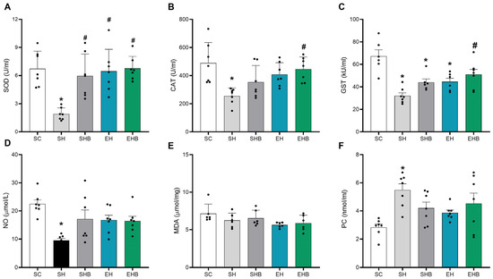
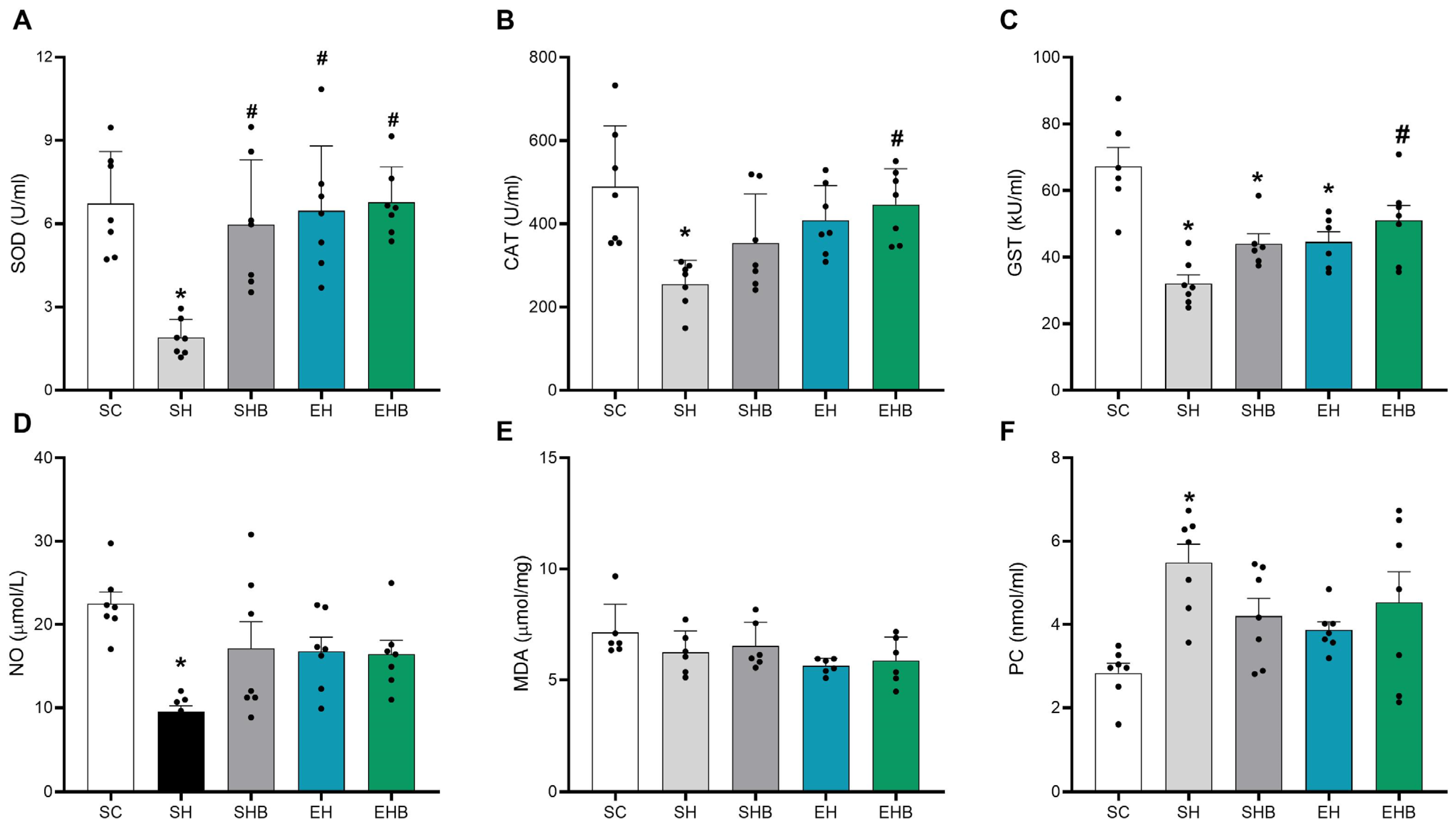
The activity of SOD was lower in the SH group than in the other groups (Figure 1A). In addition, CAT activity and NO levels were reduced in the SH group compared with the SC group (Figure 1B,D). The isolated treatments (SHB and EH) did not prevent these reductions, whereas the combined treatment prevented them (SH < EHB). Regarding GST activity, lower values were found in the SH, SHB, and EH groups compared with the SC group (Figure 1C), whereas the combined treatment prevented such a reduction (SH < EHB). Concerning the PC levels (Figure 1F), rats in the SH group exhibited greater values than those in the SC group; however, no treatment effects were observed. No differences between groups were found for the MDA levels (Figure 1E).
Figure 1.
Effects of blueberry extract and resistance training on oxidative stress markers in lung tissue. (A) SOD (superoxide dismutase), (B) CAT (catalase), (C) GST (glutathione S-transferase), (D) NO (nitric oxide), (E) MDA (malondialdehyde), (F) PC (protein carbonyl). SC, sedentary control; SH, sedentary hypertensive; SHB, sedentary hypertensive blueberry; EH, exercise hypertensive; EHB, exercise hypertensive blueberry. The data are expressed as means ± SD of five to seven rats per group. One-way ANOVA followed by Tukey’s post hoc test. * p ˂ 0.05 vs. SC; # p ˂ 0.05 vs. SH.
3.3. RV Oxidative Stress
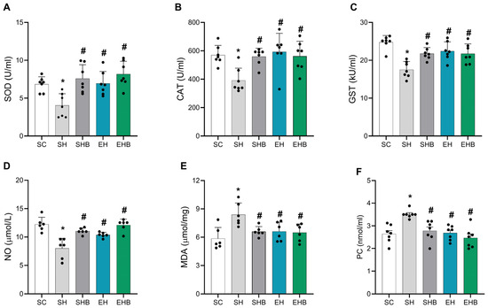
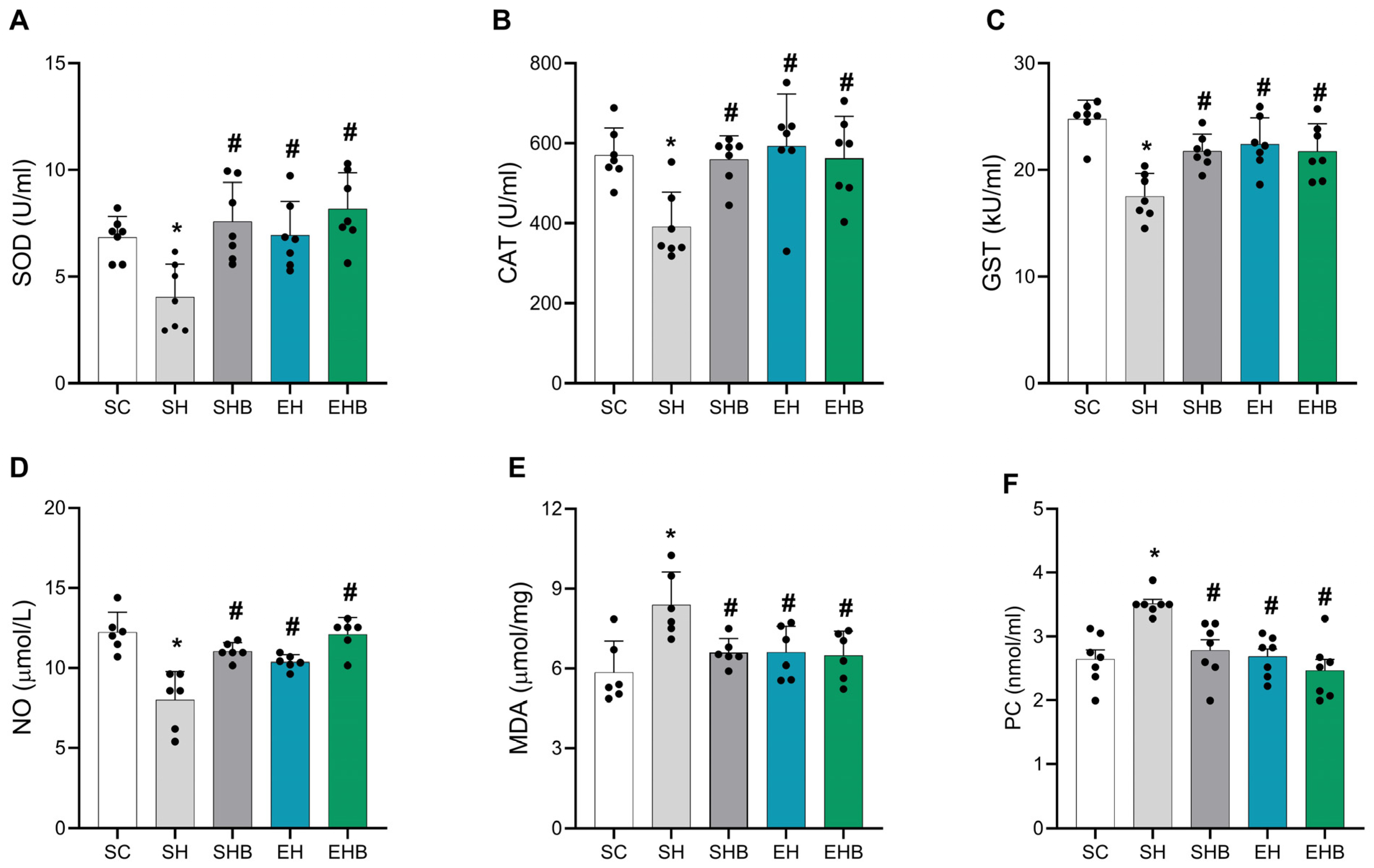
The rats in the SH group showed decreased SOD (Figure 2A), CAT (Figure 2B), and GST (Figure 2C) activities compared with those in the SC group. However, the treatments, either alone (SHB and EH groups) or in combination (EHB group), prevented such a reduction to a similar extent. The levels of NO were also lower in the SH group than in the SC and EHB groups (Figure 2D). Concerning MDA levels (Figure 2E), they were higher in the SH group than in the SC group. As for the CP levels (Figure 2F), rats in the SH group showed increased values compared with those in the SC group, while this elevation was similarly attenuated by both interventions, either alone (SHB and EH groups) or combined (EHB group).
Figure 2.
Effects of blueberry extract and resistance training on oxidative stress markers in the right ventricle. (A) SOD (superoxide dismutase), (B) CAT (catalase), (C) GST (glutathione S-transferase), (D) NO (nitric oxide), (E) MDA (malondialdehyde), (F) PC (protein carbonyl). SC, sedentary control. SH, sedentary hypertensive. SHB, sedentary hypertensive blueberry. EH, exercise hypertensive. EHB, exercise hypertensive blueberry. Data are means ± SD of five to seven rats per group. One-way ANOVA followed by Tukey’s post hoc test. * p ˂ 0.05 vs. SC; # p ˂ 0.05 vs. SH.
3.4. Single RV Myocyte Contractile Function
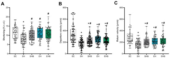
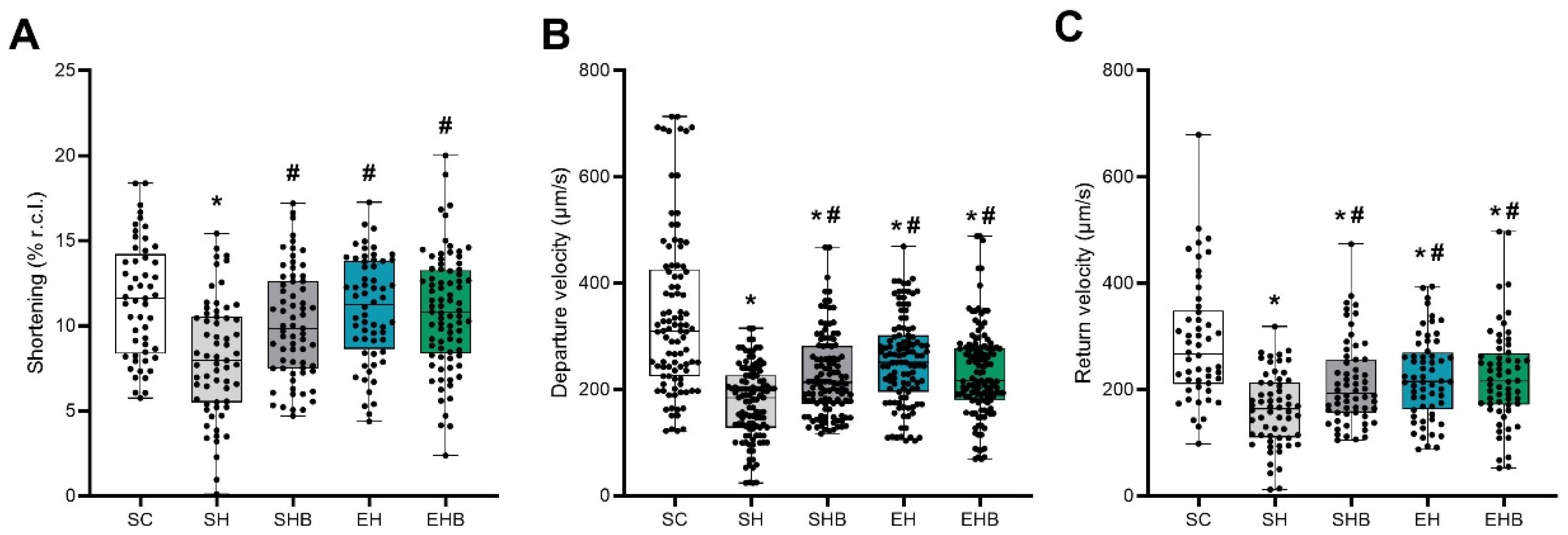
Myocytes from rats in the SH group showed a reduced contraction amplitude compared with those from the SC group (Figure 3A). However, both treatments, either alone (SHB and EH groups) or combined (EHB group), prevented such a reduction to a similar extent. The departure (Figure 3B) and return (Figure 3C) velocities of shortening were lower in myocytes from hypertensive rats (SH, SHB, EH, and EHB groups) than in those from the SC group. Nevertheless, the employed treatments either alone (SHB and EH groups) or combined (EHB groups) resulted in higher velocities compared with those observed in the SH group.
Figure 3.
Effects of blueberry extract and resistance training on contractility in single right-ventricular myocytes stimulated at 5 Hz. (A) Amplitude of shortening. (B) Departure velocity of shortening. (C) Return velocity of shortening. SC, sedentary control. SH, sedentary hypertensive. SHB, sedentary hypertensive blueberry. EH, exercise hypertensive. EHB, exercise hypertensive blueberry. Data are shown as box-and-whisker plots (median, interquartile range; whiskers = minimum–maximum) overlaid with individual cell values (10 cells per animal; 5–6 animals per group). Between-group differences were assessed with the Kruskal–Wallis test followed by Dunn’s multiple comparisons. * p < 0.05 vs. SC; # p < 0.05 vs. SH.
3.5. Expression of Ca2+ Handling Proteins
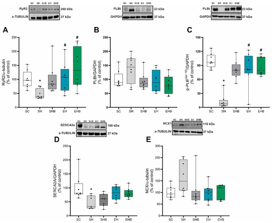
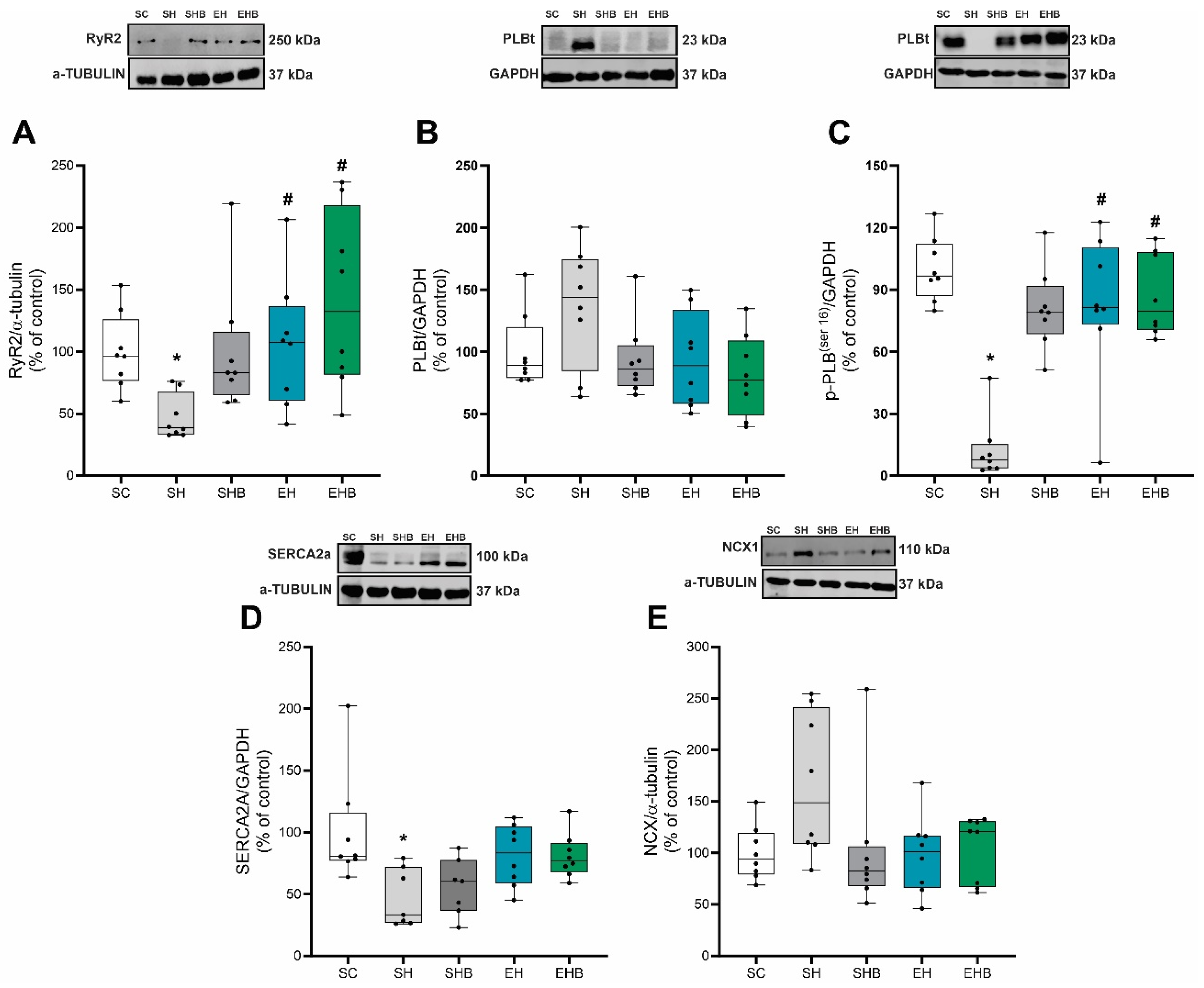
Concerning the expression of proteins involved in Ca2+ handling, the levels of RyR2 (Figure 4A) and p-PLBser16 (Figure 4C) were lower in the SH group compared with the SC, EH, and EHB groups. No differences between groups were observed in the expression of PLBt (Figure 4B) or NCX (Figure 4E). Regarding SERCA2a expression, the SH group showed reduced levels compared with those of the SC group (Figure 4D).
Figure 4.
Effects of blueberry extract and resistance training on the expression of Ca2+-handling proteins in the right ventricle. (A) Ryanodine receptor type 2 (RyR2). (B) Total phospholamban (PLB_t). (C) Phospholamban phosphorylated at Ser16 (p-PLB^Ser16). (D) Sarcoplasmic reticulum calcium ATPase type 2a (SERCA2a). (E) Na+/Ca2+ exchanger (NCX1). SC, sedentary control. SH, sedentary hypertensive. SHB, sedentary hypertensive blueberry. EH, exercise hypertensive. EHB, exercise hypertensive blueberry. Data are shown as box-and-whisker plots (median, interquartile range; whiskers = minimum–maximum) with individual animal values (n = 7–8 per group), expressed as % of SC (control). Group differences were tested with the Kruskal–Wallis test followed by Dunn’s post hoc comparisons. * p < 0.05 vs. SC; # p < 0.05 vs. SH.
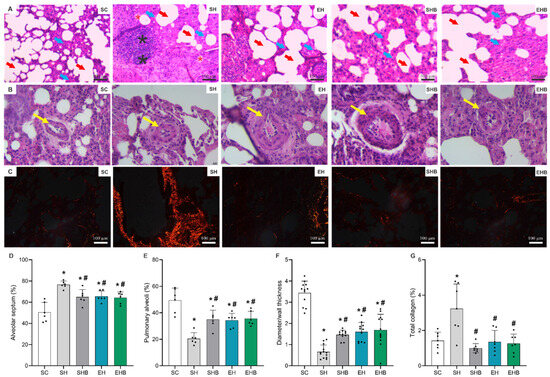
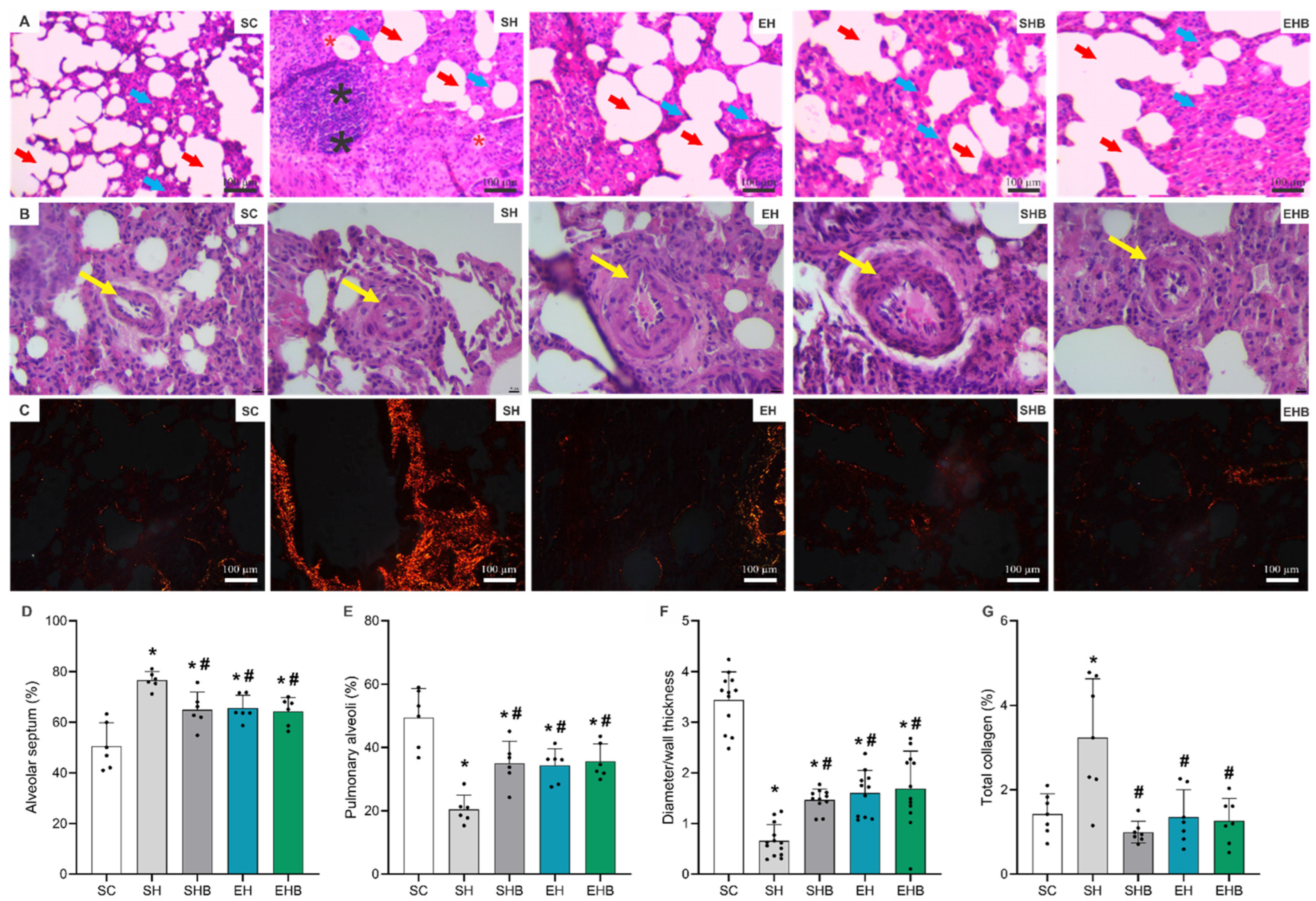
3.6. Lung Remodeling
Representative lung photomicrographs (Figure 5A) reveal maintained pulmonary architecture in the SC group, characterized by thin septa, open alveoli, and the absence of inflammatory signs. In contrast, the SH group displayed disrupted microarchitecture, with thickened alveolar septa and inflammatory infiltrates. However, these structural alterations were attenuated in all treated groups (SHB, EH, and EHB), which showed better alveolar preservation, reduced septal thickening, and less inflammation. In addition, in the lung sections in Figure 5B, it can be observed that the SH group displayed a thickened arteriolar wall compared with the SC group, while such a change was lessened in the treated groups. Moreover, lung sections in Figure 5C show augmented collagen fibers in the SH group than in the control group, and that this increase was attenuated in the treated groups. The quantitative analysis indicated that hypertensive rats (SH, SHB, EH, and EHB groups) had a higher percentage of alveolar septa (Figure 5D) and a lower percentage of pulmonary alveoli (Figure 5E) than the control rats (SC group). The diameter-to-wall thickness ratio of pulmonary arterioles was lower in rats from the SH group than in those from the SC group (Figure 5F). Nevertheless, all treatments attenuated this reduction, partially restoring the diameter-to-wall thickness ratio, although values remained lower than those observed in the SC group. Finally, total collagen was higher in rats from the SH group compared with the control ones (SC group) (Figure 5G). However, all treatments prevented such an increase.
Figure 5.
Effects of blueberry extract and resistance training on lung histomorphometry. (A) Representative photomicrograph of lung tissue stained with hematoxylin and eosin (scale bar: 100 μm). (B) Representative photomicrographs of pulmonary arterioles stained with hematoxylin and eosin (scale bar: 100 μm). (C) Representative photomicrographs of lung tissue stained with picrosirius red, with collagen fibers shown in red/orange (scale bar: 100 μm). (D) Alveolar septum (%). (E) Pulmonary alveoli (%). (F) Pulmonary arteriole diameter/wall thickness ratio. (G) Total collagen (%). Blue arrows: alveolar septum; red arrows: alveolus; red asterisks: hemorrhagic alveolus; black asterisks: inflammatory foci; yellow arrows: pulmonary arteriole. SC, sedentary control; SH, sedentary hypertensive; SHB, sedentary hypertensive blueberry; EH, exercise hypertensive; EHB, exercise hypertensive blueberry. Data are expressed as mean ± SD of 10 images per animal in each group (7 animals per group). One-way ANOVA followed by Tukey’s post hoc test. * p < 0.05 vs. SC; # p < 0.05 vs. SH.
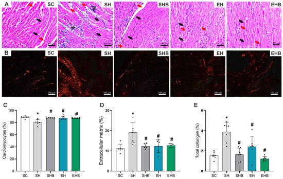
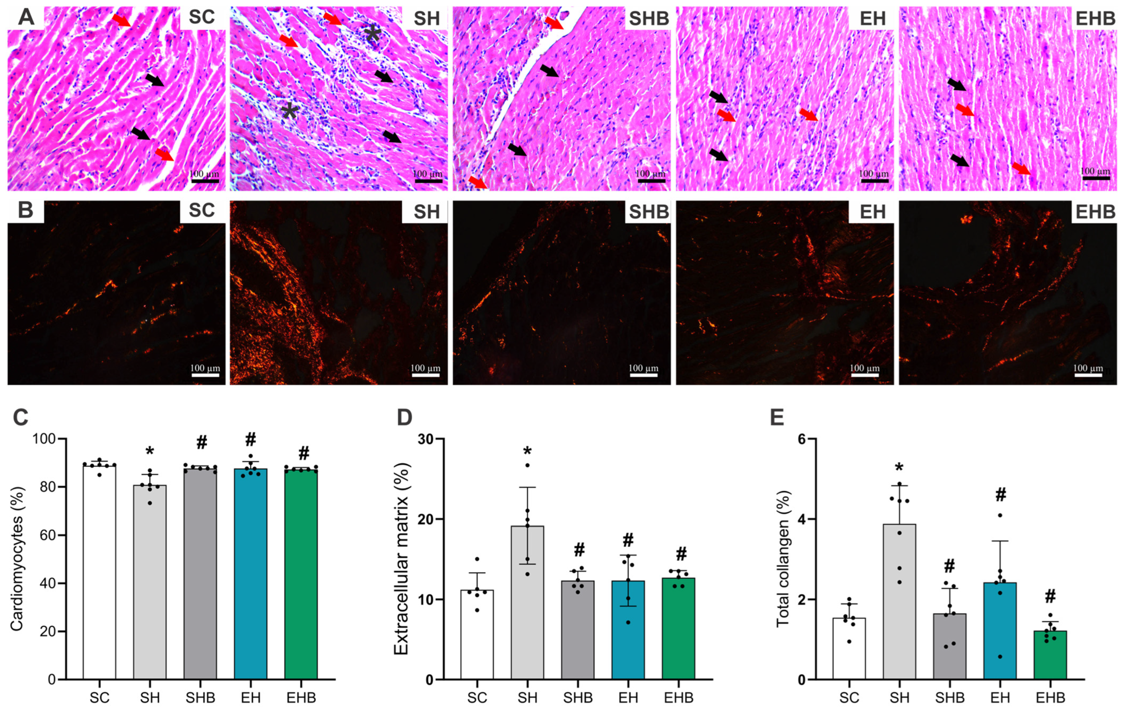
3.7. RV Remodeling
Cardiac sections from the SC group (Figure 6A,B) showed preserved RV morphology, with organized fibers, central nuclei, and homogeneous cytoplasm, without inflammatory infiltration or extracellular matrix accumulation. In contrast, SH animals presented disorganized fibers, reduced cardiomyocyte density, inflammatory foci, and marked extracellular matrix and collagen deposition. These changes were attenuated in the treated groups (SHB, EH, and EHB), which displayed better conservation of tissue architecture and less fibrosis compared with the SH group, with no prominent inflammatory infiltration observed. The quantitative analysis showed that rats in the SH group had a lower percentage of cardiomyocytes (Figure 6C), and higher percentages of extracellular matrix (Figure 6D) and total collagen (Figure 6E) than those in the SC group. However, all treated groups showed increased proportions of cardiomyocytes and reduced extracellular matrix and total collagen compared with the SH group.
Figure 6.
Effects of blueberry extract and resistance training on right ventricular remodeling. (A) Representative photomicrographs of tissue stained with hematoxylin and eosin (scale bar: 100 μm). (B) Representative photomicrographs of tissue stained with picrosirius red, with collagen fibers shown in red/orange (scale bar: 100 μm). (C) Percentage of cardiomyocytes. (D) Percentage of extracellular matrix. (E) Percentage of total collagen. Black arrows: cardiomyocytes; red arrow: extracellular matrix; asterisks: inflammatory foci. SC, sedentary control; SH, sedentary hypertensive; SHB, sedentary hypertensive blueberry; EH, hypertensive exercise; EHB, exercise hypertensive blueberry. Data are means ± SD of seven rats in each group. One-way ANOVA followed by Tukey’s post hoc test. * p ˂ 0.05 vs. SC; # p ˂ 0.05 vs. SH.
4. Discussion
We used a model of severe PAH in rats to evaluate the effects of blueberry extract and resistance exercise training on pulmonary and RV function and structure. Both interventions, either alone or in combination, mitigated cardiopulmonary oxidative imbalance and adverse structural remodeling during PAH progression. In addition, the treatments preserved RV myocyte contractile function, while resistance training, alone or combined with blueberry extract, was associated with the partial preservation of key Ca2+-handling proteins. Overall, these adaptations were accompanied by the attenuation of pulmonary vascular impairment and RV dysfunction.
The echocardiographic analyses confirmed that the injected dose of monocrotaline markedly increased the pulmonary artery resistance (i.e., a reduced AT/ET ratio) and reduced RV systolic function (i.e., TAPSE) in hypertensive rats, changes that are typically observed in disease progression [27,30,32,54]. Such RV dysfunction was followed by impaired RV single cardiomyocyte contractility, characterized by a reduced amplitude of shortening and slower velocities of shortening and relaxation.
In line with these functional disturbances, we observed that the induced PAH caused a pronounced oxidative imbalance (i.e., decreased SOD, CAT, GST and NO) and elevated oxidative damage (i.e., increased MDA and PC) in both the lung and RV. In addition, the lung of these rats exhibited inflammatory infiltrates and hemorrhagic alveoli, followed by alveolar septal thickening and increased collagen deposition, along with reduced alveolar density and arteriole diameter. Likewise, the RV of hypertensive rats showed structural changes like reduced proportion of myocytes, expansion of the extracellular matrix, and collagen accumulation. Moreover, the RV presented downregulation of Ca2+ regulatory proteins (i.e., RyR2 and SERCA2a). These changes are strongly associated with PAH progression [10,11,12,13,14,30,35]. More importantly, our results indicate that both interventions attenuated these impairments, and the observed functional benefits are believed to be associated with metabolic, mechanical and structural adaptations to the applied treatments. Initially, it is noteworthy that the resistance training protocol used here improved the rats’ exercise tolerance (i.e., maximum carrying load), although blueberry extract either alone or combined with resistance training did not affect exercise tolerance. This improvement may be attributed not only to the preservation of central parameters (i.e., lung and heart) but also to peripheral adaptations to resistance training, including the maintenance of skeletal muscle integrity and enhanced respiratory efficiency [29,55].
Regarding the cardiopulmonary functional benefits of the applied treatments (i.e., AT/ET and TAPSE), it is conceivable that the preservation of NO bioavailability provided by the treatments played a role, since resistance exercise enhances eNOS activity and antioxidants can potentiate this pathway [56,57]. Indeed, the preservation of NO levels was particularly evident with the combined intervention in both lung and RV tissues, suggesting that maintenance of NO bioavailability may contribute to the observed cardiopulmonary benefits. Moreover, the interventions attenuated the lung and RV pro-oxidant profile, with exercise showing the greatest capacity to preserve antioxidant activity. Since oxidative stress activates fibroblasts and apoptotic pathways [58,59,60], these biochemical adaptations provide a mechanistic explanation for the prevention of pathological remodeling observed in both lung and RV tissues. Really, the RV of treated rats displayed attenuated extracellular matrix and collagen accumulation and preserved architecture. Similarly, the interventions mitigated the structural changes and improved the pulmonary oxidative profile. Altogether, our data support the concept that pathological remodeling in PAH emerges as the final consequence of oxidative imbalance, and that interventions capable of restoring these upstream processes can substantially mitigate cardiopulmonary damage [30,35].
Concerning the impaired contractility of single RV myocytes and the downregulation of Ca2+ handling proteins, the employed treatments attenuated the reduction in cellular mechanical function, as well as the expression of RyR2 and p-PLB(Ser16), indicating the partial preservation of calcium release and reuptake. These findings align with previous studies showing that resistance training improves cardiomyocyte contractile function as well as intracellular calcium dynamics by upregulating regulatory proteins [29]. It is noteworthy that our resistance exercise protocol improved antioxidant defenses in the RV of trained rats and thus attenuated oxidative stress, which positively impacts the disruption of Ca2+ handling [11,58,59]. Notably, this study demonstrates for the first time that blueberry extract directly contributed to the preservation of RV myocyte function, despite having no direct impact on the expression of Ca2+ regulatory proteins. It is reasonable, nevertheless, that the antioxidant properties of blueberry favored RV redox regulation, which positively influenced intracellular Ca2+ dynamics [10,11].
Taken together, these findings indicate that blueberry extract and resistance training were associated with complementary cardiopulmonary protective responses during severe PAH progression. The combined intervention showed broader preservation of pulmonary redox markers, whereas resistance training was particularly associated with improved RV systolic function. Across the interventions, the attenuation of adverse structural remodeling and the preservation of RV myocyte contractile function support the potential relevance of these strategies in severe experimental PAH.
Limitations
This study has some limitations that should be considered when interpreting the findings. First, in the resistance training protocol, the animals’ climbing speed could not be precisely controlled, limiting the accurate determination of exercise intensity and total workload. Second, as blueberry extract is a natural product, variations in its chemical composition associated with cultivation conditions, harvesting, and processing may influence the reproducibility and consistency of the intervention. Although the extract was stored protected from light at −80 °C and fresh working solutions were prepared daily to minimize phenolic degradation, stability and degradation kinetics were not directly assessed during the experimental period. This is particularly relevant for anthocyanins and other phenolic compounds that may undergo oxidation during processing and storage. Nevertheless, previous studies have demonstrated substantial preservation of blueberry anthocyanins and other phenolic compounds under low-temperature and light-protected storage conditions [61,62,63]. In addition, circulating concentrations of anthocyanins, phenolic acids, or their metabolites were not measured; therefore, the systemic exposure and bioavailability of the administered extract could not be directly determined. Previous evidence indicates that blueberry-derived phenolics are absorbed and metabolized in the gastrointestinal tract, with parent compounds and/or their metabolites reaching the systemic circulation [64]. Moreover, specific toxicological outcomes were not assessed in the present study; therefore, potential adverse effects associated with prolonged or high-dose exposure to concentrated polyphenol-rich extracts cannot be excluded. Although available evidence suggests limited overt toxicity following the oral intake of anthocyanin-rich extracts, the safety of high-dose polyphenol exposure remains incompletely characterized. Moreover, high-dose supplementation may exert pro-oxidant and other adverse effects depending on the dose, duration of exposure, and physiological context [65,66]. Future studies should therefore include dedicated safety and dose–response assessments, particularly when considering long-term supplementation or translation to clinical settings. Third, the duration of the interventions was constrained by the time course of severe MCT-induced PAH, which limits the extrapolation of our findings to longer-term adaptations. Finally, only male rats were included, limiting the generalizability of the findings and highlighting the need for future studies investigating sex-specific responses to these interventions in severe PAH.
5. Conclusions
In conclusion, blueberry extract and resistance training, either alone or in combination, alleviate cardiopulmonary oxidative imbalance and adverse structural remodeling, sustain RV myocyte contractility and the expression of proteins involved in Ca2+ cycling, and thus conserve pulmonary and right ventricle function in severe PAH induced by monocrotaline. These findings reinforce the relevance of integrating non-pharmacological strategies involving natural antioxidants and physical exercise in mitigating cardiopulmonary dysfunctions and structural damage associated with severe PAH, which could have significant implications for disease management and prognosis.
Author Contributions
Conception and design of the research: L.B.L. and A.J.N.; Acquisition of data, analysis and interpretation of the data: L.L.S., L.O.G.-E., S.F.F.C., S.C.d.L.G., M.A.M.X., A.E.d.C.Q.; M.S.F., T.I.-L., F.G.H., P.T., A.S.d.R.A., T.F., B.R.d.A.P. and P.F.; Critically reviewed the manuscript. L.B.L. and A.J.N.; Acquisition of funding, equipment, and materials necessary for conducting the study: M.M.-N., E.M.O., E.C.C.R. and A.J.N. All authors have read and agreed to the published version of the manuscript.
Funding
This study was funded by the Fundação de Amparo à Pesquisa do Estado de Minas Gerais (FAPEMIG), Brazil (grant number APQ-01485-22); the Conselho Nacional de Desenvolvimento Científico e Tecnológico (CNPq), Brazil (grant number 306956/2021-7); and the Coordenação de Aperfeiçoamento de Pessoal de Nível Superior (CAPES), which provided a scholarship to LB Leite (Finance Code 001). The funding agencies were not involved in the study’s design, data collection, analysis, or interpretation, the writing of the manuscript, or the decision to submit it for publication. This research was funded by FCT—The Portuguese Foundation for Science and Technology, grant number UID/6157/2025 (doi: https://doi.org/10.54499/UID/06157/2025).
Institutional Review Board Statement
The animal study protocol was approved by the Ethics Committee on the Use of Animals at the Federal University of Viçosa (protocol code 11/2022 and date of approval 8 July 2022).
Informed Consent Statement
Not applicable.
Data Availability Statement
The data will be shared upon reasonable request to the corresponding author due to ethical restrictions related to the use of animals in research.
Acknowledgments
We extend our gratitude to CNPq—Brazil for awarding fellowships to A.J. Natali, E.M. Oliveira, E.C.C. Reis and M.M. Neves. We also acknowledge the Coordenação de Aperfeiçoamento de Pessoal de Nível Superior (CAPES) for awarding a D.S. scholarship awarded to L.B. Leite. We also acknowledge the MCTI-INCT Physical (In)activity and Health (No. 408899/2024-7) for a post-doc scholarship awarded to L.B. Leite.
Conflicts of Interest
The authors declare no conflicts of interest.
References
- Humbert, M.; Guignabert, C.; Bonnet, S.; Dorfmüller, P.; Klinger, J.R.; Nicolls, M.R.; Olschewski, A.J.; Pullamsetti, S.S.; Schermuly, R.T.; Stenmark, K.R.; et al. Pathology and pathobiology of pulmonary hypertension: State of the art and research perspectives. Eur. Respir. J. 2019, 53, 1801887. [Google Scholar] [CrossRef] [Scilit] [PubMed]
- Grygiel-Górniak, B.; Lucki, M.; Daroszewski, P.; Lucka, E. Prevalence, molecular mechanisms and diagnostic approaches to pulmonary arterial hypertension in connective tissue diseases. Rheumatol. Int. 2025, 45, 87. [Google Scholar] [CrossRef] [Scilit]
- Shafeghat, M.; Raza, Y.; Catania, R.; Rahsepar, A.A.; Tilkens, B.; Cuttica, M.J.; Freed, B.H.; Dai, J.; Zhao, Y.-Y.; Carr, J.C. State of the art in pulmonary arterial hypertension: Molecular basis, imaging modalities, and right heart failure treatment. Biomedicines 2025, 13, 1773. [Google Scholar] [CrossRef] [Scilit] [PubMed]
- Ryan, J.J.; Huston, J.; Kutty, S.; Hatton, N.D.; Bowman, L.; Tian, L.; Herr, J.E.; Johri, A.M.; Archer, S.L. Right ventricular adaptation and failure in pulmonary arterial hypertension. Can. J. Cardiol. 2015, 31, 391–406. [Google Scholar] [CrossRef] [Scilit] [PubMed]
- Gaikwad, A.V.; Lu, W.; Dey, S.; Bhattarai, P.; Chia, C.; Larby, J.; Haug, G.; Myers, S.; Jaffar, J.; Westall, G.; et al. Vascular remodelling in idiopathic pulmonary fibrosis patients and its detrimental effect on lung physiology: Potential role of endothelial-to-mesenchymal transition. ERJ Open Res. 2022, 8, 00571-2021. [Google Scholar] [CrossRef] [Scilit] [PubMed]
- Bowers, R.; Cool, C.; Murphy, R.C.; Tuder, R.M.; Hopken, M.W.; Flores, S.C.; Voelkel, N.F. Oxidative stress in severe pulmonary hypertension. Am. J. Respir. Crit. Care Med. 2004, 169, 764–769. [Google Scholar] [CrossRef] [Scilit] [PubMed]
- DeMarco, V.G.; Whaley-Connell, A.; Sowers, J.R.; Habibi, J.; Dellsperger, K.C. Contribution of oxidative stress to pulmonary arterial hypertension. World J. Cardiol. 2010, 2, 316–324. [Google Scholar] [CrossRef] [Scilit] [PubMed]
- Xu, D.; Hu, Y.-H.; Gou, X.; Li, F.-Y.; Yang, X.-Y.-C.; Li, Y.-M.; Chen, F. Oxidative stress and antioxidative therapy in pulmonary arterial hypertension. Molecules 2022, 27, 3724. [Google Scholar] [CrossRef] [Scilit] [PubMed]
- Freire, T.C.; Ferreira, M.S.; De Angelelis, K.; Paula-Ribeiro, M. Respiratory, cardiovascular and musculoskeletal mechanisms involved in the pathophysiology of pulmonary hypertension: An updated systematic review of preclinical and clinical studies. Heart Lung 2024, 68, 81–91. [Google Scholar] [CrossRef] [Scilit] [PubMed]
- Giordano, F.J. Oxygen, oxidative stress, hypoxia, and heart failure. J. Clin. Investig. 2005, 115, 500–508. [Google Scholar] [CrossRef] [Scilit]
- Zima, A.V.; Blatter, L.A. Redox regulation of cardiac calcium channels and transporters. Cardiovasc. Res. 2006, 71, 310–321. [Google Scholar] [CrossRef] [Scilit] [PubMed]
- Takimoto, E.; Kass, D.A. Role of oxidative stress in cardiac hypertrophy and remodeling. Hypertension 2007, 49, 241–248. [Google Scholar] [CrossRef] [Scilit] [PubMed]
- Aggarwal, S.; Gross, C.M.; Sharma, S.; Fineman, J.R.; Black, S.M. Reactive oxygen species in pulmonary vascular remodeling. Compr. Physiol. 2013, 3, 1011–1034. [Google Scholar] [CrossRef] [Scilit]
- Peng, J.-J.; Liu, B.; Xu, J.-Y.; Peng, J.; Luo, X.-J. NADPH oxidase: Its potential role in promotion of pulmonary arterial hypertension. Naunyn Schmiedebergs Arch. Pharmacol. 2017, 390, 331–338. [Google Scholar] [CrossRef] [Scilit] [PubMed]
- Crosswhite, P.; Sun, Z. Nitric oxide, oxidative stress and inflammation in pulmonary arterial hypertension. J. Hypertens. 2010, 28, 201–212. [Google Scholar] [CrossRef] [Scilit] [PubMed]
- Crosswhite, P.; Sun, Z. Molecular mechanisms of pulmonary arterial remodeling. Mol. Med. 2014, 20, 191–201. [Google Scholar] [CrossRef] [Scilit] [PubMed]
- Weinstein, A.A.; Chin, L.M.K.; Keyser, R.E.; Kennedy, M.; Nathan, S.D.; Woolstenhulme, J.G.; Connors, G.; Chan, L. Effect of aerobic exercise training on fatigue and physical activity in patients with pulmonary arterial hypertension. Respir. Med. 2013, 107, 778–784. [Google Scholar] [CrossRef] [Scilit] [PubMed]
- Galiè, N.; Humbert, M.; Vachiery, J.-L.; Gibbs, S.; Lang, I.; Torbicki, A.; Simonneau, G.; Peacock, A.; Vonk Noordegraaf, A.; Beghetti, M.; et al. 2015 ESC/ERS guidelines for the diagnosis and treatment of pulmonary hypertension: The joint task force for the diagnosis and treatment of pulmonary hypertension of the European Society of Cardiology (ESC) and the European Respiratory Society (ERS): Endorsed by the Association for European Paediatric and Congenital Cardiology (AEPC) and the International Society for Heart and Lung Transplantation (ISHLT). Eur. Heart J. 2016, 37, 67–119. [Google Scholar] [PubMed]
- Lajoie, A.-C.; Bonnet, S.; Provencher, S. Combination therapy in pulmonary arterial hypertension: Recent accomplishments and future challenges. Pulm. Circ. 2017, 7, 312–325. [Google Scholar] [CrossRef] [Scilit] [PubMed]
- Keusch, S.; Turk, A.; Saxer, S.; Ehlken, N.; Grünig, E.; Ulrich, S.; Swiss Society of Pulmonary Hypertension. Rehabilitation in patients with pulmonary arterial hypertension. Swiss Med. Wkly. 2017, 147, w14462. [Google Scholar] [CrossRef] [Scilit] [PubMed]
- Grünig, E.; Eichstaedt, C.; Barberà, J.-A.; Benjamin, N.; Blanco, I.; Bossone, E.; Cittadini, A.; Coghlan, G.; Corris, P.; D’Alto, M.; et al. ERS statement on exercise training and rehabilitation in patients with severe chronic pulmonary hypertension. Eur. Respir. J. 2019, 53, 1800332. [Google Scholar] [CrossRef] [Scilit] [PubMed]
- Grünig, E.; MacKenzie, A.; Peacock, A.J.; Eichstaedt, C.A.; Benjamin, N.; Nechwatal, R.; Ulrich, S.; Saxer, S.; Bussotti, M.; Sommaruga, M.; et al. Standardized exercise training is feasible, safe, and effective in pulmonary arterial and chronic thromboembolic pulmonary hypertension: Results from a large European multicentre randomized controlled trial. Eur. Heart J. 2021, 42, 2284–2295. [Google Scholar] [CrossRef] [Scilit] [PubMed]
- Vieira, J.S.; Cunha, T.F.; Paixão, N.A.; Dourado, P.M.; Carrascoza, L.S.; Bacurau, A.V.N.; Brum, P.C. Exercise intolerance establishment in pulmonary hypertension: Preventive effect of aerobic exercise training. Life Sci. 2020, 261, 118298. [Google Scholar] [CrossRef] [Scilit] [PubMed]
- Belló-Klein, A.; Khaper, N.; Llesuy, S.; Vassallo, D.V.; Pantos, C. Oxidative stress and antioxidant strategies in cardiovascular disease. Oxid. Med. Cell. Longev. 2014, 2014, 678741. [Google Scholar] [CrossRef] [Scilit] [PubMed]
- Mangge, H.; Becker, K.; Fuchs, D.; Gostner, J.M. Antioxidants, inflammation and cardiovascular disease. World J. Cardiol. 2014, 6, 462–477. [Google Scholar] [CrossRef] [Scilit] [PubMed]
- Jain, A.K.; Mehra, N.K.; Swarnakar, N.K. Role of antioxidants for the treatment of cardiovascular diseases: Challenges and opportunities. Curr. Pharm. Des. 2015, 21, 4441–4455. [Google Scholar] [CrossRef] [Scilit] [PubMed]
- Soares, L.L.; Drummond, F.R.; Rezende, L.M.T.; Lopes Dantas Costa, A.J.; Leal, T.F.; Fidelis, M.R.; Neves, M.M.; Prímola-Gomes, T.N.; Carneiro-Júnior, M.A.; Reis, E.C.C.; et al. Voluntary running counteracts right ventricular adverse remodeling and myocyte contraction impairment in pulmonary arterial hypertension model. Life Sci. 2019, 238, 116974. [Google Scholar] [CrossRef] [Scilit] [PubMed]
- de Jesus Silva, F.; Drummond, F.R.; Fidelis, M.R.; Freitas, M.O.; Leal, T.F.; de Rezende, L.M.T.; de Moura, A.G.; Reis, E.C.C.; Natali, A.J. Continuous aerobic exercise prevents detrimental remodeling and right heart myocyte contraction and calcium cycling dysfunction in pulmonary artery hypertension. J. Cardiovasc. Pharmacol. 2021, 77, 69–78. [Google Scholar] [CrossRef] [Scilit] [PubMed]
- Soares, L.L.; Leite, L.B.; Ervilha, L.O.G.; Pelozin, B.R.A.; Pereira, N.P.; da Silva, B.A.F.; Portes, A.M.O.; Drummond, F.R.; de Rezende, L.M.T.; Fernandes, T.; et al. Resistance exercise training benefits pulmonary, cardiac, and muscular structure and function in rats with stable pulmonary artery hypertension. Life Sci. 2023, 332, 122128. [Google Scholar] [CrossRef] [Scilit] [PubMed]
- Leite, L.B.; Soares, L.L.; Portes, A.M.O.; Soares, T.I.; da Silva, B.A.F.; Dias, T.R.; Costa, S.F.F.; Guimarães-Ervilha, L.O.; Assis, M.Q.; Lavorato, V.N.; et al. Combined physical training protects the left ventricle from structural and functional damages in experimental pulmonary arterial hypertension. Clin. Hypertens. 2024, 30, 12. [Google Scholar] [CrossRef] [Scilit] [PubMed]
- Soares, L.L.; Leite, L.B.; Ervilha, L.O.G.; da Silva, B.A.F.; de Freitas, M.O.; Portes, A.M.O.; Rezende, L.M.T.; Drummond, F.R.; Carneiro-Júnior, M.A.; Machado-Neves, M.; et al. Resistance exercise training mitigates left ventricular dysfunctions in pulmonary artery hypertension model. Arq. Bras. Cardiol. 2022, 119, 574–584. [Google Scholar] [PubMed]
- Leite, L.B.; Soares, L.L.; Portes, A.M.O.; da Silva, B.A.F.; Dias, T.R.; Soares, T.I.; Assis, M.Q.; Guimarães-Ervilha, L.O.; Carneiro-Júnior, M.A.; Forte, P.; et al. Combined exercise hinders the progression of pulmonary and right heart harmful remodeling in monocrotaline-induced pulmonary arterial hypertension. J. Appl. Physiol. 2025, 138, 182–194. [Google Scholar] [CrossRef] [Scilit] [PubMed]
- Moreira-Gonçalves, D.; Ferreira, R.; Fonseca, H.; Padrão, A.I.; Moreno, N.; Silva, A.F.; Vasques-Nóvoa, F.; Gonçalves, N.; Vieira, S.; Santos, M.; et al. Cardioprotective effects of early and late aerobic exercise training in experimental pulmonary arterial hypertension. Basic Res. Cardiol. 2015, 110, 57. [Google Scholar] [CrossRef] [Scilit] [PubMed]
- Colombo, R.; Siqueira, R.; Becker, C.U.; Fernandes, T.G.; Pires, K.M.; Valença, S.S.; Souza-Rabbo, M.P.; Araujo, A.S.; Belló-Klein, A. Effects of exercise on monocrotaline-induced changes in right heart function and pulmonary artery remodeling in rats. Can. J. Physiol. Pharmacol. 2013, 91, 38–44. [Google Scholar] [CrossRef] [Scilit] [PubMed]
- Türck, P.; Fraga, S.; Salvador, I.; Campos-Carraro, C.; Lacerda, D.; Bahr, A.; Ortiz, V.; Hickmann, A.; Koetz, M.; Belló-Klein, A.; et al. Blueberry extract decreases oxidative stress and improves functional parameters in lungs from rats with pulmonary arterial hypertension. Nutrition 2020, 70, 110579. [Google Scholar] [CrossRef] [Scilit] [PubMed]
- Türck, P.; Salvador, I.S.; Campos-Carraro, C.; Ortiz, V.; Bahr, A.; Andrades, M.; Belló-Klein, A.; da Rosa Araujo, A.S. Blueberry extract improves redox balance and functional parameters in the right ventricle from rats with pulmonary arterial hypertension. Eur. J. Nutr. 2022, 61, 373–386. [Google Scholar] [CrossRef] [Scilit] [PubMed]
- Liu, Y.; Tan, D.; Shi, L.; Liu, X.; Zhang, Y.; Tong, C.; Song, D.; Hou, M. Blueberry anthocyanins-enriched extracts attenuate cyclophosphamide-induced cardiac injury. PLoS ONE 2015, 10, e0127813. [Google Scholar] [CrossRef] [Scilit] [PubMed]
- Natali, A.J.; Fowler, E.D.; Calaghan, S.C.; White, E. Voluntary exercise delays heart failure onset in rats with pulmonary artery hypertension. Am. J. Physiol. Heart Circ. Physiol. 2015, 309, H421–H424. [Google Scholar] [CrossRef] [Scilit] [PubMed]
- Leite, L.B.; Soares, L.L.; Guimarães-Ervilha, L.O.; Costa, S.F.F.; Generoso, S.C.d.L.; Xavier, M.A.M.; Iasbik-Lima, T.; Oliveira, L.L.d.; Della Lucia, C.M.; Bianchi, S.E.; et al. Blueberry extract and resistance training prevent left ventricular redox dysregulation and pathological remodeling in experimental severe pulmonary arterial hypertension. Nutrients 2025, 17, 1145. [Google Scholar] [CrossRef] [Scilit] [PubMed]
- Hornberger, T.A., Jr.; Farrar, R.P. Physiological hypertrophy of the FHL muscle following 8 weeks of progressive resistance exercise in the rat. Can. J. Appl. Physiol. 2004, 29, 16–31. [Google Scholar] [CrossRef] [Scilit] [PubMed]
- Sanches, I.C.; Conti, F.F.; Sartori, M.; Irigoyen, M.C.; De Angelis, K. Standardization of resistance exercise training: Effects in diabetic ovariectomized rats. Int. J. Sports Med. 2014, 35, 323–329. [Google Scholar] [CrossRef] [Scilit] [PubMed]
- Sahn, D.J.; DeMaria, A.; Kisslo, J.; Weyman, A. Recommendations regarding quantitation in M-mode echocardiography: Results of a survey of echocardiographic measurements. Circulation 1978, 58, 1072–1083. [Google Scholar] [CrossRef] [Scilit] [PubMed]
- Dieterich, S.; Bieligk, U.; Beulich, K.; Hasenfuss, G.; Prestle, J. Gene expression of antioxidative enzymes in the human heart: Increased expression of catalase in the end-stage failing heart. Circulation 2000, 101, 33–39. [Google Scholar] [PubMed]
- Hadwan, M.H.; Abed, H.N. Data supporting the spectrophotometric method for the estimation of catalase activity. Data Brief 2016, 6, 194–199. [Google Scholar] [CrossRef] [Scilit] [PubMed]
- Habig, W.H.; Pabst, M.J.; Jakoby, W.B. Glutathione S-transferases: The first enzymatic step in mercapturic acid formation. J. Biol. Chem. 1974, 249, 7130–7139. [Google Scholar] [PubMed]
- Tsikas, D. Analysis of nitrite and nitrate in biological fluids by assays based on the Griess reaction: Appraisal of the Griess reaction in the L-arginine/nitric oxide area of research. J. Chromatogr. B 2007, 851, 51–70. [Google Scholar] [CrossRef] [Scilit] [PubMed]
- Buege, J.A.; Aust, S.D. Microsomal lipid peroxidation. In Methods in Enzymology; Fleischer, S., Packer, L., Eds.; Academic Press: New York, NY, USA, 1978; Volume 52, pp. 302–310. [Google Scholar]
- Levine, R.L.; Williams, J.A.; Stadtman, E.R.; Shacter, E. Carbonyl assays for determination of oxidatively modified proteins. Methods Enzymol. 1994, 233, 346–357. [Google Scholar] [CrossRef] [Scilit] [PubMed]
- Lowry, O.H.; Rosebrough, N.J.; Farr, A.L.; Randall, R.J. Protein measurement with the Folin phenol reagent. J. Biol. Chem. 1951, 193, 265–275. [Google Scholar] [CrossRef] [Scilit]
- Pelozin, B.R.A.; Soci, U.P.R.; Gomes, J.L.P.; Oliveira, E.M.; Fernandes, T. mTOR signaling-related microRNAs as cardiac hypertrophy modulators in high-volume endurance training. J. Appl. Physiol. 2022, 132, 126–139. [Google Scholar] [CrossRef] [Scilit] [PubMed]
- Wang, Z.; Patel, J.R.; Schreier, D.A.; Hacker, T.A.; Moss, R.L.; Chesler, N.C. Organ-level right ventricular dysfunction with preserved Frank-Starling mechanism in a mouse model of pulmonary arterial hypertension. J. Appl. Physiol. 2018, 124, 1244–1253. [Google Scholar] [CrossRef] [Scilit] [PubMed]
- Souza, A.C.F.; de Paiva Coimbra, J.L.; Ervilha, L.O.G.; Bastos, D.S.S.; Cossolin, J.F.S.; Santos, E.C.; de Oliveira, L.L.; Machado-Neves, M. Arsenic induces dose-dependent structural and ultrastructural pathological remodeling in the heart of Wistar rats. Life Sci. 2020, 257, 118132. [Google Scholar] [CrossRef] [Scilit] [PubMed]
- Portes, A.M.O.; de Moura, A.G.; Silva, F.J.; Guimarães-Ervilha, L.O.; de Freitas, M.O.; Soares, L.L.; da Silva, B.A.F.; Costa, S.F.F.; Generoso, S.C.L.; de Souza Silva, T.; et al. Previous resistance exercise training mitigates progression of right ventricle dysfunction and remodeling in male rats with pulmonary arterial hypertension. Physiol. Rep. 2026, 14, e70786. [Google Scholar] [CrossRef] [Scilit] [PubMed]
- Tello, K.; Axmann, J.; Ghofrani, H.A.; Naeije, R.; Narcin, N.; Rieth, A.; Seeger, W.; Gall, H.; Richter, M.J. Relevance of the TAPSE/PASP ratio in pulmonary arterial hypertension. Int. J. Cardiol. 2018, 266, 229–235. [Google Scholar] [CrossRef] [Scilit] [PubMed]
- Costa, S.F.F.; Leite, L.B.; Soares, L.L.; Generoso, S.C.d.L.; Xavier, M.A.M.; Faria, M.S.; de Carvalho Quintão, A.E.; Ervilha, L.O.G.; Lima, T.I.; Pelozin, B.R.A.; et al. Resistance exercise training attenuates skeletal muscle atrophy in experimental pulmonary arterial hypertension. Pflug. Arch. 2026, 478, 3. [Google Scholar] [CrossRef] [Scilit] [PubMed]
- Ashor, A.W.; Lara, J.; Siervo, M.; Celis-Morales, C.; Oggioni, C.; Jakovljevic, D.G.; Mathers, J.C. Exercise modalities and endothelial function: A systematic review and dose–response meta-analysis of randomized controlled trials. Sports Med. 2015, 45, 279–296. [Google Scholar] [CrossRef] [Scilit] [PubMed]
- Lee, Y.; Im, E. Regulation of miRNAs by natural antioxidants in cardiovascular diseases: Focus on SIRT1 and eNOS. Antioxidants 2021, 10, 377. [Google Scholar] [CrossRef] [Scilit] [PubMed]
- Redout, E.M.; van der Toorn, A.; Zuidwijk, M.J.; van de Kolk, C.W.A.; van Echteld, C.J.A.; Musters, R.J.P.; van Hardeveld, C.; Paulus, W.J.; Simonides, W.S. Antioxidant treatment attenuates pulmonary arterial hypertension-induced heart failure. Am. J. Physiol. Heart Circ. Physiol. 2010, 298, H1038–H1047. [Google Scholar] [CrossRef] [Scilit] [PubMed]
- Farahmand, F.; Hill, M.F.; Singal, P.K. Antioxidant and oxidative stress changes in experimental cor pulmonale. Mol. Cell. Biochem. 2004, 260, 21–29. [Google Scholar] [CrossRef] [Scilit] [PubMed]
- Thomas, T.P.; Grisanti, L.A. The dynamic interplay between cardiac inflammation and fibrosis. Front. Physiol. 2020, 11, 529075. [Google Scholar] [CrossRef] [Scilit] [PubMed]
- Song, H.-N.; Ji, S.-A.; Park, H.-R.; Kim, H.-H.; Hogstrand, C. Impact of Various Factors on Color Stability of Fresh Blueberry Juice during Storage. Prev. Nutr. Food Sci. 2018, 23, 46–51. [Google Scholar] [CrossRef] [Scilit] [PubMed]
- Srivastava, A.; Akoh, C.C.; Yi, W.; Fischer, J.; Krewer, G. Effect of Storage Conditions on the Biological Activity of Phenolic Compounds of Blueberry Extract Packed in Glass Bottles. J. Agric. Food Chem. 2007, 55, 2705–2713. [Google Scholar] [CrossRef] [Scilit] [PubMed]
- Ionescu, C.; Samide, A.; Tigae, C. Trend in Detection of Anthocyanins from Fresh Fruits and the Influence of Some Factors on Their Stability Impacting Human Health: Kinetic Study Assisted by UV–Vis Spectrophotometry. Antioxidants 2025, 14, 227. [Google Scholar] [CrossRef] [Scilit] [PubMed]
- Kuntz, S.; Rudloff, S.; Asseburg, H.; Borsch, C.; Fröhling, B.; Unger, F.; Dold, S.; Spengler, B.; Römpp, A.; Kunz, C. Uptake and Bioavailability of Anthocyanins and Phenolic Acids from Grape/Blueberry Juice and Smoothie in vitro and in vivo. Br. J. Nutr. 2015, 113, 1044–1055. [Google Scholar] [CrossRef] [Scilit] [PubMed]
- Cladis, D.P.; Weaver, C.M.; Ferruzzi, M.G. (Poly)phenol Toxicity in vivo Following Oral Administration: A Targeted Narrative Review of (Poly)phenols from Green Tea, Grape, and Anthocyanin-Rich Extracts. Phytother. Res. 2022, 36, 323–335. [Google Scholar] [CrossRef] [Scilit] [PubMed]
- Vrankova, S.; Cebova, M.; Klimentova, J.; Pechanova, O. Redox Paradox of Polyphenols: Bioavailability, Hormesis, and Safety Concerns. Phytother. Res. 2026, 40, 2437–2455. [Google Scholar] [CrossRef] [Scilit] [PubMed]

Disclaimer/Publisher’s Note: The statements, opinions and data contained in all publications are solely those of the individual author(s) and contributor(s) and not of MDPI and/or the editor(s). MDPI and/or the editor(s) disclaim responsibility for any injury to people or property resulting from any ideas, methods, instructions or products referred to in the content. |
© 2026 by the authors. Licensee MDPI, Basel, Switzerland. This article is an open access article distributed under the terms and conditions of the Creative Commons Attribution (CC BY) license.