Early Risk Stratification and Time-to-Cure Analysis in Patients with Residual Hemifacial Spasm After Microvascular Decompression
Highlights
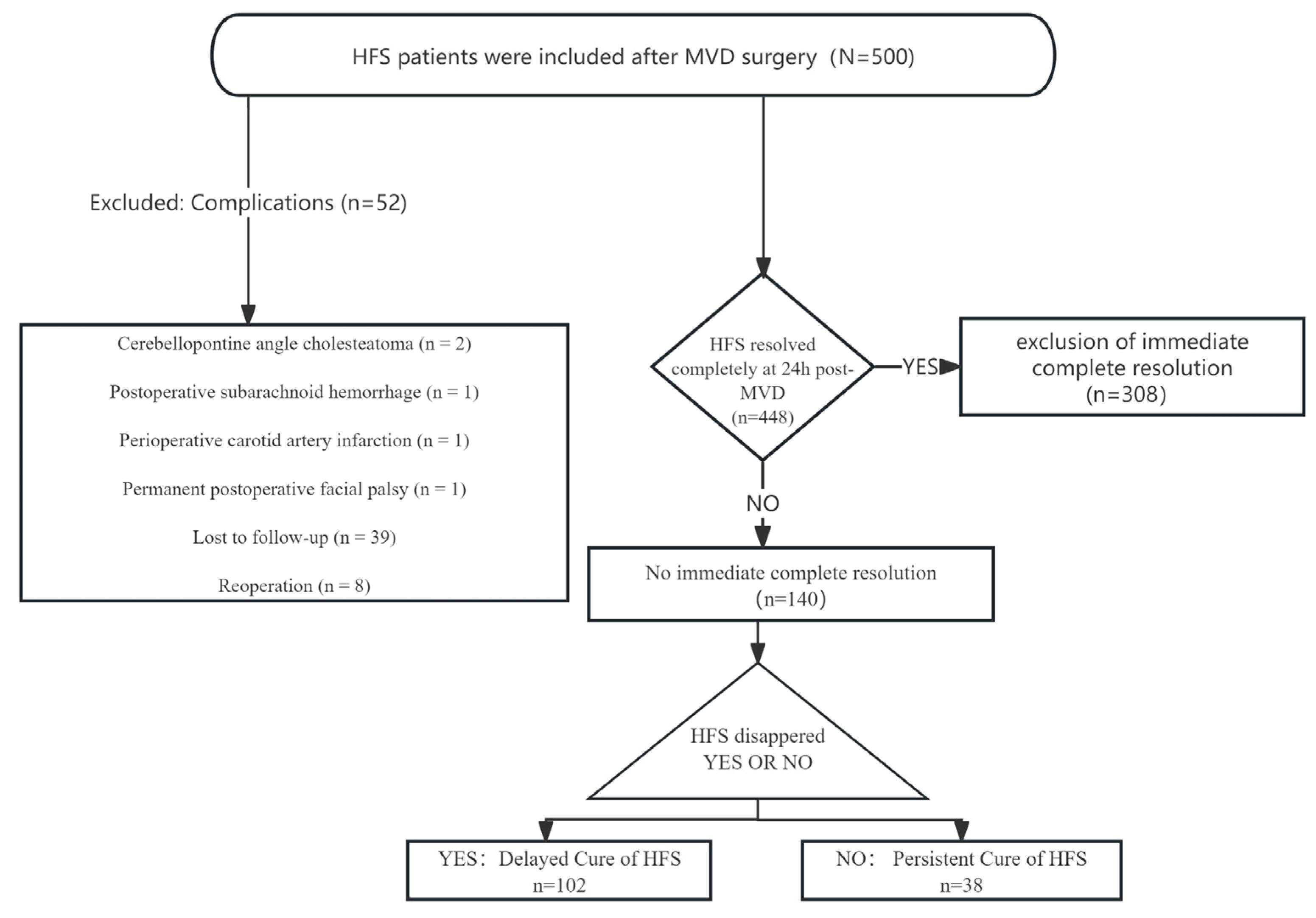
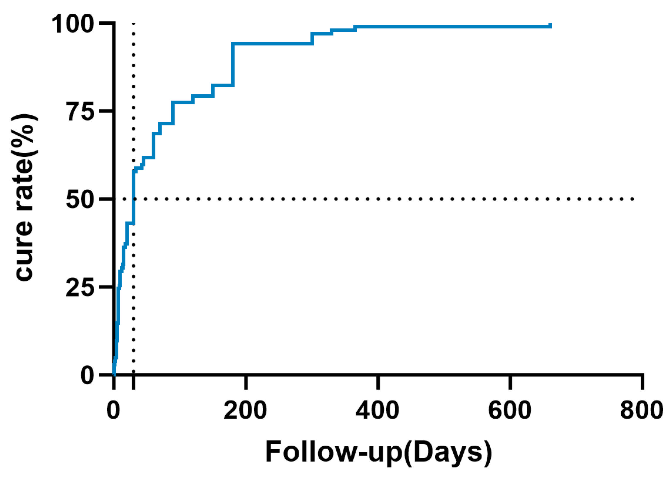
- Among 140 patients with residual hemifacial spasm 24 h after microvascular decompression, 102 (72.9%) achieved delayed cure without reoperation. The median time to delayed cure was 30 days, and 95% of delayed cures occurred within 300 days after surgery.
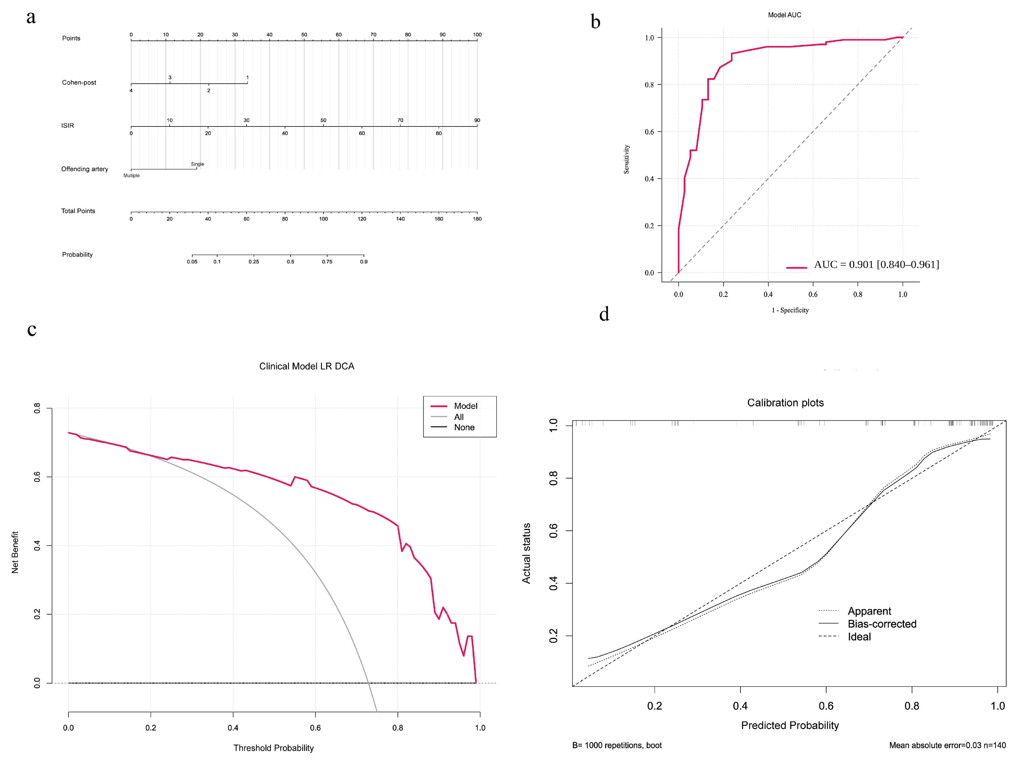
- A prediction model incorporating ISIR, Cohen-post, and offending artery status showed good discrimination, with an area under the curve of 0.901.
- Persistent spasm shortly after surgery does not necessarily indicate surgical failure, as most patients eventually achieved symptom resolution without additional surgery.
- These findings may help clinicians counsel patients after surgery and guide follow-up decisions.
Abstract
1. Introduction
2. Methods
2.1. Patients and Study Design
2.2. Patient Selection
2.3. Collection of Data
2.4. Outcome Definitions
- Delayed cure: residual symptoms at 24 h after surgery but complete resolution of HFS at the last follow-up.
- Persistent HFS: ongoing HFS at the last follow-up.
2.5. Development of Models and Survival Curves
2.6. Time-to-Event Analysis (Time to Cure)
2.7. Forest Plot Construction
3. Results
3.1. Baseline Characteristics
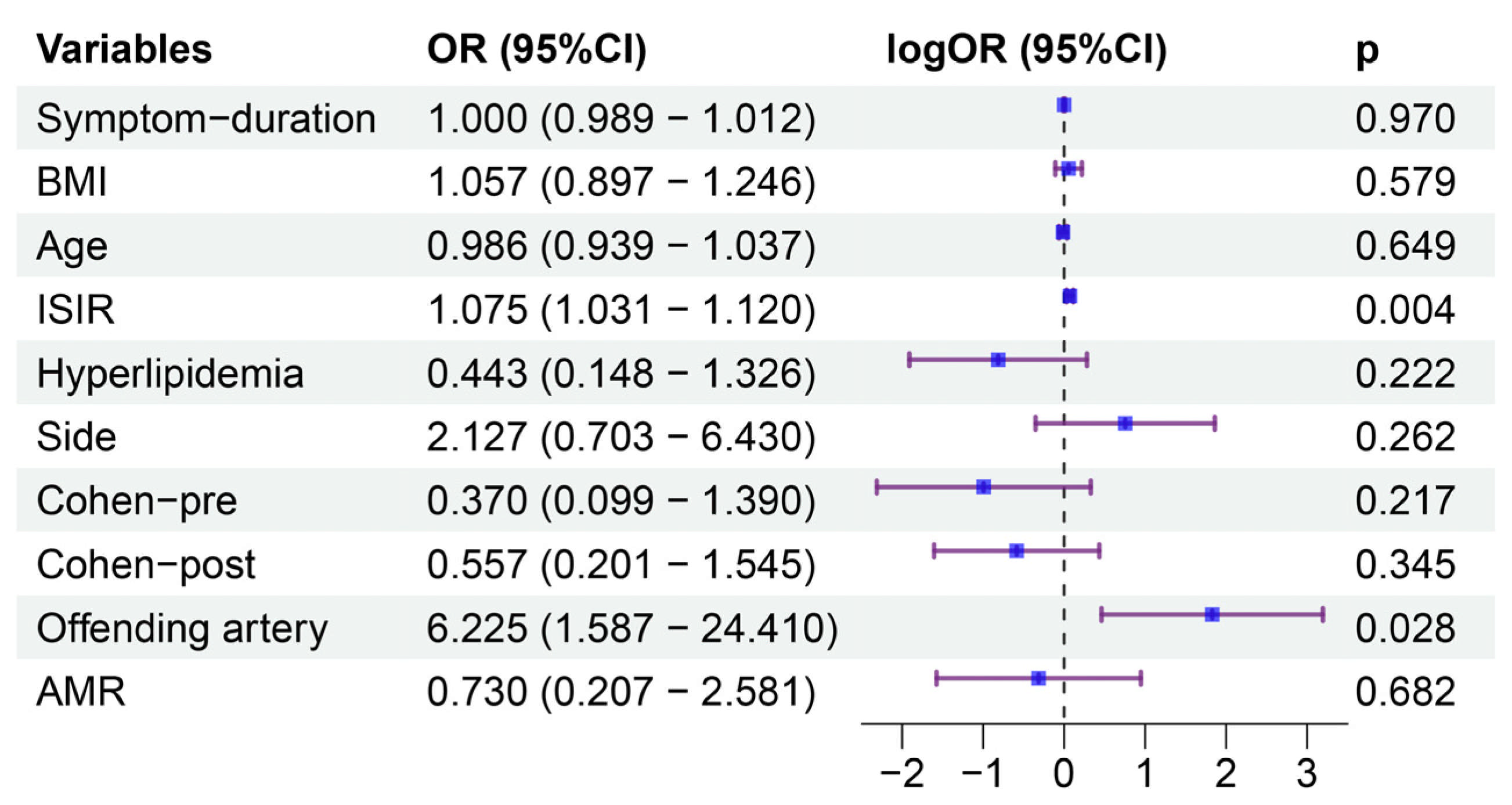
3.2. Predictor Selection for the Model
3.3. Nomogram Construction
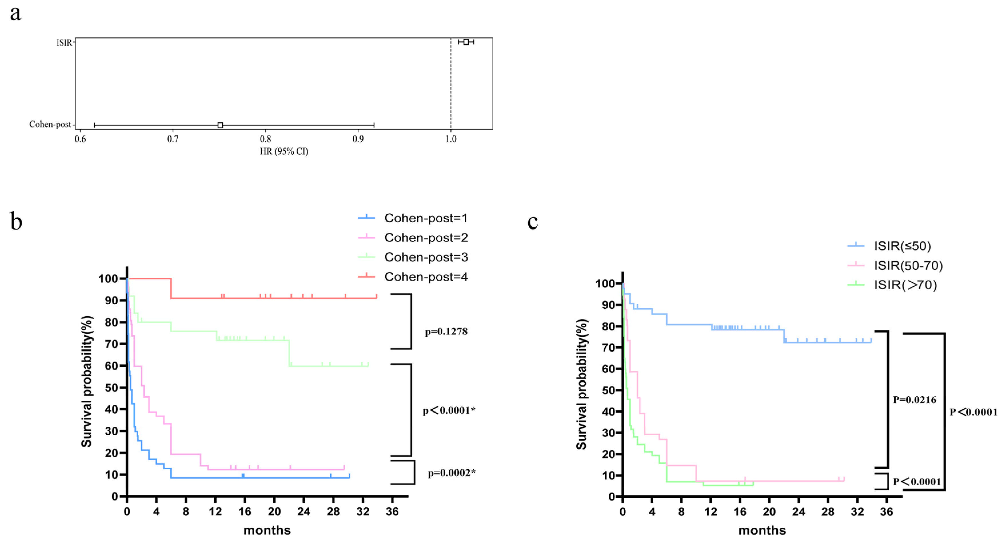
3.4. Survival Curves and Factor Analysis
4. Discussion
4.1. Summary of Key Findings
4.2. Comparison with Previous Studies
4.3. Potential Mechanisms of Key Factors
4.4. Implications of Risk Stratification for Follow-Up and Reassessment
4.5. Evidence and Clinical Considerations Regarding the Timing of Reoperation
4.6. Predictive Factors Inform but Do Not Determine Reoperation Decisions
4.7. Limitations
5. Conclusions
Supplementary Materials
Author Contributions
Funding
Institutional Review Board Statement
Informed Consent Statement
Data Availability Statement
Conflicts of Interest
Abbreviations
References
- Sindou, M.; Mercier, P. Microvascular decompression for hemifacial spasm: Outcome on spasm and complications. A review. Neurochirurgie 2018, 64, 106–116. [Google Scholar] [CrossRef] [Scilit] [PubMed]
- Xia, L.; Zhong, J.; Zhu, J.; Dou, N.N.; Liu, M.X.; Li, S.T. Delayed relief of hemifacial spasm after microvascular decompression. J. Craniofac. Surg. 2015, 26, 408–410. [Google Scholar] [CrossRef] [Scilit] [PubMed]
- Nunta-Aree, S.; Kateyoi, T.; Sitthinamsuwan, B. Delayed complete remission of hemifacial spasms following microvascular decompression and the implications for optimal time of revision surgery. Acta Neurochir. 2024, 166, 12. [Google Scholar] [CrossRef] [Scilit] [PubMed]
- Al Mutawa, M.; Matthes, M.; Schroeder, H.W.S.; Weidemeier, M.E. Etiology and management of recurrent and persistent hemifacial spasm-A single-center experience. Neurosurgery 2024, 95, 418–427. [Google Scholar] [CrossRef] [Scilit] [PubMed]
- Liu, J.; Li, F.; Wu, G.; Liu, B.; Zhou, J.; Fan, C.; Jiao, F.; Wang, D.; Wu, G.; Song, H.; et al. Long-term retrospective analysis of re-do microvascular decompression in patients with hemifacial spasm. Front. Neurol. 2021, 12, 687945. [Google Scholar] [CrossRef] [Scilit] [PubMed]
- Chen, K.; Shen, L.; Yang, J.; Zhao, J.; Wu, J.; Cai, Y.; Mei, Z.; Chai, S.; Xu, D.; Jiang, P.; et al. A nomogram based on clinical multivariate factors predicts delayed cure after microvascular decompression for hemifacial spasm. Neurosurg. Rev. 2024, 47, 44. [Google Scholar] [CrossRef] [Scilit] [PubMed]
- Al Menabbawy, A.; Refaee, E.E.; Elwy, R.; Salem, A.A.; Lehmann, S.; Vollmer, M.; Matthes, M.; Fleck, S.; Baldauf, J.; Schroeder, H.W.S. A multivariable prediction model for recovery patterns and time course of symptoms improvement in hemifacial spasm following microvascular decompression. Acta Neurochir. 2022, 164, 833–844. [Google Scholar] [CrossRef] [Scilit] [PubMed]
- Sato, Y.; Shimizu, K.; Iizuka, K.; Irie, R.; Matsumoto, M.; Mizutani, T. Factors related to the delayed cure of hemifacial spasm after microvascular decompression: An analysis of 175 consecutive patients. J. Neurol. Surg. B Skull Base 2022, 83, 548–553. [Google Scholar] [CrossRef] [Scilit] [PubMed]
- Tambasco, N.; Simoni, S.; Sacchini, E.; Eusebi, P.; Marsili, E.; Nigro, P.; Brahimi, E.; Paoletti, F.P.; Romoli, M.; Calabresi, P. Validation of the Hemifacial Spasm Grading Scale: A clinical tool for hemifacial spasm. Neurol. Sci. 2019, 40, 1887–1892. [Google Scholar] [CrossRef] [Scilit] [PubMed]
- Li, M.W.; Jiang, X.F.; Wu, M.; He, F.; Niu, C. Clinical research on delayed cure after microvascular decompression for hemifacial spasm. J. Neurol. Surg. A Cent. Eur. Neurosurg. 2020, 81, 195–199. [Google Scholar] [CrossRef] [Scilit] [PubMed]
- Zhong, J.; Xia, L.; Dou, N.N.; Ying, T.T.; Zhu, J.; Liu, M.X.; Li, S.T. Delayed relief of hemifacial spasm after microvascular decompression: Can it be avoided? Acta Neurochir. 2015, 157, 93–98; discussion 98–99. [Google Scholar] [CrossRef] [Scilit] [PubMed]
- Zhao, H.; Zhang, X.; da Tang, Y.D.; Zhu, J.; Ying, T.T.; Yan, Y.; Li, S. Factors promoting a good outcome in a second microvascular decompression operation when hemifacial spasm is not relieved after the initial operation. World Neurosurg. 2017, 98, 872.e11–872.e19. [Google Scholar] [CrossRef] [Scilit] [PubMed]
- Bonomo, G.; Bonomo, R.; Iess, G.; Alimonti, P.; Restelli, F.; Falco, J.; Mazzapicchi, E.; Schiariti, M.P.; Broggi, M.; Acerbi, F.; et al. Arterial Hypertension After Macrovascular Decompression of Vertebrobasilar Dolichoectasia: Rethinking the Cause-Effect Relationship. World Neurosurg. 2025, 198, 123974. [Google Scholar] [CrossRef] [Scilit] [PubMed]
- Lee, S.; Park, S.K.; Lee, J.A.; Joo, B.E.; Park, K. Missed culprits in failed microvascular decompression surgery for hemifacial spasm and clinical outcomes of redo surgery. World Neurosurg. 2019, 129, e627–e633, Correction in World Neurosurg. 2020, 133, 483. https://doi.org/10.1016/j.wneu.2019.09.096. [Google Scholar] [CrossRef] [Scilit] [PubMed]
- Bigder, M.G.; Kaufmann, A.M. Failed microvascular decompression surgery for hemifacial spasm due to persistent neurovascular compression: An analysis of reoperations. J. Neurosurg. 2016, 124, 90–95. [Google Scholar] [CrossRef] [Scilit] [PubMed]
- Kondo, A. Follow-up results of microvascular decompression in trigeminal neuralgia and hemifacial spasm. Neurosurgery 1997, 40, 46–51; discussion 51–52. [Google Scholar] [CrossRef] [Scilit] [PubMed]
- Engh, J.A.; Horowitz, M.; Burkhart, L.; Chang, Y.F.; Kassam, A. Repeat microvascular decompression for hemifacial spasm. J. Neurol. Neurosurg. Psychiatry 2005, 76, 1574–1580. [Google Scholar] [CrossRef] [Scilit] [PubMed]
- Marneffe, V.; Polo, G.; Fischer, C.; Sindou, M. Microsurgical vascular decompression for hemifacial spasm. Follow-up over one year, clinical results and prognostic factors. Study of a series of 100 cases. Neuro-Chirurgie 2003, 49, 527–535. [Google Scholar] [PubMed]
- Du, T.; Wang, Y.; Liu, S.; Ni, B.; Zhu, H. Revision surgery of microvascular decompression for hemifacial spasm: 2-dimensional operative video. World Neurosurg. 2025, 193, 223. [Google Scholar] [CrossRef] [Scilit] [PubMed]
- Zhang, J.; Li, Z.H.; Wang, J.F.; Chen, Y.H.; Wang, N.; Wang, Y. Prognostic value of abnormal muscle response during microvascular decompression for hemifacial spasm: A meta-analysis. World Neurosurg. 2020, 137, 8–17. [Google Scholar] [CrossRef] [Scilit] [PubMed]
- El Damaty, A.; Rosenstengel, C.; Matthes, M.; Baldauf, J.; Schroeder, H.W.S. The value of lateral spread response monitoring in predicting the clinical outcome after microvascular decompression in hemifacial spasm: A prospective study on 100 patients. Neurosurg. Rev. 2016, 39, 455–466. [Google Scholar] [CrossRef] [Scilit] [PubMed]
- Satoh, T.; Onoda, K.; Date, I. Fusion imaging of three-dimensional magnetic resonance cisternograms and angiograms for the assessment of microvascular decompression in patients with hemifacial spasms. J. Neurosurg. 2007, 106, 82–89. [Google Scholar] [CrossRef] [Scilit] [PubMed]
- D’Amico, A.; Perillo, T.; Ugga, L.; Cuocolo, R.; Brunetti, A. Intruding implements: A pictorial review of retained surgical foreign objects in neuroradiology. Insights Imaging 2019, 10, 124. [Google Scholar] [CrossRef] [Scilit] [PubMed]



| Variables | Feature | Overall (n = 140) | Delayed Cure (n = 102) | Persistent HFS (n = 38) | p |
|---|---|---|---|---|---|
| Age (years) | Mean ± SD | 52.3 ± 10.3 | 52.0 ± 10.4 | 53.2 ± 10.1 | 0.545 |
| BMI (kg/m2) | Mean ± SD | 24.7 ± 3.5 | 24.8 ± 3.7 | 24.4 ± 3.0 | 0.527 |
| Gender | M:F, n (%) | 47:93 (33.6:66.4) | 29:73 (28.4:71.6) | 18:20 (47.4:52.6) | 0.056 |
| Symptom duration (months) | Mean ± SD | 56.9 ± 56.6 | 59.0 ± 61.4 | 51.1 ± 41.0 | 0.379 |
| Hypertension | Yes, n (%) | 42 (30.0) | 27 (26.5) | 15 (39.5) | 0.199 |
| Diabetes | Yes, n (%) | 14 (10.0) | 11 (10.8) | 3 (7.9) | 0.758 |
| Hyperlipidemia | Yes, n (%) | 69 (49.3) | 49 (48.0) | 20 (52.6) | 0.769 |
| Botulinum toxin | Yes, n (%) | 23 (16.4) | 16 (15.7) | 7 (18.4) | 0.895 |
| Tinnitus | Yes, n (%) | 20 (14.3) | 15 (14.7) | 5 (13.2) | 1.000 |
| Side | L:R, n (%) | 68:72 (48.6:51.4) | 54:48 (52.9:47.1) | 14:24 (36.8:63.2) | 0.132 |
| Cohen-pre grade | n (%) | 0.191 | |||
| Grade 2 | 2 (1.4) | 2 (2.0) | 0 (0.0) | ||
| Grade 3 | 22 (15.7) | 19 (18.6) | 3 (7.9) | ||
| Grade 4 | 116 (82.9) | 81 (79.4) | 35 (92.1) | ||
| Offending artery, | S:M, n (%) | 117:23 (83.6:16.4) | 85:17 (83.3:16.7) | 32:6 (84.2:15.8) | 1.000 |
| AMR disappeared | Yes, n (%) | 109(77.9) | 80 (78.4) | 29 (76.3) | 0.969 |
| Cohen-post grade | n (%) | <0.001 | |||
| Grade 1 | 48 (34.3) | 44 (43.1) | 4 (10.5) | ||
| Grade 2 | 57 (40.7) | 49 (48.0) | 8 (21.1) | ||
| Grade 3 | 24 (17.1) | 8 (7.8) | 16 (42.1) | ||
| Grade 4 | 11 (7.9) | 1 (1.0) | 10 (26.3) | ||
| ISIR | Mean ± SD | 63.5 ± 24.6 | 73.3 ± 16.6 | 37.1 ± 23.2 | <0.001 |
| Follow-up time (months) | Mean ± SD | 21.9 ± 6.8 (12–36) | 22.6 ± 6.9 | 20.2 ± 6.5 | 0.055 |
| Coefficient | HR | SE | Coefficient (95% CI) | HR (95% CI) | z | p | |
|---|---|---|---|---|---|---|---|
| ISIR | 0.02 | 1.02 | 0 | 0.01–0.02 | 1.01–1.02 | 3.92 | <0.005 |
| Cohen-post | −0.29 | 0.75 | 0.1 | −0.49–−0.09 | 0.62–0.92 | −2.81 | <0.005 |
Disclaimer/Publisher’s Note: The statements, opinions and data contained in all publications are solely those of the individual author(s) and contributor(s) and not of MDPI and/or the editor(s). MDPI and/or the editor(s) disclaim responsibility for any injury to people or property resulting from any ideas, methods, instructions or products referred to in the content. |
© 2026 by the authors. Licensee MDPI, Basel, Switzerland. This article is an open access article distributed under the terms and conditions of the Creative Commons Attribution (CC BY) license.
Share and Cite
Liu, S.; Du, T.; Hou, F.; Jiang, Z.; He, L.; Huang, Y.; Ni, B.; Wang, X.; Wang, Y.; Zhu, H. Early Risk Stratification and Time-to-Cure Analysis in Patients with Residual Hemifacial Spasm After Microvascular Decompression. Brain Sci. 2026, 16, 978. https://doi.org/10.3390/brainsci16090978
Liu S, Du T, Hou F, Jiang Z, He L, Huang Y, Ni B, Wang X, Wang Y, Zhu H. Early Risk Stratification and Time-to-Cure Analysis in Patients with Residual Hemifacial Spasm After Microvascular Decompression. Brain Sciences. 2026; 16(9):978. https://doi.org/10.3390/brainsci16090978
Chicago/Turabian StyleLiu, Siyi, Tao Du, Feng Hou, Zaiying Jiang, Liu He, Yunjian Huang, Bing Ni, Xueyuan Wang, Yunpeng Wang, and Hongwei Zhu. 2026. "Early Risk Stratification and Time-to-Cure Analysis in Patients with Residual Hemifacial Spasm After Microvascular Decompression" Brain Sciences 16, no. 9: 978. https://doi.org/10.3390/brainsci16090978
APA StyleLiu, S., Du, T., Hou, F., Jiang, Z., He, L., Huang, Y., Ni, B., Wang, X., Wang, Y., & Zhu, H. (2026). Early Risk Stratification and Time-to-Cure Analysis in Patients with Residual Hemifacial Spasm After Microvascular Decompression. Brain Sciences, 16(9), 978. https://doi.org/10.3390/brainsci16090978