


Highlights
What are the main findings?
- Neuronavigation improves the spatial and orientational reproducibility of neuronavigated transcranial magnetic stimulation (nTMS) delivery but does not, by itself, confirm biological target engagement or response.
- Response variability arises across biophysical, cellular, plasticity, and network levels, allowing uncertainty to propagate through the stimulation-response cascade.
What are the implications of the main findings?
- Precision nTMS requires independent validation of modeled exposure, physiological and network engagement, and durable functional or clinical outcomes.
- Adaptive targeting and dosing should be guided by multimodal measurements rather than geometric accuracy alone.
Abstract
Background/Objectives: Transcranial magnetic stimulation (TMS) initiates a cascade from intracranial electric-field exposure through neural recruitment and plasticity to distributed network responses. Neuronavigation improves the geometric reproducibility of delivery but does not guarantee equivalent cortical exposure or target engagement. This narrative review integrates these levels within an operational framework for precision TMS. Methods: Six domain-specific PubMed searches covering 1 January 1985 to 31 July 2026 were supplemented by Google Scholar and citation tracking. A documented rerun on 17 August 2026 yielded 6430 records (5617 unique after cross-query deduplication). Evidence was synthesized narratively; no quantitative synthesis or formal risk-of-bias assessment was performed. Results: Neuronavigation improves geometric precision by stabilizing target definition and coil pose, whereas individualized electric-field models estimate intracranial exposure. Neither establishes biological precision, which also depends on neuronal orientation, brain state, circuit architecture, medication, and behavior. Motor-system measures are not validated as universal biomarkers for nonmotor cortex, and no single validated biomarker captures TMS-induced plasticity. Convergent, controlled multimodal evidence may strengthen inference about target engagement; adaptive and closed-loop approaches remain experimental. Conclusions: Geometric delivery, modeled exposure, biological engagement, and durable functional or clinical benefit require separate validation. Spatial accuracy alone does not establish clinical value.
1. Introduction
Transcranial magnetic stimulation (TMS) uses electromagnetic induction to probe and modulate human brain function [1,2]. Since Barker et al. first elicited motor responses in 1985, TMS has developed into a tool for functional mapping, assessment of cortical excitability and connectivity, and therapeutic neuromodulation [1,2]. A rapidly changing current in the stimulation coil generates a magnetic field that induces an electric field in brain tissue. Neural recruitment is shaped not only by stimulator output but also by pulse waveform, coil geometry and orientation, coil-to-cortex distance, tissue conductivity, cortical folding, axonal geometry, and the state of the stimulated system [2,3]. Magnetic resonance imaging (MRI)-based neuronavigation enabled TMS to be delivered while tracking coil position and orientation relative to individual anatomy. In this review, neuronavigated TMS (nTMS) refers to this image-guided mode of delivery. Neuronavigation improves spatial control and reproducibility, but it neither changes the stimulation waveform nor introduces a separate biological mechanism [4,5,6].
The biological effects of TMS extend beyond the brief induced electric field. A single pulse recruits suitably oriented axonal elements and, through them, local excitatory and inhibitory circuits [3,7,8]. Repeated stimulation may produce long-term potentiation (LTP)-like or long-term depression (LTD)-like after-effects, metaplasticity, and activity-dependent molecular or structural change [7,8,9]. These responses can propagate through connected networks [10], but their direction and magnitude depend on pulse pattern, intensity, total pulse count, temporal spacing, baseline excitability, ongoing brain state, anatomy, medication, and behavior [7,9]. Frequency-based labels are therefore useful protocol descriptors, not universal mechanistic rules [5,11,12].
Evidence for this cascade is distributed across computational biophysics, cellular and animal models, human neurophysiology, and neuroimaging [3,8,9,10,13]. In nTMS, coil pose interacts with cortical geometry and tissue heterogeneity to shape the strength, direction, and distribution of the electric field reaching excitable elements [3,4,13,14]. Neuronavigation therefore controls the geometric interface between an intended target and the delivered physical stimulus; it does not control downstream state- or network-dependent variability.
This narrative review integrates biophysical, cellular, synaptic, molecular, and network evidence into a multiscale framework for nTMS. It distinguishes geometric, biophysical, and biological precision; examines individualized modeling, mechanistic biomarkers, protocol dependence, and adaptive stimulation; and identifies the evidence required to link reproducible delivery with a durable functional or clinical response.
2. Materials and Methods
2.1. Review Design and Scope
This narrative review was prepared with reference to relevant principles of the Scale for the Assessment of Narrative Review Articles (SANRA) [15]. It was designed to integrate heterogeneous mechanistic evidence rather than estimate clinical efficacy or perform a meta-analysis. The scope included the generation and intracranial distribution of TMS-induced electric fields; recruitment of neural elements and local microcircuits; synaptic, molecular, and glial mechanisms of plasticity; propagation through distributed brain networks; and the influence of neuronavigation and individualized electric-field modeling on target engagement. Because neuronavigation constrains stimulus delivery rather than introducing a separate biological mechanism, evidence from conventional single-pulse, paired-pulse, repetitive, and patterned TMS was included whenever it informed the interpretation of nTMS.
2.2. Information Sources and Study Selection
Literature published from 1 January 1985 to 31 July 2026 was identified through domain-specific PubMed searches, supplemented by Google Scholar and backward and forward citation tracking. The PubMed searches were rerun and documented on 17 August 2026. Six searches addressed the following: (i) biophysics and electric-field distribution; (ii) neural recruitment and local microcircuits; (iii) synaptic, molecular, and glial mechanisms; (iv) network-level effects, TMS combined with electroencephalography (TMS–EEG), and neuroimaging; (v) neuronavigation, targeting, and individualized modeling; and (vi) variability, biomarkers, and adaptive stimulation. Complete search strings and domain-specific retrieval counts are provided in Supplementary Table S1. Across the six searches, PubMed returned 6430 records before cross-query deduplication and 5617 unique PubMed records. Google Scholar and citation chaining were used to identify foundational studies and newly linked sources; because their rankings and result counts are dynamic, they were not treated as countable screening sets.
Eligible sources included English-language human, animal, in vitro, and computational studies that addressed at least one level of the proposed framework. Priority was given to primary studies providing direct mechanistic evidence. Reviews, consensus statements, methodological papers, and guidelines were used to contextualize original findings, while clinical studies were included when they reported mechanistic, neurophysiological, neuroimaging, or targeting-related outcomes. Selected recent preprints were considered only when rapidly developing questions were not adequately represented in the peer-reviewed literature; their findings were identified as preliminary and were not used as the sole support for major conclusions. Studies limited to clinical efficacy, tolerability, or safety, together with conference abstracts, protocols, duplicate reports, and publications lacking substantive mechanistic information, were excluded.
The first author conducted the searches and initial eligibility assessment, and the coauthors critically reviewed the selected evidence and final synthesis. Records were not managed as a closed systematic-review cohort. Selection was iterative and purposive, prioritizing direct mechanistic evidence, replication, and convergence across complementary methods. The principal exclusion categories are stated above. Because record-level inclusion decisions and, where applicable, reasons for exclusion were not logged prospectively, an accurate Preferred Reporting Items for Systematic Reviews and Meta-Analyses (PRISMA)-style flow diagram and exclusion counts cannot be reconstructed without retrospective assumptions. We therefore report this limitation rather than provide retrospective estimates. The final synthesis comprises 131 cited sources.
2.3. Evidence-Type Stratification
To make the source and limits of each inference explicit, studies were grouped into four non-equivalent categories before findings were integrated across mechanistic levels.
2.3.1. Computational Evidence
Computational studies estimated how individual anatomy and coil configuration shape intracranial exposure. Subject-specific models showed that tissue boundaries and gyral geometry alter field magnitude and direction [13,14], while multiscale neural models generated hypotheses about orientation-dependent recruitment [3]. These studies support biophysical and hypothesis-generating cellular inferences, not direct claims of target engagement or clinical efficacy [16].
2.3.2. In Vitro Evidence
In vitro and organotypic studies directly examined synaptic efficacy, receptor-dependent plasticity, intracellular signaling, gene expression, and glial responses under controlled conditions [8,9]. The included experiments report persistent synaptic and structural changes after repeated magnetic stimulation (Section 6), but their anatomy, scale, and exposure differ from those of the intact human neocortex.
2.3.3. Animal Evidence
Animal studies linked cellular and molecular responses to changes in intact circuits, glia, structure, and behavior [8,9]. In the included corpus, these effects depended on protocol, species, disease model, and anesthesia (Section 6 and Section 7); they were therefore treated as preclinical evidence rather than assumed to be equivalent to human nTMS.
2.3.4. Human Evidence
Human studies demonstrated current-direction-dependent corticospinal recruitment [3], improved coil-pose reproducibility with neuronavigation [4], state-dependent physiological responses [9], and network propagation [10]. Their relevance to clinical nTMS varied because many studies involved small samples, healthy volunteers, motor-cortex paradigms, or short-term surrogate outcomes (Section 4, Section 5, Section 6, Section 7, Section 8, Section 9, Section 10 and Section 11).
2.4. Narrative Synthesis and Appraisal
Within each category, studies were appraised qualitatively according to reported methodological safeguards, directness, replication, consistency with complementary methods, and relevance to human nTMS. Findings were then integrated along the proposed sequence from physical delivery to neural recruitment, local circuit responses, plasticity, and distributed network effects. Discrepant results were considered in relation to species, preparation, brain state, target, stimulation parameters, coil orientation, and measurement technique. Formal study-level risk-of-bias tools were not prespecified for this narrative review. The review did not include duplicate screening, prospective protocol registration, or quantitative synthesis. This qualitative appraisal does not eliminate author judgment and remains vulnerable to selection and interpretive bias; these limitations are detailed in Section 12.1.
3. A Multiscale Framework for nTMS Mechanisms
Precision nTMS comprises distinct levels. Geometric precision concerns reproducible target definition and coil pose; biophysical precision concerns the individualized magnitude, direction, and distribution of the intracranial electric field; and biological precision concerns reproducible recruitment of the intended neural elements and circuits. Durable functional or clinical benefit is a separate endpoint. Programmed parameters, physical exposure, engagement, and outcome are related but not interchangeable [3,4,13,14,17].
Four filters connect these levels: biophysical (device output to intracranial field), cellular (field to neural and microcircuit recruitment), temporal-plasticity (integration with stimulation history and ongoing activity), and network (local response to distributed propagation) [3,8,9,10,13,14,17,18]. Neuronavigation mainly constrains upstream geometry; later links require independent physiological, network, and outcome validation. Figure 1 and Figure 2 summarize the cascade and operational hierarchy [5,12,16].
Figure 1.
Multiscale mechanistic framework for precision neuronavigated transcranial magnetic stimulation. Programmed stimulation parameters are transformed into intracranial physical exposure through a biophysical filter shaped by coil–brain geometry, individual anatomy, cortical folding, and tissue conductivity. The cellular filter determines the recruitment of axons and local microcircuits; repeated activation is integrated by temporal-plasticity mechanisms; and the network filter governs whether the perturbation remains local or propagates through distributed circuits to produce a functional response. Neuronavigation constrains upstream spatial and orientational uncertainty but does not, by itself, establish downstream biological engagement. Precision nTMS therefore requires independent validation of modeled exposure, physiological target engagement, network engagement, and durable functional or clinical outcomes, with feedback enabling adaptive refinement of targeting and stimulation parameters. Abbreviations: BDNF, brain-derived neurotrophic factor; E-field, electric field; EEG, electroencephalography; fMRI, functional magnetic resonance imaging; LTP/LTD, long-term potentiation/depression; MEP, motor evoked potential; nTMS, neuronavigated transcranial magnetic stimulation; PET, positron emission tomography; TMS–EEG, concurrent transcranial magnetic stimulation and electroencephalography.
Figure 2.
Operational hierarchy of precision in neuronavigated transcranial magnetic stimulation. Geometric precision denotes reproducible target definition and coil pose; biophysical precision denotes reproducible individualized intracranial electric-field exposure; and biological precision denotes reproducible engagement of the intended neural elements, circuit, and downstream response. Functional or clinical value is a separate validation endpoint. Evidence at one level does not establish the next, and failed downstream validation should prompt refinement of target selection, delivery, exposure modeling, or outcome measurement. Abbreviations: E-field, electric field; nTMS, neuronavigated transcranial magnetic stimulation.
5. Cellular and Microcircuit Mechanisms
5.1. Membrane Polarization and the Sites of Neural Excitation
At the cellular level, the induced field polarizes neuronal processes according to their alignment and the field variation along their trajectories [3,42]. Axons are generally more excitable than somata or dendrites, particularly at terminals, branches, bends, diameter changes, and sharp field gradients [42,43]. Early models favored the axon initial segment, bends, or the gray–white matter boundary [42,44], whereas models with richer axonal arborizations often identified intracortical terminals near gyral crowns and lips [43,45]. Somata and dendrites may undergo subthreshold polarization but are unlikely to be the principal initiation sites under conventional conditions [3,42,43]. TMS is therefore best viewed as an axonal stimulus whose site of excitation depends on axonal geometry, myelination, field direction, waveform, and cortical anatomy; initiated spikes can then recruit local and distant neurons synaptically [3].
Model disagreement largely reflects assumptions about microscopic anatomy and axonal biophysics. Activation cannot be predicted from field magnitude at the soma or a single cortical point; it depends on exposure along the excitable cable [46,47]. Field rotation can therefore change the branch that reaches threshold even when overall magnitude is unchanged [45,46]. Predictions remain sensitive to axonal diameter, myelination, internodal and branch geometry, and the completeness of reconstructed collaterals [43,45,46]. Models with limited collaterals tend to favor initial segments, bends, or the gray–white matter transition, whereas models with myelinated terminal branches more often favor intracortical terminals [42,43,44,45]. A histology-informed preprint instead reported high thresholds at incompletely myelinated cortical terminals and identified myelinated bends entering superficial white matter as plausible targets [48]. The first compartment activated by TMS thus remains unresolved. Neuronavigation constrains macroscopic exposure, not the microscopic initiation site, although small orientation changes may alter the recruited axonal subset.
5.2. Direct and Transsynaptic Recruitment of Cortical Microcircuits
After an action potential is initiated, the response is shaped by propagation through local and long-range circuits. Cervical epidural recordings distinguish an early direct wave (D-wave) from later indirect waves (I-waves) [49,50]. D-waves mainly reflect direct corticospinal-axon excitation, whereas I-waves reflect synchronized transsynaptic recruitment of corticospinal neurons through cortical interneuronal networks [50]. Their relative contribution depends on intensity, waveform, current direction, and anatomy [49,50,51,52]. The motor-evoked potential (MEP) integrates these cortical events with corticospinal transmission, spinal excitability, neuromuscular propagation, and recording conditions. It is not a direct measure of local membrane depolarization or activated cortical volume. Because this evidence derives predominantly from the motor cortex, motor threshold and MEP-based measures should not be extrapolated automatically to nonmotor targets as universal measures of cortical excitability.
I-wave periodicity is thought to emerge from recurrent interactions among supragranular excitatory neurons, inhibitory interneurons, and layer V corticospinal neurons, but the cellular composition remains unresolved [53]. Posterior-to-anterior stimulation preferentially recruits a low-threshold I1 pathway, whereas anterior-to-posterior stimulation favors later, more dispersed inputs [51,52,53]. Afferent inhibition and paired-pulse facilitation further support partly distinct early and late circuits [52,53,54]. Robot-assisted neuronavigated mapping also found spatially shifted representations for short-interval intracortical facilitation relative to single-pulse motor maps [54]. Reproducible microcircuit engagement therefore requires stable orientation, tilt, and current direction in addition to anatomical localization.
5.3. Excitatory–Inhibitory Balance Within Cortical Microcircuits
Paired-pulse paradigms probe the simultaneous recruitment of excitatory and inhibitory circuits. A subthreshold conditioning pulse produces short-interval intracortical inhibition (SICI) at approximately 1–5 ms and commonly reveals intracortical facilitation (ICF) at 6–20 ms [55]. Lorazepam enhances SICI and suppresses facilitation [56], whereas the NMDA-receptor antagonist dextromethorphan suppresses ICF [57]. At 50–200 ms, long-interval intracortical inhibition (LICI) is associated mainly with slower GABAB-receptor signaling and is enhanced by baclofen [58]. These are systems-level probes, not receptor-specific assays; their magnitude depends on pulse intensities, current direction, recruited populations, and interactions among circuits.
Inhibitory control is nonlinear. SICI and LICI involve partly distinct populations, and presynaptic GABAB activation can attenuate a GABAA-related SICI response while strengthening other inhibition [58]. SICI also preferentially affects later I-wave pathways and can reduce repetitive spinal motoneuron discharges [59]. Conversely, overlapping short-interval intracortical facilitation can mask SICI and mimic reduced inhibition [60]. Changes in SICI, ICF, or LICI therefore cannot be translated directly into cortical GABA or glutamate concentrations, nor can human TMS reliably assign them to individual interneuron classes [3,53]. Increasing intensity may change circuit composition rather than simply amplify one response; neuronavigation stabilizes the physical input, not this biological input–output relationship.
5.4. State Dependency and Nonlinear Recruitment
The response to TMS depends on circuit state at the moment of stimulation. Voluntary contraction increases descending I-waves and motor-evoked potentials [50]. Real-time electroencephalography (EEG)-triggered stimulation likewise produced LTP-like facilitation during a high-excitability phase of the sensorimotor μ-rhythm but not during a low-excitability phase [61]; oscillatory power and the μ- and β-band components further modify these relations [62,63]. Membrane potential, recent activity, oscillatory state, neuromodulatory tone, and behavior all contribute, so no single variable captures brain state. Neuronavigation reduces geometric variability but cannot control these temporal fluctuations [12,64].
State also changes over longer timescales. Motor learning can alter subsequently induced plasticity [65], preconditioning with transcranial direct current stimulation can change responses to the same repetitive transcranial magnetic stimulation (rTMS) protocol [66], and GABAergic or glutamatergic drugs can modify intracortical measures without changing resting motor threshold [56,57,58]. These observations are compatible with homeostatic metaplasticity but are not deterministic across participants or protocols. Recent motor or cognitive activity, medication, prior sessions, and interblock intervals should therefore be documented and, when feasible, standardized.
6. Synaptic, Molecular, and Glial Mechanisms
6.1. LTP- and LTD-like Synaptic Plasticity
Persistent changes after repetitive TMS are commonly described as long-term potentiation (LTP)-like or long-term depression (LTD)-like plasticity. The qualifier “-like” is essential: human studies generally infer plasticity from sustained changes in MEPs, TMS-evoked EEG responses, or behavior rather than direct measurements of synaptic strength [8,18]. Paired associative stimulation demonstrated timing-dependent facilitation or suppression [67], and theta-burst stimulation produced directionally different average after-effects under specified motor-cortex conditions [68]. Memantine blocked both effects, supporting NMDA-receptor involvement [69], but NMDA dependence and persistent output changes do not prove canonical cellular LTP or LTD. Intrinsic excitability, inhibition, network synchronization, and spinal mechanisms may also contribute. Direct synaptic and molecular mechanisms are therefore evaluated below primarily as preclinical evidence that generates hypotheses for human nTMS [11].
Experimental preparations provide a more direct bridge to synaptic mechanisms. In mouse organotypic hippocampal cultures, 10 Hz repetitive magnetic stimulation produced persistent glutamatergic potentiation, spine remodeling, and NMDA-receptor-dependent AMPA-receptor changes [70,71]. A 600-pulse intermittent theta-burst stimulation (TBS) protocol likewise potentiated dentate-granule-cell synapses and induced coordinated spine remodeling lasting up to 24 h, with an orientation-dependent c-Fos response [72]. Multiscale modeling reproduced preferential proximal dendritic potentiation and predicted engagement of both proximal and distal compartments [73]. These rodent hippocampal preparations directly demonstrate changes in synaptic efficacy and structure, but they cannot establish the corresponding mechanism in the human neocortex or clinical nTMS.
6.2. Intracellular Signaling, Brain-Derived Neurotrophic Factor (BDNF), and Activity-Dependent Gene Expression
Calcium-dependent signaling provides one link from depolarization to persistent modification. NMDA-receptor and L-type-channel calcium entry activates calcium/calmodulin-dependent protein kinase II (CaMKII), cAMP response element-binding protein (CREB), and plasticity-related transcription, including brain-derived neurotrophic factor (BDNF) [74,75]. High-frequency magnetic stimulation increased CaMKII–CREB signaling and BDNF expression in Neuro-2a cells [75], while daily 5 Hz rTMS enhanced BDNF–tropomyosin receptor kinase B (TrkB) signaling in rat prefrontal cortex [76]. In organotypic cultures, disrupting BDNF–TrkB signaling prevented 10 Hz magnetic stimulation from potentiating excitatory transmission [77]. These results support a preclinical BDNF–TrkB mechanism, but peripheral BDNF concentrations do not quantify cortical signaling in humans.
Activity-dependent gene expression can extend plasticity beyond transient receptor activation. In rats, 1 Hz, 10 Hz, and intermittent TBS produced distinct regional patterns of immediate-early gene expression [78]. Anesthesia altered the direction of BDNF- and glutamate-receptor-related responses to repeated high-frequency rTMS [79], and transcriptomic profiling suggested a progression from immediate-early genes after one session to structural and growth-related programs after five sessions [80]. Gene induction nevertheless indicates cellular activation, not beneficial or durable plasticity, and interpretation depends on sample size, protocol, tissue-collection timing, brain state, and experimental preparation. Reproducing the physical input does not guarantee a reproducible transcriptional response.
6.3. Glial and Neuroimmune Contributions to Plasticity
Lasting plasticity also develops within a neuron–glia system. In cultured mouse astrocytes, low-intensity repetitive magnetic stimulation altered calcium-signaling, inflammatory, and adhesion pathways in a frequency- and intensity-dependent manner [81]. In ischemic rats, rTMS shifted microglial and cytokine profiles toward an anti-inflammatory phenotype [82]. In adult mice, low-intensity intermittent TBS increased the survival and maturation of new oligodendrocytes and enhanced myelination, whereas continuous TBS and 10 Hz stimulation did not [83].
These findings suggest that glial cells can translate repeated neuronal activity into homeostatic, trophic, inflammatory, and myelin-related adaptations. The evidence, however, comes predominantly from cell cultures, low-intensity stimulation, and animal models. It does not establish that clinical nTMS has direct anti-inflammatory or remyelinating effects in humans. Neuronavigation can reproduce the spatial distribution of the physical stimulus but cannot confer cell-type specificity within the exposed tissue.
7. From Local Cortical Activation to Network-Level Plasticity
7.1. Transsynaptic Propagation and Distributed Network Effects
The focality of the induced electric field should not be equated with the extent of the biological response. After local recruitment, activity can propagate through horizontal, commissural, association, and cortico-subcortical pathways. Early TMS–EEG showed spread from the left sensorimotor hand area to adjacent ipsilateral regions within 5–10 ms and to homologous contralateral areas within approximately 20 ms [84]. Concurrent TMS combined with positron emission tomography (TMS–PET) likewise detected blood-flow changes in connected regions beyond the localized target [85]. In a neuronavigated TMS–EEG study with individual diffusion and functional magnetic resonance imaging (fMRI), structural connectivity predicted propagation from default-mode and dorsal-attention network nodes better than resting-state functional connectivity [86]. A navigated cortical target is therefore an entry point into a network, not an isolated functional unit.
Propagation is not a passive continuation of the induced field. Macaque single-unit recordings found direct spiking effects within a region less than 2 mm in diameter [87], distinguishing local recruitment from synaptically mediated distributed responses. In humans, combined TMS–EEG and diffusion imaging linked callosal microstructure to interhemispheric propagation between homologous motor and prefrontal regions [88].
Not every distant response represents transsynaptic cortical activity. The coil click, scalp and cranial-muscle stimulation, somatosensory input, and motor reafference can all contribute to EEG and neuroimaging signals. Realistic multisensory sham stimulation can evoke complex EEG potentials resembling active TMS responses at early and late latencies [89]. Demonstrating network engagement therefore requires auditory masking, realistic sham and peripheral sensory controls, and preferably source-resolved recordings. Multimodal evidence is most convincing when electrophysiology, imaging, and connectivity measures provide convergent but nonredundant information about engagement of the intended network.
7.2. Oscillations, Functional Connectivity, and Brain-State Dependence
Brain state influences both local excitability and the propagation of stimulation through distributed networks. EEG-triggered motor-cortex TMS demonstrated phase-dependent differences in induced plasticity [61], with modulation of motor-evoked potentials most evident at lower stimulation intensities [90]. In a small neuronavigated TMS–EEG–fMRI study, higher prestimulus alpha power attenuated evoked hemodynamic responses across cortical and subcortical motor regions among participants with measurable motor-network reactivity [91]. Open-loop nTMS can therefore reproduce target location and coil geometry without controlling the physiological state that gates network propagation. Current evidence, however, comes mainly from healthy participants and motor-system paradigms, and a clinical advantage of state-dependent stimulation has not yet been established [12,64].
Repeated stimulation can modify network coupling in patterns not predicted by a simple inhibitory–excitatory frequency distinction. After left posterior inferior parietal stimulation, 20 Hz rTMS reduced correlations among default-mode cortical components, whereas 1 Hz stimulation increased target–hippocampal connectivity [92]. In a sham-controlled study, high-frequency left dorsolateral prefrontal cortex (DLPFC) stimulation increased anterior cingulate connectivity within one mesocorticolimbic network but not the other networks examined [93]. Network plasticity is therefore circuit-dependent and may redistribute rather than uniformly increase or decrease coupling. Neuronavigation reproduces engagement of a cortical node but does not determine the downstream response; fMRI connectivity remains an indirect, correlational measure.
8. Protocol-Dependent Mechanistic Signatures
8.1. Low- and High-Frequency rTMS
Conventional rTMS protocols are commonly classified by frequency. Early motor-cortex studies found that rapid-rate stimulation could facilitate or suppress corticospinal output depending on both frequency and intensity [94], while approximately 1 Hz stimulation often reduced and 20 Hz stimulation often increased MEP amplitude [95,96]. These observations motivated the shorthand association of low frequency with inhibitory or LTD-like effects and high frequency with facilitatory or LTP-like effects. MEP amplitude, however, reflects the net output of cortical, corticospinal, spinal, and peripheral elements rather than direct synaptic depression or potentiation. Frequency labels describe protocol classes, not guaranteed biological outcomes.
Frequency–response curves differ among participants, and increasing the pulse number from 240 to 1600 did not remove this variability [97]. In a sham-controlled repeated-session study, neither 1 Hz nor 10 Hz rTMS changed corticospinal excitability significantly at group level, and active-protocol reproducibility was poor [98]. These single-session motor-cortex findings do not determine clinical efficacy, but they show why frequency cannot be interpreted without intensity, pulse number, train structure, waveform, current direction, anatomy, and prestimulation state.
Neuronavigation reduces the spatial component of variability. In a crossover study of ten healthy participants, navigated 1 Hz rTMS produced more robust physiological and behavioral effects than non-navigated stimulation, consistent with improved reproduction of coil placement and orientation [99]. This small study does not establish a general superiority of navigation for all protocols, and spatial precision cannot eliminate biological variability. Mechanistic studies should report target definition, coil pose, stimulation intensity, complete temporal structure, positioning accuracy, and, when available, the modeled electric field and relevant state variables.
8.2. Theta-Burst Stimulation
Theta-burst stimulation (TBS) delivers patterned pulses over a much shorter period than conventional rTMS. The standard pattern consists of 50 Hz triplets repeated every 200 ms. Continuous TBS (cTBS) delivers 600 uninterrupted pulses over 40 s, whereas intermittent TBS (iTBS) applies 2 s trains every 10 s and delivers 600 pulses over 190 s. In the original motor-cortex experiments, iTBS increased MEP amplitudes for approximately 15 min, while cTBS suppressed them for nearly 60 min [68]. Memantine abolished both effects, supporting NMDA-receptor involvement [69]. Epidural recordings suggested partly distinct circuit effects: cTBS preferentially suppressed the early I1-wave, whereas iTBS facilitated later transsynaptically generated I-waves [100,101]. Temporal spacing can therefore produce different responses despite identical intraburst frequency and total pulse number [102,103].
Canonical TBS after-effects are not fixed. Increasing each protocol to 1200 pulses reversed the expected direction in one study [104], and voluntary contraction during or after stimulation also modified or reversed responses [105]. The Big TMS Data Collaboration pooled 430 healthy participants from 22 studies and identified baseline MEP amplitude, target muscle, time of day, stimulation device, and measurement timing as contributors to variability, with different effects for iTBS and cTBS [106]. Accordingly, iTBS and cTBS identify temporal stimulation patterns; “facilitatory” and “inhibitory” describe conditional average responses, not deterministic biological outcomes [102,103].
Neuronavigation reduces spatial and angular targeting error [4,107] but cannot by itself ensure reproducible TBS after-effects [106]. TBS remains a multidimensional intervention defined by intensity, pulse number, temporal spacing, coil orientation, electric-field distribution, prior activity, and brain state. Its short duration is a practical advantage, not evidence of mechanistic uniformity.
8.3. Paired Associative Stimulation
Paired associative stimulation (PAS) repeatedly couples two inputs at a defined interstimulus interval to induce timing-dependent associative plasticity. In its classical sensorimotor form, electrical stimulation of a peripheral nerve is paired with TMS over the contralateral primary motor cortex so that afferent input and TMS-evoked cortical activity converge in a selected temporal order. Depending on that interval and other protocol variables, corticospinal excitability may increase or decrease for tens of minutes; these changes are commonly described as long-term potentiation (LTP)-like or long-term depression (LTD)-like after-effects. TMS–EEG findings indicate that PAS also modulates cortical responses, but these measures remain indirect and do not constitute direct observation of synaptic LTP or LTD [108].
Cortico-cortical PAS (ccPAS) replaces the peripheral input with a conditioning TMS pulse delivered to one cortical area, followed at a defined interval by a second pulse to an anatomically or functionally connected area. Repeated pairing is intended to modify pathway-specific effective connectivity through mechanisms analogous to spike-timing-dependent plasticity. ccPAS has been extended from motor circuits to sensory, parietal, prefrontal, and other higher-order networks, although protocols and outcomes remain heterogeneous and evidence for clinical efficacy is limited [109,110].
Because the intended effect depends on the order, timing, and location of stimulation at two network nodes, ccPAS places particularly high demands on spatial and temporal reproducibility. Neuronavigation can improve the reproducibility of target localization, coil position, and orientation across pairings and sessions, especially outside the motor system, but it does not control conduction delays, baseline excitability, ongoing brain state, or a pathway’s susceptibility to plastic change [19,107,109]. PAS should therefore be regarded as a timing- and pathway-defined mechanistic probe rather than a deterministic means of inducing LTP or LTD.
10. Variability and Mechanistic Biomarkers
10.1. Sources of Inter- and Intraindividual Variability
Even with reproduced coil geometry, TMS responses vary across individuals and sessions because of technical delivery, anatomy and circuit organization, dynamic physiology, medication or behavioral context, and measurement. Stable factors include cortical anatomy, network architecture, genetic background, and current-direction-sensitive recruitment; dynamic factors include attention, arousal, recent activity, sleep pressure, circadian phase, and medication [18,113,114,115]. Neuronavigation addresses only part of the delivery variance [12,64].
Evidence supports contributions from both circuit recruitment and brain state. In 56 healthy participants, responses to excitatory and inhibitory TBS were associated with the latency of MEPs produced by anterior-to-posterior currents, suggesting that different interneuronal pathways contributed to response variability [113]. Repeated TMS–EEG during sustained wakefulness also showed changes in cortical excitability related to circadian phase and accumulated sleep pressure [114]. Protocol parameters interact as well: doubling standard TBS protocols reversed their expected effects [104], while changing cTBS intensity abolished or partly reversed modulation of corticospinal excitability [116].
Mechanistic studies should report programmed parameters, target definition, recorded coil pose and positioning error, outcome procedures, and modeled electric-field exposure when available. Recent activity, medication, sleep, and time of day should be controlled or documented. Table 1 links the major variance sources with practical controls.
Table 1.
Sources of inter- and intraindividual variability in neuronavigated transcranial magnetic stimulation and strategies for their control.
10.2. Biomarkers of Target Engagement and Plasticity
Target engagement denotes a measurable response of the intended cortical region or network to stimulation, whereas plasticity requires a change that persists beyond the immediate perturbation. Although related, these concepts should not be treated as interchangeable.
Motor thresholds, MEP amplitudes, and stimulus–response curves characterize corticospinal excitability and output. Paired-pulse measures and the cortical silent period probe inhibitory and facilitatory interactions [117,118]. These measures remain indirect and depend on cortical recruitment, corticospinal and spinal excitability, peripheral conduction, recording quality, and protocol. Their utility in the motor cortex does not establish validity for nonmotor targets.
TMS–EEG records local and distributed evoked responses outside the motor system [28,84,119], but waveform, current direction, and auditory, somatosensory, muscular, ocular, and equipment-related artifacts can alter response morphology and amplitude [28,89,119]. Target-engagement inference therefore requires masking, realistic sham or sensory controls, artifact suppression, and standardized analysis [120,121,122].
EEG, functional MRI, and positron emission tomography can characterize oscillatory, hemodynamic, metabolic, and connectivity responses over time [84,85,123]. They are sensitive to acquisition, preprocessing, timing, and operational definitions and remain largely correlational. Convergence across controlled, nonredundant modalities is more informative than multiple readouts of the same artifact or physiological process [111].
Peripheral biochemical or neuroendocrine candidates remain indirect. A double-blind matched-pair study of 20 female volleyball players reported changes in salivary Orexin-A levels, attention, and reaction time after 10 Hz left DLPFC rTMS [124]. This neither validates Orexin-A as a marker of cortical engagement nor isolates a contribution from neuronavigation. Such measures require corroboration by central physiological, imaging, and functional outcomes.
No single measure is a validated universal biomarker of TMS-induced plasticity. Stronger inference requires a prespecified chain linking modeled exposure to a controlled local response, distributed engagement, and a durable behavioral or clinical outcome. Table 2 summarizes the inference and limitations for each measure.
Table 2.
Candidate measures of target engagement and plasticity in precision nTMS.
11. Translational Implications
11.1. From Mechanistic Understanding to Individualized Stimulation
Mechanistic knowledge becomes clinically useful only when it informs what should be stimulated, how the stimulus should be delivered, and whether the intended neural system responded. This requires individualized target selection, electric-field-informed exposure planning, and physiological confirmation of engagement. Computational studies show that optimal coil position and orientation vary across regions and individuals [125]; identical device settings or scalp coordinates therefore cannot be assumed to produce equivalent cortical exposure.
Computational modeling may also help predict the motor hotspot and motor threshold before stimulation. In a proof-of-concept study of ten healthy participants, predicted hotspots were located, on average, 1.3 cm from experimentally determined sites, while modeled electric-field strength was associated with the measured motor threshold [126]. These findings support further development of individualized planning, but they do not justify replacing physiological measurements or established safety procedures.
Precision nTMS is better understood as a workflow than as an inherent property of a navigation system. It requires an individually defined target, planned and reproducible stimulus delivery, and evidence that the intended neural system was engaged. Accurate return to a scalp coordinate satisfies only part of this process and does not, by itself, demonstrate biological specificity.
11.2. Toward Adaptive and Closed-Loop nTMS
Most nTMS protocols remain open-loop: target location and stimulation parameters are selected in advance and remain unchanged despite ongoing fluctuations in cortical excitability. Closed-loop approaches instead use real-time physiological signals, most commonly EEG, to determine when a pulse should be delivered or how stimulation should be adapted to the current brain state.
In the healthy motor cortex, otherwise identical rTMS produced LTP-like increases in corticospinal excitability when pulses were triggered during a high-excitability phase of the sensorimotor μ-rhythm, but not during a low-excitability phase [61]. Early clinical evidence remains preliminary. In a single-session study involving 17 patients with antidepressant-resistant major depressive disorder, real-time EEG triggering of left DLPFC stimulation was successfully achieved in 15 participants and produced phase-dependent neurophysiological effects. However, the study was designed to assess feasibility and immediate target modulation rather than therapeutic efficacy [127].
Neuronavigation and closed-loop control address complementary dimensions: navigation constrains where the field is delivered, whereas physiological feedback may refine when it is delivered and in which physiological state. A near-term experimental model could combine a stable, electric-field-informed target with brain-state-dependent timing and periodic reassessment of engagement. Translation requires validated input signals, artifact control, low-latency processing, reproducible algorithms, and explicit safety constraints [128]. No current evidence establishes general clinical superiority of closed-loop over conventional approaches; adaptive nTMS should remain an experimental strategy pending prospective comparative trials [12,64].
12. Limitations and Future Research Directions
12.1. Methodological and Mechanistic Limitations
Several limitations constrain the mechanistic interpretation of nTMS. Most cellular and molecular evidence derives from TMS experiments that were not designed to isolate the contribution of neuronavigation, while direct nTMS-specific mechanistic data remain limited. Its application to nTMS is therefore inferential and rests on the premise that neuronavigation modifies the spatial and orientational properties of stimulus delivery without creating a distinct downstream biological mechanism.
The evidence considered in this review spans in vitro experiments, animal models, human motor-cortex studies, and clinical investigations using heterogeneous coils, waveforms, targets, intensities, and outcome measures. Extrapolation from acute cellular responses to durable plasticity in human brain networks consequently remains uncertain. Human studies frequently include small samples of healthy participants, focus predominantly on the primary motor cortex, and use short-term changes in motor-evoked potentials as surrogate outcomes. A large pooled analysis of theta-burst stimulation showed that participant characteristics, baseline measurements, and methodological choices contribute substantially to response variability [106]. TMS–EEG and neuroimaging measures are additionally affected by sensory contamination, preprocessing decisions, analytic flexibility, and incomplete interlaboratory reproducibility [129]. Electric-field models depend on MRI segmentation, assumed tissue conductivities, coil representations, and numerical implementation. They estimate physical exposure but do not directly identify the neuronal elements that were recruited [16,103,120,121,130].
Greater spatial precision does not necessarily produce stronger or more durable clinical effects when disease-related network reorganization, medication, behavior, and fluctuating brain state remain uncontrolled. Several links between electric-field delivery and functional plasticity remain plausible rather than conclusively demonstrated. This narrative review used a targeted, iterative search without prospective registration, duplicate screening, a formal study-level risk-of-bias instrument, or quantitative synthesis. Records examined during the original development were not prospectively logged, so the documented rerun improves reproducibility but cannot create a retrospective systematic-review cohort or item-level exclusion flow. Study selection and evidence weighting therefore remain susceptible to selection and interpretive bias. The proposed framework is an integrative, hypothesis-generating structure, not a validated classification, taxonomy, or causal model.
12.2. Priorities for Future Research
Future studies should report enough information to reconstruct the delivered stimulus and its modeled exposure. At minimum, reports should specify the intended anatomical target, target-definition method, actual recorded coil pose, positional and angular targeting error, coil-to-cortex distance, coil and stimulator characteristics, waveform, intensity, pulse number and timing, motor-threshold procedure when used, and the individualized field metric and modeling pipeline when available [131]. Medication, behavioral or physiological state, and outcome timing should also be documented. These data distinguish intended delivery from achieved geometry and allow between-study differences to be interpreted [16,111].
Preregistered multicenter studies should directly compare standard scalp-based targeting and motor-threshold dosing with neuronavigated, electric-field-informed, and biomarker-guided strategies [4]. Such studies should incorporate credible sham conditions, blinded outcome assessment, and clinically meaningful endpoints. They should also extend beyond small studies of healthy motor cortex to disease-specific populations and nonmotor networks. Ideally, electric-field estimates, local electrophysiological responses, network-level measurements, and behavioral or clinical outcomes would be acquired in the same participants at multiple time points. This design would allow investigators to test whether early target engagement mediates subsequent network reorganization and durable functional change.
Candidate biomarkers should be externally validated, demonstrate prospective predictive value rather than merely post hoc associations, and provide reproducible criteria for meaningful target engagement [119,128]. Shared datasets, harmonized preprocessing pipelines, and transparent reporting of negative findings will likewise be necessary to distinguish robust mechanisms from laboratory-specific effects [120,121,122,129].
The objective should not be to maximize technological complexity or personalization for its own sake, but to determine which additional layer of precision produces a reproducible biological and clinical benefit. A more accurate electric-field map matters only if it ultimately contributes to a better patient outcome.
13. Conclusions
Precision nTMS is a chain linking the intended target and recorded coil pose to individualized intracranial exposure, neural and network engagement, and durable functional or clinical benefit. Neuronavigation improves geometric precision, and modeling estimates biophysical exposure; neither alone establishes biological precision or efficacy. Each link requires independent validation. Prospective comparative studies should therefore test whether electric-field-informed planning, multimodal target-engagement measures obtained under controlled conditions, or adaptive stimulation produce reproducible biological effects and clinically meaningful outcomes.
Supplementary Materials
The following supporting information can be downloaded at https://www.mdpi.com/article/10.3390/brainsci16090901/s1, Table S1: Reproducible PubMed search strategies and retrieval counts.
Author Contributions
Conceptualization, M.K.S. and B.B.; methodology, M.K.S. and B.B.; investigation, M.K.S. and B.B.; resources, M.K.S., B.B., M.W. and A.R.; writing—original draft preparation, M.K.S.; writing—review and editing, M.K.S., B.B., M.W. and A.R.; visualization, M.K.S.; supervision, A.R.; project administration, M.K.S. All authors have read and agreed to the published version of the manuscript.
Funding
This research received no external funding. The APC was funded by the authors or their institution.
Institutional Review Board Statement
Not applicable. This article is a narrative review and did not involve human participants or animals.
Informed Consent Statement
Not applicable.
Data Availability Statement
No new data were created or analyzed in this study. Data sharing is not applicable to this article.
Acknowledgments
During manuscript preparation and revision, the authors used ChatGPT (OpenAI, GPT-5.5 Thinking) and OpenAI Codex (accessed 20 August 2026) for language editing, structural refinement, and document quality control. Figure 1 and Figure 2 and the graphical abstract were developed with the assistance of ChatGPT (OpenAI, GPT-5.5 Thinking) based on the authors’ original conceptual framework. The authors reviewed and edited all AI-assisted content, verified the cited sources, and take full responsibility for the publication.
Conflicts of Interest
The authors declare no conflicts of interest.
Abbreviations
The following abbreviations are used in this manuscript:
| BDNF | brain-derived neurotrophic factor |
| ccPAS | cortico-cortical paired associative stimulation |
| cTBS | continuous theta-burst stimulation |
| DLPFC | dorsolateral prefrontal cortex |
| EEG | electroencephalography |
| E-field | electric field |
| fMRI | functional magnetic resonance imaging |
| ICF | intracortical facilitation |
| iTBS | intermittent theta-burst stimulation |
| LICI | long-interval intracortical inhibition |
| LTD | long-term depression |
| LTP | long-term potentiation |
| MEP | motor-evoked potential |
| MRI | magnetic resonance imaging |
| nTMS | neuronavigated transcranial magnetic stimulation |
| PAS | paired associative stimulation |
| PET | positron emission tomography |
| rTMS | repetitive transcranial magnetic stimulation |
| SANRA | Scale for the Assessment of Narrative Review Articles |
| SICI | short-interval intracortical inhibition |
| TBS | theta-burst stimulation |
| TMS | transcranial magnetic stimulation |
| TMS–EEG | concurrent transcranial magnetic stimulation and electroencephalography |
References
- Barker, A.T.; Jalinous, R.; Freeston, I.L. Non-invasive magnetic stimulation of human motor cortex. Lancet 1985, 1, 1106–1107. [Google Scholar] [CrossRef] [Scilit] [PubMed]
- Hallett, M. Transcranial magnetic stimulation: A primer. Neuron 2007, 55, 187–199. [Google Scholar] [CrossRef] [Scilit] [PubMed]
- Siebner, H.R.; Funke, K.; Aberra, A.S.; Antal, A.; Bestmann, S.; Chen, R.; Classen, J.; Davare, M.; Di Lazzaro, V.; Fox, P.T.; et al. Transcranial magnetic stimulation of the brain: What is stimulated?—A consensus and critical position paper. Clin. Neurophysiol. 2022, 140, 59–97. [Google Scholar] [CrossRef] [Scilit] [PubMed]
- Caulfield, K.A.; Fleischmann, H.H.; Cox, C.E.; Wolf, J.P.; George, M.S.; McTeague, L.M. Neuronavigation maximizes accuracy and precision in TMS positioning: Evidence from 11,230 distance, angle, and electric field modeling measurements. Brain Stimul. 2022, 15, 1192–1205. [Google Scholar] [CrossRef] [Scilit] [PubMed]
- Massimini, M.; Peterchev, A.V.; Vlachos, A.; Ziemann, U. Mechanisms of transcranial magnetic brain stimulation. Physiol. Rev. 2026, 106, 2267–2316. [Google Scholar] [CrossRef] [Scilit] [PubMed]
- Shirota, Y.; Ugawa, Y. Transcranial magnetic stimulation. Curr. Opin. Behav. Sci. 2024, 58, 101396. [Google Scholar] [CrossRef] [Scilit]
- Pell, G.S.; Roth, Y.; Zangen, A. Modulation of cortical excitability induced by repetitive transcranial magnetic stimulation: Influence of timing and geometrical parameters and underlying mechanisms. Prog. Neurobiol. 2011, 93, 59–98. [Google Scholar] [CrossRef] [Scilit] [PubMed]
- Müller-Dahlhaus, F.; Vlachos, A. Unraveling the cellular and molecular mechanisms of repetitive magnetic stimulation. Front. Mol. Neurosci. 2013, 6, 50. [Google Scholar] [CrossRef] [Scilit] [PubMed]
- Jannati, A.; Oberman, L.M.; Rotenberg, A.; Pascual-Leone, A. Assessing the mechanisms of brain plasticity by transcranial magnetic stimulation. Neuropsychopharmacology 2023, 48, 191–208. [Google Scholar] [CrossRef] [Scilit] [PubMed]
- Fox, M.D.; Halko, M.A.; Eldaief, M.C.; Pascual-Leone, A. Measuring and manipulating brain connectivity with resting state functional connectivity magnetic resonance imaging (fcMRI) and transcranial magnetic stimulation (TMS). NeuroImage 2012, 62, 2232–2243. [Google Scholar] [CrossRef] [Scilit] [PubMed]
- Fitzsimmons, S.M.D.D.; Oostra, E.; Postma, T.S.; van der Werf, Y.D.; van den Heuvel, O.A. Repetitive transcranial magnetic stimulation-induced neuroplasticity and the treatment of psychiatric disorders: State of the evidence and future opportunities. Biol. Psychiatry 2024, 95, 592–600. [Google Scholar] [CrossRef] [Scilit] [PubMed]
- Sack, A.T.; Paneva, J.; Küthe, T.; Dijkstra, E.; Zwienenberg, L.; Arns, M.; Schuhmann, T. Target engagement and brain state dependence of transcranial magnetic stimulation: Implications for clinical practice. Biol. Psychiatry 2024, 95, 536–544. [Google Scholar] [CrossRef] [Scilit] [PubMed]
- Opitz, A.; Windhoff, M.; Heidemann, R.M.; Turner, R.; Thielscher, A. How the brain tissue shapes the electric field induced by transcranial magnetic stimulation. NeuroImage 2011, 58, 849–859. [Google Scholar] [CrossRef] [Scilit] [PubMed]
- Thielscher, A.; Opitz, A.; Windhoff, M. Impact of the gyral geometry on the electric field induced by transcranial magnetic stimulation. NeuroImage 2011, 54, 234–243. [Google Scholar] [CrossRef] [Scilit] [PubMed]
- Baethge, C.; Goldbeck-Wood, S.; Mertens, S. SANRA-a scale for the quality assessment of narrative review articles. Res. Integr. Peer Rev. 2019, 4, 5. [Google Scholar] [CrossRef] [Scilit] [PubMed]
- Dannhauer, M.; Gomez, L.J.; Robins, P.L.; Wang, D.; Hasan, N.I.; Thielscher, A.; Siebner, H.R.; Fan, Y.; Deng, Z.D. Electric field modeling in personalizing transcranial magnetic stimulation interventions. Biol. Psychiatry 2024, 95, 494–501. [Google Scholar] [CrossRef] [Scilit] [PubMed]
- Peterchev, A.V.; Wagner, T.A.; Miranda, P.C.; Nitsche, M.A.; Paulus, W.; Lisanby, S.H.; Pascual-Leone, A.; Bikson, M. Fundamentals of transcranial electric and magnetic stimulation dose: Definition, selection, and reporting practices. Brain Stimul. 2012, 5, 435–453. [Google Scholar] [CrossRef] [Scilit] [PubMed]
- Ridding, M.C.; Ziemann, U. Determinants of the induction of cortical plasticity by non-invasive brain stimulation in healthy subjects. J. Physiol. 2010, 588, 2291–2304. [Google Scholar] [CrossRef] [Scilit] [PubMed]
- Wagner, T.; Valero-Cabre, A.; Pascual-Leone, A. Noninvasive human brain stimulation. Annu. Rev. Biomed. Eng. 2007, 9, 527–565. [Google Scholar] [CrossRef] [Scilit] [PubMed]
- Deng, Z.D.; Lisanby, S.H.; Peterchev, A.V. Electric field depth-focality tradeoff in transcranial magnetic stimulation: Simulation comparison of 50 coil designs. Brain Stimul. 2013, 6, 1–13. [Google Scholar] [CrossRef] [Scilit] [PubMed]
- Deng, Z.D.; Lisanby, S.H.; Peterchev, A.V. Coil design considerations for deep transcranial magnetic stimulation. Clin. Neurophysiol. 2014, 125, 1202–1212. [Google Scholar] [CrossRef] [Scilit] [PubMed]
- Gomez, L.J.; Goetz, S.M.; Peterchev, A.V. Design of transcranial magnetic stimulation coils with optimal trade-off between depth, focality, and energy. J. Neural Eng. 2018, 15, 046033. [Google Scholar] [CrossRef] [Scilit] [PubMed]
- Laakso, I.; Hirata, A.; Ugawa, Y. Effects of coil orientation on the electric field induced by TMS over the hand motor area. Phys. Med. Biol. 2014, 59, 203–218. [Google Scholar] [CrossRef] [Scilit] [PubMed]
- Janssen, A.M.; Oostendorp, T.F.; Stegeman, D.F. The coil orientation dependency of the electric field induced by TMS for M1 and other brain areas. J. Neuroeng. Rehabil. 2015, 12, 47. [Google Scholar] [CrossRef] [Scilit] [PubMed]
- Peterchev, A.V.; Goetz, S.M.; Westin, G.G.; Luber, B.; Lisanby, S.H. Pulse width dependence of motor threshold and input-output curve characterized with controllable pulse parameter transcranial magnetic stimulation. Clin. Neurophysiol. 2013, 124, 1364–1372. [Google Scholar] [CrossRef] [Scilit] [PubMed]
- Sommer, M.; Ciocca, M.; Chieffo, R.; Hammond, P.; Neef, A.; Paulus, W.; Rothwell, J.C.; Hannah, R. TMS of primary motor cortex with a biphasic pulse activates two independent sets of excitable neurones. Brain Stimul. 2018, 11, 558–565. [Google Scholar] [CrossRef] [Scilit] [PubMed]
- Goetz, S.M.; Luber, B.; Lisanby, S.H.; Murphy, D.L.; Kozyrkov, I.C.; Grill, W.M.; Peterchev, A.V. Enhancement of Neuromodulation with Novel Pulse Shapes Generated by Controllable Pulse Parameter Transcranial Magnetic Stimulation. Brain Stimul. 2016, 9, 39–47. [Google Scholar] [CrossRef] [Scilit] [PubMed]
- Lucarelli, D.; Pizzella, V.; Marzetti, L.; Guidali, G. Recent progress and emerging perspectives on how technical parameters of transcranial magnetic stimulation shape cortical measures derived from concurrent electroencephalography. Neuromodulation 2026. online ahead of print. [Google Scholar] [CrossRef] [Scilit] [PubMed]
- Stokes, M.G.; Chambers, C.D.; Gould, I.C.; Henderson, T.R.; Janko, N.E.; Allen, N.B.; Mattingley, J.B. Simple metric for scaling motor threshold based on scalp-cortex distance: Application to studies using transcranial magnetic stimulation. J. Neurophysiol. 2005, 94, 4520–4527. [Google Scholar] [CrossRef] [Scilit] [PubMed]
- Wagner, T.A.; Zahn, M.; Grodzinsky, A.J.; Pascual-Leone, A. Three-dimensional head model simulation of transcranial magnetic stimulation. IEEE Trans. Biomed. Eng. 2004, 51, 1586–1598. [Google Scholar] [CrossRef] [Scilit] [PubMed]
- Lee, E.G.; Rastogi, P.; Hadimani, R.L.; Jiles, D.C.; Camprodon, J.A. Impact of non-brain anatomy and coil orientation on inter- and intra-subject variability in TMS at midline. Clin. Neurophysiol. 2018, 129, 1873–1883. [Google Scholar] [CrossRef] [Scilit] [PubMed]
- Opitz, A.; Legon, W.; Rowlands, A.; Bickel, W.K.; Paulus, W.; Tyler, W.J. Physiological observations validate finite element models for estimating subject-specific electric field distributions induced by transcranial magnetic stimulation of the human motor cortex. NeuroImage 2013, 81, 253–264. [Google Scholar] [CrossRef] [Scilit] [PubMed]
- Minjoli, S.; Saturnino, G.B.; Blicher, J.U.; Stagg, C.J.; Siebner, H.R.; Antunes, A.; Thielscher, A. The impact of large structural brain changes in chronic stroke patients on the electric field caused by transcranial brain stimulation. NeuroImage Clin. 2017, 15, 106–117. [Google Scholar] [CrossRef] [Scilit] [PubMed]
- Mantell, K.E.; Sutter, E.N.; Shirinpour, S.; Nemanich, S.T.; Lench, D.H.; Gillick, B.T.; Opitz, A. Evaluating transcranial magnetic stimulation (TMS) induced electric fields in pediatric stroke. NeuroImage Clin. 2021, 29, 102563. [Google Scholar] [CrossRef] [Scilit] [PubMed]
- Opitz, A.; Zafar, N.; Bockermann, V.; Rohde, V.; Paulus, W. Validating computationally predicted TMS stimulation areas using direct electrical stimulation in patients with brain tumors near precentral regions. NeuroImage Clin. 2014, 4, 500–507. [Google Scholar] [CrossRef] [Scilit] [PubMed]
- Windhoff, M.; Opitz, A.; Thielscher, A. Electric field calculations in brain stimulation based on finite elements: An optimized processing pipeline for the generation and usage of accurate individual head models. Hum. Brain Mapp. 2013, 34, 923–935. [Google Scholar] [CrossRef] [Scilit] [PubMed]
- Saturnino, G.B.; Madsen, K.H.; Thielscher, A. Electric field simulations for transcranial brain stimulation using FEM: An efficient implementation and error analysis. J. Neural Eng. 2019, 16, 066032. [Google Scholar] [CrossRef] [Scilit] [PubMed]
- Thielscher, A.; Antunes, A.; Saturnino, G.B. Field modeling for transcranial magnetic stimulation: A useful tool to understand the physiological effects of TMS? Annu. Int. Conf. IEEE Eng. Med. Biol. Soc. 2015, 2015, 222–225. [Google Scholar] [CrossRef] [Scilit] [PubMed]
- Caulfield, K.A.; Li, X.; George, M.S. Four electric field modeling methods of Dosing Prefrontal Transcranial Magnetic Stimulation (TMS): Introducing APEX MT dosimetry. Brain Stimul. 2021, 14, 1032–1034. [Google Scholar] [CrossRef] [Scilit] [PubMed]
- Numssen, O.; Kuhnke, P.; Weise, K.; Hartwigsen, G. Electric-field-based dosing for TMS. Imaging Neurosci. 2024, 2, imag-2-00106. [Google Scholar] [CrossRef] [Scilit] [PubMed]
- Van Hoornweder, S.; Nuyts, M.; Frieske, J.; Verstraelen, S.; Meesen, R.L.J.; Caulfield, K.A. Outcome measures for electric field modeling in tES and TMS: A systematic review and large-scale modeling study. NeuroImage 2023, 281, 120379. [Google Scholar] [CrossRef] [Scilit] [PubMed]
- Pashut, T.; Wolfus, S.; Friedman, A.; Lavidor, M.; Bar-Gad, I.; Yeshurun, Y.; Korngreen, A. Mechanisms of magnetic stimulation of central nervous system neurons. PLoS Comput. Biol. 2011, 7, e1002022. [Google Scholar] [CrossRef] [Scilit] [PubMed]
- Aberra, A.S.; Peterchev, A.V.; Grill, W.M. Biophysically realistic neuron models for simulation of cortical stimulation. J. Neural Eng. 2018, 15, 066023. [Google Scholar] [CrossRef] [Scilit] [PubMed]
- Seo, H.; Schaworonkow, N.; Jun, S.C.; Triesch, J. A multi-scale computational model of the effects of TMS on motor cortex. F1000Research 2016, 5, 1945. [Google Scholar] [CrossRef] [Scilit] [PubMed]
- Aberra, A.S.; Wang, B.; Grill, W.M.; Peterchev, A.V. Simulation of transcranial magnetic stimulation in head model with morphologically-realistic cortical neurons. Brain Stimul. 2020, 13, 175–189. [Google Scholar] [CrossRef] [Scilit] [PubMed]
- Weise, K.; Worbs, T.; Kalloch, B.; Souza, V.H.; Jaquier, A.T.; Van Geit, W.; Thielscher, A.; Knösche, T.R. Directional sensitivity of cortical neurons towards TMS-induced electric fields. Imaging Neurosci. 2023, 1, imag-1-00036. [Google Scholar] [CrossRef] [Scilit] [PubMed]
- Wang, B.; Hussain, M.A.; Worbs, T.; Thielscher, A.; Grill, W.M.; Peterchev, A.V. Single pointwise samples of electric field on a neuron model cannot predict activation threshold by brain stimulation. Brain Stimul. 2025, 18, 1667–1670. [Google Scholar] [CrossRef] [Scilit] [PubMed]
- Worbs, T.H.; Nielsen, J.D.; Wang, B.; Hansen, J.W.; Grill, W.M.; Peterchev, A.V.; Thielscher, A. Histologically informed multiscale modeling of the neuronal elements activated by TMS. bioRxiv 2026, 2026.06.05.730288. [Google Scholar] [CrossRef] [Scilit] [PubMed]
- Nakamura, H.; Kitagawa, H.; Kawaguchi, Y.; Tsuji, H. Direct and indirect activation of human corticospinal neurons by transcranial magnetic and electrical stimulation. Neurosci. Lett. 1996, 210, 45–48. [Google Scholar] [CrossRef] [Scilit] [PubMed]
- Di Lazzaro, V.; Restuccia, D.; Oliviero, A.; Profice, P.; Ferrara, L.; Insola, A.; Mazzone, P.; Tonali, P.; Rothwell, J.C. Effects of voluntary contraction on descending volleys evoked by transcranial stimulation in conscious humans. J. Physiol. 1998, 508, 625–633. [Google Scholar] [CrossRef] [Scilit] [PubMed]
- Di Lazzaro, V.; Oliviero, A.; Saturno, E.; Pilato, F.; Insola, A.; Mazzone, P.; Profice, P.; Tonali, P.; Rothwell, J.C. The effect on corticospinal volleys of reversing the direction of current induced in the motor cortex by transcranial magnetic stimulation. Exp. Brain Res. 2001, 138, 268–273. [Google Scholar] [CrossRef] [Scilit] [PubMed]
- Ni, Z.; Charab, S.; Gunraj, C.; Nelson, A.J.; Udupa, K.; Yeh, I.J.; Chen, R. Transcranial magnetic stimulation in different current directions activates separate cortical circuits. J. Neurophysiol. 2011, 105, 749–756. [Google Scholar] [CrossRef] [Scilit] [PubMed]
- Di Lazzaro, V.; Ziemann, U. The contribution of transcranial magnetic stimulation in the functional evaluation of microcircuits in human motor cortex. Front. Neural Circuits 2013, 7, 18. [Google Scholar] [CrossRef] [Scilit] [PubMed]
- Madsen, M.A.J.; Christiansen, L.; Chung, C.; Jønsson, M.G.; Siebner, H.R. Single and paired TMS pulses engage spatially distinct corticomotor representations in human pericentral cortex. J. Neurophysiol. 2025, 133, 1423–1434. [Google Scholar] [CrossRef] [Scilit] [PubMed]
- Ziemann, U.; Rothwell, J.C.; Ridding, M.C. Interaction between intracortical inhibition and facilitation in human motor cortex. J. Physiol. 1996, 496, 873–881. [Google Scholar] [CrossRef] [Scilit] [PubMed]
- Ziemann, U.; Lönnecker, S.; Steinhoff, B.J.; Paulus, W. The effect of lorazepam on the motor cortical excitability in man. Exp. Brain Res. 1996, 109, 127–135. [Google Scholar] [CrossRef] [Scilit] [PubMed]
- Ziemann, U.; Chen, R.; Cohen, L.G.; Hallett, M. Dextromethorphan decreases the excitability of the human motor cortex. Neurology 1998, 51, 1320–1324. [Google Scholar] [CrossRef] [Scilit] [PubMed]
- McDonnell, M.N.; Orekhov, Y.; Ziemann, U. The role of GABA(B) receptors in intracortical inhibition in the human motor cortex. Exp. Brain Res. 2006, 173, 86–93. [Google Scholar] [CrossRef] [Scilit] [PubMed]
- Batzianouli, E.T.; Caranzano, L.; Nguepnjo Nguissi, N.A.; Miaz, B.; Herrmann, F.R.; Benninger, D.H. The paired-pulse TMS paradigm of short intracortical inhibition is mediated by a reduction of repetitive motor neuron discharges. J. Neurophysiol. 2024, 131, 541–547. [Google Scholar] [CrossRef] [Scilit] [PubMed]
- Peurala, S.H.; Müller-Dahlhaus, J.F.; Arai, N.; Ziemann, U. Interference of short-interval intracortical inhibition (SICI) and short-interval intracortical facilitation (SICF). Clin. Neurophysiol. 2008, 119, 2291–2297. [Google Scholar] [CrossRef] [Scilit] [PubMed]
- Zrenner, C.; Desideri, D.; Belardinelli, P.; Ziemann, U. Real-time EEG-defined excitability states determine efficacy of TMS-induced plasticity in human motor cortex. Brain Stimul. 2018, 11, 374–389. [Google Scholar] [CrossRef] [Scilit] [PubMed]
- Hussain, S.J.; Claudino, L.; Bönstrup, M.; Norato, G.; Cruciani, G.; Thompson, R.; Zrenner, C.; Ziemann, U.; Buch, E.; Cohen, L.G. Sensorimotor Oscillatory Phase-Power Interaction Gates Resting Human Corticospinal Output. Cereb. Cortex 2019, 29, 3766–3777. [Google Scholar] [CrossRef] [Scilit] [PubMed]
- Perera, N.D.; Wischnewski, M.; Alekseichuk, I.; Shirinpour, S.; Opitz, A. State-Dependent Motor Cortex Stimulation Reveals Distinct Mechanisms for Corticospinal Excitability and Cortical Responses. eNeuro 2024, 11, ENEURO.0450-24.2024. [Google Scholar] [CrossRef] [Scilit] [PubMed]
- Wang, Q.; Gong, A.; Feng, Z.; Bai, Y.; Ziemann, U. Interactions of transcranial magnetic stimulation with brain oscillations: A narrative review. Front. Syst. Neurosci. 2024, 18, 1489949. [Google Scholar] [CrossRef] [Scilit] [PubMed]
- Ziemann, U.; Ilić, T.V.; Pauli, C.; Meintzschel, F.; Ruge, D. Learning modifies subsequent induction of long-term potentiation-like and long-term depression-like plasticity in human motor cortex. J. Neurosci. 2004, 24, 1666–1672. [Google Scholar] [CrossRef] [Scilit] [PubMed]
- Lang, N.; Siebner, H.R.; Ernst, D.; Nitsche, M.A.; Paulus, W.; Lemon, R.N.; Rothwell, J.C. Preconditioning with transcranial direct current stimulation sensitizes the motor cortex to rapid-rate transcranial magnetic stimulation and controls the direction of after-effects. Biol. Psychiatry 2004, 56, 634–639. [Google Scholar] [CrossRef] [Scilit] [PubMed]
- Stefan, K.; Kunesch, E.; Cohen, L.G.; Benecke, R.; Classen, J. Induction of plasticity in the human motor cortex by paired associative stimulation. Brain 2000, 123, 572–584. [Google Scholar] [CrossRef] [Scilit] [PubMed]
- Huang, Y.Z.; Edwards, M.J.; Rounis, E.; Bhatia, K.P.; Rothwell, J.C. Theta burst stimulation of the human motor cortex. Neuron 2005, 45, 201–206. [Google Scholar] [CrossRef] [Scilit] [PubMed]
- Huang, Y.Z.; Chen, R.S.; Rothwell, J.C.; Wen, H.Y. The after-effect of human theta burst stimulation is NMDA receptor dependent. Clin. Neurophysiol. 2007, 118, 1028–1032. [Google Scholar] [CrossRef] [Scilit] [PubMed]
- Vlachos, A.; Müller-Dahlhaus, F.; Rosskopp, J.; Lenz, M.; Ziemann, U.; Deller, T. Repetitive magnetic stimulation induces functional and structural plasticity of excitatory postsynapses in mouse organotypic hippocampal slice cultures. J. Neurosci. 2012, 32, 17514–17523. [Google Scholar] [CrossRef] [Scilit] [PubMed]
- Lenz, M.; Platschek, S.; Priesemann, V.; Becker, D.; Willems, L.M.; Ziemann, U.; Deller, T.; Müller-Dahlhaus, F.; Jedlicka, P.; Vlachos, A. Repetitive magnetic stimulation induces plasticity of excitatory postsynapses on proximal dendrites of cultured mouse CA1 pyramidal neurons. Brain Struct. Funct. 2015, 220, 3323–3337. [Google Scholar] [CrossRef] [Scilit] [PubMed]
- Lu, H.; Garg, S.; Lenz, M.; Vlachos, A. Repetitive magnetic stimulation with iTBS600 induces persistent structural and functional plasticity in mouse organotypic slice cultures. Brain Stimul. 2025, 18, 1392–1402. [Google Scholar] [CrossRef] [Scilit] [PubMed]
- Hananeia, N.; Ebner, C.; Galanis, C.; Cuntz, H.; Opitz, A.; Vlachos, A.; Jedlicka, P. Multi-scale modelling of location- and frequency-dependent synaptic plasticity induced by repetitive magnetic stimulation in the dendrites of pyramidal neurons. PLoS Comput. Biol. 2025, 21, e1012295. [Google Scholar] [CrossRef] [Scilit] [PubMed]
- Tao, X.; Finkbeiner, S.; Arnold, D.B.; Shaywitz, A.J.; Greenberg, M.E. Ca2+ influx regulates BDNF transcription by a CREB family transcription factor-dependent mechanism. Neuron 1998, 20, 709–726. [Google Scholar] [CrossRef] [Scilit] [PubMed]
- Baek, A.; Park, E.J.; Kim, S.Y.; Nam, B.G.; Kim, J.H.; Jun, S.W.; Kim, S.H.; Cho, S.R. High-Frequency Repetitive Magnetic Stimulation Enhances the Expression of Brain-Derived Neurotrophic Factor Through Activation of Ca2+-Calmodulin-Dependent Protein Kinase II-cAMP-Response Element-Binding Protein Pathway. Front. Neurol. 2018, 9, 285. [Google Scholar] [CrossRef] [Scilit] [PubMed]
- Wang, H.Y.; Crupi, D.; Liu, J.; Stucky, A.; Cruciata, G.; Di Rocco, A.; Friedman, E.; Quartarone, A.; Ghilardi, M.F. Repetitive transcranial magnetic stimulation enhances BDNF-TrkB signaling in both brain and lymphocyte. J. Neurosci. 2011, 31, 11044–11054. [Google Scholar] [CrossRef] [Scilit] [PubMed]
- Galanis, C.; Hananeia, N.; Lenz, M.; Vasheghani Farahani, M.; Jedlicka, P.; Vlachos, A. Repetitive magnetic stimulation induces plasticity of excitatory synapses through cooperative pre- and postsynaptic activity. Brain Stimul. 2025, 18, 1641–1650. [Google Scholar] [CrossRef] [Scilit] [PubMed]
- Aydin-Abidin, S.; Trippe, J.; Funke, K.; Eysel, U.T.; Benali, A. High- and low-frequency repetitive transcranial magnetic stimulation differentially activates c-Fos and zif268 protein expression in the rat brain. Exp. Brain Res. 2008, 188, 249–261. [Google Scholar] [CrossRef] [Scilit] [PubMed]
- Gersner, R.; Kravetz, E.; Feil, J.; Pell, G.; Zangen, A. Long-term effects of repetitive transcranial magnetic stimulation on markers for neuroplasticity: Differential outcomes in anesthetized and awake animals. J. Neurosci. 2011, 31, 7521–7526. [Google Scholar] [CrossRef] [Scilit] [PubMed]
- Hwang, W.; Choi, J.K.; Bang, M.S.; Park, W.Y.; Oh, B.M. Gene Expression Profile Changes in the Stimulated Rat Brain Cortex After Repetitive Transcranial Magnetic Stimulation. Brain Neurorehabil. 2022, 15, e27. [Google Scholar] [CrossRef] [Scilit] [PubMed]
- Clarke, D.; Beros, J.; Bates, K.A.; Harvey, A.R.; Tang, A.D.; Rodger, J. Low intensity repetitive magnetic stimulation reduces expression of genes related to inflammation and calcium signalling in cultured mouse cortical astrocytes. Brain Stimul. 2021, 14, 183–191. [Google Scholar] [CrossRef] [Scilit] [PubMed]
- Luo, J.; Feng, Y.; Li, M.; Yin, M.; Qin, F.; Hu, X. Repetitive Transcranial Magnetic Stimulation Improves Neurological Function and Promotes the Anti-inflammatory Polarization of Microglia in Ischemic Rats. Front. Cell. Neurosci. 2022, 16, 878345. [Google Scholar] [CrossRef] [Scilit] [PubMed]
- Cullen, C.L.; Senesi, M.; Tang, A.D.; Clutterbuck, M.T.; Auderset, L.; O’Rourke, M.E.; Rodger, J.; Young, K.M. Low-intensity transcranial magnetic stimulation promotes the survival and maturation of newborn oligodendrocytes in the adult mouse brain. Glia 2019, 67, 1462–1477. [Google Scholar] [CrossRef] [Scilit] [PubMed]
- Ilmoniemi, R.J.; Virtanen, J.; Ruohonen, J.; Karhu, J.; Aronen, H.J.; Näätänen, R.; Katila, T. Neuronal responses to magnetic stimulation reveal cortical reactivity and connectivity. NeuroReport 1997, 8, 3537–3540. [Google Scholar] [CrossRef] [Scilit] [PubMed]
- Paus, T.; Jech, R.; Thompson, C.J.; Comeau, R.; Peters, T.; Evans, A.C. Transcranial magnetic stimulation during positron emission tomography: A new method for studying connectivity of the human cerebral cortex. J. Neurosci. 1997, 17, 3178–3184. [Google Scholar] [CrossRef] [Scilit] [PubMed]
- Momi, D.; Ozdemir, R.A.; Tadayon, E.; Boucher, P.; Di Domenico, A.; Fasolo, M.; Shafi, M.M.; Pascual-Leone, A.; Santarnecchi, E. Perturbation of resting-state network nodes preferentially propagates to structurally rather than functionally connected regions. Sci. Rep. 2021, 11, 12458. [Google Scholar] [CrossRef] [Scilit] [PubMed]
- Romero, M.C.; Davare, M.; Armendariz, M.; Janssen, P. Neural effects of transcranial magnetic stimulation at the single-cell level. Nat. Commun. 2019, 10, 2642. [Google Scholar] [CrossRef] [Scilit] [PubMed]
- Voineskos, A.N.; Farzan, F.; Barr, M.S.; Lobaugh, N.J.; Mulsant, B.H.; Chen, R.; Fitzgerald, P.B.; Daskalakis, Z.J. The role of the corpus callosum in transcranial magnetic stimulation induced interhemispheric signal propagation. Biol. Psychiatry 2010, 68, 825–831. [Google Scholar] [CrossRef] [Scilit] [PubMed]
- Conde, V.; Tomasevic, L.; Akopian, I.; Stanek, K.; Saturnino, G.B.; Thielscher, A.; Bergmann, T.O.; Siebner, H.R. The non-transcranial TMS-evoked potential is an inherent source of ambiguity in TMS-EEG studies. NeuroImage 2019, 185, 300–312. [Google Scholar] [CrossRef] [Scilit] [PubMed]
- Schaworonkow, N.; Triesch, J.; Ziemann, U.; Zrenner, C. EEG-triggered TMS reveals stronger brain state-dependent modulation of motor evoked potentials at weaker stimulation intensities. Brain Stimul. 2019, 12, 110–118. [Google Scholar] [CrossRef] [Scilit] [PubMed]
- Peters, J.C.; Reithler, J.; Graaf, T.A.; Schuhmann, T.; Goebel, R.; Sack, A.T. Concurrent human TMS-EEG-fMRI enables monitoring of oscillatory brain state-dependent gating of cortico-subcortical network activity. Commun. Biol. 2020, 3, 40. [Google Scholar] [CrossRef] [Scilit] [PubMed]
- Eldaief, M.C.; Halko, M.A.; Buckner, R.L.; Pascual-Leone, A. Transcranial magnetic stimulation modulates the brain’s intrinsic activity in a frequency-dependent manner. Proc. Natl. Acad. Sci. USA 2011, 108, 21229–21234. [Google Scholar] [CrossRef] [Scilit] [PubMed]
- Tik, M.; Hoffmann, A.; Sladky, R.; Tomova, L.; Hummer, A.; Navarro de Lara, L.; Bukowski, H.; Pripfl, J.; Biswal, B.; Lamm, C.; et al. Towards understanding rTMS mechanism of action: Stimulation of the DLPFC causes network-specific increase in functional connectivity. NeuroImage 2017, 162, 289–296. [Google Scholar] [CrossRef] [Scilit] [PubMed]
- Pascual-Leone, A.; Valls-Solé, J.; Wassermann, E.M.; Hallett, M. Responses to rapid-rate transcranial magnetic stimulation of the human motor cortex. Brain 1994, 117, 847–858. [Google Scholar] [CrossRef] [Scilit] [PubMed]
- Chen, R.; Classen, J.; Gerloff, C.; Celnik, P.; Wassermann, E.M.; Hallett, M.; Cohen, L.G. Depression of motor cortex excitability by low-frequency transcranial magnetic stimulation. Neurology 1997, 48, 1398–1403. [Google Scholar] [CrossRef] [Scilit] [PubMed]
- Maeda, F.; Keenan, J.P.; Tormos, J.M.; Topka, H.; Pascual-Leone, A. Modulation of corticospinal excitability by repetitive transcranial magnetic stimulation. Clin. Neurophysiol. 2000, 111, 800–805. [Google Scholar] [CrossRef] [Scilit] [PubMed]
- Maeda, F.; Keenan, J.P.; Tormos, J.M.; Topka, H.; Pascual-Leone, A. Interindividual variability of the modulatory effects of repetitive transcranial magnetic stimulation on cortical excitability. Exp. Brain Res. 2000, 133, 425–430. [Google Scholar] [CrossRef] [Scilit] [PubMed]
- Magnuson, J.; Ozdemir, M.A.; Mathieson, E.; Kirkman, S.; Passera, B.; Rampersad, S.; Dufour, A.B.; Brooks, D.; Pascual-Leone, A.; Fried, P.J.; et al. Neuromodulatory effects and reproducibility of the most widely used repetitive transcranial magnetic stimulation protocols. PLoS ONE 2023, 18, e0286465. [Google Scholar] [CrossRef] [Scilit] [PubMed]
- Bashir, S.; Edwards, D.; Pascual-Leone, A. Neuronavigation increases the physiologic and behavioral effects of low-frequency rTMS of primary motor cortex in healthy subjects. Brain Topogr. 2011, 24, 54–64. [Google Scholar] [CrossRef] [Scilit] [PubMed]
- Di Lazzaro, V.; Pilato, F.; Saturno, E.; Oliviero, A.; Dileone, M.; Mazzone, P.; Insola, A.; Tonali, P.A.; Ranieri, F.; Huang, Y.Z.; et al. Theta-burst repetitive transcranial magnetic stimulation suppresses specific excitatory circuits in the human motor cortex. J. Physiol. 2005, 565, 945–950. [Google Scholar] [CrossRef] [Scilit] [PubMed]
- Di Lazzaro, V.; Pilato, F.; Dileone, M.; Profice, P.; Oliviero, A.; Mazzone, P.; Insola, A.; Ranieri, F.; Meglio, M.; Tonali, P.A.; et al. The physiological basis of the effects of intermittent theta burst stimulation of the human motor cortex. J. Physiol. 2008, 586, 3871–3879. [Google Scholar] [CrossRef] [Scilit] [PubMed]
- Kirkovski, M.; Donaldson, P.H.; Do, M.; Speranza, B.E.; Albein-Urios, N.; Oberman, L.M.; Enticott, P.G. A systematic review of the neurobiological effects of theta-burst stimulation (TBS) as measured using functional magnetic resonance imaging (fMRI). Brain Struct. Funct. 2023, 228, 717–749. [Google Scholar] [CrossRef] [Scilit] [PubMed]
- Park, J.H. Reliability of theta burst stimulation as a neuromodulation tool. J. Neurophysiol. 2022, 127, 1532–1534. [Google Scholar] [CrossRef] [Scilit] [PubMed]
- Gamboa, O.L.; Antal, A.; Moliadze, V.; Paulus, W. Simply longer is not better: Reversal of theta burst after-effect with prolonged stimulation. Exp. Brain Res. 2010, 204, 181–187. [Google Scholar] [CrossRef] [Scilit] [PubMed]
- Huang, Y.Z.; Rothwell, J.C.; Edwards, M.J.; Chen, R.S. Effect of physiological activity on an NMDA-dependent form of cortical plasticity in human. Cereb. Cortex 2008, 18, 563–570. [Google Scholar] [CrossRef] [Scilit] [PubMed]
- Corp, D.T.; Bereznicki, H.G.K.; Clark, G.M.; Youssef, G.J.; Fried, P.J.; Jannati, A.; Davies, C.B.; Gomes-Osman, J.; Stamm, J.; Chung, S.W.; et al. Large-scale analysis of interindividual variability in theta-burst stimulation data: Results from the ‘Big TMS Data Collaboration’. Brain Stimul. 2020, 13, 1476–1488. [Google Scholar] [CrossRef] [Scilit] [PubMed]
- Nieminen, A.E.; Nieminen, J.O.; Stenroos, M.; Novikov, P.; Nazarova, M.; Vaalto, S.; Nikulin, V.; Ilmoniemi, R.J. Accuracy and precision of navigated transcranial magnetic stimulation. J. Neural Eng. 2022, 19, 066037. [Google Scholar] [CrossRef] [Scilit] [PubMed]
- Arrigoni, E.; Bolognini, N.; Pisoni, A.; Guidali, G. Cortical markers of PAS-induced long-term potentiation and depression in the motor system: A TMS-EEG Registered Report. Imaging Neurosci. 2026, 4, IMAG.a.1086. [Google Scholar] [CrossRef] [Scilit] [PubMed]
- Hernandez-Pavon, J.C.; San Agustín, A.; Wang, M.C.; Veniero, D.; Pons, J.L. Can we manipulate brain connectivity? A systematic review of cortico-cortical paired associative stimulation effects. Clin. Neurophysiol. 2023, 154, 169–193. [Google Scholar] [CrossRef] [Scilit] [PubMed]
- Veniero, D.; Ponzo, V.; Koch, G. Paired associative stimulation enforces the communication between interconnected areas. J. Neurosci. 2013, 33, 13773–13783. [Google Scholar] [CrossRef] [Scilit] [PubMed]
- Lioumis, P.; Rosanova, M. The role of neuronavigation in TMS-EEG studies: Current applications and future perspectives. J. Neurosci. Methods 2022, 380, 109677. [Google Scholar] [CrossRef] [Scilit] [PubMed]
- Gomez, L.J.; Dannhauer, M.; Peterchev, A.V. Fast computational optimization of TMS coil placement for individualized electric field targeting. NeuroImage 2021, 228, 117696. [Google Scholar] [CrossRef] [Scilit] [PubMed]
- Hamada, M.; Murase, N.; Hasan, A.; Balaratnam, M.; Rothwell, J.C. The role of interneuron networks in driving human motor cortical plasticity. Cereb. Cortex 2013, 23, 1593–1605. [Google Scholar] [CrossRef] [Scilit] [PubMed]
- Ly, J.Q.M.; Gaggioni, G.; Chellappa, S.L.; Papachilleos, S.; Brzozowski, A.; Borsu, C.; Rosanova, M.; Sarasso, S.; Middleton, B.; Luxen, A.; et al. Circadian regulation of human cortical excitability. Nat. Commun. 2016, 7, 11828. [Google Scholar] [CrossRef] [Scilit] [PubMed]
- Guerra, A.; López-Alonso, V.; Cheeran, B.; Suppa, A. Variability in non-invasive brain stimulation studies: Reasons and results. Neurosci. Lett. 2020, 719, 133330. [Google Scholar] [CrossRef] [Scilit] [PubMed]
- Sasaki, T.; Kodama, S.; Togashi, N.; Shirota, Y.; Sugiyama, Y.; Tokushige, S.I.; Inomata-Terada, S.; Terao, Y.; Ugawa, Y.; Hamada, M. The intensity of continuous theta burst stimulation, but not the waveform used to elicit motor evoked potentials, influences its outcome in the human motor cortex. Brain Stimul. 2018, 11, 400–410. [Google Scholar] [CrossRef] [Scilit] [PubMed]
- Rossini, P.M.; Burke, D.; Chen, R.; Cohen, L.G.; Daskalakis, Z.; Di Iorio, R.; Di Lazzaro, V.; Ferreri, F.; Fitzgerald, P.B.; George, M.S.; et al. Non-invasive electrical and magnetic stimulation of the brain, spinal cord, roots and peripheral nerves: Basic principles and procedures for routine clinical and research application. An updated report from an I.F.C.N. Committee. Clin. Neurophysiol. 2015, 126, 1071–1107. [Google Scholar] [CrossRef] [Scilit] [PubMed]
- Kujirai, T.; Caramia, M.D.; Rothwell, J.C.; Day, B.L.; Thompson, P.D.; Ferbert, A.; Wroe, S.; Asselman, P.; Marsden, C.D. Corticocortical inhibition in human motor cortex. J. Physiol. 1993, 471, 501–519. [Google Scholar] [CrossRef] [Scilit] [PubMed]
- Hernandez-Pavon, J.C.; Veniero, D.; Bergmann, T.O.; Belardinelli, P.; Bortoletto, M.; Casarotto, S.; Casula, E.P.; Farzan, F.; Fecchio, M.; Julkunen, P.; et al. TMS combined with EEG: Recommendations and open issues for data collection and analysis. Brain Stimul. 2023, 16, 567–593. [Google Scholar] [CrossRef] [Scilit] [PubMed]
- Beck, M.M.; Heyl, M.; Mejer, L.; Vinding, M.C.; Christiansen, L.; Tomasevic, L.; Siebner, H.R. Methodological choices matter: A systematic comparison of TMS-EEG studies targeting the primary motor cortex. Hum. Brain Mapp. 2024, 45, e70048. [Google Scholar] [CrossRef] [Scilit] [PubMed]
- Parmigiani, S.; Ross, J.M.; Cline, C.C.; Minasi, C.B.; Gogulski, J.; Keller, C.J. Reliability and validity of transcranial magnetic stimulation-electroencephalography biomarkers. Biol. Psychiatry Cogn. Neurosci. Neuroimaging 2023, 8, 805–814. [Google Scholar] [CrossRef] [Scilit] [PubMed]
- Kallioniemi, E.; Daskalakis, Z.J. Identifying novel biomarkers with TMS-EEG—Methodological possibilities and challenges. J. Neurosci. Methods 2022, 377, 109631. [Google Scholar] [CrossRef] [Scilit] [PubMed]
- Bergmann, T.O.; Karabanov, A.; Hartwigsen, G.; Thielscher, A.; Siebner, H.R. Combining non-invasive transcranial brain stimulation with neuroimaging and electrophysiology: Current approaches and future perspectives. NeuroImage 2016, 140, 4–19. [Google Scholar] [CrossRef] [Scilit] [PubMed]
- Moscatelli, F.; Monda, A.; Messina, A.; Monda, M.; Monda, V.; Villano, I.; De Maria, A.; Mancini, N.; Marsala, G.; de Stefano, M.I.; et al. Evaluation of Orexin-A salivary levels and its correlation with attention after non-invasive brain stimulation in female volleyball players. Sports Med. Open 2024, 10, 32. [Google Scholar] [CrossRef] [Scilit] [PubMed]
- Gomez-Tames, J.; Hamasaka, A.; Laakso, I.; Hirata, A.; Ugawa, Y. Atlas of optimal coil orientation and position for TMS: A computational study. Brain Stimul. 2018, 11, 839–848. [Google Scholar] [CrossRef] [Scilit] [PubMed]
- Matilainen, N.; Kataja, J.; Laakso, I. Predicting the hotspot location and motor threshold prior to transcranial magnetic stimulation using electric field modelling. Phys. Med. Biol. 2024, 69, 015012. [Google Scholar] [CrossRef] [Scilit] [PubMed]
- Zrenner, B.; Zrenner, C.; Gordon, P.C.; Belardinelli, P.; McDermott, E.J.; Soekadar, S.R.; Fallgatter, A.J.; Ziemann, U.; Müller-Dahlhaus, F. Brain oscillation-synchronized stimulation of the left dorsolateral prefrontal cortex in depression using real-time EEG-triggered TMS. Brain Stimul. 2020, 13, 197–205. [Google Scholar] [CrossRef] [Scilit] [PubMed]
- Wischnewski, M.; Shirinpour, S.; Alekseichuk, I.; Lapid, M.I.; Nahas, Z.; Lim, K.O.; Croarkin, P.E.; Opitz, A. Real-time TMS-EEG for brain state-controlled research and precision treatment: A narrative review and guide. J. Neural Eng. 2024, 21, 061001. [Google Scholar] [CrossRef] [Scilit] [PubMed]
- Belardinelli, P.; Biabani, M.; Blumberger, D.M.; Bortoletto, M.; Casarotto, S.; David, O.; Desideri, D.; Etkin, A.; Ferrarelli, F.; Fitzgerald, P.B.; et al. Reproducibility in TMS-EEG studies: A call for data sharing, standard procedures and effective experimental control. Brain Stimul. 2019, 12, 787–790. [Google Scholar] [CrossRef] [Scilit] [PubMed]
- Gomez, L.J.; Dannhauer, M.; Koponen, L.M.; Peterchev, A.V. Conditions for numerically accurate TMS electric field simulation. Brain Stimul. 2020, 13, 157–166. [Google Scholar] [CrossRef] [Scilit] [PubMed]
- Chipchase, L.; Schabrun, S.; Cohen, L.; Hodges, P.; Ridding, M.; Rothwell, J.; Taylor, J.; Ziemann, U. A checklist for assessing the methodological quality of studies using transcranial magnetic stimulation to study the motor system: An international consensus study. Clin. Neurophysiol. 2012, 123, 1698–1704. [Google Scholar] [CrossRef] [Scilit] [PubMed]
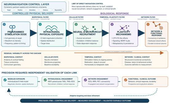
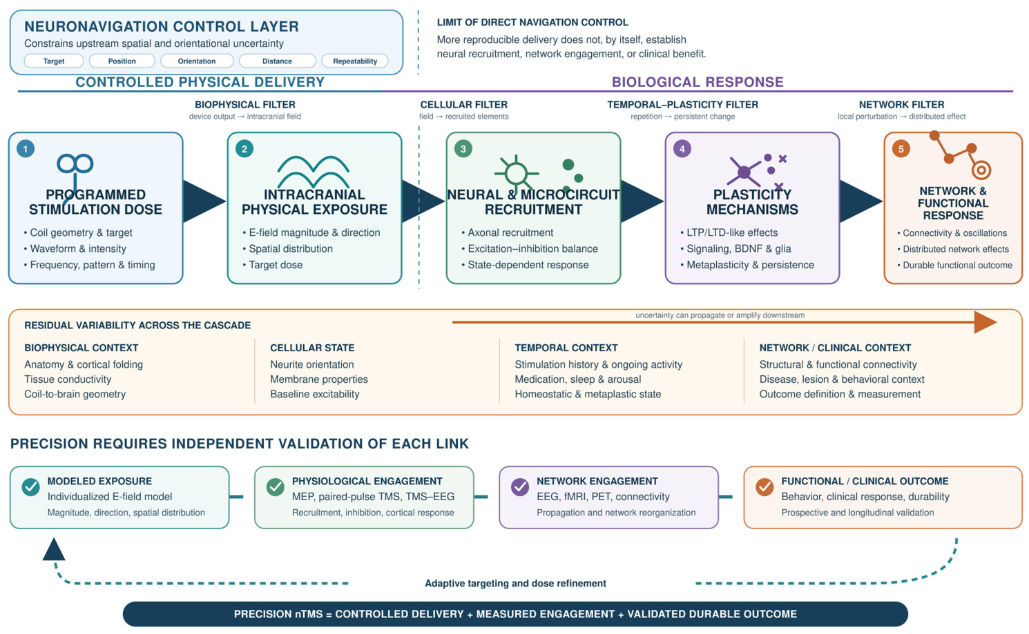
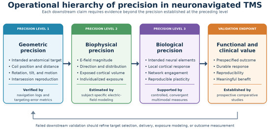
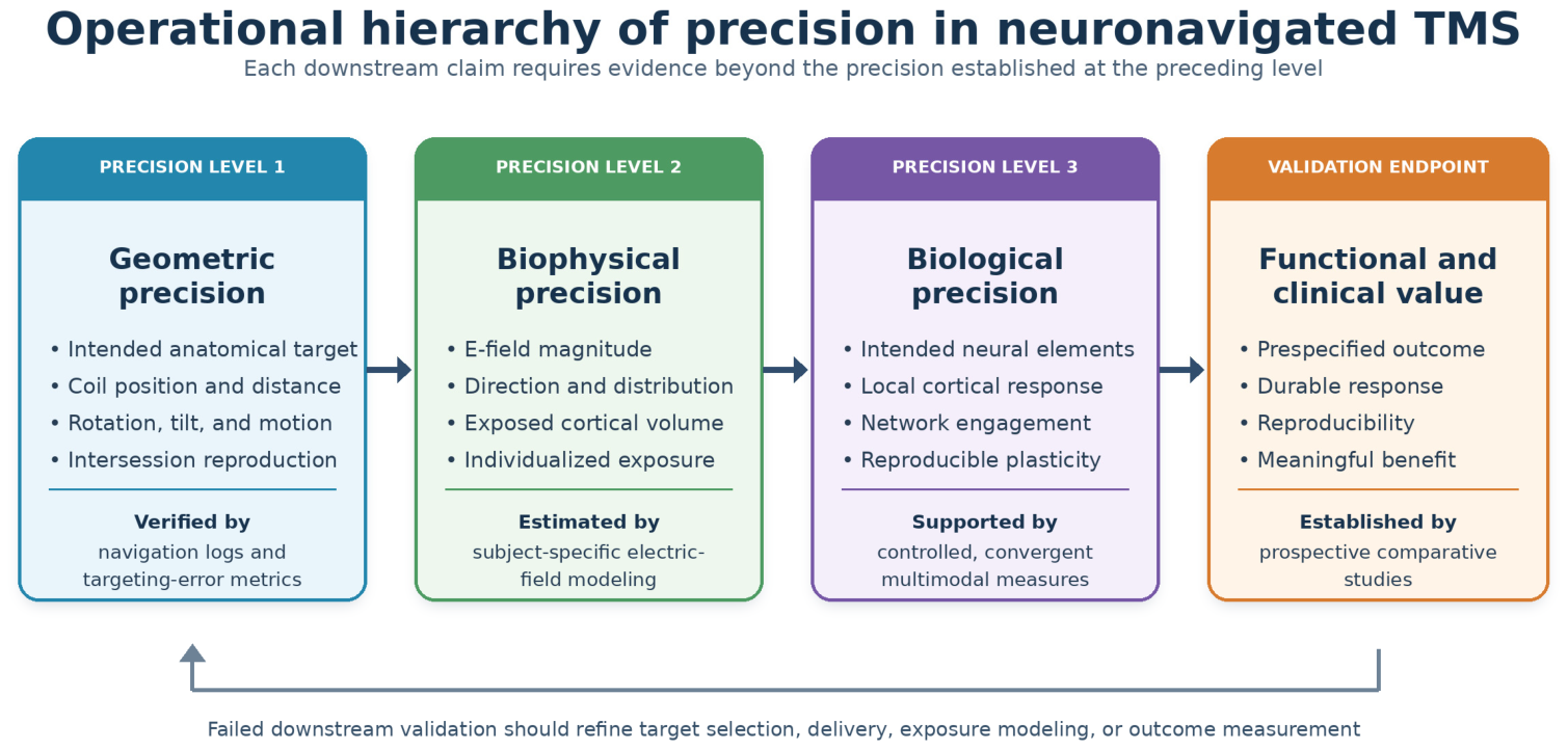

Disclaimer/Publisher’s Note: The statements, opinions and data contained in all publications are solely those of the individual author(s) and contributor(s) and not of MDPI and/or the editor(s). MDPI and/or the editor(s) disclaim responsibility for any injury to people or property resulting from any ideas, methods, instructions or products referred to in the content. |
© 2026 by the authors. Licensee MDPI, Basel, Switzerland. This article is an open access article distributed under the terms and conditions of the Creative Commons Attribution (CC BY) license.