


Abstract
Background: People with stroke (PwS) fall nearly twice as often as healthy counterparts, which may reflect deficits in reactive balance control. Those with greater motor impairment fall more often and tolerate lower perturbation intensities, so they may need supplemental agents within perturbation-based training. Our recent work showed that functional electrical stimulation (FES) during unpredicted stance slips immediately improved reactive balance biomechanics and reduced laboratory falls. Whether these peripheral and behavioral effects are accompanied by cortical changes remains unknown. Using mobile brain imaging via electroencephalography (EEG), we examined sensorimotor beta power during slips with and without FES. Methods: Nineteen participants (75.35 ± 12.62 years) with moderate-to-high motor impairment (Chedoke McMaster Leg ≤ 4/7) experienced two unpredicted stance slips in a randomized order: one with FES to the paretic quadriceps 50–500 ms after perturbation onset and one with sub-sensory sham stimulation (Sham-FES). Fifteen participants had artifact-free recordings. Primary outcomes were beta power in the 400 ms windows before and after perturbation onset. Secondary outcomes were laboratory falls and reactive stability; tertiary outcomes were compensatory step length, initiation time, and execution time. We compared conditions using McNemar and paired t-tests. Results: Pre-perturbation beta power was similar across conditions (p > 0.05). Beta power increased from the pre- to the post-perturbation window in both conditions (p < 0.01). Post-perturbation, PwS showed lower sensorimotor beta power during slips with FES than with Sham-FES (p < 0.05), accompanied by fewer laboratory falls, higher reactive stability, and shorter step initiation time. Conclusion: FES to the paretic quadriceps was accompanied by lower post-perturbation sensorimotor beta power and improved reactive balance control, with no pre-perturbation differences. These preliminary findings suggest a promising role for FES in reactive balance recovery among PwS and support further mechanistic study.
1. Introduction
Falls are one of the most common secondary complications in community-dwelling people with chronic stroke (PwS) [1,2,3], which often occur on exposure to environmental perturbations like slips and trips [4,5]. About 40% of falls resulting in serious injuries occur during slips while performing dynamic activities [6,7]. Compared to healthy counterparts, PwS exhibit twofold higher fall risk [3], which is postulated to be associated with sensorimotor and balance deficits [3,8]. Specifically, PwS demonstrate impaired reactive balance control (i.e., sensorimotor responses recruited following unpredicted perturbations to the body to maintain/restore balance) executed via change-in-support strategies (compensatory stepping/grasping) [9,10], which are necessary to avoid falls from large-magnitude environmental slips/trips [11,12].
Mechanisms of failed reactive balance control responses resulting in a fall have been commonly studied using biomechanical [13] and neuromuscular variables [14]. Specifically, studies have shown that adults who experience a fall due to an unexpected slip demonstrate poorer reactive control of the center-of-mass state and lower-limb support at touchdown of the recovery limb post-slip than those who recover balance without falling [13]. Such biomechanical differences are postulated to arise from variations in neuromuscular control of reactive balance recovery. For instance, studies have shown that adults who fell on unpredicted slips demonstrated delayed onset times of knee muscle activation and higher levels of agonist-antagonist coactivation about the knee joint of the slipping limb, and fewer between-limb muscle synergies than those who recovered successfully [14]. These differential neuromuscular activation patterns observed between fallers and non-fallers could be attributed to differential modulation of cortical processes involved in reactive balance recovery [15,16]. However, the cortical mechanisms governing these reactive balance failures have been comparatively understudied until recently.
Recent evidence from mobile brain imaging has suggested a notable role for the cerebral cortex in the mechanisms governing reactive balance control [11,15]. Studies using electroencephalography (EEG) have demonstrated that sensorimotor beta oscillations (13–30 Hz), which represent sensorimotor processing and the status quo of the current postural state, are modulated by perturbation magnitude, balance ability, and perturbation outcome [17,18,19]. Specifically, higher post-perturbation beta power at the midline sensorimotor electrode (Cz) has been associated with worse reactive balance performance and a greater risk of falling, especially among vulnerable populations like healthy older adults and those with stroke [18,19,20,21]. In our recent study of healthy young adults exposed to novel gait slips, individuals who fell exhibited significantly greater early post-perturbation beta power (0–150 ms) than those who recovered, suggesting heightened cortical error detection and greater dependence on cortical resources for sensorimotor processing [16]. In contrast, successful recovery showed a delayed increase in beta power (150–300 ms), consistent with cortical command generation for compensatory stepping [16]. To date, however, comparable EEG investigations of slip-induced perturbations in PwS, a population with substantially greater cortical and sensorimotor deficits, are lacking. Understanding these cortical contributions also raises the question of how reactive balance deficits in PwS, which are largely attributed to impaired paretic limb control, can be addressed using neuromodulatory therapeutic agents. One such low-cost neuromodulatory agent commonly used in clinical and research settings is functional electrical stimulation (FES).
Functional electrical stimulation (FES) is peripheral stimulation of innervated muscles during functional movements (e.g., grasping or walking) typically used in upper motor neuron lesions (e.g., stroke/spinal cord injuries) for either replacing lost function (as an orthotic/prosthesis) [22,23,24] or restoring lost function and promoting motor relearning (therapeutic agent) [25,26,27]. In recent decades, FES applied to the paretic limb, combined with functional tasks, has been extensively used in clinical and research settings. Several studies have suggested the effectiveness of long-term FES in conjunction with conventional therapies for improving gait parameters like cadence, gait speed, walking endurance, and symmetry in PwS [28,29,30,31,32]. In a recent study by our lab, we delivered FES to the paretic quadriceps during treadmill-induced stance slips among ambulatory PwS [33]. FES to the paretic quadriceps at slip onset resulted in fewer laboratory falls associated with higher reactive stability and increased vertical limb support compared to slips without FES [33]. Further, with FES, PwS executed a longer, faster compensatory step. However, these findings were limited to biomechanical outcomes, and it remains unknown whether FES improves reactive balance and fall risk through a central component of its mechanism of action. In the current preliminary study, we aimed to examine differences in sensorimotor beta power at Cz between unpredicted stance slips delivered with and without FES to the paretic quadriceps among PwS. We hypothesized that FES delivered at slip onset would be associated with lower post-perturbation sensorimotor beta power than slips without FES. We further hypothesized that lower post-perturbation beta power would parallel improvements in biomechanical outcomes of reactive balance (i.e., fewer laboratory-induced falls, higher reactive stability, and more effective compensatory stepping) during FES-assisted slips.
2. Methods
2.1. Participants
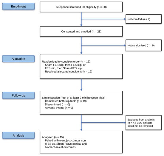
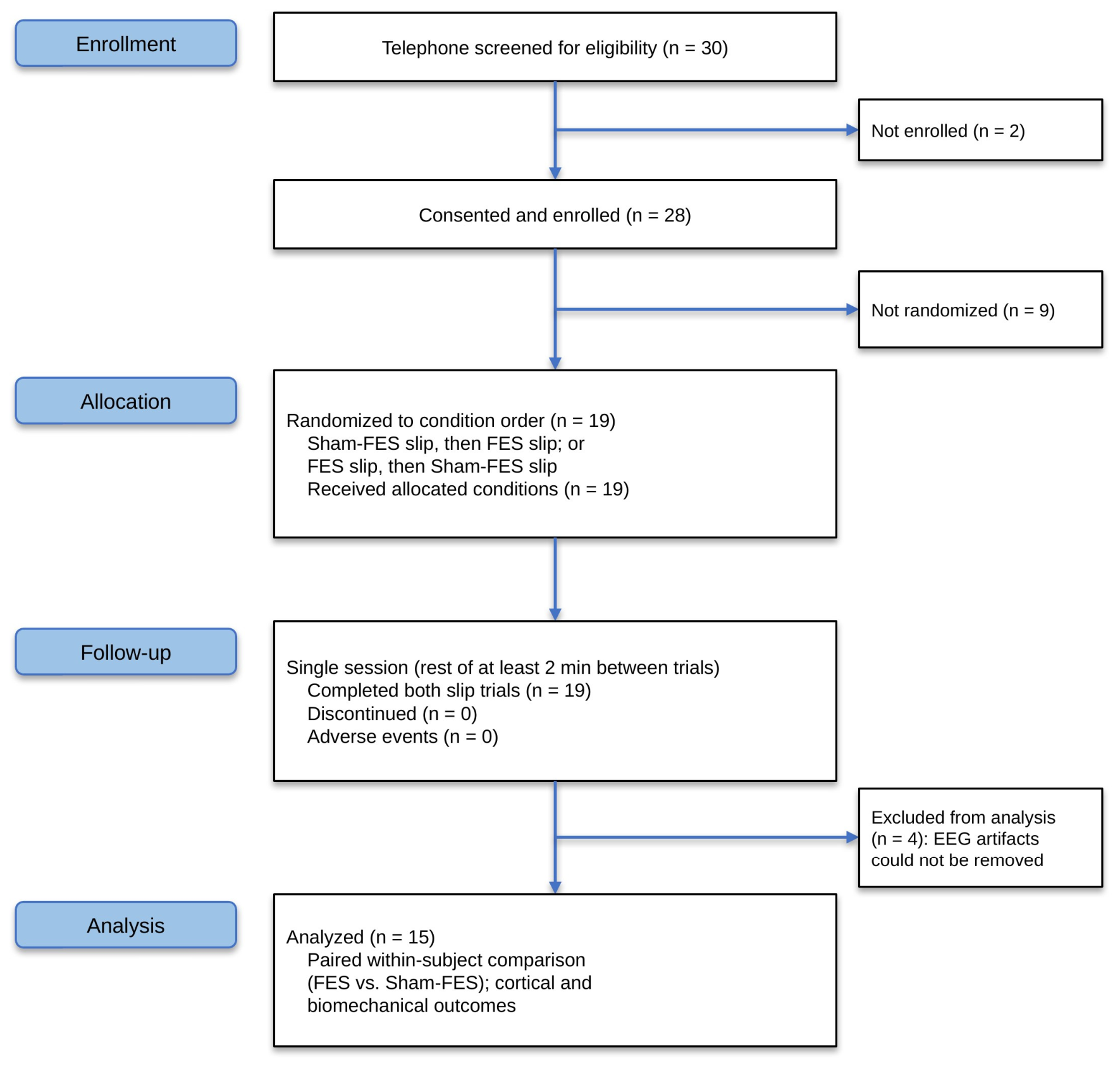
This preliminary study used a single-session, within-subject crossover design in which each participant experienced an unpredicted stance slip with and without FES in a randomized order, thereby serving as their own control. Nineteen community-dwelling older adults with chronic hemiparetic stroke were recruited via stroke support groups, organizational databases, and university hospital collaborations between August 2023 and August 2025. Inclusion criteria were: (1) age 40–90 years; (2) a confirmed diagnosis of unilateral stroke with onset greater than six months prior to enrollment; (3) moderate-to-high lower-limb motor impairment (Chedoke McMaster Stroke Assessment Leg score ≤ 4/7); and (4) ability to walk independently with or without an assistive device for at least 10 m. Participants were excluded if they had any additional neurological conditions, cardiovascular or musculoskeletal diagnoses that would preclude safe participation, severe cognitive impairment (Mini-Mental State Examination score < 25), significant aphasia (Mississippi Aphasia Screening Test ≤ 71/100), severe lower-limb spasticity (Modified Ashworth Scale > 2), or a history of epilepsy or seizure disorder. Participants who used a wearable FES device for foot drop (e.g., a peroneal nerve stimulator) as their primary walking aid in daily life, or who had any implanted or powered electronic device (e.g., implanted stimulator, pacemaker, or powered orthosis), were excluded to prevent carry-over effects of chronic stimulation and electrical interference with EEG signals. Written informed consent was obtained from all participants prior to enrollment. The preliminary study was approved by the Institutional Review Board at the University of Illinois Chicago (IRB # 2023-1233). The consort flow diagram in Figure 1 provides the participant information from recruitment through completion.
Figure 1.
CONSORT flow diagram of participant enrollment, allocation, follow-up, and analysis. Thirty individuals were screened by telephone for eligibility; 2 were not enrolled and 28 provided consent and were enrolled. Of these, 19 were randomized to a condition order (Sham-FES slip followed by FES slip, or FES slip followed by Sham-FES slip) and all received both allocated conditions. All 19 participants completed both slip trials within a single session, with at least 2 min of rest between trials, and there were no discontinuations or adverse events. Four participants were excluded from analysis because EEG artifacts could not be removed, leaving 15 participants for the paired within-subject comparison of cortical and biomechanical outcomes between the FES and Sham-FES conditions. FES, functional electrical stimulation; EEG, electroencephalography.
2.2. Experimental Setup
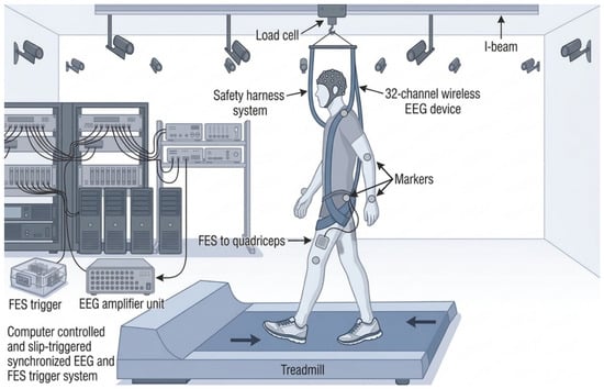
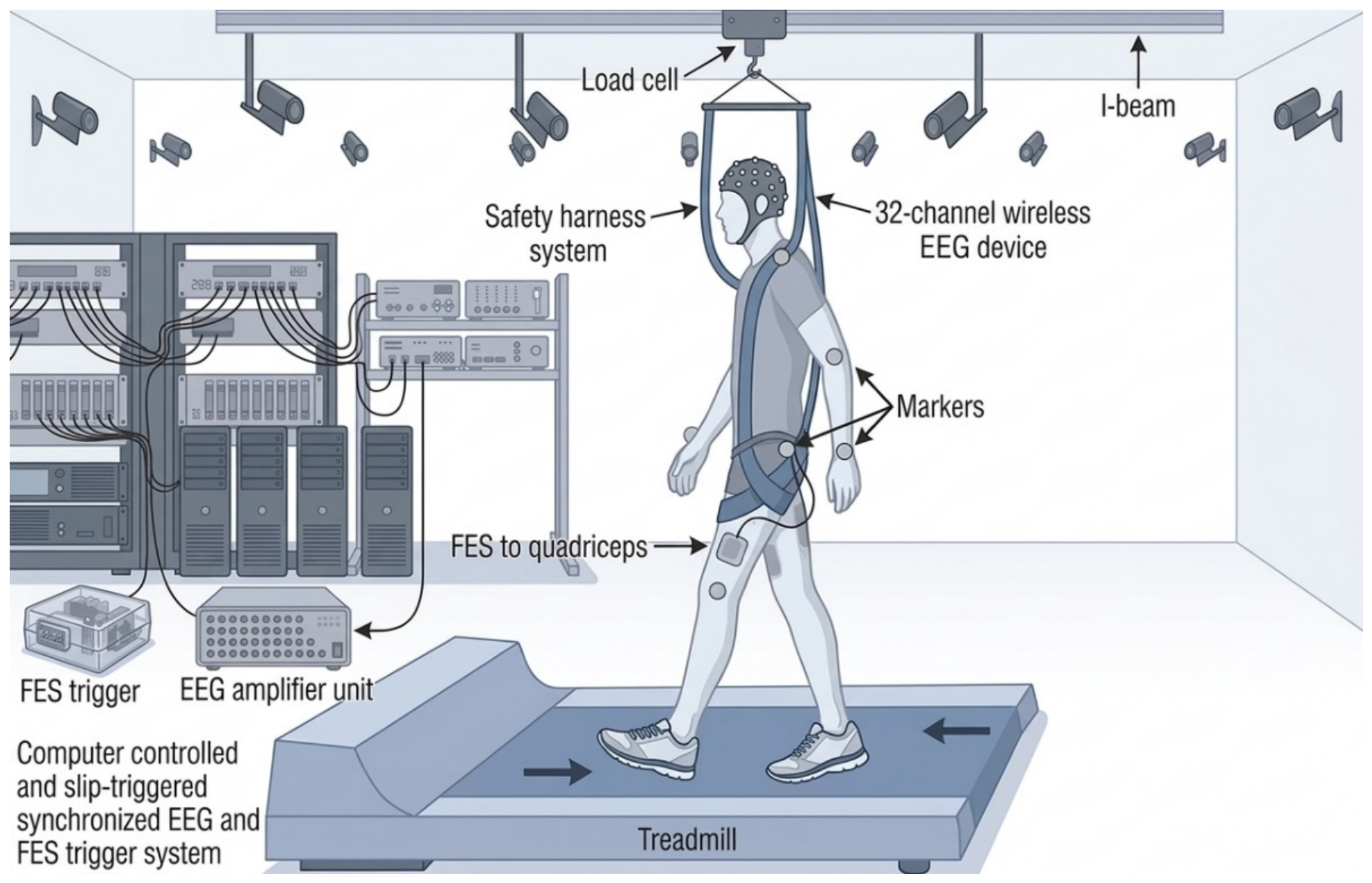
The experimental setup consisted of the ActiveStep motorized treadmill system (Simbex, Lebanon, NH, USA), which was used to deliver stance slip-like perturbations (Figure 2). The treadmill induces multidirectional perturbations via customized parameters of displacement, velocity, and acceleration, and was configured to deliver a forward, unilateral slip-like perturbation under the paretic limb. A customized heel switch, consisting of a thin force-sensitive resistor taped to the sole of the participant’s own shoe under the paretic heel, detected heel strike and triggered the perturbation during the early paretic stance phase. The heel switch served only as a sensor and trigger and provided no support or assistive function. Participants wore a full-body safety harness attached to an overhead load cell (600 Hz) to prevent falls to the ground and to classify perturbation outcomes. Full-body kinematics were recorded using a set of 30 light-reflective markers and an eight-camera 3D motion capture system (Motion Analysis, Qualisys AB, Gothenburg, Sweden) at 120 frames per second. Videos were recorded and synchronized with kinematic and load cell data.
Figure 2.
Experimental setup for mobile brain and biomechanical data acquisition during slip-perturbation walking. Participants walked on a motorized treadmill while secured in a full-body safety harness connected to an overhead bearing-and-load-cell system to prevent falls. A 32-channel wireless EEG device recorded cortical activity, and 3D kinematic data were captured by a Qualisys motion capture system. Reflective body markers were used for motion tracking, and perturbations were delivered as computer-controlled, unpredictable treadmill slips. The computer system simultaneously triggered the slip event and EEG recording to ensure precise temporal synchronization across neural and biomechanical signals. The heel-strike trigger (a force-sensitive resistor taped under the paretic heel) served only to detect heel-strike and provided no assistive function. Surface EMG was not recorded in the present study.
Functional electrical stimulation was delivered to the paretic quadriceps using the TrainFES Stimulation System (Biomedical Devices SpA, Santiago, Chile), a multi-channel FES device consisting of a control unit and cables connected to hypoallergenic surface stimulation electrodes. Two electrodes were placed on the muscle belly of the rectus femoris, vastus medialis, and vastus lateralis of the paretic limb (standard rectangular self-adhesive surface electrodes; TrainFES, Biomedical Devices SpA, Santiago, Chile). The FES device was connected to a wireless, triaxial Bluetooth sensor attached to the treadmill belt; this sensor detected perturbation onset and triggered the stimulation train. Stimulation parameters were individualized prior to the slip trials based on the participant’s maximal tolerable motor threshold (strong motor but not painful). FES was delivered as a fixed-duration train that began 50 milliseconds and ended 500 milliseconds after perturbation onset at a moderate-to-high intensity (30–50 mA), low frequency (20–45 Hz), and pulse duration sufficient to elicit a visible contraction of the paretic quadriceps (i.e., the slipping limb, which was the leading limb at perturbation onset) [34].
2.3. Experimental Protocol
All testing was completed in a single laboratory session, which included informed consent, clinical assessments, EEG and reflective marker set-up, a 10-min warm-up of low-intensity lower-limb exercises, and slip trials. Participants wore their regular orthotic device (e.g., ankle-foot orthosis) as used in daily life but did not use their habitual walking aid (e.g., cane) during treadmill trials. Following the warm-up, participants were informed that a slip might occur without being told its timing or nature and were instructed to attempt to regain balance and to continue walking if a slip occurred.
Each participant experienced one unpredicted stance slip in each of two conditions, delivered in a randomized order: Sham-FES and FES. Perturbations were delivered as large-magnitude forward slips (acceleration: −16 m/s2) under the paretic limb during stance. In the FES condition, stimulation was delivered to the paretic quadriceps as a fixed 50–500 ms post-perturbation train at the individualized parameters described in Section 2.2. In the Sham-FES condition, electrodes were placed identically, and the device was switched on, but stimulation was delivered at a sub-sensory intensity (0–10 mA, 50–100 Hz) that produced no perceptible sensation or visible quadriceps contraction; this was done to blind participants to the condition rather than to deliver a therapeutic dose. Participants were not informed which condition was being delivered. A seated rest of at least two minutes was provided between trials.
2.4. EEG Data Collection and Processing
Continuous cortical activity was recorded using a 32-channel mobile EEG system (g.Nautilus Research, g.tec neurotechnology, USA Inc., Castleton-on-Hudson, NY, USA) with active electrodes placed according to the international 10–20 system. Signals were referenced to the earlobe electrode, and the ground electrode was positioned at the anterior fontanelle. EEG was recorded at 500 Hz with a 24-bit resolution and an in-built anti-aliasing filter. All electrode impedances were maintained below 10 kΩ. A transistor-transistor logic signal synchronized the EEG data with the kinematic and load cell data.
Offline EEG preprocessing and analysis were performed using EEGLAB version 2023.0 functions and custom MATLAB R2024b scripts. Raw signals were band-pass filtered between 0.5 and 100 Hz to remove baseline drift and high-frequency noise. Channels with excessive noise (standard deviation > 4), flat signals (>5 s duration), or poor cross-channel correlation (r < 0.8) were rejected using automated EEGLAB functions (pop_clean_rawdata). Independent component analysis (ICA) was performed to identify and remove non-neural artifacts, including eye blinks, cardiac signals, muscle activity, and movement-related artifacts. Corrected signals were divided into 10-s single-trial epochs aligned to perturbation onset via the transistor-transistor logic signal. Recordings in which artifacts could not be removed (i.e., more than 25% of channels rejected, or movement/EMG artifact persisting after ICA within the post-perturbation window) were excluded from all analyses. To keep the cortical and biomechanical outcomes within an identical paired sample, secondary and tertiary outcomes were analyzed only for participants with artifact-free EEG recordings.
Consistent with our previous work [16], the present preliminary study focused beta power analysis on the Cz electrode, which overlies the midline sensorimotor cortex and has been shown to reliably capture beta oscillatory dynamics during reactive balance perturbations in both healthy and clinical populations [17,18,19]. We selected sensorimotor beta power (13–30 Hz) as our primary cortical outcome for three reasons. First, beta is the frequency band most consistently linked to reactive balance control and postulated to represent sensorimotor processing and the status quo of the current postural state, and studies have shown that it is reliably modulated by perturbation magnitude, balance ability, and perturbation outcome [17,18,19]. Second, post-perturbation beta power at the midline sensorimotor electrode (Cz) is sensitive to balance performance and fall risk, with greater beta power associated with worse reactive recovery and a higher likelihood of falling, making it a functionally meaningful marker for detecting intervention-related differences [17,18,19,20,21]. Third, the timing and magnitude of beta modulation are postulated to be able to distinguish cortical error detection from cortical command generation for compensatory stepping [16] and thus could provide a mechanistic measure of whether FES has a central nervous system effect or cortical mechanism of action in enhancing reactive balance control and reducing fall risk.
A time-frequency analysis was conducted using wavelet-based event-related spectral perturbation (ERSP) analysis (EEGLAB function: pop_newtimef) over the beta frequency range (13–30 Hz). A tapered Morlet wavelet was used to quantify power (energy per unit time), calculated as the square of the FFT amplitude at each frequency. ERSP values were computed across 200 time points (10 milliseconds apart) and 10 linearly spaced frequencies from 13 to 30 Hz. The mean baseline log-spectrum (from −1000 to −800 milliseconds prior to perturbation onset) was subtracted from each spectral estimate to produce baseline-normalized time-frequency distributions. Single-trial ERSP values across four sampled beta frequencies (centered on 15, 20, 24, and 28 Hz) were averaged to yield a single waveform of beta power per trial and condition.
2.5. Outcome Measures
2.5.1. Primary Outcome Measures
The primary cortical outcomes were peak sensorimotor beta power (dB) extracted at Cz across two time windows: (1) pre-perturbation: peak beta power during the 400-millisecond window preceding perturbation onset; and (2) post-perturbation: peak beta power during the 400-millisecond window following perturbation onset. These windows were selected based on established literature on reactive balance and our prior work examining beta dynamics at pre- and post-perturbation time points [17,18,19].
2.5.2. Secondary Outcome Measures
Laboratory-induced fall: The outcome of a slip trial was classified as a fall if the force detected by the load cell exceeded 30% of the participant’s body weight after perturbation [13,35] and was confirmed by video recording [36]. Otherwise, it was regarded as a balanced recovery.
Reactive stability: calculated from the COM state (position and velocity) and computed at two instants post-slipping: liftoff (LO) of the compensatory (non-paretic) limb and the instant immediately preceding its first touchdown (pre-TD). The COM position was obtained relative to the rear edge of the base of support and normalized by participants’ foot length [37]. The COM velocity was calculated as the first-order derivative of the COM position and was expressed relative to the heel velocity of the base of support [38] and normalized by a dimensionless factor of √(g·h), here ‘g’ represents the acceleration of gravity and ‘h’ represents the height of the participant [39]. COM stability, expressed as the margin of stability (MOS), was computed as the shortest distance of the instantaneous COM state (position and velocity) to the predetermined computational boundary for backward balance loss [12,40]. Values < 0 indicated a greater possibility of experiencing backward loss of balance and vice versa for values > 0.
2.5.3. Tertiary Outcome Measures
Compensatory step length (meters): assessed as the distance from the paretic slipping heel to the non-paretic heel at the instant of recovery touchdown [33,41]. A more negative step length indicated a longer backward compensatory step and vice versa.
Compensatory step initiation time (seconds): defined as the time taken for liftoff of the non-paretic limb after the perturbation onset [33].
Compensatory step execution time (seconds): defined as the time taken from liftoff of the non-paretic limb to its first touchdown after perturbation [33].
Proactive stability: computed as the MOS at the instant of paretic (slipping) touchdown immediately before the perturbation onset. Proactive stability was assessed to rule out COM state changes occurring in anticipation of a perturbation and confirm that differences in COM state were purely attributed to reactive balance variables.
2.6. Statistical Analysis
Our analysis plan was structured to address two questions: whether FES applied at slip onset modulates sensorimotor cortical activity, and whether any cortical changes parallel changes in biomechanical outcomes of reactive balance. Descriptive statistics (means and standard deviations or medians and interquartile ranges) were first computed for participant demographics, clinical characteristics (Chedoke McMaster Stroke Assessment, Fugl-Meyer Lower Extremity, Mini-Mental State Examination), and all primary and secondary outcomes. The distribution of all outcome variables was then assessed using the Shapiro–Wilk test of normality with an alpha level of 0.05. Parametric statistics were used for variables meeting the normality assumption (p > 0.05); otherwise, non-parametric equivalents (e.g., Wilcoxon signed-rank test) were applied.
For the primary cortical outcome, peak sensorimotor beta power at Cz was compared between conditions using paired-samples t-tests. Because the hypotheses were directional and specified a priori (i.e., lower post-perturbation beta power with FES than with Sham-FES), one-tailed tests were used for the between-condition comparisons of the primary outcome. Pre- versus post-perturbation beta power within each condition was compared using paired-samples t-tests to characterize the effect of the perturbation itself. For the secondary biomechanical outcomes, paired t-tests (or Wilcoxon signed-rank tests for non-normally distributed data) were used to compare reactive stability between conditions. The percentage of laboratory-induced falls was compared using the exact McNemar test, as each participant experienced both conditions and the perturbation outcome (fall vs. recovery) was a dichotomous variable. The tertiary outcomes, including the compensatory step length, step initiation time, and step execution time, were similarly compared using paired t-tests (or Wilcoxon signed-rank tests where applicable). Effect sizes were calculated as Cohen’s d, with values of 0.2, 0.5, and 0.8 interpreted as small, medium, and large effects, respectively [42]. This analysis was guided by previous studies examining brain–behavior relationships during reactive balance tasks [16,20]. All statistical analyses were performed using the Statistical Package for the Social Sciences (SPSS) software, version 27 (IBM Corporation, Armonk, NY, USA), with the alpha level set at 0.05.
3. Results
Participant characteristics. Nineteen participants enrolled in the study, and all completed both slip trials without adverse events and tolerated the FES well. Four participants were excluded from the EEG analysis because artifacts could not be removed (Section 2.4); thus, the analytic sample comprised 15 participants (age 75.35 ± 12.62 years; 6 women, 9 men; 5 left/10 right hemiparesis; time since stroke 8.25 ± 5.6 years; Chedoke McMaster Leg 3.0 ± 1.0/7; Fugl-Meyer Lower Extremity 24.5 ± 3.5/34; MMSE 28.5 ± 1.5/30) (Table 1). Excluded and included participants did not differ in age, sex distribution, or motor impairment.
Table 1.
Demographic and clinical characteristics of the analytic sample. Values are mean ± SD or n. Participants who habitually used an ankle-foot orthosis wore it during all slip trials (Section 2.3). The analytic sample comprises participants with artifact-free EEG recordings.
3.1. Primary Measures Results
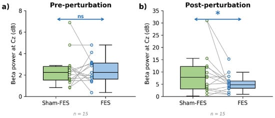
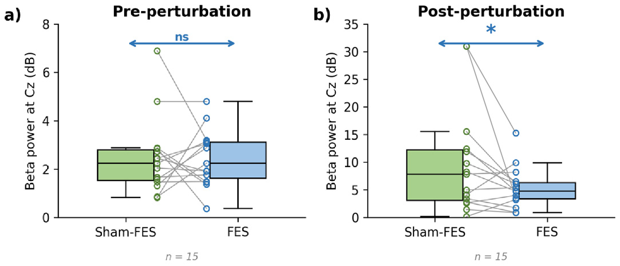
Within each condition, post-perturbation beta power was higher than pre-perturbation beta power (Sham-FES: t(14) = 2.80, p = 0.007, d = 0.72; FES: t(14) = 3.23, p = 0.003, d = 0.83; Figure 3 and Figure 4). As expected, pre-perturbation beta power did not differ between the Sham-FES and FES conditions (t(14) = 0.038, p = 0.49, d = 0.01; Figure 3a and Figure 4a). Post-perturbation beta power was lower during slips with FES than with Sham-FES (t(14) = 2.055, p = 0.03, d = 0.53; Figure 3b and Figure 4b).
Figure 3.
Sensorimotor beta power before and after perturbation onset. Peak beta power (13–30 Hz, dB relative to baseline) at the Cz electrode during unpredicted stance slips delivered with sham stimulation (Sham-FES; green) and with FES to the paretic quadriceps 50–500 ms after slip onset (FES; blue). (a) Pre-perturbation window (−400 to 0 ms). (b) Post-perturbation window (0–400 ms). Box plots show the median and interquartile range (whiskers: most extreme non-outlier values); circles show individual participants, and grey lines connect each participant’s paired values across conditions (n = 15). Arrows denote the between-condition comparison (one-tailed paired t-test; * p < 0.05; ns, not significant). Note that the y-axis range differs between panels (0–8 dB in (a); 0–35 dB in (b)) to display the pre-perturbation values, which are an order of magnitude smaller.
Figure 4.
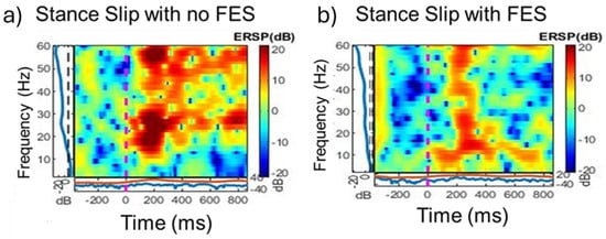
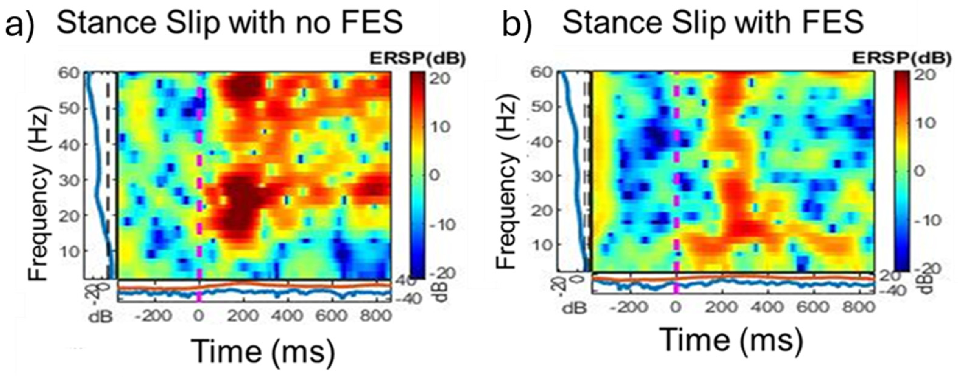
Event-related spectral perturbation (ERSP) at the Cz electrode during stance-slip perturbations with and without FES. Time-frequency representations of cortical activity recorded at the Cz electrode during single-task stance-slip trials, displayed as ERSP (in dB) relative to a pre-perturbation baseline. The (a) panel shows a representative single trial with functional electrical stimulation (FES) delivered to the paretic quadriceps 50–500 ms after slip onset, and the (b) panel shows a representative single trial with sham stimulation (Sham-FES) from the same participant (group n = 15). The x-axis represents time (ms) relative to perturbation onset (0 ms; magenta dashed line), and the y-axis represents frequency (Hz) from 5 to 60 Hz. Warmer colors (red) indicate event-related synchronization (increased spectral power), while cooler colors (blue) indicate event-related desynchronization (decreased spectral power). The black dashed line marks the pre-perturbation reference window. Visual inspection of the post-perturbation period (0–500 ms) reveals a more pronounced and sustained increase in beta-band (13–30 Hz) power during the Sham-FES trial compared to the FES trial, consistent with greater cortical engagement for error detection and sensorimotor processing in the absence of FES-assisted recovery.
3.2. Secondary Measures Results
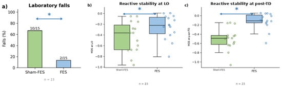
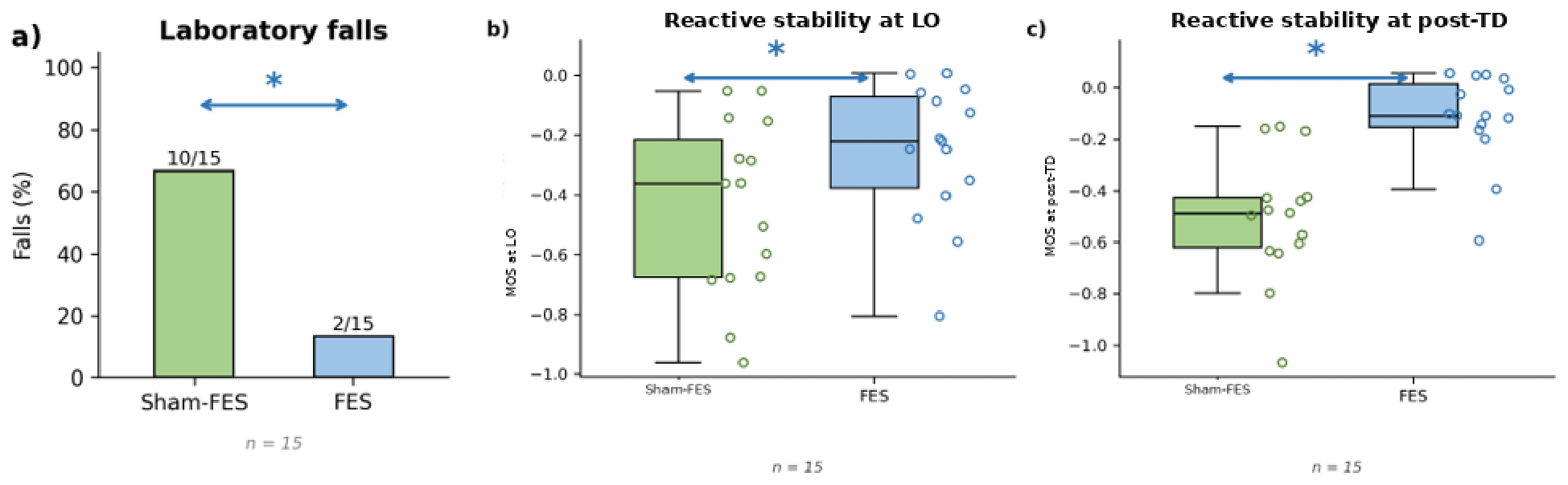
A lower percentage of participants fell during stance slips with FES (2/15, 13.3%) than with Sham-FES (10/15, 66.7%; exact McNemar test, p = 0.008; Figure 5a). Both participants who fell with FES also fell with Sham-FES; eight participants fell with Sham-FES but recovered with FES, and none showed the reverse pattern. Proactive stability (i.e., MOS at paretic touchdown before slip onset) did not differ between conditions (t(14) = 0.87, p = 0.40, d = 0.22), confirming that between-condition differences in reactive stability were not attributable to anticipatory changes in the COM state. Participants displayed greater reactive stability (MOS) at compensatory step liftoff (LO) with FES than with Sham-FES (t(14) = 3.83, p = 0.002, d = 0.99; Figure 5b). Stability at pre-touchdown (pre-TD) was likewise higher with FES than with Sham-FES (t(14) = 7.62, p < 0.001, d = 1.97; Figure 5c).
Figure 5.
Laboratory fall outcomes and stability during stance slips with sham stimulation (Sham-FES) and with FES. (a) Percentage of trials classified as a fall versus a recovery in each condition (exact McNemar test). (b) Reactive stability, expressed as the margin of stability (MOS), at compensatory step liftoff (LO). (c) Reactive stability at the instant immediately before compensatory step touchdown (pre-TD). Higher (less negative) MOS values indicate a lower likelihood of backward balance loss. Box plots show the median and interquartile range; circles show individual participants, jittered horizontally for visibility (n = 15). * p < 0.05; ns, not significant.
3.3. Tertiary Measures Results
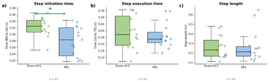
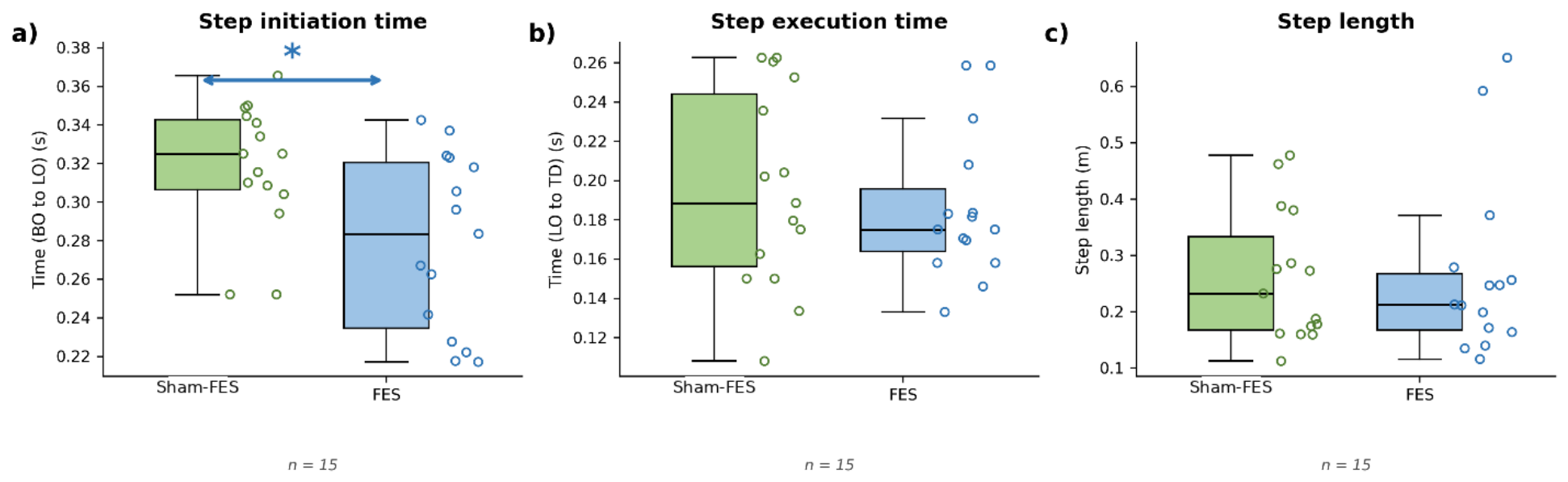
Participants showed a reduced step initiation time during stance slips with FES compared to Sham-FES (t(14) = 4.02, p = 0.001, d = 1.04) (Figure 6a). There were no differences in step execution time (t(14) = 0.93, p = 0.37, d = 0.24) (Figure 6b) or in compensatory step length (t(14) = 0.21, p = 0.84, d = 0.05) (Figure 6c).
Figure 6.
Step kinematic outcomes during stance slips with sham stimulation (Sham-FES) and with FES. (a) Step initiation time (time from perturbation onset to foot lift-off) was significantly shorter during FES-assisted slips, indicating faster initiation of compensatory stepping. (b) Step execution time (the time from foot lift-off to touchdown) did not differ between conditions. (c) Compensatory step length did not differ between conditions. Box plots display the median and interquartile range; individual participant values are overlaid, jittered horizontally for visibility (n = 15). Asterisks (*) denote statistically significant differences between conditions (p < 0.05). Green circles represent individual participant data for Sham-FES condition and blue circles represent individual participant data for FES-condition.
4. Discussion
This study is, to our knowledge, the first to examine the cortical correlates of FES-induced effects on reactive balance control among PwS. Our results showed that PwS with moderate-to-high motor impairment exhibited lower post-perturbation sensorimotor beta power during slips with FES than during slips with sham stimulation (Sham-FES), with no differences in beta power before the onset of the perturbation. The changes in post-perturbation sensorimotor beta power were accompanied by biomechanical changes, including a lower percentage of laboratory-induced falls, higher reactive stability, and shorter step initiation time during FES-assisted slips compared to Sham-FES. Together, these findings suggest that FES delivered to the paretic quadriceps at slip onset is associated with enhanced reactive balance recovery, with concurrent behavioral (biomechanical) and neural (cortical) correlates.
Sensorimotor beta power at Cz was selected because it has been repeatedly shown to index cortical engagement during reactive balance: post-perturbation beta power increases with perturbation magnitude, is higher in individuals with poorer balance ability and in those who fall, and is delayed in PwS relative to controls [16,18,19,20,21]. Hence, a change in post-perturbation beta power between conditions can be interpreted as a change in the cortical demand for sensorimotor processing during recovery. The biomechanical outcomes, in turn, are established indices of reactive balance in PwS: the MOS at compensatory step liftoff and pre-touchdown discriminates fallers from non-fallers [43], and the harness load-cell criterion for a laboratory fall has been validated against video [35]. Because both were measured within the same slip trial, the lower beta power observed with FES can be interpreted as cortical modulation that accompanied a functional improvement in reactive balance, rather than as an isolated electrophysiological finding. Whether this coupling may be causal is addressed below.
Pre-perturbation beta power was comparable between the two conditions, indicating that participants’ initial cortical state did not differ before slip onset. This null finding is consistent with our previous work in healthy young adults exposed to unexpected slips while walking, in which pre-perturbation beta power did not differentiate between those who fell and those who recovered [16]. Two factors likely account for this. First, the slips were unpredictable: participants knew a slip was possible, but not when or which limb would be affected, so the cerebral cortex had no opportunity to adjust the central set or internal model in advance of the perturbation [44]. Second, FES was triggered at slip onset rather than before it, so any FES-induced modulation of cortical activity would, by design, emerge only after the perturbation. Together, these results indicate that the effect of FES on cortical activity is event-related rather than a shift in baseline cortical state. The slip itself increased sensorimotor beta power from the pre- to the post-perturbation window in both conditions. This is consistent with prior EEG studies in which post-perturbation beta power at Cz increased relative to baseline and scaled with perturbation magnitude and balance ability [19,20], and with our previous work in young adults exposed to gait slips [16]. Hence, FES did not abolish the cortical response to the perturbation; rather, the perturbation-evoked beta response was smaller in magnitude with FES than with sham stimulation. In other words, the sensorimotor cortex still responded to the slip when FES was delivered, just to a lesser degree.
Post-perturbation sensorimotor beta power was lower during FES-assisted slips than during slips without FES. To interpret this difference, it helps to consider what cortical beta power reflects during a reactive balance event. When an unpredicted perturbation occurs, afferent inputs from somatosensory, visual, and vestibular receptors are relayed to the primary somatosensory cortex for sensory reweighting and multisensory integration [45,46,47]. A central component of this processing is error detection, the cortical recognition of a mismatch between the expected postural state (based on the internal model) and the ongoing postural state (based on real-time sensory feedback) [48,49]. Studies in healthy young and older adults have shown that higher post-perturbation sensorimotor beta power tracks with worse reactive balance performance, higher perturbation magnitude, and greater fall risk [16,19,20]. Lower beta power with FES, then, is consistent with (although does not demonstrate) reduced cortical error detection and less cortical demand for sensorimotor processing during reactive balance recovery. An alternative interpretation is that the synchronous FES volley could act as a competing afferent input, gating rather than reducing cortical error detection. However, our biomechanical findings argue against pure sensory gating, given that the compensatory response was more, not less, effective with FES. Differentiating reduced cortical demand from afferent gating would require future paradigms with parametric variation in FES intensity and timing relative to perturbation onset to understand the mechanisms of action of FES.
Two mechanisms could explain the observed reduction in sensorimotor beta power. The first relates to peripheral afferent feedback. PwS exhibit altered sensorimotor processing due to lesion-induced disruptions in cortical and corticospinal pathways [50,51,52], and during a slip without FES the cerebral cortex must compensate for deficient afferent feedback from the paretic limb and altered descending motor commands. FES applied to the paretic quadriceps at slip onset could have provided immediate, synchronized peripheral input by activating muscle spindles, neuromuscular junctions, and cutaneous afferents. This supplemental afferent input could lessen the cortical demand for postural error detection, i.e., the difference between the expected and actual postural state, thus displaying as lower post-perturbation beta power. The second mechanism relates to the success of the compensatory response itself. By facilitating the timely activation of the paretic quadriceps, FES could support a more effective compensatory step, again reducing the gap between expected and ongoing postural states. A smaller mismatch translates into less work for the cortex, reflected in lower beta power. Both mechanisms are consistent with the conceptual framework proposed below on the neuromodulatory effects of FES, which act through peripheral pathways (enhanced afferent feedback) and central pathways (increased cerebral blood perfusion and neuronal activity in sensorimotor cortices) [53,54].
The cortical changes paralleled improvements in biomechanical outcomes, including fewer laboratory-induced falls, higher reactive stability, and shorter step initiation times during FES-assisted slips. These results extend our previous work in which FES applied to the paretic quadriceps during stance slips reduced falls and improved reactive stability and vertical limb support [34]. The biomechanical improvements have a straightforward neuromuscular explanation. PwS exhibit weakness and delayed activation of the paretic quadriceps, which contributes to limb collapse and inadequate weight transfer during reactive balance responses [14,43]. Delivering FES to the paretic quadriceps within 50–500 ms after slip onset directly addresses these deficits, supporting timely muscle activation and a more effective compensatory response. The shorter step initiation time during FES-assisted slips also points to improved temporal control of the compensatory step, likely mediated by enhanced proprioceptive feedback and motor control of the paretic limb. The cortical and biomechanical findings point to a brain–behavior coupling in FES-assisted reactive balance recovery. With FES, enhanced peripheral afferent feedback and a more effective compensatory step could reduce the cortical demand for error detection and sensorimotor processing, reflected as lower post-perturbation beta power. Without FES (i.e., with sham stimulation), the cortex appears to compensate for deficient peripheral feedback and altered motor control by increasing cortical engagement, as reflected in higher beta power and poorer reactive balance performance. Beyond its peripheral neuromuscular effects, then, FES may also act as a neuromodulatory adjunct that shapes the cortical correlates of reactive balance. This interpretation is consistent with evidence that long-term FES therapy can induce neuroplastic changes via enhanced cerebral blood perfusion, increased brain-derived neurotrophic factor (BDNF), and synaptic plasticity [54,55], as well as increased corticospinal excitability measured with transcranial magnetic stimulation [56] and normalized spinal reciprocal inhibition between ankle antagonists [57]. Whether the immediate cortical changes observed here translate to lasting plasticity with repeated exposure is a question for future longitudinal work.
4.1. Proposed Conceptual Framework for the Central Effects of FES
Taken together, our findings allow us to propose a preliminary framework for how FES could enhance reactive balance control via a central pathway in PwS. First, FES provides a strong, time-locked somatosensory volley from the paretic limb at perturbation onset, which could enhance somatosensory contribution to sensory reweighting and thereby support more balanced multisensory integration during recovery [58,59]. Second, by delivering supplemental afferent input that compensates for deficient peripheral sensory feedback after stroke, FES could reduce the cortical demand for postural error detection (i.e., the mismatch between the expected and existing postural state), which would be observed as a smaller post-perturbation rise in sensorimotor beta power at the midline electrode [19,60]. We emphasize that this framework is derived from the associations observed here and remains to be tested directly.
Clinical relevance: Training program design. Perturbation-based balance training is effective for reducing falls but is often delivered at reduced intensity, or not at all, to PwS with moderate-to-high motor impairment due to the risk of repeated falls into the harness. Our findings suggest that FES to the paretic quadriceps, time-locked to perturbation onset, could serve as a scaffold that allows these individuals to safely experience large-magnitude perturbations and generate an effective compensatory step. In practice, FES could be applied during the early phase of training and progressively withdrawn as reactive responses improve, in a manner analogous to fading physical assistance. Risk screening. Post-perturbation sensorimotor beta power indexed the cortical demand of the slip and was sensitive to a single-trial intervention; hence, it could serve as a marker of an individual’s reliance on cortical resources during reactive balance and their response to an intervention. However, its single-trial reliability and predictive validity for real-world falls have not been established, and it should presently be regarded as a research measure rather than a clinical screening tool. Optimization of interventions. Our data support three parameters for FES-assisted reactive balance training: (1) targeting the paretic quadriceps to support limb loading and weight transfer; (2) triggering stimulation within 50–500 ms after perturbation onset, i.e., within the latency of the compensatory step; and (3) individualizing intensity to the maximal tolerable motor threshold. Given the inter-individual variability observed in the cortical response, concurrent EEG could in future be used to titrate stimulation dose to the individual. Whether these immediate effects translate to lasting improvements in reactive balance and community falls requires longitudinal, sham-controlled training studies.
4.2. Study Limitations
Our results should be interpreted in light of several limitations. First, the sample size was relatively small for this preliminary study, and no a priori power analysis was conducted to estimate sample size, which limits the ability to perform subgroup analyses by side of hemiparesis, lesion location, or severity of motor impairment. However, the significant between-condition differences were accompanied by moderate-to-large effect sizes (d = 0.53–1.97), and these results can serve as pilot data to launch larger future studies. Although the sample size was sufficient to detect within-subject differences in beta power and biomechanical outcomes, larger, more diverse cohorts are needed to confirm these patterns and link them to fall risk in real-world settings. Further, our sample included 6 women and 9 men, which did not allow us to examine sex as a factor. Sex-related differences in functional recovery after stroke have been reported [61,62], and it could be postulated that such differences contributed to the inter-individual variability in beta power modulation observed here. Larger, sex-stratified samples are needed to examine this possibility. Second, we examined only the immediate effects of FES during a single session. Whether repeated exposure to FES-assisted slips can produce lasting cortical and behavioral changes, and whether such changes carry over to community ambulation, remains unknown and is the natural next step for longitudinal training studies.
Third, our cortical analyses focused on the Cz electrode, which overlies the midline sensorimotor cortex and reliably captures beta oscillatory dynamics during reactive balance perturbations [19,21]. This single-electrode approach, however, cannot address activity in lateral motor areas (C3, C4), prefrontal areas, or parietal areas that have also been implicated in reactive balance control [63,64]. Multichannel analyses or data-driven spatial filtering would provide a more complete spatial picture of where FES exerts its cortical effects. Fourth, we focused on beta oscillations because of their established role in sensorimotor processing during reactive balance [20,65], but theta, alpha, and gamma bands likely contribute as well. Pairing beta with other frequency bands, perturbation-evoked potentials (e.g., the N1), and functional connectivity measures would build a more complete picture of the cortical mechanisms behind FES-assisted recovery. Fifth, our sample was restricted to ambulatory PwS with moderate-to-high motor impairment (Chedoke McMaster Leg score ≤ 4/7), so the results may not generalize to those with severe impairment, non-ambulatory status, or acute or subacute stroke. Sixth, we used unpredicted stance slips, which differ from the perturbations PwS most commonly encounter outside the laboratory. Most falls in this population occur during dynamic activities such as walking [5,66], so testing FES during gait perturbations is a clear next step for ecological validity. Finally, we did not record surface EMG, which would have allowed us to directly map the neuromuscular activation patterns of paretic and non-paretic muscles. Pairing EEG with EMG and biomechanical measures in future work would tie cortical, neuromuscular, and behavioral effects of FES into a single unified account of how FES enhances reactive balance. The findings reported here are associations observed within a single slip trial per condition. Our design shows that FES delivered at slip onset was accompanied by lower post-perturbation sensorimotor beta power and by better reactive balance outcomes; it does not establish that the cortical change caused the biomechanical improvement, that the biomechanical improvement caused the cortical change, or that both were driven by a common factor (e.g., the afferent volley itself). Similarly, reduced cortical error detection and restored sensory reweighting are proposed as candidate mechanisms consistent with the data, not as demonstrated mechanisms. Establishing causality would require designs in which FES intensity and timing are varied parametrically, cortical responses are related to trial-level biomechanics, and repeated exposure is compared against a sham-controlled arm. Thus, our conclusions should be read as preliminary evidence that FES has a central correlate during reactive balance recovery in PwS.
5. Conclusions
FES delivered to the paretic quadriceps at slip onset was associated with lower post-perturbation sensorimotor beta power, fewer laboratory-induced falls, and improved reactive stability and compensatory step initiation in PwS with moderate-to-high motor impairment. The lower beta power is consistent with a reduced cortical demand for error detection and sensorimotor processing during reactive balance recovery, while the biomechanical improvements suggest a more effective compensatory step. Together, these findings offer preliminary evidence of a cortical correlate of FES-assisted reactive balance recovery among PwS and motivate future longitudinal work testing whether repeated exposure to FES-assisted reactive balance training produces lasting cortical and behavioral change in those at highest risk of falling.
Author Contributions
Conceptualization, R.P. and T.B.; methodology, R.P., S.W., T.K., M.B., and T.B.; formal analysis, R.P. and S.W.; investigation, R.P.; writing (original draft preparation), R.P.; writing (review and editing), R.P., S.W., T.K., M.B., and T.B.; supervision, T.B.; funding acquisition, T.B. All authors have read and agreed to the published version of the manuscript.
Funding
This study was funded by the National Institutes of Health (NIH) [R01HD088543-01A1] awarded to Dr. Tanvi Bhatt.
Institutional Review Board Statement
This study was conducted in accordance with the ethical principles of the Declaration of Helsinki. All procedures were approved by the University of Illinois Chicago Institutional Review Board (Protocol #2023-1233). Clinical Trial Registration: This trial is registered at ClinicalTrials.gov (Identifier: NCT03205527, initial registration or release date: 06/23/17, prospective registration). IRB: 2023-1233.
Informed Consent Statement
Written informed consent was obtained from all participants prior to enrollment.
Data Availability Statement
The original contributions presented in this study are included in the article. Further inquiries can be directed to the corresponding author.
Conflicts of Interest
The authors declare that the research was conducted in the absence of any commercial or financial relationships that could be construed as a potential conflict of interest.
References
- Batchelor, F.A.; Hill, K.D.; Mackintosh, S.F.; Said, C.M.; Whitehead, C.H. Effects of a multifactorial falls prevention program for people with stroke returning home after rehabilitation: A randomized controlled trial. Arch. Phys. Med. Rehabil. 2012, 93, 1648–1655. [Google Scholar] [CrossRef] [Scilit] [PubMed]
- Davenport, R.; Dennis, M.; Wellwood, I.; Warlow, C.P. Complications after acute stroke. Stroke 1996, 27, 415–420. [Google Scholar] [CrossRef] [Scilit] [PubMed]
- Jørgensen, L.; Engstad, T.; Jacobsen, B.K. Higher incidence of falls in long-term stroke survivors than in population controls: Depressive symptoms predict falls after stroke. Stroke 2002, 33, 542–547. [Google Scholar] [PubMed]
- Schmid, A.A.; Yaggi, H.K.; Burrus, N.; McClain, V.; Austin, C.; Ferguson, J.; Fragoso, C.; Sico, J.J.; Miech, E.J.; Matthias, M.S. Circumstances and consequences of falls among people with chronic stroke. J. Rehabil. Res. Dev. 2013, 50, 1277–1286. [Google Scholar] [CrossRef] [Scilit] [PubMed]
- Weerdesteyn, V.; de Niet, M.; van Duijnhoven, H.J.; Geurts, A.C. Falls in individuals with stroke. J. Rehabil. Res. Dev. 2008, 45, 1195–1213. [Google Scholar] [PubMed]
- Luukinen, H.; Herala, M.; Koski, K.; Honkanen, R.; Laippala, P.; Kivelä, S.-L. Fracture risk associated with a fall according to type of fall among the elderly. Osteoporos. Int. 2000, 11, 631–634. [Google Scholar] [CrossRef] [Scilit] [PubMed]
- Sterling, D.A.; O’Connor, J.A.; Bonadies, J. Geriatric falls: Injury severity is high and disproportionate to mechanism. J. Trauma 2001, 50, 116–119. [Google Scholar] [CrossRef] [Scilit] [PubMed]
- Harris, J.E.; Eng, J.J.; Marigold, D.S.; Tokuno, C.D.; Louis, C.L. Relationship of balance and mobility to fall incidence in people with chronic stroke. Phys. Ther. 2005, 85, 150–158. [Google Scholar] [CrossRef] [Scilit]
- Maki, B.E.; McIlroy, W.E. The role of limb movements in maintaining upright stance: The “change-in-support” strategy. Phys. Ther. 1997, 77, 488–507. [Google Scholar] [CrossRef] [Scilit] [PubMed]
- McIlroy, W.E.; Maki, B.E. Task constraints on foot movement and the incidence of compensatory stepping following perturbation of upright stance. Brain Res. 1993, 616, 30–38. [Google Scholar] [CrossRef] [Scilit] [PubMed]
- Monaghan, A.S.; Takla, T.; Ofori, E.; Peterson, D.S.; Wu, W.; Fritz, N.E.; Longhurst, J.K. The Neural Contributions to Reactive Balance Control: A Scoping Review of EEG, fNIRS, MRI, and PET Studies. Brain Sci. 2025, 15, 1330. [Google Scholar] [CrossRef] [Scilit] [PubMed]
- Pai, Y.C. Movement termination and stability in standing. Exerc. Sport Sci. Rev. 2003, 31, 19–25. [Google Scholar] [CrossRef] [Scilit] [PubMed]
- Yang, F.; Bhatt, T.; Pai, Y.C. Role of stability and limb support in recovery against a fall following a novel slip induced in different daily activities. J. Biomech. 2009, 42, 1903–1908. [Google Scholar] [CrossRef] [Scilit] [PubMed]
- Sawers, A.; Bhatt, T. Neuromuscular determinants of slip-induced falls and recoveries in older adults. J. Neurophysiol. 2018, 120, 1534–1546. [Google Scholar] [CrossRef] [Scilit] [PubMed]
- Purohit, R.; Bhatt, T. Mobile brain imaging to examine task-related cortical correlates of reactive balance: A systematic review. Brain Sci. 2022, 12, 1487. [Google Scholar] [CrossRef] [Scilit] [PubMed]
- Purohit, R.; Wang, S.; Bhatt, T. Differential Cortical Activations Among Young Adults Who Fall Versus Those Who Recover Successfully Following an Unexpected Slip During Walking. Brain Sci. 2025, 15, 765. [Google Scholar] [CrossRef] [Scilit] [PubMed]
- Kerr, K.G.; Boebinger, S.E.; Mirdamadi, J.L.; Protzak, J.; Borich, M.R.; Ting, L.H. Cortical response to balance perturbation is more sensitive in modern dancers than nondancers during biomechanically similar balance recovery. J. Neurophysiol. 2026, 135, 842–851. [Google Scholar] [CrossRef] [Scilit] [PubMed]
- Palmer, J.A.; Payne, A.M.; Mirdamadi, J.L.; Ting, L.H.; Borich, M.R. Delayed cortical responses during reactive balance after stroke associated with slower kinetics and clinical balance dysfunction. Neurorehabilit. Neural Repair 2025, 39, 16–30. [Google Scholar] [CrossRef] [Scilit] [PubMed]
- Palmer, J.A.; Payne, A.M.; Ting, L.H.; Borich, M.R. Cortical engagement metrics during reactive balance are associated with distinct aspects of balance behavior in older adults. Front. Aging Neurosci. 2021, 13, 684743. [Google Scholar] [CrossRef] [Scilit] [PubMed]
- Ghosn, N.J.; Palmer, J.A.; Borich, M.R.; Ting, L.H.; Payne, A.M. Cortical beta oscillatory activity evoked during reactive balance recovery scales with perturbation difficulty and individual balance ability. Brain Sci. 2020, 10, 860. [Google Scholar] [CrossRef] [Scilit] [PubMed]
- Payne, A.M.; Ting, L.H. Worse balance is associated with larger perturbation-evoked cortical responses in healthy young adults. Gait Posture 2020, 80, 324–330. [Google Scholar] [CrossRef] [Scilit] [PubMed]
- Bhattacharya, S.D.; Manjunatha, M. Functional electrical stimulation on improving foot drop gait in poststroke rehabilitation: A review of its technology and clinical efficacy. Crit. Rev. Biomed. Eng. 2013, 41, 149–160. [Google Scholar] [CrossRef] [Scilit] [PubMed]
- Kottink, A.I.; Oostendorp, L.J.; Buurke, J.H.; Nene, A.V.; Hermens, H.J.; IJzerman, M.J. The orthotic effect of functional electrical stimulation on the improvement of walking in stroke patients with a dropped foot: A systematic review. Artif. Organs 2004, 28, 577–586. [Google Scholar] [CrossRef] [Scilit] [PubMed]
- Thrasher, T.A.; Popovic, M.R. Functional electrical stimulation of walking: Function, exercise and rehabilitation. Ann. Réadapt. Méd. Phys. 2008, 51, 452–460. [Google Scholar] [CrossRef] [Scilit] [PubMed]
- da Cunha, M.J.; Rech, K.D.; Salazar, A.P.; Pagnussat, A.S. Functional electrical stimulation of the peroneal nerve improves post-stroke gait speed when combined with physiotherapy. A systematic review and meta-analysis. Ann. Phys. Rehabil. Med. 2021, 64, 101388. [Google Scholar] [CrossRef] [Scilit] [PubMed]
- Kafri, M.; Laufer, Y. Therapeutic effects of functional electrical stimulation on gait in individuals post-stroke. Ann. Biomed. Eng. 2015, 43, 451–466. [Google Scholar] [CrossRef] [Scilit] [PubMed]
- Shin, H.E.; Kim, M.; Lee, D.; Jang, J.Y.; Soh, Y.; Yun, D.H.; Kim, S.; Yang, J.; Kim, M.K.; Lee, H. Therapeutic effects of functional electrical stimulation on physical performance and muscle strength in post-stroke older adults: A review. Ann. Geriatr. Med. Res. 2022, 26, 16–24. [Google Scholar] [CrossRef] [Scilit] [PubMed]
- Kesar, T.M.; Reisman, D.S.; Perumal, R.; Jancosko, A.M.; Higginson, J.S.; Rudolph, K.S.; Binder-Macleod, S.A. Combined effects of fast treadmill walking and functional electrical stimulation on post-stroke gait. Gait Posture 2011, 33, 309–313. [Google Scholar] [CrossRef] [Scilit] [PubMed]
- Sabut, S.K.; Sikdar, C.; Kumar, R.; Mahadevappa, M. Improvement of gait & muscle strength with functional electrical stimulation in sub-acute & chronic stroke patients. In Proceedings of the 2011 Annual International Conference of the IEEE Engineering in Medicine and Biology Society; IEEE: Piscataway, NJ, USA, 2011; pp. 2085–2088. [Google Scholar]
- Sabut, S.K.; Sikdar, C.; Mondal, R.; Kumar, R.; Mahadevappa, M. Restoration of gait and motor recovery by functional electrical stimulation therapy in persons with stroke. Disabil. Rehabil. 2010, 32, 1594–1603. [Google Scholar] [CrossRef] [Scilit] [PubMed]
- Satow, T.; Kawase, T.; Kitamura, A.; Kajitani, Y.; Yamaguchi, T.; Tanabe, N.; Otoi, R.; Komuro, T.; Kobayashi, A.; Nagata, H. Combination of transcranial direct current stimulation and neuromuscular electrical stimulation improves gait ability in a patient in chronic stage of stroke. Case Rep. Neurol. 2016, 8, 39–46. [Google Scholar] [CrossRef] [Scilit] [PubMed]
- Sharif, F.; Ghulam, S.; Malik, A.N.; Saeed, Q. Effectiveness of functional electrical stimulation (FES) versus conventional electrical stimulation in gait rehabilitation of patients with stroke. J. Coll. Physicians Surg. Pak. 2017, 27, 703–706. [Google Scholar] [PubMed]
- Varas-Diaz, G.; Bhatt, T. Application of neuromuscular electrical stimulation on the support limb during reactive balance control in persons with stroke: A pilot study. Exp. Brain Res. 2021, 239, 3635–3647. [Google Scholar] [CrossRef] [Scilit] [PubMed]
- Purohit, R.; Varas-Diaz, G.; Bhatt, T. Functional electrical stimulation to enhance reactive balance among people with hemiparetic stroke. Exp. Brain Res. 2024, 242, 559–570. [Google Scholar] [CrossRef] [Scilit] [PubMed]
- Yang, F.; Pai, Y.C. Automatic recognition of falls in gait-slip training: Harness load cell based criteria. J. Biomech. 2011, 44, 2243–2249. [Google Scholar] [CrossRef] [Scilit] [PubMed]
- Patel, P.J.; Bhatt, T. Fall risk during opposing stance perturbations among healthy adults and chronic stroke survivors. Exp. Brain Res. 2018, 236, 619–628. [Google Scholar] [CrossRef] [Scilit] [PubMed]
- Pai, Y.C.; Patton, J. Center of mass velocity-position predictions for balance control. J. Biomech. 1997, 30, 347–354. [Google Scholar] [CrossRef] [Scilit] [PubMed]
- Bhatt, T.; Wening, J.D.; Pai, Y.C. Influence of gait speed on stability: Recovery from anterior slips and compensatory stepping. Gait Posture 2005, 21, 146–156. [Google Scholar] [CrossRef] [Scilit] [PubMed]
- Lee, A.; Bhatt, T.; Liu, X.; Wang, Y.; Wang, S.; Pai, Y.C. Can Treadmill Slip-Perturbation Training Reduce Longer-Term Fall Risk Upon Overground Slip Exposure? J. Appl. Biomech. 2020, 36, 298–306. [Google Scholar] [CrossRef] [Scilit] [PubMed]
- Yang, F.; Anderson, F.C.; Pai, Y.C. Predicted threshold against backward balance loss in gait. J. Biomech. 2007, 40, 804–811. [Google Scholar] [CrossRef] [Scilit] [PubMed]
- Patel, P.; Bhatt, T. Adaptation to large-magnitude treadmill-based perturbations: Improvements in reactive balance response. Physiol. Rep. 2015, 3, e12247. [Google Scholar] [CrossRef] [Scilit] [PubMed]
- Cohen, J. Statistical Power Analysis for the Behavioral Sciences; Lawrence Erlbaum Associates: Hillsdale, NJ, USA, 1988. [Google Scholar]
- Salot, P.; Patel, P.; Bhatt, T. Reactive balance in individuals with chronic stroke: Biomechanical factors related to perturbation-induced backward falling. Phys. Ther. 2016, 96, 338–347. [Google Scholar] [CrossRef] [Scilit] [PubMed]
- Jacobs, J.V.; Horak, F.B. Cortical control of postural responses. J. Neural Transm. 2007, 114, 1339–1348. [Google Scholar] [CrossRef] [Scilit] [PubMed]
- Allum, J.H.; Bloem, B.R.; Carpenter, M.G.; Hulliger, M.; Hadders-Algra, M. Proprioceptive control of posture: A review of new concepts. Gait Posture 1998, 8, 214–242. [Google Scholar] [CrossRef] [Scilit] [PubMed]
- Borich, M.; Brodie, S.; Gray, W.; Ionta, S.; Boyd, L. Understanding the role of the primary somatosensory cortex: Opportunities for rehabilitation. Neuropsychologia 2015, 79, 246–255. [Google Scholar] [CrossRef] [Scilit] [PubMed]
- Peterka, R.J. Sensorimotor integration in human postural control. J. Neurophysiol. 2002, 88, 1097–1118. [Google Scholar] [CrossRef] [Scilit] [PubMed]
- Seidler, R.D.; Kwak, Y.; Fling, B.W.; Bernard, J.A. Neurocognitive mechanisms of error-based motor learning. In Progress in Motor Control: Neural, Computational and Dynamic Approaches; Springer: Berlin/Heidelberg, Germany, 2013; pp. 39–60. [Google Scholar]
- Shadmehr, R.; Mussa-Ivaldi, F.A. Adaptive representation of dynamics during learning of a motor task. J. Neurosci. 1994, 14, 3208–3224. [Google Scholar] [CrossRef] [Scilit] [PubMed]
- Bolognini, N.; Russo, C.; Edwards, D.J. The sensory side of post-stroke motor rehabilitation. Restor. Neurol. Neurosci. 2016, 34, 571–586. [Google Scholar] [CrossRef] [Scilit] [PubMed]
- Campfens, S.F.; Zandvliet, S.B.; Meskers, C.G.; Schouten, A.C.; van Putten, M.J.; van der Kooij, H. Poor motor function is associated with reduced sensory processing after stroke. Exp. Brain Res. 2015, 233, 1339–1349. [Google Scholar] [CrossRef] [Scilit] [PubMed]
- Hordacre, B.; Austin, D.; Brown, K.E.; Graetz, L.; Pareés, I.; De Trane, S.; Vallence, A.-M.; Koblar, S.; Kleinig, T.; McDonnell, M.N.; et al. Evidence for a window of enhanced plasticity in the human motor cortex following ischemic stroke. Neurorehabilit. Neural Repair 2021, 35, 307–320. [Google Scholar] [CrossRef] [Scilit] [PubMed]
- Blickenstorfer, A.; Kleiser, R.; Keller, T.; Keisker, B.; Meyer, M.; Riener, R.; Kollias, S. Cortical and subcortical correlates of functional electrical stimulation of wrist extensor and flexor muscles revealed by fMRI. Hum. Brain Mapp. 2009, 30, 963–975. [Google Scholar] [CrossRef] [Scilit] [PubMed]
- Hara, Y.; Obayashi, S.; Tsujiuchi, K.; Muraoka, Y. The effects of electromyography-controlled functional electrical stimulation on upper extremity function and cortical perfusion in stroke patients. Clin. Neurophysiol. 2013, 124, 2008–2015. [Google Scholar] [CrossRef] [Scilit] [PubMed]
- Ke, Z.; Yip, S.P.; Li, L.; Zheng, X.-X.; Tong, K.-Y. The effects of voluntary, involuntary, and forced exercises on brain-derived neurotrophic factor and motor function recovery: A rat brain ischemia model. PLoS ONE 2011, 6, e16643. [Google Scholar] [CrossRef] [Scilit] [PubMed]
- Everaert, D.G.; Thompson, A.K.; Chong, S.L.; Stein, R.B. Does functional electrical stimulation for foot drop strengthen corticospinal connections? Neurorehabilit. Neural Repair 2010, 24, 168–177. [Google Scholar] [CrossRef] [Scilit] [PubMed]
- Thompson, A.K.; Estabrooks, K.L.; Chong, S.; Stein, R.B. Spinal reflexes in ankle flexor and extensor muscles after chronic central nervous system lesions and functional electrical stimulation. Neurorehabilit. Neural Repair 2009, 23, 133–142. [Google Scholar] [CrossRef] [Scilit] [PubMed]
- Bonan, I.; Marquer, A.; Eskiizmirliler, S.; Yelnik, A.; Vidal, P.-P. Sensory reweighting in controls and stroke patients. Clin. Neurophysiol. 2013, 124, 713–722. [Google Scholar] [CrossRef] [Scilit] [PubMed]
- Shindo, M.; Isezaki, T.; Koike, Y.; Aoki, R. Induced effects of electrical muscle stimulation and visual stimulation on visual sensory reweighting dynamics during standing on a balance board. PLoS ONE 2023, 18, e0285831. [Google Scholar] [CrossRef] [Scilit] [PubMed]
- Adkin, A.L.; Quant, S.; Maki, B.E.; McIlroy, W.E. Cortical responses associated with predictable and unpredictable compensatory balance reactions. Exp. Brain Res. 2006, 172, 85–93. [Google Scholar] [CrossRef] [Scilit] [PubMed]
- Gargano, J.W.; Reeves, M.J. Sex differences in stroke recovery and stroke-specific quality of life: Results from a statewide stroke registry. Stroke 2007, 38, 2541–2548. [Google Scholar] [PubMed]
- Reeves, M.J.; Bushnell, C.D.; Howard, G.; Gargano, J.W.; Duncan, P.W.; Lynch, G.; Khatiwoda, A.; Lisabeth, L. Sex differences in stroke: Epidemiology, clinical presentation, medical care, and outcomes. Lancet Neurol. 2008, 7, 915–926. [Google Scholar] [CrossRef] [Scilit] [PubMed]
- Mihara, M.; Miyai, I.; Hatakenaka, M.; Kubota, K.; Sakoda, S. Role of the prefrontal cortex in human balance control. NeuroImage 2008, 43, 329–336. [Google Scholar] [CrossRef] [Scilit] [PubMed]
- Solis-Escalante, T.; van der Cruijsen, J.; de Kam, D.; van Kordelaar, J.; Weerdesteyn, V.; Schouten, A.C. Cortical dynamics during preparation and execution of reactive balance responses with distinct postural demands. NeuroImage 2019, 188, 557–571. [Google Scholar] [CrossRef] [Scilit] [PubMed]
- Varghese, J.P.; Marlin, A.; Beyer, K.B.; Staines, W.R.; Mochizuki, G.; McIlroy, W.E. Frequency characteristics of cortical activity associated with perturbations to upright stability. Neurosci. Lett. 2014, 578, 33–38. [Google Scholar] [CrossRef] [Scilit] [PubMed]
- Hyndman, D.; Ashburn, A.; Stack, E. Fall events among people with stroke living in the community: Circumstances of falls and characteristics of fallers. Arch. Phys. Med. Rehabil. 2002, 83, 165–170. [Google Scholar] [CrossRef] [Scilit] [PubMed]

Disclaimer/Publisher’s Note: The statements, opinions and data contained in all publications are solely those of the individual author(s) and contributor(s) and not of MDPI and/or the editor(s). MDPI and/or the editor(s) disclaim responsibility for any injury to people or property resulting from any ideas, methods, instructions or products referred to in the content. |
© 2026 by the authors. Licensee MDPI, Basel, Switzerland. This article is an open access article distributed under the terms and conditions of the Creative Commons Attribution (CC BY) license.