


Highlights
What are the main findings?
- In this review, we propose a hypothetical muscle–microglia axis conceptual framework, synthesizing predominantly preclinical evidence on how exercise-induced factors may signal across the blood–brain barrier to modulate microglial reactivity and synaptic remodeling relevant to ASD and ADHD.
- Drawing on preclinical neuroendocrine and developmental studies, we hypothesize that the pubertal transition represents a potential high-sensitivity window during which surging sex hormones may interact with exercise-induced signaling and neuroepigenetic regulation, while emphasizing that direct confirmation in pediatric clinical cohorts remains a future research priority.
What are the implications of the main findings?
- This conceptual framework offers a hypothesis-generating basis for exploring candidate exercise parameters under the FITT-VP model in future clinical trials, aiming to align exercise modalities with putative molecular pathways and distinct clinical phenotypes in ASD and ADHD.
- We outline a prospective conceptual proposal integrating candidate biomarkers, wearable sensors, and artificial intelligence as a future translational direction for personalized exercise monitoring in neurodevelopmental disorders.
Abstract
Autism spectrum disorder (ASD) and attention-deficit/hyperactivity disorder (ADHD) are prevalent neurodevelopmental disorders in childhood and adolescence in which neuroimmune dysregulation and altered microglial function have been increasingly implicated, albeit with substantially different levels of human versus preclinical evidence. Preclinical (in vitro and rodent) studies indicate that contracting skeletal muscle secretes bioactive exercise-induced factors (exerkines)—including irisin, cathepsin B, lactate, and extracellular vesicles—that can directly cross or indirectly signal across the blood–brain barrier, modulate microglial inflammatory states, and support synaptic plasticity. However, whether this complete peripheral-to-central cascade operates to normalize synaptic pruning in children and adolescents with ASD or ADHD remains unconfirmed. In this review, we propose the developmental muscle–microglia axis as a hypothesis-generating conceptual framework and specifically focus on its intersection with pubertal maturation. We critically synthesize in vitro, animal, and human evidence on how key exerkines and multi-pathway brain-derived neurotrophic factor (BDNF) regulation may interact with surging sex hormones during the pubertal transition to influence microglial activity and neuroepigenetic states. Furthermore, we frame modality-specific exercise parameters (FITT-VP model) and multimodal wearable/biomarker closed-loop monitoring as candidate hypotheses and conceptual proposals for future clinical trials in youth with ASD and ADHD, while highlighting methodological limitations, safety considerations, and translational gaps.
1. Introduction
Autism spectrum disorder (ASD) and attention-deficit/hyperactivity disorder (ADHD) are two of the most prevalent neurodevelopmental disorders in childhood and adolescence worldwide [1,2]. Epidemiological data have revealed a steady rise in the prevalence of both disorders over recent decades, representing a major challenge for public health systems and affected families [1,3]. Although diagnostic criteria and symptomatic treatments have improved, therapy is largely palliative, with an emphasis on reducing behavioral manifestations and not on the underlying neuropathological substrates [4,5]. Central to the pathophysiology of ASD and ADHD is a disruption of synaptic homeostasis, that is, irregular synaptic pruning and neuroinflammation (NI), with resident immune cells in the central nervous system (CNS) being involved [6]. These crucial pathological features highlight the need for new therapeutic strategies that will target the molecular and cellular mechanisms involved in disease pathogenesis, as opposed to simply managing symptoms [7,8].
Traditional pharmacological therapies (such as stimulants and antipsychotics) have been shown to be effective for symptom control but are limited in their ability to modulate underlying neurobiological abnormalities, including deficits in synaptic remodeling and chronic neuroinflammation [9]. This gap in therapy has spurred research to investigate these endogenous repair mechanisms from a systems biology perspective to integrate knowledge from several fields, including neuroimmunology, endocrinology, and exercise physiology. Recent advances indicate that contracting skeletal muscle functions not merely as a locomotor effector, but as a dynamic secretory organ [10,11]. Terminologically, it is important to distinguish between myokines—which refer specifically to cytokines, peptides, and growth factors synthesized and secreted by skeletal muscle fibers (such as irisin)—and the broader umbrella term exerkines (exercise-induced factors), which encompasses all humoral signaling moieties (including peptides, metabolites such as lactate, nucleic acids, and extracellular vesicles) released into circulation in response to exercise by skeletal muscle as well as other tissues (e.g., liver, adipose tissue, and brain endothelium) [11,12,13,14]. These molecules, which are secreted as a response to physical activity, have pleiotropic effects on distant tissues, such as the brain, and affect neuroplasticity, immune regulation, and metabolic homeostasis [13,15]. How peripheral signals can orchestrate central immune responses and synaptic remodeling is a new avenue of understanding, stimulated by the recognition of skeletal muscle as a key modulator of CNS function [16,17].
Recent developments have further clarified the ability of some muscle-derived factors, such as exosomes and protein mediators, to penetrate the blood–brain barrier (BBB), which is traditionally considered a formidable obstacle to peripheral–central communication [18]. The BBB is not fixed and is influenced by developmental stage, hormonal conditions, and environmental factors such as exercise. This dynamic permeability permits the entry of specific circulating molecules into the CNS microenvironment and their interaction with resident cells of the immune system, including microglia [18,19,20]. Microglia serve as the brain’s innate immune sentinels, playing indispensable roles in maintaining neural circuit integrity through synaptic pruning during critical periods of neurodevelopment [21]. Dysregulation of microglial developmental programs—encompassing both neuroinflammatory reactivity and stage-specific alterations in synaptic engulfment—has been implicated in the pathophysiology of neurodevelopmental disorders, contributing to disrupted neural connectivity and cognitive–behavioral deficits [22,23].
The adolescent transition is a sensitive period of neurodevelopment characterized by extensive cortical synaptic remodeling and surging sex steroid hormones [20]. Preclinical studies suggest that pubertal hormonal fluctuations may influence BBB properties and microglial receptor expression, potentially altering microglial responsiveness to peripheral signals [20,24]. Accordingly, we hypothesize that physical exercise during this developmental transition might interact with neuroendocrine maturation to support homeostatic microglial function and synaptic remodeling, offering potential therapeutic implications for youth with ASD and ADHD [25,26].
In summary, although previous reviews have separately examined broad muscle-to-brain communication [17,27,28], the biology of exerkines and extracellular vesicles (EVs) [12,13,29], and exercise-induced regulation of microglia in general health and neurodegeneration [30,31,32], the specific intersection integrating exercise-induced peripheral factors, microglial synaptic remodeling, and the neuroendocrine dynamics of pubertal development remains largely unexplored in ASD and ADHD. By examining how muscle-derived signals might interact with hormone-sensitive microglial states during the adolescent transition, this review proposes a developmental muscle–microglia framework for hypothesis-driven neuromodulation. Specifically, we aim to critically synthesize preclinical and clinical evidence at the convergence of exercise physiology, microglial biology, and pubertal maturation, while evaluating the evidence hierarchy, disease-specific differences, and translational challenges of applying these insights to ASD and ADHD.
To ensure a transparent, rigorous, and balanced synthesis in this narrative review, systematic literature searches were conducted across the PubMed, Web of Science, and Scopus databases from database inception through August 2026 (with a primary emphasis on recent advances published between January 2020 and August 2026, supplemented by foundational mechanistic publications prior to 2020 where historically or biologically necessary). Search queries combined keywords and Medical Subject Headings (MeSH) across four core domains: (1) skeletal muscle and exercise factors (“exercise”, “physical activity”, “exerkines”, “myokines”, “irisin”, “FNDC5”, “cathepsin B”, “lactate”, “extracellular vesicles”, “exosomes”, “miR-146a”); (2) neuroimmune and synaptic mechanisms (“microglia”, “neuroinflammation”, “synaptic pruning”, “BDNF”, “blood–brain barrier”); (3) developmental window (“puberty”, “adolescence”, “Tanner stage”, “sex hormones”, “estrogen”, “testosterone”, “epigenetics”); and (4) target clinical conditions (“autism spectrum disorder”, “ASD”, “attention-deficit/hyperactivity disorder”, “ADHD”, “neurodevelopmental disorders”). Eligibility criteria included peer-reviewed original research articles (in vitro cellular assays, preclinical animal models, and human clinical studies), systematic reviews, and meta-analyses published in English, whereas non-peer-reviewed preprints, conference abstracts, and single case reports were excluded. Regarding the study selection process, two authors (X.W. and J.L.) independently screened titles, abstracts, and full texts of retrieved records against the eligibility criteria, with any discrepancies resolved through discussion and consensus with the senior corresponding author (M.Z.). Methodological quality and internal validity were critically appraised according to study type: (1) preclinical animal and in vitro studies were evaluated based on disease/developmental model validity, sample size adequacy, blinding of outcome assessment, physiological versus supraphysiological dosing regimens, and molecular assay specificity (e.g., distinguishing quantitative mass spectrometry from commercial ELISAs); and (2) human clinical studies were evaluated based on study design hierarchy (prioritizing systematic reviews, network meta-analyses, and randomized controlled trials over observational cohorts), sample size, control group rigor, supervision of exercise protocols, and the use of validated behavioral scales or biomarker endpoints. Both supporting and contradictory findings were purposefully retained to provide a balanced appraisal, and the specific level of evidence is explicitly annotated throughout the text.
2. Conceptual Reconstruction: From Locomotor Organ to Neuroimmune Regulatory Hub—The Theoretical Basis of the Muscle–Microglia Axis
2.1. Historical Evolution of Skeletal Muscle Role Recognition and Contemporary Endocrinological Evidence
Skeletal muscle has long been considered to be predominantly a mechanical effector that is used to produce force and promote movement. Older physiological thinking limited it to the execution of motor commands and focused attention on its contractile function, ignoring interactions with other systems [33]. In the last decade, however, advances in proteomics and secretomics have turned this picture upside down, as it is now known that contracting skeletal muscle actively transcribes and secretes hundreds of bioactive peptides, metabolites, and extracellular vesicles [34]. These secreted factors are collectively known as “myokines” and have been shown to change dynamically according to the type, intensity, and duration of exercise, demonstrating the plasticity of the muscle secretome [35]. This paradigm shift acknowledges that skeletal muscle is an endocrine organ with systemic effects that go beyond locomotion [36].
One of the most striking examples is the myokine irisin, which has been reported to increase in circulating blood following exercise, though systemic response magnitudes exhibit considerable heterogeneity across human trials [37]. Preclinical and human CSF studies suggest that circulating irisin may cross the blood–brain barrier (BBB) via a saturable transendothelial transport mechanism, pointing to a potential humoral link between skeletal muscle activity and central nervous system (CNS) function, although the exact endothelial transporter remains to be fully identified [38,39]. This finding contradicts the existing belief that exercise benefits for the brain are due to a higher firing rate in the neurons or synapses. Rather, it promotes a model in which muscle’s molecular signals are conveyed via humoral mechanisms, which affect neuroplasticity and neuroprotection [27,40].
These findings have tremendous endogenous pharmacological implications. They propose that the effect of exercise on the brain may be more mediated by specific factors secreted by the muscles than by the neural activity itself [28]. This perspective may give rise to molecularly targeted therapies that mimic or potentiate the effects of myokines induced by exercise, which may be beneficial to those unable to exercise. Overall, there is solid endocrinological evidence supporting the historical shift from perceiving skeletal muscle as a static “locomotor organ” to viewing it as a dynamic endocrine entity, altering the way that muscle–microglia communication is understood [11].
2.2. The Proposal and Extension of the “Muscle–Microglia Axis” Concept
Previous reviews have extensively characterized broad skeletal-muscle-to-brain communication [17,27,28], the systemic biology of exerkines and exercise-induced extracellular vesicles (EVs) [12,13,29], and the general capacity of physical exercise to modulate microglial reactivity in healthy and aging brains [30,31,32]. Building upon these foundational concepts, we propose the developmental muscle–microglia axis not as a newly discovered anatomical pathway, but as an integrative, problem-oriented conceptual framework [41]. Compared with existing muscle–brain models that treat the CNS as a homogeneous target or focus primarily on adult neurodegeneration [16,17,31], the distinct scientific contribution of our framework lies in coupling four specific dimensions: (1) isolating microglia-mediated synaptic pruning and neuroinflammation as a tractable cellular interface for exercise-induced peripheral signals; (2) embedding this peripheral–central signaling within the neuroendocrine dynamics of the pubertal transition [20]; (3) contrasting its mechanistic relevance across the distinct neuroimmune profiles of ASD and ADHD; and (4) using these developmental and molecular hypotheses to inform candidate parameters for phenotype-adapted exercise interventions. Importantly, although broader muscle–brain communication involves bidirectional neuromuscular and autonomic feedback [10], the evidence synthesized in the present framework predominantly reflects unidirectional endocrine and paracrine signaling from contracting skeletal muscle to CNS microglia rather than direct microglia-to-muscle communication.
The basic mechanism of this axis is that the skeletal muscle contraction releases exercise factors (such as myokines, metabolites, and extracellular vesicles) into the bloodstream; these factors then cross the BBB and bind to specific receptors on the surface of microglia [32,42]. This receptor engagement inhibits pro-inflammatory NF-κB signaling and shifts microglia from a pro-inflammatory, cytokine-secreting reactive state toward a homeostatic, anti-inflammatory, and tissue-supportive transcriptional profile [43,44]. In accordance with contemporary microglial nomenclature consensus [43], we avoid treating microglial states as a rigid M1/M2 binary and instead describe specific functional states—such as pro-inflammatory reactive, homeostatic, and phagocytic/synapse-remodeling states—retaining “M1-like” or “M2-like” only when citing original preclinical studies that explicitly measured those specific marker panels [43,45].
By specifying candidate cellular effectors and receptor pathways, this conceptual model provides testable hypotheses for future mechanistic investigation [31]. Within this heuristic framework, exercise-induced peripheral factors can be viewed as candidate endogenous modulators whose potential to attenuate neuroinflammation and support circuit maturation in neurodevelopmental disorders such as ASD and ADHD warrants rigorous clinical evaluation [46].
2.3. Critical Comparison of Microglial Dysfunction, Synaptic Pruning, and Exercise Responses in ASD Versus ADHD
Although ASD and ADHD frequently co-occur and share overlapping executive and behavioral features, they exhibit fundamental differences in neurobiological substrates, the strength and directionality of microglial and synaptic pruning evidence, and clinical exercise response profiles that preclude merging them into a single uniform entity.
In ASD, direct human neuropathological and neuroimaging studies provide relatively robust evidence for central neuroinflammation and microglial alterations. Primary post-mortem histological analyses in individuals with ASD have demonstrated increased microglial density, morphological reactivity, and altered inflammatory profiles across the dorsolateral prefrontal cortex, cerebellum, fronto-insular cortex, and visual cortex [47,48,49]. However, in vivo translocator protein (TSPO) positron emission tomography (PET) imaging studies in ASD have yielded inconsistent findings [50]: while an early first-generation tracer study reported elevated regional TSPO binding in young adults with ASD [51], subsequent second-generation TSPO PET investigations reported either lower regional TSPO expression [50] or no significant group-level differences compared with neurotypical controls [52]. Moreover, TSPO is not specific to microglia—as it is also expressed by reactive astrocytes and vascular endothelial cells—and TSPO PET does not directly measure developmental synaptic pruning [50,52,53]. Crucially, however, mechanistic animal models of ASD reveal that developmental synaptic pruning defects are heterogeneous and bidirectional rather than uniform: for instance, mice with autism-associated Scn2a deficiency and human cerebral organoids exhibit excessive microglial phagocytosis (over-pruning) of postsynaptic density-95 (PSD-95)-positive synapses [54], whereas microglial Tmem59 deficiency in mice impairs synaptic engulfment (under-pruning), leading to an excess of excitatory synapses and autism-like behavioral rigidity [22]. Thus, microglial dysfunction in ASD involves complex, region- and genotype-dependent disruptions in developmental synaptic pruning rather than a uniform change in a single direction, ultimately disturbing the excitatory/inhibitory (E/I) synaptic balance [8,22,54,55].
In ADHD, by contrast, direct human post-mortem or in vivo PET evidence of central microglial activation and aberrant synaptic pruning remains scarce. Instead, the hypothesis of neuroimmune involvement in ADHD is supported primarily by indirect clinical observations—such as altered peripheral inflammatory markers and epidemiological associations with prenatal or early-life immune challenges [56]—combined with preclinical rodent models. In rodent models of ADHD-relevant phenotypes (such as striatal/cortical dopamine depletion or route-selection impairment models), increased numbers of Iba-1+ reactive microglia and elevated pro-inflammatory cytokines, including tumor necrosis factor-alpha (TNF-α) and interleukin-1 beta (IL-1β), have been observed in frontostriatal and hippocampal circuits [57,58]. Preclinical evidence suggests that such inflammatory signaling may interfere with dopaminergic neurotransmission, delayed myelination, and the maturation of prefrontal–subcortical networks governing executive function and impulse control [5,56,58,59].
Correspondingly, clinical responses to exercise differ between the two disorders: meta-analyses in pediatric ASD indicate that structured, predictable, skill-based or group movement programs primarily improve social interaction, communication, and stereotypic behaviors [60,61,62], whereas network meta-analyses in pediatric ADHD demonstrate that moderate-to-vigorous aerobic and cognitively engaging exercises predominantly enhance catecholaminergic-dependent executive functions (inhibitory control, working memory, cognitive flexibility) and reduce core inattention and hyperactivity symptoms [46,63,64,65]. At the intracellular level, a key molecular node linking neurodevelopmental synaptic pathology to exercise-mediated regulation is the phosphoinositide 3-kinase (PI3K)/AKT/tuberous sclerosis complex (TSC1/2)/mechanistic target of rapamycin (mTOR) signaling cascade (Figure 1): in the CNS, hyperactivation of mTORC1 and consequent suppression of ULK1/ATG13-mediated autophagy in microglia and neurons are well-established drivers of impaired synaptic pruning and excitatory/inhibitory imbalance in ASD and related neurodevelopmental models [4,24,66], whereas in peripheral skeletal muscle, mechanical and metabolic modulation of mTOR and autophagy pathways governs protein synthesis and the release of candidate exerkines and extracellular vesicles during exercise [24,67]. Recognizing both the clinical divergences between ASD and ADHD and these shared intracellular regulatory hubs provides a rigorous foundation for evaluating how exercise-induced peripheral factors might act across neurodevelopmental trajectories [68].
Figure 1.
Dual role of the intracellular PI3K/AKT/TSC1-2/mTOR signaling network in neurodevelopmental synaptic pruning pathology and exercise-induced skeletal muscle adaptation. This schematic illustrates the core regulatory architecture of the mTORC1 and mTORC2 pathways, which serve as a critical intracellular nexus in both central neurodevelopmental pathology (Section 2.3) and peripheral exercise physiology (Section 5.1). Upstream signals, such as insulin, IGF-1, and growth factors, activate the RTK/PI3K/PDK1/AKT cascade to inhibit the TSC1/TSC2/TBC1D7 complex, thereby activating mTORC1 via RHEB alongside lysosomal amino acid sensing mediated by SLC38A9, V-ATPase, and Rag GTPases. In the central nervous system (particularly in ASD models such as TSC1/2 or PTEN deficiency), pathological hyperactivation of mTORC1 suppresses ULK1/ATG13-dependent autophagy and dysregulates S6K1/4E-BP1-mediated translation in microglia and neurons, leading to impaired developmental synaptic pruning and neuroinflammation. Conversely, in peripheral skeletal muscle, exercise dynamically modulates mTORC1, mitochondrial biogenesis, and autophagic flux, which is hypothesized to coordinate myofiber remodeling with the synthesis and release of muscle-associated exerkines (e.g., cathepsin B, CTSB) and extracellular vesicles. In parallel, mTORC2 regulates cell survival, proliferation, and glucose metabolism via AKT and SGK1. Abbreviations: AKT (p-AKT), protein kinase B (phosphorylated protein kinase B); CTSB, cathepsin B; DEPTOR, DEP domain-containing mTOR-interacting protein; GF, growth factor; IGF-1, insulin-like growth factor 1; LAMTOR, late endosomal/lysosomal adaptor, MAPK and mTOR activator; mLST8, mammalian lethal with SEC13 protein 8; mSIN1, mammalian stress-activated protein kinase-interacting protein 1; mTOR, mechanistic target of rapamycin; mTORC1/2, mechanistic target of rapamycin complex 1/2; p-4E-BP1, phosphorylated eukaryotic translation initiation factor 4E-binding protein 1; p-ATG13, phosphorylated autophagy-related protein 13; PDK1, 3-phosphoinositide-dependent protein kinase 1; PI3K, phosphoinositide 3-kinase; PIP3, phosphatidylinositol (3,4,5)-trisphosphate; PROTOR1/2, protein observed with RICTOR 1/2; p-S6K1, phosphorylated ribosomal protein S6 kinase beta-1; p-SGK1, phosphorylated serum and glucocorticoid-regulated kinase 1; p-ULK1, phosphorylated Unc-51-like autophagy-activating kinase 1; p18, late endosomal/lysosomal adaptor, MAPK and mTOR activator 1 (LAMTOR1); RagA/B and RagC/D, Ras-related GTP-binding protein A/B and C/D; RAPTOR, regulatory-associated protein of mTOR; RHEB, Ras homolog enriched in brain; RICTOR, rapamycin-insensitive companion of mTOR; RTK, receptor tyrosine kinase; Ser473, serine 473; SLC38A9, solute carrier family 38 member 9; TBC1D7, TBC1 domain family member 7; Thr308, threonine 308; TSC1/2, tuberous sclerosis complex 1/2; V-ATPase, vacuolar-type H+-ATPase.
3. Molecular Decoding of Cross-Organ Communication: How Exercise Factors Reprogram the Brain Immune Microenvironment
3.1. Biological Characteristics and Putative Blood–Brain Barrier Transport Versus Endothelial Signaling of Representative Exerkines
Physical exercise induces the release of diverse exerkines into the systemic circulation [13,69]. When evaluating how these peripheral factors influence the CNS, it is critical to distinguish between molecules that directly traverse the blood–brain barrier (BBB) into the brain parenchyma and those that act indirectly by binding to receptors on brain microvascular endothelial cells to trigger secondary central signaling cascades [28,38,70].
Irisin, a 12 kDa polypeptide fragment cleaved from the membrane protein fibronectin type III domain-containing protein 5 (FNDC5) [71], is detectable in human cerebrospinal fluid (CSF) [39,72] and has been proposed in preclinical studies to cross the BBB via saturable transendothelial transport, although exercise also directly upregulates endogenous FNDC5 expression within hippocampal neurons [73,74].
Cathepsin B (CTSB) is a lysosomal cysteine protease whose systemic levels increase following aerobic exercise in mice and adult humans [75,76,77]. Importantly, although running increases Ctsb gene expression in murine gastrocnemius muscle and AMPK activation stimulates CTSB secretion in cultured myotubes [75], CTSB is ubiquitously expressed across many tissues (including immune cells, liver, and brain parenchyma) and has also been identified within circulating extracellular vesicles (EVs) [78,79]. Therefore, exercise-induced elevations in circulating CTSB should be viewed as a muscle-associated exerkine response rather than a strictly muscle-exclusive signal [13,75]. Rather than diffusing passively through intact paracellular tight junctions, circulating EVs and exosomes are thought to interact with or traverse the BBB primarily via transcellular routes across brain microvascular endothelial cells—including receptor-mediated transcytosis, clathrin- or caveolae-dependent endocytosis, macropinocytosis, and adsorptive-mediated transcytosis—or by delivering their cargo directly to endothelial cells to modulate neurovascular signaling [13,29,30,80], whereas free CTSB has been shown in mouse models to cross the BBB and stimulate hippocampal neurogenesis [75].
Lactate, a glycolytic metabolite abundantly released by contracting skeletal muscle, influences the brain via both direct transport and indirect neurovascular receptor signaling. Circulating lactate can directly cross the BBB through proton-linked monocarboxylate transporters (e.g., MCT1) expressed on cerebral endothelial cells to enter neurons and glia, while simultaneously acting as an extracellular signaling ligand for G protein-coupled receptor 81 (GPR81/HCAR1) on brain vascular endothelial and perivascular cells to stimulate cerebral VEGF expression and angiogenesis [81,82,83].
Crucially, central brain-derived neurotrophic factor (BDNF) is not merely a simple linear downstream product of peripheral lactate or CTSB; rather, central BDNF expression is governed by a complex, multi-source regulatory network [73,84]. During exercise, hippocampal and cortical BDNF transcription is driven primarily by intrinsic neuronal synaptic activity (via calcium/calmodulin-dependent kinases and CREB activation), local neuronal PGC-1α/FNDC5 signaling [73], and epigenetic remodeling of Bdnf promoters [85,86], with peripheral lactate (acting via an endothelial/neuronal SIRT1–PGC-1α–FNDC5 axis [81]) and CTSB [75] serving as complementary systemic modulators rather than sole determinants [84,87]. Moreover, exercise alters the circulating pool of extracellular vesicles containing microRNAs, such as miR-146a [80]. These exosomal miRNAs are hypothesized to participate in peripheral-to-central signaling where they may target neuroinflammatory pathways such as the Toll-like receptor (TLR)/nuclear factor kappa-light-chain-enhancer of activated B cells (NF-κB) cascade in microglia [88]. Preserved encapsulation within extracellular vesicles provides molecular stability during systemic transit, representing a candidate mechanism by which peripheral factors might influence central immune homeostasis [32,89]. Together, these representative exercise factors possess distinct molecular properties and putative BBB transport or endothelial signaling mechanisms that may allow them to influence the central immune microenvironment (Figure 2). Nevertheless, substantial methodological heterogeneity, contradictory findings, and biological uncertainties surround exerkine biology and BBB transport in humans. First, regarding FNDC5/irisin, human translation has been debated because the human FNDC5 gene contains an atypical ATA start codon that may lower translation efficiency compared with the canonical ATG codon in rodents, and commercial enzyme-linked immunosorbent assays (ELISAs) for circulating irisin have historically suffered from nonspecific cross-reactivity and wide inter-assay variability compared with quantitative tandem mass spectrometry [39,72]. Consequently, human exercise studies have reported inconsistent acute and chronic irisin responses depending on assay methodology, sampling timing, fitness status, and age [37]. Second, circulating CTSB is not a strictly muscle-exclusive signal; as a ubiquitously expressed lysosomal protease, systemic CTSB elevations may reflect combined contributions from skeletal muscle, immune cells, and other tissues, and under certain neuropathological conditions (such as neuroinfectious or neurodegenerative states), excessive extracellular CTSB can even exert neurotoxic rather than neuroprotective effects [78,79]. Third, direct in vivo evidence demonstrating intact trans-BBB transport of skeletal-muscle-derived EVs in humans is lacking, as conventional tetraspanin markers (CD63, CD81) cannot reliably distinguish muscle-derived vesicles from the abundant pool of platelet-, leukocyte-, and endothelial-derived EVs in peripheral blood [89,90]. Finally, although endogenous irisin is detectable in human CSF at nanogram-to-picogram per milliliter levels [39,72], many preclinical studies demonstrating robust microglial modulation rely on supraphysiological exogenous recombinant doses or viral overexpression [18,41,70,91]; thus, whether normal physiological exercise in children and adolescents can generate sufficient central concentrations of irisin, CTSB, or muscle-derived EVs in the human brain to replicate experimental effects remains a major unresolved question.
Figure 2.
Proposed model of the muscle–microglia axis: hypothetical direct BBB transport and indirect endothelial signaling pathways of exercise-induced factors (based primarily on preclinical evidence). This schematic illustrates the proposed unidirectional communication network linking contracting skeletal myofibers to the central nervous system. Irisin is released into the circulation and is hypothesized to traverse the blood–brain barrier (BBB) via saturable transendothelial transport. Meanwhile, circulating lactate both crosses the BBB via monocarboxylate transporters (MCTs) to stimulate hippocampal BDNF expression via a neuronal SIRT1–PGC-1α–FNDC5 pathway and activates HCAR1 (GPR81) receptors on cerebral perivascular and endothelial cells to promote cerebral VEGFA signaling and angiogenesis; together with intrinsic neuronal activity, these pathways support neurovascular remodeling and central BDNF regulation. Concurrently, circulating extracellular vesicles (exosomes) carrying cargo—such as miRNAs (e.g., miR-146a) and muscle-associated cathepsin B (CTSB)—are proposed to cross or signal across the intact BBB primarily via transcellular endothelial pathways (including receptor-mediated transcytosis, clathrin- or caveolae-dependent endocytosis, and macropinocytosis) rather than paracellular tight junctions, while free CTSB can also cross the BBB directly. Within the brain microenvironment, these signals are hypothesized to interact with astrocytes, neurons, and microglia to attenuate neuroinflammation and support neuroprotection. Abbreviations: BBB, blood–brain barrier; BDNF, brain-derived neurotrophic factor; CTSB, cathepsin B; FNDC5, fibronectin type III domain-containing protein 5; GPR81, G protein-coupled receptor 81 (hydroxycarboxylic acid receptor 1, HCAR1); MCTs, monocarboxylate transporters; miRNAs (miR-146a), microRNAs (microRNA-146a); PGC-1α, peroxisome proliferator-activated receptor gamma coactivator 1-alpha.
3.2. Receptor Mechanisms Underlying Microglial Functional Modulation: From NF-κB Inhibition to Homeostatic and Anti-Inflammatory Reprogramming
The intracellular signaling cascades linking exerkines to microglial modulation have been characterized primarily in preclinical rodent models of neurological injury and in vitro microglial cultures, rather than in human pediatric neurodevelopmental cohorts [32]. In mouse models of intracerebral hemorrhage, postoperative cognitive dysfunction, and acute glaucoma, as well as in cultured microglia, irisin binds to the integrin αVβ5 receptor on microglial membranes to activate AMP-activated protein kinase (AMPK) or STAT6 signaling, thereby inhibiting NF-κB nuclear translocation and downregulating pro-inflammatory gene expression [41,70,91]. Regarding lactate signaling, in vitro studies in primary microglia demonstrate that L-lactate acts via Gi-coupled GPR81 (HCAR1) receptors to suppress microglial phagocytosis [92]. Although canonical Gi-coupled GPR81 activation inhibits the cyclic AMP (cAMP)/protein kinase A (PKA) pathway in skeletal muscle and cerebral models [82,93] and high-intensity interval training (HIIT) promotes mitochondrial biogenesis via the lactate–GPR81–ERK1/2 and PGC-1α cascade in bulk hippocampal tissue and Neuro-2A neuronal cells [94], whether this complete GPR81–cAMP/PKA–PGC-1α intracellular cascade operates directly within microglia to drive metabolic reprogramming remains a hypothesis extrapolated from non-microglial tissues. This metabolic reprogramming shifts microglia from a glycolysis-dominated pro-inflammatory reactive state toward an oxidative phosphorylation-supported homeostatic and tissue-supportive profile (characterized in rodent studies by markers such as Arg-1, Ym-1, and IL-10) [32,70,95]. In addition, in rodent models of ischemic stroke and Alzheimer’s disease using mesenchymal stem cell-derived exosomes, exosomal miR-146a-5p taken up by microglia post-transcriptionally silences interleukin-1 receptor-associated kinase 1 (IRAK1) and tumor necrosis factor receptor-associated factor 6 (TRAF6) in the TLR4/NF-κB pathway [88,96], although direct evidence of skeletal-muscle-derived exosomal miR-146a targeting microglia in adolescents with ASD or ADHD has not yet been established. This silencing effectively reduces inflammatory signaling cascades and promotes the maintenance of the quiescent and anti-inflammatory status of microglia [32,96]. These signaling pathways converge to drive a metabolic and functional switch in microglia: from a glycolysis-dependent pro-inflammatory phenotype to an oxidative phosphorylation-dependent anti-inflammatory phenotype that promote tissue repair and synaptic homeostasis [95]. Crucially, however, contemporary consensus emphasizes that the classical M1/M2 dichotomy is an outdated oversimplification that fails to capture the multidimensional, context-, region-, and stage-dependent transcriptional states of microglia in vivo; throughout this review, we therefore adopt functional state descriptors (e.g., homeostatic, pro-inflammatory reactive, and phagocytic states) and mention “M1/M2-like” only when referring to historical in vitro/rodent marker panels or schematic labels [43]. During neurodevelopment, microglial synaptic pruning is a highly orchestrated physiological event governed by neuronal activity patterns, complement cascades (e.g., C1q/C3), fractalkine (CX3CL1–CX3CR1) signaling, and inhibitory “don’t-eat-me” cues (e.g., CD47–SIRPα) [21,43,97]. Therefore, merely reducing neuroinflammation or shifting microglia toward an anti-inflammatory state does not automatically restore normal developmental synaptic pruning or proper neuronal connectivity; indeed, indiscriminate dampening of microglial phagocytic or immune activity during critical windows can itself impair circuit refinement [43,98]. Consequently, the proposed benefit of exercise-induced exerkines is hypothesized to lie not in a simplistic binary “M1-to-M2 switch,” but in mitigating pathological neuroinflammation while preserving the metabolic and homeostatic competence required for activity-dependent neuron–microglia communication (Figure 3).
Figure 3.
Proposed intracellular signaling pathways mediating exerkine-induced microglial modulation (synthesized from preclinical models). This diagram illustrates putative intracellular signaling cascades within microglial cells triggered by exercise-induced factors based on preclinical studies. Upon binding to the integrin αVβ5 receptor, irisin activates the AMPK pathway, which inhibits pro-inflammatory NF-κB signaling. Concurrently, lactate binds to the GPR81 (HCAR1) receptor, which has been shown to suppress phagocytosis in primary microglia; the downstream cAMP/PKA and PGC-1α mitochondrial biogenesis pathways (indicated by dashed arrows) represent hypothetical cascades extrapolated from skeletal muscle and hippocampal/neuronal models rather than direct measurements in isolated microglia. Additionally, exosome-delivered miR-146a can target IRAK1 and TRAF6 adaptors to attenuate TLR/NF-κB activation. Together, these preclinical pathways are hypothesized to promote an anti-inflammatory, homeostatic microglial state (“M2-like” is shown solely as a schematic shorthand), which may help support synaptic homeostasis and CNS immune balance, although reducing inflammation alone does not automatically guarantee normal developmental synaptic pruning. Abbreviations: AMPK, AMP-activated protein kinase; cAMP, cyclic adenosine monophosphate; CNS, central nervous system; GPR81, G protein-coupled receptor 81; Integrin αVβ5, integrin alpha-V beta-5; IRAK1, interleukin-1 receptor-associated kinase 1; M1/M2, pro-inflammatory (M1-like)/reparative anti-inflammatory (M2-like) microglial signaling state; miR-146a, microRNA-146a; NF-κB, nuclear factor kappa-light-chain-enhancer of activated B cells; OXPHOS, oxidative phosphorylation; PGC-1α, peroxisome proliferator-activated receptor gamma coactivator 1-alpha; PKA, protein kinase A; TRAF6, tumor necrosis factor receptor-associated factor 6.
3.3. Restoring Precision in Developmental Synaptic Pruning: The Temporal and Spatial Regulatory Advantages of Exercise Factors
Microglia contribute to neural circuit maturation by pruning synapses in a “use-it-or-lose-it” competitive fashion during neurodevelopment [97]. This process can become bidirectionally dysregulated in neurodevelopmental disorders—manifesting either as aberrant over-pruning when protective “don’t-eat-me” signals (such as CD47) are disrupted [54,99], or as impaired microglial engulfment (under-pruning) resulting in a surplus of weak synapses [22,100]. Modulating microglial states through exercise is hypothesized not simply to unidirectionally suppress or elevate phagocytosis, but to help restore activity-dependent fine-tuning of synaptic pruning, allowing for the selective elimination of less active synaptic terminals while preserving functional connectivity [100]. This concept is supported by preclinical evidence in rodent models: in mice exposed to maternal immune activation (MIA, an established environmental risk model for ASD-like phenotypes) that exhibit impaired microglial synaptic engulfment and a consequent surplus of synapses in the hippocampal CA3 region, voluntary wheel running (VWR) in adulthood reversed behavioral abnormalities by stimulating dentate gyrus neuronal activity and promoting microglial engulfment of surplus weaker synapses in CA3, thereby normalizing synaptic density [100]. Separately, in valproic acid (VPA)-induced autistic rats, treadmill exercise was shown to ameliorate motor dysfunction by inhibiting cerebellar Purkinje cell loss [101]. Importantly, the temporal modulation provided by exercise-induced factors differs from pharmacological immunosuppressants, which have been shown to indiscriminately inhibit synaptic elimination and interfere with the integrity of neural networks [98]. By preserving activity-dependent neuronal competition while attenuating excessive inflammation, exercise may support more balanced circuit refinement than indiscriminate pharmacological immunosuppression [98,100]. Key molecular and cellular components of this proposed axis, along with their experimental evidence levels, are summarized in Table 1.
Table 1.
Key molecular and cellular components of the proposed muscle–microglia axis.
4. Developmental Critical Window: Hypothesized Sensitivity of the Muscle–Microglia Axis During the Pubertal Transition
4.1. Key Biological Events in Pubertal Neurodevelopment and Microglial Functional Switching
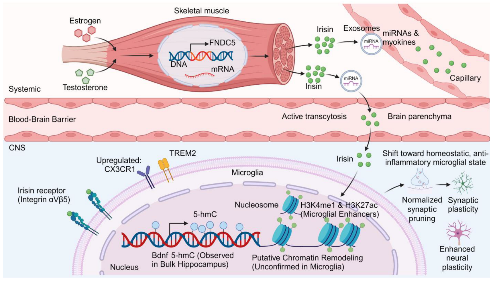
Puberty is a critical period in brain maturation, with a second surge of growth and remodeling, especially in the prefrontal cortex, where the maximum number of excitatory synapses is reached and then rapidly pruned [113]. This synaptic remodeling is vital to the development of effective neural networks responsible for higher cognitive processes. The resident immune cells of the central nervous system, microglia, are important during this time, acting as “architects” in the precise remodeling of synapses [106]. Their synaptic pruning activity is highly correlated with adolescent cognitive flexibility and impulse control, highlighting their functional importance in neurodevelopmental trajectories [106]. In humans, the pubertal transition (clinically categorized across Tanner stages II–IV based on secondary sexual characteristics) is marked by a surge in sex steroid hormones, such as estradiol and testosterone, alongside pronounced cortical restructuring [113]. Crucially, however, clinical Tanner-stage terminology cannot be directly extrapolated from rodent developmental stages (typically defined by postnatal days, vaginal opening, or preputial separation), because rodents lack a prolonged human-like juvenile hiatus and adrenarche and exhibit compressed neurodevelopmental timelines [20,106,113]. Direct mechanistic evidence linking pubertal sex hormone fluctuations to microglial receptor expression—such as CX3CR1 (involved in neuron–microglia signaling) and TREM2 (involved in phagocytosis)—derives primarily from preclinical rodent models rather than human pediatric studies [20,106,108,110]. In adolescent rodents, sex-specific hormonal milieus modulate microglial morphology, immune reactivity, and prefrontal circuit refinement [20,106,108]. While these rodent findings cannot be directly equated to specific human Tanner stages, they support the hypothesis that the pubertal transition may represent a period of heightened neuroimmune plasticity during which microglia could be particularly responsive to systemic signals, including exercise-induced factors [20,106,110,113].
4.2. Hypothesized Interactions Between Pubertal Sex Hormones and Exercise Factors
We hypothesize that the signaling activity of the proposed muscle–microglia axis may be dynamically modulated by the changing endocrine milieu of puberty, although direct clinical evidence comparing exerkine responses across human Tanner stages (e.g., stages II–IV versus prepubertal stage I or adulthood) is currently lacking. This hypothesis is informed by preclinical endocrinological studies demonstrating that sex steroids influence both skeletal muscle plasticity and FNDC5/irisin regulation. In rodent models, hormonal status (such as gonadal steroid manipulation) significantly alters FNDC5 expression and circulating irisin levels across skeletal muscle, adipose tissue, and the hypothalamus [112], and irisin has been shown to interact with neuroendocrine pathways in the primate hypothalamus in vitro [114]. Concurrently, androgens such as testosterone promote myogenesis and activate quiescent satellite cells in rodent skeletal muscle [109,111], while biological sex and sex hormones strongly influence putative myokine signaling networks [107] and microglial developmental trajectories [108]. However, whether rising estradiol or testosterone during human puberty directly enhances exercise-induced FNDC5 transcription, facilitates exerkine transport across the human blood–brain barrier, or amplifies microglial responsiveness remains an untested hypothesis. Given substantial differences in pubertal timing, endocrine profiles, and BBB physiology between rodents and humans, longitudinal pediatric studies stratified by Tanner stage and sex steroid levels are required to test whether human puberty truly constitutes a high-sensitivity window for exercise-induced neuromodulation in ASD and ADHD (Figure 4).
4.3. Long-Term Neuroepigenetic Memory Effects Induced by Pubertal Exercise Interventions
Preclinical studies indicate that physical exercise during juvenile or adolescent developmental windows in rodents (which, as noted above, cannot be directly equated to specific human Tanner stages such as stage III) can induce neuroepigenetic modifications; however, it is essential to specify whether these changes were measured in bulk brain tissue, neurons, or specifically in microglia, as well as across distinct brain regions. In juvenile mice, early-life voluntary wheel running primes the hippocampal neural epigenome (assessed in hippocampal neuronal and bulk tissue) to facilitate activity-dependent gene expression and memory consolidation [85]. Similarly, forced running exercise in mice mitigates radiation-induced cognitive deficits by regulating DNA hydroxymethylation (5-hydroxymethylcytosine, 5-hmC) and BDNF expression in bulk hippocampal tissue [86], while treadmill exercise in rats exerts age-dependent effects on histone deacetylase (HDAC) enzymatic activity measured in bulk striatal tissue [115]. Regarding glial-specific epigenetic regulation, high-intensity interval training (HIIT) in adolescent rodents exposed to postnatal immune activation was shown to ameliorate mood-related behaviors by modulating histone demethylase KDM6B-regulated glial activation [116]. Separately, the concept that isolated microglia can retain long-lasting epigenetic innate immune memory—characterized specifically by enhancer histone modifications such as monomethylation of lysine 4 on histone H3 (H3K4me1) and acetylation of lysine 27 on histone H3 (H3K27ac)—has been established in adult mouse models of systemic immune challenge [117], whereas promoter H3K4me3 modifications have been examined at the Tnf-α locus in primary rat microglia exposed to lipopolysaccharide and dextromethorphan in vitro [118]. Crucially, neither of these microglial studies [117,118] investigated exercise or demonstrated promoter remodeling at BDNF, TGF-β, or IL-10 loci in microglia. Therefore, because current exercise epigenetics studies examine heterogeneous bulk tissues or neurons across distinct brain regions (e.g., bulk hippocampal 5-hmC at the Bdnf promoter [86] and neuronal H3K27ac [85], or bulk striatal HDAC activity [115]) rather than isolated pubertal microglia, whether adolescent exercise directly establishes persistent H3K4me1, H3K27ac, or 5-hmC marks specifically within microglial chromatin remains an unconfirmed hypothesis that warrants future cell-type-specific epigenomic investigation.
Figure 4.
Hypothetical model of the pubertal window: proposed interactions between sex hormones and the muscle–microglia axis. This schematic depicts a theoretical hypothesis extrapolated from preclinical rodent studies regarding how pubertal sex hormones might interact with exercise-induced factors. Based on rodent evidence, systemic estrogen and testosterone are hypothesized to modulate skeletal muscle plasticity, satellite cell activation, and FNDC5/irisin or exosome release. After crossing or signaling across the blood–brain barrier, these factors are proposed to interact with adolescent microglia—which exhibit hormone-sensitive expression of receptors such as CX3CR1 and TREM2 in rodent models—as well as integrin αVβ5. The lower panel illustrates a strictly hypothetical convergence of neuroepigenetic mechanisms—extrapolating from bulk hippocampal DNA hydroxymethylation (5-hmC at the Bdnf promoter in rodent exercise models) and enhancer histone modifications (H3K4me1 and H3K27ac) established in microglial innate immune memory. Note: Direct exercise-induced chromatin remodeling at specific gene promoters within isolated pubertal microglia has not yet been experimentally demonstrated. Abbreviations: 5-hmC, 5-hydroxymethylcytosine; BBB, blood–brain barrier; BDNF, brain-derived neurotrophic factor; CNS, central nervous system; CX3CR1, C-X3-C motif chemokine receptor 1; DNA, deoxyribonucleic acid; FNDC5, fibronectin type III domain-containing protein 5; H3K4me1, monomethylation of lysine 4 on histone H3; H3K27ac, acetylation of lysine 27 on histone H3; Integrin αVβ5, integrin alpha-V beta-5; miRNA (miRNAs), microRNA (microRNAs); mRNA, messenger ribonucleic acid; TREM2, triggering receptor expressed on myeloid cells 2.
4.4. Comparative Analysis of Dose–Response Relationships Across Developmental Stages
Preclinical studies suggest that the neurobiological and neuroimmune effects of exercise can vary depending on the developmental stage at which the intervention is initiated [119]. For instance, comparative studies in rodents have demonstrated differential effects of wheel running on hypothalamic and hippocampal neuroinflammation and microglial profiles between adolescent and adult cohorts [119]. In mice exposed to chronic unpredictable stress, running exercise alleviates hippocampal neuroinflammation and shifts microglial polarization toward an anti-inflammatory phenotype via adiponectin/AdipoR1 pathway activation [45], while voluntary exercise specifically during the pubertal period in mice promotes spatial memory and enhances hippocampal dentate gyrus (DG)/CA3 synaptic transmission [120]. Furthermore, early-life and adolescent exercise paradigms in rodents have been shown to induce persistent enhancements in hippocampal memory consolidation and glial-mediated behavioral resilience [85,116]. While these preclinical observations suggest that the adolescent neurodevelopmental window may exhibit heightened biological responsiveness to physical activity (note that formal health-economic cost-effectiveness analyses do not exist), direct head-to-head dose–response comparisons across human developmental stages remain limited. Consequently, calibrating exercise intensity and volume to avoid both undertraining and excessive fatigue in youth with ASD or ADHD requires individualized clinical monitoring rather than direct extrapolation from rodent timelines [65,121].
5. Hypothesis-Driven Framework for Candidate Exercise Prescription Parameters (FITT-VP) in Future Clinical Trials
5.1. Exercise Modality Specificity and Molecular Target Matching: Comparison of Aerobic, Resistance, and HIIT
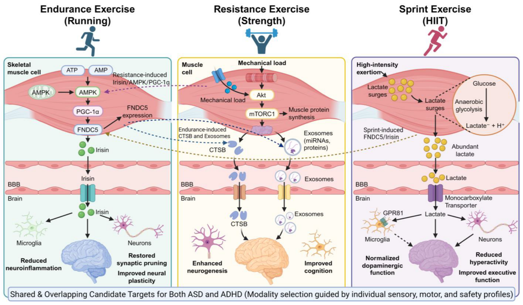
Exploring individualized exercise strategies for adolescents with ASD and ADHD requires examining how different exercise modalities may preferentially engage distinct skeletal muscle signaling pathways while recognizing that current mechanistic evidence derives primarily from preclinical models and healthy adult physiology rather than pediatric clinical trials [65]. In rodent models and adult human studies, sustained endurance (aerobic) exercise stimulates the AMP-activated protein kinase (AMPK)/peroxisome proliferator-activated receptor gamma coactivator 1-alpha (PGC-1α) signaling axis [122] and upregulates skeletal muscle Fndc5 expression and hippocampal BDNF in mice [73], as well as circulating irisin in human endurance cohorts [37]. Notably, however, these exerkine pathways are substantially overlapping rather than exclusive to a single exercise modality: for instance, acute and chronic functional and traditional resistance training also increase circulating AMPK, PGC-1α, and irisin levels in young adult males [123], and HIIT similarly upregulates hippocampal FNDC5/irisin and BDNF in preclinical models [124]. Conversely, while resistance training and HIIT robustly activate the mechanistic target of rapamycin complex 1 (mTORC1) and hypoxia-inducible factor-1 alpha (HIF-1α) pathways in skeletal muscle [67,125], aerobic running also elevates systemic cathepsin B (CTSB) [75,76]. Therefore, endurance training is not proven to be uniquely optimal for ASD via irisin, nor is HIIT uniquely specific for ADHD via lactate; rather, aerobic, resistance, and intermittent modalities engage overlapping candidate exerkine networks that may collectively support neuroimmune homeostasis and executive function across both disorders [62,65,126]. In adult exercise studies and preclinical models, resistance exercise and endurance training have been reported to modulate circulating neuroprotective biomarkers—such as cathepsin B (CTSB)—and extracellular vesicle release [75,127,128]. Given preclinical links between CTSB and hippocampal neurogenesis, resistance exercise could theoretically contribute to neurotrophic support, though whether resistance training reliably elevates central CTSB or alters clinical outcomes in pediatric ASD or ADHD has not yet been established.
During HIIT (such as repeated short bouts of high-intensity sprinting), anaerobic glycolysis generates acute, pulsatile elevations in systemic lactate [102]. Preclinical rodent studies suggest that elevated lactate can activate cerebral G protein-coupled receptor 81 (GPR81/HCAR1) and support mitochondrial and neurotrophic adaptations [83,94]. However, whether intermittent lactate surges are more effective than moderate continuous exercise at modulating microglial function in humans—and specifically in children and adolescents with ASD or ADHD—remains unconfirmed and requires direct clinical investigation.
Implementing HIIT, however, presents a major clinical paradox regarding neurodivergent populations. For teens who are showing signs of sensory hyper-reactivity, the high impact and sympathetic activation of HIIT exercises can cause psychological distress and associated cortisol release, potentially worsening neuroinflammation rather than preventing it [129,130]. Likewise, in ADHD groups, the basic symptoms of poor motor control and impulsivity increase the likelihood of injuries during high-intensity activities. Therefore, the biochemical benefits that can be obtained from lactate-mediated neural adaptations may be negated by physiological stress responses, necessitating caution and phenotype-specific intensity scaling [131].
Taken together, rather than serving as validated clinical prescriptions, these modality-specific molecular associations offer a hypothesis-generating framework for selecting candidate exercise parameters in future pediatric clinical trials. Within this conceptual model, moderate-intensity continuous aerobic exercise is hypothesized to preferentially engage AMPK/PGC-1α/FNDC5 signaling [73,122], resistance exercise to engage mechanotransduction and CTSB/EV release [67,127,128], and carefully scaled intermittent exercise to engage lactate-mediated signaling [83,94], each warranting prospective biomarker-stratified testing in youth with ASD and ADHD (Figure 5).
5.2. Distinguishing Acute Molecular Responses from Chronic Therapeutic Adaptations: Candidate Dose Parameters
When evaluating exercise dose–response relationships, it is essential to clearly distinguish between acute, transient molecular responses to a single bout of exercise and the cumulative, long-term neurobiological adaptations required for sustained clinical therapeutic effects in ASD and ADHD [64,65]. In healthy adults and preclinical models, a single acute bout of exercise triggers transient elevations in circulating lactate, irisin, and tetraspanin-positive EVs—typically peaking within minutes to 2 h post-exercise before returning to baseline [90,102,132,133]—whereas clinically meaningful improvements in executive function, synaptic remodeling, or core behavioral symptoms require chronic, multi-week training regimens (typically 6–12 weeks or longer) [62,65,75,76,121].
In adult exercise physiology and preclinical studies, acute exerkine kinetics vary with exercise intensity and mechanical load: for example, in healthy young adults, higher-intensity exercise elicits a greater acute circulating irisin response than low-intensity exercise under matched energy expenditure [132], and resistance training near muscular failure acutely modulates circulating neuroprotective biomarkers and EV subpopulations in healthy men and women [127,128]. Regarding lactate–BDNF coupling, preclinical studies show that exercise-induced lactate contributes to hippocampal BDNF induction via SIRT1/PGC-1α/FNDC5 signaling [73,81,84] and activates cerebral HCAR1 (GPR81) [83,94], while voluntary running in mice rescues metabolic signaling in cerebellar Purkinje cells [66] and short-term high-intensity exercise promotes ceramide-dependent EV secretion in the mouse CNS [133]. In early adolescents, acute resistance and HIIT sessions elicit transient metabolic and blood lactate responses [102].
Figure 5.
Hypothetical framework illustrating overlapping candidate molecular pathways across endurance, resistance, and high-intensity interval exercise modalities for future clinical exploration in ASD and ADHD. Rather than representing validated, disorder-exclusive treatment choices, endurance (aerobic), resistance, and sprint/high-intensity interval training (HIIT) engage overlapping peripheral signaling cascades (indicated by cross-connecting dashed arrows). Endurance exercise activates the AMPK/PGC-1α/FNDC5 pathway and also elevates systemic CTSB; resistance exercise activates mechanical Akt/mTORC1 signaling and modulates circulating EVs and CTSB while also elevating circulating AMPK, PGC-1α, and irisin; and HIIT generates pulsatile lactate release (acting via monocarboxylate transporters and GPR81) while also upregulating hippocampal FNDC5/irisin and BDNF in preclinical models. Collectively, these overlapping candidate pathways converge on shared neurodevelopmental targets across both ASD and ADHD—attenuating neuroinflammation and supporting neurogenesis, synaptic plasticity, and executive function—while clinical modality selection must be guided by individual sensory tolerance, motor coordination, and safety profiles rather than rigid phenotype-to-exerkine matching. Abbreviations: ADHD, attention-deficit/hyperactivity disorder; Akt, protein kinase B; AMP, adenosine monophosphate; AMPK, AMP-activated protein kinase; ASD, autism spectrum disorder; ATP, adenosine triphosphate; BBB, blood–brain barrier; CTSB, cathepsin B; FNDC5, fibronectin type III domain-containing protein 5; GPR81, G protein-coupled receptor 81; H+, hydrogen ion; HIIT, high-intensity interval training; miRNAs, microRNAs; mTORC1, mechanistic target of rapamycin complex 1; PGC-1α, peroxisome proliferator-activated receptor gamma coactivator 1-alpha.
Crucially, however, no clinical studies have established quantitative biochemical thresholds (such as specific VO2max percentages, resistance set volumes, blood lactate concentrations, or post-exercise feeding intervals) in children or adolescents with ASD or ADHD. Adult or rodent metabolic thresholds cannot be directly extrapolated to neurodivergent youth without rigorous pediatric validation. Instead, recent systematic reviews and meta-analyses in pediatric ADHD and ASD populations indicate that structured moderate-to-vigorous aerobic or multicomponent exercise programs—commonly implemented for approximately 30–60 min per session, 2–5 sessions per week over 6–12 weeks, alongside variable regimen durations and frequencies across trials—are associated with clinically meaningful improvements in executive functions and core behavioral symptoms, although optimal dose–response parameters vary by symptom domain and exercise modality [60,61,62,63,64,65]. Whether acute post-exercise exerkine surges directly mediate these long-term clinical gains in youth with ASD and ADHD remains a working hypothesis for future longitudinal trials.
5.3. Conceptual Considerations and Candidate Parameters for Adapting Exercise Prescriptions (FITT-VP) in Adolescents with ASD and ADHD
When translating preclinical exerkine hypotheses into clinical trial design for children and adolescents with ASD and ADHD, candidate parameters structured around the FITT-VP (Frequency, Intensity, Time, Type, Volume, Progression) framework must prioritize clinical feasibility, behavioral tolerance, and existing pediatric evidence over unvalidated theoretical cutoffs [62,64,65]. Recent systematic reviews and network meta-analyses performed directly in pediatric ADHD and ASD cohorts indicate that regular, structured physical activity—typically conducted 3 to 5 times per week in sessions of 30 to 60 min over 6 to 12 weeks—significantly improves executive functions, social interaction, and core behavioral symptoms [60,62,64,65].
For children with ASD, predictable routines and structured group or skill-based activities (such as gymnastics or adapted team sports) support both physical activity engagement and social communication [60,61], whereas maintaining moderate intensities helps avoid sensory over-responsivity and psychosocial stress [129,134]. For youth with ADHD, network meta-analytic evidence demonstrates that moderate-to-vigorous aerobic and multicomponent cognitively engaging exercises effectively improve core symptoms and executive function deficits [63,64,65]. Although we hypothesize that pubertal maturation (across Tanner stages) may influence physiological responsiveness to exercise, specific prescriptions dictating exact heart rate reserve cutoffs by Tanner stage have not yet been empirically tested in pediatric ASD or ADHD cohorts and should be treated purely as candidate variables for future stratified trials.
Regarding exercise timing (chronobiology), scheduling sessions in the late afternoon (approximately 15:00–17:00) versus the morning should likewise be viewed as a candidate parameter governed by both circadian physiology and clinical routines. Physiologically, core body temperature, skeletal muscle contractile performance, and glycolytic capacity typically peak in the late afternoon, and after-school hours provide a practical window for structured group physical activity in school-aged youth [60,135]. In addition, because sleep disturbances are prevalent in youth with ASD and ADHD—and preclinical evidence in sleep-deprived adolescent mice demonstrates that sleep loss impairs microglial synaptic pruning whereas voluntary exercise during the active phase ameliorates these pruning deficits [136]—completing exercise during daytime or afternoon hours rather than close to bedtime is a practical clinical consideration to avoid pre-sleep sympathetic arousal, although direct pediatric trials evaluating exercise timing on melatonin onset in ASD or ADHD are still needed. Conversely, for adolescents with ADHD receiving morning stimulant pharmacotherapy or experiencing classroom attentional deficits, morning aerobic exercise sessions have also demonstrated clinical efficacy in enhancing daytime executive control and frontal brain activation [137,138]. Because direct head-to-head pediatric trials comparing morning versus afternoon exerkine responses in ASD and ADHD are lacking, exercise timing and volume progression should be individualized to each child’s sleep–wake schedule, school routine, motor competence, and standardized symptom assessments [64,65,121,139]. A summary of representative preclinical and clinical exercise studies—explicitly distinguishing animal models from human trials—is provided in Table 2.
Table 2.
Representative preclinical and clinical exercise studies relevant to neuroimmune modulation and ASD/ADHD.
6. Candidate Digital and Molecular Biomarkers: A Conceptual Proposal for Closed-Loop Exercise Monitoring in ASD and ADHD
6.1. Circulating Extracellular Vesicles and Exerkines as Candidate Liquid Biopsy Biomarkers
Circulating extracellular vesicles (EVs) and exosomes have been proposed as candidate blood-based biomarkers for exploring systemic exercise responsiveness in preclinical and adult studies [140,141]. In healthy human adults, high-intensity intermittent exercise triggers an acute release of tetraspanin-positive (CD81+/CD63+) EVs into the circulation [90], providing a potential temporal window for sampling acute peripheral exercise responses. Beyond surface markers, preclinical studies indicate that EV cargo—including microRNAs (miRNAs)—may participate in peripheral-to-central signaling [142], and specific miRNAs such as miR-146a and miR-21-5p have been hypothesized as candidate indicators of inflammatory pathway modulation. While mathematical modeling and profiling studies implicate miRNAs such as miR-146a in NF-κB-mediated inflammatory regulation and ASD pathophysiology [103,104,143], and aerobic exercise trials in late middle-aged adults at risk for Alzheimer’s disease (26-week intervention) [76] as well as healthy young adults [75,77] have reported correlations between circulating cathepsin B (CTSB) increments and memory performance, these peripheral molecular markers have not yet been validated in pediatric ASD or ADHD exercise trials. These results suggest the potential of circulating extracellular vesicles and exercise-inducible proteins as a blood-based platform for exploring dynamic molecular responses to exercise in real time [90,141]. Crucially, however, peripheral biomarker changes reflect systemic physiological responsiveness to exercise and should not be assumed to directly mirror central microglial remodeling or automatically predict improvements in complex clinical and behavioral outcomes in ASD or ADHD. Rather, combining exosomal miRNA profiling with candidate proteins such as CTSB represents an exploratory biomarker panel whose clinical predictive utility must be rigorously tested in future longitudinal pediatric trials [77,89,141].
6.2. Wearable Devices and AI-Driven Real-World Exercise Monitoring Systems
With the emergence of sophisticated wearable technologies and artificial intelligence (AI), objective measurements of physical activity in daily life have been transformed beyond the limitations of subjective questionnaires [144]. Tri-axial accelerometers combined with AI algorithms for posture recognition can accurately capture the times spent at moderate-to-vigorous physical activity (MVPA) levels by children in their daily activities, as well as the proportions of physical activities performed [145]. This method reduces recall bias and increases data fidelity in populations of children with neurodevelopmental disorders [146]. The latest smartwatch and patch-based heart rate monitors deliver real-time physiological data, including estimated oxygen consumption (VO2), heart rate recovery times after activity, and blood oxygen saturation levels [147]. These are indirect indicators of lactate production and metabolic stress and provide information on exercise intensity and recovery. Moreover, recent feasibility and case–control studies have begun applying machine learning models to multimodal wearable and physiological data to assist in symptom tracking and activity classification in adolescents with ADHD [144,146,148]. Although wearable sensors cannot directly measure central microglial states, we hypothesize that longitudinal tracking of physical activity, sleep–wake regularity, and autonomic recovery could serve as a non-invasive behavioral and physiological proxy to help clinicians monitor exercise adherence and tolerance in real-world settings [144,146,147]. If validated in future pediatric trials, such digital tools may eventually support timely adjustments to individualized exercise regimens.
6.3. Conceptual Proposal for a Multimodal Closed-Loop Exercise Framework: Feasibility, Ethical, and Algorithmic Challenges
Integrating candidate molecular biomarkers, standardized behavioral evaluations (e.g., SNAP-IV and ADOS), and continuous physiological streams from wearable sensors into an AI-assisted closed-loop framework represents an appealing future direction for personalized neurorehabilitation [149,150,151]. Crucially, however, such a multimodal closed-loop system currently constitutes a prospective conceptual proposal rather than an established or clinically validated platform in pediatric ASD or ADHD. Within this proposed architecture, longitudinal wearable metrics and periodic clinical ratings could theoretically be analyzed by machine learning models to construct individualized response profiles and guide gradual, clinician-supervised adjustments to FITT-VP parameters [150,151]. Preliminary clinical evidence supporting mobile-health-assisted personalization comes from a recent 12-week randomized controlled trial in children with ADHD, in which a tailored mobile-health exercise rehabilitation program significantly improved hyperactivity and core behavioral scores compared with empirical control routines [121]; nevertheless, that trial did not incorporate exosomal liquid biopsies or automated central neuroimmune tracking. The pathological features and corresponding candidate digital or molecular biomarkers proposed for monitoring the functional status of the muscle–microglia axis are systematically summarized in Table 3.
Before any multimodal closed-loop model can transition from a theoretical concept to clinical practice, substantial barriers regarding clinical feasibility, validation, data quality, algorithmic reliability, ethics, and healthcare accessibility must be addressed: (1) Feasibility and invasiveness: Repeated venipuncture for profiling circulating exosomes or proteins (such as CTSB) is invasive, costly, and poorly tolerated by children with ASD or ADHD who exhibit severe sensory hypersensitivity and needle phobia [139], underscoring the need for truly non-invasive alternatives (e.g., validated salivary or sweat-based sensors) [152,153]. (2) Data quality and sensor compliance: In naturalistic settings, sensory hypersensitivity and tactile defensiveness frequently lead to wearable device removal or non-compliance [62,139], while motor stereotypies, hyperactivity, and rapid rest–exercise transitions introduce severe motion artifacts that degrade the accuracy of wrist-worn photoplethysmography (PPG) and accelerometry streams [154,155]. (3) Validation and algorithmic limitations: Machine learning models trained on small, homogeneous datasets are prone to overfitting, algorithmic bias, and poor generalizability across heterogeneous neurodevelopmental subtypes, ages, and medication states [144,148,151]. Moreover, peripheral biomarker fluctuations do not necessarily reflect central microglial states or predict clinical behavioral improvements. (4) Ethical, privacy, and regulatory challenges: Continuous digital phenotyping and biological profiling of minors with neurodevelopmental disabilities raise critical ethical concerns regarding pediatric assent, parental informed consent, cybersecurity, data ownership, and the risk of algorithmic stigmatization. (5) Economic cost and accessibility: Multi-omic liquid biopsies, specialized wearables, and AI infrastructure entail high financial and technical burdens that could exacerbate healthcare disparities, limiting real-world accessibility in routine community and school-based rehabilitation settings [135,144].
Table 3.
Candidate molecular, neuroimaging, and digital biomarkers for monitoring exercise responses and neuroimmune status.
7. Translational Medicine Prospects and Key Barriers
7.1. Current Status Assessment and Limitations of Clinical Research Evidence
The field of clinical research on exercise interventions for ASD and ADHD has expanded rapidly, with a growing number of randomized controlled trials (RCTs) investigating behavioral efficacy [61,63]. Despite notable progress, most clinical studies rely on subjective behavioral ratings and symptom checklists, which—while clinically useful—provide limited insight into underlying neurobiological mechanisms [156]. There is a noticeable lack of research that uses biomarkers, such as neuroimaging or molecular assays, to explore the mediating pathways through which exercise affects neurodevelopmental disorders. Moreover, substantial methodological heterogeneity exists across pediatric exercise trials regarding sample sizes, blinding procedures, control group designs, exercise supervision, and self-reported adherence, contributing to variable effect sizes across studies [62,64,65,156].
Crucially, directly extrapolating mechanistic findings from preclinical models (such as microglial remodeling and synaptic pruning rescue) to human neurodevelopment faces major translational barriers [39,156]. First, binary M1/M2 classifications fail to capture the complex, overlapping transcriptional states of human microglia [43]. Second, quantitative proteomic atlases demonstrate substantial interspecies differences in the abundance of BBB transporters, receptors, and tight junction proteins between rodents and primates [157], and preclinical studies frequently employ supraphysiological recombinant exerkine doses that far exceed endogenous post-exercise plasma or CSF increments in humans [39,72]. Because paired plasma-to-CSF pharmacokinetic studies and in vivo microglial imaging (such as TSPO PET) following exercise interventions are lacking in pediatric cohorts, it remains unknown whether normal, clinically feasible exercise in children and adolescents can produce sufficient central concentrations of irisin and other exerkines to meaningfully modulate microglial activity in the human brain. Therefore, it is now necessary to conduct well-designed mechanistic clinical trials that integrate multiple types of biomarkers—neuroimaging, peripheral molecular signatures, and neurophysiological measures—to verify the biological effects of exercise intervention and establish a rigorous evidence base for personalized therapeutic strategies [36,158].
7.2. Evaluation of Interference from Age Stratification, Sex Differences, and Comorbidities on Intervention Efficacy
Accumulating evidence has highlighted that the heterogeneous nature of ASD and ADHD populations presents major issues in the design and implementation of exercise-based interventions, as age, sex, and comorbid conditions may be modulatory factors in therapeutic outcome [62]. Adolescence is a critical developmental period characterized by dynamic fluctuations in sex steroid hormones—particularly across the female menstrual cycle—that modulate responsiveness to exercise-induced molecular mediators such as myokines and neurotrophic factors. This cyclical hormonal environment may explain why the effectiveness of exercise interventions may vary across the menstrual cycle, and future studies of exercise interventions in female adolescents should consider adding menstrual phase tracking and hormone profiles to guide the optimal timing and dosing of exercise interventions [107,159]. Furthermore, rigorous clinical safety protocols are paramount when prescribing exercise for youth with ASD and ADHD due to the high prevalence of neurological, motor, cognitive, and sensory comorbidities [62,139,160]. First, for children and adolescents with co-occurring epilepsy (which is prevalent in ASD), exercise recommendations should follow established International League Against Epilepsy (ILAE) clinical guidance, which emphasizes that physical activity and sports are generally safe and beneficial for individuals with epilepsy and that unnecessary blanket restrictions should be avoided; following an individualized risk assessment (considering seizure control, seizure type, and supervision needs, particularly for water- or height-related activities), youth with epilepsy can safely participate in physical activities with appropriate medical clearance and a seizure action plan [161]. Second, for individuals with significant intellectual disability or sensory intolerance (e.g., auditory, tactile, or proprioceptive over-responsivity), high-intensity or chaotic group settings can provoke severe distress and behavioral dysregulation [129,134]. These populations require predictable, low-sensory-stimulation environments, visual schedules, simplified step-by-step instructions, and gradual intensity titration; for individuals unable to tolerate active aerobic regimens, low-impact or passive modalities such as supervised whole-body vibration therapy have been explored in preclinical and preliminary studies as potential alternatives [162,163]. Third, in youth with ADHD or ASD who exhibit developmental coordination deficits, postural instability, or high impulsivity, the elevated baseline risk of unintentional musculoskeletal and head injuries [131,160] necessitates structured supervision, simplified motor tasks, protective equipment, and avoidance of high-collision activities.
Equally critical is the management of energy balance at the intersection of stimulant pharmacotherapy and pubertal growth. Physiological calorimetry studies demonstrate that central nervous system stimulants prescribed for ADHD directly alter daily energy balance by modifying basal metabolic rate, total energy expenditure, and spontaneous physical activity levels [164], while clinically, stimulant pharmacotherapy is also well recognized to cause appetite suppression that can reduce daytime caloric intake [5,9]. Importantly, adolescence (particularly during the pubertal growth spurt) is characterized by substantially increased basal metabolic requirements and heightened energy and protein demands to support rapid somatic growth, bone mineral accrual, and brain maturation. Adding high-volume or high-intensity exercise to a medicated adolescent experiencing stimulant-induced appetite suppression can precipitate a state of relative energy deficiency, potentially impairing linear growth, disrupting pubertal endocrine axes, increasing musculoskeletal injury risk, and blunting skeletal muscle anabolic and myokine responses. Therefore, pediatric exercise interventions must incorporate routine monitoring of body mass index (BMI) percentiles, growth velocity, and dietary intake, alongside practical nutritional strategies—such as scheduling nutrient-dense meals prior to morning stimulant administration and after evening medication wear-off, and moderating exercise volume during periods of inadequate caloric intake—to safeguard adolescent development [139,164].
7.3. Prospective Perspectives on Exercise as a Fundamental Adjuvant in Combination with Pharmacological and Non-Pharmacological Therapies
Emerging clinical and preclinical evidence suggests that structured physical exercise may serve as a valuable non-pharmacological adjuvant alongside standard pharmacological and behavioral therapies in ASD and ADHD [137,138,156]. Pharmacologically, first-line stimulants for ADHD such as methylphenidate act primarily presynaptically by blocking dopamine and norepinephrine transporters (DAT and NET) to increase synaptic monoamine availability, whereas exercise modulates overlapping frontostriatal and catecholaminergic networks, neurotrophic support [105], and neuroimmune homeostasis. Clinically, a randomized trial in adolescents with ADHD demonstrated that adding a 6-week aerobic exercise program to methylphenidate pharmacotherapy yielded greater improvements in clinical symptoms, perseverative errors, and right frontotemporal brain activity (on fMRI) than methylphenidate alone [137], while an acute 25-min cycling bout independently enhanced executive control and task-related cortical activation (on fNIRS) in youth with ADHD [138]. Although these clinical trials do not demonstrate direct exercise-induced modifications of postsynaptic drug-receptor binding, they support the clinical rationale for investigating complementary multimodal interventions combining exercise with standard pharmacotherapy. Preclinical evidence further suggests that exercise can attenuate systemic and central inflammation and promote a homeostatic, anti-inflammatory microglial state, which may help alleviate common comorbidities such as anxiety and gastrointestinal dysfunction frequently associated with these conditions [15]. Upstream modification of the neuroimmune and synaptic environment by exercise provides favorable conditions for downstream targeted intervention, and a stratified treatment approach that aims for pathological repair rather than mere symptom relief has been proposed [15]. Framing exercise as a foundational adjunctive strategy aligns with the goals of personalized medicine, offering a conceptual basis for future trials investigating combined exercise, pharmacological, and behavioral interventions in ASD and ADHD [15,36,158,162].
8. Conclusions and Perspectives
In this review, we have proposed the developmental muscle–microglia axis as a hypothesis-generating conceptual framework linking unidirectional humoral signaling from contracting skeletal muscle to central neuroimmune modulation in ASD and ADHD. Synthesizing predominantly preclinical evidence, we highlight how exercise-induced factors—including irisin, CTSB, lactate, and exosomal miRNAs—may cross or signal across the blood–brain barrier to attenuate pro-inflammatory microglial reactivity and support activity-dependent synaptic remodeling. Rather than operating as a simplistic binary M1-to-M2 switch, these peripheral signals are hypothesized to interact with intrinsic neuronal activity and complex developmental pruning programs across the distinct neuroimmune landscapes of ASD and ADHD. A central hypothesis of this framework is that the pubertal transition represents a period of heightened neuroimmune and endocrine plasticity during which surging sex steroids may interact with exercise-induced pathways and neuroepigenetic regulation. While these developmental insights offer a theoretical rationale for exploring phenotype-adapted exercise strategies (under the FITT-VP model) and candidate digital/molecular monitoring tools, specific biochemical thresholds, Tanner-stage adjustments, and multimodal AI closed-loop systems currently remain conceptual proposals and candidate parameters rather than validated clinical protocols. The review acknowledges current knowledge gaps and clearly outlines future research needs. It proposes mechanistic human studies with molecular endpoints, including synaptic proteins and neuroinflammatory markers, alongside standardized behavioral scales to validate the proposed pathway. Future pediatric trials must establish age- and sex-specific dose–response relationships, account for stimulant medication–energy balance interactions during adolescent growth, and enforce rigorous clinical safety adaptations for comorbid epilepsy, sensory intolerance, and motor coordination deficits. Through such multidisciplinary validation, exercise-based interventions can be progressively refined to support neurodevelopmental health in youth with ASD and ADHD.
Author Contributions
Conceptualization, X.W., J.L., Y.L. (Ying Li) and Y.L. (Yanyan Liu); methodology, X.W. and J.L.; validation, X.W., J.L. and Y.L. (Ying Li); formal analysis, J.L. and Y.J.; investigation, X.W. and J.L.; writing—original draft preparation, X.W., J.L., Y.L. (Ying Li), Y.L. (Yanyan Liu), Y.W. and M.Z.; writing—review and editing, M.Z.; supervision, M.Z.; project administration, M.Z.; funding acquisition, M.Z. All authors have read and agreed to the published version of the manuscript.
Funding
Natural Science Foundation of Heilongjiang Province (Project No. LH2021C054); Harbin Sport University Experimental Platform project (Project No. LAB2021-04); Harbin Sport University Talent Introduction Research Start-up Fund Project (RC20-201906).
Data Availability Statement
No new data were created or analyzed in this study. Data sharing is not applicable to this article.
Acknowledgments
We extend our gratitude to all authors for their dedication and contributions. We also thank the BioRender website for providing us with a platform for figure creation.
Conflicts of Interest
The authors declare no conflicts of interest.
Abbreviations
The following abbreviations are used in this manuscript:
| ASD | Autism spectrum disorder |
| ADHD | Attention deficit hyperactivity disorder |
| BBB | Blood–brain barrier |
| BDNF | Brain-derived neurotrophic factor |
| FITT-VP | Frequency, Intensity, Time, Type, Volume, Progression |
| HIIT | High-intensity interval training |
| AMPK/NF-κB | AMP-activated protein kinase/nuclear factor kappa-light-chain-enhancer of activated B cells |
| PGC-1α/FNDC5 | Peroxisome proliferator-activated receptor gamma coactivator 1-alpha/Fibronectin type III domain-containing protein 5 |
| CTSB/GPR81 | Cathepsin B/G protein-coupled receptor 81 |
| AI | Artificial intelligence |
| mTORC1/mTORC2 | Mechanistic target of rapamycin complex 1/2 |
| PI3K/AKT | Phosphoinositide 3-kinase/protein kinase B |
| IGF-1 | Insulin-like growth factor 1 |
| TSC1/2 | Tuberous sclerosis complex 1/2 |
| RHEB | Ras homolog enriched in brain |
| LTP | Long-term potentiation |
| PSD-95 | Postsynaptic density protein 95 |
| VPA | Valproic acid |
| MVPA | Moderate-to-vigorous physical activity |
| ADOS/SNAP-IV | Autism Diagnostic Observation Schedule/Swanson, Nolan, and Pelham-IV Rating Scale |
| TSPO PET | Translocator protein positron emission tomography |
| EV | Extracellular vesicle |
References
- Global Burden of Disease Study 2021 Autism Spectrum Collaborators. The Global Epidemiology and Health Burden of the Autism Spectrum: Findings from the Global Burden of Disease Study 2021. Lancet Psychiatry 2025, 12, 111–121. [Google Scholar] [CrossRef] [Scilit] [PubMed]
- Salari, N.; Ghasemi, H.; Abdoli, N.; Rahmani, A.; Shiri, M.H.; Hashemian, A.H.; Akbari, H.; Mohammadi, M. The Global Prevalence of ADHD in Children and Adolescents: A Systematic Review and Meta-Analysis. Ital. J. Pediatr. 2023, 49, 48. [Google Scholar] [CrossRef] [Scilit] [PubMed]
- Wang, C.; Hou, L.; Zhou, H.; Wang, Q.; Yan, H.; Lang, Y.; Tan, J.; Liu, Y.; Ge, Y.; Lin, Y.; et al. The Evolving Global Burden of ADHD: A Comprehensive Analysis and Future Projections (1990–2046). J. Affect. Disord. 2025, 391, 120037. [Google Scholar] [CrossRef] [Scilit] [PubMed]
- Baribeau, D.; Anagnostou, E. Novel Treatments for Autism Spectrum Disorder Based on Genomics and Systems Biology. Pharmacol. Ther. 2022, 230, 107939. [Google Scholar] [CrossRef] [Scilit] [PubMed]
- Shaw, P.; Sudre, G. Adolescent Attention-Deficit/Hyperactivity Disorder: Understanding Teenage Symptom Trajectories. Biol. Psychiatry 2021, 89, 152–161. [Google Scholar] [CrossRef] [Scilit] [PubMed]
- Kaur, T.; Dushyant; Yadav, N.; Saini, V.; Devi, R.; Dhingra, A.K. Neuroinflammatory and Synaptic Roles of Glial Cells in Autism Spectrum Disorder. CNS Neurol. Disord. Drug Targets, 2026; in press. [CrossRef] [Scilit] [PubMed]
- Tan, Z.; Xia, R.; Zhao, X.; Yang, Z.; Liu, H.; Wang, W. Potential Key Pathophysiological Participant and Treatment Target in Autism Spectrum Disorder: Microglia. Mol. Cell. Neurosci. 2024, 131, 103980. [Google Scholar] [CrossRef] [Scilit] [PubMed]
- Mordelt, A.; de Witte, L.D. Microglia-Mediated Synaptic Pruning as a Key Deficit in Neurodevelopmental Disorders: Hype or Hope? Curr. Opin. Neurobiol. 2023, 79, 102674. [Google Scholar] [CrossRef] [Scilit] [PubMed]
- Persico, A.M.; Ricciardello, A.; Lamberti, M.; Turriziani, L.; Cucinotta, F.; Brogna, C.; Vitiello, B.; Arango, C. The Pediatric Psychopharmacology of Autism Spectrum Disorder: A Systematic Review—Part I: The Past and the Present. Prog. Neuropsychopharmacol. Biol. Psychiatry 2021, 110, 110326. [Google Scholar] [CrossRef] [Scilit] [PubMed]
- Ibeas, K.; Herrero, L.; Mera, P.; Serra, D. Hypothalamus-Skeletal Muscle Crosstalk during Exercise and Its Role in Metabolism Modulation. Biochem. Pharmacol. 2021, 190, 114640. [Google Scholar] [CrossRef] [Scilit] [PubMed]
- Berg, R.M.G. The Enduring Quest for an ‘Exercise Factor’: A Historical Account of Skeletal Muscle as an Endocrine Organ. J. Physiol. 2026, 604, 44–65. [Google Scholar] [CrossRef] [Scilit] [PubMed]
- Dai, Y.; Dou, X.; Nie, B.; Sun, Y.; Chen, P.; Fu, C.; Zhang, X.; Chu, Y.; Gao, Q.; Ge, Y.; et al. Exercise and the Organ-Brain Axis: Regulation of Neurological Disorders by Emerging Exerkines. Pharmacol. Res. 2025, 219, 107913. [Google Scholar] [CrossRef] [Scilit] [PubMed]
- Chow, L.S.; Gerszten, R.E.; Taylor, J.M.; Pedersen, B.K.; van Praag, H.; Trappe, S.; Febbraio, M.A.; Galis, Z.S.; Gao, Y.; Haus, J.M.; et al. Exerkines in Health, Resilience and Disease. Nat. Rev. Endocrinol. 2022, 18, 273–289. [Google Scholar] [CrossRef] [Scilit] [PubMed]
- Brooks, G.A.; Osmond, A.D.; Arevalo, J.A.; Duong, J.J.; Curl, C.C.; Moreno-Santillan, D.D.; Leija, R.G. Lactate as a Myokine and Exerkine: Drivers and Signals of Physiology and Metabolism. J. Appl. Physiol. 2023, 134, 529–548. [Google Scholar] [CrossRef] [Scilit] [PubMed]
- Toscano, C.V.A.; Barros, L.; Lima, A.B.; Nunes, T.; Carvalho, H.M.; Gaspar, J.M. Neuroinflammation in Autism Spectrum Disorders: Exercise as a “Pharmacological” Tool. Neurosci. Biobehav. Rev. 2021, 129, 63–74. [Google Scholar] [CrossRef] [Scilit] [PubMed]
- Taha, H.B.; Birnbaum, A.; Matthews, I.; Aceituno, K.; Leon, J.; Thorwald, M.; Godoy-Lugo, J.; Cortes, C.J. Activation of the Muscle-to-Brain Axis Ameliorates Neurocognitive Deficits in an Alzheimer’s Disease Mouse Model via Enhancing Neurotrophic and Synaptic Signaling. Geroscience 2025, 47, 1593–1613. [Google Scholar] [CrossRef] [Scilit] [PubMed]
- Burtscher, J.; Millet, G.P.; Place, N.; Kayser, B.; Zanou, N. The Muscle-Brain Axis and Neurodegenerative Diseases: The Key Role of Mitochondria in Exercise-Induced Neuroprotection. Int. J. Mol. Sci. 2021, 22, 6479. [Google Scholar] [CrossRef] [Scilit] [PubMed]
- Guo, P.; Jin, Z.; Wang, J.; Sang, A.; Wu, H. Irisin Rescues Blood-Brain Barrier Permeability Following Traumatic Brain Injury and Contributes to the Neuroprotection of Exercise in Traumatic Brain Injury. Oxidative Med. Cell. Longev. 2021, 2021, 1118981. [Google Scholar] [CrossRef] [Scilit] [PubMed]
- Lu, D.; Qu, C.; Fang, M.; Zhang, J. Exercise Rescues Cognitive Impairment through Inhibiting the Fibrinogen Neuroinflammative Pathway in Diabetes. Metab. Brain Dis. 2024, 40, 2. [Google Scholar] [CrossRef] [Scilit] [PubMed]
- Kolmogorova, D.; Ah-Yen, E.G.; Taylor, B.C.; Vaggas, T.; Liang, J.; Davis, T.; Ismail, N. Sex-Specific Responses of the Pubertal Neuroimmune Axis in CD-1 Mice. Brain Behav. Immun. Health 2021, 13, 100229. [Google Scholar] [CrossRef] [Scilit] [PubMed]
- Favuzzi, E.; Huang, S.; Saldi, G.A.; Binan, L.; Ibrahim, L.A.; Fernández-Otero, M.; Cao, Y.; Zeine, A.; Sefah, A.; Zheng, K.; et al. Gaba-Receptive Microglia Selectively Sculpt Developing Inhibitory Circuits. Cell 2021, 184, 5686. [Google Scholar] [CrossRef] [Scilit] [PubMed]
- Meng, J.; Han, L.; Zheng, N.; Wang, T.; Xu, H.; Jiang, Y.; Wang, Z.; Liu, Z.; Zheng, Q.; Zhang, X.; et al. Microglial Tmem59 Deficiency Impairs Phagocytosis of Synapse and Leads to Autism-like Behaviors in Mice. J. Neurosci. 2022, 42, 4958–4979. [Google Scholar] [CrossRef] [Scilit] [PubMed]
- Guedes, J.R.; Ferreira, P.A.; Costa, J.; Laranjo, M.; Pinto, M.J.; Reis, T.; Cardoso, A.M.; Lebre, C.; Casquinha, M.; Gomes, M.; et al. IL-4 Shapes Microglia-Dependent Pruning of the Cerebellum during Postnatal Development. Neuron 2023, 111, 3435–3449.e8. [Google Scholar] [CrossRef] [Scilit] [PubMed]
- Abdulghani, A.; Poghosyan, M.; Mehren, A.; Philipsen, A.; Anderzhanova, E. Neuroplasticity to Autophagy Cross-Talk in a Therapeutic Effect of Physical Exercises and Irisin in ADHD. Front. Mol. Neurosci. 2022, 15, 997054. [Google Scholar] [CrossRef] [Scilit] [PubMed]
- Zong, W.; Lu, X.; Dong, G.; Zhang, L.; Li, K. Molecular Mechanisms of Exercise Intervention in Alleviating the Symptoms of Autism Spectrum Disorder: Targeting the Structural Alterations of Synapse. Front. Psychiatry 2023, 14, 1096503. [Google Scholar] [CrossRef] [Scilit] [PubMed]
- Wang, C.; Wang, X.; Sun, S.; Chang, Y.; Lian, P.; Guo, H.; Zheng, S.; Ma, R.; Li, G. Irisin Inhibits Microglial Senescence via Tfam-Mediated Mitochondrial Metabolism in a Mouse Model of Tauopathy. Immun. Ageing 2024, 21, 30. [Google Scholar] [CrossRef] [Scilit] [PubMed]
- Kostka, M.; Morys, J.; Małecki, A.; Nowacka-Chmielewska, M. Muscle-Brain Crosstalk Mediated by Exercise-Induced Myokines—Insights from Experimental Studies. Front. Physiol. 2024, 15, 1488375. [Google Scholar] [CrossRef] [Scilit] [PubMed]
- Isaac, A.R.; Lima-Filho, R.A.S.; Lourenco, M.V. How Does the Skeletal Muscle Communicate with the Brain in Health and Disease? Neuropharmacology 2021, 197, 108744. [Google Scholar] [CrossRef] [Scilit] [PubMed]
- Safdar, A.; Tarnopolsky, M.A. Exosomes as Mediators of the Systemic Adaptations to Endurance Exercise. Cold Spring Harb. Perspect. Med. 2018, 8, a029827. [Google Scholar] [CrossRef] [Scilit] [PubMed]
- Li, C.; Hu, J.; Liu, W.; Ke, C.; Huang, C.; Bai, Y.; Pan, B.; Wang, J.; Wan, C. Exercise Intervention Modulates Synaptic Plasticity by Inhibiting Excessive Microglial Activation via Exosomes. Front. Cell. Neurosci. 2022, 16, 953640. [Google Scholar] [CrossRef] [Scilit] [PubMed]
- Cheon, J.; Kwon, S.; Kim, M. Exerkines Mitigating Alzheimer’s Disease Progression by Regulating Inflammation: Focusing on Macrophage/Microglial NLRP3 Inflammasome Pathway. Alzheimer’s Dement. 2025, 21, e14432. [Google Scholar] [CrossRef] [Scilit] [PubMed]
- Strohm, A.O.; Majewska, A.K. Physical Exercise Regulates Microglia in Health and Disease. Front. Neurosci. 2024, 18, 1420322. [Google Scholar] [CrossRef] [Scilit] [PubMed]
- Bosco, F.; Musolino, V.; Gliozzi, M.; Nucera, S.; Carresi, C.; Zito, M.C.; Scarano, F.; Scicchitano, M.; Reale, F.; Ruga, S.; et al. The Muscle to Bone Axis (and Viceversa): An Encrypted Language Affecting Tissues and Organs and yet to Be Codified? Pharmacol. Res. 2021, 165, 105427. [Google Scholar] [CrossRef] [Scilit] [PubMed]
- Leuchtmann, A.B.; Adak, V.; Dilbaz, S.; Handschin, C. The Role of the Skeletal Muscle Secretome in Mediating Endurance and Resistance Training Adaptations. Front. Physiol. 2021, 12, 709807. [Google Scholar] [CrossRef] [Scilit] [PubMed]
- Miyamoto, T.; Shimizu, Y.; Matsuo, Y.; Otaru, T.; Kanzawa, Y.; Miyamae, N.; Yamada, E.; Katsuno, T. Effects of Exercise Intensity and Duration on a Myokine, Secreted Protein Acidic and Rich in Cysteine. Eur. J. Sport Sci. 2022, 22, 1401–1410. [Google Scholar] [CrossRef] [Scilit] [PubMed]
- Ma, M.; Zhang, Z.; Bo, H.; Zhang, Y. Skeletal Muscle as Endocrine Organ. Adv. Exp. Med. Biol. 2025, 1478, 513–543. [Google Scholar] [CrossRef] [Scilit] [PubMed]
- Jandova, T.; Buendía-Romero, A.; Polanska, H.; Hola, V.; Rihova, M.; Vetrovsky, T.; Courel-Ibáñez, J.; Steffl, M. Long-Term Effect of Exercise on Irisin Blood Levels-Systematic Review and Meta-Analysis. Healthcare 2021, 9, 1438. [Google Scholar] [CrossRef] [Scilit] [PubMed]
- Gonçalves, R.A.; De Felice, F.G. The Crosstalk between Brain and Periphery: Implications for Brain Health and Disease. Neuropharmacology 2021, 197, 108728. [Google Scholar] [CrossRef] [Scilit] [PubMed]
- Ruan, Q.; Zhang, L.; Ruan, J.; Zhang, X.; Chen, J.; Ma, C.; Yu, Z. Detection and Quantitation of Irisin in Human Cerebrospinal Fluid by Tandem Mass Spectrometry. Peptides 2018, 103, 60–64. [Google Scholar] [CrossRef] [Scilit] [PubMed]
- Zhang, J.; Zhang, W. Can Irisin Be a Linker between Physical Activity and Brain Function? Biomol. Concepts 2016, 7, 253–258. [Google Scholar] [CrossRef] [Scilit] [PubMed]
- Wang, Y.; Tian, M.; Tan, J.; Pei, X.; Lu, C.; Xin, Y.; Deng, S.; Zhao, F.; Gao, Y.; Gong, Y. Irisin Ameliorates Neuroinflammation and Neuronal Apoptosis through Integrin αVβ5/AMPK Signaling Pathway after Intracerebral Hemorrhage in Mice. J. Neuroinflamm. 2022, 19, 82. [Google Scholar] [CrossRef] [Scilit] [PubMed]
- Ma, E.B.; Hong, S.J.; Kang, H.; Sohn, E.J.; Choi, K.H.; Huh, J.Y. Exercise-Induced Myokine Irisin Protects against Obesity-Associated Ischemic Stroke via Integrin αVβ5 Signaling. Front. Pharmacol. 2026, 17, 1893339. [Google Scholar] [CrossRef] [Scilit] [PubMed]
- Paolicelli, R.C.; Sierra, A.; Stevens, B.; Tremblay, M.E.; Aguzzi, A.; Ajami, B.; Amit, I.; Audinat, E.; Bechmann, I.; Bennett, M.; et al. Microglia States and Nomenclature: A Field at Its Crossroads. Neuron 2022, 110, 3458–3483. [Google Scholar] [CrossRef] [Scilit] [PubMed]
- Xu, H.; Tian, X.; Wang, Y.; Lin, J.; Zhu, B.; Zhao, C.; Wang, B.; Zhang, X.; Sun, Y.; Li, N.; et al. Exercise Promotes Hippocampal Neurogenesis in T2DM Mice via Irisin/TLR4/MyD88/NF-κB-Mediated Neuroinflammation Pathway. Biology 2024, 13, 809. [Google Scholar] [CrossRef] [Scilit] [PubMed]
- Liu, L.; Tang, J.; Liang, X.; Li, Y.; Zhu, P.; Zhou, M.; Qin, L.; Deng, Y.; Li, J.; Wang, Y.; et al. Running Exercise Alleviates Hippocampal Neuroinflammation and Shifts the Balance of Microglial M1/M2 Polarization through Adiponectin/Adipor1 Pathway Activation in Mice Exposed to Chronic Unpredictable Stress. Mol. Psychiatry 2024, 29, 2031–2042. [Google Scholar] [CrossRef] [Scilit] [PubMed]
- Vergara Nieto, Á.A.; Diaz, A.H.; Millán, M.H.; Oyarzo, D.S.; Gacitúa, J.A. Physical Exercise as a Non-Pharmacological Strategy for ADHD Considering Neurobiological Mechanisms, Cognitive Benefits, and Practical Recommendations: A Narrative Review. Eur. Arch. Psychiatry Clin. Neurosci. 2026, 276, 2387–2399. [Google Scholar] [CrossRef] [Scilit] [PubMed]
- Vargas, D.L.; Nascimbene, C.; Krishnan, C.; Zimmerman, A.W.; Pardo, C.A. Neuroglial Activation and Neuroinflammation in the Brain of Patients with Autism. Ann. Neurol. 2005, 57, 67–81. [Google Scholar] [CrossRef] [Scilit] [PubMed]
- Morgan, J.T.; Chana, G.; Pardo, C.A.; Achim, C.; Semendeferi, K.; Buckwalter, J.; Courchesne, E.; Everall, I.P. Microglial Activation and Increased Microglial Density Observed in the Dorsolateral Prefrontal Cortex in Autism. Biol. Psychiatry 2010, 68, 368–376. [Google Scholar] [CrossRef] [Scilit] [PubMed]
- Tetreault, N.A.; Hakeem, A.Y.; Jiang, S.; Williams, B.A.; Allman, E.; Wold, B.J.; Allman, J.M. Microglia in the Cerebral Cortex in Autism. J. Autism Dev. Disord. 2012, 42, 2569–2584. [Google Scholar] [CrossRef] [Scilit] [PubMed]
- Zürcher, N.R.; Loggia, M.L.; Mullett, J.E.; Tseng, C.; Bhanot, A.; Richey, L.; Hightower, B.G.; Wu, C.; Parmar, A.J.; Butterfield, R.I.; et al. [11C]PBR28 MR-PET Imaging Reveals Lower Regional Brain Expression of Translocator Protein (TSPO) in Young Adult Males with Autism Spectrum Disorder. Mol. Psychiatry 2021, 26, 1659–1669. [Google Scholar] [CrossRef] [Scilit] [PubMed]
- Suzuki, K.; Sugihara, G.; Ouchi, Y.; Nakamura, K.; Futatsubashi, M.; Takebayashi, K.; Yoshihara, Y.; Omata, K.; Matsumoto, K.; Tsuchiya, K.J.; et al. Microglial Activation in Young Adults with Autism Spectrum Disorder. JAMA Psychiatry 2013, 70, 49–58. [Google Scholar] [CrossRef] [Scilit] [PubMed]
- Simpson, D.; Gharehgazlou, A.; Da Silva, T.; Labrie-Cleary, C.; Wilson, A.A.; Meyer, J.H.; Mizrahi, R.; Rusjan, P.M. In Vivo Imaging Translocator Protein (TSPO) in Autism Spectrum Disorder. Neuropsychopharmacology 2022, 47, 1421–1427. [Google Scholar] [CrossRef] [Scilit] [PubMed]
- Ryu, J.; Oh, M. Positron Emission Tomography in Autism Spectrum Disorder: Current Status and Future Perspectives. Semin. Nucl. Med. 2025, 55, 605–615. [Google Scholar] [CrossRef] [Scilit] [PubMed]
- Wu, J.; Zhang, J.; Chen, X.; Wettschurack, K.; Que, Z.; Deming, B.A.; Olivero-Acosta, M.I.; Cui, N.; Eaton, M.; Zhao, Y.; et al. Microglial over-Pruning of Synapses during Development in Autism-Associated SCN2A-Deficient Mice and Human Cerebral Organoids. Mol. Psychiatry 2024, 29, 2424–2437. [Google Scholar] [CrossRef] [Scilit] [PubMed]
- Neniskyte, U.; Gross, C.T. Errant Gardeners: Glial-Cell-Dependent Synaptic Pruning and Neurodevelopmental Disorders. Nat. Rev. Neurosci. 2017, 18, 658–670. [Google Scholar] [CrossRef] [Scilit] [PubMed]
- Vázquez-González, D.; Carreón-Trujillo, S.; Alvarez-Arellano, L.; Abarca-Merlin, D.M.; Domínguez-López, P.; Salazar-García, M.; Corona, J.C. A Potential Role for Neuroinflammation in ADHD. Adv. Exp. Med. Biol. 2023, 1411, 327–356. [Google Scholar] [CrossRef] [Scilit] [PubMed]
- Skubic, S.G.; Wynter, Z.; Kramer, M.M.; Hales, J.B. Route Selection Impairment and Microglia Activation in a Rodent Model of Attention-Deficit Hyperactivity Disorder. Front. Cell. Neurosci. 2026, 20, 1797607. [Google Scholar] [CrossRef] [Scilit] [PubMed]
- Meseguer-Beltrán, M.; Sánchez-Sarasúa, S.; Kerekes, N.; Landry, M.; Real-López, M.; Sánchez-Pérez, A.M. Abscisic Acid Rescues Behavior in Adult Female Mice in Attention Deficit Disorder with Hyperactivity Model of Dopamine Depletion by Regulating Microglia and Increasing Vesicular Gaba Transporter Expression. J. Neuroimmune Pharmacol. 2025, 20, 39. [Google Scholar] [CrossRef] [Scilit] [PubMed]
- Amiri, D.; Briziarelli, L.; Shah, S.J.; Amiri, S.; Vakili, A.; Jalali Nadoushan, A.H. From Brain Circuits to Self-Regulation: An Integrative Neurocognitive Model of Metacognition and Emotion Regulation in ADHD. Front. Psychiatry 2026, 17, 1830766. [Google Scholar] [CrossRef] [Scilit] [PubMed]
- Xing, Y.; Huang, S.; Zhao, Y.; Wu, X. Effects of Group Sports Activities on Physical Activity and Social Interaction Abilities of Children with Autism Spectrum Disorders. Front. Psychol. 2024, 15, 1496660. [Google Scholar] [CrossRef] [Scilit] [PubMed]
- Deng, C.; Zhu, Z.; Luo, G.; Zeng, H.; Jiang, X.; Yu, Q.; Pan, X. Effects of Gymnastics Exercises on Executive Function in Children with Autism Spectrum Disorder Aged 6 to 9 Years: A Pilot Study of a Randomized Controlled Trial. Front. Psychol. 2025, 16, 1660305. [Google Scholar] [CrossRef] [Scilit] [PubMed]
- Li, Q.; Tuerdi, Z.; Li, B.; Guan, H.; Zhang, W.; Wang, Y.; He, Y. Effects of Exercise on Core Symptoms in Children with Autism Spectrum Disorder: A Systematic Review and Three-Level Meta-Analysis. BMC Psychiatry 2026, 26, 709. [Google Scholar] [CrossRef] [Scilit] [PubMed]
- Liang, X.; Xu, R.H.; Zhao, M.; Qu, L.; Shum, D.H.K. Impact of a Physical Exercise Intervention on Psychological Resilience and Mental Ill-Being in Medical-Naïve Children with Attention-Deficit/Hyperactivity Disorder: A Pilot Randomised Controlled Trial. Psychol. Health Med. 2024, 29, 1764–1775. [Google Scholar] [CrossRef] [Scilit] [PubMed]
- Liu, C.; Liu, R.; Tao, R.; Feng, J.; Liang, X.; Yang, Y.; Sit, C.H. Physical Activity and Executive Functions in Children and Adolescents with Neurodevelopmental Disorders: A Systematic Review and Meta-Analysis. Prev. Med. 2025, 193, 108246. [Google Scholar] [CrossRef] [Scilit] [PubMed]
- Zhang, M.; He, C.; Gao, X.; Fan, Z.; Pan, K.; Huang, H.; Jin, S.; Wei, Y.; Xu, W.; Ling, J.; et al. Exercise for Core ADHD Symptoms and Executive Functions in Children and Adolescents with ADHD: A Network and Dose-Response Meta-Analysis. J. Sport Health Sci. 2026; in press. [CrossRef] [Scilit] [PubMed]
- Brazier, S.R.; Ditrano, K.L.; Betancourt-Trompa, D.S.; Belt, G.A.; Barbarini, O.R.; Pena, U.M.; Espinal-San Miguel, I.M.; Rodriguez, A.V.; Immerman, C.N.; Petrelli, C.J.; et al. Voluntary Running Reduces Loss of PTEN-Deficient Purkinje Cells, Modulates Their Metabolic Signaling, and Improves Motor Deficits in Mice. Front. Cell. Neurosci. 2026, 20, 1865302. [Google Scholar] [CrossRef] [Scilit] [PubMed]
- Roberts, M.D.; McCarthy, J.J.; Hornberger, T.A.; Phillips, S.M.; Mackey, A.L.; Nader, G.A.; Boppart, M.D.; Kavazis, A.N.; Reidy, P.T.; Ogasawara, R.; et al. Mechanisms of Mechanical Overload-Induced Skeletal Muscle Hypertrophy: Current Understanding and Future Directions. Physiol. Rev. 2023, 103, 2679–2757. [Google Scholar] [CrossRef] [Scilit] [PubMed]
- King, C.; Rogers, L.G.; Jansen, J.; Sivayokan, B.; Neyhard, J.; Warnes, E.; Hall, S.E.; Plakke, B. Adolescent Treadmill Exercise Enhances Hippocampal Brain-Derived Neurotrophic Factor (BDNF) Expression and Improves Cognition in Autism-Modeled Rats. Physiol. Behav. 2024, 284, 114638. [Google Scholar] [CrossRef] [Scilit] [PubMed]
- Sabaratnam, R.; Wojtaszewski, J.F.P.; Højlund, K. Factors Mediating Exercise-Induced Organ Crosstalk. Acta Physiol. 2022, 234, e13766. [Google Scholar] [CrossRef] [Scilit] [PubMed]
- Wang, J.; Gao, S.; Fu, S.; Li, Y.; Su, L.; Li, X.; Wu, G.; Jiang, J.; Zhao, Z.; Yang, C.; et al. Irisin Reprograms Microglia through Activation of Stat6 and Prevents Cognitive Dysfunction after Surgery in Mice. Brain Behav. Immun. 2025, 125, 68–91. [Google Scholar] [CrossRef] [Scilit] [PubMed]
- Jodeiri Farshbaf, M.; Alviña, K. Multiple Roles in Neuroprotection for the Exercise Derived Myokine Irisin. Front. Aging Neurosci. 2021, 13, 649929. [Google Scholar] [CrossRef] [Scilit] [PubMed]
- Ruan, Q.; Huang, Y.; Yang, L.; Ruan, J.; Gu, W.; Zhang, X.; Zhang, Y.; Zhang, W.; Yu, Z. The Effects of Both Age and Sex on Irisin Levels in Paired Plasma and Cerebrospinal Fluid in Healthy Humans. Peptides 2019, 113, 41–51. [Google Scholar] [CrossRef] [Scilit] [PubMed]
- Wrann, C.D.; White, J.P.; Salogiannnis, J.; Laznik-Bogoslavski, D.; Wu, J.; Ma, D.; Lin, J.D.; Greenberg, M.E.; Spiegelman, B.M. Exercise Induces Hippocampal BDNF through a PGC-1α/FNDC5 Pathway. Cell Metab. 2013, 18, 649–659. [Google Scholar] [CrossRef] [Scilit] [PubMed]
- Gupta, R.; Khan, R.; Cortes, C.J. Forgot to Exercise? Exercise Derived Circulating Myokines in Alzheimer’s Disease: A Perspective. Front. Neurol. 2021, 12, 649452. [Google Scholar] [CrossRef] [Scilit] [PubMed]
- Moon, H.Y.; Becke, A.; Berron, D.; Becker, B.; Sah, N.; Benoni, G.; Janke, E.; Lubejko, S.T.; Greig, N.H.; Mattison, J.A.; et al. Running-Induced Systemic Cathepsin B Secretion Is Associated with Memory Function. Cell Metab. 2016, 24, 332–340. [Google Scholar] [CrossRef] [Scilit] [PubMed]
- Gaitán, J.M.; Moon, H.Y.; Stremlau, M.; Dubal, D.B.; Cook, D.B.; Okonkwo, O.C.; van Praag, H. Effects of Aerobic Exercise Training on Systemic Biomarkers and Cognition in Late Middle-Aged Adults at Risk for Alzheimer’s Disease. Front. Endocrinol. 2021, 12, 660181. [Google Scholar] [CrossRef] [Scilit] [PubMed]
- Gökçe, E.; Gün, N. The Relationship between Exercise, Cathepsin B, and Cognitive Functions: Systematic Review. Percept. Mot. Ski. 2023, 130, 1366–1385. [Google Scholar] [CrossRef] [Scilit] [PubMed]
- Yuyama, K.; Sun, H.; Fujii, R.; Hemmi, I.; Ueda, K.; Igeta, Y. Extracellular Vesicle Proteome Unveils Cathepsin B Connection to Alzheimer’s Disease Pathogenesis. Brain 2024, 147, 627–636. [Google Scholar] [CrossRef] [Scilit] [PubMed]
- Cantres-Rosario, Y.M.; Ortiz-Rodríguez, S.C.; Santos-Figueroa, A.G.; Plaud, M.; Negron, K.; Cotto, B.; Langford, D.; Melendez, L.M. HIV Infection Induces Extracellular Cathepsin B Uptake and Damage to Neurons. Sci. Rep. 2019, 9, 8006. [Google Scholar] [CrossRef] [Scilit] [PubMed]
- Maggio, S.; Canonico, B.; Ceccaroli, P.; Polidori, E.; Cioccoloni, A.; Giacomelli, L.; Ferri Marini, C.; Annibalini, G.; Gervasi, M.; Benelli, P.; et al. Modulation of the Circulating Extracellular Vesicles in Response to Different Exercise Regimens and Study of Their Inflammatory Effects. Int. J. Mol. Sci. 2023, 24, 3039. [Google Scholar] [CrossRef] [Scilit] [PubMed]
- El Hayek, L.; Khalifeh, M.; Zibara, V.; Abi Assaad, R.; Emmanuel, N.; Karnib, N.; El-Ghandour, R.; Nasrallah, P.; Bilen, M.; Ibrahim, P.; et al. Lactate Mediates the Effects of Exercise on Learning and Memory through SIRT1-Dependent Activation of Hippocampal Brain-Derived Neurotrophic Factor (BDNF). J. Neurosci. 2019, 39, 2369–2382. [Google Scholar] [CrossRef] [Scilit] [PubMed]
- Colucci, A.C.M.; Tassinari, I.D.; Loss, E.D.S.; de Fraga, L.S. History and Function of the Lactate Receptor GPR81/HCAR1 in the Brain: A Putative Therapeutic Target for the Treatment of Cerebral Ischemia. Neuroscience 2023, 526, 144–163. [Google Scholar] [CrossRef] [Scilit] [PubMed]
- Morland, C.; Andersson, K.A.; Haugen, Ø.P.; Hadzic, A.; Kleppa, L.; Gille, A.; Rinholm, J.E.; Palibrk, V.; Diget, E.H.; Kennedy, L.H.; et al. Exercise Induces Cerebral VEGF and Angiogenesis via the Lactate Receptor HCAR1. Nat. Commun. 2017, 8, 15557. [Google Scholar] [CrossRef] [Scilit] [PubMed]
- Müller, P.; Duderstadt, Y.; Lessmann, V.; Müller, N.G. Lactate and BDNF: Key Mediators of Exercise Induced Neuroplasticity? J. Clin. Med. 2020, 9, 1136. [Google Scholar] [CrossRef] [Scilit] [PubMed]
- Raus, A.M.; Fuller, T.D.; Nelson, N.E.; Valientes, D.A.; Bayat, A.; Ivy, A.S. Early-Life Exercise Primes the Murine Neural Epigenome to Facilitate Gene Expression and Hippocampal Memory Consolidation. Commun. Biol. 2023, 6, 18. [Google Scholar] [CrossRef] [Scilit] [PubMed]
- Zhang, J.; Li, J.; Zhu, Y.; Miao, Z.; Tian, Y. Forced Running Exercise Mitigates Radiation-Induced Cognitive Deficits via Regulated DNA Hydroxymethylation. Epigenomics 2020, 12, 385–396. [Google Scholar] [CrossRef] [Scilit] [PubMed]
- Hah, Y.S.; Hwang, J.; Lee, S.J.; Kwag, S.J. Decoding the Endocrine Code of Skeletal Muscle: Myokines, Exerkines, and Inter-Organ Crosstalk in Metabolic Health and Disease. Cells 2026, 15, 318. [Google Scholar] [CrossRef] [Scilit] [PubMed]
- Zhang, Z.; Zou, X.; Zhang, R.; Xie, Y.; Feng, Z.; Li, F.; Han, J.; Sun, H.; Ouyang, Q.; Hua, S.; et al. Human Umbilical Cord Mesenchymal Stem Cell-Derived Exosomal miR-146a-5p Reduces Microglial-Mediated Neuroinflammation via Suppression of the IRAK1/TRAF6 Signaling Pathway after Ischemic Stroke. Aging 2021, 13, 3060–3079. [Google Scholar] [CrossRef] [Scilit] [PubMed]
- Estébanez, B.; Jiménez-Pavón, D.; Huang, C.J.; Cuevas, M.J.; González-Gallego, J. Effects of Exercise on Exosome Release and Cargo in In Vivo and Ex Vivo Models: A Systematic Review. J. Cell. Physiol. 2021, 236, 3336–3353. [Google Scholar] [CrossRef] [Scilit] [PubMed]
- McIlvenna, L.C.; Parker, H.J.; Seabright, A.P.; Sale, B.; Anghileri, G.; Weaver, S.R.C.; Lucas, S.J.E.; Whitham, M. Single Vesicle Analysis Reveals the Release of Tetraspanin Positive Extracellular Vesicles into Circulation with High Intensity Intermittent Exercise. J. Physiol. 2023, 601, 5093–5106. [Google Scholar] [CrossRef] [Scilit] [PubMed]
- Zhang, Q.; Xiang, S.; Chen, X.; Rong, Y.; Huang, L.; Chen, Z.; Yao, K.; Chen, W.; Deng, C.; Wang, J. Irisin Attenuates Acute Glaucoma-Induced Neuroinflammation by Activating Microglia-Integrin αVβ5/AMPK and Promoting Autophagy. Int. Immunopharmacol. 2024, 138, 112545. [Google Scholar] [CrossRef] [Scilit] [PubMed]
- Nicola, R.; Madar, R.; Okun, E. HCAR1-Mediated L-Lactate Signaling Suppresses Microglial Phagocytosis. Neuromol. Med. 2022, 24, 399–404. [Google Scholar] [CrossRef] [Scilit] [PubMed]
- Chen, S.; Zhou, L.; Sun, J.; Qu, Y.; Chen, M. The Role of cAMP-PKA Pathway in Lactate-Induced Intramuscular Triglyceride Accumulation and Mitochondria Content Increase in Mice. Front. Physiol. 2021, 12, 709135. [Google Scholar] [CrossRef] [Scilit] [PubMed]
- Shang, Q.; Bian, X.; Zhu, L.; Liu, J.; Wu, M.; Lou, S. Lactate Mediates High-Intensity Interval Training-Induced Promotion of Hippocampal Mitochondrial Function through the GPR81-ERK1/2 Pathway. Antioxidants 2023, 12, 2087. [Google Scholar] [CrossRef] [Scilit] [PubMed]
- Li, X.; Fang, C.; Li, Y.; Xiong, X.; Xu, X.; Gu, L. Glycolytic Reprogramming during Microglial Polarization in Neurological Diseases. Front. Immunol. 2025, 16, 1648887. [Google Scholar] [CrossRef] [Scilit] [PubMed]
- Zhang, J.; He, S.; Xiao, Q.; Jiang, W.; Wang, B.; Lu, J.; Gu, H.; Liao, Y.; Wang, Z.; Xu, Y.; et al. Engineered Mesenchymal Stem Cell-Derived Extracellular Vesicles Overexpressing miR-146a Alleviate Neuroinflammation in Alzheimer’s Disease. Neural Regen. Res. 2026, 21, 4989–4999. [Google Scholar] [PubMed]
- Schafer, D.P.; Lehrman, E.K.; Kautzman, A.G.; Koyama, R.; Mardinly, A.R.; Yamasaki, R.; Ransohoff, R.M.; Greenberg, M.E.; Barres, B.A.; Stevens, B. Microglia Sculpt Postnatal Neural Circuits in an Activity and Complement-Dependent Manner. Neuron 2012, 74, 691–705. [Google Scholar] [CrossRef] [Scilit] [PubMed]
- Inta, D.; Lang, U.E.; Borgwardt, S.; Meyer-Lindenberg, A.; Gass, P. Microglia Activation and Schizophrenia: Lessons from the Effects of Minocycline on Postnatal Neurogenesis, Neuronal Survival and Synaptic Pruning. Schizophr. Bull. 2017, 43, 493–496. [Google Scholar] [CrossRef] [Scilit] [PubMed]
- Andoh, M.; Ikegaya, Y.; Koyama, R. Microglia as Possible Therapeutic Targets for Autism Spectrum Disorders. Prog. Mol. Biol. Transl. Sci. 2019, 167, 223–245. [Google Scholar] [CrossRef] [Scilit] [PubMed]
- Andoh, M.; Shibata, K.; Okamoto, K.; Onodera, J.; Morishita, K.; Miura, Y.; Ikegaya, Y.; Koyama, R. Exercise Reverses Behavioral and Synaptic Abnormalities after Maternal Inflammation. Cell Rep. 2019, 27, 2817–2825.e5. [Google Scholar] [CrossRef] [Scilit] [PubMed]
- Cho, H.S.; Kim, T.W.; Ji, E.S.; Park, H.S.; Shin, M.S.; Baek, S.S. Treadmill Exercise Ameliorates Motor Dysfunction through Inhibition of Purkinje Cell Loss in Cerebellum of Valproic Acid-Induced Autistic Rats. J. Exerc. Rehabil. 2016, 12, 293–298. [Google Scholar] [CrossRef] [Scilit] [PubMed]
- Harris, N.K.; Dulson, D.K.; Logan, G.R.M.; Warbrick, I.B.; Merien, F.L.R.; Lubans, D.R. Acute Responses to Resistance and High-Intensity Interval Training in Early Adolescents. J. Strength Cond. Res. 2017, 31, 1177–1186. [Google Scholar] [CrossRef] [Scilit] [PubMed]
- Nguyen, L.S.; Lepleux, M.; Makhlouf, M.; Martin, C.; Fregeac, J.; Siquier-Pernet, K.; Philippe, A.; Feron, F.; Gepner, B.; Rougeulle, C.; et al. Profiling Olfactory Stem Cells from Living Patients Identifies miRNAs Relevant for Autism Pathophysiology. Mol. Autism 2016, 7, 1. [Google Scholar] [CrossRef] [Scilit] [PubMed]
- Huang, Z.X.; Chen, Y.; Guo, H.R.; Chen, G.F. Systematic Review and Bioinformatic Analysis of microRNA Expression in Autism Spectrum Disorder Identifies Pathways Associated with Cancer, Metabolism, Cell Signaling, and Cell Adhesion. Front. Psychiatry 2021, 12, 630876. [Google Scholar] [CrossRef] [Scilit] [PubMed]
- Lee, J.M.; Sim, T.H.; Kim, S.H.; Choi, Y.J.; Lee, J.H.; Yeo, S.G.; Kim, Y.J. Exercise-Induced FNDC5/Irisin Ameliorates Cognitive Impairment in Aged Mice, Associated with Antioxidant and Neurotrophic Responses. Antioxidants 2025, 14, 1239. [Google Scholar] [CrossRef] [Scilit] [PubMed]
- Schalbetter, S.M.; von Arx, A.S.; Cruz-Ochoa, N.; Dawson, K.; Ivanov, A.; Mueller, F.S.; Lin, H.Y.; Amport, R.; Mildenberger, W.; Mattei, D.; et al. Adolescence Is a Sensitive Period for Prefrontal Microglia to Act on Cognitive Development. Sci. Adv. 2022, 8, eabi6672. [Google Scholar] [CrossRef] [Scilit] [PubMed]
- Velez, L.M.; Van, C.; Moore, T.; Zhou, Z.; Johnson, C.; Hevener, A.L.; Seldin, M.M. Genetic Variation of Putative Myokine Signaling Is Dominated by Biological Sex and Sex Hormones. eLife 2022, 11, e76887. [Google Scholar] [CrossRef] [Scilit] [PubMed]
- Davis, L.K.; Anders, M.M.; Guerin, S.P.; Khoury, S.E.; Thompson, L.M.; Darling, J.S.; Gore, A.C.; Fonken, L.K. Sex Differences in Microglia Morphology and Function across the Lifespan Are Mediated by the Early Hormone Environment. Brain Behav. Immun. 2026, 132, 106187. [Google Scholar] [CrossRef] [Scilit] [PubMed]
- Joubert, Y.; Tobin, C. Testosterone Treatment Results in Quiescent Satellite Cells Being Activated and Recruited into Cell Cycle in Rat Levator Ani Muscle. Dev. Biol. 1995, 169, 286–294. [Google Scholar] [CrossRef] [Scilit] [PubMed]
- Pramanik, S.; Devi, M.H.; Chakrabarty, S.; Paylar, B.; Pradhan, A.; Thaker, M.; Ayyadhury, S.; Manavalan, A.; Olsson, P.E.; Pramanik, G.; et al. Microglia Signaling in Health and Disease—Implications in Sex-Specific Brain Development and Plasticity. Neurosci. Biobehav. Rev. 2024, 165, 105834. [Google Scholar] [CrossRef] [Scilit] [PubMed]
- Barsky, S.T.; Monks, D.A. Androgen Action on Myogenesis Throughout the Lifespan; Comparison with Neurogenesis. Front. Neuroendocrinol. 2023, 71, 101101. [Google Scholar] [CrossRef] [Scilit] [PubMed]
- Varela-Rodríguez, B.M.; Pena-Bello, L.; Juiz-Valiña, P.; Vidal-Bretal, B.; Cordido, F.; Sangiao-Alvarellos, S. FNDC5 Expression and Circulating Irisin Levels Are Modified by Diet and Hormonal Conditions in Hypothalamus, Adipose Tissue and Muscle. Sci. Rep. 2016, 6, 29898. [Google Scholar] [CrossRef] [Scilit] [PubMed]
- Juraska, J.M. The Last Stage of Development: The Restructuring and Plasticity of the Cortex during Adolescence Especially at Puberty. Dev. Psychobiol. 2024, 66, e22468. [Google Scholar] [CrossRef] [Scilit] [PubMed]
- Wahab, F.; Khan, I.U.; Polo, I.R.; Zubair, H.; Drummer, C.; Shahab, M.; Behr, R. Irisin in the Primate Hypothalamus and Its Effect on GnRH In Vitro. J. Endocrinol. 2019, 241, 175–187. [Google Scholar] [CrossRef] [Scilit] [PubMed]
- Elsner, V.R.; Basso, C.; Bertoldi, K.; de Meireles, L.C.; Cechinel, L.R.; Siqueira, I.R. Differential Effect of Treadmill Exercise on Histone Deacetylase Activity in Rat Striatum at Different Stages of Development. J. Physiol. Sci. 2017, 67, 387–394. [Google Scholar] [CrossRef] [Scilit] [PubMed]
- Yao, Y.; Du, J.; Wang, D.; Li, N.; Tao, Z.; Wu, D.; Peng, F.; Shi, J.; Zhou, W.; Zhao, T.; et al. High-Intensity Interval Training Ameliorates Postnatal Immune Activation-Induced Mood Disorders through KDM6B-Regulated Glial Activation. Brain Behav. Immun. 2024, 120, 290–303. [Google Scholar] [CrossRef] [Scilit] [PubMed]
- Wendeln, A.C.; Degenhardt, K.; Kaurani, L.; Gertig, M.; Ulas, T.; Jain, G.; Wagner, J.; Häsler, L.M.; Wild, K.; Skodras, A.; et al. Innate Immune Memory in the Brain Shapes Neurological Disease Hallmarks. Nature 2018, 556, 332–338. [Google Scholar] [CrossRef] [Scilit] [PubMed]
- Yang, Y.N.; Yang, Y.S.H.; Wu, P.L.; Yang, C.H.; Kuo, K.C.; Yang, S.N. Dextromethorphan Suppresses Lipopolysaccharide-Induced Epigenetic Histone Regulation in the Tumor Necrosis Factor-A Expression in Primary Rat Microglia. Mediat. Inflamm. 2020, 2020, 9694012. [Google Scholar] [CrossRef] [Scilit] [PubMed]
- Soch, A.; Bradburn, S.; Sominsky, L.; De Luca, S.N.; Murgatroyd, C.; Spencer, S.J. Effects of Exercise on Adolescent and Adult Hypothalamic and Hippocampal Neuroinflammation. Hippocampus 2016, 26, 1435–1446. [Google Scholar] [CrossRef] [Scilit] [PubMed]
- Wan, C.; Song, X.; Zhang, Z.; Hu, W.; Chen, Y.; Sun, W.; Liu, Z.; Wang, S.; Meng, W. Voluntary Exercise during Puberty Promotes Spatial Memory and Hippocampal DG/CA3 Synaptic Transmission in Mice. Cereb. Cortex 2024, 34, bhad497. [Google Scholar] [CrossRef] [Scilit] [PubMed]
- Zhu, F.; Kuang, D.; Zhu, X.; Xu, B.; Lin, S.; Chen, J.; Bi, X.; Li, Z.; Yang, L.; Wang, Y.; et al. Efficacy of a Mobile Health with Tailored Personalized Exercise Rehabilitation Program for ADHD Children: Randomized Controlled Trial. J. Affect. Disord. 2025, 387, 119495. [Google Scholar] [CrossRef] [Scilit] [PubMed]
- Gurd, B.J.; Menezes, E.S.; Arhen, B.B.; Islam, H. Impacts of Altered Exercise Volume, Intensity, and Duration on the Activation of AMPK and CaMKII and Increases in PGC-1α mRNA. Semin. Cell Dev. Biol. 2023, 143, 17–27. [Google Scholar] [CrossRef] [Scilit] [PubMed]
- Zuo, C.; Ma, X.; Ye, C.; Zheng, Z.; Bo, S. Acute and Chronic Functional and Traditional Resistance Training Improve Muscular Fitness in Young Males via the AMPK/PGC-1α/Irisin Signaling Pathway. Environ. Health Prev. Med. 2023, 28, 69. [Google Scholar] [CrossRef] [Scilit] [PubMed]
- Puoyan-Majd, S.; Parnow, A.; Rashno, M.; Heidarimoghadam, R.; Komaki, A. Effects of Pretreatment with Coenzyme Q10 (CoQ10) and High-Intensity Interval Training (HIIT) on FNDC5, Irisin, and BDNF Levels, and Amyloid-Beta (Aβ) Plaque Formation in the Hippocampus of Aβ-Induced Alzheimer’s Disease Rats. CNS Neurosci. Ther. 2025, 31, e70221. [Google Scholar] [CrossRef] [Scilit] [PubMed]
- Abe, T.; Kitaoka, Y.; Kikuchi, D.M.; Takeda, K.; Numata, O.; Takemasa, T. High-Intensity Interval Training-Induced Metabolic Adaptation Coupled with an Increase in Hif-1α and Glycolytic Protein Expression. J. Appl. Physiol. 2015, 119, 1297–1302. [Google Scholar] [CrossRef] [Scilit] [PubMed]
- Tatode, A.; Gupta, T.; Qutub, M.; Umekar, M.; Taksande, B.; Premchandani, T. Targeting Exercise Triggered Irisin for Therapeutic Intervention of Autism-Associated Social Anxiety. J. Psychiatr. Res. 2026, 193, 247–260. [Google Scholar] [CrossRef] [Scilit] [PubMed]
- Conkright, W.R.; Beckner, M.E.; Sterczala, A.J.; Mi, Q.; Lovalekar, M.; Sahu, A.; Krajewski, K.T.; Martin, B.J.; Flanagan, S.D.; Greeves, J.P.; et al. Resistance Exercise Differentially Alters Extracellular Vesicle Size and Subpopulation Characteristics in Healthy Men and Women: An Observational Cohort Study. Physiol. Genom. 2022, 54, 350–359. [Google Scholar] [CrossRef] [Scilit] [PubMed]
- Benitez, B.; Juber, M.C.; Macarilla, C.T.; Robinson, Z.P.; Pelland, J.C.; Remmert, J.F.; Hinson, S.R.; Visavadiya, N.P.; Zourdos, M.C. Effect of Proximity to Failure in Resistance Training on Circulating Levels of Neuroprotective Biomarkers. Biology 2025, 14, 1756. [Google Scholar] [CrossRef] [Scilit] [PubMed]
- Yuan, H.L.; Lai, C.Y.Y.; Wong, M.N.K.; Kwong, T.C.; Choy, Y.S.; Mung, S.W.Y.; Chan, C.C.H. Interventions for Sensory over-Responsivity in Individuals with Autism Spectrum Disorder: A Narrative Review. Children 2022, 9, 1584. [Google Scholar] [CrossRef] [Scilit] [PubMed]
- Woodburn, S.C.; Bollinger, J.L.; Wohleb, E.S. The Semantics of Microglia Activation: Neuroinflammation, Homeostasis, and Stress. J. Neuroinflamm. 2021, 18, 258. [Google Scholar] [CrossRef] [Scilit] [PubMed]
- Grigorian, A.; Nahmias, J.; Dolich, M.; Barrios, C., Jr.; Schubl, S.D.; Sheehan, B.; Lekawa, M. Increased Risk of Head Injury in Pediatric Patients with Attention Deficit Hyperactivity Disorder. J. Child Adolesc. Psychiatr. Nurs. 2019, 32, 171–176. [Google Scholar] [CrossRef] [Scilit] [PubMed]
- Tsuchiya, Y.; Ando, D.; Goto, K.; Kiuchi, M.; Yamakita, M.; Koyama, K. High-Intensity Exercise Causes Greater Irisin Response Compared with Low-Intensity Exercise under Similar Energy Consumption. Tohoku J. Exp. Med. 2014, 233, 135–140. [Google Scholar] [CrossRef] [Scilit] [PubMed]
- Zhang, R.; Liang, X.; Tang, S.; Song, L.; Zhang, J.; Du, Y. Short-Term High-Intensity Treadmill Exercise Promotes Ceramide-Dependent Extracellular Vesicle Secretion in the Central Nervous System of Mice. Med. Sci. Monit. 2021, 27, e929609. [Google Scholar] [CrossRef] [Scilit] [PubMed]
- Jansen, L.M.; Gispen-de Wied, C.C.; van der Gaag, R.J.; van Engeland, H. Differentiation between Autism and Multiple Complex Developmental Disorder in Response to Psychosocial Stress. Neuropsychopharmacology 2003, 28, 582–590. [Google Scholar] [CrossRef] [Scilit] [PubMed]
- Rico-González, M.; Holsbrekken, E.; Gómez-Carmona, C.D.; Ardigò, L.P. Machine Learning Applications for in-School Physical Activity Data Using Imus in Children and Adolescents: A Systematic Review for Health Promotion. J. Prim. Care Community Health 2026, 17, 21501319261431024. [Google Scholar] [CrossRef] [Scilit] [PubMed]
- Tuan, L.H.; Tsao, C.Y.; Lee, L.J.; Lee, L.J. Voluntary Exercise Ameliorates Synaptic Pruning Deficits in Sleep-Deprived Adolescent Mice. Brain Behav. Immun. 2021, 93, 96–110. [Google Scholar] [CrossRef] [Scilit] [PubMed]
- Choi, J.W.; Han, D.H.; Kang, K.D.; Jung, H.Y.; Renshaw, P.F. Aerobic Exercise and Attention Deficit Hyperactivity Disorder: Brain Research. Med. Sci. Sports Exerc. 2015, 47, 33–39. [Google Scholar] [CrossRef] [Scilit] [PubMed]
- Van Riper, S.M.; Tempest, G.D.; Piccirilli, A.; Ma, Q.; Reiss, A.L. Aerobic Exercise, Cognitive Performance, and Brain Activity in Adolescents with Attention-Deficit/Hyperactivity Disorder. Med. Sci. Sports Exerc. 2023, 55, 1445–1455. [Google Scholar] [CrossRef] [Scilit] [PubMed]
- Ludyga, S.; Pühse, U.; Gerber, M.; Kamijo, K. How Children with Neurodevelopmental Disorders Can Benefit from the Neurocognitive Effects of Exercise. Neurosci. Biobehav. Rev. 2021, 127, 514–519. [Google Scholar] [CrossRef] [Scilit] [PubMed]
- Lin, J.; Shao, X.; Shi, T.; Wang, H.; Zhang, P.; Zhou, Y.; Liu, N.; He, Y.; Fang, D.; Shi, Y.; et al. Author Correction: Exercise Alleviates Cognitive Dysfunction in Alzheimer’s Disease Mice via Skeletal Muscle-Derived Extracellular Vesicles That Enhance Plaque Clearance by Microglia. Nat. Aging 2026, 6, 1733. [Google Scholar] [CrossRef] [Scilit] [PubMed]
- Ünal, D.; Sarıköse Özgüven, A. Exosomes: New Biomarker and Therapeutic Candidates in Autism Spectrum Disorder Research. Acta Neuropsychiatr. 2025, 38, e6. [Google Scholar] [CrossRef] [Scilit] [PubMed]
- Huang, M.; Cheng, S.; Li, Z.; Chen, J.; Wang, C.; Li, J.; Zheng, H. Preconditioning Exercise Inhibits Neuron Ferroptosis and Ameliorates Brain Ischemia Damage by Skeletal Muscle-Derived Exosomes via Regulating miR-484/ACSL4 Axis. Antioxid. Redox Signal 2024, 41, 769–792. [Google Scholar] [CrossRef] [Scilit] [PubMed]
- Xue, X.; Xia, W.; Wenzhong, H. A Modeled Dynamic Regulatory Network of NF-κB and IL-6 Mediated by miRNA. Biosystems 2013, 114, 214–218. [Google Scholar] [CrossRef] [Scilit] [PubMed]
- Jiang, Z.; Chan, A.Y.L.; Lum, D.; Wong, K.; Leung, J.C.N.; Ip, P.; Coghill, D.; Wong, R.S.; Ngai, E.C.H.; Wong, I.C.K. Wearable Signals for Diagnosing Attention-Deficit/Hyperactivity Disorder in Adolescents: A Feasibility Study. JAACAP Open 2025, 3, 875–889. [Google Scholar] [CrossRef] [Scilit] [PubMed]
- Christian, H.E.; Adams, E.K.; Moore, H.L.; Nathan, A.; Murray, K.; Schipperijn, J.; Trost, S.G. Developmental Trends in Young Children’s Device-Measured Physical Activity and Sedentary Behaviour. Int. J. Behav. Nutr. Phys. Act. 2024, 21, 97. [Google Scholar] [CrossRef] [Scilit] [PubMed]
- Rahman, M.M. Unlocking the Potential of Wearable Technology: Fitbit-Derived Measures for Predicting ADHD in Adolescents. Front. Child Adolesc. Psychiatry 2025, 4, 1504323. [Google Scholar] [CrossRef] [Scilit] [PubMed]
- Faqar Uz Zaman, S.F.; Sliwinski, S.; Mohr-Wetzel, L.; Dreilich, J.; Filmann, N.; Detemble, C.; Zmuc, D.; Chun, F.; Derwich, W.; Schreiner, W.; et al. Validity, Accuracy, and Safety Assessment of an Aerobic Interval Training Using an App-Based Prehabilitation Program (PROTEGO MAXIMA Trial) before Major Surgery: Prospective, Interventional Pilot Study. JMIR Mhealth Uhealth 2025, 13, e55298. [Google Scholar] [CrossRef] [Scilit] [PubMed]
- Andrikopoulos, D.; Vassiliou, G.; Fatouros, P.; Tsirmpas, C.; Pehlivanidis, A.; Papageorgiou, C. Machine Learning-Enabled Detection of Attention-Deficit/Hyperactivity Disorder with Multimodal Physiological Data: A Case-Control Study. BMC Psychiatry 2024, 24, 547. [Google Scholar] [CrossRef] [Scilit] [PubMed]
- Zhao, L.; Sun, H.; Yang, F.; Wang, Z.; Zhao, Y.; Tang, W.; Bu, L. A Multimodal Data Driven Rehabilitation Strategy Auxiliary Feedback Method: A Case Study. IEEE Trans. Neural Syst. Rehabil. Eng. 2022, 30, 1181–1190. [Google Scholar] [CrossRef] [Scilit] [PubMed]
- Viken, J.K.; Olsen, T.; Drevon, C.A.; Hjorth, M.; Birkeland, K.I.; Norheim, F.; Lee-Ødegård, S. Intra-Individual Variations in How Insulin Sensitivity Responds to Long-Term Exercise: Predictions by Machine Learning Based on Large-Scale Serum Proteomics. Metabolites 2024, 14, 335. [Google Scholar] [CrossRef] [Scilit] [PubMed]
- Greco, G.; Petrelli, A.; Poli, L.; Fischetti, F.; Cataldi, S. Machine Learning in Adapted Physical Activity: Clinical Applications, Monitoring, and Implementation Pathways for Personalized Exercise in Chronic Conditions: A Narrative Review. J. Funct. Morphol. Kinesiol. 2026, 11, 106. [Google Scholar] [CrossRef] [Scilit] [PubMed]
- Ding, Z.; Zhu, L.; Zhou, Y.; Li, M.; Yao, Y.; Su, M.; Wu, Z.; Wang, L.; Yu, C. In Situ Growth of Au Nanowires Based Wearable Sensor for Real-Time Monitoring of Uric Acid Metabolite in Sweat. Anal. Bioanal. Chem. 2026, 418, 2551–2564. [Google Scholar] [CrossRef] [Scilit] [PubMed]
- Qi, P.; Hu, C.; Yu, Z.; Chen, Y.; Zhu, C.; Liu, L. A Flexible Multichannel Wearable Sensor for Real-Time Monitoring of Exercise-Induced Sweat Composition. Talanta 2026, 302, 129355. [Google Scholar] [CrossRef] [Scilit] [PubMed]
- Hsiao, C.T.; Tong, C.; Coté, G.L. Machine Learning-Based Vo2 Estimation Using a Wearable Multiwavelength Photoplethysmography Device. Biosensors 2025, 15, 208. [Google Scholar] [CrossRef] [Scilit] [PubMed]
- Bretonneau, Q.; Peruque-Gayou, E.; Wolfs, E.; Bosquet, L. Accuracy of Heart-Rate-Recovery Parameters Assessed from a Wrist-Worn Photoplethysmography Monitor (Polar Unite). Int. J. Sports Physiol. Perform. 2024, 19, 13–18. [Google Scholar] [CrossRef] [Scilit] [PubMed]
- Vancampfort, D.; Firth, J.; Stubbs, B.; Schuch, F.; Rosenbaum, S.; Hallgren, M.; Deenik, J.; Ward, P.B.; Mugisha, J.; Van Damme, T.; et al. The Efficacy, Mechanisms and Implementation of Physical Activity as an Adjunctive Treatment in Mental Disorders: A Meta-Review of Outcomes, Neurobiology and Key Determinants. World Psychiatry 2025, 24, 227–239. [Google Scholar] [CrossRef] [Scilit] [PubMed]
- Hoshi, Y.; Uchida, Y.; Tachikawa, M.; Inoue, T.; Ohtsuki, S.; Terasaki, T. Quantitative Atlas of Blood-Brain Barrier Transporters, Receptors, and Tight Junction Proteins in Rats and Common Marmoset. J. Pharm. Sci. 2013, 102, 3343–3355. [Google Scholar] [CrossRef] [Scilit] [PubMed]
- Zhang, Y. Exercise-Mediated Modulation of the NLRP3 Inflammasome in Aging and Neurodegenerative Diseases: Mechanisms, Microglial Crosstalk, and Translational Perspectives. Mol. Neurobiol. 2026, 63, 813. [Google Scholar] [CrossRef] [Scilit] [PubMed]
- Schlie, J.; Krassowski, V.; Schmidt, A. Effects of Menstrual Cycle Phases on Athletic Performance and Related Physiological Outcomes: A Systematic Review of Studies Using High Methodological Standards. J. Appl. Physiol. 2025, 139, 650–667. [Google Scholar] [CrossRef] [Scilit] [PubMed]
- Ruiz-Goikoetxea, M.; Cortese, S.; Aznarez-Sanado, M.; Magallón, S.; Alvarez Zallo, N.; Luis, E.O.; de Castro-Manglano, P.; Soutullo, C.; Arrondo, G. Risk of Unintentional Injuries in Children and Adolescents with ADHD and the Impact of ADHD Medications: A Systematic Review and Meta-Analysis. Neurosci. Biobehav. Rev. 2018, 84, 63–71. [Google Scholar] [CrossRef] [Scilit] [PubMed]
- Capovilla, G.; Kaufman, K.R.; Perucca, E.; Moshé, S.L.; Arida, R.M. Epilepsy, Seizures, Physical Exercise, and Sports: A Report from the ILAE Task Force on Sports and Epilepsy. Epilepsia 2016, 57, 6–12. [Google Scholar] [CrossRef] [Scilit] [PubMed]
- Oroszi, T.; de Boer, S.F.; Nyakas, C.; Schoemaker, R.G.; van der Zee, E.A. Chronic Whole Body Vibration Ameliorates Hippocampal Neuroinflammation, Anxiety-like Behavior, Memory Functions and Motor Performance in Aged Male Rats Dose Dependently. Sci. Rep. 2022, 12, 9020. [Google Scholar] [CrossRef] [Scilit] [PubMed]
- Ahuja, G.; Arauz, Y.L.A.; van Heuvelen, M.J.G.; Kortholt, A.; Oroszi, T.; van der Zee, E.A. The Effects of Whole-Body Vibration Therapy on Immune and Brain Functioning: Current Insights in the Underlying Cellular and Molecular Mechanisms. Front. Neurol. 2024, 15, 1422152. [Google Scholar] [CrossRef] [Scilit] [PubMed]
- Butte, N.F.; Treuth, M.S.; Voigt, R.G.; Llorente, A.M.; Heird, W.C. Stimulant Medications Decrease Energy Expenditure and Physical Activity in Children with Attention-Deficit/Hyperactivity Disorder. J. Pediatr. 1999, 135, 203–207. [Google Scholar] [CrossRef] [Scilit] [PubMed]
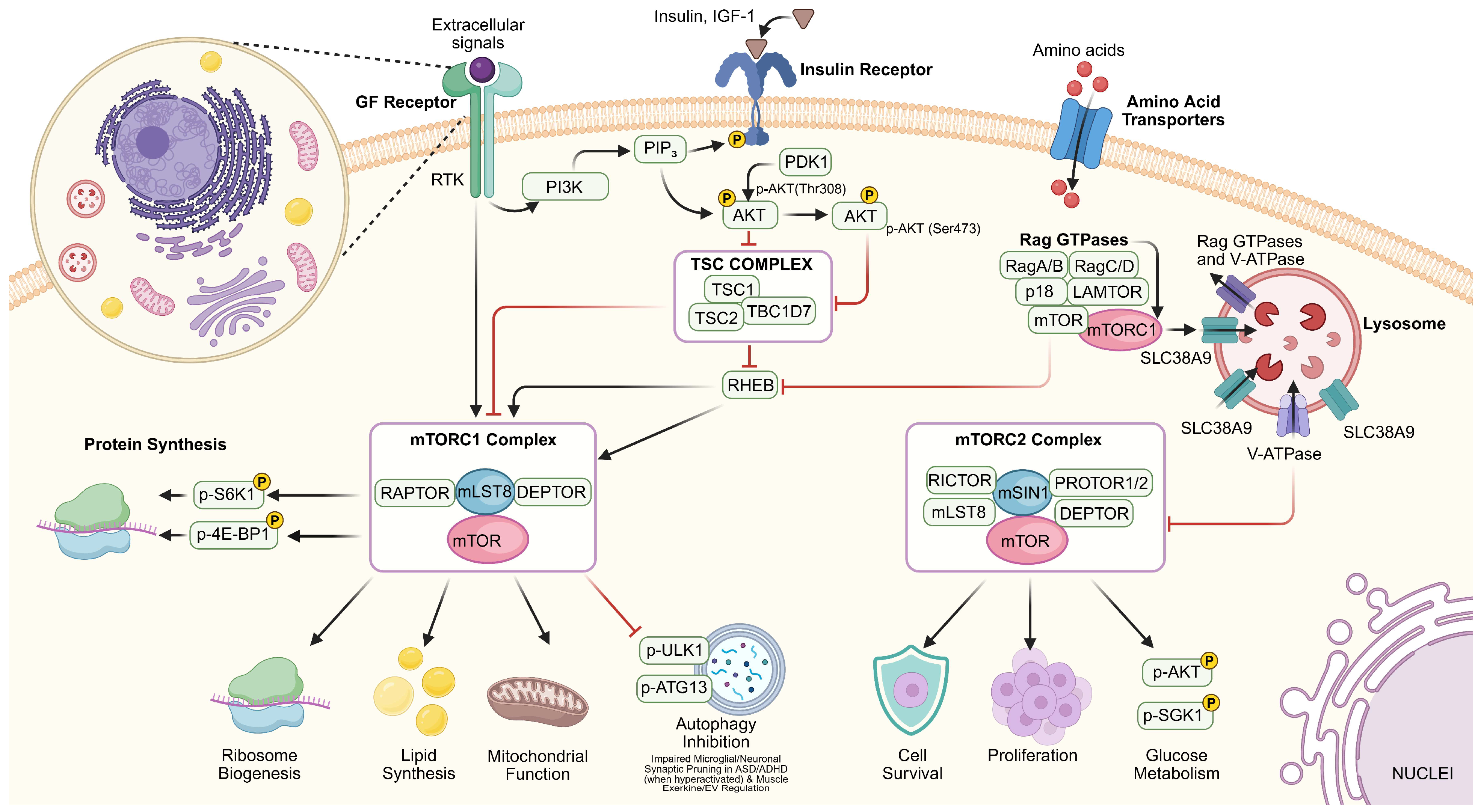
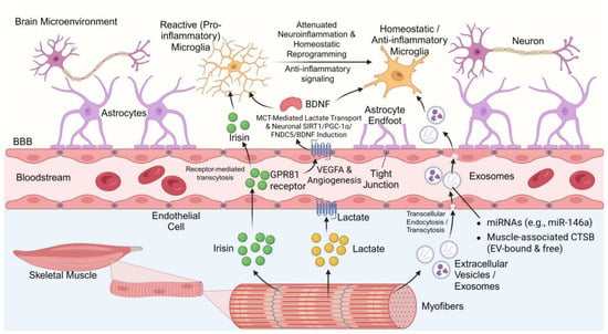
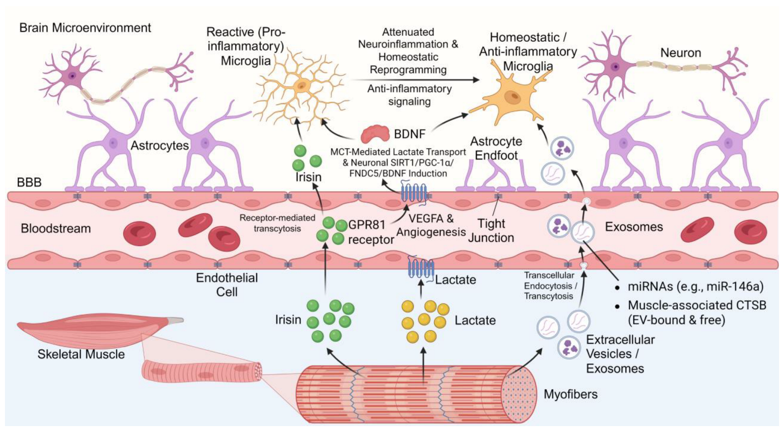
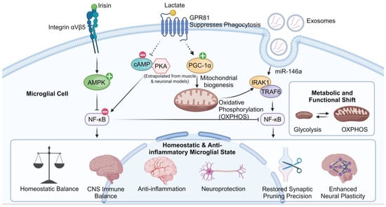
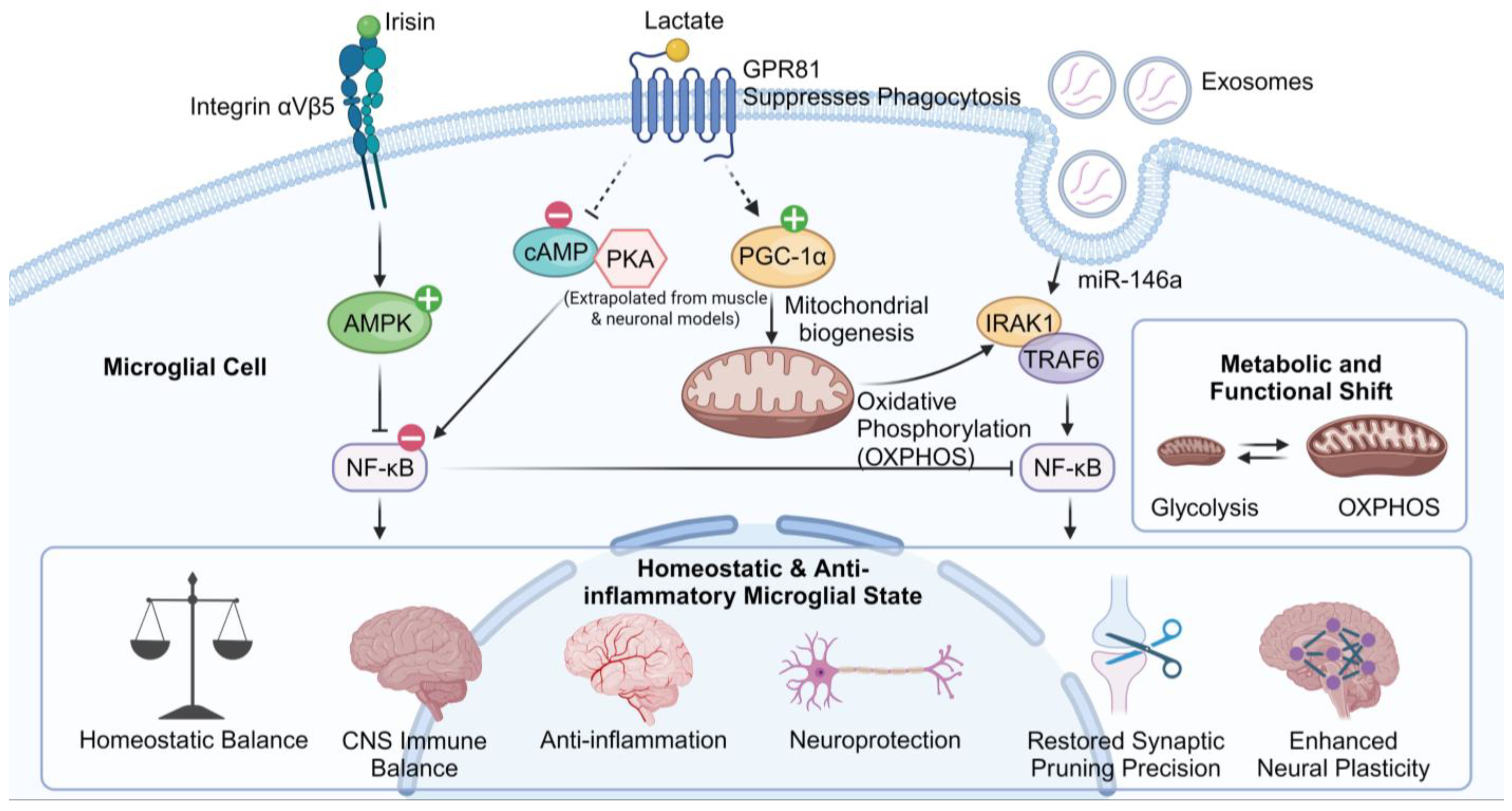

Disclaimer/Publisher’s Note: The statements, opinions and data contained in all publications are solely those of the individual author(s) and contributor(s) and not of MDPI and/or the editor(s). MDPI and/or the editor(s) disclaim responsibility for any injury to people or property resulting from any ideas, methods, instructions or products referred to in the content. |
© 2026 by the authors. Licensee MDPI, Basel, Switzerland. This article is an open access article distributed under the terms and conditions of the Creative Commons Attribution (CC BY) license.