


Highlights
What are the main findings?
- Postoperative SSST is uncommon but can occur after both cranial and spinal neurosurgical procedures.
- Presentation ranges from asymptomatic thrombosis to severe neurological complications.
What are the implications of the main findings?
- Postoperative SSST should be considered when postoperative neurological deterioration occurs, particularly after surgery near the superior sagittal sinus or with CSF loss.
- Prospective studies are needed to establish optimal surveillance and treatment strategies.
Abstract
Background/Objectives: Postoperative superior sagittal sinus thrombosis (SSST) is an uncommon neurosurgical complication with variable clinical severity. This scoping review summarized its frequency, mechanisms, clinical presentation, management, and outcomes. Methods: MEDLINE/PubMed, Scopus, Web of Science Core Collection, and Google Scholar were searched up to 11 May 2026. Original studies reporting postoperative SSST were included and synthesized descriptively. Results: Twenty-four studies comprising 1084 patients were included, with 79 postoperative SSST cases with confirmed or extractable SSS involvement. Among studies with operative denominators, SSST frequency ranged from 0.8% to 10.0%. Reported mechanisms included local sinus injury or manipulation, cerebrospinal fluid loss or diversion, infectious/inflammatory conditions, and systemic prothrombotic factors. Clinical presentation ranged from incidental thrombosis to neurological deterioration with edema, venous infarction, or hemorrhage. Management included observation, anticoagulation, and, in selected severe cases, surgical or endovascular treatment. In small, heterogeneous cohorts with SSST-specific outcomes, mortality ranged from 0% to 33.3% and should be interpreted as a descriptive, not pooled, estimate. Conclusions: Postoperative SSST is heterogeneous in mechanism and severity. Current evidence is predominantly retrospective and case-based, limiting firm treatment recommendations.
1. Introduction
Cerebral venous thrombosis (CVT) accounts for about 0.5% to 3% of all strokes and typically affects younger adults, with a median age of 37 years and a marked female predominance of about 75% in large cohorts [1,2,3]. Within the spectrum of CVT, the superior sagittal sinus is one of the most commonly affected sites, involved in approximately 62% of cases, compared with 41% to 45% for the transverse sinus and about 18% for the straight sinus. Superior sagittal sinus thrombosis (SSST) is therefore not an exceptional anatomical subtype, but a major component of CVT overall [4].
In neurosurgical practice, this issue deserves particular attention because successful surgery does not eliminate the risk of postoperative SSST [5]. Procedures performed close to the midline, falx, parasagittal dura, or major dural sinuses may disturb venous outflow even when the primary surgical objective has been achieved [6]. This makes SSST especially relevant in neurosurgery, where a technically satisfactory resection or decompression may still be followed by a venous complication capable of changing postoperative recovery and outcome [7].
Available data suggest that postoperative venous sinus thrombosis is more common radiographically than it appears clinically [8]. In a prospective study of 74 patients undergoing surgery adjacent to major dural sinuses, postoperative cerebral venous sinus thrombosis (CVST) was found in 24 patients (32.4%), and all were asymptomatic at diagnosis [7]. After posterior fossa tumor surgery, asymptomatic postoperative CVST was reported in 33 of 266 patients (12.4%) [8]. In patients undergoing resection of parasagittal or parafalcine meningiomas, postoperative superior sagittal sinus thrombosis was identified in 15 of 266 patients (5.6%), while only 1.9% of the total cohort were symptomatic [5]. These figures show that postoperative SSST is not merely anecdotal, especially in procedures performed in close relationship to the superior sagittal sinus [5,7].
Early recognition remains difficult. In spontaneous CVT, the median delay to diagnosis is 7 days from symptom onset [9]. In postoperative neurosurgical patients, diagnosis may be even more challenging because headache, nausea, seizures, drowsiness, and focal neurological deficits can be misattributed to the expected postoperative course rather than evolving venous thrombosis [2]. This is one reason why postoperative SSST may remain underrecognized, particularly when its course is clinically silent. At the same time, SSST cannot be considered harmless. Although many postoperative cases remain asymptomatic, others are associated with venous infarction, intracranial hemorrhage, raised intracranial pressure, neurological deterioration, and death [10]. In the series by Sturiale et al., radiological signs of venous congestion or hemorrhagic infarction were present in 19/59 patients (32.2%), 78% remained asymptomatic, and 5/59 (8.5%) developed hemorrhagic infarction [6]. The clinical importance of this complication lies in its dual nature. It is often silent and easily missed, yet in selected patients it may have serious consequences despite an otherwise successful neurosurgical procedure.
The aim of this scoping review was to map the available literature on superior sagittal sinus thrombosis after neurosurgical procedures and to summarize the current evidence on this clinically important postoperative complication.
2. Materials and Methods
2.1. Study Design and Reporting
This scoping review was conducted according to the methodological guidance of the Joanna Briggs Institute (JBI) [11] for scoping reviews and reported in accordance with the Preferred Reporting Items for Systematic Reviews and Meta-Analyses extension for Scoping Reviews (PRISMA-ScR) ( Supplementary Materials) [12]. The review protocol was registered in the Open Science Framework (OSF) under registration ID xbfvt.
2.2. Eligibility Criteria
Studies were eligible according to the PICOS framework if they included patients who developed superior sagittal sinus thrombosis after a neurosurgical procedure. Postoperative SSST was defined as a newly diagnosed postoperative thrombus involving the SSS, either in isolation or together with other venous sinuses, with a reported temporal relationship to the neurosurgical procedure. Studies reporting broader postoperative cerebral venous sinus thrombosis cohorts were eligible only when SSS involvement was explicitly reported, or the number of patients with SSS involvement was extractable. For this review, isolated SSST denoted thrombosis confined to the SSS; SSS-predominant thrombosis denoted reports in which the SSS was explicitly described as the principal involved sinus; and multisinus SSST denoted SSS involvement together with at least one additional dural sinus or jugular venous segment. Each patient was counted once irrespective of the number of venous segments involved. No SSS-primary origin or direction of thrombus propagation was inferred unless demonstrated in the source report. A comparator group was not required. Eligible studies had to report at least one relevant outcome, including incidence, clinical presentation, timing of diagnosis, imaging findings, complications, treatment, recanalization, morbidity, or mortality. Original clinical studies published in English, including cohort studies, case–control studies, cross-sectional studies, case series, and case reports, were considered. Reviews, editorials, commentaries, conference proceedings, experimental studies, non-English publications, and reports without sufficient evidence of a temporal relationship between neurosurgery and SSST were excluded (Table 1). For terminology consistency, “postoperative SSST” is used when referring specifically to SSST occurring after a neurosurgical procedure, whereas “SSST” alone is reserved for general or anatomical references.
Table 1.
PICOS framework for defining eligibility criteria.
2.3. Data Source and Search Strategy
A literature search was performed in MEDLINE/PubMed, Scopus, Web of Science Core Collection, and Google Scholar from database inception to 11 May 2026. Reference lists of eligible studies and relevant reviews were manually screened to identify additional publications. The database strategies used database-specific free-text terms and field syntax related to the superior sagittal sinus, cerebral venous sinus thrombosis, and neurosurgical procedures; no MeSH-based controlled-vocabulary component was used in the PubMed strategy. The Google Scholar query shown in Appendix A identified 89 records, all of which were entered into the same deduplication and screening workflow as records from the bibliographic databases. Detailed search strings are presented in Appendix A, Table A1.
2.4. Study Selection and Data Extraction
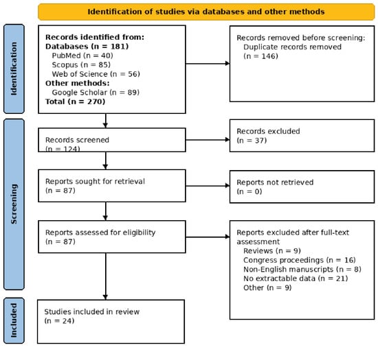
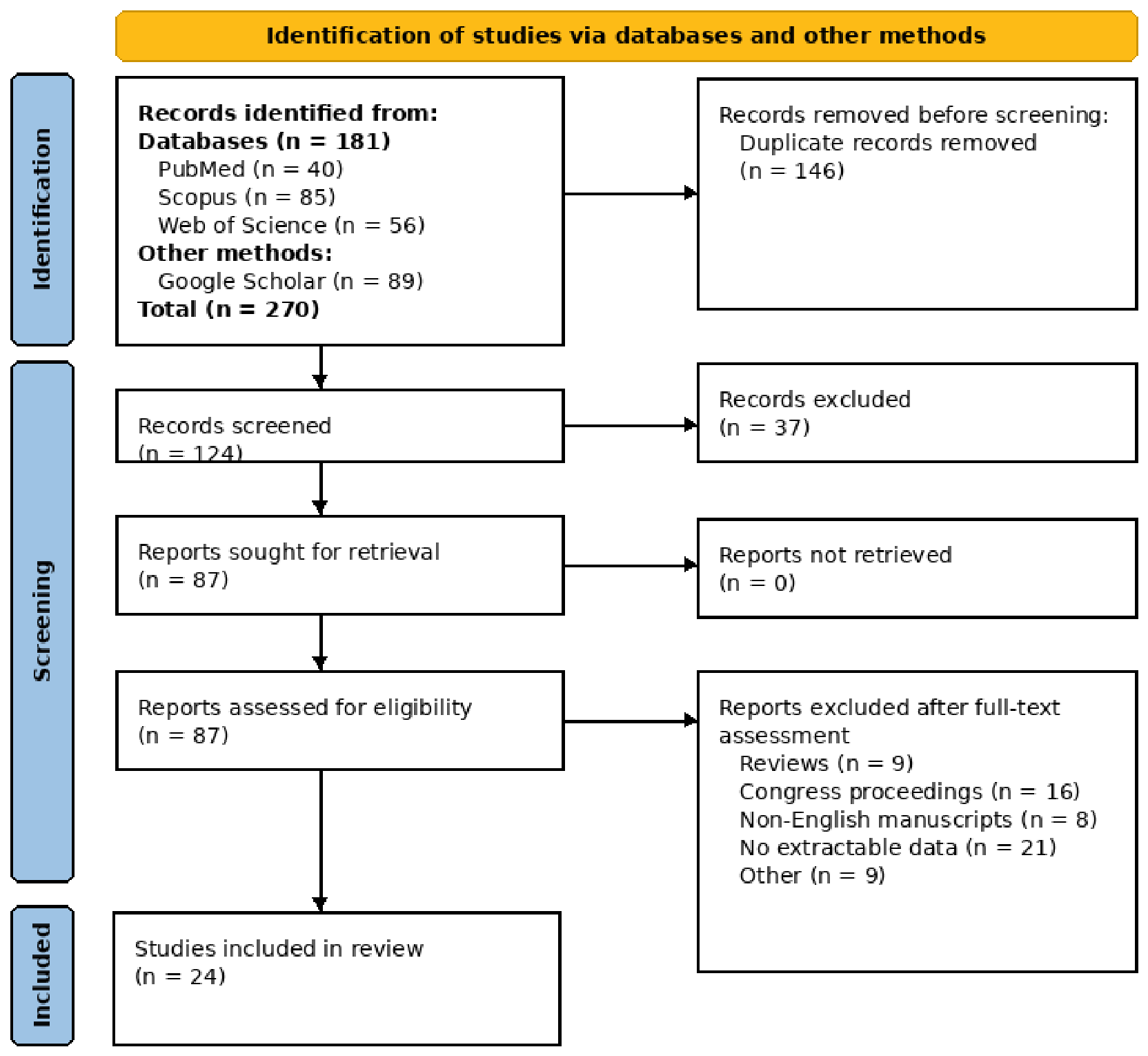
The database and supplementary searches identified 270 records, including 40 from PubMed, 85 from Scopus, 56 from Web of Science, and 89 from Google Scholar. After removal of 146 duplicate records, 124 unique records remained for title and abstract screening. Of these, 37 records were excluded because they did not meet the predefined eligibility criteria, leaving 87 reports for full-text retrieval and assessment. All 87 reports were successfully retrieved and assessed for eligibility. Following full-text review, 63 reports were excluded, including reviews (n = 9), congress proceedings (n = 16), non-English manuscripts (n = 8), reports without extractable data (n = 21), and studies excluded for other eligibility-related reasons (n = 9). Finally, 24 studies met the inclusion criteria and were included in the scoping review (Figure 1).
Figure 1.
PRISMA flow diagram.
Title and abstract screening and full-text assessment were performed independently by two researchers (E.B. and H.B.) according to the predefined eligibility criteria. Disagreements between the two researchers were resolved through discussion and consensus.
Data were independently extracted by two researchers (E.B. and M.P.) using a standardized data-extraction form developed for this review. Extracted variables included study characteristics, country, study design and cohort size, neurosurgical indication and procedure, relationship of the operative field to the superior sagittal sinus, number and proportion of postoperative SSST cases, timing of diagnosis, clinical presentation, symptomatic or asymptomatic course, anatomical extent of thrombosis, associated venous infarction or intracranial hemorrhage, and relevant perioperative or patient-related factors. Data on management included conservative treatment, anticoagulation and its regimen when reported, and surgical or endovascular interventions. Radiological recanalization, neurological recovery, residual deficits, and mortality were also recorded. For broader postoperative CVST cohorts, SSS-specific data were extracted whenever they were explicitly identifiable. When management or outcome data were reported only for the entire CVST cohort and could not be assigned to the SSS subgroup, they were retained only as contextual cohort-level information, explicitly labelled as non-SSST-specific, and were not used to calculate SSST-specific patient-level proportions. Anatomical extent was classified as isolated SSST, SSS-predominant thrombosis, or multisinus thrombosis with SSS involvement according to the definitions above. Any discrepancies between the two researchers were resolved through discussion and consensus.
2.5. Data Synthesis
A descriptive synthesis was performed because of substantial heterogeneity in study design, surgical indication, cohort definition, and outcome reporting. For evidence mapping, study-level observations were summarized primarily as counts of reports rather than clinical frequencies, because a case report and a large cohort should not contribute equal weight to a patient-level interpretation. Patient-level proportions were reported only when an explicit numerator and denominator were available. Procedure-specific postoperative SSST frequencies were reported only when an operative denominator was available. For clinical synthesis, studies were grouped into four non-mutually exclusive mechanistic contexts: local sinus injury/exposure or manipulation, CSF-loss/diversion related, infectious/inflammatory, and systemic/prothrombotic. Clinical phenotype was described as incidental/asymptomatic, symptomatic without major parenchymal injury, or complicated SSST with edema, venous infarction, hemorrhage, thrombus progression, or marked neurological deterioration. No pooled prevalence estimate or meta-analysis was performed.
3. Results
3.1. Characteristics of Included Studies
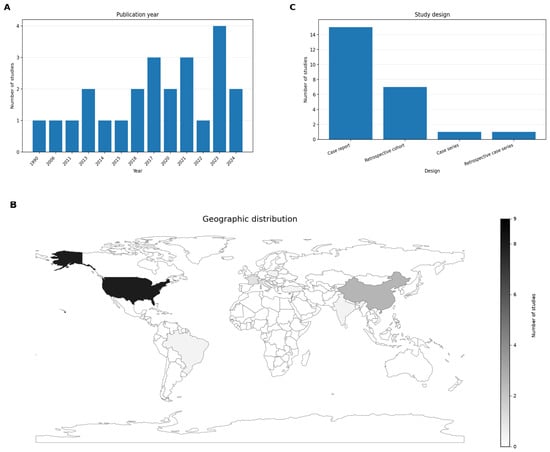
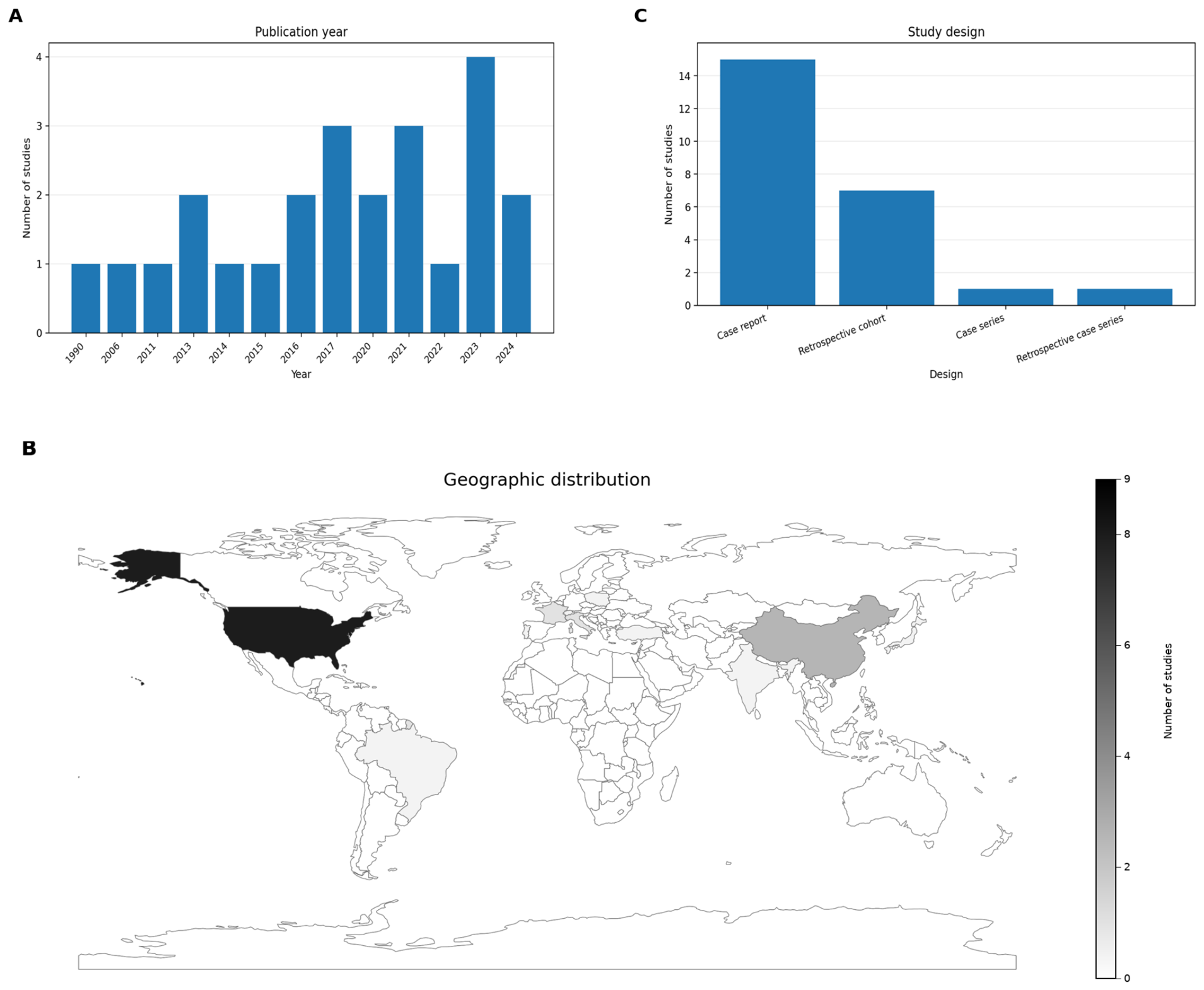
A total of 24 studies were included in the final synthesis [5,6,10,13,14,15,16,17,18,19,20,21,22,23,24,25,26,27,28,29,30,31,32,33,34]. The included studies were published between 1990 and 2024, with a clear increase in publications in recent years. The highest number of studies was published in 2023 (n = 4, 16.7%), followed by 2017 (n = 3, 12.5%). Two studies each were published in 2013, 2016, 2020, 2021, 2022, and 2024 (n = 2, 8.3% each), while one study each was published in 1990, 2006, 2011, 2014, and 2015 (n = 1, 4.2% each) (Figure 2A).
Figure 2.
Temporal, geographic, and methodological characteristics of the included studies. (A) Distribution by publication year. (B) Geographic distribution by country. (C) Distribution by study design.
The studies originated from 11 countries. The largest proportion came from the United States (n = 9, 37.5%), followed by China (n = 4, 16.7%). France and Italy each contributed two studies (n = 2, 8.3%), while Switzerland, Brazil, Japan, India, Portugal, Turkey, and Poland each contributed one study (n = 1, 4.2%) (Figure 2B). Regarding study design, the evidence base was dominated by case reports (n = 15, 62.5%), followed by retrospective cohort studies (n = 7, 29.2%). One case series (n = 1, 4.2%) and one retrospective case series (n = 1, 4.2%) were also included (Figure 2C). Detailed study characteristics are presented in Table 2.
Table 2.
Characteristics of included studies reporting postoperative superior sagittal sinus thrombosis.
Across the 24 included studies, the combined study populations comprised 1084 patients undergoing the relevant neurosurgical procedures or represented in selected postoperative CVST cohorts. A total of 79 postoperative SSST cases with explicit or extractable SSS involvement were reported. Because the included studies differed substantially in design, patient selection, surgical indication, and denominator definition, an overall postoperative SSST prevalence was not calculated. Among studies providing an operative cohort denominator, postoperative SSST or SSS-region thrombosis occurred in 1/75 patients (1.3%) in Ricci et al. [23], 14/401 (3.5%) in Planet et al. [33], 3/71 (4.2%) in Ying et al. [24], 15/266 (5.6%) in Jimenez et al. [5], and 3/44 (6.8%) in Wang et al. [20]. Raza et al. [15] reported postoperative venous thrombosis/infarction in approximately 7.0% of a cohort of 110 patients with parasagittal meningiomas invading the SSS; however, the involved venous segment was not reported at the patient level. These events were therefore not counted as confirmed postoperative SSST cases and were excluded from SSST-specific tables, figures, and anatomical analyses.
3.2. Mechanistic and Neurosurgical Context of Postoperative SSST
The included reports represented heterogeneous postoperative mechanisms rather than a single etiological entity. Local sinus-related injury, exposure, manipulation, reconstruction, or surgery immediately adjacent to a dural sinus was the most common context in cranial reports, particularly after parasagittal/parafalcine meningioma surgery and other operations near major venous sinuses. A distinct CSF-loss/diversion context was represented by spinal surgery with durotomy or CSF leakage, intracranial hypotension, burr-hole drainage, and ventriculoperitoneal shunting. Infectious/inflammatory postoperative settings included intracranial infection, subdural empyema, or abscess, whereas systemic prothrombotic susceptibility included inherited or acquired thrombophilia and other patient-related risk factors. These categories were not mutually exclusive; several patients had both a local surgical trigger and systemic or CSF-related susceptibility.
Procedure-specific frequencies were interpreted separately from mechanism. Among parasagittal and parafalcine meningioma cohorts, postoperative SSST or SSS-region thrombosis was reported in 15/266 patients (5.6%) by Jimenez et al. [5], 3/44 (6.8%) by Wang et al. [20], 3/71 (4.2%) by Ying et al. [24], and 1/75 (1.3%) by Ricci et al. [23]. Outside this setting, Planet et al. [33] identified SSS involvement in 14/401 patients (3.5%) after awake supratentorial craniotomy, Przepiorka et al. [30] reported clinically apparent SSST in 1/130 patients (0.8%) after retrosigmoid procedures, and Skochdopole et al. [34] reported one SSST case in a 10-patient cranio-orbital reconstruction series (10.0%).
Selected postoperative CVST cohorts showed higher proportions of SSS involvement but did not represent procedure-specific SSST incidence. Sturiale et al. [6] reported SSS involvement in 20/59 postoperative CVST cases (33.9%), including isolated SSS and multisinus thromboses with SSS involvement; 8/20 of these SSS-involved cases were symptomatic. Petrov et al. [22] reported SSS involvement in 6/13 pediatric postoperative CVST cases (46.2%). Accordingly, among studies with an operative denominator, reported postoperative SSST frequency ranged from 0.8% to 10.0%, whereas higher proportions in selected postoperative CVST populations describe the anatomical distribution of thrombosis after CVST had already occurred.
3.3. Timing and Clinical Phenotype of Postoperative SSST
Timing of postoperative SSST was heterogeneous, but when a specific postoperative day (POD) was reported, thrombosis was most often detected early. Postoperative SSST was diagnosed on POD 0 in two reports, POD 1 in two reports, approximately POD 1.5 in one report, and POD 3 in two reports (Table 3). Petrov et al. [22] reported a median diagnosis on POD 3 for their overall pediatric postoperative CVST cohort. Later presentations were described on POD 9, POD 10, POD 14, and POD 26. In several additional studies, postoperative SSST was described as early, but the exact day was not reported. Clinically, the evidence mapped onto three phenotypes: incidental/asymptomatic thrombosis detected on surveillance imaging; symptomatic SSST without major parenchymal injury, presenting with headache, nausea/vomiting, seizure, or focal neurological deficit; and complicated SSST associated with marked neurological deterioration, edema, venous infarction, intracranial hemorrhage, thrombus propagation, or coma. Among individual case reports with explicit symptoms, headache was described in seven reports, altered consciousness or marked neurological deterioration in eight, seizures in four, focal neurological deficits in five, and nausea/vomiting in four. These are report counts rather than patient-level frequencies.
Table 3.
Clinical and radiological characteristics of postoperative superior sagittal sinus thrombosis.
Larger cohorts confirmed that postoperative SSST can be clinically silent. Planet et al. [33] reported no symptomatic cases among the 14 patients with SSS involvement. In Jimenez et al. [5], 5/15 patients with postoperative SSST were symptomatic, and 10/15 were asymptomatic. In Sturiale et al. [6], symptoms occurred in 8/20 patients with SSS involvement (40.0%). Petrov et al. [22] reported 9/13 patients as asymptomatic in the overall pediatric postoperative CVST cohort, but the symptomatic proportion was not separately reported for the six-patient SSS subset. Thus, postoperative SSST spans incidental imaging findings, symptomatic disease without major parenchymal injury, and complicated phenotypes with neurological deterioration or parenchymal injury.
3.4. Anatomical Extent and Radiological Complications
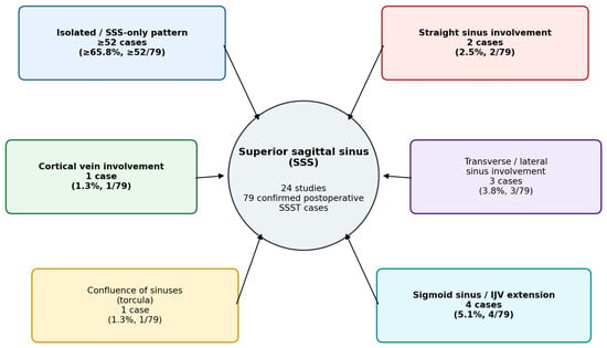
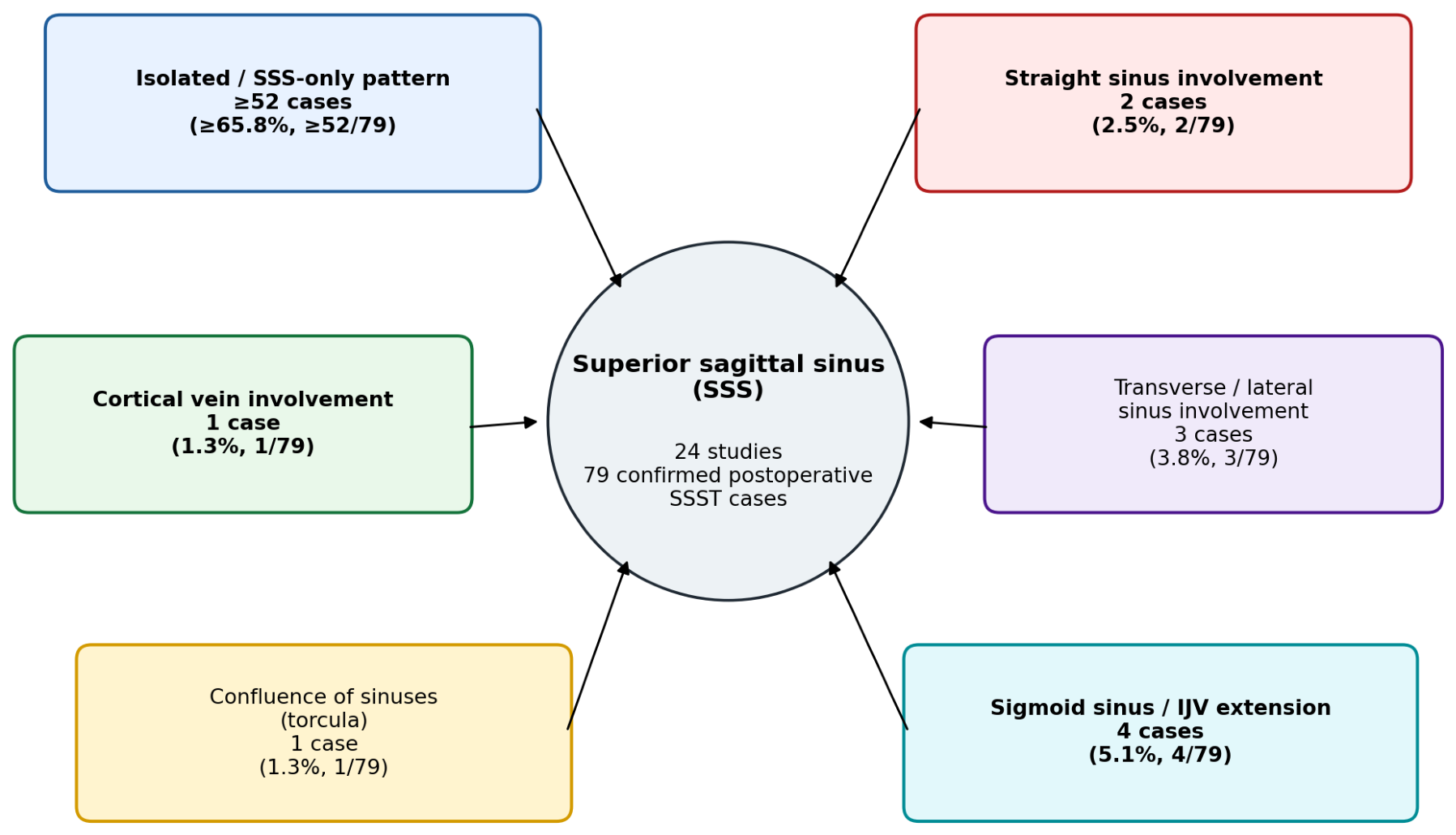
Among explicitly classifiable postoperative SSST cases, an SSS-only or SSS-predominant pattern was documented in at least 52/79 cases (≥65.8%). Additional venous involvement included the sigmoid sinus and/or internal jugular vein in 4/79 cases (5.1%), the transverse or lateral sinus in 3/79 (3.8%), the straight sinus in 2/79 (2.5%), and the torcula (confluence of sinuses) and cortical veins in 1/79 each (1.3%). Categories were non-mutually exclusive, and some cases could not be further anatomically subclassified from the published data. Accordingly, these proportions represent minimum documented proportions among confirmed postoperative SSST cases rather than the true anatomical distribution of postoperative SSST. Extensive multisinus thrombosis was particularly described after CSF-loss-related procedures, skull-base surgery, and CSF diversion. In the larger cohorts, Sturiale et al. [6] documented SSS involvement in 20/59 postoperative CVST patients, including 16 isolated SSS cases and four multisinus cases involving the SSS, while Planet et al. [33] reported SSS involvement in 14/16 postoperative CVST cases (Figure 3). These categories describe anatomical distribution only; serial imaging establishing the site of thrombus initiation was generally unavailable, and no SSS-primary origin was inferred.
Figure 3.
Anatomical distribution patterns of postoperative SSST.
Radiological severity ranged from isolated thrombosis without parenchymal injury to extensive venous infarction and hemorrhage. Cerebral edema or brain swelling was described in six reports and hemorrhagic complications in five; these report counts are descriptive rather than patient-level frequencies. Hemorrhagic venous infarction was documented in Garrido et al. [13] and Sawarkar et al. [10]. Sturiale et al. [6] reported edema and hemorrhagic infarction at the overall postoperative CVST-cohort level, but did not provide SSS-specific radiological complication counts. Ricci et al. [23] reported one patient with SSS occlusion accompanied by venous infarction and brain swelling. Xie et al. [32] described bilateral frontoparietal hemorrhages, subarachnoid hemorrhage, and cerebral edema associated with acute postoperative SSST. In contrast, absence of infarction or intracranial hemorrhage was explicitly reported in several SSST-specific reports, including Jimenez et al. [5], Planet et al. [33], and individual cases.
3.5. Reported Predisposing and Perioperative Factors
Local operative factors were frequently reported as potential contributors to postoperative SSST. Direct sinus exposure, manipulation, reconstruction, or surgery immediately adjacent to the SSS was explicitly described in 10 reports. In the pediatric CVST series of Petrov et al. [22], a dural sinus was exposed in 11/13 patients (84.6%), and the operative field was adjacent to a sinus in 8/13 (61.5%); these data refer to the full CVST cohort rather than the six-patient SSS subset. Wang et al. [20] reported SSS transection in 9/44 patients (20.5%), opening and repair in 3/44 (6.8%), and sinus-wall coagulation in 32/44 (72.7%). All three postoperative venous sinus thromboses occurred after the more extensive SSS manipulation categories. Ricci et al. [23] reconstructed the SSS in 21/75 patients (28.0%), while Ying et al. [24] reported SSS excision in 15/71 (21.1%), reconstruction in 8/71 (11.3%), and electrocoagulation in 48/71 (67.6%). In Planet et al. [33], a lesion located within 1 cm of a major venous sinus was independently associated with postoperative CVST (adjusted OR 10.58, 95% CI 2.93–38.20; p < 0.001), and female sex was also associated with increased risk (adjusted OR 4.00, 95% CI 1.24–12.91; p = 0.021).
CSF-related mechanisms were explicitly described in seven reports, with two additional reports describing suspected CSF loss or intracranial hypotension. These contexts included postoperative CSF leakage, durotomy, excessive CSF drainage, and intracranial hypotension following spinal surgery, burr-hole drainage, or ventriculoperitoneal shunting.
Systemic prothrombotic factors were less consistently assessed. Lega et al. reported previous recurrent deep-vein thrombosis, factor V Leiden, and antiphospholipid antibodies, while Sommerstein et al. [16] described oral contraceptive use and heterozygous factor V Leiden. Costa et al. [19] reported prothrombin G20210A and homozygous MTHFR variants with oral contraceptive use. In the pediatric cohort of Petrov et al. [22], prothrombotic abnormalities were identified in 10/13 patients (76.9%). Other reported associated factors included dehydration, obesity, infection, hypertension, diabetes, and smoking, although these were not consistently demonstrated as independent predictors across studies (Table 4).
Table 4.
Reported predisposing and perioperative factors in studies of postoperative superior sagittal sinus thrombosis.
3.6. Management Strategies and Outcomes
Management varied according to clinical severity, thrombus extent, associated hemorrhage, and the postoperative setting. Across the literature, approaches included observation/supportive care, therapeutic anticoagulation, prophylactic anticoagulation without therapeutic escalation, and surgical or endovascular rescue. Importantly, management data were not always SSS-specific. In Petrov et al. [22], 7/13 patients in the overall postoperative pediatric CVST cohort received anticoagulation and 6/13 were observed; treatment was not separately reported for the six patients with SSS involvement. Similarly, Sturiale et al. [6] treated 39/59 patients in the overall postoperative CVST cohort with therapeutic LMWH and managed 20/59 conservatively; these proportions cannot be assigned specifically to the 20 SSS-involved cases. By contrast, Jimenez et al. [5] provided SSST-specific management: 2/15 patients received anticoagulation, both symptomatic, whereas asymptomatic thromboses were generally observed. Planet et al. [33] reported standard prophylactic-dose anticoagulation across the postoperative CVST cohort without treatment-dose escalation. Anticoagulation was withheld in the hemorrhagic case reported by Sawarkar et al. [10].
Surgical or endovascular interventions were described in severe individual cases and included mechanical or aspiration thrombectomy, venous sinus thrombectomy, decompressive craniectomy, repeat surgery, and reoperation for thrombotic SSS occlusion. Radiological outcomes also required separation of SSST-specific from whole-CVST-cohort data. Planet et al. [33] reported spontaneous recanalization of all postoperative CVSTs; because 14/16 involved the SSS, these SSS-involved cases were included among the recanalized thromboses. Sturiale et al. [6] reported persistent thrombosis in 36/59 patients (61.0%) at follow-up, but this was a whole-CVST-cohort outcome and was not available separately for the SSS subgroup. Petrov et al. [22] reported a median time to partial or complete recanalization of 2.4 months (IQR 0.7–5.1) for the overall pediatric CVST cohort, not specifically for the SSS subset. Complete recanalization was also reported in several SSST-specific individual reports. Among multi-patient cohorts with clearly SSST-specific outcome data, mortality ranged from 0% in Jimenez et al. [5] to 33.3% in Wang et al. [20] and Ying et al. [24]. Because these estimates derive from small samples and clinically heterogeneous study populations, the observed range should be interpreted cautiously as descriptive evidence rather than as a reliable estimate of postoperative SSST mortality risk. Fatal outcomes were also described in individual case reports. Cohort-level mortality from broader CVST series was not used to estimate SSST-specific mortality (Table 5).
Table 5.
Management strategies and outcomes of postoperative superior sagittal sinus thrombosis.
4. Discussion
Postoperative SSST should not be regarded as a uniform complication. The available evidence spans incidental, segmental thrombosis detected on surveillance imaging to extensive venous occlusion associated with edema, hemorrhagic venous infarction, seizures, impaired consciousness, and death. This distinction is clinically important because studies using routine postoperative venographic imaging detect substantially more thrombosis than symptom-driven series [7,8,30]. The practical implication is that radiological SSST and clinically significant venous outflow obstruction should not be considered equivalent entities. Because the evidence base is heterogeneous, retrospective, and heavily influenced by case reports, the management discussion below is intended as a conceptual framework for risk stratification rather than a set of definitive postoperative SSST treatment recommendations.
This discrepancy is particularly relevant for the superior sagittal sinus because limited segmental thrombosis may remain compensated through cortical and parasagittal collateral drainage. Conversely, progression into cortical veins, the confluence of sinuses, straight sinus, transverse-sigmoid system, or jugular venous outflow may markedly increase the physiological consequences of the same primary lesion. Observational CVT studies consistently identify parenchymal lesions, cortical venous involvement, hemorrhage, and impaired consciousness as markers of a more severe phenotype [35,36,37,38]. Thus, anatomical extent should be interpreted together with collateral drainage and parenchymal consequences rather than as an isolated measure of severity [38].
4.1. Surgical and Perioperative Mechanisms
The most intuitive mechanism is direct local injury. Parasagittal and parafalcine meningioma surgery frequently requires exposure, coagulation, opening, suturing, reconstruction, or even partial sacrifice of the SSS, all of which may produce endothelial injury and transient or persistent flow reduction [15,20,23]. Similar mechanisms are plausible in skull-base and posterior fossa procedures where venous sinuses are skeletonized, retracted, thermally injured, or compressed by postoperative changes [6,30].
The relationship, however, is not purely mechanical. Many extensively manipulated sinuses remain patent, while SSST has been reported after procedures with no direct contact with the SSS. This suggests that local surgical injury acts together with systemic and hemodynamic factors rather than functioning as an isolated cause [5,15,20,23,24].
The spinal cases are especially instructive because they point toward a different mechanism. CSF loss after durotomy, spinal surgery, lumbar drainage, or persistent postoperative leakage may cause intracranial hypotension, compensatory venous engorgement, slower venous flow, and downward traction on intracranial venous structures [39,40]. Cerebral venous thrombosis following intracranial hypotension has repeatedly been described after lumbar puncture, spinal anesthesia, spinal surgery, and CSF diversion [41,42].
Clinically, the transition from low-pressure headache to venous thrombosis may be subtle. A typical orthostatic headache that becomes persistent, loses its postural character, progressively worsens, or is accompanied by vomiting, diplopia, papilledema, seizure, or focal neurological deficit should prompt consideration of venous imaging rather than continued attribution to CSF loss alone [39,40,41,42].
Systemic predisposition probably determines why only some patients develop thrombosis after comparable surgical stress. Factor V Leiden, prothrombin G20210A, antiphospholipid antibodies, previous venous thromboembolism, oral contraceptive use, malignancy, obesity, dehydration, and infection are established CVT-associated factors, and several were represented in the included postoperative cases [14,16,19]. Meta-analytic data have demonstrated strong associations between CVT and prothrombin G20210A, factor V Leiden, hyperhomocysteinemia, and hormonal contraceptive exposure [43,44]. Even so, current recommendations do not support universal thrombophilia screening after an obviously provoked postoperative event. Testing is most relevant when it may alter duration or choice of anticoagulation, particularly in recurrent thrombosis, unexplained events, a strong family history, younger patients without a clear trigger, or suspected antiphospholipid syndrome [45,46,47,48].
4.2. Clinical Presentation and Diagnostic Interpretation
The postoperative setting reduces the specificity of many classic CVT symptoms. Headache is present in the great majority of spontaneous CVT cases, while seizures, papilledema, focal deficits, and encephalopathy occur less consistently [2,49,50]. After neurosurgery, however, headache, nausea, somnolence, and mild confusion may be expected postoperative findings. The more important signal is therefore a deviation from the expected recovery trajectory: progressive headache, delayed emergence, new seizure, focal deficit, or deterioration after an initially stable postoperative course [41,42].
Seizures appear to mark a more complicated venous phenotype. In a meta-analysis of 1244 patients, early seizures were associated with supratentorial lesions, cortical vein involvement, motor deficits, and hemorrhagic lesions [36]. In a large international cohort, SSS thrombosis, intracerebral hemorrhage, cerebral edema or infarction, and cortical vein thrombosis were independently associated with acute symptomatic seizures [37]. These findings are concordant with the severe postoperative cases in which seizures accompanied hemorrhagic venous infarction or extensive thrombosis rather than an isolated radiological defect [37].
Imaging should reflect clinical suspicion rather than screening laboratory results. CT venography and MR venography are both accepted diagnostic tools, while MRI offers better characterization of edema, venous infarction, cortical vein involvement, and small hemorrhagic lesions [46]. D-dimer has limited value after surgery because specificity is reduced and false-negative results may occur with limited thrombosis or delayed presentation. A normal D-dimer therefore should not defer venographic imaging when postoperative SSST is clinically suspected [51].
4.3. Anticoagulation: The Central Management Dilemma
For spontaneous symptomatic CVT, therapeutic anticoagulation is well established. Randomized studies of UFH and LMWH showed clinical benefit without a convincing increase in symptomatic intracranial hemorrhage, even in patients presenting with venous hemorrhagic lesions [52,53]. On this basis, major contemporary guidelines recommend therapeutic anticoagulation for symptomatic CVT and generally favor LMWH in clinically stable patients [45,46].
Postoperative SSST is more difficult because the bleeding substrate is different. A hemorrhagic venous infarct caused by elevated venous pressure is not equivalent to a fresh craniotomy bed, tumor cavity, dural repair, reconstructed sinus, or recently manipulated venous surface. The standard CVT principle that intracranial hemorrhage is not by itself a contraindication to anticoagulation cannot therefore be applied mechanically to the immediate postoperative period [45,46].
The neurosurgical evidence supports this caution. Several postoperative series have shown that asymptomatic thrombosis can remain stable or recanalize without therapeutic anticoagulation [5,6,30,33]. In the parasagittal/parafalcine meningioma cohort, anticoagulation was largely reserved for symptomatic SSST [5]. In another cohort, SSS-predominant postoperative thromboses were managed without therapeutic escalation and recanalized spontaneously [33]. Shabo et al. observed more intracranial hemorrhage among anticoagulated asymptomatic postoperative CVST patients, although confounding by indication prevents a causal conclusion [8].
This favors a selective rather than automatic approach. Stable, nonprogressive, asymptomatic SSST without edema, infarction, hemorrhage, or cortical venous congestion may reasonably be observed when bleeding risk is high. By contrast, neurological deterioration, thrombus propagation, cortical venous compromise, progressive edema, venous infarction, or expanding multisinus disease provide a stronger rationale for full anticoagulation once operative hemostasis is considered secure [6,46].
Drug choice should also reflect the postoperative context. LMWH is generally preferred in stable CVT because of predictable pharmacokinetics, whereas UFH has an advantage when rapid reversal or reoperation may be required [46,54]. In the randomized trial by de Bruijn and Stam, nadroparin was administered at 180 anti-Xa IU/kg per 24 h [52]. In contemporary VTE practice, enoxaparin is commonly used at 1 mg/kg subcutaneously every 12 h, with renal adjustment when appropriate, while UFH is titrated according to institutional aPTT or anti-Xa protocols. These are standard therapeutic regimens rather than postoperative SSST-specific dosing recommendations [53,55].
Timing remains unresolved. Observational neurosurgical series suggest that therapeutic anticoagulation during the first postoperative week is feasible in selected patients but carries a measurable hemorrhagic risk [56,57,58]. In one series of 19 patients anticoagulated within 10 days after craniotomy, three developed intracerebral hemorrhage, although none required surgical treatment [57]. In another, anticoagulation started at a median of postoperative day 5, and one of 18 patients required evacuation of a postoperative hemorrhage [58]. These studies do not define a universally safe postoperative day. The decision should instead incorporate operative hemostasis, postoperative CT findings, residual surgical-bed blood, type of procedure, sinus reconstruction, renal function, and the likelihood that another operation may be necessary [58,59].
4.4. Maintenance Anticoagulation and Duration
Once the immediate postoperative bleeding risk has subsided, treatment can more closely follow conventional CVT practice. Vitamin K antagonists with a target INR of 2.0–3.0 remain acceptable, and DOACs are increasingly supported for clinically stable patients. Treatment duration is usually at least three months and should depend on whether the event was transiently provoked, recurrent, or associated with persistent thrombophilia or active malignancy [9,46].
The evidence for DOACs has become substantially stronger. In RE-SPECT CVT, dabigatran 150 mg twice daily produced similarly low rates of recurrent thrombosis and major bleeding compared with dose-adjusted warfarin after initial parenteral therapy [60]. ACTION-CVT, which included 845 patients, found similar rates of recurrent thrombosis, death, and recanalization between DOACs and warfarin, but fewer major hemorrhages with DOAC therapy [61]. SECRET further supported rivaroxaban as a feasible option, and subsequent meta-analyses have generally found comparable efficacy and safety between DOACs and vitamin K antagonists [62,63,64].
These data are most relevant after stabilization. They do not support replacing short-acting, reversible parenteral anticoagulation with a DOAC during the immediate post-craniotomy period [60,62,63,64].
4.5. Endovascular Treatment and Decompression
Endovascular treatment should remain a rescue strategy. Mechanical thrombectomy, aspiration, stent-retriever techniques, and combined approaches can achieve rapid recanalization, but technical success does not guarantee functional recovery [35,49,65]. In TO-ACT, excellent functional outcome at 12 months occurred in 67% of patients treated endovascularly and 68% of patients receiving standard treatment, providing no evidence of routine benefit [65].
The TO-ACT trial was small and cannot exclude benefit in carefully selected deteriorating patients. Observational series and meta-analyses report high recanalization and favorable-outcome rates after thrombectomy, but these studies are heavily affected by severity and selection bias [49]. Contemporary CVT guidance generally considers endovascular treatment only for selected patients with clinical deterioration despite adequate anticoagulation or severe disease in whom rescue therapy is being considered [45,46,54,65]. These recommendations derive from spontaneous CVT and should not be interpreted as postoperative SSST-specific evidence.
Decompressive surgery addresses a different problem. When venous infarction or hemorrhage produces malignant edema, mass effect, or impending herniation, the aim is survival rather than thrombus removal [50,66]. In a multicenter registry and systematic review of severe CVT, more than half of surgically treated patients regained functional independence despite very severe initial presentation [50]. DECOMPRESS2 similarly showed that meaningful recovery remains possible after aggressive surgical management [66].
Thus, neurological deterioration from malignant venous edema should not be managed by anticoagulation alone when a clear decompressive indication exists [10,28,50,66].
4.6. Recanalization and Follow-Up
Recanalization is frequent after CVT, but complete reopening is not synonymous with clinical recovery. Most venous recanalization occurs during the first several months [61,67]. Meta-analytic data indicate that failure to recanalize is associated with worse outcomes and recurrent CVT, while the difference between partial and complete recanalization is less consistent [67,68].
This matters in postoperative SSST because persistent residual thrombus should not automatically trigger prolonged anticoagulation. Current recommendations emphasize recurrent thrombotic risk rather than complete radiological normalization when deciding treatment duration. Repeat CT venography or MR venography at approximately three to six months is reasonable after treated or clinically significant SSST, whereas earlier imaging is justified if symptoms worsen or thrombus propagation is suspected [46,54,67,68].
4.7. Implications and Evidence Gaps
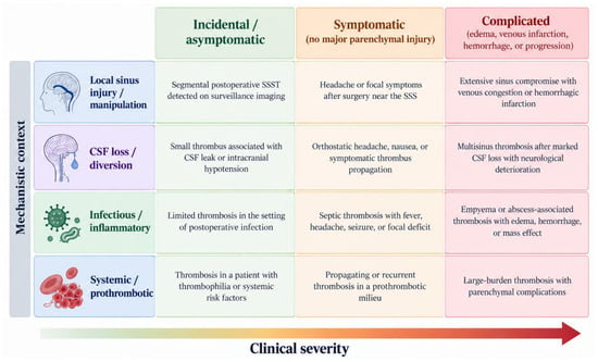
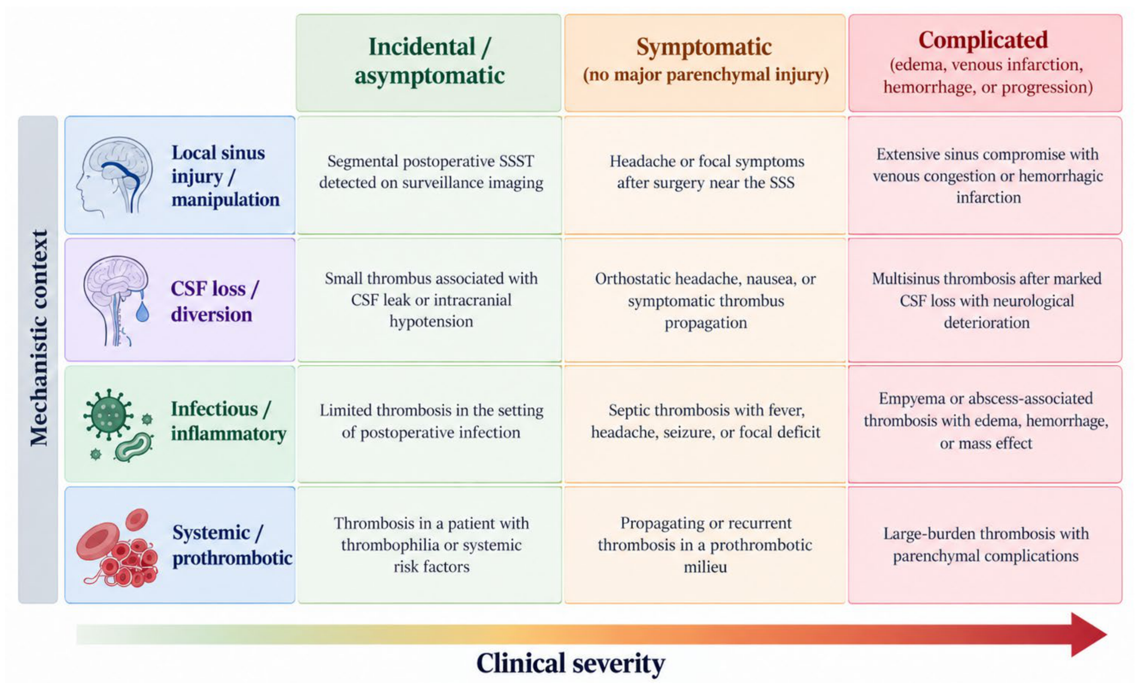
A practical synthesis of the available evidence can be represented as a two-axis conceptual framework combining mechanism and clinical severity (Figure 4). On the mechanism axis, postoperative SSST may arise in local sinus-injury/manipulation, CSF-loss/diversion, infectious/inflammatory, or systemic/prothrombotic contexts, often with overlap. On the clinical-severity axis, patients range from incidental/asymptomatic thrombosis to symptomatic disease without major parenchymal injury and to complicated SSST with edema, infarction, hemorrhage, propagation, or neurological deterioration. Management decisions in the published literature broadly intensified across this severity spectrum, but the evidence is insufficient to define a validated treatment algorithm.
Figure 4.
Two-axis conceptual framework for postoperative SSST, integrating mechanistic context and clinical severity. Mechanistic categories are non-mutually exclusive. The framework summarizes the descriptive evidence base and is not a validated treatment algorithm.
The main limitations of the literature are small sample size, inconsistent case definition, and substantial selection bias. Screening cohorts preferentially identify benign radiological thrombosis, whereas case reports preferentially capture catastrophic complications [5,7,8,30]. Study-level counts therefore map where a feature has been reported but do not estimate patient-level frequency. In addition, several broader postoperative CVST cohorts reported management and outcomes only for the full CVST population; these data cannot be assumed to represent the SSS subgroup.
Imaging protocols, thresholds for anticoagulation, duration of follow-up, and definitions of “symptomatic” thrombosis vary considerably. These differences probably explain part of the apparent disagreement between studies [30,33].
Future prospective studies should apply standardized anatomical definitions and distinguish at least three clinical phenotypes: incidental asymptomatic postoperative SSST, symptomatic postoperative SSST without major parenchymal injury, and complicated postoperative SSST with hemorrhage, venous infarction, edema, propagation, or marked neurological deterioration. At minimum, studies should report the operative relationship to the SSS, degree of luminal occlusion, cortical vein involvement, mechanism/context, timing and dose of anticoagulation, postoperative hemorrhage, serial venographic evolution, and standardized functional outcome [30,33,46].
The key unanswered question is no longer whether postoperative SSST occurs. It is which patients are more likely to be harmed by thrombus progression than by anticoagulation itself. Existing CVT guidelines cannot answer that question directly because their evidence base is derived predominantly from spontaneous symptomatic disease rather than postoperative neurosurgical thrombosis [45,54].
5. Conclusions
Postoperative SSST is a heterogeneous complication that ranges from incidental, clinically silent thrombosis to extensive venous occlusion with edema, hemorrhagic infarction, neurological deterioration, and death. The available literature supports distinguishing both the underlying postoperative mechanism and the clinical phenotype when interpreting individual cases. The risk-based approach proposed in this review is a conceptual framework derived from descriptive, predominantly retrospective evidence and should not be regarded as a definitive treatment recommendation. Observation, anticoagulation, endovascular therapy, and decompression have all been used in selected settings, but postoperative SSST-specific comparative evidence is insufficient to establish standardized management pathways. Prospective studies using consistent anatomical definitions, SSST-specific outcome reporting, and standardized treatment variables are needed.
Supplementary Materials
The following supporting information can be downloaded at: https://www.mdpi.com/article/10.3390/brainsci16111198/s1, Table S1: Preferred Reporting Items for Systematic reviews and Meta-Analyses extension for Scoping Reviews (PRISMA-ScR) Checklist.
Author Contributions
Conceptualization, E.B. and H.B.; methodology, E.B., M.P., and H.B.; validation, H.B. and K.A.; formal analysis, E.B. and M.P.; investigation, E.B., M.P., and H.B.; resources, E.B., H.B., and K.A.; data curation, E.B. and M.P.; writing—original draft preparation, E.B. and M.P.; writing—review and editing, M.P., H.B., and K.A.; visualization, E.B.; supervision, H.B. and K.A.; project administration, M.P. All authors have read and agreed to the published version of the manuscript.
Funding
This research received no external funding.
Institutional Review Board Statement
Not applicable.
Informed Consent Statement
Not applicable.
Data Availability Statement
No new datasets were generated or analyzed in this study. All data presented in this scoping review were extracted from publicly available published studies included in the review.
Conflicts of Interest
The authors declare no conflicts of interest.
Abbreviations
AC, anticoagulation; aPTT, activated partial thromboplastin time; CI, confidence interval; cCVST, clinical cerebral venous sinus thrombosis; CSF, cerebrospinal fluid; CT, computed tomography; CVST, cerebral venous sinus thrombosis; CVT, cerebral venous thrombosis; DOAC, direct oral anticoagulant; ICH, intracranial hemorrhage; IJV, internal jugular vein; INR, international normalized ratio; IQR, interquartile range; IU, international unit; IV, intravenous; JBI, Joanna Briggs Institute; LMWH, low-molecular-weight heparin; MR, magnetic resonance; MRI, magnetic resonance imaging; mRS, modified Rankin Scale; MTHFR, methylenetetrahydrofolate reductase; NR, not reported; OR, odds ratio; OSF, Open Science Framework; PICOS, Population, Intervention or Exposure, Comparator, Outcomes, and Study Design; POD, postoperative day; PRISMA-ScR, Preferred Reporting Items for Systematic Reviews and Meta-Analyses extension for Scoping Reviews; rCVST, radiological cerebral venous sinus thrombosis; SAH, subarachnoid hemorrhage; SSS, superior sagittal sinus; SSST, superior sagittal sinus thrombosis; UFH, unfractionated heparin; VTE, venous thromboembolism.
Appendix A
Table A1.
Search strategy and strings for databases.
References
- Bushnell, C.; Saposnik, G. Evaluation and management of cerebral venous thrombosis. Continuum 2014, 20, 335–351. [Google Scholar] [CrossRef] [Scilit] [PubMed]
- Saposnik, G.; Bushnell, C.; Coutinho, J.M.; Field, T.S.; Furie, K.L.; Galadanci, N.; Kam, W.; Kirkham, F.C.; McNair, N.D.; Singhal, A.B.; et al. Diagnosis and Management of Cerebral Venous Thrombosis: A Scientific Statement From the American Heart Association. Stroke 2024, 55, e77–e90. [Google Scholar] [CrossRef] [Scilit] [PubMed]
- Ranjan, R.; Ken-Dror, G.; Sharma, P. Pathophysiology, diagnosis and management of cerebral venous thrombosis: A comprehensive review. Medicine 2023, 102, e36366. [Google Scholar] [CrossRef] [Scilit] [PubMed]
- Liu, J.; Yang, J.; Fan, Y.; Jiang, C. Cerebral venous sinus thrombosis and exploration of prognostic factors: A case series. Front. Neurol. 2024, 15, 1359786. [Google Scholar] [CrossRef] [Scilit] [PubMed]
- Jimenez, A.E.; Khalafallah, A.M.; Botros, D.; Horowitz, M.A.; Azmeh, O.; Lam, S.; Oliveira, L.A.P.; Chakravarti, S.; Liu, S.; Wu, E.; et al. The role of anticoagulation for superior sagittal sinus thrombosis following craniotomy for resection of parasagittal/parafalcine meningiomas. J. Neuro-Oncol. 2022, 156, 341–352. [Google Scholar] [CrossRef] [Scilit] [PubMed]
- Sturiale, C.L.; Auricchio, A.M.; Valente, I.; Vacca, A.; Pennisi, G.; Ciaffi, G.; Albanese, A.; Olivi, A.; Trevisi, G. Post-operative segmental cerebral venous sinus thrombosis: Risk factors, clinical implications, and therapeutic considerations. Neurosurg. Rev. 2023, 46, 161. [Google Scholar] [CrossRef] [Scilit] [PubMed]
- Benjamin, C.G.; Sen, R.D.; Golfinos, J.G.; Sen, C.; Roland, J.T.; McMenomey, S.; Pacione, D. Postoperative cerebral venous sinus thrombosis in the setting of surgery adjacent to the major dural venous sinuses. J. Neurosurg. 2019, 131, 1317–1323. [Google Scholar] [CrossRef] [Scilit] [PubMed]
- Shabo, E.; Wach, J.; Hamed, M.; Güresir, Á.; Weinhold, L.; Vatter, H.; Güresir, E. Asymptomatic Postoperative Cerebral Venous Sinus Thrombosis After Posterior Fossa Tumor Surgery: Incidence, Risk Factors, and Therapeutic Options. Neurosurgery 2023, 92, 1171–1176. [Google Scholar] [CrossRef] [Scilit] [PubMed]
- Ferro, J.M.; Canhão, P.; Stam, J.; Bousser, M.G.; Barinagarrementeria, F.; Massaro, A.; Ducrocq, X.; Kasner, S.E. Delay in the diagnosis of cerebral vein and dural sinus thrombosis: Influence on outcome. Stroke 2009, 40, 3133–3138. [Google Scholar] [CrossRef] [Scilit] [PubMed]
- Sawarkar, D.P.; Verma, S.K.; Singh, P.K.; Doddamani, R.; Kumar, A.; Sharma, B.S. Fatal Superior Sagittal Sinus and Torcular Thrombosis After Vestibular Schwannoma Surgery: Report of a Rare Complication and Review of the Literature. World Neurosurg. 2016, 96, 607.e19–607.e24. [Google Scholar] [CrossRef] [Scilit] [PubMed]
- Peters, M.D.J.; Godfrey, C.; McInerney, P.; Munn, Z.; Tricco, A.C.; Khalil, H. Chapter 11: Scoping reviews. In JBI Manual for Evidence Synthesis; Aromataris, E., Munn, Z., Eds.; JBI: Adelaide, Australia, 2020. [Google Scholar]
- Tricco, A.C.; Lillie, E.; Zarin, W.; O’Brien, K.K.; Colquhoun, H.; Levac, D.; Moher, D.; Peters, M.D.J.; Horsley, T.; Weeks, L.; et al. PRISMA Extension for Scoping Reviews (PRISMA-ScR): Checklist and Explanation. Ann. Intern. Med. 2018, 169, 467–473. [Google Scholar] [CrossRef] [Scilit] [PubMed]
- Garrido, E.; Fahs, G.R. Cerebral venous and sagittal sinus thrombosis after transcallosal removal of a colloid cyst of the third ventricle: Case report. Neurosurgery 1990, 26, 540–542. [Google Scholar] [CrossRef] [Scilit] [PubMed]
- Lega, B.C.; Yoshor, D. Postoperative dural sinus thrombosis in a patient in a hypercoagulable state: Case report. J. Neurosurg. 2006, 105, 772–774. [Google Scholar] [CrossRef] [Scilit] [PubMed]
- Raza, S.M.; Gallia, G.L.; Brem, H.; Weingart, J.D.; Long, D.M.; Olivi, A. Perioperative and long-term outcomes from the management of parasagittal meningiomas invading the superior sagittal sinus. Neurosurgery 2010, 67, 885–893. [Google Scholar] [CrossRef] [Scilit] [PubMed]
- Sommerstein, R.; Jung, H.H.; Knoblauch, C. Cerebral venous sinus thrombosis as a complication of a microendoscopic discectomy. Eur. Neurol. 2011, 65, 72–73. [Google Scholar] [CrossRef] [Scilit] [PubMed]
- Ghizoni, E.; Raposo-Amaral, C.A.; Mathias, R.; Denadai, R.; Raposo-Amaral, C.E. Superior sagittal sinus thrombosis as a treatment complication of nonsyndromic Kleeblattschädel. J. Craniofacial Surg. 2013, 24, 2030–2033. [Google Scholar] [CrossRef] [Scilit] [PubMed]
- Kozasa, Y.; Takaseya, H.; Koga, Y.; Hiraki, T.; Mishima, Y.; Niiyama, S.; Ushijima, K. A case of delayed emergence from anesthesia caused by postoperative brain edema associated with unexpected cerebral venous sinus thrombosis. J. Anesth. 2013, 27, 764–767. [Google Scholar] [CrossRef] [Scilit] [PubMed]
- Costa, B.L.; Shamasna, M.; Nunes, J.; Magalhães, F.; Peliz, A.J. Cerebral venous thrombosis: An unexpected complication from spinal surgery. Eur. Spine J. 2014, 23, 253–256. [Google Scholar] [CrossRef] [Scilit] [PubMed][Green Version]
- Wang, S.; Ying, J.; Wei, L.; Li, S.; Jing, J. Guidance value of intracranial venous circulation evaluation to parasagittal meningioma operation. Int. J. Clin. Exp. Med. 2015, 8, 13508–13515. [Google Scholar] [PubMed]
- Manzoor, N.F.; Ray, A.; Singer, J.; Nord, R.; Sunshine, J.; Megerian, C.A.; Bambakidis, N.C.; Semaan, M.T. Successful endovascular management of venous sinus thrombosis complicating trans-labyrinthine removal of vestibular schwanomma. Am. J. Otolaryngol. 2016, 37, 379–382. [Google Scholar] [CrossRef] [Scilit] [PubMed]
- Petrov, D.; Uohara, M.Y.; Ichord, R.; Ali, Z.; Jastrzab, L.; Lang, S.-S.; Billinghurst, L. Pediatric cerebral sinovenous thrombosis following cranial surgery. Child’s Nerv. Syst. 2017, 33, 491–497. [Google Scholar] [CrossRef] [Scilit] [PubMed]
- Ricci, A.; Di Vitantonio, H.; De Paulis, D.; Del Maestro, M.; Gallieni, M.; Raysi Dechcordi, S.; Marzi, S.; Galzio, R.J. Parasagittal meningiomas: Our surgical experience and the reconstruction technique of the superior sagittal sinus. Surg. Neurol. Int. 2017, 8, 1. [Google Scholar] [CrossRef] [Scilit] [PubMed]
- Ying, J.; Li, J.; Lin, Z.; Li, S.; Wang, S. Clinical value of collateral venous circulation assessment in operations of parasagittal meningiomas. Chin. J. Neurosurg. 2017, 33, 377–382. [Google Scholar] [CrossRef]
- Chang, T.; Yang, Y.-L.; Gao, L.; Li, L.-H. Cerebral venous sinus thrombosis following transsphenoidal surgery for craniopharyngioma: A case report. World J. Clin. Cases 2020, 8, 1158–1163. [Google Scholar] [CrossRef] [Scilit] [PubMed]
- Wong, A.K.; Wong, R.H. Successful treatment of superior sagittal sinus thrombosis after translabyrinthine resection of metastatic neuroendocrine tumor: A case report and review of literature. Surg. Neurol. Int. 2020, 11, 410. [Google Scholar] [CrossRef] [Scilit] [PubMed]
- Luckett, J.P.; Sotoudeh, H.; Tabibian, B.E.; Markert, J.M.; Kline, L.B. Cerebral Venous Sinus Thrombosis following Ventriculoperitoneal Shunting for Idiopathic Intracranial Hypertension. Neuro-Ophthalmology 2021, 46, 91–94. [Google Scholar] [CrossRef] [Scilit] [PubMed]
- Yeşilbaş, O.; Tahaoğlu, I.; Yozgat, C.Y.; Duramaz, B.B.; Türel, Ö.; Tekin, N.; Uzuner, S.; Abdallah, A. Subdural empyema, brain abscess, and superior sagittal sinus venous thrombosis secondary to Streptococcus anginosus. Turk. Arch. Pediatr. 2021, 56, 88–91. [Google Scholar] [CrossRef] [Scilit] [PubMed]
- Ng, S.; Poulen, G.; Lonjon, N.; Boetto, J.; Le Corre, M. Cerebral venous sinus thrombosis following post-neurosurgical intracranial hypotension: A case report and systematic review of the literature. Neurochirurgie 2022, 68, 117–122. [Google Scholar] [CrossRef] [Scilit] [PubMed]
- Przepiorka, L.; Wójtowicz, K.; Camlet, K.; Jankowski, J.; Kujawski, S.; Grabowska-Derlatka, L.; Marchel, A.; Kunert, P. Postoperative Cerebral Venous Sinus Thrombosis Following a Retrosigmoid Craniotomy—A Clinical and Radiological Analysis. Brain Sci. 2023, 13, 1039. [Google Scholar] [CrossRef] [Scilit] [PubMed]
- Saffarini, S.; Kamil, S.; Suradi, Y. Superior Sagittal Sinus Thrombosis in a Patient With Iatrogenic Intracranial Hypotension: A Case Report. Cureus 2023, 15, e42787. [Google Scholar] [CrossRef] [Scilit] [PubMed]
- Xie, Y.; Mei, X.; Liu, S.; Fiani, B.; Fan, X.; Yu, Y. Postoperative Intracranial Hemorrhage after an Endoscopic L5-S1 Laminectomy and Discectomy: A Case Report and Literature Review. J. Pers. Med. 2023, 13, 196. [Google Scholar] [CrossRef] [Scilit] [PubMed]
- Planet, M.; Roux, A.; Elia, A.; Moiraghi, A.; Leclerc, A.; Aboubakr, O.; Bedioui, A.; Simboli, G.A.; Benzakoun, J.; Parraga, E.; et al. Presentation and Management of Cerebral Venous Sinus Thrombosis After Supratentorial Craniotomy. Neurosurgery 2024, 95, 34–45. [Google Scholar] [CrossRef] [Scilit] [PubMed]
- Skochdopole, A.J.; Layon, S.A.; Hashemi, A.S.A.; Yim, N.H.; McCarter, J.H.; Chelius, D.C.; Pederson, W.C.; Buchanan, E.P. Cranio-Orbital Oncoplastic Reconstruction in Pediatric Population: Single-Institution’s Experience of 10 Cases. Face 2024, 5, 88–98. [Google Scholar] [CrossRef] [Scilit]
- Naik, A.; Smith, E.; Dharnipragada, R.; Catapano, J.S.; Cramer, S.W.; Johnson, R.; Khanam, R.; Hassaneen, W.; Lawton, M.T.; Arnold, P.M. Endovascular and Medical Management of Cerebral Venous Thrombosis: A Systematic Review and Network Meta-Analysis. World Neurosurg. 2022, 165, e197–e205. [Google Scholar] [CrossRef] [Scilit] [PubMed]
- Li, H.; Cui, L.; Chen, Z.; Chen, Y. Risk factors for early-onset seizures in patients with cerebral venous sinus thrombosis: A meta-analysis of observational studies. Seizure 2019, 72, 33–39. [Google Scholar] [CrossRef] [Scilit] [PubMed]
- Lindgren, E.; Silvis, S.M.; Hiltunen, S.; Heldner, M.R.; Serrano, F.; de Scisco, M.; Zelano, J.; Zuurbier, S.M.; Sánchez van Kammen, M.; Mansour, M.; et al. Acute symptomatic seizures in cerebral venous thrombosis. Neurology 2020, 95, e1706–e1715. [Google Scholar] [CrossRef] [Scilit] [PubMed]
- Canhão, P.; Ferro, J.M.; Lindgren, A.G.; Bousser, M.G.; Stam, J.; Barinagarrementeria, F. Causes and predictors of death in cerebral venous thrombosis. Stroke 2005, 36, 1720–1725. [Google Scholar] [CrossRef] [Scilit] [PubMed]
- Honig, A.; Eliahou, R.; Pikkel, Y.Y.; Leker, R.R. Iatrogenic intracranial hypotension and cerebral venous thrombosis. J. Neurol. Sci. 2016, 366, 191–194. [Google Scholar] [CrossRef] [Scilit] [PubMed]
- Miglis, M.G.; Levine, D.N. Intracranial venous thrombosis after placement of a lumbar drain. Neurocrit. Care 2010, 12, 83–87. [Google Scholar] [CrossRef] [Scilit] [PubMed]
- Mehta, A.; Danesh, J.; Kuruvilla, D. Cerebral Venous Thrombosis Headache. Curr. Pain Headache Rep. 2019, 23, 47. [Google Scholar] [CrossRef] [Scilit] [PubMed]
- Cumurciuc, R.; Crassard, I.; Sarov, M.; Valade, D.; Bousser, M.G. Headache as the only neurological sign of cerebral venous thrombosis: A series of 17 cases. J. Neurol. Neurosurg. Psychiatry 2005, 76, 1084–1087. [Google Scholar] [CrossRef] [Scilit] [PubMed]
- Dentali, F.; Crowther, M.; Ageno, W. Thrombophilic abnormalities, oral contraceptives, and risk of cerebral vein thrombosis: A meta-analysis. Blood 2006, 107, 2766–2773. [Google Scholar] [CrossRef] [Scilit] [PubMed]
- Amoozegar, F.; Ronksley, P.E.; Sauve, R.; Menon, B.K. Hormonal contraceptives and cerebral venous thrombosis risk: A systematic review and meta-analysis. Front. Neurol. 2015, 6, 7. [Google Scholar] [CrossRef] [Scilit] [PubMed]
- Ferro, J.M.; Bousser, M.G.; Canhão, P.; Coutinho, J.M.; Crassard, I.; Dentali, F.; di Minno, M.; Maino, A.; Martinelli, I.; Masuhr, F.; et al. European Stroke Organization guideline for the diagnosis and treatment of cerebral venous thrombosis—Endorsed by the European Academy of Neurology. Eur. J. Neurol. 2017, 24, 1203–1213. [Google Scholar] [CrossRef] [Scilit] [PubMed]
- Field, T.S.; Lindsay, M.P.; Wein, T.; Debicki, D.B.; Gorman, J.; Heran, M.K.S.; Levin, L.A.; Lund, R.; Moharir, M.; Peeling, L.; et al. Canadian Stroke Best Practice Recommendations, 7th Edition: Cerebral Venous Thrombosis, 2024. Can. J. Neurol. Sci. 2024, 52, 353–369. [Google Scholar] [CrossRef] [Scilit] [PubMed]
- Silvis, S.M.; Middeldorp, S.; Zuurbier, S.M.; Cannegieter, S.C.; Coutinho, J.M. Risk Factors for Cerebral Venous Thrombosis. Semin. Thromb. Hemost. 2016, 42, 622–631. [Google Scholar] [CrossRef] [Scilit] [PubMed]
- Silvis, S.M.; de Sousa, D.A.; Ferro, J.M.; Coutinho, J.M. Cerebral venous thrombosis. Nat. Rev. Neurol. 2017, 13, 555–565. [Google Scholar] [CrossRef] [Scilit] [PubMed]
- Nepal, G.; Kharel, S.; Bhagat, R.; Coghlan, M.A.; Yadav, J.K.; Goeschl, S.; Lamichhane, R.; Phuyal, S.; Ojha, R.; Shrestha, G.S. Safety and efficacy of endovascular thrombectomy in patients with severe cerebral venous thrombosis: A meta-analysis. J. Cent. Nerv. Syst. Dis. 2022, 14, 11795735221131736. [Google Scholar] [CrossRef] [Scilit] [PubMed]
- Ferro, J.M.; Crassard, I.; Coutinho, J.M.; Canhão, P.; Barinagarrementeria, F.; Cucchiara, B.; Derex, L.; Lichy, C.; Masjuan, J.; Massaro, A.; et al. Decompressive surgery in cerebrovenous thrombosis: A multicenter registry and a systematic review of individual patient data. Stroke 2011, 42, 2825–2831. [Google Scholar] [CrossRef] [Scilit] [PubMed]
- Dentali, F.; Squizzato, A.; Marchesi, C.; Bonzini, M.; Ferro, J.M.; Ageno, W. D-dimer testing in the diagnosis of cerebral vein thrombosis: A systematic review and a meta-analysis of the literature. J. Thromb. Haemost. 2012, 10, 582–589. [Google Scholar] [CrossRef] [Scilit] [PubMed]
- de Bruijn, S.F.; Stam, J. Randomized, placebo-controlled trial of anticoagulant treatment with low-molecular-weight heparin for cerebral sinus thrombosis. Stroke 1999, 30, 484–488. [Google Scholar] [CrossRef] [Scilit] [PubMed]
- Einhäupl, K.M.; Villringer, A.; Meister, W.; Mehraein, S.; Garner, C.; Pellkofer, M.; Haberl, R.L.; Pfister, H.W.; Schmiedek, P. Heparin treatment in sinus venous thrombosis. Lancet 1991, 338, 597–600. [Google Scholar] [CrossRef] [Scilit] [PubMed]
- Weimar, C.; Beyer-Westendorf, J.; Bohmann, F.O.; Hahn, G.; Halimeh, S.; Holzhauer, S.; Kalka, C.; Knoflach, M.; Koennecke, H.C.; Masuhr, F.; et al. New recommendations on cerebral venous and dural sinus thrombosis from the German consensus-based (S2k) guideline. Neurol. Res. Pract. 2024, 6, 23. [Google Scholar] [CrossRef] [Scilit] [PubMed]
- Misra, U.K.; Kalita, J.; Chandra, S.; Kumar, B.; Bansal, V. Low molecular weight heparin versus unfractionated heparin in cerebral venous sinus thrombosis: A randomized controlled trial. Eur. J. Neurol. 2012, 19, 1030–1036. [Google Scholar] [CrossRef] [Scilit] [PubMed]
- Salottolo, K.; Bartt, R.; Frei, D.F.; Bellon, R.J.; Atchie, B.; McCarthy, K.; Bar-Or, D. Timing of Anticoagulation in Patients with Cerebral Venous Thrombosis Requiring Decompressive Surgery: Systematic Review of the Literature and Case Series. World Neurosurg. 2020, 137, 408–414. [Google Scholar] [CrossRef] [Scilit] [PubMed]
- Wilson, J.M.; Shelvin, K.B.; Lawhon, S.E.; Crabill, G.A.; Hayden, E.A.; Velander, A.J. Safety and timing of early therapeutic anticoagulation therapy after craniotomy. Surg. Neurol. Int. 2024, 15, 31. [Google Scholar] [CrossRef] [Scilit] [PubMed]
- Riviere-Cazaux, C.; Naylor, R.M.; Van Gompel, J.J. Ultra-early therapeutic anticoagulation after craniotomy—A single institution experience. J. Clin. Neurosci. 2022, 100, 46–51. [Google Scholar] [CrossRef] [Scilit] [PubMed]
- Davison, M.A.; Patel, A.A.; Lilly, D.T.; Shost, M.D.; Kashkoush, A.I.; Krishnaney, A.A.; Kshettry, V.R.; Moore, N.Z.; Rasmussen, P.A.; Bain, M.D. Risk assessment of early therapeutic anticoagulation following cranial surgery: An institutional case series. J. Neurosurg. 2024, 141, 1138–1146. [Google Scholar] [CrossRef] [Scilit] [PubMed]
- Ferro, J.M.; Coutinho, J.M.; Dentali, F.; Kobayashi, A.; Alasheev, A.; Canhão, P.; Karpov, D.; Nagel, S.; Posthuma, L.; Roriz, J.M.; et al. Safety and Efficacy of Dabigatran Etexilate vs Dose-Adjusted Warfarin in Patients With Cerebral Venous Thrombosis: A Randomized Clinical Trial. JAMA Neurol. 2019, 76, 1457–1465. [Google Scholar] [CrossRef] [Scilit] [PubMed]
- Yaghi, S.; Shu, L.; Bakradze, E.; Salehi Omran, S.; Giles, J.A.; Amar, J.Y.; Henninger, N.; Elnazeir, M.; Liberman, A.L.; Moncrieffe, K.; et al. Direct Oral Anticoagulants Versus Warfarin in the Treatment of Cerebral Venous Thrombosis (ACTION-CVT): A Multicenter International Study. Stroke 2022, 53, 728–738. [Google Scholar] [CrossRef] [Scilit] [PubMed]
- Field, T.S.; Dizonno, V.; Almekhlafi, M.A.; Bala, F.; Alhabli, I.; Wong, H.; Norena, M.; Villaluna, M.K.; King-Azote, P.; Ratnaweera, N.; et al. Study of Rivaroxaban for Cerebral Venous Thrombosis: A Randomized Controlled Feasibility Trial Comparing Anticoagulation With Rivaroxaban to Standard-of-Care in Symptomatic Cerebral Venous Thrombosis. Stroke 2023, 54, 2724–2736. [Google Scholar] [CrossRef] [Scilit] [PubMed]
- Chen, X.; Guo, L.; Lin, M. Efficacy and Safety of Direct Oral Anticoagulants in Cerebral Venous Thrombosis: Meta-Analysis of Randomized Clinical Trials. Clin. Appl. Thromb. Hemost. 2024, 30, 10760296241256360. [Google Scholar] [CrossRef] [Scilit] [PubMed]
- Ranjan, R.; Ken-Dror, G.; Sharma, P. Direct oral anticoagulants compared to warfarin in long-term management of cerebral venous thrombosis: A comprehensive meta-analysis. Health Sci. Rep. 2024, 7, e1869. [Google Scholar] [CrossRef] [Scilit] [PubMed]
- Coutinho, J.M.; Zuurbier, S.M.; Bousser, M.G.; Ji, X.; Canhão, P.; Roos, Y.B.; Crassard, I.; Nunes, A.P.; Uyttenboogaart, M.; Chen, J.; et al. Effect of Endovascular Treatment With Medical Management vs Standard Care on Severe Cerebral Venous Thrombosis: The TO-ACT Randomized Clinical Trial. JAMA Neurol. 2020, 77, 966–973. [Google Scholar] [CrossRef] [Scilit] [PubMed]
- Aaron, S.; Ferreira, J.M.; Coutinho, J.M.; Canhão, P.; Conforto, A.B.; Arauz, A.; Carvalho, M.; Masjuan, J.; Sharma, V.K.; Putaala, J.; et al. Outcomes of Decompressive Surgery for Patients With Severe Cerebral Venous Thrombosis: DECOMPRESS2 Observational Study. Stroke 2024, 55, 1218–1226. [Google Scholar] [CrossRef] [Scilit] [PubMed]
- Aguiar de Sousa, D.; Lucas Neto, L.; Canhão, P.; Ferro, J.M. Recanalization in Cerebral Venous Thrombosis. Stroke 2018, 49, 1828–1835. [Google Scholar] [CrossRef] [Scilit] [PubMed]
- Kim, D.J.; Honig, A.; Alimohammadi, A.; Sepehry, A.A.; Zhou, L.W.; Field, T.S. Recanalization and outcomes after cerebral venous thrombosis: A systematic review and meta-analysis. Res. Pract. Thromb. Haemost. 2023, 7, 100143. [Google Scholar] [CrossRef] [Scilit] [PubMed]

Disclaimer/Publisher’s Note: The statements, opinions and data contained in all publications are solely those of the individual author(s) and contributor(s) and not of MDPI and/or the editor(s). MDPI and/or the editor(s) disclaim responsibility for any injury to people or property resulting from any ideas, methods, instructions or products referred to in the content. |
© 2026 by the authors. Licensee MDPI, Basel, Switzerland. This article is an open access article distributed under the terms and conditions of the Creative Commons Attribution (CC BY) license.