


Highlights
What are the main findings?
- FAT reconstruction was successful in 94.2% of attempts, with moderate-to-good inter-rater spatial agreement.
- Intra-rater reproducibility was higher than inter-rater agreement, while FA and MD were more reproducible than tract volume.
What are the implications of the main findings?
- A standardized ROI-based protocol demonstrated moderate-to-good reproducibility, although measurable operator-dependent variability remained.
- The findings provide a methodological baseline in healthy individuals, while further validation in neurosurgical patients is required before clinical applicability can be established.
Abstract
Background/Objectives: Diffusion tensor tractography is used to visualize white matter pathways relevant to neurosurgical planning, but manual ROI-based reconstruction may be operator-dependent. This study assessed the inter-rater and intra-rater reproducibility of a standardized deterministic protocol for reconstruction of the frontal aslant tract (FAT). Methods: Diffusion-weighted MRI datasets from 30 neurologically healthy adults were retrospectively reanalyzed. Three independent raters reconstructed the left and right FAT using the same standardized protocol, with identical predefined ROI locations and fixed tracking parameters; however, each rater independently delineated the ROIs. Reconstructions were repeated after 3 weeks. Spatial agreement was assessed using the Dice similarity coefficient and HD95, while reliability of fractional anisotropy (FA), mean diffusivity (MD), and tract volume were evaluated using intraclass correlation coefficients (ICC). Results: FAT reconstruction was successful in 339 of 360 attempts (94.2%), with a higher success rate for the left than for the right FAT (96.7% vs. 91.7%). Mean inter-rater Dice similarity was 0.74 for the left FAT and 0.69 for the right FAT. Inter-rater reliability was highest for FA (ICC 0.88–0.90), followed by MD (ICC 0.85–0.88) and tract volume (ICC 0.79–0.84). Intra-rater agreement was consistently higher than inter-rater agreement. Conclusions: Standardized deterministic FAT reconstruction demonstrated moderate-to-good reproducibility, although operator-dependent variability remained. These findings provide baseline estimates of procedural reliability and support further evaluation of standardized ROI-based tractography approaches.
1. Introduction
Diffusion-weighted magnetic resonance imaging and tractography enable three-dimensional reconstruction of white matter pathways and are increasingly used to assess white matter anatomy relevant to neurosurgical procedures. Their potential clinical value is particularly important in patients with lesions located close to functionally relevant fibre systems, where information concerning the relationship between a lesion and adjacent white matter pathways may support surgical planning [1,2,3,4,5,6,7].
Nevertheless, tractography does not provide direct anatomical visualization of axons. The resulting reconstruction represents a computational estimate based on local diffusion properties and is influenced by image acquisition, mathematical modelling, tracking algorithms, termination criteria, reconstruction thresholds, and anatomical definition of regions of interest [8,9,10]. Importantly, reproducibility should be distinguished from anatomical accuracy. Although a tractography protocol may produce highly consistent reconstructions across different operators or repeated sessions, this does not necessarily indicate that the reconstructed trajectories accurately represent the underlying white matter anatomy. Even highly reproducible reconstructions may contain false-positive trajectories or fail to identify existing fiber pathways. Therefore, reproducibility should be considered a measure of procedural reliability rather than direct anatomical validation. Nevertheless, establishing the reproducibility of a tractography protocol represents an important prerequisite for its potential application in quantitative research and clinical imaging [9,10].
In manual ROI-based tractography, an additional source of variability is introduced by the operator. The use of anatomically defined regions of interest (ROIs) remains a widely adopted approach in clinical diffusion tractography. Even when the same diffusion dataset and tracking algorithm are used, relatively small differences in ROI localization and boundaries may alter the extent and quantitative characteristics of the reconstructed pathway. Consequently, the reliability of quantitative tractography measurements depends not only on technical acquisition parameters but also on the reproducibility of the reconstruction procedure itself [11,12].
The frontal aslant tract (FAT) is a bilateral frontal association pathway connecting the inferior frontal gyrus with medial frontal regions, including the supplementary motor area and medial superior frontal gyrus. The FAT has been associated with speech initiation, motor planning, executive functions, and other higher-order processes. Its anatomical relationship with eloquent frontal regions has resulted in increasing interest in FAT reconstruction in the context of neurosurgical planning [13,14,15,16].
The aim of this study was to evaluate the inter-rater and intra-rater reproducibility of a standardized deterministic ROI-based protocol for FAT reconstruction performed independently by three operators.
We hypothesized that standardized ROI placement would provide good inter- and intra-rater reproducibility of FAT reconstruction, although measurable operator-related variability would remain.
2. Materials and Methods
2.1. Study Design and Participants
This retrospective methodological study included diffusion-weighted MRI datasets from 30 neurologically healthy adults aged 32–68 years, including 16 women and 14 men. Twenty-one participants were right-handed and nine were left-handed. None had a history of neurological or psychiatric disease or structural abnormalities identified on neuroimaging.
Of the 30 participants included in the present study, 26 had also been included in a previously published study addressing a different methodological research question [17]. The previous study focused on the comparison of different approaches to frontal aslant tract reconstruction and did not investigate inter-rater or intra-rater reproducibility. Importantly, all tract reconstructions and measurements used in the present reproducibility analysis were generated de novo specifically for the current study by three independent raters in two separate reconstruction sessions. No tract reconstructions, tract masks, or operator-dependent measurements from the previous study were reused in the present analysis.
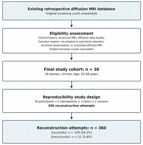
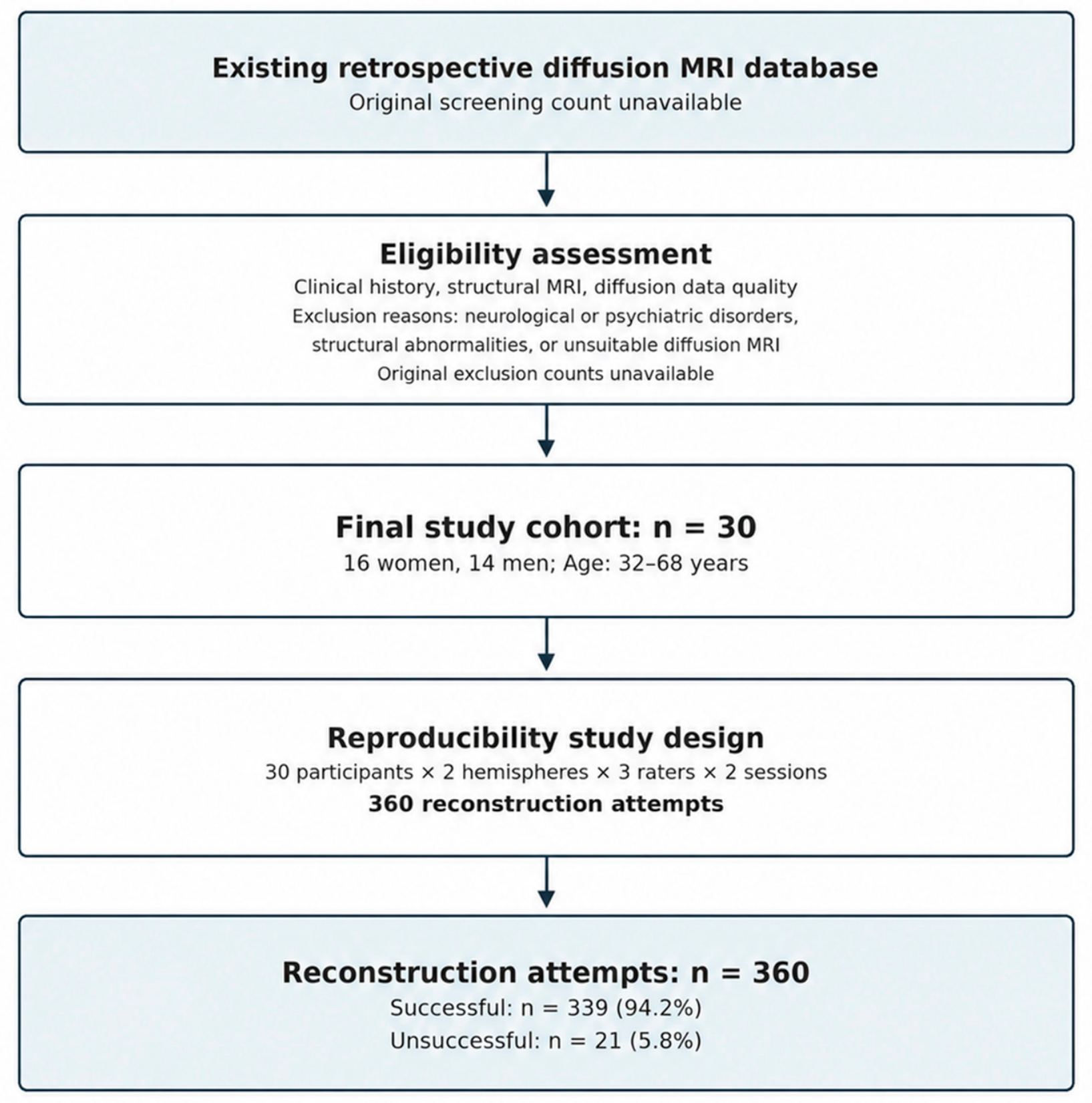
Participants were selected retrospectively from an existing database of diffusion-weighted MRI examinations. Eligibility was based on the absence of known neurological or psychiatric disorders, absence of structural brain abnormalities, and availability of diffusion MRI data suitable for tractography analysis. A flow diagram illustrating the retrospective participant selection process is presented in Figure 1.
Figure 1.
Flow diagram illustrating retrospective participant selection and the reproducibility study design. The final cohort consisted of 30 participants. Three independent raters performed bilateral FAT reconstructions in two sessions, resulting in 360 reconstruction attempts (339 successful and 21 unsuccessful). Original screening and exclusion counts were unavailable.
2.2. Ethics
The original study was approved by the Bioethics Committee of Collegium Medicum, Nicolaus Copernicus University (KB 316/2022; 17 May 2022). All participants provided informed consent prior to imaging.
2.3. MRI Acquisition
All examinations were performed using a 3.0 T Philips Ingenia MRI scanner (Germany) equipped with a 32-channel head coil. Diffusion-weighted data were obtained using a single-shot spin-echo echo-planar imaging sequence with 60 diffusion directions and b-values of 0 and 1000 s/mm2. The isotropic voxel size was 2.5 mm.
2.4. Diffusion Preprocessing
Diffusion MRI datasets were processed using DSI Studio (2026 release, USA). The preprocessing workflow included correction for subject motion and eddy-current-related distortions, followed by diffusion tensor reconstruction. A standardized processing procedure was applied to all 30 datasets to minimize methodological variability.
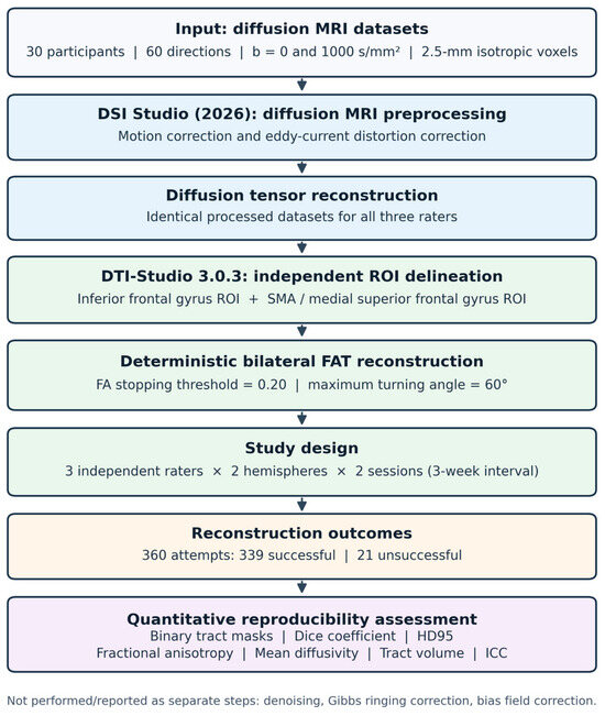
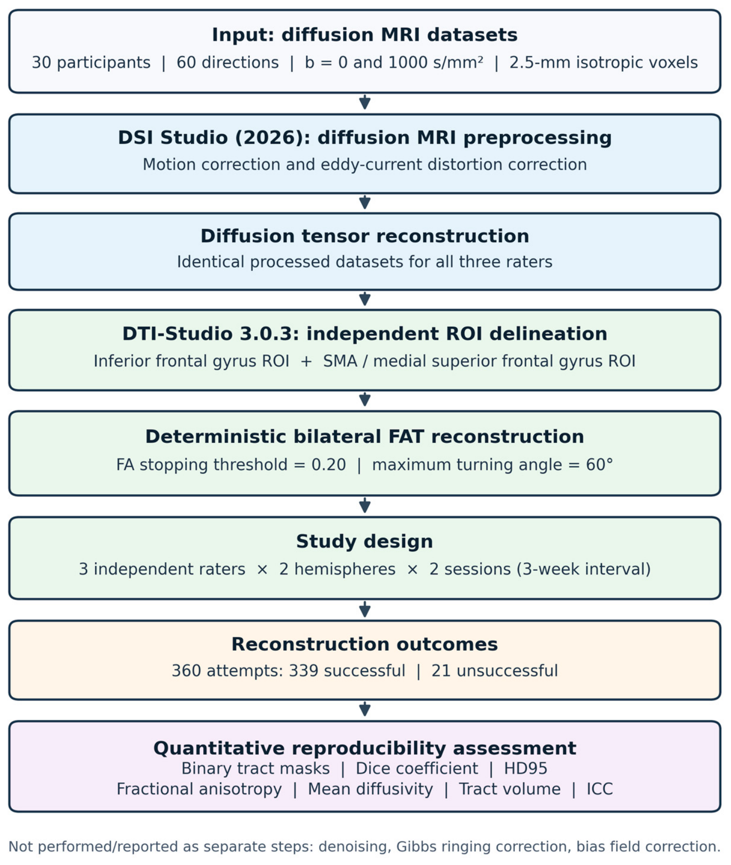
All datasets were processed using the same workflow before operator-dependent tractography analysis. Identically processed diffusion data were subsequently provided to the three independent raters. No additional preprocessing was performed individually by the raters. The processing workflow is illustrated in Figure 2.
Figure 2.
Diffusion MRI preprocessing and frontal aslant tract (FAT) reconstruction workflow. The diagram illustrates the main processing stages, standardized tracking parameters, independent bilateral FAT reconstruction by three raters in two sessions, and subsequent quantitative reproducibility assessment.
Additional denoising, Gibbs ringing correction, and bias field correction were not performed. The same preprocessing workflow was applied to all datasets to maintain consistent methodological conditions. The potential influence of these omitted correction procedures on diffusion parameter estimation and tract reconstruction was not independently evaluated.
2.5. Deterministic FAT Reconstruction
Deterministic diffusion tensor tractography was performed using DTI-Studio 3.0.3. A standardized single-tensor tracking approach was applied to all participants. Fibre tracking was terminated when fractional anisotropy (FA) decreased below 0.20 or when the angular deviation between successive tracking steps exceeded 60°. The same tracking parameters were maintained across all participants, hemispheres, raters, and reconstruction sessions.
For each participant, the left and right frontal aslant tracts were reconstructed independently using two anatomically predefined regions of interest (ROIs). The inferior frontal ROI encompassed the pars opercularis and pars triangularis of the inferior frontal gyrus, whereas the medial frontal ROI included the supplementary motor area and the medial portion of the superior frontal gyrus. Only trajectories passing through both ROIs were retained.
Although anatomical ROI locations were predefined, their boundaries were manually delineated independently by each rater for each participant, hemisphere, and reconstruction session. Previously defined ROI masks were not transferred or reused between raters or sessions. This procedure allowed operator-dependent variability in manual ROI placement to be assessed while maintaining consistent anatomical definitions and tracking parameters.
2.6. Operators
Three operators with different levels of previous experience in diffusion MRI tractography independently performed FAT reconstruction. Before the study, all operators received the same written reconstruction protocol specifying anatomical landmarks, ROI definitions, tracking parameters, and criteria for successful reconstruction. No joint training or calibration session was conducted.
All operators followed identical methodological instructions throughout the study. ROI placement and tract reconstruction were performed independently, without access to the reconstructions or results obtained by the other operators. The study was designed to assess inter-rater and intra-rater reproducibility under standardized methodological conditions rather than to directly evaluate the influence of operator experience on reconstruction performance.
2.7. Reconstruction Sessions
During the first reconstruction session, each operator independently reconstructed the left and right FAT in all 30 participants.
Each operator therefore performed:
30 participants × 2 hemispheres = 60 reconstruction attempts per operator per session; 180 attempts per session; 360 attempts overall.
Following an interval of 3 weeks, all operators repeated the complete reconstruction procedure.
During Session 2, they were blinded to their previous reconstructions and to the results obtained by the other operators.
The three-week interval between reconstruction sessions was selected to reduce the likelihood of operators recalling their previous ROI placements and tract reconstructions. Although this interval was intended to minimize recall bias, the possibility of residual recall effects cannot be completely excluded.
2.8. Definition of Successful Reconstruction
Successful reconstruction was defined as identification of a continuous tract connecting the predefined inferior frontal and medial frontal ROIs while satisfying the fixed tracking criteria. Anatomical plausibility was assessed based on the expected oblique course of the frontal aslant tract between the inferior frontal gyrus and the supplementary motor area/medial superior frontal gyrus.
Reconstructions showing discontinuous trajectories, fibers extending substantially beyond the expected anatomical course, or trajectories inconsistent with the known anatomy of the FAT were considered anatomically implausible. A reconstruction was classified as unsuccessful when no continuous tract fulfilling these criteria could be obtained. Tracking parameters were not modified to achieve reconstruction in unsuccessful cases.
Anatomical plausibility was assessed according to the predefined criteria described above. The assessment included verification of tract continuity, the expected anatomical trajectory, and the absence of aberrant fiber pathways. This qualitative assessment represented an additional potential source of operator-dependent variability.
2.9. Outcome Measures
The primary outcome was inter-rater spatial agreement, quantified using the Dice similarity coefficient:
where A and B represent binary tract masks obtained from two independent raters, |A ∩ B| denotes the number of overlapping voxels, and |A| and |B| represent the total numbers of voxels in each mask.
Pairwise comparisons were performed for:
- Rater 1 versus Rater 2,
- Rater 1 versus Rater 3,
- Rater 2 versus Rater 3.
Analyses were performed separately for the left and right FAT.
The 95th-percentile Hausdorff distance (HD95) was additionally calculated to assess the spatial distance between tract boundaries.
Binary tract masks were represented in a common image space using an identical voxel grid. Voxels belonging to the reconstructed tract were assigned a value of 1, whereas background voxels were assigned a value of 0. Dice similarity coefficients and HD95 were calculated using the corresponding binary masks. Tract volume was expressed in mm3 and calculated as the number of voxels included in the binary tract mask multiplied by the volume of a single voxel. Quantitative outcomes included: fractional anisotropy, mean diffusivity, and tract volume.
Dice similarity coefficients and HD95 values were calculated only for paired reconstructions in which both operators successfully identified the FAT. Reconstruction failure was analyzed as a separate methodological endpoint rather than being assigned a Dice coefficient of zero, because failure to generate a tract and spatial disagreement between two successfully reconstructed tracts represent different aspects of procedural reproducibility. Assigning a Dice value of zero to unsuccessful reconstructions would combine reconstruction feasibility with spatial overlap and could therefore complicate the interpretation of the latter. Consequently, Dice and HD95 values reported in the present study represent spatial agreement conditional on successful paired reconstruction and should not be interpreted as measures of overall protocol performance across all reconstruction attempts.
Intra-rater reproducibility was evaluated by comparing FAT reconstructions obtained by the same operator during Session 1 and Session 2. Spatial agreement was summarized using the available Dice similarity coefficients, while the reliability of quantitative tractography parameters (FA, MD, and tract volume) was assessed using intraclass correlation coefficients. Results were summarized separately by operator and hemisphere where corresponding estimates were available.
2.10. Statistical Analysis
Continuous variables were assessed for normality and summarized as mean ± standard deviation when appropriate. Reconstruction success was expressed as the number and percentage of successful attempts. Dice similarity coefficients and HD95 values were summarized as mean ± standard deviation. Inter-rater analyses were based on reconstructions obtained during Session 1, whereas Sessions 1 and 2 were used to assess intra-rater reproducibility. Inter-rater reliability of FA, MD, and tract volume was evaluated using a two-way random-effects intraclass correlation coefficient (ICC) for absolute agreement of single measurements [ICC(2,1)], with 95% confidence intervals. ICC values <0.50 were considered poor, 0.50–0.75 moderate, 0.75–0.90 good, and >0.90 excellent. Intra-rater reproducibility was assessed separately for each operator by comparing reconstructions obtained during Session 1 and Session 2. Intra-rater reliability was assessed separately for each operator using a two-way mixed-effects model for absolute agreement of single measurements. Differences in quantitative tractography parameters were assessed using linear mixed-effects models to account for the repeated-measures structure of the data. Each participant contributed measurements from two hemispheres, three raters, and two reconstruction sessions. Rater, reconstruction session, and hemisphere were included as categorical fixed effects, while participant identity was included as a random intercept to account for correlations among repeated observations obtained from the same individual. Separate models were fitted for FA, MD, and tract volume.
Spatial agreement was additionally evaluated using pairwise Dice similarity coefficients and HD95 for all three possible rater combinations. The participant, rather than the individual reconstruction attempt, was considered the independent sampling unit. Accordingly, the repeated measurements obtained within each participant were not treated as statistically independent observations. A two-sided p-value < 0.05 was considered statistically significant.
3. Results
FAT reconstruction was successful in 339 of 360 attempts (94.2%). The success rate was higher for the left FAT than for the right FAT (174/180, 96.7% vs. 165/180, 91.7%). Unsuccessful reconstructions occurred across all three raters and in both reconstruction sessions (Table 1). Inter-rater spatial agreement was moderate-to-good. The mean pairwise Dice similarity coefficient was 0.74 ± 0.09 for the left FAT and 0.69 ± 0.10 for the right FAT. The corresponding HD95 values were 4.5 ± 1.5 mm and 5.2 ± 1.8 mm, respectively. Pairwise Dice coefficients for the left FAT were 0.76 ± 0.08 for Rater 1 versus Rater 2, 0.73 ± 0.10 for Rater 1 versus Rater 3, and 0.74 ± 0.09 for Rater 2 versus Rater 3. For the right FAT, the corresponding values were 0.71 ± 0.09, 0.67 ± 0.11, and 0.69 ± 0.10.
Table 1.
FAT reconstruction success according to rater, session, and hemisphere.
Differences between the left and right FAT in reconstruction success rates and spatial agreement measures were evaluated descriptively. No formal statistical hypothesis testing was performed for these hemispheric comparisons; therefore, the observed differences should not be interpreted as statistically significant.
Intra-rater reliability of quantitative tractography parameters was generally high. ICC values ranged from 0.88 to 0.94 for FA and from 0.85 to 0.92 for MD. Tract volume demonstrated comparatively lower reproducibility, with ICC values ranging from 0.78 to 0.88. These findings indicate that FA and MD were more reproducible than tract volume within the evaluated reconstruction protocol. However, the magnitude of intra-rater agreement varied across operators and hemispheres. Tract volume demonstrated lower inter-rater reliability, with ICC values of 0.84 (95% CI 0.70–0.91) for the left FAT and 0.79 (95% CI 0.61–0.89) for the right FAT (Table 2).
Table 2.
Inter-rater and intra-rater reproducibility of FAT reconstruction.
Linear mixed-effects analysis showed no significant systematic effect of rater on FA (p = 0.38) or MD (p = 0.31). A greater between-rater effect was observed for tract volume, although it did not reach statistical significance (p = 0.09). No significant effect of reconstruction session was identified for FA (p = 0.62), MD (p = 0.67), or tract volume (p = 0.41). Hemisphere was not significantly associated with FA (p = 0.08), MD (p = 0.11), or tract volume (p = 0.14).
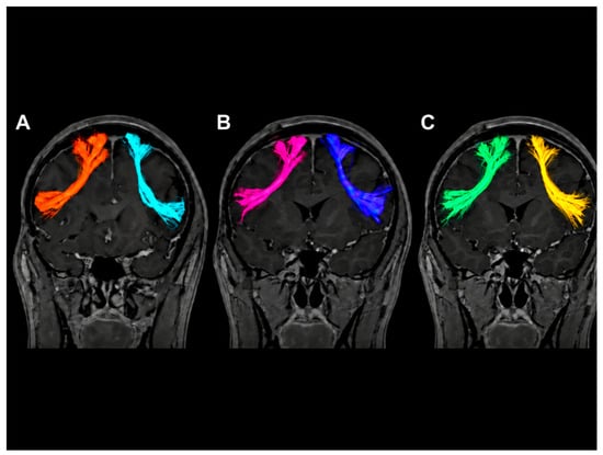
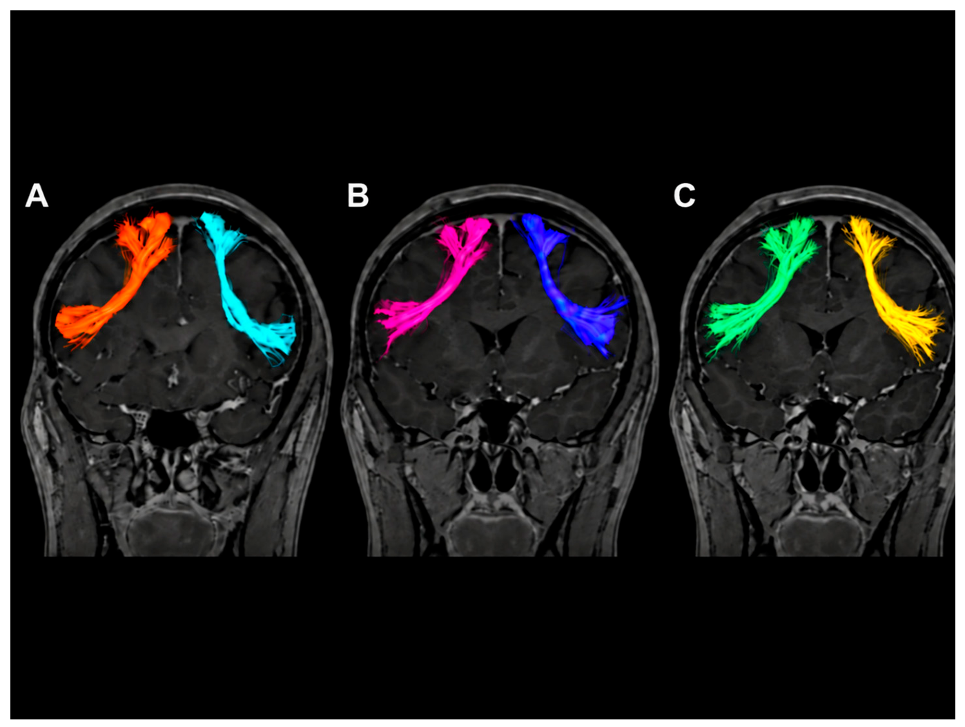
Representative bilateral frontal aslant tract reconstructions obtained by three independent raters in the same participant are presented in Figure 3.
Figure 3.
Representative bilateral frontal aslant tract (FAT) reconstructions obtained independently by three raters in the same participant. Panels (A–C) illustrate reconstructions generated by Raters 1–3, respectively, using identical tracking parameters and predefined anatomical ROI criteria. The panels allow visual comparison of the general tract trajectory and differences in the spatial extent of reconstructed bundles. Quantitative between-rater spatial agreement was assessed separately using Dice similarity coefficients and HD95. The colors are used solely for visual differentiation of the left and right FAT reconstructions across the three raters and do not represent different anatomical structures or quantitative parameters.
4. Discussion
The present study showed that standardized ROI-based deterministic tractography provides moderate-to-good reproducibility of frontal aslant tract reconstruction, although measurable operator-dependent variability remains. FAT reconstruction was successful in most attempts, while a small proportion of reconstructions remained unsuccessful despite preserved brain anatomy and identical tracking parameters. Under a standardized protocol, such failures should be considered an outcome of the reconstruction procedure rather than corrected by modifying FA or angular thresholds, as this would reduce comparability between operators and datasets.
Spatial agreement was numerically higher for the left than for the right FAT, and intra-rater reproducibility consistently exceeded inter-rater agreement. These findings indicate that individual operators reproduce their own ROI placement more consistently than different operators reproduce one another [12,18]. The principal spatial course of the FAT was generally preserved, whereas the peripheral extent of reconstructed bundles showed greater variability. This is relevant to neurosurgical imaging, where tractography may assist in estimating the course of a white matter pathway, but the borders of the reconstructed bundle should not be interpreted as precise anatomical boundaries [10,16,19,20].
The clinical implications of the observed reproducibility require careful consideration. Although the mean inter-rater Dice coefficients indicated moderate-to-good spatial agreement, no universally accepted Dice, HD95, or ICC thresholds establish the suitability of FAT tractography for individual neurosurgical decision-making. The observed HD95 values of 4.5–5.2 mm indicate measurable spatial differences between independently reconstructed tracts. These values should not be interpreted as direct anatomical localization errors or as acceptable surgical safety margins.
Previous studies have demonstrated that tractography reproducibility depends on the anatomical pathway, reconstruction methodology, and degree of protocol standardization. Wakana et al. [8] emphasized the importance of standardized anatomical ROI placement for improving inter- and intra-rater reproducibility. Besseling et al. reported Dice coefficients of 0.70–0.92 for proximal white matter tract segmentations and demonstrated that diffusion metrics generally exhibited higher reproducibility than tract volume. These observations are broadly consistent with our findings, in which FA and MD showed higher reliability than tract volume [21].
Furthermore, Rheault et al. highlighted the influence of operator-dependent decisions on tract dissection reproducibility and the importance of standardized reconstruction protocols. Nevertheless, direct numerical comparisons should be interpreted cautiously because previous investigations differed in the anatomical pathways examined, reconstruction algorithms, acquisition parameters, and reproducibility measures. The present study specifically extends this methodological perspective to standardized deterministic FAT reconstruction performed independently by three raters in two sessions [18].
In the context of preoperative planning and intraoperative neuronavigation, such variability may be relevant when lesions are located close to functionally important white matter pathways. Furthermore, reconstruction failure, particularly for the right FAT (8.3% of attempts), highlights the need for cautious interpretation. Failure to reconstruct the FAT should not be considered evidence of anatomical absence, and alternative imaging approaches or functional assessment may be necessary [12].
Operator-dependent variability may arise at several stages of manual FAT reconstruction, particularly during ROI delineation within the inferior frontal gyrus and supplementary motor area/medial superior frontal gyrus. Minor differences in the interpretation of anatomical landmarks and ROI boundaries may influence streamline selection and the spatial extent of reconstructed bundles. In addition, the qualitative assessment of anatomical plausibility may introduce further variability between operators.
Although the use of a standardized written protocol may reduce methodological inconsistencies, it cannot completely eliminate differences associated with manual ROI placement. Future improvements may include joint operator calibration, semi-automated ROI delineation, and atlas-based approaches to increase procedural consistency. However, these strategies were not evaluated in the present study.
Although FA and MD demonstrated higher reproducibility than tract volume, these findings alone do not establish their validity as clinical or longitudinal biomarkers. Additional studies involving neurosurgical patients and independent functional or anatomical validation are required before specific clinical recommendations can be formulated.
The numerically lower reproducibility observed for the right FAT may reflect several anatomical and methodological factors. Differences in local fiber architecture, tract geometry, and the spatial relationship between the predefined ROIs may influence deterministic fiber tracking. In particular, crossing fiber configurations and small differences in ROI delineation may affect the continuity and spatial extent of reconstructed trajectories. Operator-dependent differences in the interpretation of anatomical landmarks may also contribute to the observed asymmetry. However, these explanations remain hypothetical, as the present study was not specifically designed to investigate the anatomical or technical determinants of hemispheric differences. Therefore, the observed asymmetry should be interpreted cautiously.
Quantitative tractography measures also differed in reproducibility. FA and MD demonstrated relatively high inter-rater reliability, whereas tract volume was more sensitive to operator-dependent variability. This difference may be related to the way these parameters are derived. Mean FA and MD values represent diffusion characteristics averaged across the reconstructed tract and may therefore remain relatively stable despite minor differences in ROI placement or streamline selection. In contrast, tract volume depends more directly on the spatial extent of the reconstructed bundle and the distribution of retained trajectories. Even small differences in ROI boundaries may result in the inclusion or exclusion of peripheral fibers, leading to measurable changes in tract volume without substantially altering mean diffusion metrics. These findings suggest that FA and MD may be less sensitive to minor variations in manual ROI delineation than tract volume within the standardized protocol evaluated in the present study. However, their higher reproducibility does not necessarily indicate greater anatomical accuracy or clinical validity. An important methodological consideration is that spatial agreement was evaluated only when both raters successfully reconstructed the FAT. Consequently, the reported Dice and HD95 values may overestimate overall protocol reproducibility by excluding technically challenging cases. Nevertheless, reconstruction success was treated as a separate outcome because failure to reconstruct a tract and disagreement between successfully reconstructed tracts represent distinct aspects of procedural performance. The reported spatial agreement measures should therefore be interpreted together with reconstruction success rates rather than as independent indicators of overall protocol reliability [21,22,23].
According to established ICC interpretation criteria, the observed inter-rater reliability ranged from moderate to excellent, depending on the parameter and hemisphere. In particular, the ICC for right FAT volume (0.79, 95% CI: 0.61–0.89) indicates good reliability based on the point estimate, although its confidence interval extends into the moderate range. Dice coefficients should be interpreted separately as measures of spatial overlap rather than direct indicators of anatomical accuracy [24].
Reproducibility should not, however, be equated with anatomical accuracy. Agreement between operators does not demonstrate that all true fibres are identified or that false-positive trajectories are absent. Tractography remains an indirect representation of white matter architecture and should be interpreted together with structural imaging, functional information, and, when appropriate, intraoperative mapping [9,10].
The inclusion of neurologically healthy participants allowed operator-dependent variability to be evaluated under relatively homogeneous anatomical conditions, without the confounding effects of intracranial lesions. However, the reproducibility estimates obtained in this population cannot be directly extrapolated to neurosurgical patients. In the presence of brain tumors or other space-occupying lesions, peritumoral edema, mass effect, tumor infiltration, and displacement or distortion of white matter pathways may complicate anatomical landmark identification and ROI delineation. These factors may increase operator-dependent variability and reduce reconstruction success compared with the results observed in the present study. Therefore, the current findings should be considered a methodological baseline, and further validation in clinical populations is necessary before their potential application to individual neurosurgical decision-making [14,17,20].
An important methodological limitation of the present study is the use of deterministic single-tensor tractography. This approach cannot adequately resolve complex fiber configurations, particularly crossing, kissing, or branching fibers, which may influence FAT reconstruction in frontal white matter regions. More advanced reconstruction methods, including generalized q-sampling imaging and constrained spherical deconvolution, may provide improved characterization of complex fiber architecture. However, the present study was designed to evaluate the reproducibility of a specific standardized deterministic protocol rather than to compare different tractography algorithms. Consequently, the reported reproducibility estimates should be considered specific to the evaluated reconstruction approach and cannot be directly generalized to GQI- or CSD-based tractography. Future comparative studies are warranted to determine whether more advanced reconstruction techniques improve inter-rater and intra-rater reproducibility of FAT delineation [25,26,27].
The operators differed in their previous tractography experience, and no joint calibration session was conducted. The retrospective nature of the study and limited availability of the complete reconstruction-level dataset restricted the possibility of conducting additional exploratory analyses and independently reassessing selected statistical estimates. These limitations should be considered when interpreting the results and their potential clinical applicability.
Rater 3 demonstrated numerically lower reconstruction success rates and spatial agreement than the other operators. These differences may reflect individual variation in manual ROI delineation, anatomical landmark interpretation, or previous tractography experience. However, because the study was not designed to examine the relationship between operator experience and reconstruction performance, no causal explanation can be established.
Several limitations should be acknowledged. The study included a relatively small sample of 30 neurologically healthy participants, and no formal a priori sample size calculation was performed. All diffusion MRI data were acquired using a single scanner and acquisition protocol, and only one deterministic single-tensor tractography approach was evaluated. The absence of an independent anatomical reference standard prevented assessment of anatomical accuracy. Furthermore, spatial agreement was evaluated only for successfully reconstructed tract pairs, potentially leading to overestimation of overall protocol reproducibility.
Overall, the study provides a baseline estimate of operator-dependent variability in standardized FAT reconstruction and supports further evaluation of the protocol in neurosurgical patients and comparison with more automated reconstruction approaches.
5. Conclusions
Standardized ROI-based deterministic tractography enabled successful FAT reconstruction in most cases and demonstrated moderate-to-good inter-rater reproducibility, with higher intra-rater agreement. FA and MD showed relatively high reliability, whereas tract volume was more sensitive to operator-dependent variability.
Although standardization of ROI placement and tracking parameters may improve procedural consistency, it does not eliminate operator-related differences. Importantly, the observed reproducibility should not be interpreted as evidence of anatomical accuracy or sufficient validation for independent clinical decision-making.
The present findings provide a methodological baseline for standardized FAT reconstruction in healthy individuals and support further investigations in neurosurgical populations, including comparisons with more automated and advanced tractography approaches.
Author Contributions
Conceptualization, S.K.-S. and P.F.; methodology, S.K.-S.; software, S.K.-S.; validation, P.F., A.Y. and D.G.; formal analysis, S.K.-S.; investigation, S.K.-S.; resources, P.F.; data curation, D.G.; writing—original draft preparation, S.K.-S.; writing—review and editing, P.F.; visualization, A.Y.; supervision, D.G.; project administration, S.K.-S.; funding acquisition, S.K.-S. All authors have read and agreed to the published version of the manuscript.
Funding
This research received no external funding.
Institutional Review Board Statement
The original study was approved by the Bioethics Committee of Collegium Medicum, Nicolaus Copernicus University (KB 316/2022; 17 May 2022). All participants provided informed consent prior to imaging.
Informed Consent Statement
Informed consent was obtained from all subjects involved in the study.
Data Availability Statement
The data presented in this study are available on reasonable request from the corresponding author. The data are not publicly available due to ethical restrictions and the need to protect patient confidentiality and privacy.
Acknowledgments
During the preparation of this manuscript, the authors used OpenAI ChatGPT (GPT-5.6) for language editing, refinement of manuscript structure, and assistance in drafting selected sections and preparing illustrative statistical and graphical templates. All scientific content, methodological decisions, statistical analyses, and interpretation of the study data were independently reviewed and verified by the authors. The authors take full responsibility for the content of the publication.
Conflicts of Interest
The authors declare no conflicts of interest.
References
- Catani, M.; Howard, R.J.; Pajevic, S.; Jones, D.K. Virtual in Vivo Interactive Dissection of White Matter Fasciculi in the Human Brain. Neuroimage 2002, 17, 77–94. [Google Scholar] [CrossRef] [Scilit] [PubMed]
- Abdullah, K.G.; Lubelski, D.; Nucifora, P.G.P.; Brem, S. Use of Diffusion Tensor Imaging in Glioma Resection. Neurosurg. Focus 2013, 34, E1. [Google Scholar] [CrossRef] [Scilit] [PubMed]
- Henderson, F.; Abdullah, K.G.; Verma, R.; Brem, S. Tractography and the Connectome in Neurosurgical Treatment of Gliomas: The Premise, the Progress, and the Potential. Neurosurg. Focus 2020, 48, E6. [Google Scholar] [CrossRef] [Scilit] [PubMed]
- Szmuda, T.; Kierońska, S.; Ali, S.; Słoniewski, P.; Pacholski, M.; Dzierzanowski, J.; Sabisz, A.; Szurowska, E. Tractography-Guided Surgery of Brain Tumours: What Is the Best Method to Outline the Corticospinal Tract? Folia Morphol. 2021, 80, 40–46. [Google Scholar] [CrossRef] [Scilit] [PubMed]
- Reisch, K.; Böttcher, F.; Tuncer, M.S.; Schneider, H.; Vajkoczy, P.; Picht, T.; Fekonja, L.S. Tractography-Based Navigated TMS Language Mapping Protocol. Front. Oncol. 2022, 12, 1008442. [Google Scholar] [CrossRef] [Scilit] [PubMed]
- Sammartino, F.; Krishna, V.; King, N.K.K.; Lozano, A.M.; Schwartz, M.L.; Huang, Y.; Hodaie, M. Tractography-Based Ventral Intermediate Nucleus Targeting: Novel Methodology and Intraoperative Validation. Mov. Disord. 2016, 31, 1217–1225. [Google Scholar] [CrossRef] [Scilit] [PubMed]
- Kierońska, S.; Sokal, P.; Dura, M.; Jabłońska, M.; Rudaś, M.; Jabłońska, R. Tractography-Based Analysis of Morphological and Anatomical Characteristics of the Uncinate Fasciculus in Human Brains. Brain Sci. 2020, 10, 709. [Google Scholar] [CrossRef] [Scilit] [PubMed]
- Jones, D.K.; Knösche, T.R.; Turner, R. White Matter Integrity, Fiber Count, and Other Fallacies: The Do’s and Don’ts of Diffusion MRI. Neuroimage 2013, 73, 239–254. [Google Scholar] [CrossRef] [Scilit] [PubMed]
- Maier-Hein, K.H.; Neher, P.F.; Houde, J.C.; Côté, M.A.; Garyfallidis, E.; Zhong, J.; Chamberland, M.; Yeh, F.C.; Lin, Y.C.; Ji, Q.; et al. The Challenge of Mapping the Human Connectome Based on Diffusion Tractography. Nat. Commun. 2017, 8, 1349. [Google Scholar] [CrossRef] [Scilit] [PubMed]
- Schilling, K.G.; Nath, V.; Hansen, C.; Parvathaneni, P.; Blaber, J.; Gao, Y.; Neher, P.; Aydogan, D.B.; Shi, Y.; Ocampo-Pineda, M.; et al. Limits to Anatomical Accuracy of Diffusion Tractography Using Modern Approaches. Neuroimage 2019, 185, 1–11. [Google Scholar] [CrossRef] [Scilit] [PubMed]
- Wasserthal, J.; Neher, P.; Maier-Hein, K.H. TractSeg—Fast and Accurate White Matter Tract Segmentation. Neuroimage 2018, 183, 239–253. [Google Scholar] [CrossRef] [Scilit] [PubMed]
- Wakana, S.; Caprihan, A.; Panzenboeck, M.M.; Fallon, J.H.; Perry, M.; Gollub, R.L.; Hua, K.; Zhang, J.; Jiang, H.; Dubey, P.; et al. Reproducibility of Quantitative Tractography Methods Applied to Cerebral White Matter. Neuroimage 2007, 36, 630–644. [Google Scholar] [CrossRef] [Scilit] [PubMed]
- Catani, M.; Dell’Acqua, F.; Vergani, F.; Malik, F.; Hodge, H.; Roy, P.; Valabregue, R.; Thiebaut de Schotten, M. Short Frontal Lobe Connections of the Human Brain. Cortex 2012, 48, 273–291. [Google Scholar] [CrossRef] [Scilit] [PubMed]
- Tagliaferri, M.; Amorosino, G.; Voltolini, L.; Giampiccolo, D.; Avesani, P.; Cattaneo, L. A Revision of the Dorsal Origin of the Frontal Aslant Tract (FAT) in the Superior Frontal Gyrus: A DWI-Tractographic Study. Brain Struct. Funct. 2024, 229, 987–999. [Google Scholar] [CrossRef] [Scilit] [PubMed]
- Burkhardt, E.; Kinoshita, M.; Herbet, G. Functional Anatomy of the Frontal Aslant Tract and Surgical Perspectives. J. Neurosurg. Sci. 2021, 65, 566–580. [Google Scholar] [CrossRef] [Scilit] [PubMed]
- La Corte, E.; Eldahaby, D.; Greco, E.; Aquino, D.; Bertolini, G.; Levi, V.; Ottenhausen, M.; Demichelis, G.; Romito, L.M.; Acerbi, F.; et al. The Frontal Aslant Tract: A Systematic Review for Neurosurgical Applications. Front. Neurol. 2021, 12, 641586. [Google Scholar] [CrossRef] [Scilit] [PubMed]
- Kierońska-Siwak, S.; Mackiewicz-Nartowicz, H.; Sinkiewicz, A.; Kozakiewicz-Rutkowska, A.; Jaroch, A.; Bracha, M.; Zwierko, B.; Grzanka, D. Three-Dimensional Identification of the Frontal Aslant Tract in the Human Brain: Tractography Study. Brain Sci. 2026, 16, 885. [Google Scholar] [CrossRef] [Scilit] [PubMed]
- Rheault, F.; De Benedictis, A.; Daducci, A.; Maffei, C.; Tax, C.M.W.; Romascano, D.; Caverzasi, E.; Morency, F.C.; Corrivetti, F.; Pestilli, F.; et al. Tractostorm: The What, Why, and How of Tractography Dissection Reproducibility. Hum. Brain Mapp. 2020, 41, 1859–1874. [Google Scholar] [CrossRef] [Scilit] [PubMed]
- Yang, J.Y.M.; Yeh, C.H.; Poupon, C.; Calamante, F. Diffusion MRI Tractography for Neurosurgery: The Basics, Current State, Technical Reliability and Challenges. Phys. Med. Biol. 2021, 66, 15TR01. [Google Scholar] [CrossRef] [Scilit] [PubMed]
- Essayed, W.I.; Zhang, F.; Unadkat, P.; Cosgrove, G.R.; Golby, A.J.; O’Donnell, L.J. White Matter Tractography for Neurosurgical Planning: A Topography-Based Review of the Current State of the Art. Neuroimage Clin. 2017, 15, 659–672. [Google Scholar] [CrossRef] [Scilit] [PubMed]
- Besseling, R.M.H.; Jansen, J.F.A.; Overvliet, G.M.; Vaessen, M.J.; Braakman, H.M.H.; Hofman, P.A.M.; Aldenkamp, A.P.; Backes, W.H. Tract Specific Reproducibility of Tractography Based Morphology and Diffusion Metrics. PLoS ONE 2012, 7, e34125. [Google Scholar] [CrossRef] [Scilit] [PubMed][Green Version]
- Legarreta, J.H.; Schiavi, S.; Tang, W.; Banks, G.; Cieslak, M.; Schilling, K.; De Luca, A.; Tournier, J.D.; Kruper, J.; Rheault, F.; et al. What Needs to Be Standardized for Reliable, Reproducible, and Robust Tractography? Gigascience 2026, 15, giag034. [Google Scholar] [CrossRef] [Scilit] [PubMed]
- Heiervang, E.; Behrens, T.E.J.; Mackay, C.E.; Robson, M.D.; Johansen-Berg, H. Between Session Reproducibility and between Subject Variability of Diffusion MR and Tractography Measures. Neuroimage 2006, 33, 867–877. [Google Scholar] [CrossRef] [Scilit] [PubMed]
- Koo, T.K.; Li, M.Y. A Guideline of Selecting and Reporting Intraclass Correlation Coefficients for Reliability Research. J. Chiropr. Med. 2016, 15, 155–163. [Google Scholar] [CrossRef] [Scilit] [PubMed]
- Schlaier, J.R.; Beer, A.L.; Faltermeier, R.; Fellner, C.; Steib, K.; Lange, M.; Greenlee, M.W.; Brawanski, A.T.; Anthofer, J.M. Probabilistic vs. Deterministic Fiber Tracking and the Influence of Different Seed Regions to Delineate Cerebellar-Thalamic Fibers in Deep Brain Stimulation. Eur. J. Neurosci. 2017, 45, 1623–1633. [Google Scholar] [CrossRef] [Scilit] [PubMed]
- Descoteaux, M.; Deriche, R.; Knösche, T.R.; Anwander, A. Deterministic and Probabilistic Tractography Based on Complex Fibre Orientation Distributions. IEEE Trans. Med. Imaging 2009, 28, 269–286. [Google Scholar] [CrossRef] [Scilit] [PubMed]
- Behrens, T.E.J.; Berg, H.J.; Jbabdi, S.; Rushworth, M.F.S.; Woolrich, M.W. Probabilistic Diffusion Tractography with Multiple Fibre Orientations: What Can We Gain? Neuroimage 2007, 34, 144–155. [Google Scholar] [CrossRef] [Scilit] [PubMed]

Disclaimer/Publisher’s Note: The statements, opinions and data contained in all publications are solely those of the individual author(s) and contributor(s) and not of MDPI and/or the editor(s). MDPI and/or the editor(s) disclaim responsibility for any injury to people or property resulting from any ideas, methods, instructions or products referred to in the content. |
© 2026 by the authors. Licensee MDPI, Basel, Switzerland. This article is an open access article distributed under the terms and conditions of the Creative Commons Attribution (CC BY) license.