


Abstract
To address the inherent inefficiencies and high error rates associated with manual biomass estimation in Litopenaeus vannamei aquaculture, this study presents a highly sophisticated automated framework based on an enhanced U-Net framework. We propose the DCRAG-UNetmodel, which integrates Dilated Convolutions to expand the receptive field, and a Residual-hybrid Attention Gate (RAG) to effectively suppress underwater noise while reinforcing edge features. The framework integrates a morphological skeleton extraction algorithm with sub-pixel geometric correction to achieve precise body length measurements, which are subsequently used for weight prediction via a robust regression model. Experimental results demonstrate that DCRAG-UNet achieves a Dice coefficient of 98.55% and an mIoU of 98.72%. Furthermore, the average relative errors for body length and weight estimation were 1.12% and 6.92%, respectively. These findings validate the model’s reliability and effectiveness for non-invasive biomass monitoring in intelligent aquaculture environments.
1. Introduction
Litopenaeus vannamei is an economically vital species that is extensively distributed along the tropical and subtropical Pacific coasts of South America. Characterized by its rapid growth rate, exceptional environmental adaptability, and versatility across diverse aquaculture systems, it has emerged as a cornerstone of global shrimp farming [1,2]. In high-density intensive aquaculture, the continuous monitoring of L. vannamei growth is fundamental to precision feeding, proactive disease prevention, and the determination of optimal harvesting schedules [3,4,5]. Specifically, the high-fidelity and rapid acquisition of individual body length data constitutes the core of efficient growth management [5,6]. However, conventional manual measurement practices currently prevalent in the industry exhibit several critical limitations: the handling process induces significant physiological stress and physical injuries to the shrimp, operational efficiency remains low, and high inter-operator subjectivity leads to substantial measurement disparities. These factors collectively hinder the implementation of large-scale automation and high-frequency monitoring, thereby restricting the advancement of smart fisheries and digital aquaculture management [7].
In recent years, computer vision and deep learning technologies have been increasingly integrated into aquaculture to facilitate non-contact phenotypic measurements [8,9,10,11,12,13].
In the domain of object detection and keypoint identification, Gong [14] utilized Faster R-CNN to detect multiple anatomical regions of shrimp, achieving precise parameter conversion via reference scaling. Qin [15] coupled Mask R-CNN with a Cascade Pyramid Network (CPN), significantly enhancing the precision of keypoint recognition with an average error of less than 3.51% for carapace and abdominal measurements. Similarly, Li et al. [16] employed a High-Resolution Network (HRNet) for keypoint localization in Penaeus monodon, achieving a mean accuracy of 93.79%. Furthermore, Xi et al. [17] investigated automated monitoring by integrating wearable sensors, depth cameras, and machine learning. Regarding image segmentation, Liu et al. [18] applied an improved AlexNet for soft-shell shrimp identification, while Liu et al. [19] developed adaptive multi-scale background modeling for fish segmentation in deep-sea cage environments. Additionally, Liu et al. [20] introduced ShrimpNet, a specialized convolutional neural network based on LeNet-5, for targeted shrimp image segmentation.
Despite these technological strides, existing methodologies encounter significant challenges related to environmental interference and feature degradation in complex underwater settings. Turbid water, non-uniform illumination, and background noise from suspended debris frequently compromise the ability of conventional convolutional neural networks (CNNs) to accurately delineate target boundaries. Moreover, the slender and semi-transparent nature of shrimp appendages often causes fragmented or lost structures when relying on single-scale convolutions or deep semantic features, directly undermining the accuracy of subsequent biophysical measurements.
To address these challenges, this paper proposes an enhanced U-Net architecture termed DCRAG-UNet (U-Net with fused Dilated Convolution and Residual-hybrid Attention Gate). The model expands the effective receptive field to capture multi-scale contextual information through the integration of Dilated Convolutions. Furthermore, it incorporates a Residual-hybrid Attention Gate (RAG) to effectively suppress background noise while reinforcing boundary feature extraction. This approach significantly improves the segmentation precision of L. vannamei within complex underwater backgrounds. Building upon high-quality segmentation masks, a framework for automated body length measurement and a length-weight regression model were developed by integrating a morphological skeleton extraction algorithm with a sub-pixel geometric correction strategy. This study provides a high-precision, automated biomass estimation framework that minimizes manual intervention while enhancing the robustness and efficiency of growth monitoring in intelligent aquaculture scenarios.
2. Materials and Methods
This section provides a comprehensive overview of the experimental methodology, model architecture, and phenotypic estimation techniques developed for L. vannamei. Section 2.1 details the data acquisition process, hardware configuration, and the rigorous pixel-level annotation and augmentation strategies employed to ensure dataset quality. Section 2.2 introduces the proposed DCRAG-UNet architecture, specifically focusing on the integration of multi-scale Dilated Convolutions and the Residual-hybrid Attention Gate (RAG) for robust feature extraction in underwater environments. Section 2.3 describes the morphological parameter estimation workflow, utilizing the Zhang–Suen thinning algorithm and a geometric correction factor to extract precise body length measurements. Section 2.4 establishes the biomass regression model based on allometric growth power laws to derive weight from skeletal length. Finally, Section 2.5 outlines the experimental environment, including the hardware specifications and hyperparameter configurations used for model training.
2.1. Experimental Dataset and Data Preprocessing
2.1.1. Data Sources and Acquisition Context
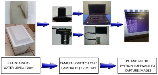
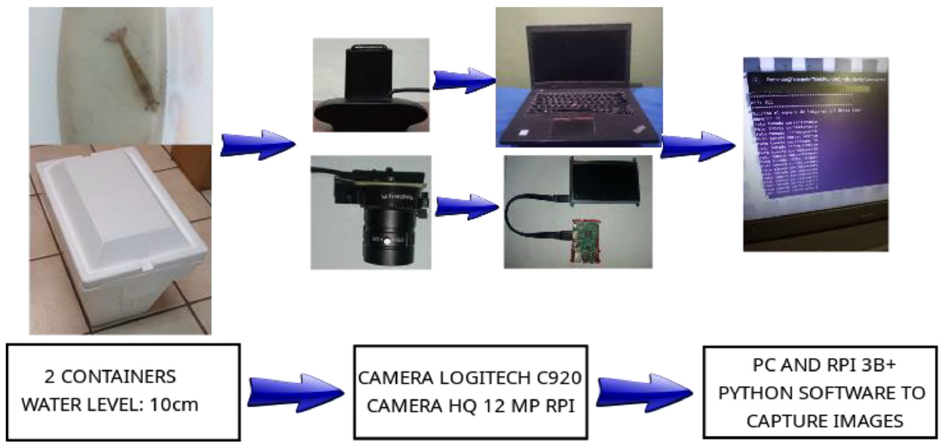
The primary experimental data for this study were sourced from a comprehensive public dataset of Litopenaeus vannamei published by Ramírez-Coronel et al. [21] in the Mendeley Data repository. The imagery was captured at industrialized shrimp farms and academic experimental facilities in Sonora, Mexico, under stable natural illumination. To replicate the natural swimming posture of the specimens, samples were positioned in a controlled observation tank with a water depth of 10 cm. The imaging system utilized a dual-hardware configuration: a Logitech C920 camera and a 12-megapixel high-definition (HD) camera integrated with a Raspberry Pi B3. This setup provided a heterogeneous range of image qualities and optical perspectives, thereby enriching the representativeness of the sample population used for training. The detailed hardware configuration and acquisition process are illustrated in Figure 1.
Figure 1.
Hardware diagram of the image capturing process [21].
2.1.2. Data Selection and Pixel-Level Annotation
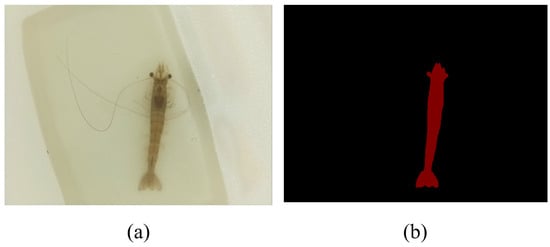
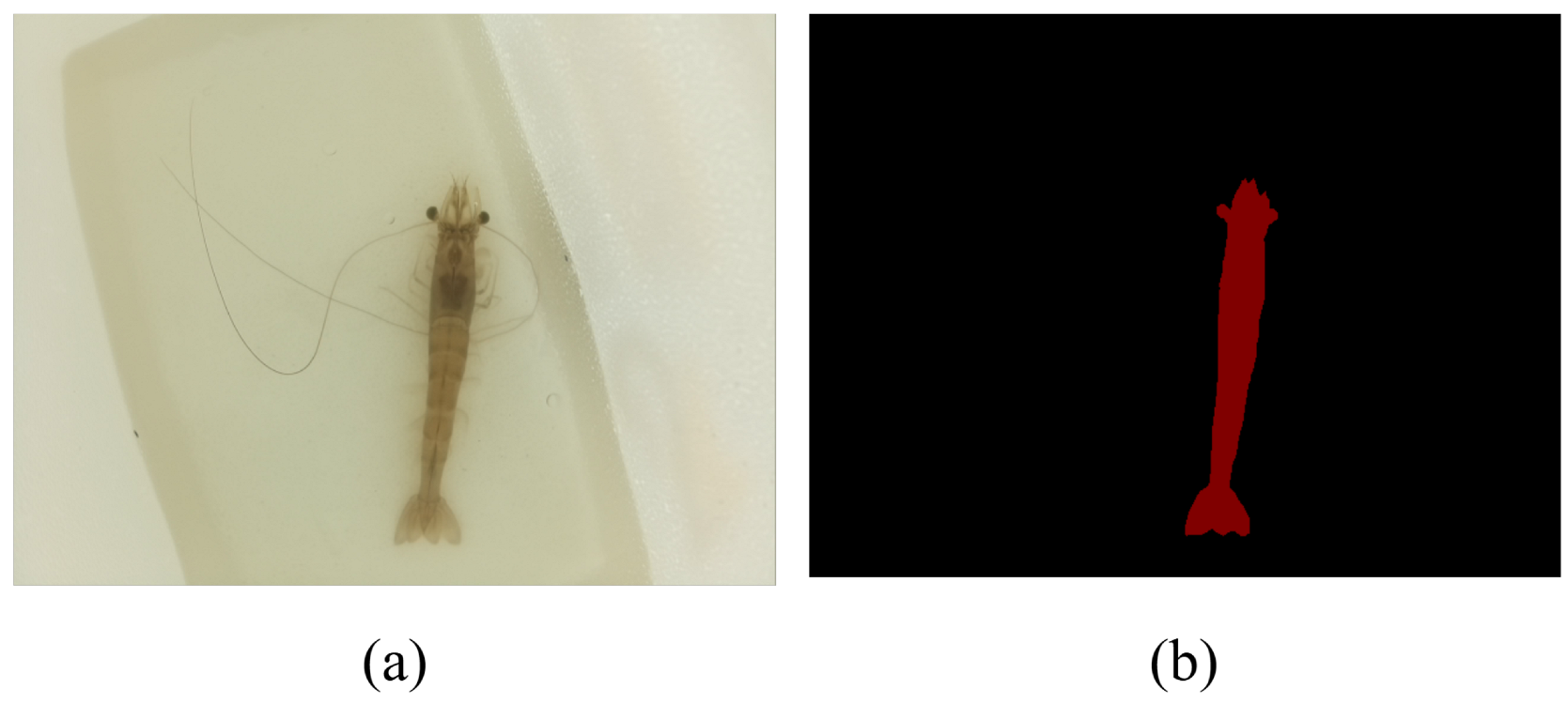
From the initial repository containing 5507 images of 170 individuals, a refined subset consisting of 58 individuals from a single industrial farm was rigorously selected to ensure environmental and phenotypical consistency. To adapt the dataset for high-precision semantic segmentation, secondary processing was performed: only high-clarity images featuring complete, non-occluded single shrimp were retained, resulting in a final dataset of 798 images comprising 50 individual prawns. We subsequently conducted pixel-level semantic annotation using the Labelme tool to generate high-fidelity binary masks, which served as the ground truth (GT) for the DCRAG-UNet model. Unlike the bounding-box or point annotations provided in the original study, this manual re-annotation ensures that the network accurately captures the fine-grained morphological boundaries and slender appendages of the shrimp. A comparison between the raw underwater imagery and the annotated masks is presented in Figure 2.
Figure 2.
Comparison of the original image and the annotated mask: (a) original underwater image from the database; (b) pixel-level ground truth mask generated using Labelme software.
2.1.3. Data Augmentation and Dataset Partitioning
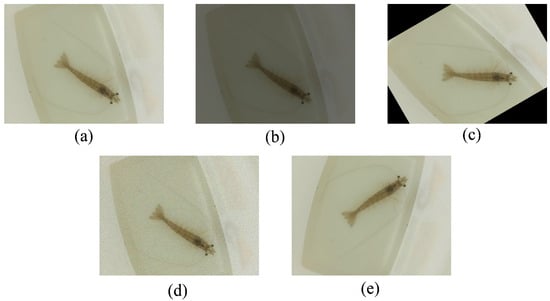
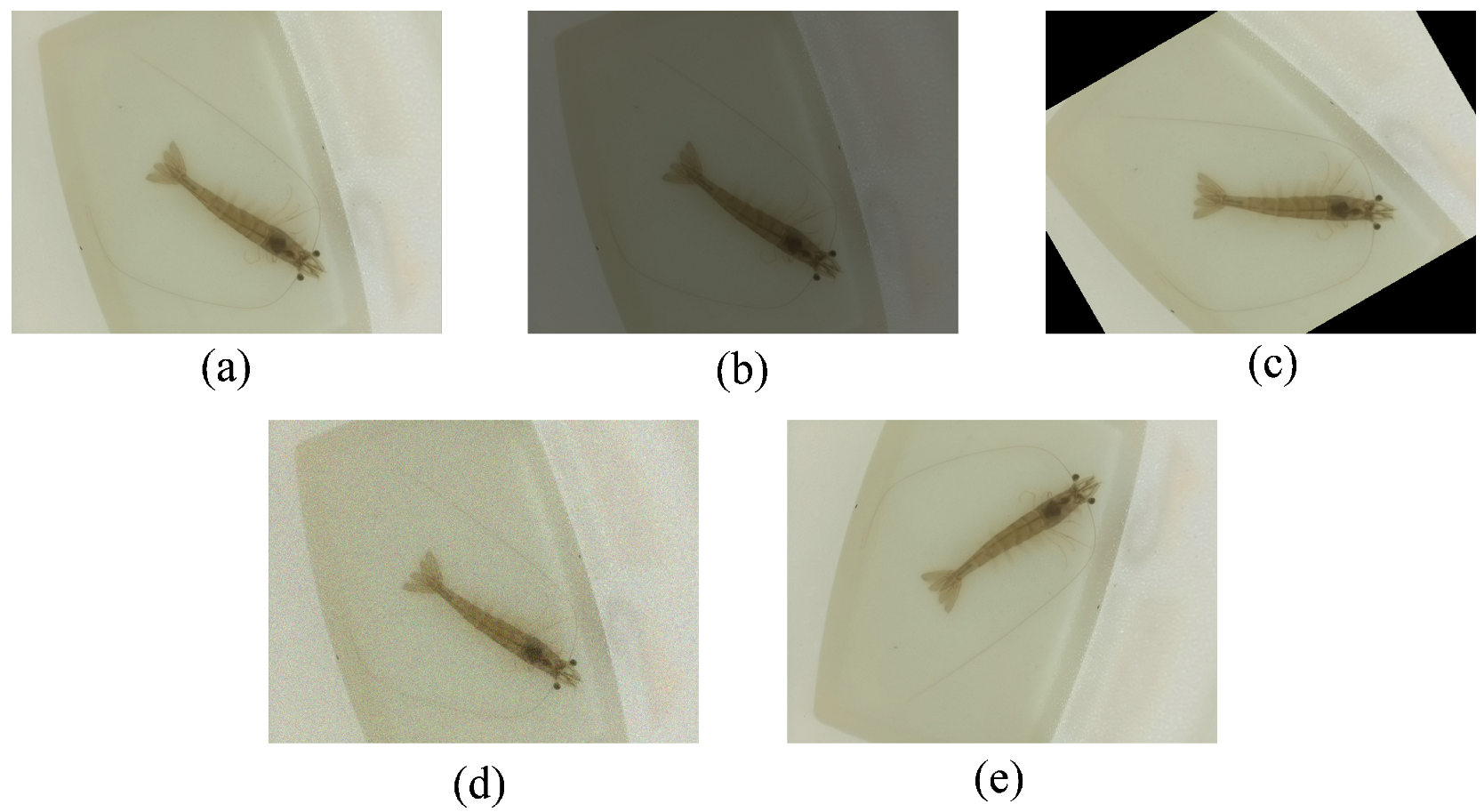
To mitigate the risk of model overfitting and enhance the generalization capacity in complex underwater scenarios, a multi-dimensional data augmentation strategy was implemented. This included geometric transformations (rotation and random flipping), pixel-level intensity adjustments (brightness and contrast variations), and noise injection (Gaussian blurring) to simulate varying water turbidity and lighting conditions typical of intensive aquaculture (Figure 3). These operations expanded the dataset to five times its original volume, resulting in a total of 3990 images. To support a rigorous evaluation framework, the total augmented image library was bifurcated into training and testing subsets: a training set of 3,192 images and a test set of 798 images. This allocation follows a standard 8:2 ratio, providing a sufficiently large sample size for both model optimization and objective verification.
Figure 3.
Visualization of data augmentation strategies: (a) Original image; (b) Brightness adjustment; (c) Random rotation; (d) Gaussian noise injection for turbidity simulation; Brightness adjustment; (e) Horizontal flip.
2.2. DCRAG-UNet Model Architecture
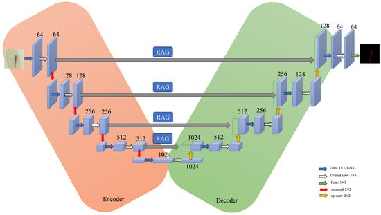
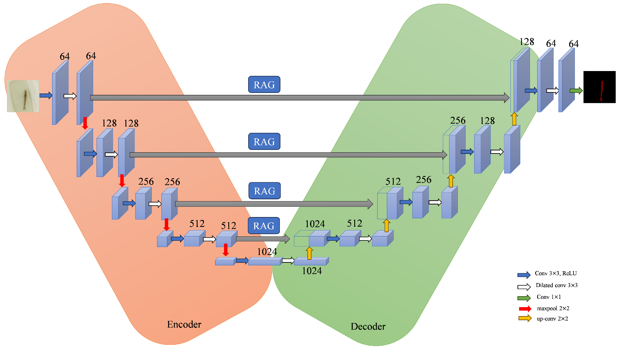
The DCRAG-UNet architecture was specifically engineered to address the inherent challenges of low-contrast underwater imagery and the intricate morphological features of L. vannamei. The complete architecture is depicted in Figure 4.
Figure 4.
Architecture of the proposed DCRAG-UNet model.
2.2.1. Multi-Scale Feature Extraction via Dilated Convolution
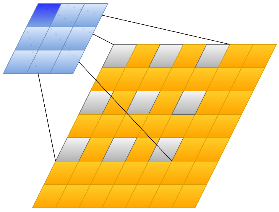
To capture global contextual information while preserving the spatial resolution often lost during standard pooling operations, Dilated Convolutions (also formally known as atrous convolutions, an approach widely popularized by architectures such as DeepLabv3+ [22]) were integrated into the bottleneck layer. For a 2D input signal x, the output of a Dilated Convolution with a filter w and dilation rate r is defined as
This mechanism effectively expands the receptive field to , where k is the kernel size. This enables the network to perceive the entire longitudinal span of the shrimp even within high-resolution feature maps, ensuring structural continuity during segmentation. Architecture of the Dilated Convolution is shown in Figure 5.
Figure 5.
Architecture of the Dilated Convolution.
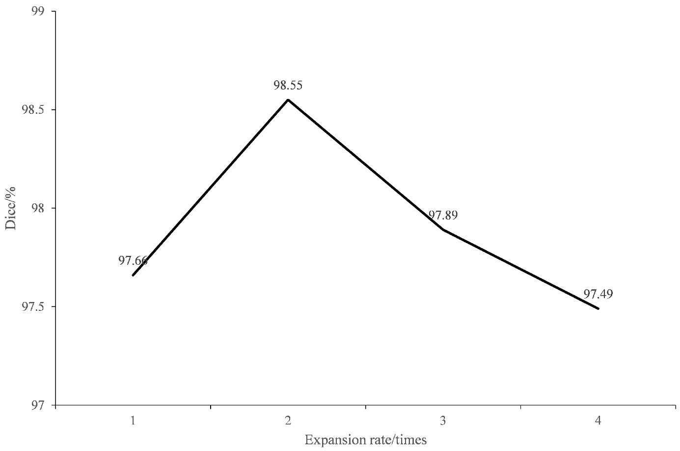
To determine the optimal configuration for the Dilated Convolution layers, comparative experiments were conducted across various dilation rates (r). The empirical results indicate that the segmentation performance reaches its peak at a dilation rate of (Figure 6). This optimality is attributed to the fact that a moderate dilation rate ensures a sufficiently expanded receptive field to capture global contextual information, while simultaneously mitigating the risk of “gridding artifacts” or information loss associated with excessively sparse spatial sampling. Consequently, this configuration maintains a superior balance between multi-scale feature integration and the preservation of local spatial details, which is critical for the accurate delineation of the slender morphological features of L. vannamei.
Figure 6.
Comparison of Different Expansion Rates.
2.2.2. Residual-Hybrid Attention Gate (RAG)
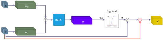
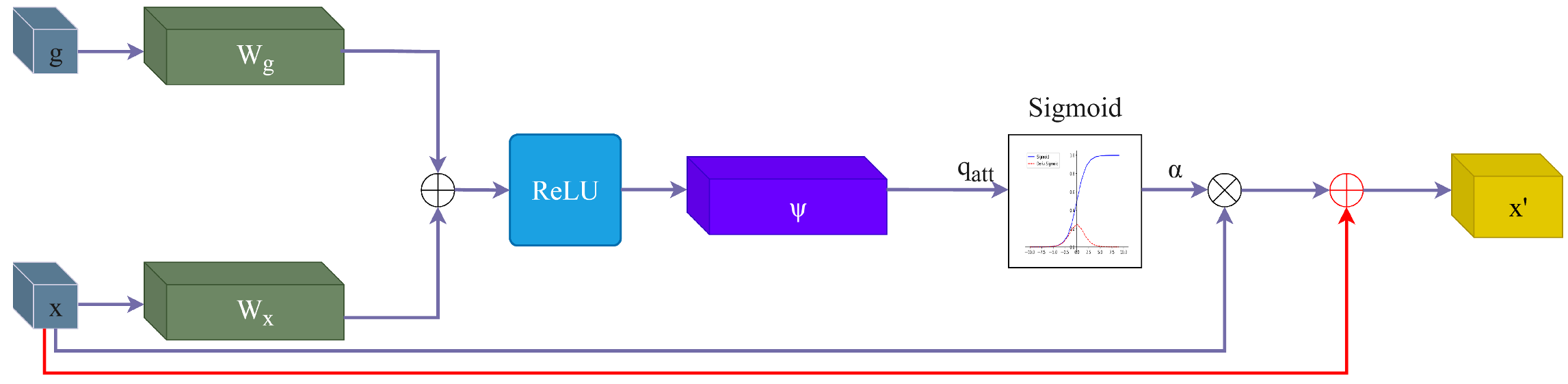
Unlike conventional Attention Gates (such as those in Attention U-Net) that multiply the gating coefficient directly with the input features, leading to the potential degradation of fine-grained boundary gradients in low-contrast environments, the proposed RAG incorporates a skip-residual pathway (). Theoretically, this dual-path approach ensures that the attention mechanism functions exclusively as a background-suppression filter ( for noise) while the identity mapping preserves the high-frequency spatial gradients essential for reconstructing the semi-transparent appendages of L. vannamei. This makes RAG fundamentally more suited for turbid underwater imaging than standard multiplicative attention.
The gating coefficient is computed as
where g is the gating signal from the coarser scale and represents the features from the encoder. By adding a residual path, the final output becomes , ensuring that critical edge features are preserved rather than filtered out during the attention process. The structural logic and operational principle of the proposed Residual-hybrid Attention Gate (RAG) are schematically illustrated in Figure 7.
Figure 7.
Schematic diagram of residual-hybrid attention gate.
2.3. Morphological Parameter Estimation
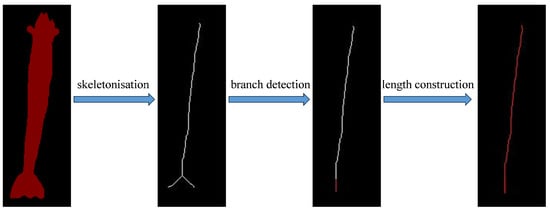
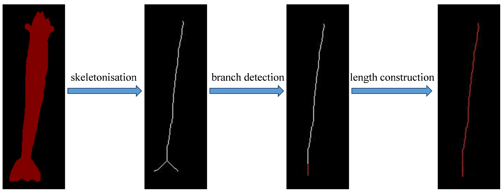
The segmentation mask is processed using the Zhang–Suen thinning algorithm [23] to extract the central skeleton, including skeletonisation, branch detection, length construction, and total length calculation (Figure 8). To account for the natural curvature of the shrimp, a geometric correction factor is applied:
where C is the pixel-to-metric calibration constant.
Figure 8.
The process of segmentation mask.
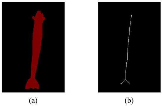
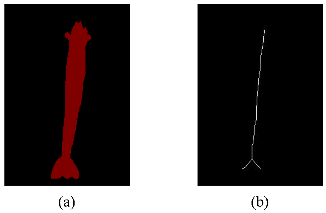
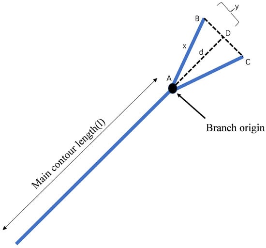
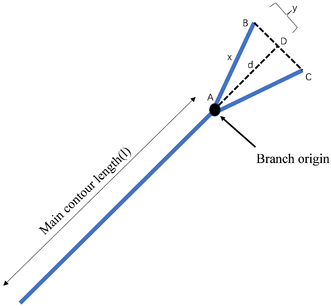
During the skeletonization of the binarized L. vannamei images, redundant branches frequently emerge near the cephalothorax and uropod regions due to the intricate morphological structures of the shrimp (Figure 9). Consequently, implementing a robust post-processing workflow—comprising branch detection, pruning, and total length reconstruction—is essential to ensure measurement accuracy. In this study, the final body length is estimated by integrating the primary skeleton length with a specific branch compensation factor, as illustrated in Figure 10. This additive approach effectively rectifies the underestimation caused by non-linear skeletal segments, thereby enhancing the precision of the automated phenotypic analysis.
Figure 9.
Comparison of shrimp skeletons before and after extraction: (a) before extracting the skeleton; (b) after extracting the skeleton.
Figure 10.
Schematic The process of constructing the total length.
Specifically, the primary contour length of the L. vannamei skeleton is first extracted. To address potential branching at the cephalothorax and uropod extremities, the algorithm detects the triangle formed by the branch origin and the respective endpoints. To more accurately reflect the length loss attributed to the natural curvature and morphological complexity of the shrimp body, the perpendicular distance from the branch origin (A) to the line connecting the branch endpoints (B and C) is calculated. The compensation length is defined as
where represents the length of the non-hypotenuse side of the branch (segment or ), and denotes the distance from the endpoint (B or C) to the foot of the perpendicular (D), as illustrated in Figure 9 (specifically segments or ). Consequently, the total body length is derived using the following integrated formula:
2.4. Biomass Regression
Drawing upon the methodology for the L. vannamei length–weight (L–W) regression model proposed by Yang et al. [24], this study established an allometric growth model based on empirical measurements of individual body length (L) and wet weight (W). The exponential relationship is expressed as
To facilitate parameter estimation and ensure statistical robustness, the power-law model was linearized via logarithmic transformation, yielding
The Ordinary Least Squares (OLS) method was subsequently employed to perform linear regression, from which the estimated values for parameters a and b, along with the coefficient of determination (), were derived. The final regression equation is formulated as
where W denotes the body weight (g) and L represents the body length (cm). A higher value indicates a superior goodness of fit for the regression model within the context of biomass estimation.
2.5. Experimental Environment and Hyperparameters
The computational execution of the proposed architecture was performed via PyTorch 2.5.0, utilizing the hardware acceleration provided by an NVIDIA GeForce RTX 4060 Ti GPU (16 GB VRAM). Over a duration of 200 epochs, the model was optimized using the Adam algorithm with a fixed batch size of 16 and a starting learning rate of . To mitigate the challenges posed by pixel-class imbalances in subaquatic imagery, a composite objective function—integrating Binary Cross-Entropy and Dice Loss—was adopted.
3. Results
3.1. Segmentation Accuracy
To comprehensively evaluate the performance advantages of the proposed DCRAG-UNet model in the image segmentation of L. vannamei, comparative experiments were conducted against several state-of-the-art models, including the original U-Net [25], UNet++ [26], ResUNet [27], TransUNet [28], Swin-UNet [29], VM-UNet [30], DeepLabv3+ [22], LightM-UNet [31] and Attention U-Net [32]. To ensure the integrity of the comparative analysis, all evaluated models were subjected to uniform training and testing datasets, standardized hyperparameter initialization, and a synchronized hardware environment. As detailed in Table 1—where the most favorable outcomes are highlighted in bold—the various network architectures exhibited substantial disparities in efficacy. Notably, the DCRAG-UNet surpassed conventional models across all quantitative indices. Analysis of the performance metrics in Table 1 reveals that the proposed architecture consistently exceeds alternative frameworks in both Dice and mIoU values, demonstrating particular resilience within the challenging subaquatic conditions inherent to L. vannamei farming.
Table 1.
Performance comparison of segmentation models.
To assess the deployment feasibility of DCRAG-UNet in real-time edge computing environments, an empirical evaluation of computational cost was conducted. The model contains 17.44M parameters and requires 163.26 GFLOPs. Peak GPU memory consumption during inference on the NVIDIA RTX 4060 Ti is 1.8 GB. The average inference time per frame is 27.71 ms (approx. 36.07 FPS), demonstrating that the introduction of Dilated Convolutions and the RAG module imposes a negligible computational overhead (a 0.16 ms increase compared to standard U-Net) while satisfying real-time processing requirements for intelligent aquaculture systems.
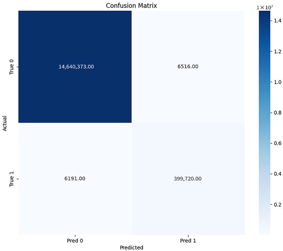
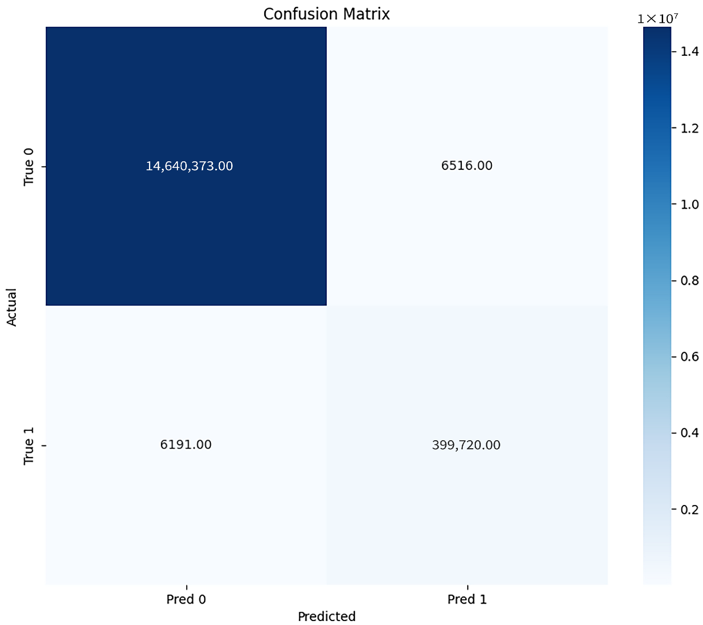
To further evaluate the pixel-level classification accuracy and the model’s ability to distinguish shrimp bodies from complex underwater backgrounds, a confusion matrix was generated (Figure 11). As shown in the matrix, the DCRAG-UNet demonstrated exceptional specificity, with very low misclassification rates between the background and shrimp classes, thereby validating the effectiveness of the RAG module in suppressing turbid background interference.
Figure 11.
Confusion matrix.
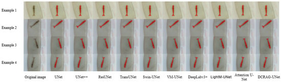
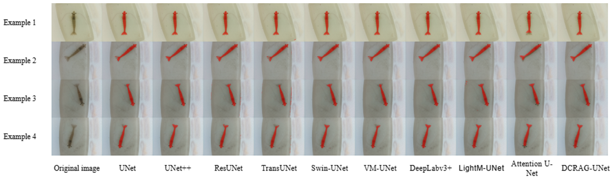
As evidenced by the results in Table 1, the developed DCRAG-UNet model demonstrated high-fidelity results, specifically reaching a mean Intersection over Union (mIoU) of 98.72% and a Dice score of 98.55%. The experimental results indicate that the proposed framework outperforms the state-of-the-art comparative models in both segmentation precision and estimation accuracy. Compared with classical architectures such as ResUNet and DeepLabv3+, our model exhibits consistent improvements across all performance metrics, underscoring its superior capability in capturing morphological details. To further visualize the segmentation performance of the DCRAG-UNet, representative results are presented in Figure 12.
Figure 12.
The segmentation effects of different models.
3.2. Ablation Study
To assess the individual contributions and architectural logic of the model’s components, ablation studies were conducted to isolate the influence of Dilated Convolutions and the Residual-hybrid Attention Gate (RAG) on overall efficacy. This investigation utilized three distinct benchmarks: DC-UNet (relying solely on Dilated Convolutions), RAG-UNet (utilizing only the attention mechanism), and a standard U-Net (functioning as the control baseline).
The quantitative outcomes, detailed in Table 2 and Table 3, indicate that the inclusion of Dilated Convolutions in isolation produced a Dice coefficient of 97.02% and an mIoU of 97.07%. In parallel, the independent RAG module attained a Dice score of 97.66% and an mIoU of 97.72%. While both independent enhancements surpassed the baseline’s capabilities, the most significant performance gains were realized through their combined integration in the DCRAG-UNet. This configuration achieved a Dice coefficient of 98.55% and an mIoU of 98.72%, confirming that the structural modifications operate synergistically to optimize segmentation results.
Table 2.
Ablation experiment design of the proposed DCRAG-UNet.
Table 3.
Ablation study of the proposed DCRAG-UNet.
3.3. Biomass Parameter Estimation Results
3.3.1. Body Length Estimation Performance
To validate the practical application of the proposed system, 30 L. vannamei specimens were randomly selected within a body length range of 105.00–122.00 mm. The comparison between the body length estimated by the proposed DCRAG-UNet algorithm and the manual ground truth is presented in Table 4. The results demonstrate a high degree of concordance with manual measurements, with the maximum relative error and maximum absolute error recorded at 1.83% and 0.204 cm, respectively. Furthermore, the minimum relative error and absolute error were as low as 0.37% and 0.045 cm (Table 5), confirming the precision of the skeleton-based measurement strategy.
Table 4.
Length measurement results of randomly selected L. vannamei specimens.
Table 5.
Comparison between manually measured and algorithm-estimated body length for 30 randomly selected samples.
To investigate the impact of components such as Dilated Convolution, residual-attention gates, morphological skeleton extraction and sub-pixel geometric correction on the body length estimation task, a series of ablation experiments were designed and validated (Table 6 and Table 7). Here, DC denotes Dilated Convolution, RAG denotes residual hybrid attention gates, and SeGc denotes morphological skeleton extraction and sub-pixel geometric correction. Specifically, DCRAG-UNet refers to the error estimated without geometric correction, whilst SeGc-DCRAG-UNet refers to the error estimated with geometric correction. The results indicate that the error produced by the proposed model, when combined with morphological skeleton extraction and sub-pixel geometric correction for body length estimation, is minimal, at just 0.112 cm.
Table 6.
Ablation experiment design of the length measurement.
Table 7.
Ablation Experiments Result.
3.3.2. Body Weight Prediction Accuracy
The estimated body length values were subsequently utilized as inputs for the biomass weight estimation model. The comparison between the predicted weights and the manually measured true values is summarized in Table 8. The performance analysis indicates that the weight estimation, derived from the established length–weight regression model, achieved an average relative error of 6.92% and a mean absolute weight error of 2.163 g (Table 9). These results validate the reliability of the integrated framework for non-invasive biomass monitoring in intelligent aquaculture scenarios.
Table 8.
Statistical validation of body length and body weight estimation models based on measured data.
Table 9.
Evaluation results of the weight estimation model based on segmented morphological features.
4. Discussion
The experimental results demonstrate that the proposed DCRAG-UNet framework significantly enhances the precision of biomass estimation for L. vannamei in complex underwater environments. Conventional segmentation networks often struggle with the low-contrast boundaries and background noise typical of intensive aquaculture. By integrating Dilated Convolutions, our model effectively expands the receptive field while preserving spatial resolution, which is essential for capturing the longitudinal continuity and fine-grained features of the shrimp.
The superior performance of DCRAG-UNet, reflected in a Dice coefficient of 98.55%, is primarily attributed to the Residual-hybrid Attention Gate (RAG). Unlike standard multiplicative attention mechanisms, the RAG incorporates a residual pathway that ensures high-frequency spatial gradients are preserved even when suppressing underwater noise. This architectural choice successfully mitigates interference from suspended solids and light refraction, overcoming the segmentation fragmentation often observed in mainstream models like U-Net or TransUNet. Furthermore, the synergistic effect of the sub-pixel geometric correction and the morphological skeleton extraction algorithm reduced the body length measurement error to 1.12%. This confirms that compensating for the natural curvature of L. vannamei is a prerequisite for high-fidelity biological phenotyping.
A critical challenge in vision-based aquaculture is maintaining model robustness under varying water conditions. In this study, while the dataset was derived from a controlled 10 cm observation tank to ensure high-fidelity ground truth, we accounted for environmental degradation through computational simulations. By applying Gaussian noise and contrast perturbations during data augmentation, we simulated the visual effects of turbidity and varying illumination levels. However, it must be acknowledged that real-world commercial ponds present more chaotic backgrounds, including algae clusters and irregular benthic topography. Subsequent refinements of the proposed architecture might integrate domain adaptation strategies to mitigate the statistical discrepancies between datasets derived from laboratory tanks and those from field-based open ponds.
The potential for industrial-scale monitoring is supported by the model’s computational efficiency, with an inference time of 18.2 ms (approx. 55 FPS). While the current study focuses on isolated individuals to establish a precise morphological baseline, practical factory farming involves high-density populations. To extend the proposed framework to real-world industrial scenarios, a multi-stage pipeline is envisioned. This involves integrating DCRAG-UNet with multi-target tracking algorithms (e.g., ByteTrack) to isolate individuals from clusters before performing segmentation. Alternatively, the framework could be deployed within a dedicated automated sampling chamber or observation channel. Such a hardware–software synergy would allow for high-throughput, non-invasive measurements without disrupting continuous farming operations.
Despite the high reliability of the weight estimation (6.92% error), biological variability remains a source of uncertainty. Future research will explore non-parametric statistical models and multi-view imaging to further reduce estimation errors. Additionally, we aim to investigate the model’s performance under ultra-high-density conditions and its adaptability to other aquatic species, eventually evolving this framework into a comprehensive intelligent monitoring technology for shrimp growth and health.
5. Conclusions
This study proposed an automated biomass estimation framework based on an improved DCRAG-UNet and morphological analysis. With an mIoU of 98.72% and a Dice coefficient of 98.55%, the architecture demonstrated exceptional precision in delineating targets, outperforming standard benchmarks in the specific segmentation tasks evaluated.
The findings confirm that combining deep semantic segmentation with geometric morphological measurements is a highly effective technical route for intelligent aquaculture. This approach provides critical real-time growth data for precision feeding and harvesting decisions, demonstrating significant potential for industrial promotion and the advancement of smart fisheries management.
Author Contributions
Conceptualization, L.M. and M.C.; methodology, L.M. and M.C.; software, L.M.; validation, L.M. and M.C.; data curation, L.M. and M.C.; writing—original draft preparation, L.M.; writing review and editing, L.M. and M.C.; visualization, L.M.; supervision, M.C.; project administration, M.C.; resources, M.C. All authors have read and agreed to the published version of the manuscript.
Funding
This research received no external funding.
Data Availability Statement
The data presented in this study are available upon request from the corresponding author.
Acknowledgments
The authors acknowledge the support by the Information College of Shanghai Ocean University and the Key Laboratory of Fisheries Information of the Ministry of Agriculture and Rural Affairs for their support.
Conflicts of Interest
The authors declare that they have no known competing financial interests or personal relationships that could have appeared to influence the work reported in this paper.
References
- Mandal, A.; Singh, P. Global Scenario of Shrimp Industry: Present Status and Future Prospects. In Shrimp Culture Technology: Farming, Health Management and Quality Assurance; Springer: Singapore, 2025; pp. 1–23. [Google Scholar]
- Aquaculture Snapshot. Industry Summary. Available online: https://www.trade.gov/aquaculture-industry-summary (accessed on 25 November 2023).
- Ran, X.; Liu, Y.; Pan, H.; Wang, J.; Duan, Q. Shrimp phenotypic data extraction and growth abnormality identification method based on instance segmentation. Comput. Electron. Agric. 2025, 229, 109701. [Google Scholar] [CrossRef] [Scilit]
- Wang, G.; Van Stappen, G.; De Baets, B. Automated Artemia length measurement using U-shaped fully convolutional networks and second-order anisotropic Gaussian kernels. Comput. Electron. Agric. 2020, 168, 105102. [Google Scholar] [CrossRef] [Scilit]
- Puig-Pons, V.; Muñoz-Benavent, P.; Espinosa, V.; Andreu-García, G.; Valiente-González, J.M.; Estruch, V.D.; Ordóñez, P.; Pérez-Arjona, I.; Atienza, V.; Mèlich, B.; et al. Automatic Bluefin Tuna (Thunnus thynnus) biomass estimation during transfers using acoustic and computer vision techniques. Aquac. Eng. 2019, 85, 22–31. [Google Scholar] [CrossRef] [Scilit]
- Saleh, A.; Hasan, M.M.; Raadsma, H.W.; Khatkar, M.S.; Jerry, D.R.; Azghadi, M.R. Prawn morphometrics and weight estimation from images using deep learning for landmark localization. Aquac. Eng. 2024, 106, 102391. [Google Scholar] [CrossRef] [Scilit]
- Marshall, S.; Warburton, K.; Paterson, B.; Mann, D. Cannibalism in juvenile blue-swimmer crabs Portunus pelagicus (Linnaeus, 1766): Effects of body size, moult stage, and refuge availability. Appl. Anim. Behav. Sci. 2005, 90, 65–82. [Google Scholar] [CrossRef] [Scilit]
- Shi, C.; Wang, Q.; He, X.; Zhang, X.; Li, D. An automatic method of fish length estimation using underwater stereo system based on LabVIEW. Comput. Electron. Agric. 2020, 173, 105419. [Google Scholar] [CrossRef] [Scilit]
- Hao, M.; Yu, H.; Li, D. The measurement of fish size by machine vision-a review. In Proceedings of the International Conference on Computer and Computing Technologies in Agriculture, Dongying, China, 19–21 October 2016; pp. 15–32. [Google Scholar]
- Heng, Z.; Hoon, K.S.; Cheol, K.S.; Won, K.C.; Won, K.S.; Hyongsuk, K. Instance segmentation of shrimp based on contrastive learning. Appl. Sci. 2023, 13, 6979. [Google Scholar] [CrossRef] [Scilit]
- Zhou, H.; Kim, S.; Kim, S.C.; Kim, C.W.; Kang, S.W. Size estimation for shrimp using deep learning method. Smart Media J. 2023, 12, 112. [Google Scholar] [CrossRef] [Scilit]
- Ramírez-Coronel, F.J.; Rodríguez-Elías, O.M.; Esquer-Miranda, E.; Pérez-Patricio, M.; Pérez-Báez, A.J.; Hinojosa-Palafox, E.A. Non-Invasive Fish Biometrics for Enhancing Precision and Understanding of Aquaculture Farming through Statistical Morphology Analysis and Machine Learning. Animals 2024, 14, 1850. [Google Scholar] [CrossRef] [Scilit]
- Cong, X.; Tian, Y.; Quan, J.; Qin, H.; Li, Q.; Li, R. Machine vision-based estimation of body size and weight of pearl gentian grouper. Aquac. Int. 2024, 32, 5325–5351. [Google Scholar] [CrossRef] [Scilit]
- Gong, R. Fish and Shrimp Recognition and Morphological Parameter Measurement Based on Computer Vision. Ph.D. Thesis, Xiamen University, Xiamen, China, 2018. [Google Scholar]
- Qin, P.F. Measurement of Body Shape Parameters for Marine Aquatic Breeding Based on Deep Learning. Ph.D. Thesis, Xiamen University, Xiamen, China, 2021. [Google Scholar]
- Li, X.; Liu, R.; Wang, Z.; Zheng, G.; Lv, J.; Fan, L.; Guo, Y.; Gao, Y. Automatic Penaeus Monodon Larvae Counting via Equal Keypoint Regression with Smartphones. Animals 2023, 13, 2036. [Google Scholar] [CrossRef] [Scilit]
- Xi, M.; Rahman, A.; Nguyen, C.; Arnold, S.; McCulloch, J. Smart headset, computer vision and machine learning for efficient prawn farm management. Aquac. Eng. 2023, 102, 102339. [Google Scholar] [CrossRef] [Scilit]
- Liu, Z. Soft-shell shrimp recognition based on an improved AlexNet for quality evaluations. J. Food Eng. 2020, 266, 109698. [Google Scholar]
- Liu, T.; Li, P.; Liu, H.; Deng, X.; Liu, H.; Zhai, F. Multi-class fish stock statistics technology based on object classification and tracking algorithm. Ecol. Inform. 2021, 63, 101240. [Google Scholar]
- Liu, Z.; Jia, X.; Xu, X. Study of shrimp recognition methods using smart networks. Comput. Electron. Agric. 2019, 165, 104926. [Google Scholar] [CrossRef] [Scilit]
- Ramírez-Coronel, F.J.; Esquer-Miranda, E.; Rodríguez-Elias, O.M.; García-Hinostro, P.; Parra-Salazar, G.C. A Litopenaeus vannamei shrimp dataset with images and corresponding weight-size measurements. Mendeley Data 2024. [Google Scholar] [CrossRef]
- Chen, L.C.; Zhu, Y.; Papandreou, G.; Schroff, F.; Adam, H. Encoder-decoder with atrous separable convolution for semantic image segmentation. In Proceedings of the European Conference on Computer Vision (ECCV), Munich, Germany, 8–14 September 2018; pp. 801–818. [Google Scholar]
- Zhang, T.Y.; Suen, C.Y. A fast parallel algorithm for thinning digital patterns. Commun. ACM 1984, 27, 236–239. [Google Scholar] [CrossRef] [Scilit]
- Yang, M.; Xu, J.B.; Shi, Y.H.; Shui, C.; Xie, Y.D. Estimation of growth parameters of Litopenaeus vannamei under pond polyculture conditions based on multi-model inference. Fish. Qual. Stand. China 2024, 14, 45–53. (In Chinese) [Google Scholar]
- Ronneberger, O.; Fischer, P.; Brox, T. U-Net: Convolutional Networks for Biomedical Image Segmentation. arXiv 2015, arXiv:1505.04597. [Google Scholar] [CrossRef] [Scilit]
- Zhou, Z.; Siddiquee, M.M.R.; Tajbakhsh, N.; Liang, J. UNet++: A Nested U-Net Architecture for Medical Image Segmentation. In Deep Learning in Medical Image Analysis and Multimodal Learning for Clinical Decision Support; Stoyanov, D., Taylor, Z., Carneiro, G., Syeda-Mahmood, T., Martel, A., Maier-Hein, L., Eds.; Springer: Cham, Switzerland, 2018; pp. 3–11. [Google Scholar]
- Diakogiannis, F.I.; Waldner, F.; Caccetta, P.; Wu, C. ResUNet-a: A deep learning framework for semantic segmentation of remotely sensed data. ISPRS J. Photogramm. Remote Sens. 2020, 162, 94–114. [Google Scholar]
- Chen, J.; Lu, Y.; Yu, Q.; Luo, X.; Adeli, E.; Wang, Y.; Lu, L.; Yuille, A.L.; Zhou, Y. TransUNet: Transformers make strong encoders for medical image segmentation. arXiv 2021, arXiv:2102.04306. [Google Scholar] [CrossRef] [Scilit]
- Cao, H.; Wang, Y.; Chen, J.; Jiang, D.; Zhang, X.; Tian, Q.; Wang, M. Swin-UNet: UNet-like pure transformer for medical image segmentation. In Proceedings of the Computer Vision—ECCV 2022 Workshops; Springer Nature: Cham, Switzerland, 2022; pp. 205–218. [Google Scholar]
- Ruan, J.; Li, J.; Xiang, S. VM-UNet: Vision Mamba UNet for medical image segmentation. arXiv 2024, arXiv:2402.02491. [Google Scholar] [CrossRef] [Scilit]
- Liao, W.; Zhu, Y.; Wang, X.; Pan, C.; Ma, Y.; Qian, R. LightM-UNet: Mamba assists in lightweight UNet for medical image segmentation. arXiv 2024, arXiv:2403.05246. [Google Scholar]
- Oktay, O.; Schlemper, J.; Folgoc, L.L.; Lee, M.; Heinrich, M.; Misawa, K.; Mori, K.; McDonagh, S.; Hammerla, N.Y.; Kainz, B.; et al. Attention U-Net: Learning Where to Look for the Pancreas. arXiv 2018, arXiv:1804.03999. [Google Scholar] [CrossRef] [Scilit]

Disclaimer/Publisher’s Note: The statements, opinions and data contained in all publications are solely those of the individual author(s) and contributor(s) and not of MDPI and/or the editor(s). MDPI and/or the editor(s) disclaim responsibility for any injury to people or property resulting from any ideas, methods, instructions or products referred to in the content. |
© 2026 by the authors. Licensee MDPI, Basel, Switzerland. This article is an open access article distributed under the terms and conditions of the Creative Commons Attribution (CC BY) license.