


Abstract
Nasal tumors in dogs are most frequently found in long-nosed breeds aged 10 to 15 years, especially among urban dogs. This case report describes a dog with recurrent bloody nasal discharge. Diagnostic tests, including laboratory analysis, CT, MRI, and histopathology, confirmed the diagnosis of nasal squamous cell carcinoma. The dog was treated with surgical resection and chemotherapy over 21 weeks. At the 21-week follow-up, nasal discharge had returned, prompting external carotid artery ligation, which reopened the nasopharynx.
1. Introduction
Canine nasal tumors are among the common types of cancers in dogs, making up a significant part of canine tumor cases and posing serious risks to their health and lives [1,2]. Nasal squamous cell carcinoma (SCC) accounts for approximately 10–20% of all canine nasal tumors, making it the second most common nasal malignancy after adenocarcinoma. The disease occurs more frequently in long-nosed, mesaticephalic and dolichocephalic breeds, with a median age at diagnosis of 10–12 years, and no clear sex predilection has been established [3]. Clinical signs typically include unilateral epistaxis, nasal discharge, sneezing, and facial deformity, with many dogs presenting at an advanced stage due to the tumor’s locally aggressive behavior. These tumors are mostly malignant, invasive, and destructive locally, often leading to symptoms like nosebleeds, nasal discharge, congestion, and facial changes. These symptoms significantly impair respiratory function and reduce the quality of life for affected dogs [4].
Canine nose tumors primarily include squamous cell carcinoma, adenocarcinoma, lymphoma, and others [5]. These tumors vary in biological behavior, treatment response, and prognosis depending on their pathological type. Currently, treatment options for canine nasal tumors mainly involve surgery, radiotherapy, and chemotherapy. Surgical resection is preferred for early, localized nasal cavity tumors. However, due to the nasal cavity’s unique structure, achieving clear surgical margins is challenging, increasing the risk of tumor recurrence [6]. Radiotherapy is crucial, especially for tumors that are inoperable or have residual postoperative tissue [7], as it effectively controls local tumor growth. Chemotherapy serves both as an adjuvant therapy after surgery or radiotherapy and as palliative treatment for advanced or metastatic nasal tumors [8,9]. Despite these approaches, the prognosis for canine nasal tumors remains poor, with high rates of recurrence and metastasis. This drives veterinarians to seek new treatments and strategies to enhance outcomes and quality of life [10,11]. This article discusses the diagnosis and treatment of a case involving canine nasal squamous cell carcinoma.
2. Materials and Methods
Basic Information of the Cases
The 8-year-old Pembroke Welsh Corgi presented before 30 January 2024, with chronic sneezing and right nasal bleeding. The dog had no trauma and was in good physical condition, with normal temperature, heart rate, and respiratory rate (38.5 °C, 120 beats/min, and 28 breaths per minute, respectively). There was no apparent discomfort on palpation of the nose and surrounding tissues.
3. Case Diagnosis Process
To better understand the pathology and disease of the affected dog and to provide evidence for subsequent treatment and prognosis, a multimodal diagnostic examination (clinical, laboratory, and imaging) was performed.
3.1. Hematological Examination
The results of blood routine examination showed that the red blood cell (RBC) count [5.23 × 10¹²/L; reference range: 5.65–8.87 × 10¹²/L], hematocrit (HCT) [35.1%; reference range: 37.3–61.7%] and hemoglobin concentration (HGB) [11.6 g/dL; reference range: 13.1–20.5 g/dL] of the affected dog were abnormally decreased, indicating mild anemia. Combined with the preoperative chief complaint of epistaxis for two weeks, it could be judged that the anemia was caused by blood loss. The absolute number of basophils (BASO) was slightly increased [0.15 × 10⁹/L; reference range: 0.00–0.10 × 10⁹/L], and their number increases in the presence of parasitic infections, tumors and allergic reactions [12,13].
The results of biochemical examination showed that Alkaline Phosphatase (ALP) was increased [511 U/L; reference range: 23–212 U/L]. While the exact cause of ALP elevation in this case remains speculative, potential differentials include a paraneoplastic process, hepatic reaction to chronic inflammation or neoplasia, or a consequence of tumor lysis or bone involvement. The hypothesis that destruction of the cribriform plate directly leads to increased ALP is not supported by the current veterinary literature and should be interpreted with caution [14,15].
3.2. CT Scanning Situations
The dogs were scanned with a 16-row whole-body X-ray computed tomography device.
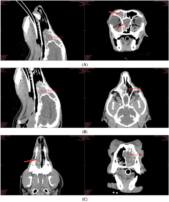
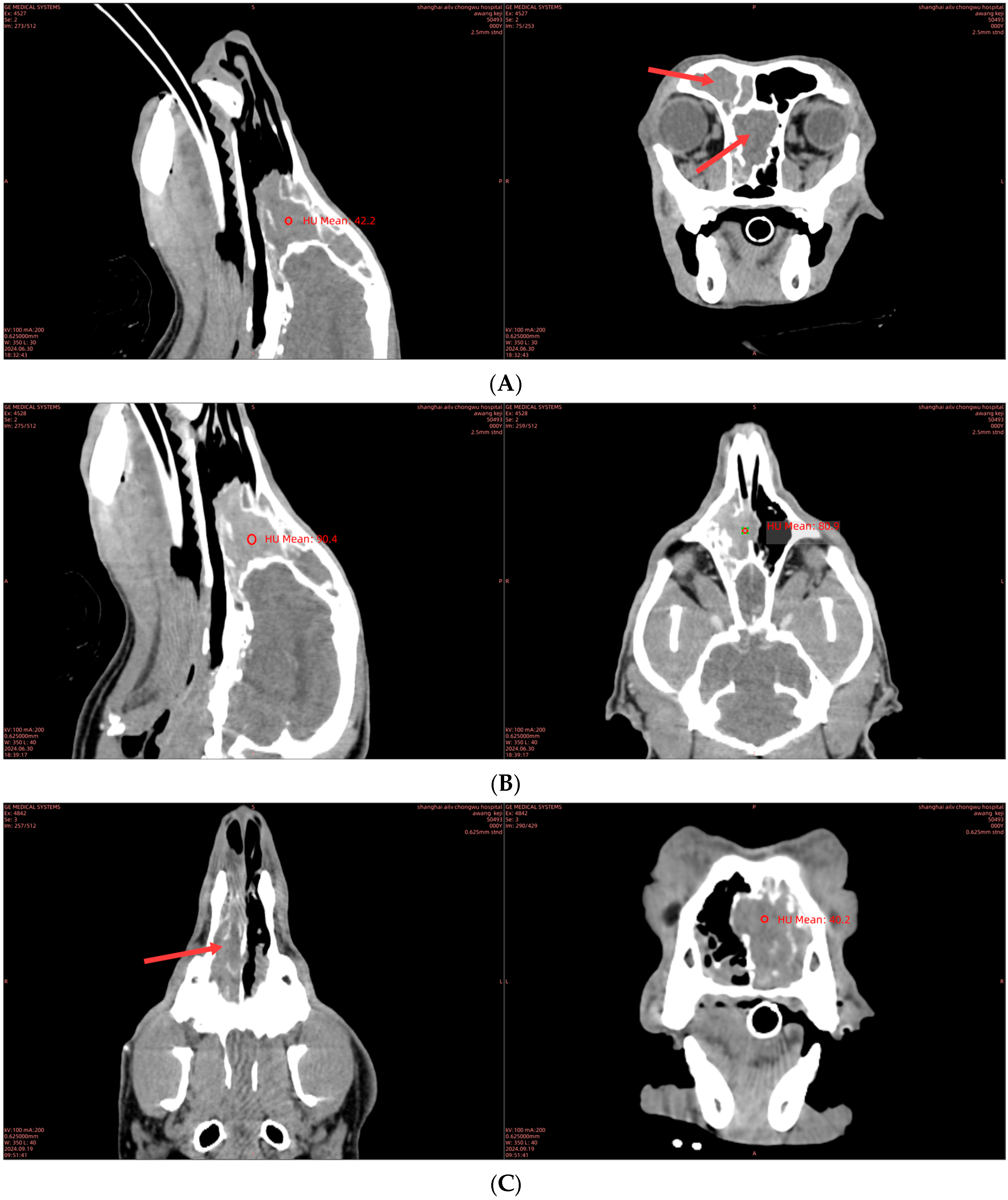
CT findings at the initial visit are shown in Figure 1A. The upper, middle, and lower nasal airways of the right nasal cavity were covered by irregular, non-characteristic, soft tissue attenuation, and the original normal turbinate structure was not visible. The nasal septum was destroyed in the middle of the tumor, and part of the tumor invaded into the left nasal cavity. The lesion did not cross the contralateral frontal sinus cavity. The caudal margin of the nasal mass was close to the lamina cribrosa, and the structure of the lamina cribrosa was destroyed in the ventral position close to the choanal foramen, resulting in no block between the mass and the frontal lobe of the brain. After angiography, as shown in Figure 1B, the mass in the nasal cavity was homogeneous with mild enhancement, and the higher the CT value, the more vascular the structure of the mass is suggested to be.
Figure 1.
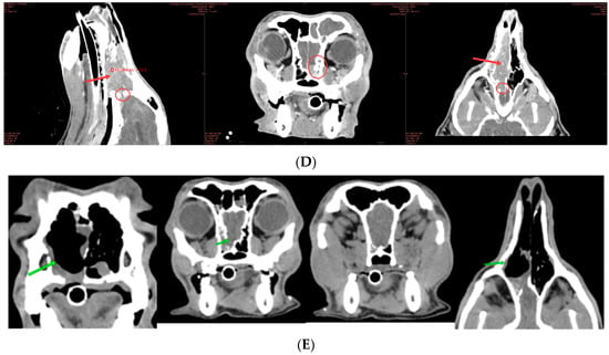
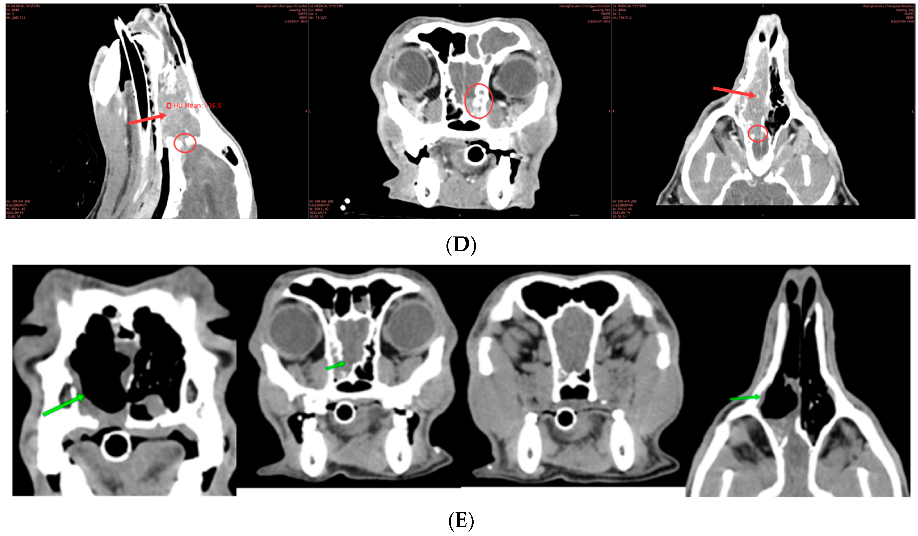
CT scan images of the affected dog at different time points. (A) Transverse plane, initial visit, soft tissue window, showing a mass filling the right nasal cavity (red arrow). (B) Transverse plane, initial visit, post-contrast, showing mild homogeneous enhancement. (C) Transverse plane, pre-operative, showing mass extending to the cribriform plate (red arrow). (D) Transverse plane, pre-operative, post-contrast, showing destruction of the cribriform plate (red arrow) and extension to the olfactory lobe (red circle). (E) Transverse plane, post-operative, showing post-surgical changes and residual soft tissue (green arrow).
The preoperative CT results are shown in Figure 1C. The right nasal cavity was filled with abnormal images of soft tissue attenuation from the entrance of the nasal cavity to the lamina cribrosa of the posterior segment of the nose, which distributed along the nasal bone and septum, destroyed part of the septum at the middle position, and entered the contralateral nasal cavity. After angiography, as shown in Figure 1D, the mass in the nasal cavity showed moderate homogeneous enhancement (red arrow), destruction of bone structure at the cribroidal plate, and a small amount of spread of the lesion to the olfactory lobe (red circle).
As shown in Figure 1E, the postoperative CT results showed that the lateral nasal cavity was filled with air shadow, the nostril to choanal was basically unobstructed, ethmoid bone and soft tissue mass shadow mostly disappeared, a small amount of soft tissue mass was still seen around the right olfactory bulb, and osteolysis was seen on the ventral side of the right lamina cribrosa. The right sphenoid sinus was filled with fluid or soft tissue shadow (green arrow).
3.3. Tumor Staging
Based on the CT findings, the tumor was staged as T3 (tumor invading the cribriform plate and extending into the left nasal cavity), N0 (no evidence of regional lymph node metastasis on CT), and M0 (no distant metastasis on thoracic CT). This staging is consistent with a modified Adams’ CT staging system for canine nasal tumors [16].
3.4. Histopathological Examination
After CT of the affected dog, puncture sampling was performed. After sampling, a portion of the sample in the needle was blown onto the slide, and a thin smear was immediately made. After air drying, Wright-Giemsa staining was used. The other part was sent for tissue sections for examination [17,18].
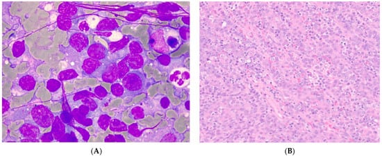
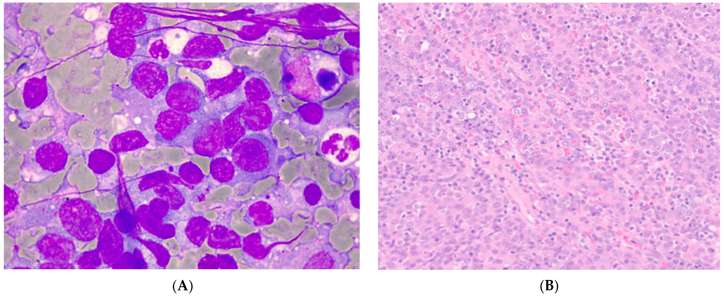
The results of needle cytology microscopy are shown in Figure 2A: The smear was rich in cellular components, mainly atypical epithelial cells, mixed with a small number of inflammatory cells. The cells were scattered, with some clumped. Tadpole-like cells were seen, and occasionally, intercellular Bridges were seen. The cell boundaries were clear, and some cell edges were serrated, with obvious keratosis. The nuclei were significantly different in size, the nuclear cytoplasmic ratio was increased, the shape of the nuclei was irregular, and the nuclear membrane was thickened and distorted. A large amount of necrotic debris, dissolved cell debris, and cellulose-like material was seen in the background. Emperipolesis was observed, that is, the migration of neutrophils and malignant cells into larger malignant cells.
Figure 2.
Histopathological examination results. (A) Needle cytology smear showing atypical epithelial cells with tadpole forms and keratinization (Wright-Giemsa stain, 1000× magnification). (B) Histopathological section showing sheets of polygonal neoplastic cells with squamous differentiation, infiltrating the nasal mucosa (Hematoxylin and Eosin stain, 400× magnification).
The results of microscopic examination of the pathological sections are shown in Figure 2B: the tumor was composed of polygonal cells arranged in sheets, cords, islands, and trabeculae, with manifestation of squamous differentiation. Microscopic observation showed that the lesions involved the nasal mucosa. The lesions were indistinct in border and showed moderately infiltrative growth.
Histopathologic grading of the squamous cell carcinoma was performed, characterizing the tumor as Grade 2 (moderately differentiated) based on the degree of keratinization, cellular pleomorphism, and mitotic count (approximately 8 mitotic figures per 10 high-power fields). Surgical margins were reported as “incomplete” or “close” due to the presence of neoplastic cells at the deep surgical margin adjacent to the cribriform plate. Immunohistochemistry (IHC) for pan-cytokeratin (AE1/AE3) was considered but not performed due to cost constraints; however, histomorphology alone was considered diagnostic.
3.5. Comprehensive Diagnostic Results
During the comprehensive case review, the dog was conclusively diagnosed with initial nasal squamous cell carcinoma through multiple examination methods. The screening results aligned closely with the disease indicators, providing clear evidence for the diagnosis. Clinically, the dog exhibited prolonged bleeding from the right nasal meatus, a key clue to nasal abnormalities. This symptom matched the pathological features of nasal squamous cell carcinoma, known for causing tissue damage and bleeding.
Specifically, the two-week history of nasal bleeding suggested that the anemia most likely resulted from ongoing blood loss. The elevated ALP, while documented, warrants further investigation to rule out a paraneoplastic syndrome or concurrent hepatobiliary disease, but a direct causal link to the nasal tumor remains unproven.
The CT scan revealed blurred boundaries of soft tissue within the upper, middle, and lower parts of the right nasal cavity. The nasal turbinates appeared dispersed, and a mass invaded the nasal septum, with some lesions affecting the left nasal area. Abnormal density changes were noted in the right frontal sinus, and the filter plate was damaged, indicating a significant tumor occupying the nasal cavity. These imaging findings clearly depicted the tumor’s proliferation and infiltration levels.
Histopathological analysis was crucial for differential diagnosis. Cytology and tissue examination showed atypical epithelial cells, tadpole-shaped cell bodies, bridging cell connections, and keratinization, indicative of squamous cell carcinoma. The lesion affected the nasal surface, showing moderate invasive growth, often exerting pressure on surrounding tissues and destroying local structures, leading to bleeding and respiratory difficulties. This study provides a foundation for future rehabilitation planning.
4. Disposal Situation
4.1. Chemotherapy
The first chemotherapy was carboplatin monotherapy (200 mg/m2, IV, q21d). After five cycles of treatment, the evaluation showed that the tumor regression was not significant, and the response was judged as disease progression, suggesting that the tumor was less sensitive to carboplatin monotherapy. Due to the poor effect of chemotherapy, surgical treatment was performed. To prevent recurrence, the chemotherapy regimen was adjusted to carboplatin (200 mg/m2, iv, q21d) combined with paladin (50 mg/m2, oral, q48h). This combination regimen is designed to enhance the antitumor effect and delay recurrence.
4.2. Surgical Reduction of the Tumor
Animals were placed in the prone position, and a longitudinal incision was made in the midline of the hard palate. Using periosteal stripper, subperiosteal dissection was performed against the bony surface of the hard palate to expose the planned resection of the maxillary region. According to the preoperative CT, the location and extent of the tumor were accurately determined, and the size and shape of the bone resection were planned. After removal of the bone fragment, the base of the nasal cavity was directly exposed to the surgical field. Using the tip and curvature of the nerve hook, a gentle blunt separation was made between the tumor and the surrounding normal mucosa of the turbinate, septum, and lateral wall of the nasal cavity. After removal of the primary tumor, complete hemostasis of the tumor bed is necessary. All peeling surfaces were examined. The nasal cavity and surgical incision cavity were rinsed thoroughly with a large amount of warm normal saline or lactated Ringer’s solution to remove blood clots, bone debris, and tumor debris. The previously stripped mucoperiosteal flap was repositioned to cover the bone defect area, and the mucoperiosteal layer was sutured. After successful surgery, the affected dog should be closely monitored for signs of choking or dyspnea due to nasal bleeding into the oropharynx. The surgical margins were evaluated histologically and deemed incomplete due to tumor cell infiltration at the deep margin near the cribriform plate.
The patient was hospitalized for 1 week after surgery, and daily wound care was provided with hemostatic drugs (ethamsylate), antibacterials, anti-inflammatory drugs (amoxicillin clavulanate potassium, metronidazole, enrofloxacin), and analgesics (teletamine hydrochloride, zolazepam hydrochloride, dexmedetomidine hydrochloride).
When the stitches were removed two weeks after the operation, there was no obvious nasal bleeding in the affected dog, and the surgical wound healed well.
4.3. Radiotherapy
Considering the highly aggressive nature of nasal squamous cell carcinoma, and that the risk of tumor recurrence still exists after previous surgical debulking, to further control the local lesions and reduce the probability of recurrence, the affected dog was transferred to Shanghai Dibaoche Animal Tumor Radiotherapy Center for radiotherapy on 15 October 2024. Radiotherapy was given every other day for a total of 18 times, aiming to precisely target the tumor area, destroy the DNA structure of residual tumor cells, inhibit their proliferation activity, and make up for the limitation that it is difficult to completely remove minimal residual disease by surgery.
During radiotherapy, intensity-modulated radiation therapy (IMRT) was delivered using a Veterinary TrueBeam linear accelerator. The radiotherapy protocol was as follows: total dose of 48 Gy delivered in 18 fractions (2.67 Gy per fraction), administered every other day, using intensity-modulated radiation therapy (IMRT). The planning target volume (PTV) encompassed the original tumor bed in the right nasal cavity, the residual lesion around the olfactory bulb, and a 5 mm margin. Organs at risk (OARs) included the brain, eyes (globes and lenses), optic nerves, and oral mucosa. Dose constraints were set to keep the maximum lens dose below 8 Gy and the mean brain dose below 30 Gy. The treatment intent was definitive (curative) for residual microscopic disease [19,20,21]. The dogs were subjected to general anesthesia before each radiotherapy to ensure the accuracy of irradiation. After radiotherapy, the mental state, appetite, and local tissue reactions were closely monitored. No serious adverse reactions, such as radiation mucositis and skin damage, were observed.
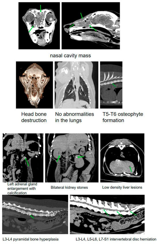
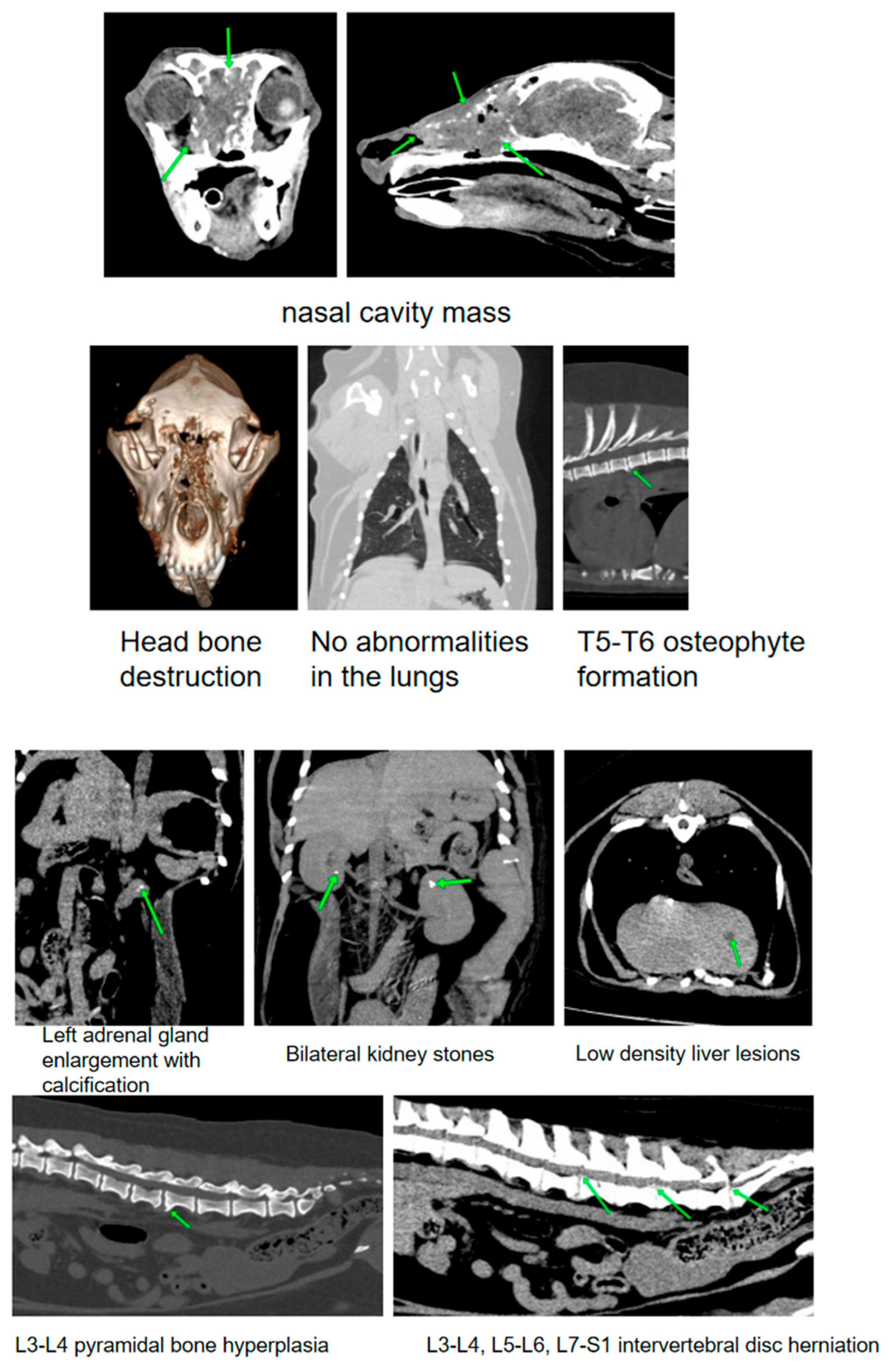
On 13 September 2025, the affected dog, Awang, underwent a whole-body CT examination at Shanghai Ruichen Huanuo Eros Pet Hospital to evaluate the efficacy and general condition following radiotherapy in October 2024. The results showed that the soft tissue mass in the original nasal cavity area of the head increased in size, the ethmoid bone and lamina cribrosa osteolysis expanded, and invaded the right orbit to compress the eyeball and the skull cavity to compress the olfactory bulb, but there was no local lymph node metastasis. There was no abnormality in the chest and lungs, except for degenerative bone hyperplasia in T5–T6 vertebral bodies. There was a 7 mm low-density lesion in the left lobe of the liver in the abdomen, bilateral kidney stones, left adrenal gland enlargement with calcification, and spinal canal stenosis caused by hyperostosis and intervertebral disk herniation at L3–L4. In general, tumors are still aggressive after short-term tumor control after radiotherapy, and there are multiple abnormalities in the whole body. It is necessary to further adjust the treatment plan and monitor the relevant lesions, as shown in Figure 3.
Figure 3.
Post-radiotherapy CT image (13 September 2025). Transverse plane, soft tissue window, showing progressive disease with an increased soft tissue mass invading the right orbit and skull cavity (green arrow).
4.4. Prognosis and Treatment
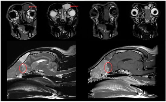
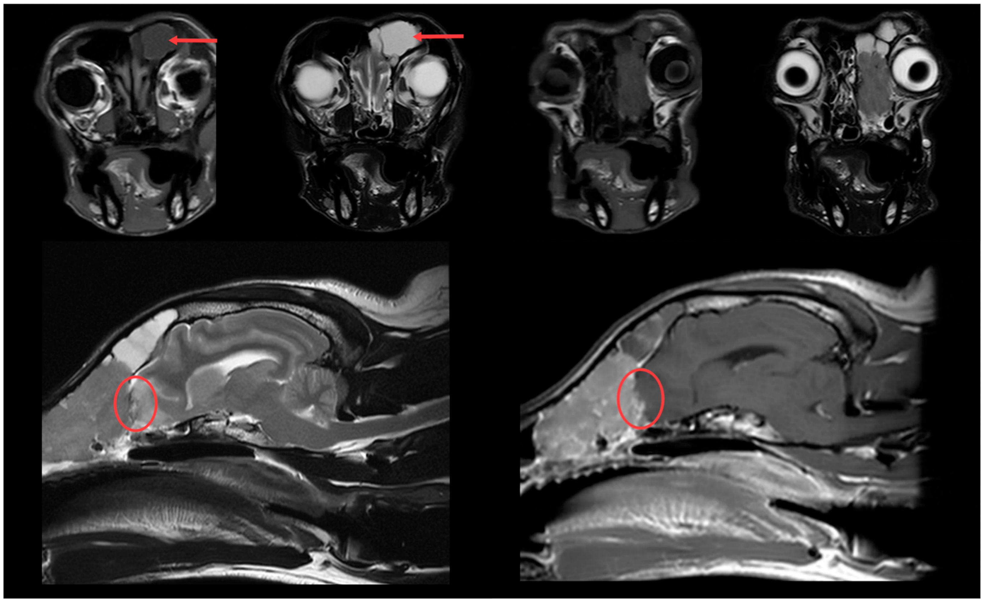
The results of the MRI reexamination five months after the operation are shown in Figure 4. Part of the nasal cavity tumor caused mild space-occupying compression on the olfactory lobe area of the brain, with local brain compression and edema. The tumor did not cause obvious extrusion space and did not show invasive growth into the brain. The symptoms of nasal bleeding recurred eight months after the operation, and the second operation was performed with external carotid artery ligation, which could last for three months or more without causing brain damage [22]. Since the second operation, the dog has shown no signs of nasal bleeding and has maintained a good appetite [23].
Figure 4.
MRI review image (T1-weighted post-contrast, transverse plane, red arrow) obtained eight months post-operatively, showing mild space-occupying compression of the right olfactory lobe by the recurrent tumor (red circle).
5. Discussion
Canine nasal squamous cell carcinoma is a highly aggressive malignant tumor. Diagnosing and treating this condition should consider the tumor’s features, the dog’s overall health, and current clinical circumstances. In this case, the initial approach of surgical removal combined with chemotherapy, along with external carotid artery ligation after postoperative recurrence, offers a useful reference for clinical practice. However, its effectiveness and limitations require further evaluation in light of existing research.
5.1. Evaluation of Treatment Effectiveness in This Case
This case used a comprehensive approach combining surgical cytoreduction with chemotherapy, yielding positive results initially. The surgery was performed via the hard palate approach, successfully removing the primary tumor in the nasal cavity. Postoperative CT scans showed that nasal ventilation largely recovered, and there was a significant reduction or disappearance of the abnormal mass in the ethmoid bone and surrounding soft tissues. The affected dogs showed improvements in mental status and feeding behavior, and nasal bleeding symptoms were notably alleviated. These findings suggest that surgical intervention can promptly alleviate tumor-induced nasal obstruction, significantly decrease tumor burden, and provide additional time for subsequent adjuvant therapy. Chemotherapy primarily works by inhibiting the growth of residual tumor cells through cytotoxic effects, aiming to delay disease recurrence and extend progression-free survival. This chemotherapy regimen aligns with the core concept of “postoperative adjuvant chemotherapy to eliminate minimal residual disease (MRD),” seeking to improve local control and long-term outcomes.
Radiotherapy is crucial for treating canine nasal squamous cell carcinoma, but its benefits and limitations should be evaluated in the context of the patient’s condition, treatment strategy, and clinical observations. Palpation indicated possible spinal disk issues or tumor-related lesions in the neck, with a diagnosis of nasal squamous cell carcinoma, which is known to be locally aggressive. Complete removal of the tumor through surgery or medication alone is challenging; however, radiotherapy can precisely target the tumor, inhibit cell growth, reduce pressure and irritation of surrounding tissues, and enhance clinical symptoms. Blood tests revealed mild anemia, decreased red blood cells and hemoglobin, and low lymphocyte levels, indicating a weakened immune system (body condition score 4). Given the patient’s condition, radiotherapy poses less trauma than secondary surgery.
While this case report demonstrates the feasibility of a sequential approach, it does not directly compare outcomes with radiotherapy as a primary modality. Published retrospective series report median survival times (MST) of 12–18 months for definitive radiotherapy alone for canine nasal tumors, which surpasses the progression-free interval observed here. The decision to delay radiotherapy in favor of initial chemotherapy was based on the owner’s preference and initial concerns about anesthesia risks, but in retrospect, earlier referral might have improved local control.
Existing studies confirm that radiotherapy is the preferred treatment for canine nasal squamous cell carcinoma, effectively destroying cancer cells by precisely targeting tumor tissue. Its response in local lesions surpasses that of surgery and chemotherapy, making it particularly suitable for invasive tumors that are difficult to remove entirely. Such tumors can also be treated with argon–helium cryoablation, which uses low temperatures to destroy tumor cells, offering benefits such as minimal invasion and a quick recovery [24]. In this case, treatment involved surgery combined with chemotherapy, without photon therapy or cryoablation. This approach has several advantages: for clinics lacking radiotherapy or argon-helium targeting, surgery and drug treatment are easier to control. Performing a hard palate operation clearly reveals tumors in the middle and posterior nasal cavity, while transnasal endoscopic procedures are better suited for large solid lesions. Patients with malignant tumors destroying the nasal septum and spreading to both nasal cavities tend to have better outcomes. For a dog with severe nasal obstruction and recurrent bleeding due to tumor compression, surgery quickly relieved pain and prevented further deterioration caused by the ineffective results of radiotherapy and chemotherapy. The dog immediately regained nasal breathing and showed significant improvement in quality of life, highlighting the rapid benefits of surgical intervention. Regarding dynamic adjustment strategies, if tumor recurrence involves bleeding, immediate occlusion of the external carotid artery can help control bleeding. Prioritizing quality of life is crucial, especially in end-stage cases, to minimize the harm caused by aggressive treatments. Studies indicate that combining surgery with medication can temporarily stabilize canine nasal squamous cell carcinoma, although the lack of radiotherapy limits long-term efficacy. Notably, this study introduces the novel use of external carotid artery ligation to manage recurrent nosebleeds by blocking main blood vessels feeding the tumor, aligning with Yang X and colleagues’ “vascular embolization step” approach. This method is less time-consuming and cost-effective, suitable for primary care settings. The findings underscore the importance of symptom-focused therapy for late-stage cancer, especially for patients unable to tolerate additional surgery or radiotherapy. Extracranial artery occlusion is an innovative way to control rebleeding, offering an actionable treatment option for advanced cases. Future treatments should incorporate radiotherapy within an integrated pathway, combining molecular targeted agents to develop personalized protocols and optimize symptom management throughout the entire treatment process.
5.2. Limitations and Directions for Future Improvements
Current research indicates that radiotherapy is the primary treatment for canine nasal squamous cell carcinoma. It precisely targets tumor tissue, destroying cancer cell DNA, and demonstrates better control over local lesions than surgery and chemotherapy, particularly for aggressive tumors that cannot be entirely removed [25,26]. In clinical settings, intensity-modulated radiation therapy (IMRT) is commonly employed, using a Veterinary TrueBeam linear accelerator and preoperative and postoperative CT and MRI scans to accurately identify the tumor target. This allows radiation to be focused on the tumor while protecting surrounding normal tissues such as the brain, eyes, and oral mucosa, thereby reducing the risk of radiotherapy-related complications. The incidence of severe adverse effects, like radiation mucositis and skin injury, can be minimized by administering general anesthesia to fix the dog’s posture during treatment to ensure precise irradiation. Post-therapy, it is essential to monitor the dog’s normal behavior state, appetite, and local tissue reactions. Additionally, argon–helium cryoablation has been used to treat such tumors, utilizing low temperatures to destroy tumor cells, resulting in less trauma and quicker recovery.
This case initially involved surgery combined with chemotherapy, a straightforward approach that requires no special equipment and is suitable for primary clinics. The hard palate approach aligns with nasal anatomy, allowing direct access to the middle and posterior tumor regions. It effectively clears large lesions that have damaged the nasal septum and invaded both nasal cavities. This method quickly alleviates nasal obstruction, bleeding, and other emergencies, with the affected dogs experiencing immediate recovery of nasal ventilation, making it practical for clinical use. When tumor recurrence and bleeding occur post-operation, ligation of the external carotid artery helps control symptoms and prevents over-treatment. The procedure is simple, inexpensive, and ideal for promotion in primary care settings. Postoperative radiotherapy plays a crucial role, targeting residual local lesions while protecting normal tissues such as the brain, eyes, and oral mucosa, leading to minimal side effects, including mucositis and skin damage. This approach effectively manages residual disease and preserves normal physiological functions, demonstrating a focus on the dogs’ health and safety, and improving upon previous methods.
However, this study has notable limitations. First, the treatment sequence deviated from current evidence-based recommendations that prioritize radiotherapy for inoperable or incompletely resected nasal tumors. Second, the failure to perform a thorough systemic evaluation (including abdominal ultrasound and thoracic CT) prior to the first surgery delayed the detection of multi-site abnormalities, such as the liver lesion and adrenal enlargement noted later. Third, the chemotherapeutic adjustment after carboplatin failure was made without guidance from drug-sensitivity testing, which might have optimized the selection of second-line agents. Fourth, while external carotid artery ligation provided effective hemostasis, this technique is palliative and does not address tumor progression. Its novelty in this context lies primarily in its use as a salvage procedure for recurrent epistaxis following multimodal therapy, rather than as a first-line anticancer treatment.
Compared to prior veterinary case reports and series, this case is notable for the explicit use of external carotid artery ligation for hemorrhage control. While Yang X et al. [27] described vascular embolization in human nasopharyngeal cancer, this report provides a simpler, low-cost alternative applicable in primary care settings for managing a terminal complication. However, the lack of long-term follow-up and the absence of a control group prevent any conclusion regarding its impact on overall survival.
Considering these limitations, the timing and combination of treatments should be optimized. Radiotherapy and cryoablation should be prioritized early after diagnosis. A comprehensive disease assessment and monitoring system needs to be improved, involving preoperative screening and regular systemic follow-up. To tailor treatment plans, chemotherapy should be based on drug-sensitivity testing, with exploration of new methods and dynamic adjustments upon recurrence to enhance outcomes [28].
5.3. Summary
An 8-year-old Pembroke Welsh Corgi was diagnosed with nasal squamous cell carcinoma. Multimodal therapy eased the dog’s symptoms and enhanced its quality of life, addressing emergencies and recurrent bleeding. However, delays in radiotherapy, incomplete systemic assessments, and late chemotherapy adjustments contributed to tumor invasion and progression. Future approaches should implement an “early accurate diagnosis + priority radiotherapy + multimodal treatment + continuous monitoring” framework, complemented by personalized plans and improved owner education for early detection and intervention. This strategy aims to enhance diagnostic accuracy, treatment success, and survival rates in affected dogs.
Author Contributions
T.L. and X.Z.: Visualization, Formal analysis, original draft, Methodology. All authors have read and agreed to the published version of the manuscript.
Funding
This research received no external funding.
Institutional Review Board Statement
Not applicable.
Informed Consent Statement
Written informed consent was obtained from the dog’s owner for the publication of this case report and any accompanying images.
Data Availability Statement
The datasets generated and/or analyzed during the current case study are available from the corresponding author on reasonable request.
Acknowledgments
We would like to thank Shanghai Ailv Pet Hospital for their help in this study.
Conflicts of Interest
The authors declare no competing interests.
References
- Dos Santos, A.; Lamego, E.C.; Eisenhardt, L.M.; de Vargas, I.; Flores, M.M.; Fighera, R.A.; Kommers, G.D. Prevalence and anatomopathological characterization of cutaneous squamous cell carcinomas with regional and distant metastases in dogs and cats: 20 cases (1985–2020). Vet. Comp. Oncol. 2023, 21, 291–301. [Google Scholar] [CrossRef] [Scilit]
- Snyder, J.M.; Shofer, F.S.; Van Winkle, T.J.; Massicotte, C. Canine intracranial primary neoplasia: 173 cases (1986–2003). J. Vet. Intern. Med. 2006, 20, 669–675. [Google Scholar] [CrossRef] [Scilit]
- Supsavhad, W.; Dirksen, W.P.; Martin, C.K.; Rosol, T.J. Animal models of head and neck squamous cell carcinoma. Vet. J. 2016, 210, 7–16. [Google Scholar] [CrossRef] [Scilit]
- Wypij, J.M. A naturally occurring feline model of head and neck squamous cell carcinoma. Pathol. Res. Int. 2013, 2013, 502197. [Google Scholar] [CrossRef] [Scilit]
- Ninomiya, F.; Suzuki, S.; Tanaka, H.; Hayashi, S.; Ozaki, K.; Narama, I. Nasal and paranasal adenocarcinomas with neuroendocrine differentiation in dogs. Vet. Pathol. 2008, 45, 181–187. [Google Scholar] [CrossRef] [Scilit]
- Steinmetz, A. Superior orbitectomy and chemotherapy in a dog with frontal sinus squamous cell carcinoma: A case report and review of the literature. Clin. Case Rep. 2017, 5, 513–520. [Google Scholar] [CrossRef] [Scilit]
- Ramos, M.; Benavents, S.; Giralt, J. Management of squamous cell carcinoma of the head and neck: Updated European treatment recommendations. Expert Rev. Anticancer Ther. 2010, 10, 339–344. [Google Scholar] [CrossRef] [Scilit]
- Choong, N.; Vokes, E. Expanding role of the medical oncologist in the management of head and neck cancer. CA Cancer J. Clin. 2008, 58, 32–53. [Google Scholar] [CrossRef] [Scilit]
- Zibelman, M.; Mehra, R. Overview of Current Treatment Options and Investigational Targeted Therapies for Locally Advanced Squamous Cell Carcinoma of the Head and Neck. Am. J. Clin. Oncol. 2016, 39, 396–406. [Google Scholar] [CrossRef] [Scilit]
- Elliot, K.M.; Mayer, M.N. Radiation therapy for tumors of the nasal cavity and paranasal sinuses in dogs. Can. Vet. J. 2009, 50, 309–312. [Google Scholar]
- Wang, Y.; Yang, R.; Zhao, M.; Guo, W.; Zhang, L.; Zhang, W.; Wang, X. Retrospective analysis of 98 cases of maxillary sinus squamous cell carcinoma and therapeutic exploration. World J. Surg. Oncol. 2020, 18, 90. [Google Scholar] [CrossRef] [Scilit]
- Kotamaryi, V.S.; Feintisch, A.M.; Ciminello, F. Invasive Squamous Cell Carcinoma of the Scalp. Eplasty 2015, 15, ic39. [Google Scholar]
- Neves-Motta, R.; Ferry, F.R.; Basilio-de-Oliveira, C.A.; Carvalho, R.d.S.; Martins, C.J.; Eyer-Silva, W.A.; Morais-De-Sá, C.A. Highly aggressive squamous cell carcinoma in an HIV-infected patient. Rev. Soc. Bras. Med. Trop. 2004, 37, 496–498. [Google Scholar]
- Neubauer, K.E.; Goldstein, G.D.; Plumb, S.J. Squamous cell carcinoma of the scalp in organ transplant recipients: Exploring mechanisms for recurrence and treatment guidelines. Dermatol. Surg. 2010, 36, 185–193. [Google Scholar] [CrossRef] [Scilit]
- Gorza, L.L.; Oliveira, A.R.; Porto, M.G.; Santos, V.C.; Hardt, I.; Maestri, L.F.P.; Souza, T.D.; Flecher, M.C. Clinical, anatomopathological, and immunohistochemical findings of a transitional cell carcinoma from nasal cavity, frontal and ethmoidal sinus with meningoencephalic invasion in a dog. Open Vet. J. 2021, 11, 1–5. [Google Scholar]
- Adams, W.M.; Kleiter, M.M.; Thrall, D.E.; Klauer, J.M.; Forrest, L.J.; LA Due, T.A.; Havighurst, T.C. Prognostic significance of tumor histology and computed tomographic staging for radiation treatment response of canine nasal tumors. Vet. Radiol. Ultrasound 2009, 50, 330–335. [Google Scholar] [CrossRef] [Scilit]
- Li, P.; Jiang, C.; Wang, W.L.; Liu, M.R. CT diagnosis of a dog with choanal tumor. Shandong Anim. Husb. Vet. Med. 2012, 43, 47–48. [Google Scholar]
- Cai, X.; Xie, F. CT imaging analysis of the characteristics of nasal diseases in dogs. Chin. J. Vet. Med. 2019, 55, 82–85. [Google Scholar]
- Liu, H.Q.; Sun, J.X.; He, X.H.; Gan, J.Y.; Li, J.; Liu, B.; Yu, F. Application of radiotherapy in tumors in dogs and cats. Chin. J. Vet. Med. 2024, 60, 110–117. [Google Scholar]
- Loddo, P.; Schiavo, L.; Dobson, J.; Marcinowska, O. Clinical Presentation of Frontal Sinus Squamous Cell Carcinoma in the Dog and Response to Treatment with Radiation Therapy in Eight Dogs. Vet. Radiol. Ultrasound 2025, 66, e70000. [Google Scholar]
- Mizuno, R.; Mori, T. Prognostic factors and survival following radiation therapy for canine nasal tumors: A single-institution retrospective study of 166 cases. Open Vet. J. 2024, 14, 1538–1552. [Google Scholar] [CrossRef] [Scilit]
- Weisse, C.; Nicholson, M.E.; Rollings, C.; Hammer, K.; Hurst, R.; Solomon, J.A. Use of percutaneous arterial embolization for treatment of intractable epistaxis in three dogs. J. Am. Vet. Med. Assoc. 2004, 224, 1307–1311. [Google Scholar] [CrossRef] [Scilit]
- Goodman, A.E.; Goodman, A.R. Common Carotid Artery Ligation to Minimize Blood Loss During Oral and Maxillofacial Surgery. J. Vet. Dent. 2016, 33, 195–200. [Google Scholar] [CrossRef] [Scilit]
- Xie, Y.; Wu, Z.; Li, S. Application of argon-helium cryoablation in the treatment of nasal squamous cell carcinoma in dogs. Chin. Vet. J. 2022, 58, 114–117. [Google Scholar]
- Elmslie, R.E.; Glawe, P.; Dow, S.W. Metronomic therapy with cyclophosphamide and piroxicam effectively delays tumor recurrence in dogs with incompletely resected soft tissue sarcomas. J. Vet. Intern. Med. 2008, 22, 1373–1379. [Google Scholar] [CrossRef] [Scilit]
- De Vos, J.; Ramos Vega, S.; Noorman, E.; de Vos, P. Primary frontal sinus squamous cell carcinoma in three dogs treated with piroxicam combined with carboplatin or toceranib. Vet. Comp. Oncol. 2012, 10, 206–213. [Google Scholar] [CrossRef] [Scilit]
- Yang, X.; Ren, H.; Li, M.; Zhu, Y.; Zhang, W.; Fu, J. Treatment of intractable epistaxis in patients with nasopharyngeal cancer. Ann. Med. 2023, 55, 2200257. [Google Scholar] [CrossRef] [Scilit]
- Mei, X.T.; Yang, Q.Q.; Yang, D.J.; Hou, X.P.; Deng, Y.F.; Zhao, Y.B.; Su, J.; Yao, D.W. Application of CT in the diagnosis and differential diagnosis of canine epistaxis. J. Anim. Sci. Vet. Med. 2019, 51, 108–114. [Google Scholar]
Disclaimer/Publisher’s Note: The statements, opinions and data contained in all publications are solely those of the individual author(s) and contributor(s) and not of MDPI and/or the editor(s). MDPI and/or the editor(s) disclaim responsibility for any injury to people or property resulting from any ideas, methods, instructions or products referred to in the content. |
© 2026 by the authors. Licensee MDPI, Basel, Switzerland. This article is an open access article distributed under the terms and conditions of the Creative Commons Attribution (CC BY) license.