


Abstract
This systematic review critically examines the use of 2D and 3D digital imaging technologies of the face and teeth, with and without integration of artificial intelligence, for human identification in forensic and medicolegal contexts. Following PRISMA 2020 guidelines, Scopus, PubMed and Web of Science were systematically searched, identifying 26 studies published between 2011 and 2025 that met predefined eligibility criteria framed by a PECO-style question. Eighteen studies focused on facial imaging, six on dental imaging and two on integrated orofacial workflows, using digital photography, CCTV/video, 3D surface imaging, intraoral scanners, and three-dimensional superimposition methods, sometimes combined with classical algorithms and deep learning models. In controlled or semi-controlled settings, state-of-the-art facial algorithms often reported very high accuracy, with values up to 99.85%. By contrast, studies using real CCTV or other challenging forensic imagery showed more variable performance, with accuracies ranging from about 72.8% to 96.6%. Dental and orofacial studies reported 100% correct identifications for 3D superimposition of intraoral scans in small samples, and around 83% accuracy for automated AI-based dental identification. Crucially, fulfilling the promise of a true orofacial approach, this review proposes a structured research agenda focused on creating realistic multi-modal databases, standardizing protocols, and implementing probabilistic reporting (likelihood ratios) to guide future validation and legal admissibility.
1. Introduction
Forensic human identification increasingly relies on digital imaging of the face and teeth, driven by widespread availability of imaging equipment and software applications in clinical, security, and medicolegal settings [1,2].
1.1. Digital Technologies
Digital single-lens reflex (DSLR) cameras are used to obtain high-resolution, well-lit frontal and profile photographs of the face under controlled conditions, often following standardized protocols [3,4]. These images preserve fine details of soft-tissue contours, skin texture and facial landmarks, making them suitable both for qualitative comparison by experts and for quantitative measurements [3,4]. Closed-circuit television (CCTV) and surveillance video systems continuously capture facial images, typically at lower resolution and under less controlled conditions than DSLR photography [4]. The scores produced from CCTV-based recognition could be embedded into a Bayesian likelihood-ratio (LR) framework, illustrating how noisy video data can still contribute quantitatively to forensic evidence [5]. Three-dimensional (3D) surface scanners are dedicated devices that reconstruct the external soft-tissue surface as a dense point cloud or polygonal mesh [6]. Photogrammetric systems reconstruct 3D head or facial models from multiple overlapping 2D photographs, using computer-vision algorithms to estimate camera positions and triangulate the location of surface points [7]. Multi-camera rigs or rotating camera setups can be designed specifically for forensic “3D mugshots”, enabling rapid acquisition of a full-head model with minimal subject cooperation and without the need for specialized laser or structured-light hardware [8].
In the dental and orofacial domain, intraoral scanners (IOS) are handheld optical devices that acquire a series of overlapping images inside the mouth and stitch them into detailed 3D models of dental arches and adjacent soft tissues [9]. When combined with standardized scanning protocols and appropriate metadata, IOS offer a repeatable, non-invasive method to document individual dentitions in vivo, bridging clinical practice and forensic odontology and enabling later comparison with post-mortem scans or 3D dental photo-scans [9,10].
Across these facial and dental modalities, digital orofacial identification workflows typically begin with automatic detection of the relevant anatomy (face, ear or dentition) and localization of landmarks, followed by extraction of geometric and texture-based descriptors such as Local Binary Patterns (LBP), Histogram of Oriented Gradients (HOG), Gabor filters, geodesic distance maps and 2D–3D proportionality indices that encode both fine-scale appearance and overall shape relationships [11,12]. These feature representations are then analyzed using a spectrum of models, ranging from principal-component-analysis (PCA)-based neural networks and support vector machines (SVM) in earlier systems to more recent deep convolutional neural network (CNN) architectures and metric-learning models specifically tailored to unconstrained or multimodal recognition scenarios, including near-infrared-visible (NIR–VIS) face matching and combined face–ear biometrics, which aim to keep genuine samples close and impostor samples far apart in an optimized embedding space [13,14,15,16]. For 3D dental and orofacial data, pre-processed intraoral scans, and 3D dental photo-scans are aligned using iterative closest point (ICP) algorithm and related registration methods, and similarity between ante-mortem (AM) and post-mortem (PM) models is quantified through root-mean-square (RMS) distances or curvature-based discrepancies across the surfaces, allowing empirically derived decision thresholds to separate matches from non-matches [17]. Building on these components, several studies integrate convolutional neural networks (CNNs) trained on intraoral or facial images, and recurrent neural networks (RNNs) applied to enhanced two-dimensional (2D) dental views, into end-to-end pipelines in which numerical similarity scores and associated error metrics can ultimately be mapped onto calibrated LR frameworks within structured, expert-driven reporting protocols for forensic and medico-legal use [17,18].
1.2. Evidential Value and Current Challenges
However, the evidential value of these technologies in real forensic scenarios remains uncertain because many systems are validated only under controlled or semi-controlled conditions [19,20]. International guidelines emphasize that forensic facial comparison must follow structured methodologies, such as Analysis, Comparison, Evaluation, and Verification (ACE-V), with rigorous documentation and, where appropriate, probabilistic evaluation of evidence using LR [21,22]. Similarly, forensic odontology has moved toward digital workflows integrating intraoral scanners, cone-beam computed tomography (CBCT), and AI-driven analysis, yet standardization, validation, and legal admissibility remain challenges [23,24]. Furthermore, these specific challenges must be understood within the broader context of human identification, where the use of physical and behavioral biometric traits increasingly intersects with complex issues of digital privacy and international regulation [25].
Several narrative and domain-specific reviews have begun to map this changing landscape. In 2020, Jacquet and Champod reviewed automated face recognition in forensic science, highlighting both the investigative utility of facial images and the lack of empirical validation under genuinely forensic conditions such as CCTV footage and identity documents [2]. In 2025, Radu and colleagues synthesized current methods and emerging trends in digital forensic odontology, underscoring how Cone Beam Computed Tomography (CBCT), 3D scanning, intraoral imaging and artificial intelligence can enhance diagnostic consistency while raising questions about standardization, ethics and admissibility [23]. These and other contributions provide valuable overviews of specific subfields, but they typically focus on either facial or dental evidence in isolation, and many adopt narrative rather than systematic approaches to literature identification and appraisal.
1.3. Scope of the Review
There is thus a clear need for a systematic synthesis that jointly considers digital 2D and 3D imaging of the face and teeth, with and without AI integration, in explicitly forensic or medicolegal contexts. Such a synthesis should critically appraise methodological quality, transparency of reporting, bias considerations and alignment with emerging legal standards. In addition, because the field is rapidly evolving, there is value in moving beyond descriptive summaries to articulate a structured research agenda to guide the development and validation of robust orofacial identification workflows.
This systematic review addresses that gap by synthesizing studies on digital 2D and 3D orofacial imaging, alone or combined with artificial intelligence, used for personal identification or identity verification in forensic and medicolegal settings. Specifically, it aimed to: (i) map imaging modalities and anatomical focus (face, teeth, or both); (ii) summarize AI and computational methods and their reported performance; (iii) assess methodological quality, transparency, and bias considerations; and (iv) identify priorities for future research, with emphasis on probabilistic reporting, legal admissibility, and real-world validation. By integrating evidence across facial, dental, and orofacial workflows, this review seeks to inform best practices and research directions for robust digital approaches to forensic orofacial identification.
2. Materials and Methods
This systematic review was conducted according to PRISMA [26,27] and designed to identify and synthesize evidence on digital imaging technologies for forensic orofacial identification in humans. The review was registered in PROSPERO under the identifier CRD420261304016. There were no deviations from the initially registered protocol. The review question was framed using a PECO approach Population (humans, living or deceased), Exposure (digital orofacial imaging technologies, including AI-enabled methods), Comparator (where applicable, alternative imaging modalities or workflows, such as 2D versus 3D imaging), and Outcomes (methods and systems aimed at personal identification or identity verification, and their reported performance). The review focused on digital 2D and 3D imaging of the face and/or teeth used in forensic and medicolegal contexts.
2.1. Search Strategy
Three electronic databases (Scopus, PubMed and Web of Science) were used. Following preliminary scoping searches to refine the terminology, the final database searches were conducted on 27 September 2026. The predefined Boolean query (“Artificial intelligence” OR “AI” OR “Machine learning” OR Digital) AND (Identification OR Identity*) AND (Scan* OR Imag* OR Photo* OR “3D”) AND (Face OR Facial OR Oral) AND (“Forensic Dentistry” OR Forensic OR “Legal medicine”) was tailored to the specific search requirements of each database. These specific medical databases were selected to ensure scientific rigor within the medical literature, focusing on the medicolegal applications of these novel concepts rather than highly technical AI-specific repositories. Furthermore, the search strategy deliberately utilized broader terms to encompass the entire orofacial region, avoiding highly specific dental terms (e.g., odontology, bite) to capture the comprehensive scope of both facial and dental identification without inappropriately narrowing the focus. All records retrieved from the three databases were exported and imported into Rayyan (Rayyan Systems Inc., Cambridge, MA, USA), an online platform for systematic review management, which was used to handle deduplication and to support blinded, independent screening by two reviewers. Titles and abstracts were first screened in Rayyan against the eligibility criteria, followed by full-text assessment of potentially relevant articles. Conflicts between reviewers were highlighted within Rayyan and resolved through discussion; when necessary, a third reviewer was consulted to reach consensus. More specific clinical dental terms (e.g., ‘odontology’, ‘CBCT’, ‘intraoral scan’) and broad biometric face-comparison terms were purposely excluded from the search query to prevent a disproportionate influx of purely clinical or non-forensic biometric literature. This targeted approach ensured the review remained strictly focused on the direct intersection of digital imaging technologies and practical forensic human identification.
2.2. Eligibility Criteria
Inclusion and exclusion criteria were applied at both title/abstract and full-text screening stages. Studies were included if they met all of the following: (i) Population: human subjects (living or deceased) with imaging of the face and/or teeth; (ii) Exposure/technology: use of digital imaging technologies, including 2D photographs, video, intraoral scanners, 3D surface or volume scanners, or other digital image-based modalities applicable to forensic orofacial identification; (iii) Context: explicit forensic or medicolegal relevance (e.g., forensic dentistry, forensic facial comparison, biometric identification used or usable in legal settings); (iv) Outcome: methods, workflows, or systems aimed at personal identification or identity verification (one-to-one comparison, one-to-many search, or ante-mortem/post-mortem matching); (v) Reporting quality: studies were required to explicitly report a predefined minimum set of methodological variables to ensure reproducibility and prevent selection bias. Specifically, a study was deemed to have ‘sufficient methodological detail’ only if it clearly documented all of the following core elements: the specific imaging modality and equipment used; the AI architecture, software, or analytical method applied; the exact sample size; and quantitative performance metrics. Studies failing to report one or more of these mandatory elements were excluded.
The search excluded studies that: (i) used only simulated, synthetic, or phantom data without real human images; (ii) Did not involve the face and/or teeth (e.g., purely cranial, skeletal, or non-orofacial biometrics); (iii) Were reviews, editorials, opinion pieces, conference abstracts without full methodological reporting, or purely conceptual works without primary data; (iv) Failed to provide enough methodological description to reliably fill the columns of the extraction table (e.g., missing imaging modality, equipment, or core details of the analytical pipeline). The search was restricted to studies involving human subjects and digital imaging data, without an a priori limit on publication year. No language restrictions were applied during the search and screening processes.
2.3. Data Extraction
For each included study, two reviewers independently extracted data into a structured spreadsheet developed a priori. The table included: title, main author, year, country, anatomical region of interest (face, teeth, or both), imaging modality, specific equipment/technology, AI software or computational method, type of study, sample size category, statistical performance metrics, main application, and reported limitations and advantages. Any discrepancies in extraction were checked against the original articles and resolved by consensus. This dataset, further detailed in the results section, underpins the descriptive synthesis and the proposed research agenda.
2.4. Quality Assessment
Methodological quality and reporting robustness of the included studies were appraised using a checklist inspired by the Joanna Briggs Institute (JBI) approach to quality assessment of diagnostic and technical studies. Due to the high heterogeneity of the included study designs, this appraisal was explicitly conducted as a rigorous reporting-completeness exercise rather than a standard risk-of-bias assessment. The adapted tool comprised ten questions covering: clarity of imaging context, sample description, reporting of quantitative performance metrics, transparency of methods and AI models, recency of the work, adequacy of performance, explicit forensic application, explicit use of AI/machine learning, and clear description of limitations and advantages. Each criterion was scored dichotomously (1 point for “yes”, 0 points for “no”, yielding a maximum possible score of 6). Studies achieving a total score of 5 to 6 were classified as high quality, while those scoring 3 to 4 were classified as moderate quality, and 0 to 2 as low quality. The appraisal was conducted independently by two reviewers, and inter-rater reliability was maintained by resolving any scoring discrepancies through discussion and consensus. Furthermore, it is acknowledged that this adapted checklist serves as a descriptive tool to gauge reporting completeness rather than a fully validated psychometric framework for biometric performance, which represents a limitation of the current appraisal process. Furthermore, it should be noted that this review did not use a standard risk-of-bias framework tailored specifically to biometrics and forensics (e.g., evaluating dataset subject overlap leakage, uncontrolled covariates, or threshold selection), which represents a limitation of the current appraisal.
3. Results
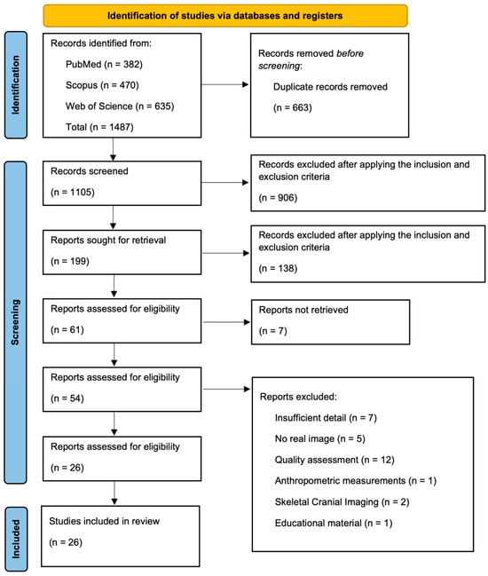
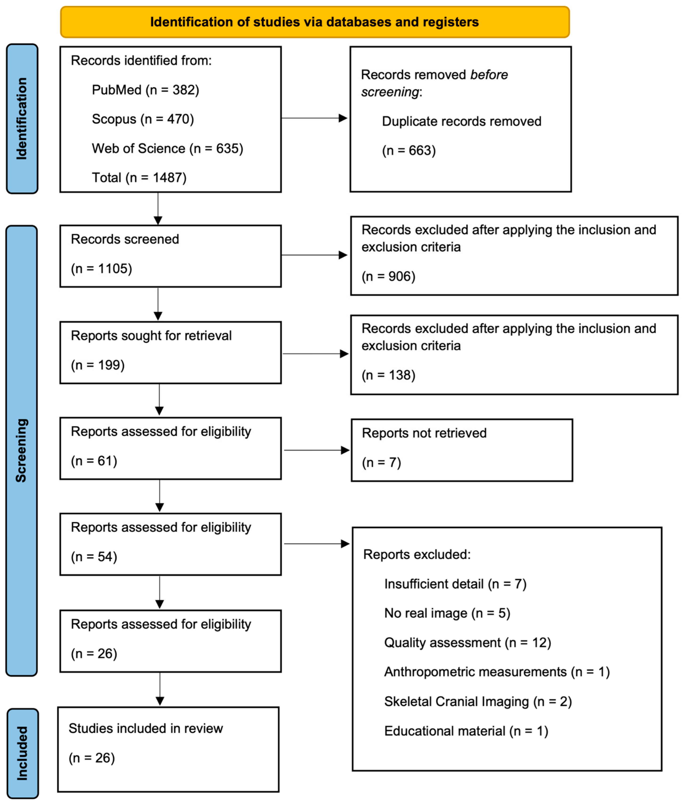
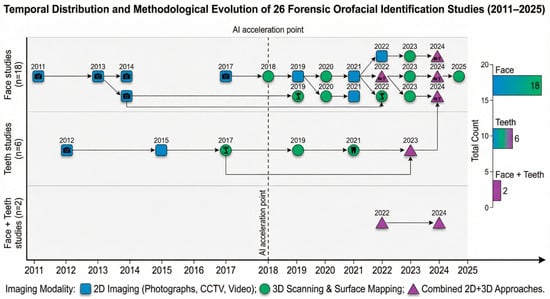
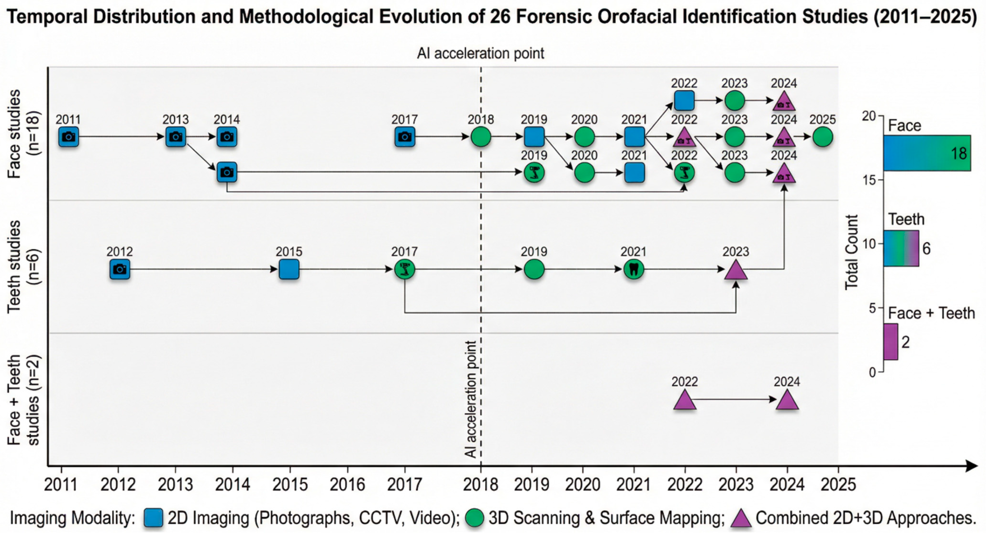
Twenty-six studies were included, addressing forensic identification based on digital imaging of the face, teeth, or both structures, with publication years between 2011 and 2025 and a clear concentration after 2018, especially in AI-supported approaches (Figure 1 and Table 1).
Figure 1.
Flow diagram based on the PRISMA flowchart template of the search and selection process.
Of these, 18 focused primarily on the face [1,28,29,30,31,32,33,34,35,36,37,38,39,40,41,42,43,44]. Six studies focused on the teeth [45,46,47,48,49,50], and two explicitly addressed combined face and teeth in orofacial workflows [51,52]. Study designs were mainly technical developments or experimental studies, with sample sizes varying from small pilots (<30 subjects) to large-scale datasets (>500 subjects or images). The overall selection process is summarized in a PRISMA-style flow diagram (Figure 1), and the temporal distribution and methodological evolution by anatomical focus and imaging modality are presented in Figure 2.
Table 1.
Data extraction of included article.
Figure 2.
Temporal distribution and methodological evolution of forensic orofacial identification studies (2011–2025) based on anatomical focus and imaging modality.
3.1. Imaging Modalities and Anatomical Focus
The 18 face-focused studies covered a wide spectrum of modalities: 2D still images and CCTV/video [28,29,31,36,37,41,42,43,44], 3D surface imaging and photogrammetry [29,31,32,33,37,38], and methodological work on facial measurements and landmarks using mixed 2D/3D data [1,35,40]. These studies addressed contexts from controlled biometric capture to challenging forensic scenarios such as variable pose, expression, occlusion, low image quality and AM/PM comparisons of deceased migrants.
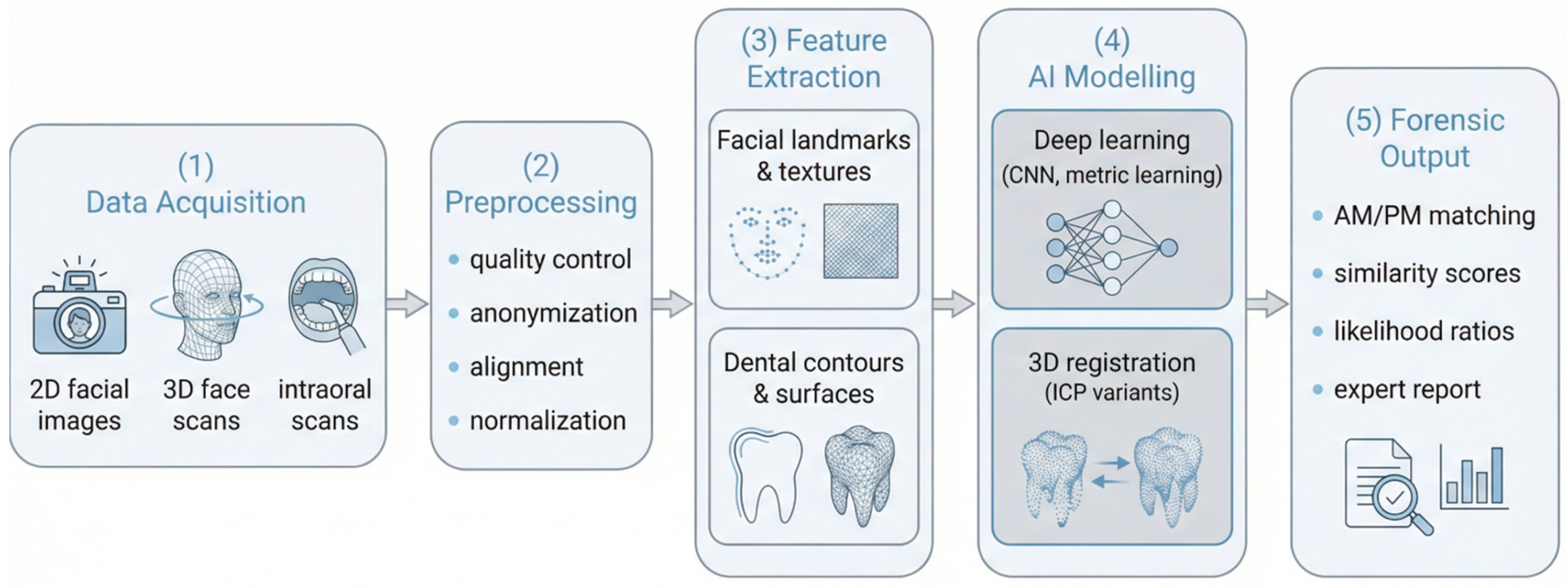
The two orofacial studies by Corte-Real [51] and Nunes [52] combined DSLR facial photography with intraoral scanning (Medit i700, Seoul, Republic of Korea Canon 5D/5D Mark series, Tokyo, Japan) to document faces, smiles and dental arches in a single digital workflow aimed at forensic or legal evidence recording. The six dental studies used 3D intraoral scans and scanned plaster models (iTero, Arizona, USA, TRIOS, Copenhagen, Denmark, Primescan, North Carolina, USA) or 2D dental images to evaluate 3D superimposition, segmentation and automated classification of dentitions for individual identification. This group includes work on automatic soft-tissue removal in 3D scans [45], 3D superimposition of dentition contours [46,48,50], CNN-based classification of molar images from IOS data [47] and 2D image-based identification using RNNs and corner detection [49]. A schematic end-to-end AI methodology for forensic orofacial identification, from data acquisition to forensic output, is illustrated in Figure 3.
Figure 3.
Proposed AI methodology for forensic orofacial identification, illustrating the end-to-end pipeline from data acquisition (2D facial images, 3D facial scans, intraoral scans) through preprocessing (quality control, anonymization, alignment, normalization), feature extraction (facial landmarks and textures, dental contours and surfaces), AI modelling (deep learning and 3D registration algorithms), and generation of the forensic output (AM/PM comparison, similarity scores, likelihood ratios, and expert report).
3.2. AI Methods and Performance in Facial Studies
The facial studies show a progression from classical pattern-recognition approaches to deep learning. Early or more traditional methods include PCA with back-propagation neural networks [43], LBP combined with SVM and Gabor filtering [41], and Viola–Jones, PCA, FLD and LBP pipelines for low-quality images [44]. More recent work uses deep architectures and metric learning: Omara [29] proposed a hybrid learning-distance-metric plus DAG-SVM model for multimodal face–ear recognition with reported accuracy of 99.85%, He [31] introduced a Wasserstein CNN for NIR–VIS face recognition with 98.7% accuracy, and Abate [37] used a MobileNetV2-based network with clustering algorithms to analyze facial attributes.
Several studies explicitly targeted forensic conditions. Urbanova [30] quantified the impact of head pose and grimaces on automated recognition, reporting accuracy between 72.8% and 96.6% depending on pose. Ciampini [28] evaluated automated face recognition on bank CCTV data and integrated scores into a Bayesian likelihood-ratio framework, with overall accuracy of 88.6% and emphasis on providing statistically interpretable evidence. Wilkinson [1] assessed AM/PM facial comparison for deceased migrants using a FISWG-style morphological feature list, achieving 85% correct classification, while Balasubramanian [42] and Kristanto [44] focused on surveillance-video analysis and low-quality imagery using combinations of Viola–Jones, HOG, CSLBP and classical classifiers. Methodological studies by Martos [35], Fourie [38], Leipner [39] and Chia [40] addressed 2D–3D facial measurement, scanner reliability, 3D photogrammetric “mugshots” and semi-supervised landmark detection, providing tools that underpin structured facial comparison.
3.3. AI Methods and Performance in Dental and Orofacial Studies
In the dental subset, Mou [48] applied a correntropy-based ICP algorithm to 3D superimposition of complete maxillary dentitions from IOS and scanned casts, obtaining 100% correct identifications in 28 volunteers and defining an RMS threshold (~0.27 mm) separating matches from mismatches with clear statistical separation. Yu [46] used iTero-based 3D dentition contours and curvature analysis in MeshLab to perform automated superimposition and reported low registration errors and robust discrimination across a medium-sized sample. Perkins [50] introduced a segmentation and registration method using a Trios 4 scanner and CloudCompare, with low RMS values and reduced operator-dependent error in a pilot sample.
Eto [47] trained a LeNet-based CNN with transfer learning to classify IOS-acquired images of individual molars from 34 cadavers (1080 images), reporting high precision, recall and F-measure for three clinically defined categories. Dela Cruz [49] developed a 2D system using adaptive Harris corner detection and an RNN on images acquired with a low-cost USB camera, achieving 83.33% accuracy in automated dental identification. Petersen [45] proposed a grid-cutting algorithm for automatic removal of soft tissue from 3D dental photo scans (Primescan), showing visually acceptable tooth preservation and demonstrating feasibility for preprocessing scans with missing teeth and malocclusions. Complementary orofacial workflows by Corte-Real [51] and Nunes [52] illustrated how combined facial photography and IOS data can be used for comparative dental identification of smiles and arches in real-world scenarios, including cadaveric cases.
3.4. Main Forensic Applications and Study Quality
Across the 26 studies, four main lines of application emerged: forensic facial examinations and criminal identification [28,30,33,34,39,42,44], biometric surveillance and access control with forensic implications [29,31,36,37,41,43], AM/PM facial comparison in humanitarian identification [1], and digital dental/orofacial identification using 3D models, smile photographs and automated dental analysis [45,46,47,48,49,50,51,52].
Quality appraisal indicated that most studies clearly described their imaging context, sample size, core methods and main limitations and advantages, and around two-thirds reported quantitative performance metrics (accuracy, error rates, RMS, ICC or equivalent). However, the considerable heterogeneity in study designs, imaging modalities (e.g., 2D CCTV vs. 3D intraoral scans), and reported statistical metrics (ranging from accuracy percentages to RMS errors) precluded the pooling of data, making it impossible to perform a quantitative meta-analysis. Furthermore, relatively few works evaluated performance under fully operational forensic conditions (real CCTV casework, post-mortem material, uncontrolled online imagery).
3.5. Key Studies and Forensic Implications
Beyond the broad patterns summarized above, a subset of studies in this review are particularly informative regarding how digital orofacial imaging can be translated into day-to-day forensic practice. On the facial side, Ciampini’s study [28] integrated automated face-recognition scores from real bank CCTV footage into a Bayesian likelihood-ratio framework, explicitly treating algorithmic output as quantitative evidence rather than as a binary decision tool. This work shows how high-throughput pattern-recognition systems can be embedded within probabilistic reporting structures that are more compatible with legal standards and with existing practice in other forensic domains such as DNA and fingerprints. Similarly, Wilkinson and colleagues [1] examined AM/PM facial comparisons for deceased migrants, highlighting both the potential and the pitfalls of facial image comparison under highly challenging humanitarian conditions, including decomposed remains and heterogeneous AM sources. Together, these studies illustrate that realistic forensic applications demand not only high algorithmic accuracy, but also robust frameworks for handling uncertainty, degraded material and complex casework narratives.
In dental and orofacial imaging, the work of Mou, Yu and Perkins studies [46,48,50] demonstrates that 3D superimposition of digital dental models can reach very high discriminative power, with clear separation between matches and mismatches when appropriate root-mean-square thresholds are defined. These studies move beyond qualitative dental charting by treating dentitions as 3D anatomical structures that can be compared using rigorous geometric metrics, pointing toward a future in which automated or semi-automated 3D–3D matching becomes a routine component of dental identification workflows. Eto and Dela Cruz studies [47,49] complement this perspective by focusing on AI-based analysis of intraoral and 2D dental images, respectively, showing that convolutional and recurrent neural networks can be trained to classify teeth or to perform automated dental identification with promising levels of accuracy. Although most of these works remain at the proof-of-concept or pilot stage, they collectively outline the technical building blocks for scalable, digitally driven dental identification systems.
The two integrated orofacial workflows proposed by Corte-Real and Nunes studies [51,52] are particularly relevant because they are grounded in real forensic and medico-legal scenarios, including cadaveric examinations and documentation in hospital settings. By combining DSLR-based facial photography with intraoral scanning using widely available clinical devices, these studies demonstrate how rich AM records can be generated in a way that is compatible with routine dental and medical practice, without requiring exotic or purely research-grade equipment. They also show that the same digital datasets can serve multiple purposes, from clinical diagnosis to legal documentation and subsequent forensic comparison, thereby improving the continuity of the evidence chain. As such, these orofacial workflows can be seen as early examples of a more integrated ecosystem in which facial morphology, smile characteristics and dentition are captured together and later interrogated using 3D registration and AI-based analysis.
4. Discussion
The mapped evidence shows a rapidly evolving field in which AI-enabled digital imaging is increasingly applied to forensic orofacial identification. Facial imaging dominates the literature, demonstrating a strong shift from hand-crafted features toward deep learning and multimodal integration.
4.1. Summary of Main Findings
Since 2015, five studies collectively demonstrate that state-of-the-art algorithms can achieve very high accuracy; hybrid DAG-SVM models and Wasserstein CNNs achieved accuracies of 99.85% and 98.7%, respectively, on large-scale datasets under relatively controlled conditions (intact dentitions, short AM/PM intervals, curated image sets) [29,31,37,41,43]. Without a tailored biometric risk-of-bias assessment, these peak accuracies must be interpreted with caution, as they are likely inflated by these non-forensic conditions and potential dataset leakages. However, a critical finding of this review is that algorithmic performance becomes considerably more variable when exposed to realistic forensic conditions, such as pose variations, low-quality CCTV, or challenging post-mortem comparisons. Consequently, the evidence strongly supports that facial recognition algorithms should function as decision-support tools that complement, rather than replace, structured expert analysis.
In both facial and dental contexts, the most forensically relevant studies demonstrated that modern digital workflows must move beyond raw binary similarity scores [28]. Whether by embedding facial recognition outputs into Bayesian likelihood-ratio frameworks [28,53], establishing rigorous quantitative 3D thresholds for dental superimposition [46,48,50], or proposing integrated workflows that capture both facial and intraoral data simultaneously [51,52], the field is transitioning toward highly structured, objective methodologies. These holistic approaches are essential for meeting the rigorous standards required for legal admissibility [21,22].
4.2. Technological Advances and Methodological Depth in Digital Orofacial Identification
The transition from traditional digital photography to automated forensic pipelines is underpinned by significant technological leaps [2,10,12,23]. In facial identification, classical pattern-recognition approaches, such as Principal Component Analysis (PCA), Support Vector Machines (SVM), and Local Binary Patterns (LBP), have been largely superseded by sophisticated deep learning architectures. Recent advancements leverage deep Convolutional Neural Networks (CNNs) to optimize embedding spaces for unconstrained recognition [12,14,15,18]. Notably, technologies like Wasserstein CNNs [14,31] are applied to complex tasks such as near-infrared to visible (NIR-VIS) face matching, while hybrid Directed Acyclic Graph (DAG) SVM models enable highly accurate multimodal face-ear recognition [16,29].
For dental and orofacial analysis, technological depth is primarily achieved through 3D superimposition and image classification algorithms. The alignment of 3D intraoral scans and dental photo-scans heavily relies on Iterative Closest Point (ICP) algorithms and correntropy-based registration. These methods quantify the similarity between ante-mortem and post-mortem models through Root Mean Square (RMS) distances and curvature-based discrepancies, allowing for empirically derived thresholds that separate matches from mismatches [11,23,33,46,48,50]. Furthermore, AI has been successfully applied to automate preprocessing, such as using grid-cutting algorithms for soft-tissue removal from 3D scans, and to classify 2D dental images utilizing Recurrent Neural Networks (RNNs) combined with adaptive corner detection [45,47,49]. Together, these technological building blocks outline the shift toward highly automated, multi-modal pipelines capable of interrogating both facial and dental morphology.
4.3. Regulatory and Legal Considerations for Human Identification
The implementation of these AI-driven imaging methods must be framed within existing legal and regulatory standards that govern forensic evidence [21,22,51,52,54,55,56]. Scientific techniques used to support an identification are expected to satisfy general admissibility tests, such as Daubert or Frye, and comply with discipline-specific guidance like those from FISWG, IOFOS, and INTERPOL [3,4,21,22,52].
For digital orofacial methods, algorithms and 3D acquisition protocols must be formally validated on representative forensic datasets, implemented in accredited laboratories, and governed by strict data-protection legislation (e.g., GDPR-style frameworks [55].
Crucially, the bridge between technological accuracy and legal admissibility lies in probabilistic evaluation [18,28,53,54,57]. Several studies, including those by Ciampini and Mou [28,48], point toward a cautious integration of AI, moving beyond raw similarity scores by embedding automated face-recognition outputs into Bayesian LR frameworks. This approach aligns algorithmic evidence with probabilistic modes of reporting that are statistically sound and compatible with legal standards [53,54,57,58,59]. Consequently, algorithms should function as decision-support tools that complement, rather than replace, structured expert analysis, ensuring that human examiners remain responsible for weighing outputs and communicating uncertainties to courts [1,2,3,4,10,18,21,22,28,52,54,56,57,58,59].
4.4. Research Agenda for Digital Orofacial Identification
Consolidating digital orofacial identification as scientifically robust and court-admissible evidence requires a coordinated research agenda focused on five axes: realistic multimodal datasets, standardized acquisition and reporting, probabilistic evaluation, fairness, and operational integration of facial, dental, and combined workflows. These five axes were directly derived from the methodological gaps and limitations identified in the 26 studies included in this review.
4.4.1. Realistic, Diverse Forensic Datasets
As identified in this review, a primary limitation is that many facial and dental studies [30,33,34,46,48,50] relied on small samples, standardized poses, intact dentitions, or short AM/PM intervals, limiting generalizability to routine casework. To address this specific gap, future work should prioritize large, multi-center datasets that reflect genuine forensic conditions, including low-quality CCTV, uncontrolled online images, post-mortem material, long AM/PM intervals, and heterogeneous treatment histories [1,2,5,10,11,12,15,16,19,23,24,29,31]. Many facial and dental studies, such as those by Urbanova, Zeng, Pinto, Mou, Yu and Perkins studies, still rely on small samples, standardized poses, intact dentitions, or short AM/PM intervals, limiting generalizability to routine casework [30,33,34,46,48,50]. Collaborative databases should encode detailed forensic context (camera or scanner type, time since death, trauma, restorations, orthodontic treatment), and be designed to support independent external validation and head-to-head comparison of algorithms [1,2,5,6,7,10,11,12,18,23,28,29,30,31,34,35,51,52].
4.4.2. Standardized Acquisition, Preprocessing, and Reporting
Our data extraction revealed that current studies use highly heterogeneous imaging devices, acquisition protocols, preprocessing pipelines, and performance metrics, which hampers cumulative synthesis. Furthermore, AI-based studies [29,31,37,41,47,49] often lack standardized AI reporting. Building on PRISMA/PRISMA-DTA and STARD 2015, and on domain-specific guidance such as FISWG and OSAC documents for facial comparison, there is a need for orofacial-specific standards that mandate detailed reporting of hardware (e.g., Vectra, TRIOS, iTero, Primescan, Medit i700), software versions, calibration procedures, and alignment methods (including ICP variants and registration parameters) [3,4,8,9,10,11,17,21,22,23,33,46,48,50]. For AI systems used by Omara, He, Abate, Bhargava, Eto or Dela Cruz studies, minimum reporting should include dataset composition, train/validation/test splits, prevention of data leakage, model architecture and hyperparameters, and full error-rate profiles (ROC/DET curves, confidence intervals) rather than isolated accuracy values [26,27,29,31,37,41,47,49,60].
4.4.3. Probabilistic Evaluation, Calibration, and Multimodal Fusion
Our review found that, with rare exceptions [28], the vast majority of included studies reported raw accuracy or similarity scores rather than probabilistic metrics. Because this limits legal admissibility, future research should systematically investigate logistic-regression calibration of scores from facial, dental, and 3D superimposition systems, assess calibration quality using cross-entropy or reliability plots, and explore score-based fusion of multiple modalities (2D/3D face, intraoral scans, 3D dental models) into a single, calibrated LR [11,18,19,28,33,46,48,50,54,56,57,59]. These probabilistic frameworks must be aligned with established principles in forensic statistics that separate the LR (evidence) from case-specific prior odds, and should be translated into reporting formats that are intelligible for judges and jurors [18,53,54,57,59,61].
4.4.4. Bias, Fairness, and Inclusivity
A critical shortcoming identified across the reviewed articles is that most orofacial studies do not report performance stratified by age, sex, or ancestry. Future datasets and analyses should be explicitly designed with representativeness in mind, documenting demographic composition and routinely publishing subgroup-specific metrics and statistical tests for differences in error rates [53,61,62,63]. In addition, fairness-aware training and mitigation strategies, such as those developed in broader biometric and machine-learning research, should be tested on orofacial data, while also acknowledging structural inequalities that affect access to digital dental records and intraoral scanning technology [64,65,66].
4.4.5. Practical Clinical, Interdisciplinary and Global Integration
Finally, our synthesis highlighted that most studies remain at the proof-of-concept stage, lacking real-world application. To bridge this gap, the transition to operational practice requires prospective studies in hospitals and medico-legal institutes that implement standardized orofacial protocols, such as the combined DSLR photography and IOS workflows proposed by Corte-Real and Nunes pilot studies [51,52]. These pilots should evaluate feasibility, costs, data-protection issues, and how experts interact with 3D models and AI-generated suggestions in real workflows, including effects on inter-observer agreement, decision times, and susceptibility to automation bias [10,20,21,22,51,52,57,66,67]. However, the widespread adoption of these advanced digital technologies faces significant operational and financial barriers, particularly the high costs of equipment acquisition, software maintenance, and operator training in low- and middle-income regions [68]. To mitigate these constraints, future frameworks should explore the use of portable imaging devices and shared-resource models between clinical healthcare facilities and forensic institutions to balance financial sustainability with diagnostic accuracy [68].
Parallel efforts should develop and test training curricula and governance frameworks, covering accreditation, quality assurance, audit trails, and clear division of roles between automated systems and human experts, to ensure that digital orofacial identification remains scientifically robust, legally defensible, and ethically acceptable [3,4,20,21,22,52,57,66,67]. Furthermore, operationalizing these technologies demands a shift towards robust interdisciplinary collaboration [68]. The complexity of modern AI-driven imaging requires seamless teamwork and structured cross-training curricula among forensic odontologists, pathologists, radiologists, and data scientists, overcoming historical operational silos [68]. As these collaborative digital workflows often involve the remote transmission of highly sensitive post-mortem data, they must be underpinned by stringent cybersecurity measures to safeguard the chain of custody, ensure data integrity, and protect digital privacy against cyber vulnerabilities [68].
Finally, the non-invasive nature of 3D orofacial and body imaging provides a culturally respectful alternative to traditional dissection, which is particularly valuable in communities or regions with strict religious beliefs regarding bodily integrity after death, such as in Islamic and Middle Eastern contexts [68]. Capitalizing on this cultural acceptability, future research should explore the creation of secure, decentralized international collaborations to enable real-time expert consultations across borders, democratizing access to high-quality forensic identification globally and ensuring that digital orofacial identification remains scientifically robust, legally defensible, and ethically acceptable [68,69]. This would enable real-time expert consultations across borders, democratizing access to high-quality forensic identification globally and ensuring that digital orofacial identification remains scientifically robust, legally defensible, and ethically acceptable [69].
5. Conclusions
This systematic review of digital orofacial identification highlights three core findings. First, regarding technological strengths, the field demonstrates significant diversity and potential, with deep learning and 3D registration methods achieving high identification accuracy in controlled facial and dental workflows. Second, despite these advances, the evidential weight of these systems is currently constrained by systematic methodological shortcomings, notably a reliance on restricted datasets, limited external validation, scarce fairness and bias analysis, and insufficient probability reporting for legal contexts. Finally, to ensure legal admissibility and scientific integrity, future research must prioritize the creation of realistic, diverse validation datasets, standardized protocols, transparent AI systems, and robust likelihood-ratio frameworks.
Author Contributions
Conceptualization, S.V., R.A.-G., A.C.-R., J.A. and T.N.; methodology, A.C.-R. and T.N.; validation, S.V. and R.A.-G.; formal analysis, S.V. and R.A.-G.; data curation, S.V., R.A.-G., A.C.-R. and T.N.; writing—original draft preparation, A.C.-R.; writing—review and editing, S.V., R.A.-G., A.C.-R. and T.N.; visualization, S.V.; supervision, A.C.-R.; project administration, A.C.-R. and T.N. All authors have read and agreed to the published version of the manuscript.
Funding
This research received no external funding.
Institutional Review Board Statement
Not applicable.
Informed Consent Statement
Not applicable.
Data Availability Statement
The original contributions presented in the study are included in the article, further inquiries can be directed to the corresponding author.
Conflicts of Interest
The authors declare no conflicts of interest.
Abbreviations
The following abbreviations are used in this manuscript:
| ACE-V | Analysis, Comparison, Evaluation, and Verification |
| AI | Artificial Intelligence |
| AM/PM | Ante-mortem/Post-mortem |
| CBCT | Cone-beam Computed Tomography |
| CCTV | Closed-circuit Television |
| CNN | Convolutional Neural Network |
| DSLR | Digital Single-Lens Reflex |
| DVI | Disaster Victim Identification |
| FISWG | Facial Identification Scientific Working Group |
| FLD | Fisher’s Linear Discriminant |
| GDPR | General Data Protection Regulation |
| HOG | Histogram of Oriented Gradients |
| ICC | Intraclass Correlation Coefficient |
| ICP | Iterative Closest Point |
| IOFOS | International Organisation for Forensic Odonto-Stomatology |
| IOS | Intraoral Scanners/Intraoral Scanning |
| JBI | Joanna Briggs Institute |
| LBP | Local Binary Patterns |
| LR | Likelihood Ratio |
| PCA | Principal Component Analysis |
| PECO | Population, Exposure, Comparison, and Outcome |
| PRISMA | Preferred Reporting Items for Systematic Reviews and Meta-Analyses |
| RMS | Root Mean Square |
| RNN | Recurrent Neural Network |
| SVM | Support Vector Machine |
References
- Wilkinson, C.; Tillotson, S.J.; Smith, K.; Meikle, B. Post-mortem to ante-mortem facial image comparison for deceased migrant identification. Int. J. Leg. Med. 2024, 138, 1877–1889. [Google Scholar] [CrossRef] [Scilit]
- Jacquet, C.; Champod, C. Automated face recognition in forensic science: Review and perspectives. Forensic Sci. Int. 2020, 307, 110124. [Google Scholar] [CrossRef] [Scilit] [PubMed]
- American Board of Forensic Odontology. ABFO Body ID Information Guidelines. Available online: https://abfo.org/wp-content/uploads/2012/08/ABFO-Body-ID-Information-Guidelines-Feb-2017.pdf (accessed on 4 March 2026).
- ANSI/ASTM E3148-18; Standard Guide for Postmortem Facial Image Capture. National Institute of Standards and Technology: Gaithersburg, MD, USA, 2023. Available online: https://www.nist.gov/standard/1521 (accessed on 4 March 2026).
- Seckiner, D.; Mallett, X.; Roux, C.; Meuwly, D.; Maynard, P. Forensic image analysis—CCTV distortion and artefacts. Forensic Sci. Int. 2018, 285, 77–85. [Google Scholar] [CrossRef] [Scilit] [PubMed]
- Dandekar, P.; Aote, S.S.; Raipurkar, A. Low-resolution face recognition: Review, challenges and research directions. Comput. Electr. Eng. 2024, 120, 109846. [Google Scholar] [CrossRef] [Scilit]
- Kau, C.H.; Richmond, S.; Incrapera, A.; English, J.; Xia, J.J. Three-dimensional surface acquisition systems for the study of facial morphology and their application to maxillofacial surgery. Int. J. Med. Robot. 2007, 3, 97–110. [Google Scholar] [CrossRef] [Scilit]
- Donato, L.; Cecchi, R.; Goldoni, M.; Ubelaker, D.H. Photogrammetry vs CT Scan: Evaluation of Accuracy of a Low-Cost Three-Dimensional Acquisition Method for Forensic Facial Approximation. J. Forensic Sci. 2020, 65, 1260–1265. [Google Scholar] [CrossRef] [Scilit] [PubMed]
- Navic, P.; Inthasan, C.; Chaimongkhol, T.; Mahakkanukrauh, P. Facial reconstruction using 3-D computerized method: A scoping review of Methods, current Status, and future developments. Leg. Med. 2023, 62, 102239. [Google Scholar] [CrossRef] [Scilit]
- Corte-Real, A.; Ribeiro, R.; Machado, R.; Silva, A.M.; Nunes, T. Digital intraoral and radiologic records in forensic identification: Match with disruptive technology. Forensic Sci. Int. 2024, 361, 112104. [Google Scholar] [CrossRef] [Scilit]
- Perkins, H.; Chiam, T.L.; Forrest, A.; Higgins, D. 3D dental images in forensic odontology: A scoping review of superimposition approaches utilizing 3D imaging. Forensic Imaging 2025, 40, 200622. [Google Scholar] [CrossRef] [Scilit]
- Tome, P.; Vera-Rodriguez, R.; Fierrez, J.; Ortega-Garcia, J. Facial soft biometric features for forensic face recognition. Forensic Sci. Int. 2015, 257, 271–284. [Google Scholar] [CrossRef] [Scilit]
- Jain, A.K.; Ross, A.; Prabhakar, S. An introduction to biometric recognition. IEEE Trans. Circuits Syst. Video Technol. 2004, 14, 4–20. [Google Scholar] [CrossRef] [Scilit]
- Yi, D.; Lei, Z.; Liao, S.; Li, S.Z. Shared Representation Learning for Heterogeneous Face Recognition. arXiv 2014, arXiv:1406.1247. [Google Scholar] [CrossRef] [Scilit]
- Schroff, F.; Kalenichenko, D.; Philbin, J. FaceNet: A Unified Embedding for Face Recognition and Clustering. arXiv 2015, arXiv:1503.03832. [Google Scholar] [CrossRef] [Scilit]
- Emeršič, Ž.; Štruc, V.; Peer, P. Ear recognition: More than a survey. Neurocomputing 2017, 255, 103–139. [Google Scholar] [CrossRef] [Scilit]
- Saleh, O.; Nozaki, K.; Matsumura, M.; Yanaka, W.; Miura, H.; Fueki, K. Texture-Based Neural Network Model for Biometric Dental Applications. J. Pers. Med. 2022, 12, 1954. [Google Scholar] [CrossRef] [Scilit] [PubMed]
- Li, Z.; Xie, L.; Wang, G. Deep learning features in facial identification and the likelihood ratio bound. Forensic Sci. Int. 2023, 344, 111576. [Google Scholar] [CrossRef] [Scilit] [PubMed]
- Rodriguez, A.M.; Geradts, Z.; Worring, M.; Unzueta, L. Improved likelihood ratios for face recognition in surveillance video by multimodal feature pairing. Forensic Sci. Int. Synerg. 2024, 8, 100458. [Google Scholar] [CrossRef] [Scilit]
- Saks, M.J.; Koehler, J.J. The coming paradigm shift in forensic identification science. Science 2005, 309, 892–895. [Google Scholar] [CrossRef] [Scilit]
- FISWG. Facial Comparison Overview and Methodology Guidelines, Version 2.0; Facial Identification Scientific Working Group: Phoenix, AZ, USA, 2022; Available online: https://fiswg.org/ (accessed on 2 March 2026).
- OSAC. Facial Identification Subcommittee Standards and Guidelines; Organization of Scientific Area Committees for Forensic Science: Gaithersburg, MD, USA, 2019. Available online: https://www.nist.gov/osac (accessed on 2 March 2026).
- Radu, C.C.; Hogea, T.; Carașca, C.; Radu, C.M. Forensic Odontology in the Digital Era: A Narrative Review of Current Methods and Emerging Trends. Diagnostics 2025, 15, 2550. [Google Scholar] [CrossRef] [Scilit]
- Pretty, I.A.; Sweet, D. A look at forensic dentistry—Part 1: The role of teeth in the determination of human identity. Br. Dent. J. 2001, 190, 359–366. [Google Scholar] [CrossRef] [Scilit]
- Abbas, F.; Gattal, A.; Djeddi, C.; Bensefia, A.; Jamil, A.; Saoudi, K. Offline Writer Identification Based on CLBP and VLBP. In Pattern Recognition and Artificial Intelligence; Djeddi, C., Kessentini, Y., Siddiqi, I., Jmaiel, M., Eds.; Springer: Cham, Switzerland, 2021. [Google Scholar] [CrossRef] [Scilit]
- Page, M.J.; McKenzie, J.E.; Bossuyt, P.M.; Boutron, I.; Hoffmann, T.C.; Mulrow, C.D.; Shamseer, L.; Tetzlaff, J.M.; Akl, E.A.; Brennan, S.E.; et al. The PRISMA 2020 statement: An updated guideline for reporting systematic reviews. BMJ 2021, 372, n71. [Google Scholar] [CrossRef] [Scilit]
- Moher, D.; Liberati, A.; Tetzlaff, J.; Altman, D.G.; PRISMA Group. Preferred reporting items for systematic reviews and meta-analyses: The PRISMA statement. PLoS Med. 2009, 6, e1000097. [Google Scholar] [CrossRef] [Scilit]
- Ciampini, C.; Franco, A.; Frosini, A. Evaluating match confidence in automated face recognition via likelihood ratio in a forensic setting. J. Forensic Sci. 2025, 70, 234–247. [Google Scholar] [CrossRef] [Scilit]
- Omara, H.; Lazaar, M. A Hybrid Model Combining Learning Distance Metric and DAG Support Vector Machine for Multimodal Biometric Recognition. IEEE Access 2020, 8, 200543–200554. [Google Scholar] [CrossRef] [Scilit]
- Urbanova, P.; Goldmann, T.; Cerny, D.; Drahansky, M. Head poses and grimaces: Challenges for automated face identification algorithms? Sci. Justice 2024, 64, 421–442. [Google Scholar] [CrossRef] [Scilit] [PubMed]
- He, R.; Wu, X.; Sun, Z.; Tan, T. Wasserstein CNN: Learning invariant features for NIR-VIS face recognition. IEEE Trans. Pattern Anal. Mach. Intell. 2019, 41, 1761–1773. [Google Scholar] [CrossRef] [Scilit]
- Sanil, V.; Kini, U.J.; Alva, H. Facial Similarity Measure for Recognizing Monozygotic Twins Utilizing 3D Facial Landmarks, Efficient Geodesic Distance Computation, and Machine Learning Algorithms. IEEE Access 2024, 12, 111100–111116. [Google Scholar] [CrossRef] [Scilit]
- Zeng, J.; Qiu, X.; Shi, S.; Bian, X.; Zhu, H. 3D Imaging Techniques for Forensic Identification of Human Images. In Proceedings of the 4th International Conference on Computer Science and Application Engineering (CSAE ‘20), Sanya, China, 20–22 October 2020; Association for Computing Machinery: New York, NY, USA, 2020; pp. 1–5. [Google Scholar] [CrossRef] [Scilit]
- Pinto, P.H.V.; Rodrigues, C.H.P.; Rozatto, J.R.; Silva, A.M.B.R.; Bruni, A.T.; Silva, M.A.M.R. Can a spontaneous smile invalidate facial identification by photo-anthropometry? Imaging Sci. Dent. 2021, 51, 279–290. [Google Scholar] [CrossRef] [Scilit]
- Martos, R.; Valsecchi, A.; Ibáñez, O.; Alemán, I. Estimation of 2D to 3D dimensions and proportionality indices for facial examination. Forensic Sci. Int. 2018, 287, 142–152. [Google Scholar] [CrossRef] [Scilit]
- Whitelam, C.; Bourlai, T. On designing an unconstrained tri-band pupil detection system for human identification. Mach. Vis. Appl. 2015, 26, 1007–1025. [Google Scholar] [CrossRef] [Scilit]
- Abate, A.F.; Barra, S.; Gallo, L.; Narducci, F.; Ricciardi, S. Clustering facial attributes: Narrowing the path from soft to hard biometrics. IEEE Access 2019, 7, 9037–9049. [Google Scholar] [CrossRef] [Scilit]
- Fourie, Z.; Damstra, J.; Gerrits, P.O.; Ren, Y. Evaluation of anthropometric accuracy and reliability using different three-dimensional scanning systems. Forensic Sci. Int. 2011, 207, 127–134. [Google Scholar] [CrossRef] [Scilit]
- Leipner, A.; Obertová, Z.; Wermuth, M.; Thali, M.; Ottiker, T.; Sieberth, T. 3D mug shot—3D head models from photogrammetry for forensic identification. Forensic Sci. Int. 2019, 300, 6–12. [Google Scholar] [CrossRef] [Scilit]
- Chia, H.-W.; Ding, J.-J. Semi-Supervised Learning for Facial Landmarks with Confidence and Augmentation Sifting Mechanisms. In Proceedings of the 2021 Asia-Pacific Signal and Information Processing Association Annual Summit and Conference (APSIPA ASC), Tokyo, Japan, 14–17 December 2021; pp. 1658–1661. [Google Scholar]
- Bhargava, P.N.; Rathore, P.S.; Saxena, D.; Goswami, A. Improving Facial Recognition Accuracy Using Local Binary Patterns. In Proceedings of the 2023 3rd Asian Conference on Innovation in Technology (ASIANCON), Ravet, India, 25–27 August 2023; pp. 1–5. [Google Scholar]
- Balasubramanian, Y.; Sivasankaran, K.; Krishraj, S.P. Forensic video solution using facial feature-based synoptic Video Footage Record. IET Comput. Vis. 2016, 10, 315–322. [Google Scholar] [CrossRef] [Scilit]
- Mahmud, F.; Afroge, S.; Mamun, M.A.; Matin, A. PCA and back-propagation neural network based face recognition system. In Proceedings of the 2015 18th International Conference on Computer and Information Technology (ICCIT), Dhaka, Bangladesh, 21–23 December 2015; pp. 582–587. [Google Scholar]
- Kristanto, V.N.; Riadi, I.; Prayudi, Y. Forensic Analysis of Faces on Low-Quality Images using Detection and Recognition Methods. J. RESTI (Rekayasa Sist. Teknol. Inf.) 2023, 7, 218–225. [Google Scholar] [CrossRef] [Scilit]
- Petersen, A.K.; Forgie, A.; Bindslev, D.A.; Spin-Neto, R. Automatic removal of soft tissue from 3D dental photo scans; an important step in automating future forensic odontology identification. Sci. Rep. 2024, 14, 12421. [Google Scholar] [CrossRef] [Scilit] [PubMed]
- Yu, J.; Wang, M.; Zhou, Y.; Jin, X.; Wang, F.; Sun, J.; Hao, W.; Yuan, L.; Li, Y. 3D superimposition of human dentition contours in personal identification: A preliminary study. J. Forensic Sci. 2024, 69, 329–336. [Google Scholar] [CrossRef] [Scilit]
- Eto, N.; Yamazoe, J.; Tsuji, A.; Wada, N.; Ikeda, N. Development of an artificial intelligence-based algorithm to classify images acquired with an intraoral scanner of individual molar teeth into three categories. PLoS ONE 2022, 17, e0261870. [Google Scholar] [CrossRef] [Scilit]
- Mou, Q.-N.; Ji, L.-L.; Liu, Y.; Zhou, P.-R.; Han, M.-Q.; Zhao, J.-M.; Cui, W.-T.; Chen, T.; Du, S.-Y.; Hou, Y.-X.; et al. Three-dimensional superimposition of digital models for individual identification. Forensic Sci. Int. 2021, 318, 110597. [Google Scholar] [CrossRef] [Scilit]
- Dela Cruz, J.C.; Garcia, R.G.; Cueto, J.C.C.V.; Pante, S.C.; Toral, C.G.V. Automated Human Identification through Dental Image Enhancement and Analysis. In Proceedings of the 2019 IEEE 11th International Conference on Humanoid, Nanotechnology, Information Technology, Communication and Control, Environment, and Management (HNICEM), Laoag, Philippines, 29 November–1 December 2019; pp. 1–6. [Google Scholar]
- Perkins, H.; Rohrlach, A.B.; Hughes, T.; Forrest, A.; Higgins, D. 3D imaging for dental identification: A pilot investigation of a novel segmentation method using an intra oral scanning device. Forensic Sci. Med. Pathol. 2025, 21, 1213–1221. [Google Scholar] [CrossRef] [Scilit]
- Corte-Real, A.; Ribeiro, R.; Almiro, P.A.; Nunes, T. Digital Orofacial Identification Technologies in Real-World Scenarios. Appl. Sci. 2024, 14, 5892. [Google Scholar] [CrossRef] [Scilit]
- Nunes, T.; Ribeiro, R.; Almiro, P.A.; Fontes, R.; Machado, R.; Abreu, J.; Corte-Real, A. Orofacial assessment as digital path for forensic and legal evidence record. Forensic Sci. Res. 2024, 9, owae006. [Google Scholar] [CrossRef] [Scilit]
- Meester, R.; Sjerps, M. Why the effect of prior odds should accompany the likelihood ratio when reporting DNA evidence. Law Probab. Risk 2004, 3, 51–62. [Google Scholar] [CrossRef] [Scilit]
- Ramos, D.; Gonzalez-Rodriguez, J. Reliable support: Measuring calibration of likelihood ratios. Forensic Sci. Int. 2013, 230, 156–169. [Google Scholar] [CrossRef] [Scilit] [PubMed]
- Klare, B.F.; Burge, M.J.; Klontz, J.C.; Bruegge, R.W.V.; Jain, A.K. Face recognition performance: Role of demographic information. IEEE Trans. Inf. Forensics Secur. 2012, 7, 1789–1801. [Google Scholar] [CrossRef] [Scilit]
- Morrison, G.S. Tutorial on logistic-regression calibration and fusion: Converting a score to a likelihood ratio. Aust. J. Forensic Sci. 2013, 45, 173–197. [Google Scholar] [CrossRef] [Scilit]
- Morrison, G.S. The likelihood-ratio framework and forensic evidence in court: A response to R v T. Int. J. Evid. Proof 2012, 16, 1–29. [Google Scholar] [CrossRef] [Scilit]
- Martire, K.A.; Growns, B.; Navarro, D.J. What do the experts know? Calibration, precision, and the wisdom of crowds among forensic handwriting experts. Psychon. Bull. Rev. 2018, 25, 2346–2355. [Google Scholar] [CrossRef] [Scilit]
- Neumann, C.; Evett, I.W.; Skerrett, J.; Mateos-Garcia, I. Quantifying the weight of evidence from a forensic fingerprint comparison: A new paradigm. J. R. Stat. Soc. Ser. A Stat. Soc. 2012, 175, 371–415. [Google Scholar] [CrossRef] [Scilit]
- Bossuyt, P.M.; Reitsma, J.B.; Bruns, D.E.; Gatsonis, C.A.; Glasziou, P.P.; Irwig, L.; Lijmer, J.G.; Moher, D.; Rennie, D.; de Vet, H.C.W.; et al. STARD 2015: An updated list of essential items for reporting diagnostic accuracy studies. BMJ 2015, 351, h5527. [Google Scholar] [CrossRef] [Scilit]
- Aitken, C.; Taroni, F. Statistics and the Evaluation of Evidence for Forensic Scientists, 2nd ed.; John Wiley & Sons: Chichester, UK, 2004. [Google Scholar]
- Buolamwini, J.; Gebru, T. Gender shades: Intersectional accuracy disparities in commercial gender classification. In Proceedings of the 1st Conference on Fairness, Accountability and Transparency, New York, NY, USA, 23–24 February 2018; Volume 81, pp. 1–15. [Google Scholar]
- Grother, P.; Ngan, M.; Hanaoka, K. Face Recognition Vendor Test (FRVT) Part 3: Demographic Effects. In NIST Interagency Report 8280; National Institute of Standards and Technology: Gaithersburg, MD, USA, 2019. [Google Scholar] [CrossRef] [Scilit]
- Najibi, A. Racial Discrimination in Face Recognition Technology; Science & Society; Harvard University, Graduate School of Arts and Sciences: Cambridge, MA, USA, 2020; Volume 24, Available online: https://sciencepolicy.hsites.harvard.edu/blog/racial-discrimination-face-recognition-technology?page=2 (accessed on 3 March 2026).
- Benjamin, R. Race After Technology: Abolitionist Tools for the New Jim Code; Polity Press: Cambridge, UK, 2019. [Google Scholar]
- Selbst, A.D.; Boyd, D.; Friedler, S.A.; Venkatasubramanian, S.; Vertesi, J. Fairness and abstraction in sociotechnical systems. In Proceedings of the 2019 Conference on Fairness, Accountability, and Transparency (FAT)*, Atlanta, GA, USA, 29–31 January 2019; pp. 59–68. [Google Scholar] [CrossRef] [Scilit]
- Garvie, C.; Bedoya, A.M.; Frankle, J. The Perpetual Line-Up: Unregulated Police Face Recognition in America; Georgetown Law Center on Privacy & Technology: Washington, DC, USA, 2016; Available online: https://www.perpetuallineup.org/ (accessed on 3 March 2026).
- Alafer, F. Emerging Imaging Technologies in Forensic Medicine: A Systematic Review of Innovations, Ethical Challenges, and Future Directions. Diagnostics 2025, 15, 1410. [Google Scholar] [CrossRef] [Scilit] [PubMed]
- Matlin, S.A.; Hanefeld, J.; Corte-Real, A.; da Cunha, P.R.; de Gruchy, T.; Manji, K.N.; Netto, G.; Nunes, T.; Şanlıer, I.; Takian, A.; et al. Digital solutions for migrant and refugee health: A framework for analysis and action. Lancet Reg. Health Eur. 2024, 50, 101190. [Google Scholar] [CrossRef] [Scilit] [PubMed]
Disclaimer/Publisher’s Note: The statements, opinions and data contained in all publications are solely those of the individual author(s) and contributor(s) and not of MDPI and/or the editor(s). MDPI and/or the editor(s) disclaim responsibility for any injury to people or property resulting from any ideas, methods, instructions or products referred to in the content. |
© 2026 by the authors. Licensee MDPI, Basel, Switzerland. This article is an open access article distributed under the terms and conditions of the Creative Commons Attribution (CC BY) license.