


Abstract
Accurate brain tumor segmentation from magnetic resonance imaging (MRI) is crucial for brain tumor diagnosis, clinical treatment decisions, and advancing research. CNNs and Transformers have dominated this area, but CNNs struggle with long-range modeling, whereas Transformers are limited by the high computational costs of self-attention. Recently, Mamba has garnered significant attention due to its remarkable performance in long sequence modeling. However, the original Mamba architecture, designed primarily for 1D sequence modeling, fails to effectively capture the spatial and structural relationships essential for brain tumor segmentation. In this paper, we propose FocusMamba, a Mamba-based model inspired by human visual observation patterns, which jointly enhances local detail modeling and global contextual understanding. FocusMamba consists of three components: (i) a novel hierarchical and tri-directional Mamba unit that elevates attention from the global to the window level, reinforcing local semantic feature extraction, while simultaneously achieving window-level interactions to maintain broader global awareness, (ii) a large kernel convolution unit that captures long-range dependencies within whole-volume features, overcoming the limitations of Mamba’s single-scale context modeling, and (iii) a fusion unit that enhances the overall feature representation by fusing information from different levels. Extensive experiments on the BraTS 2023 and BraTS 2020 datasets demonstrate that FocusMamba achieves superior segmentation performance compared with several advanced methods.
1. Introduction
Brain tumors, characterized by their high mortality and recurrence rates, remain one of the major threats to human health and survival. In the United States, tumors of the brain and nervous system are the leading cause of cancer-related deaths among children and adolescents under the age of 20 [1]. Accurate diagnosis of brain tumors is crucial for formulating effective treatment strategies and guiding surgical procedures. Magnetic resonance imaging (MRI) includes four modalities which are widely utilized in the diagnosis of brain tumors due to their high resolution and distinct contrast differences among various tissues: T1, T2, T1 with contrast enhancement (T1ce), and FLAIR. The goal of brain tumor segmentation is to accurately delineate distinct tumor regions, including the whole tumor (WT), tumor core (TC), and enhancing tumor (ET). However, manual segmentation of brain tumors is labor-intensive and time-consuming, making it prone to human errors and biases. Therefore, the development of accurate and fully automatic brain tumor segmentation techniques is essential for improving diagnostic precision and clinical decision-making.
Convolutional Neural Network (CNN) [2] and Transformer [3] have become increasingly influential in the field of medical image segmentation. CNN-based models, such as U-Net [4], SegResNet [5], and nnUNet [6], have made significant advancements. These models have achieved notable success in brain tumor segmentation, highlighting the impressive capacity of CNN to capture semantic information. However, CNN-based methods struggle to model global relationships due to the inherent locality of convolutional layers, making it challenging for them to capture long-range dependencies effectively.
In recent years, Transformers have attracted significant attention in the field of medical image segmentation. With the emergence of Transformers, which can effectively model long-range dependencies that CNNs struggle with, various Transformer-based and CNN-Transformer hybrid networks have been introduced, such as UNETR [7], UNETR++ [8], and TransBTS [9]. These models result in further enhancements in brain tumor segmentation, improving diagnostic accuracy and facilitating more effective treatment planning. However, they face the challenge of quadratic complexity due to self-attention modules, leading to high computational costs, especially when processing high-resolution 3D medical images. Although these CNN-Transformer hybrid networks alleviate some limitations of pure CNNs or Transformers, many still rely on computationally expensive attention operations, complicated multi-branch fusion strategies, or exhibit limited computational scalability when applied to high-resolution 3D volumes. As a result, achieving both fine-grained local representation and effective global dependency modeling with acceptable computational cost remains challenging in 3D brain tumor segmentation.
The state space model (SSM) [10] fundamentally connects input and output sequences through a latent state. As state space models have evolved, a new category known as ‘Selective SSMs’ or Mamba [11] has emerged to effectively address the inherent limitations of earlier architectures. Mamba is characterized by core principles that include a selection mechanism, a hardware-aware algorithm, and a simplified end-to-end neural network architecture. A wide range of studies have investigated the applications of Mamba in computer vision (CV) tasks. Vision Mamba [12] proposes a new generic vision backbone with bidirectional Mamba blocks (Vim), which incorporates bidirectional SSM for data-dependent global visual context modeling and position embeddings for location-aware visual understanding. U-Mamba [13] is designed to combine the strengths of U-Net and Mamba for global context understanding in medical image segmentation. The results demonstrate that U-Mamba surpasses both state-of-the-art CNN-based and Transformer-based segmentation networks across all tasks. SegMamba [14], an innovative 3D medical image segmentation model, introduces a tri-orientated Mamba module that computes feature dependencies from three directions, effectively capturing long-range dependencies across whole-volume features at all scales. These studies suggest that Mamba provides a promising alternative for 3D medical image segmentation, particularly because it enables effective long-range dependency modeling with linear computational complexity, offering highly favorable scalability compared with self-attention-based designs. This property makes Mamba exceptionally attractive for high-resolution volumetric brain tumor segmentation.
However, the original Mamba architecture was designed for 1D sequence modeling, limiting its effectiveness in brain tumor segmentation tasks that require comprehension of spatial and structural relationships. The discontinuity of adjacent spatial voxels can lead to inefficient local modeling capabilities in Mamba. To address this issue, the 3D feature map is divided into regular sub-cubes, enabling localized Mamba processing that captures fine-grained voxel relationships and improves spatial and structural comprehension. Building on this, we introduce the FocusMamba Block, a segmentation-oriented design inspired by human visual observation patterns. Rather than explicitly modeling biological visual mechanisms or eye movements, the block draws high-level inspiration from human visual behavior. In natural visual observation, individuals typically focus on specific regions to capture fine-grained details while maintaining awareness of the broader scene, and subsequently perform a broader scan to achieve a more precise and holistic understanding. Motivated by this observation pattern, we abstract the design into four functional concepts—Focusing, Background, Scanning, and Aggregation—and formulate them as a structured computational architecture to enhance both local detail modeling and global contextual understanding in 3D medical images.
The FocusMamba Block is composed of three innovative units that work together to enhance performance. We design a novel hierarchical and tri-directional Mamba unit, referred to as the Local–Global Integration Mamba Unit. The unit consists of two types of 3D Mamba operations: (i) voxel-level Mamba (VoM) and (ii) window-based Mamba (WiM). VoM utilizes Mamba’s robust sequence modeling and linear complexity to refine local modeling by assigning local windows to Mamba modules, effectively capturing fine-grained voxel interactions to fully extract the spatial and structural information of tumor regions. WiM achieves window-level interactions while maintaining broader global awareness. To address the limitations of the Local–Global Integration Mamba Unit’s single-scale context modeling, the large kernel convolution, embodied in the Holistic Precision Attention Unit, captures long-range dependencies within whole-volume features, facilitating precise global modeling and effectively complementing LGIMU’s local modeling capability. The Unified Mamba Kernel Fusion Unit effectively integrates the outputs of the Local–Global Integration Mamba Unit and the Holistic Precision Attention Unit, enhancing the overall feature representation by fusing information from different levels.
The main contributions in this paper are summarized as follows:
- We introduce FocusMamba, a novel framework for brain tumor segmentation that draws high-level inspiration from human visual observation patterns. By abstracting four functional concepts—Focusing, Background, Scanning, and Aggregation—into a structured computational architecture, the proposed framework enhances spatial information perception and jointly improves local detail modeling and global contextual understanding.
- We propose the FocusMamba Block, which consists of three complementary units: a hierarchical and tri-directional Mamba unit for fine-grained local feature extraction and broader contextual awareness, a large-kernel convolution unit for capturing long-range whole-volume dependencies, and a fusion unit for enhancing overall feature representation by integrating information from different levels.
- Extensive comparative experiments on the BraTS 2023 and BraTS 2020 datasets demonstrate the effectiveness of the proposed method, showing superior segmentation performance over four CNN-based models, four Transformer-based models, and three Mamba-based models.
2. Related Work
2.1. U-Net and Its Variants
Since its debut in 2015, the U-Net [4] network has held a crucial position in the field of medical image segmentation. The U-Net network consists of a contracting path on the left side, which captures context, and a symmetric expanding path on the right side that enables precise localization, giving the architecture its characteristic U-shaped structure. The 3D U-Net [15] is an extension of the original U-Net architecture, designed to perform 3D volumetric segmentation. The 3D U-Net has been widely used in the segmentation of brain tumors in MRI images [16]. UNETR [7] utilizes a transformer as the encoder to learn sequence representations of the input volume, while adhering to the successful U-shaped network design. It combines the strengths of the transformer and U-Net architectures, but this also results in quadratic complexity and a large model size. Slim UNETR [17] offers a resource-efficient 3D U-net segmentation framework for medical imaging on limited-resource devices by integrating a block that efficiently exchanges information through contextual integration.
2.2. Mamba Architecture
Recently, Mamba [11] incorporates a selection mechanism into structured state space models, enabling context-dependent reasoning while maintaining linear scalability with respect to sequence length. nnMamba [18] integrates CNN-based local representation learning with Mamba-based long-range dependency modeling, and demonstrates strong performance across multiple 3D biomedical image analysis tasks, including segmentation, classification, and landmark detection. LKM-UNet [19] is a large-kernel Mamba-based U-shaped network designed for medical image segmentation, which leverages hierarchical and bidirectional Mamba blocks to enhance local spatial modeling and global context capture, achieving efficient long-range dependency modeling with linear complexity. GL-MambaNet [20], a dual-path CNN-Mamba network with global-local feature fusion and attention modules, further demonstrates the Mamba framework’s strength in achieving accurate and efficient medical image segmentation. These models highlight the impactful contributions of the Mamba framework in medical tumor segmentation.
2.3. Large Kernel Attention
Large Kernel Attention (LKA) [21] combines the advantages of 2D convolution and self-attention, enabling the modeling of both local structural information and long-range dependencies. It improves parameter efficiency and reduces computational complexity by decomposing a large 2D kernel convolution into three components: a 2D spatial local convolution (depthwise convolution), a 2D spatial long-range convolution (depthwise dilated convolution), and a pointwise convolution. Li et al. [22] propose a novel 3D LKA module based on decomposed large-kernel convolutions, which is integrated into a U-Net to improve the accuracy of 3D medical image segmentation by enhancing salient features and reducing noise. D-LKA Net [23], a hierarchical Vision Transformer architecture, utilizes Deformable Large Kernel Attention to efficiently capture volumetric context and adapt to diverse data patterns, overcoming the computational limitations of traditional transformer models. The use of Large Kernel Attention significantly improves 3D convolutional networks by decomposing large kernel convolutions into more efficient spatial and channel components.
3. Our Approach
3.1. Framework Overview
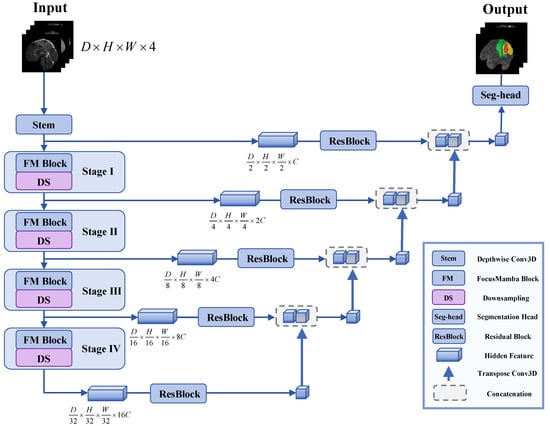
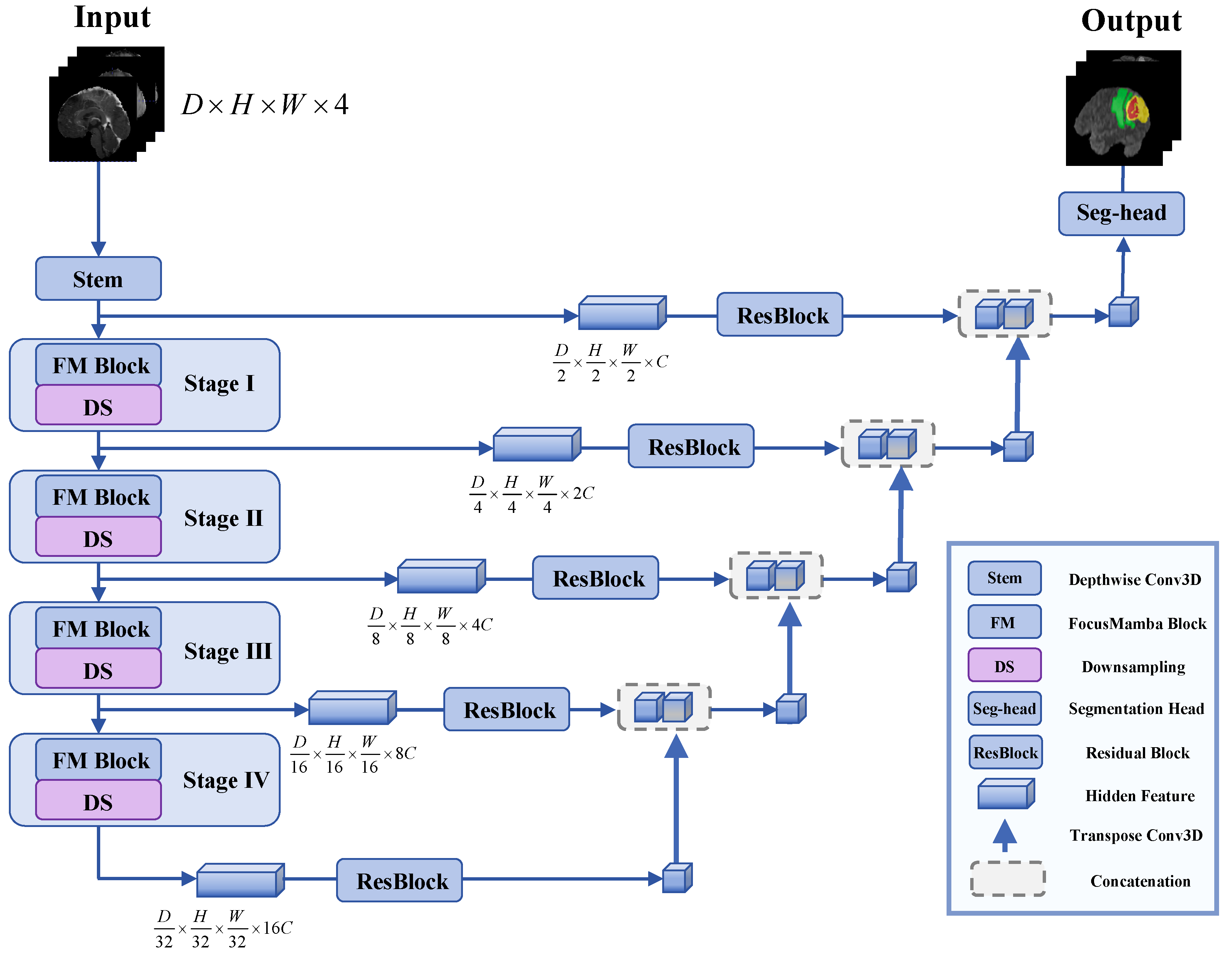
The FocusMamba framework features a U-shaped architecture, as illustrated in Figure 1, which includes an encoder and decoder connected via skip connections. The encoder employs convolutional layers to downsample input data with dimensions , while the decoder reconstructs the data to its original dimensions.
Figure 1.
The overview of the proposed FocusMamba framework. Our approach utilizes a U-shaped encoder–decoder architecture with skip connections to generate hierarchical feature maps.
Given a 3D multimodal input volume , the stem layer extracts the first-scale feature , where denotes the channel dimension of the stem output. The feature is then fed into each FocusMamba block and the corresponding downsampling layers. In the stem layer, we employ a depth-wise convolution with a kernel size of , a padding of , and a stride of . A central component of this model is the FocusMamba Block, which consists of three key units: (1) Local–Global Integration Mamba Unit, (2) Holistic Precision Attention Unit, and (3) Unified Mamba Kernel Fusion Unit.
3.2. FocusMamba Block
We develop a Mamba-based encoding mechanism that ensures that each voxel captures fine-grained local perception while maintaining global context. As shown in Figure 2, the FocusMamba Block is strategically deployed across four stages of the encoder, allowing critical features to be progressively captured and refined at multiple levels of abstraction.
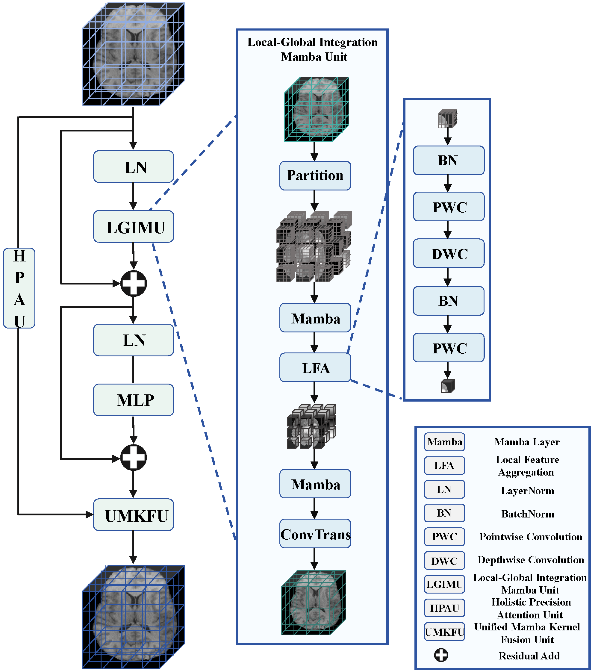
Figure 3.
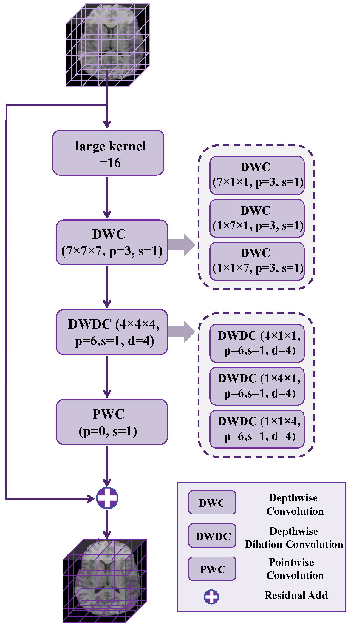
Illustration of the proposed Holistic Precision Attention Unit: This unit enables each voxel to gain fine-grained global perception, facilitating precise and detailed understanding of spatial contexts across the entire input volume.
3.2.1. Local–Global Integration Mamba Unit
The Local–Global Integration Mamba Unit (LGIMU) is designed to jointly capture fine-grained local voxel interactions and coarse-grained global dependencies in volumetric features. Its key idea is to decompose 3D context modeling into two complementary levels: voxel-level modeling within local windows and window-level modeling across the whole feature map. Specifically, LGIMU incorporates two types of 3D Mamba operations: (i) voxel-level Mamba (VoM) and (ii) window-based Mamba (WiM). Within the Mamba layer of each operation, we incorporate the tri-orientated Mamba module proposed in SegMamba [14] to capture comprehensive feature dependencies. This module captures feature relationships from three distinct directions—forward, reverse, and inter-slice—by transforming the 3D input features into sequences, enabling efficient feature interactions and seamless 3D feature fusion, as shown in Equation (1):
where f represents the forward direction, r indicates the reverse direction, and s refers to the inter-slice direction.
Given an input volumetric feature map , VoM partitions X into a regular grid of non-overlapping 3D windows of size along the spatial dimensions. By design, the feature resolutions satisfy that D, H, and W are divisible by m at all stages, enabling a clean division into a regular window grid and simplifying the processing structure. Consequently, the number of windows is . We denote the partition operator as , where each window . In this paper, we set , as determined by the window-size ablation study presented in the subsequent experiments.
Figure 4.
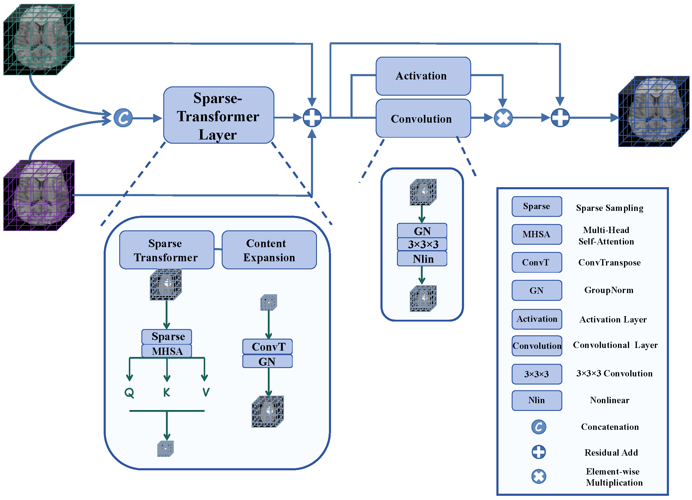
Illustration of the proposed Unified Mamba Kernel Fusion Unit: This unit ensures rich feature representation by integrating local and global outputs, enhancing the ability to capture complex patterns in high-dimensional medical image data.
The partitioned sub-cubes are directly processed by the Mamba layer. During this stage, the contiguous local voxels within each sub-cube are sequentially processed by the state-space model (SSM), enabling voxel-level Mamba (VoM) to capture detailed local interactions within each region. This process enhances the precision of voxel-wise relational modeling and strengthens feature extraction, which is essential for understanding spatial and structural dependencies in brain tumor segmentation. In particular, VoM embodies the focusing mechanism of FocusMamba, allowing each voxel to maintain fine-grained attention to its immediate neighbors for refined local representation.
To allow interaction between non-overlapping sub-windows and accurately capture long-range dependencies, WiM aggregates the information from all voxels within each local window processed by the Mamba layer using a Local Feature Aggregation (LFA) Unit. In each local window, information is collected from neighboring tokens toward the center using the LFA unit. In other words, the updated center voxel gathers information exclusively from its adjacent local voxels. The first pointwise convolution projects and mixes channel information, the depthwise convolution aggregates local spatial information from adjacent voxels toward the center, and the second pointwise convolution further refines the aggregated feature to form a more informative center representation. This process is illustrated by the following Equations (2)–(4):
where BN refers to batch normalization, and DWC and PWC stand for 3D depthwise convolution and 3D pointwise convolution, respectively.
In this way, the information from all voxels within the local window is aggregated into a single voxel, known as the focal voxel. The focal voxel from each local window serves as the overall input to the Mamba layer, contributing to maintaining a coarse-grained awareness of global information. Content Expansion, involving ConvTranspose, restores each local window from the focal voxel to its original size, ensuring that the entire feature map is reconstructed to its initial dimensions while each voxel maintains a coarse-grained awareness of global information. In the FocusMamba method, WiM implements the background mechanism.
Consequently, each voxel integrates fine-grained local details while retaining a broader understanding of the global context. This dual-level perception enables the unit to simultaneously capture intricate local structures and overarching global patterns across the entire feature map.
3.2.2. Holistic Precision Attention Unit
To facilitate global modeling with an extensive receptive field while maintaining parameter efficiency for 3D volumetric data, we adopt a hierarchical two-stage factorization design for ultra-large kernels, as illustrated in Figure 3. This design enhances spatial context modeling across the network without introducing excessive computational complexity. In the first stage, inspired by the large-kernel decomposition design [22], a 3D large-kernel convolution can be represented by a depthwise convolution, a depthwise dilated convolution, and a pointwise convolution. Specifically, a convolution can be decomposed into a depthwise dilated convolution with dilation d, a depthwise convolution, and a pointwise convolution (). In the second stage, to further reduce the computational burden of the intermediate 3D spatial convolutions, we adopt a factorized implementation following prior 3D convolution decomposition methods [24]. Instead of directly using dense 3D spatial kernels, each kernel is decomposed into three sequential one-dimensional convolutions along the depth, height, and width axes. Consequently, a 3D convolution with kernel size is realized as three 1D convolutions, namely , , and .
In our Holistic Precision Attention Unit (HPAU), we employ ultra-large kernel separable attention across stages I, II, III, and IV, with kernel sizes of 64, 32, 16, and 8, respectively. Figure 3 illustrates the schematic of the HPAU with a kernel size of 16. After convolution, each voxel attains an enhanced global receptive perception, enabling a precise and detailed understanding of spatial relationships throughout the entire input volume.
This component integrates the Scanning Mechanism within the FocusMamba framework, thereby complementing the local–global modeling process. Together with the Local–Global Integration Mamba Unit, which captures fine-grained local representation while preserving a broader global awareness, the HPAU forms a three-tiered information extraction hierarchy.
3.2.3. Unified Mamba Kernel Fusion Unit
In line with the dual-path design, the outputs from both the Local–Global Integration Mamba Unit and the Holistic Precision Attention Unit are integrated within the Unified Mamba Kernel Fusion Unit (fully illustrated in Figure 4). We concatenate the outputs from these two units to aggregate multi-level representations. Self-attention mechanisms, inherently central to Transformers, have proven highly effective in modeling long-range dependencies and capturing global context, making them ideal for fusing the distinct representations from the LGIMU and HPAU into a unified feature space. However, directly applying a standard multi-head self-attention mechanism to the concatenated outputs of these two feature-extraction units would be computationally prohibitive for high-resolution 3D volumetric data. Standard self-attention suffers from quadratic computational complexity with respect to the sequence length N, severely exacerbating memory constraints. To leverage the powerful feature-fusion capabilities of Transformers while mitigating this computational bottleneck, our module introduces a spatially reduced Sparse-Transformer layer. The mathematical operations are defined in Equations (5)–(8).
where r denotes the kernel size and stride of the 3D average pooling operator, M denotes the number of heads in the Sparse-Transformer, and indexes the attention head, with being the output of the m-th head.
Specifically, we apply a 3D average pooling operation using a spatial reduction ratio r as the stride on the concatenated input feature map . This step spatially aggregates local features to yield a compact representation , reducing the token sequence length by a factor of . To accommodate our four-stage multi-scale architecture, the reduction ratio r is stage-dependent, set to and 1 across the respective stages. This configuration effectively manages the massive spatial dimensions in early stages while preserving the spatial integrity of abstracted features in the final stage. Following the spatial reduction, the compact representation Z is linearly projected into query, key, and value matrices to perform multi-head self-attention. By computing attention weights on this downsampled grid rather than the original input, the attention complexity is reduced from to . The outputs from all attention heads are then concatenated and linearly projected into a fused representation. In this way, the spatially reduced Sparse-Transformer performs feature fusion between LGIMU and HPAU while reducing the computational and memory burden of self-attention on high-resolution 3D volumetric data. This enables more effective integration of the fine-grained local information extracted by LGIMU and the global contextual information captured by HPAU, thereby improving segmentation performance.
The Sparse-Transformer’s output undergoes Content Expansion via ConvTranspose to restore its original dimensions, which is then fused with the concatenated dual-path features X via a residual addition.
The Gated Convolution Layer processes 3D input features through two convolutional blocks, each including a normalization layer, a convolutional layer, and a nonlinear activation function. Element-wise multiplication of these block outputs acts as a gating mechanism to regulate information flow. The addition of these resulting features to the original input via a residual connection enhances the module’s ability to capture critical information, preserving the original input while incorporating the newly transformed features.
By merging the representations from the LGIMU and HPAU, the UMKFU functions as the Aggregation Mechanism for the framework. Through the integration of attention, residual, and gating operations, it empowers the module to preserve critical information while maintaining computational efficiency during complex data processing.
4. Experiment
In this section, we carry out a series of experiments to assess the performance of the proposed FocusMamba framework on two 3D medical imaging datasets. The achieved results are compared with other state-of-the-art methods in the field, highlighting the framework’s outstanding precision.
4.1. Datasets
Over the past decade, the Brain Tumor Segmentation Benchmark Challenge (BraTS) has offered several benchmark datasets to enable fair comparison of state-of-the-art segmentation methods. The BraTS datasets consist of multiparametric MRI scans collected from various clinical institutions, which have been meticulously reviewed by expert neuroradiologists and come with detailed pixel-level segmentation labels. BraTS 2020 [25,26,27] datasets and BraTS 2023 [28,29] datasets contain a total of 369 and 1251 3D brain MRI volumes, respectively. Each volume includes four modalities, namely T1, T1Gd, T2, and T2-FLAIR, and has already been resampled and co-registered with the same dimensions (i.e., 240 × 240 × 155). The segmentation task aims to segment different sub-regions, including the enhancing tumor (ET), tumor core (TC), and the whole tumor (WT). To evaluate segmentation performance, we adopted two standard metrics: the Dice coefficient and the 95% Hausdorff Distance (HD95).
4.2. Implementation Details
Our model is implemented in PyTorch 2.3.1-Cuda12.1 and Monai 1.3.2. During training, we apply a random crop of size 128 × 128 × 128 and use a batch size of 2 per GPU for each dataset. For all experiments, we utilize cross-entropy loss and employ an SGD optimizer with a polynomial learning rate scheduler (initial learning rate of 1 × 10⁻² and a decay of 1 × 10⁻⁵). Each dataset is trained for 1000 epochs, with the following data augmentations applied: additive brightness, gamma, rotation, scaling, mirror, and elastic deformation. For both the BraTS 2023 and BraTS 2020 datasets, we randomly split the 3D volumes into training, validation, and testing sets in an 8:1:1 ratio.All experiments are performed on a cloud computing platform using two NVIDIA GeForce RTX 3090 GPUs made by the NVIDIA Corporation, Santa Clara, CA, USA. The implementation of FocusMamba is publicly available at https://github.com/KWEO/Code-for-FocusMamba (accessed on 30 March 2026).
4.3. Results and Discussion
To validate the effectiveness of the proposed FocusMamba model, we compare it with several state-of-the-art segmentation methods on the BraTS 2023 and BraTS 2020 datasets. For each baseline model, we used the official implementation whenever available. Otherwise, we reproduced the model according to the details described in the corresponding paper. All baseline methods, together with our proposed model, were trained on the same hardware platform and evaluated using the same dataset split. To ensure a fair training protocol, all methods shared the same data preprocessing pipeline, the same spatial crop size of 128 × 128 × 128, and the same data augmentation strategy. In addition, rather than directly adopting the default settings, we made comparable efforts to tune the key hyperparameters of each baseline on the validation set, such as the learning rate, to ensure a fair and comparable evaluation. The best-performing model for each method was selected based on validation performance, and the final quantitative and qualitative results were reported on the test set. Moreover, all reported results are direct model outputs without any post-processing.
4.3.1. Quantitative Results
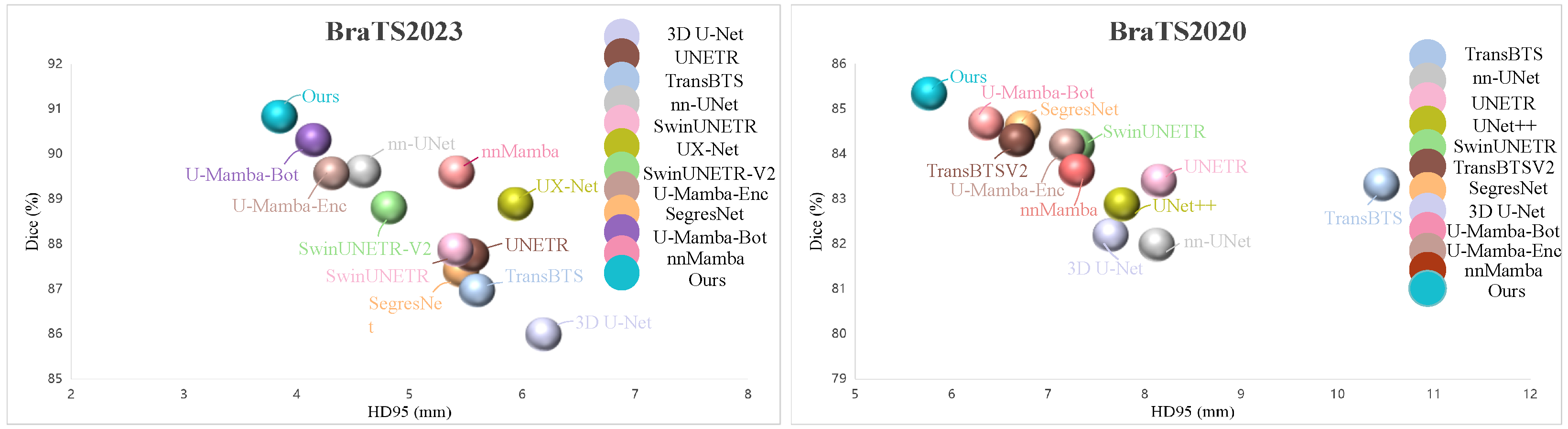
Table 1 and Figure 5 demonstrate a quantitative comparison with state-of-the-art methods, including four CNN-based models (SegresNet [5], 3D U-Net [15], UX-Net [30], and nn-UNet [6]), four transformer-based models (TransBTS [9], UNETR [7], SwinUNETR [31], and SwinUNETR-V2 [32]), and three Mamba-based models (U-Mamba-Enc [13], U-Mamba-Bot [13], and nnMamba [18]). The upper section features CNN-based models (marked by †), the middle section displays transformer-based models (marked by ‡), and the lower section shows Mamba-based models (marked by §). The top three results for each score are highlighted using color coding: the first is represented in red, the second in blue, and the third in green. The evaluation results are reported as mean ± standard deviation. For WT, TC, and ET, the standard deviations were computed across all test cases. For the overall Mean, the score for each test case was first calculated as the average of the WT, TC, and ET scores, and then the standard deviation was computed across all test cases. Statistical significance was assessed using a paired t-test across all test cases, where an asterisk (*) indicates a statistically significant difference () between our model and the compared method.
Table 1.
Quantitative comparison with SOTA methods on BraTS 2023 dataset (†: CNN-based, ‡: Transformer-based, §: Mamba-based). Top three results are shown in red, blue, and green. Results are presented as mean ± standard deviation. * indicates a statistically significant difference compared with our method (, paired t-test).
Figure 5.
Scatter plots demonstrating the performance of different brain tumor segmentation models on the BraTS 2023 and BraTS 2020 benchmarks.
By comparing the average Dice scores, our proposed FocusMamba model achieved the best performance, with Dice scores of 94.03%, 91.32%, and 87.10% for WT, TC, and ET, respectively. In terms of Hausdorff distances (95%), we achieved distances of 3.74 mm, 3.71 mm, and 4.11 mm for WT, TC, and ET, respectively, ranking first among the evaluated models and outperforming the U-Mamba-Bot model by 0.3 mm. Among the CNN-based methods, nnU-Net achieves strong and stable performance, demonstrating balanced Dice scores across tumor subregions. For Transformer-based methods, SwinUNETR-V2 delivers the best results within its category. Within the Mamba-based group, U-Mamba-Bot achieves the overall best Dice and HD95, reflecting the strength of combining Mamba with a U-Net–style encoder–decoder architecture. However, our proposed FocusMamba consistently achieves superior mean Dice and competitive HD95 compared with these baselines, confirming its effectiveness in balancing fine-grained local detail modeling with global context integration. This improvement can be attributed to its structured design, which captures fine-grained voxel interactions for spatial and structural characterization of tumor regions, preserves long-range contextual dependencies within whole-volume features, and further integrates multi-level representations through the fusion unit. In addition to achieving the best mean Dice scores, our proposed FocusMamba also exhibited the lowest standard deviations for all Dice metrics and for TC, ET, and Mean in HD95. This demonstrates that our method is not only highly accurate but also exceptionally robust and stable across diverse and challenging patient cases. Overall, both Dice scores and Hausdorff distances demonstrate the advantage of our approach.
Similarly, Table 2 and Figure 5 demonstrate a quantitative comparison with state-of-the-art methods, including four CNN-based models (SegresNet [5], 3D U-Net [15], UNet++ [33], and nn-UNet [6]), four transformer-based models (TransBTS [9], TransBTSV2 [34], UNETR [7], and SwinUNETR [31]), and three Mamba-based models (U-Mamba-Enc [13], U-Mamba-Bot [13], and nnMamba [18]). Specifically, the average Dice scores and Hausdorff distances (95%) of the proposed FocusMamba model for ET, TC, and WT all surpass those of the competitors. Our FocusMamba model achieves Dice scores of 90.14%, 85.62%, and 80.33%, along with Hausdorff distances (95%) of 5.28 mm, 5.92 mm, and 6.11 mm for WT, TC, and ET, respectively. These boundary results indicate that the model is capable of maintaining accurate contour predictions even in the challenging enhancing tumor region. Overall, the performance highlights the robustness of FocusMamba in handling heterogeneous tumor structures and complex spatial variations, confirming its reliability for clinical brain tumor segmentation tasks.
Table 2.
Quantitative comparison with SOTA methods on BraTS 2020 dataset (†: CNN-based, ‡: Transformer-based, §: Mamba-based). Top three results are shown in red, blue, and green. Results are presented as mean ± standard deviation. * indicates a statistically significant difference compared with our method (, paired t-test).
As shown in Table 3, we further analyze the architectural complexity and computational efficiency of all baseline models. Compared with CNN-based models, FocusMamba has a larger number of parameters but clear advantages in GFLOPs, while maintaining strong segmentation performance. Compared with Transformer-based approaches, FocusMamba shows advantages in both parameter count and computational cost, together with high segmentation accuracy. Among Mamba-based methods, U-Mamba-Bot, U-Mamba-Enc, and nnMamba all contain fewer parameters than FocusMamba; however, U-Mamba-Bot and U-Mamba-Enc incur higher computational cost, while nnMamba achieves inferior segmentation performance. Overall, these results indicate that FocusMamba provides a favorable balance between segmentation accuracy and computational efficiency. The relatively low GFLOPs of FocusMamba mainly stem from its efficient design, where window-based Mamba restricts sequence modeling to compact local regions, the separable large-kernel unit enlarges the receptive field with controlled cost, and the pooled sparse Transformer performs feature interaction on compressed representations, thereby reducing the computational burden of global modeling. Nevertheless, we also note that the parameter count of FocusMamba remains relatively large, and improving parameter efficiency will be an important direction of our future work. Given the specific architectural adaptations of FocusMamba, several targeted reduction strategies present viable future directions. First, structured pruning could be applied to the heavier components. For the large-kernel convolutional modules, we can evaluate channel importance and systematically remove redundant feature channels. Similarly, less informative attention heads can be pruned based on their contribution to the final output. Second, quantization offers a practical approach to accelerate inference and reduce memory footprint. We plan to explore quantization-aware training to convert full-precision weights and activations into lower bit-width representations such as INT8. This conversion is particularly beneficial for accelerating the extensive computational operations in our large kernels. Regarding the quantization of Mamba units, we will draw inspiration from recently emerging studies. Finally, exploring lightweight architectural redesigns through more efficient information aggregation methods will further streamline the overall framework. These strategic directions will help improve parameter efficiency while maintaining the robust segmentation performance of FocusMamba.
Table 3.
Parameter count and computational complexity of the compared models.
4.3.2. Qualitative Results
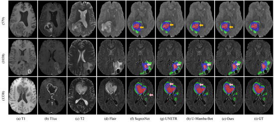
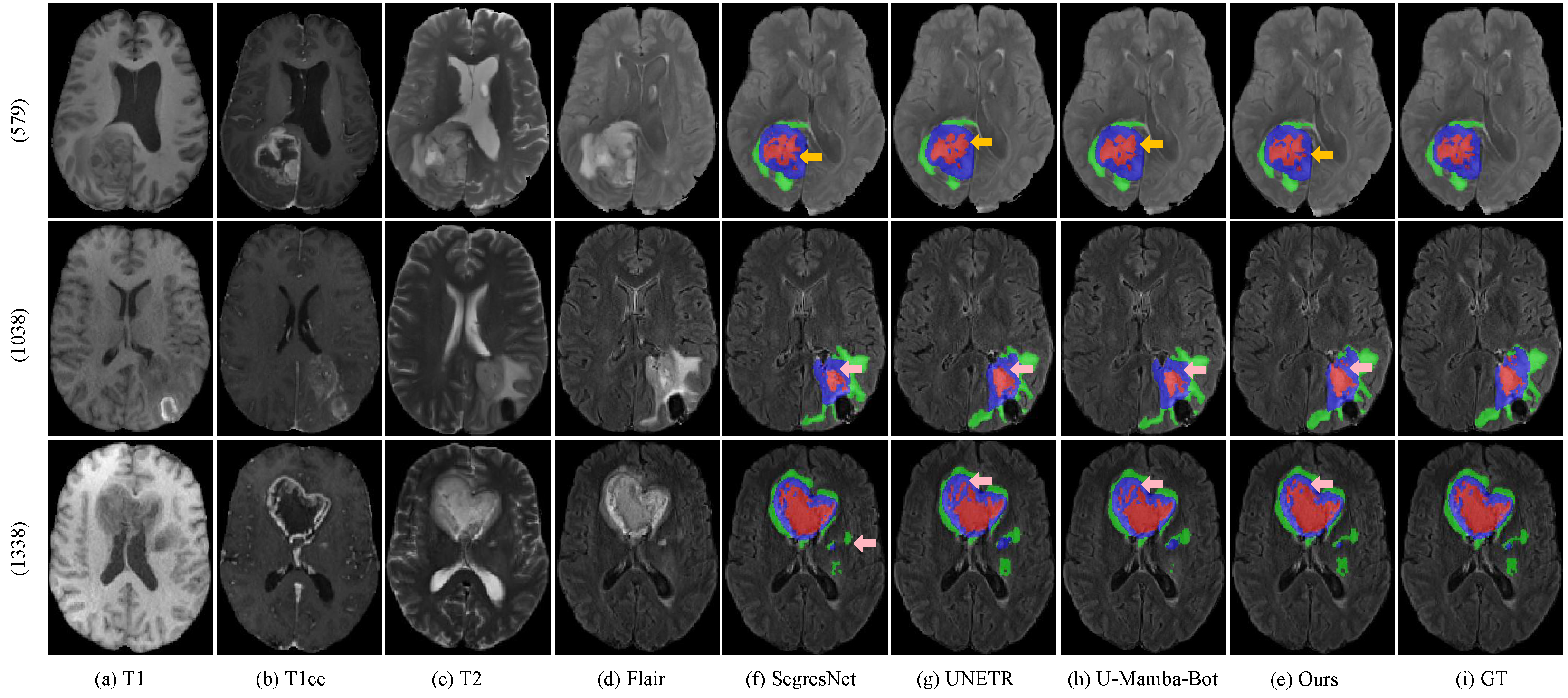
Figure 6 demonstrates the qualitative results compared with previous representative methods (including SegresNet, UNETR and U-Mamba-Bot), where rows represent three different cases from the BraTS 2023 dataset, and columns show the segmentation results from various methods. By comparing with the ground truth (GT), our proposed method delivers more precise segmentation outcomes than other approaches, particularly in cases where subtle abnormal regions are missed by alternative segmentation models. Our model effectively reduces false positives results and produces more accurate and complete boundaries, highlighting the importance of semantic context-aware feature extraction in segmentation tasks. Our model demonstrates improved segmentation accuracy in tumor areas with diverse shapes and sizes, owing to its proposed components that effectively capture shape-aware and scale-aware features of tumors.
Figure 6.
Visualization of qualitative comparison of SOTA methods on the BraTS 2023 dataset. BraTS_579, BraTS_1038, and BraTS_1338 are arranged from top to bottom. The red mask represents the necrotic tumor core (NCR), the green mask denotes peritumoral edema (ED), and the blue mask indicates the enhancing tumor (ET). Yellow arrows highlight the false positive results, which are fewer in ours compared to other models. Pink arrows emphasize the superior regions in ours when compared to other models.
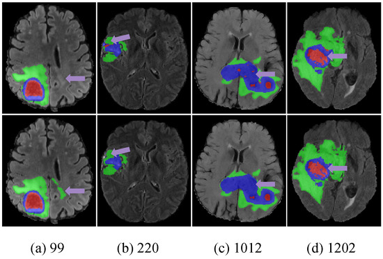
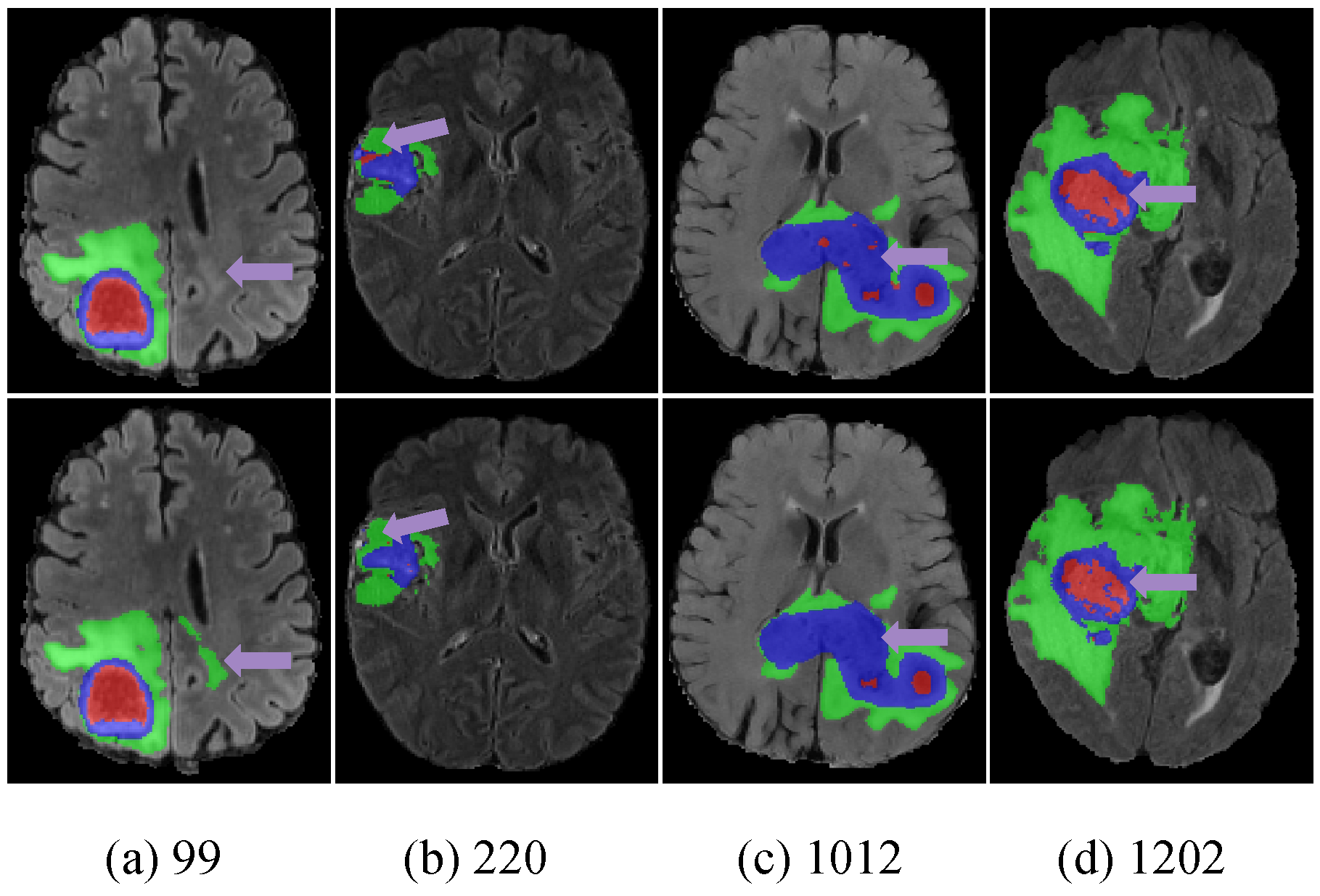
Although our model demonstrates robust performance overall, there are still several challenging cases in which the segmentation results remain suboptimal, particularly in subtle edge regions. To further analyze these limitations, we provide representative visual examples in Figure 7. The first case shows a false negative result, where an isolated abnormal region is missed by the network. The second and third cases present false positive results, in which the prediction extends into adjacent non-tumorous tissues. The final case exhibits both false positive and false negative errors, mainly associated with imprecise delineation along complex tumor boundaries. These failure cases may be related to two factors. First, the low contrast and ambiguous intensity transitions in magnetic resonance images make boundary identification inherently difficult. Second, these failure cases may also be related to limitations in feature extraction and feature fusion. Although our model is capable of capturing fine-grained local details and global contextual relationships, some subtle boundary cues may still be weakened during feature extraction. In addition, the fusion of local and global features may remain insufficient in some challenging cases, resulting in insufficient attention to these subtle regions. In future work, we will explore boundary-aware enhancement strategies, such as edge-guided refinement or boundary-aware supervision inspired by gradient-based operators like Sobel or Laplacian. Concurrently, the adoption of a more sophisticated feature fusion strategy will be investigated to effectively consolidate these features and enhance the segmentation accuracy for these highly challenging boundaries.
Figure 7.
Visualization of representative failure cases in the segmentation results of the proposed FocusMamba model. The top row presents the segmentation results of our model, and the bottom row shows the corresponding ground truth. The purple arrows highlight challenging regions with imprecise boundary delineation, including false positive and false negative predictions.
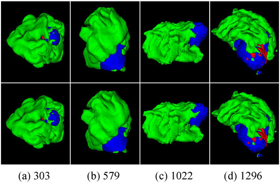
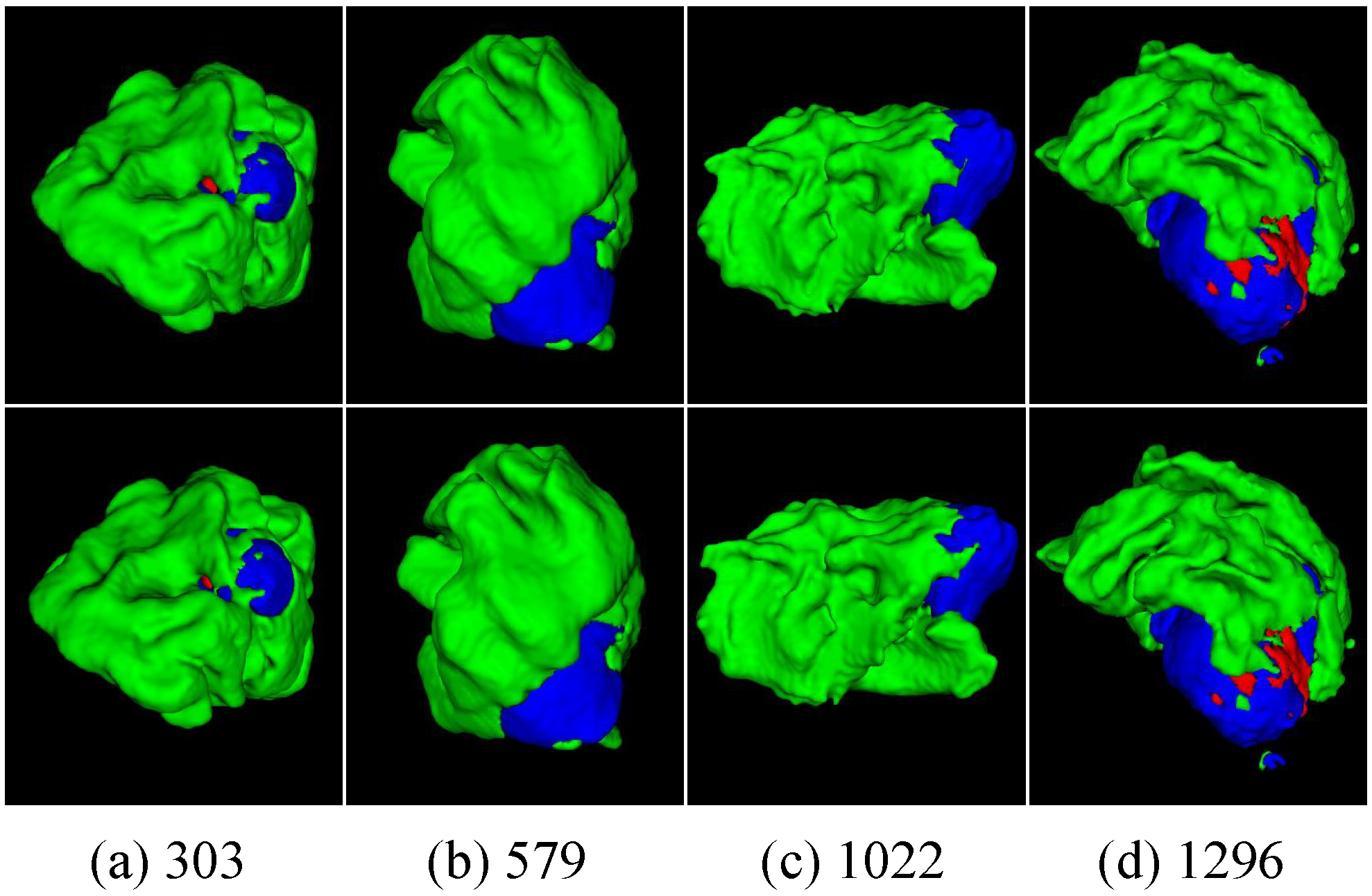
We also showcase the 3D volumetric segmentation results in comparison with the ground truth. As shown in Figure 8, our model generates 3D segmentation outputs that closely align with the ground truth, highlighting its accuracy and effectiveness. Importantly, our model is more closely aligned with ground truth in each tumor region, benefiting from the semantic context-aware feature extraction driven by both shape and scale-sensitive information.
Figure 8.
3D visualization of our tumor segmentation results. The first row displays our segmentation outcomes, while the second row shows the ground truth. The numbers correspond to the sample indices.
4.4. Ablation Study Results
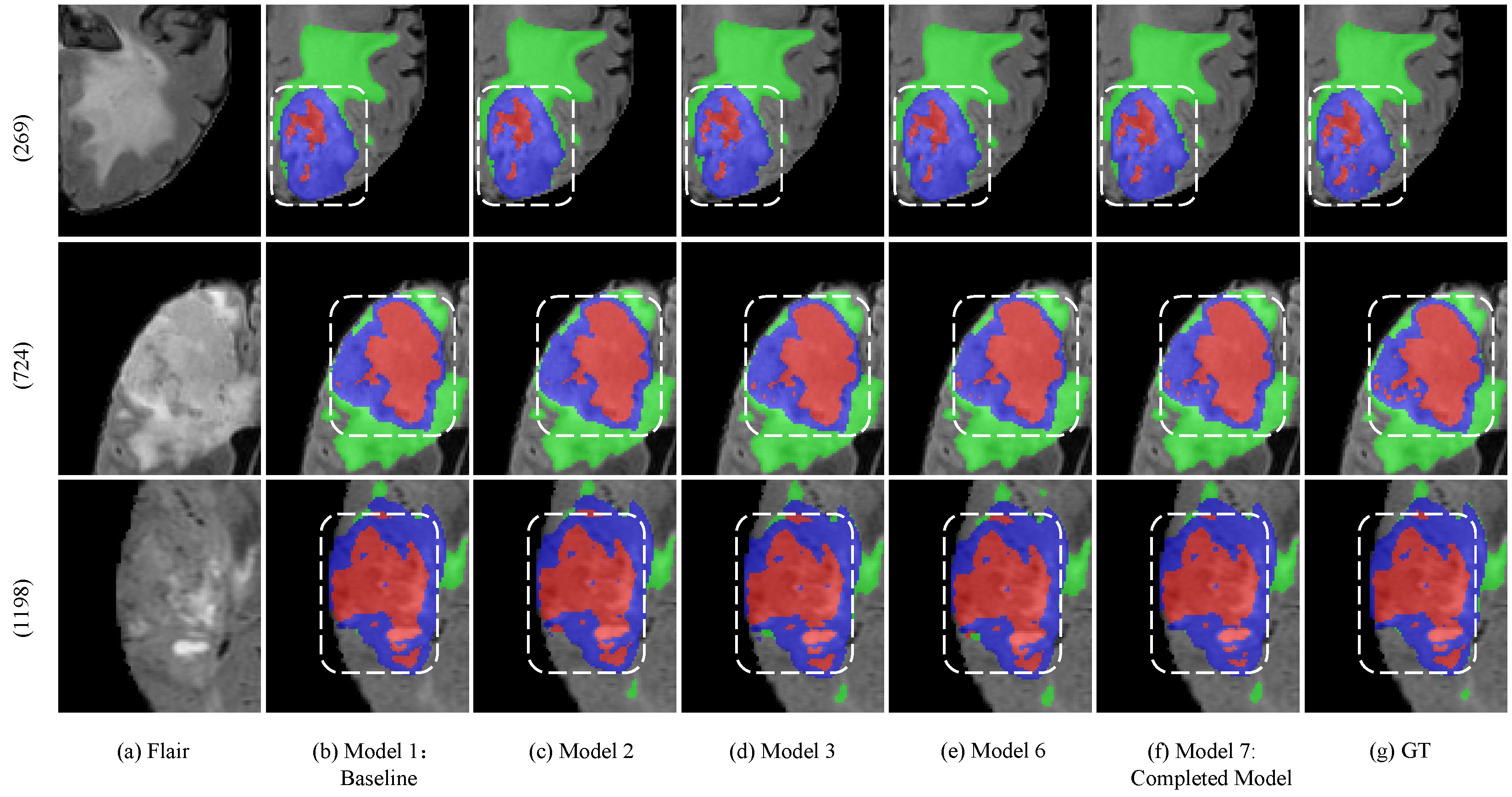
We also conducted several ablation studies on the BraTS 2023 dataset to evaluate the effectiveness of each technical component in our proposed model, including the Local–Global Integration Mamba Unit (LGIMU), the Holistic Precision Attention Unit (HPAU), and the Unified Mamba Kernel Fusion Unit (UMKFU). Table 4 illustrates the quantitative results, while Figure 9 provides a visual comparison of the segmentation results from different models in the ablation study. For the ablation study, we adopted a modified 3D U-Net as the baseline, since it is a widely recognized and streamlined architecture that provides a stable foundation for comparison. As LGIMU, HPAU, and UMKFU are developed under the unified design philosophy of the FocusMamba framework, this simple baseline is more suitable for evaluating their respective contributions and overall effectiveness. We then introduced LGIMU, HPAU, and UMKFU progressively to assess the role of each component.
Table 4.
Ablation study on LGIMU (VoM and WiM), HPAU, and UMKFU. Dice (%) and HD95 (mm) are reported on WT, TC, and ET regions. The columns denote the performance differences relative to the baseline model (1), where red and green values indicate improvements in Dice and reductions in HD95, respectively.
Figure 9.
Visual comparison of the segmentation results from different models in the ablation study. The red mask represents the necrotic tumor core (NCR), the green mask denotes peritumoral edema (ED), and the blue mask indicates the enhancing tumor (ET). White dotted boxes highlight brain tumor regions with large segmentation differences.
4.4.1. Effectiveness of LGIMU
To address the original Mamba’s limitation in 1D sequence modeling, we introduce the LGIMU, which consists of two parts: (i) voxel-level Mamba (VoM) and (ii) window-based Mamba (WiM). A careful examination reveals that each component significantly enhances segmentation outcomes. We evaluate the impact of the LGIMU by comparing the baseline model with its LGIMU-enhanced counterpart. As shown in Table 4, Compared to Model (1), Model (2) with VoM demonstrates a significant improvement in both Dice score and HD95 distance. In Model (3), the inclusion of LGIMU (comprising VoM and WiM) leads to significant improvements in Dice scores for ET, TC, and WT on the BraTS 2023 dataset, with increases of 3.11%, 3.99%, and 4.43%, respectively. This improvement highlights the effectiveness of LGIMU in enabling global-local interactions and semantic context extraction. Furthermore, Model (5) solely integrates VoM with HPAU, directly combining precise local modeling with precise global modeling. In this configuration, VoM acts as a focusing mechanism to extract fine-grained details within isolated windows, while HPAU serves as a scanning mechanism to capture precise global context. Although this dual-scale approach already achieves strong performance, Model (6) further introduces WiM, outperforming Model (5) by 0.95% in mean Dice. This significant boost highlights the highly effective synergy specifically between VoM and WiM. By acting as a background mechanism, WiM enables vital interaction between windows to provide a coarse global perception, seamlessly bridging the localized details with the broader global scan. Ultimately, this performance differential underscores the efficacy of the LGIMU architecture, wherein the simultaneous inclusion of VoM and WiM yields an optimized design framework for enhanced segmentation accuracy.
4.4.2. Effectiveness of HPAU
We introduce HPAU, which is centered around large kernel convolutions, to complement and address the limitations of LGIMU in global modeling. The quantitative results, presented in Table 4, show that HPAU (Model (4)) consistently enhances segmentation performance, achieving higher Dice scores in comparison to the baseline model. Notable improvements are observed in both the Dice score (Model (4): 0.8818, Model (5): 0.8938) and HD95 distance (Model (4): 5.10, Model (5): 4.70) when combining VoM and HPAU (Model (5)). It is essential to note that the WiM module is not standalone and must be integrated with the VoM module; therefore, WiM cannot be associated independently with HPAU. When associating LGIMU (including VoM and WiM) with HPAU in Model (6), both the Dice score and HD95 distance are further enhanced, establishing it as the final selected configuration. The results demonstrate that the lack of an explicit global modeling strategy can cause inaccuracies in contextual understanding, particularly by reducing attention to tumor boundaries.Incorporating large-kernel convolutions effectively mitigates this issue by expanding the receptive field and enhancing the model’s capacity to capture long-range spatial dependencies. Consequently, this strategy leads to more accurate boundary delineation and improves the overall robustness of the segmentation outcomes.
4.4.3. Effectiveness of UMKFU
We propose a fusion unit called Unified Mamba Kernel Fusion Unit (UMKFU), strategically incorporated to enhance the integration of outputs from two distinct units, facilitating a more effective combination of local and global information extraction. As illustrated in Table 4, integrating the UMKFU results in substantial improvements in segmentation performance. Specifically, on the BraTS 2023 dataset, Dice scores improve across all subregions—WT by 0.12%, TC by 0.80%, and ET by 0.54%—with HD reduced by 0.32 mm. This indicates that the fusion unit can more accurately focus on the regions to be segmented while filtering out irrelevant semantic information. Figure 9 demonstrates that, with the integration of UMKFU, the model achieves more robust predictions, particularly for necrotic tumor core, a challenging region to delineate due to its complexity.
4.4.4. Determining the Optimal Window Size in LGIMU
Using various window sizes within the LGIMU can potentially result in more heterogeneous outcomes. To evaluate the impact of window sizes on the performance of LGIMU, we systematically assess window sizes of 2 × 2 × 2, 4 × 4 × 4, and 8 × 8 × 8 within the VoM and WiM modules, followed by validation on the BraTS 2023 dataset. As shown in Table 5, we report the mean performance alongside the difference from our default baseline, as well as the associated computational costs. The results demonstrate that the window size yields the optimal segmentation performance. Beyond segmentation metrics, varying the window size directly impacts computational efficiency. Although the configuration requires the least resources, it results in a decrease in segmentation accuracy. The configuration offers the optimal balance, achieving the highest accuracy with moderate resource requirements. Conversely, further expanding the window to increases memory consumption to 4.95 GB and latency to 3.50 s without yielding additional performance gains. Our observations reveal that scaling up the window size does not uniformly improve segmentation performance. This phenomenon can be attributed to the intrinsic trade-off between the window scale and the Mamba module’s capacity for local interaction modeling. Although larger windows provide a broader receptive field and enhance global awareness, they inevitably weaken the model’s ability to capture fine-grained, voxel-level dependencies. The expanded scope disperses the model’s focus over a wider region, thereby diminishing sensitivity to subtle, localized features that are critical for precise boundary delineation. Consequently, an intermediate window size of provides the most effective balance, ensuring both comprehensive local voxel interactions and manageable computational overhead.
Table 5.
Performance and computational cost on BraTS 2023 with varying window sizes in LGIMU. The columns denote the performance differences relative to the baseline model (row (2)). Peak Memory represents the maximum GPU memory usage during inference. Inference Time denotes the inference time per 3D MRI volume.
5. Conclusions
In this paper, we propose FocusMamba, a novel Mamba-based framework for brain tumor segmentation. Inspired by human visual observation patterns, the proposed framework abstracts four functional concepts, namely Focusing, Background, Scanning, and Aggregation, to enhance both local detail modeling and global contextual understanding in 3D medical images. Specifically, the proposed FocusMamba Block integrates a local–global Mamba unit, which employs voxel-level Mamba for refined local feature extraction and window-level interactions to maintain broader global awareness, a large-kernel convolution unit for capturing long-range dependencies, and a fusion unit that enhances the overall feature representation by integrating information from different levels. Experimental results on the public BraTS 2023 and BraTS 2020 datasets demonstrate that FocusMamba achieves superior segmentation performance compared with several state-of-the-art methods. These results validate the effectiveness of the proposed framework and highlight the potential of Mamba-based architectures for 3D medical image analysis. Nevertheless, FocusMamba still contains a relatively large number of parameters, which may limit its applicability in resource-constrained scenarios. In future work, we will explore a lighter version of FocusMamba to improve parameter efficiency while preserving segmentation accuracy.
Author Contributions
Conceptualization, Q.L.; methodology, T.N.; software, T.N.; validation, T.N.; formal analysis, Q.L.; investigation, Q.L.; resources, Q.L.; data curation, T.N.; writing—original draft preparation, T.N.; writing—review and editing, X.W. and H.L.; visualization, T.N.; supervision, H.L.; project administration, X.W. and H.L.; funding acquisition, Q.L. All authors have read and agreed to the published version of the manuscript.
Funding
This work was supported by the National Natural Science Foundation of China (62071323), Tianjin Natural Science Foundation (22JCZDJC00220), and the Foundation of State Key Laboratory of Ultrasound in Medicine and Engineering (2022KFKT004).
Institutional Review Board Statement
Not applicable.
Informed Consent Statement
Not applicable.
Data Availability Statement
The datasets are provided by the BraTS 2023 and BraTS 2020 Challenges and are made available for personal academic research. The specific link to the datasets is https://www.med.upenn.edu/cbica/brats/ (accessed on 25 March 2026).
Conflicts of Interest
The authors declare no conflicts of interest.
References
- Siegel, R.L.; Miller, K.D.; Wagle, N.S.; Jemal, A. Cancer statistics, 2023. CA Cancer J. Clin. 2023, 73, 17–48. [Google Scholar] [CrossRef] [Scilit] [PubMed]
- LeCun, Y.; Bengio, Y. Convolutional networks for images, speech, and time series. In The Handbook of Brain Theory and Neural Networks; MIT Press: Cambridge, MA, USA, 1998; pp. 255–258. [Google Scholar]
- Vaswani, A.; Shazeer, N.; Parmar, N.; Uszkoreit, J.; Jones, L.; Gomez, A.N.; Kaiser, Ł.; Polosukhin, I. Attention is all you need. Adv. Neural Inf. Process. Syst. 2017, 30, 5998–6008. Available online: https://proceedings.neurips.cc/paper/2017/file/3f5ee243547dee91fbd053c1c4a845aa-Paper.pdf (accessed on 20 March 2026).
- Ronneberger, O.; Fischer, P.; Brox, T. U-Net: Convolutional networks for biomedical image segmentation. In Proceedings of the Medical Image Computing and Computer-Assisted Intervention (MICCAI), Munich, Germany, 5–9 October 2015; pp. 234–241. [Google Scholar] [CrossRef] [Scilit]
- Myronenko, A. 3D MRI brain tumor segmentation using autoencoder regularization. In Proceedings of the International MICCAI Brainlesion Workshop, Granada, Spain, 16 September 2018; pp. 311–320. [Google Scholar] [CrossRef] [Scilit]
- Isensee, F.; Jäger, P.F.; Full, P.M.; Vollmuth, P.; Maier-Hein, K.H. nnU-net for brain tumor segmentation. In Proceedings of the International MICCAI Brainlesion Workshop, Lima, Peru, 4–8 October 2020; pp. 118–132. [Google Scholar] [CrossRef] [Scilit]
- Hatamizadeh, A.; Tang, Y.; Nath, V.; Yang, D.; Myronenko, A.; Landman, B.; Roth, H.; Xu, D. UNETR: Transformers for 3D medical image segmentation. arXiv 2021, arXiv:2103.10504. [Google Scholar] [CrossRef] [Scilit]
- Shaker, A.M.; Maaz, M.; Rasheed, H.; Khan, S.; Yang, M.-H.; Khan, F.S. UNETR++: Delving into efficient and accurate 3D medical image segmentation. IEEE Trans. Med. Imaging 2024, 43, 3377–3390. [Google Scholar] [CrossRef] [Scilit] [PubMed]
- Wang, W.; Chen, C.; Ding, M.; Li, J.; Yu, H.; Zha, S. TransBTS: Multi-modal brain tumor segmentation using transformer. In Proceedings of the Medical Image Computing and Computer-Assisted Intervention (MICCAI), Strasbourg, France, 27 September–1 October 2021; pp. 109–119. [Google Scholar] [CrossRef] [Scilit]
- Kalman, R.E. A new approach to linear filtering and prediction problems. J. Basic Eng. 1960, 82, 35–45. [Google Scholar] [CrossRef] [Scilit]
- Gu, A.; Dao, T. Mamba: Linear-time sequence modeling with selective state spaces. arXiv 2023, arXiv:2312.00752. [Google Scholar] [CrossRef] [Scilit]
- Zhu, L.; Liao, B.; Zhang, Q.; Wang, X.; Liu, W.; Wang, X. Vision mamba: Efficient visual representation learning with bidirectional state space model. In Proceedings of the 41st International Conference on Machine Learning (ICML), Vienna, Austria, 21–27 July 2024; Volume 235, pp. 62429–62442. [Google Scholar]
- Ma, J.; Li, F.; Wang, B. U-mamba: Enhancing long-range dependency for biomedical image segmentation. arXiv 2024, arXiv:2401.04722. [Google Scholar] [CrossRef] [Scilit]
- Xing, Z.; Ye, T.; Yang, Y.; Liu, G.; Zhu, L. SegMamba: Long-range sequential modeling mamba for 3D medical image segmentation. In Proceedings of the Medical Image Computing and Computer-Assisted Intervention (MICCAI) 2024, Marrakesh, Morocco, 6–10 October 2024; pp. 578–588. [Google Scholar] [CrossRef] [Scilit]
- Çiçek, Ö.; Abdulkadir, A.; Lienkamp, S.S.; Brox, T.; Ronneberger, O. 3D U-Net: Learning dense volumetric segmentation from sparse annotation. In Proceedings of the Medical Image Computing and Computer-Assisted Intervention (MICCAI), Athens, Greece, 17–21 October 2016; pp. 424–432. [Google Scholar] [CrossRef] [Scilit]
- Mehta, R.; Arbel, T. 3D U-Net for brain tumour segmentation. In Proceedings of the International MICCAI Brainlesion Workshop, Granada, Spain, 16 September 2018; pp. 254–266. [Google Scholar] [CrossRef] [Scilit]
- Pang, Y.; Liang, J.; Huang, T.; Chen, H.; Li, Y.; Li, D.; Huang, L.; Wang, Q. Slim UNETR: Scale hybrid transformers to efficient 3D medical image segmentation under limited computational resources. IEEE Trans. Med. Imaging 2024, 43, 994–1005. [Google Scholar] [CrossRef] [Scilit] [PubMed]
- Gong, H.; Kang, L.; Wang, Y.; Wang, Y.; Wan, X.; Wu, X.; Li, H. nnMamba: 3D biomedical image segmentation, classification and landmark detection with state space model. In Proceedings of the 2025 IEEE 22nd International Symposium on Biomedical Imaging (ISBI), Houston, TX, USA, 14–17 April 2025; pp. 1–5. [Google Scholar] [CrossRef] [Scilit]
- Wang, J.; Chen, J.; Chen, D.; Wu, J. LKM-UNet: Large Kernel Vision Mamba UNet for medical image segmentation. In Proceedings of the Medical Image Computing and Computer-Assisted Intervention (MICCAI) 2024, Marrakesh, Morocco, 6–10 October 2024; pp. 360–370. [Google Scholar] [CrossRef] [Scilit]
- Kui, X.; Jiang, S.; Li, Q.; Peng, Y.; Hu, Z.; Zou, B. GL-MambaNet: A global-local hybrid Mamba network for medical image segmentation. Neurocomputing 2025, 626, 129580. [Google Scholar] [CrossRef] [Scilit]
- Guo, M.-H.; Lu, C.-Z.; Liu, Z.-N.; Cheng, M.-M.; Hu, S.-M. Visual attention network. Comput. Vis. Media 2023, 9, 733–752. [Google Scholar] [CrossRef] [Scilit]
- Li, H.; Nan, Y.; Del Ser, J.; Yang, G. Large-kernel attention for 3D medical image segmentation. Cogn. Comput. 2024, 16, 2063–2077. [Google Scholar] [CrossRef] [Scilit] [PubMed]
- Azad, R.; Niggemeier, L.; Hüttemann, M.; Kazerouni, A.; Aghdam, E.K.; Velichko, Y.; Bagci, U.; Merhof, D. Beyond self-attention: Deformable large kernel attention for medical image segmentation. In Proceedings of the IEEE/CVF Winter Conference on Applications of Computer Vision (WACV), Waikoloa, HI, USA, 4–8 January 2024; pp. 1276–1286. [Google Scholar] [CrossRef] [Scilit]
- Zhu, X.; Zhou, H.; Wang, T.; Hong, F.; Ma, Y.; Li, W.; Li, H.; Lin, D. Cylindrical and asymmetrical 3D convolution networks for LiDAR segmentation. In Proceedings of the IEEE/CVF Conference on Computer Vision and Pattern Recognition (CVPR) 2021, Nashville, TN, USA, 19–25 June 2021; pp. 9939–9948. [Google Scholar] [CrossRef] [Scilit]
- Menze, B.H.; Jakab, A.; Bauer, S.; Kalpathy-Cramer, J.; Farahani, K.; Kirby, J.; Burren, Y.; Porz, N.; Slotboom, J.; Wiest, R.; et al. The multimodal brain tumor image segmentation benchmark (BRATS). IEEE Trans. Med. Imaging 2015, 34, 1993–2024. [Google Scholar] [CrossRef] [Scilit] [PubMed]
- Bakas, S.; Akbari, H.; Sotiras, A.; Bilello, M.; Rozycki, M.; Kirby, J.S.; Freymann, J.B.; Farahani, K.; Davatzikos, C. Advancing the Cancer Genome Atlas glioma MRI collections with expert segmentation labels and radiomic features. Sci. Data 2017, 4, 170117. [Google Scholar] [CrossRef] [Scilit] [PubMed]
- Bakas, S.; Reyes, M.; Jakab, A.; Bauer, S.; Rempfler, M.; Crimi, A.; Shinohara, R.T.; Berger, C.; Ha, S.M.; Rozycki, M.; et al. Identifying the best machine learning algorithms for brain tumor segmentation, progression assessment, and overall survival prediction in the BRATS challenge. arXiv 2018, arXiv:1811.02629. [Google Scholar] [CrossRef] [Scilit]
- Kazerooni, A.F.; Khalili, N.; Liu, X.; Haldar, D.; Jiang, Z.; Anwar, S.M.; Albrecht, J.; Adewole, M.; Anazodo, U.; Anderson, H.; et al. The brain tumor segmentation (BRATS) challenge 2023: Focus on pediatrics (CBTN-CONNECT-DIPGR-ASNR-MICCAI BraTS-PEDs). arXiv 2023, arXiv:2305.17033. [Google Scholar] [CrossRef] [Scilit]
- LaBella, D.; Adewole, M.; Alonso-Basanta, M.; Altes, T.; Anwar, S.M.; Baid, U.; Bergquist, T.; Bhalerao, R.; Chen, S.; Chung, V.; et al. The ASNR-MICCAI brain tumor segmentation (BRATS) challenge 2023: Intracranial meningioma. arXiv 2023, arXiv:2305.07642. [Google Scholar] [CrossRef] [Scilit]
- Lee, H.H.; Bao, S.; Huo, Y.; Landman, B.A. 3D UX-Net: A large kernel volumetric ConvNet modernizing hierarchical transformer for medical image segmentation. arXiv 2022, arXiv:2209.15076. [Google Scholar]
- Hatamizadeh, A.; Nath, V.; Tang, Y.; Yang, D.; Roth, H.R.; Xu, D. Swin UNETR: Swin transformers for semantic segmentation of brain tumors in MRI images. In Proceedings of the International MICCAI Brainlesion Workshop, Virtual, 27 September 2021; pp. 272–284. [Google Scholar] [CrossRef] [Scilit]
- He, Y.; Nath, V.; Yang, D.; Tang, Y.; Myronenko, A.; Xu, D. SwinUNETR-v2: Stronger Swin transformers with stagewise convolutions for 3D medical image segmentation. In Proceedings of the Medical Image Computing and Computer-Assisted Intervention (MICCAI), Vancouver, BC, Canada, 8–12 October 2023; pp. 416–426. [Google Scholar] [CrossRef] [Scilit]
- Zhou, Z.; Siddiquee, M.M.R.; Tajbakhsh, N.; Liang, J. UNet++: A nested U-Net architecture for medical image segmentation. In Proceedings of the Medical Image Computing and Computer-Assisted Intervention (MICCAI), Granada, Spain, 16–20 September 2018; pp. 3–11. [Google Scholar]
- Li, J.; Wang, W.; Chen, C.; Zhang, T.; Zha, S.; Wang, J.; Yu, H. TransBTSv2: Towards better and more efficient volumetric segmentation of medical images. arXiv 2022, arXiv:2201.12785. [Google Scholar] [CrossRef] [Scilit]

Disclaimer/Publisher’s Note: The statements, opinions and data contained in all publications are solely those of the individual author(s) and contributor(s) and not of MDPI and/or the editor(s). MDPI and/or the editor(s) disclaim responsibility for any injury to people or property resulting from any ideas, methods, instructions or products referred to in the content. |
© 2026 by the authors. Licensee MDPI, Basel, Switzerland. This article is an open access article distributed under the terms and conditions of the Creative Commons Attribution (CC BY) license.