


Abstract
This systematic review aimed to assess whether the initial apical position of impacted maxillary canines, evaluated using cone-beam computed tomography [CBCT], influences three-dimensional root displacement during orthodontic traction. An extensive literature search was conducted in PubMed/MEDLINE, Web of Science, Embase, Scopus, and the Cochrane Library up to November 2025. Prospective and retrospective clinical studies including pre-treatment CBCT assessment and reporting either direct apical displacement or CBCT-derived three-dimensional position parameters were considered eligible. Study selection, data extraction, and quality appraisal were carried out independently by two reviewers. Seven studies met the inclusion criteria. Substantial heterogeneity was observed in imaging protocols, reference systems, traction mechanics, and outcome measures, precluding quantitative synthesis. Only two studies directly quantified three-dimensional apical displacement using CBCT–CBCT or CBCT–STL superimposition methods, predominantly suggesting bodily movement patterns; although, this is based on limited direct evidence, with velocities ranging from 0.29 to 0.84 mm/month. The remaining studies provided indirect evidence based on angular changes, positional parameters, or traction duration. Taken together, the available evidence suggests that unfavorable initial apical positions, including palatal or bicortical impactions and increased root angulation, may be associated with greater biomechanical complexity and longer traction duration. Although CBCT-based three-dimensional evaluation provides clinically relevant diagnostic information, standardized measurement protocols are required to improve comparability and reproducibility across studies.
1. Introduction
Impaction of the permanent maxillary canine affects approximately 1–3% of the population, and it is more prevalent in females and in the maxilla than in the mandible [1]. Among patients with maxillary canine impaction, about 8% present bilateral involvement [2]. Two-thirds of impacted canines are located palatally, whereas only one-third are positioned buccally [3].
If left undiagnosed or untreated, this condition can lead to significant clinical complications, including root resorption of adjacent teeth, loss of arch space, and functional and esthetic alterations [4].
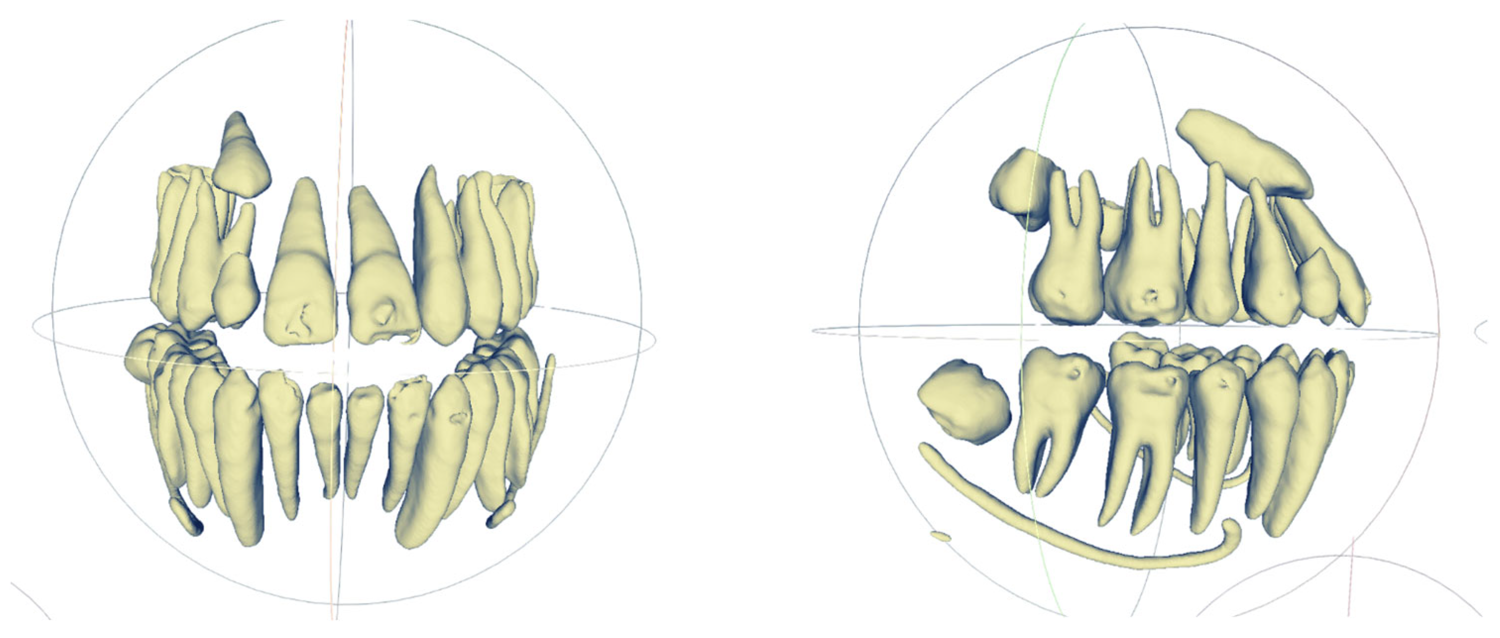
Early detection and interceptive intervention, such as extraction of the deciduous canine or orthodontic space creation, can greatly increase the likelihood of spontaneous eruption and reduce the complexity of subsequent treatment [2,5]. Diagnosis relies on clinical examination, panoramic radiography, and cone-beam computed tomography (CBCT) (Figure 1), which provides a reliable three-dimensional (3D) evaluation of tooth position, proximity to adjacent roots, and eruption direction [6,7,8,9].
Figure 1.
Segmented CBCT tracking of an impacted canine initially appearing malpositioned but showing passive eruption due to a favorable apical position.
The prognosis of impacted canines depends on multiple anatomical and temporal factors [4].
Based on two-dimensional (2D) radiographs, several authors have proposed positional classifications incorporating horizontal position, angulation, and vertical height [10]. Ericson and Kurol [2,11], divided the lateral incisor into four sectors, reporting that a cusp position distal to the incisor (sector I) indicated a favorable prognosis, while a mesial location (sector IV) carried a 99% risk of impaction. Other authors evaluated the angle between the canine’s long axis and the midline: values greater than 31° were associated with a poor prognosis [11,12]. Warford et al. [13] measured the angle between the canine long axis and the bicondylar line, finding that angles <59° correlated with unfavorable outcomes.
With the advent of 3D imaging, CBCT has emerged as a key diagnostic modality, offering detailed 3D information on tooth position and degree of impaction [14]. This imaging method has demonstrated higher diagnostic accuracy than 2D imaging for assessing impacted canines, particularly for detecting root resorption and evaluating spatial relationships with adjacent teeth. However, variability in acquisition parameters, voxel size, and segmentation methods may affect measurement reliability and inter-study comparability [15,16,17,18,19]. Nonetheless, Kau et al. [9] developed the KPG index, which scores the cusp and root apex positions in the three spatial planes using panoramic and axial CBCT slices. Later, San Martín et al. [20] suggested that the KPG index could estimate treatment duration for successful alignment of impacted canines. However, available evidence does not confirm the accuracy or reproducibility of this index as a reliable prognostic tool.
Previous studies have explored the relationship between 3D positional parameters of impacted canines and treatment duration or even complexity of the treatment. Variables such as angulation of the tooth, sector location, and depth of impaction have been associated with increased treatment difficulty; however, findings remain inconsistent due to methodological variability and the use of indirect outcome measures [2,13,20].
From a biomechanical perspective, the trajectory followed by an impacted canine during orthodontic traction depends on the relationship between the applied force vector and the tooth’s center of resistance [21].
Because the center of resistance of a single-rooted tooth lies approximately between the cervical and middle thirds of the root, the spatial position of the root apex may strongly influence the resulting type of movement (tipping, translation, or complex 3D displacement) [22]. These biomechanical considerations are presented as a theoretical framework and should not be interpreted as evidence derived from the included studies.
Therefore, it remains unclear whether a detailed CBCT-based evaluation of the apical position improves the prediction of traction complexity and the optimization of orthodontic force vectors [1,14,18].
Volumetric CBCT data enable multiplanar and 3D reconstructions, facilitating comprehensive evaluation of impaction [9]. Although the duration of orthodontic traction is primarily estimated from the 3D position of the impacted tooth, other anatomical factors, such as the presence or absence of the dental follicle, its size and volume, bone quality surrounding the tooth, and the stage of root development, may also significantly influence treatment planning [23].
Management of impacted canines involves combined surgical–orthodontic approaches aimed at restoring the tooth to a functional and esthetic position [4]. The choice of surgical technique, open or closed exposure, depends on the tooth location, amount of keratinized gingiva, and esthetic considerations [24,25]. Controlled orthodontic traction subsequently guides the tooth into the arch while minimizing root damage to adjacent teeth. In recent years, the use of temporary anchorage devices (TADs) has improved vector control and reduced unwanted side effects on anchorage teeth [26,27,28]. Despite these advances, the current literature shows considerable heterogeneity in traction protocols and in the characterization of the initial position of impacted canines. Most studies focus on the crown position, whereas the apical position, which may be associated with the direction, distance, and duration of tooth movement, has received much less attention [26,29,30,31,32,33,34].
Variability in CBCT-based measurement protocols and reference systems across studies is expected and may influence the comparability of results [15,25,35]. Therefore, it is essential to synthesize the available evidence to understand how the three-dimensional characteristics of the impacted canine, particularly the initial apical position assessed through CBCT, influence the traction distance required to achieve proper alignment within the dental arch. Consequently, the primary research question of this systematic review is to evaluate and compare, in patients with impacted maxillary canines, how the initial apical position may be associated with the traction distance needed for successful orthodontic alignment.
2. Materials and Methods
2.1. Protocol and Registration
This systematic review was carried out following the Preferred Reporting Items for Systematic Reviews and Meta-Analyses (PRISMA) 2020 guidelines [36] and was not registered in the International Prospective Register of Systematic Reviews (PROSPERO). The absence of prior registration may represent a limitation in terms of methodological transparency.
2.2. Search Strategy
A structured and comprehensive search strategy was implemented in the following electronic databases: PubMed/MEDLINE, Web of Science, Embase, Scopus, and the Cochrane Library. The search included all studies published up to November 2025, with no lower date restriction and no language limitations.
The search strategy was developed to identify clinical studies evaluating the influence of the initial three-dimensional apical position of impacted maxillary canines, assessed through CBCT, on root displacement and orthodontic traction outcomes.
To maximize sensitivity, both free-text terms and controlled vocabulary [MeSH terms, when applicable] were used. Search terms were combined using Boolean operators (“AND”, “OR”) and truncation symbols (*). The search strategy was adapted to the syntax and indexing systems of each database.
Example of search syntax for PubMed:
(“impacted canine*” OR “canine impaction” OR “ectopic canine*” OR “unerupted canine*” OR “impacted cuspid*” OR “ectopic cuspid*” OR “displaced canine*” OR “transposed canine*”) AND (“orthodontic traction” OR “forced eruption” OR “orthodontic extrusion”) AND (“cone-beam computed tomography” OR “CBCT”) AND (“apical position” OR “root displacement” OR “three-dimensional analysis” OR “3D movement” OR “root angulation”)
In addition to the electronic search, a manual search was performed in the reference lists of all included studies and in leading orthodontic journals (e.g., European Journal of Orthodontics, American Journal of Orthodontics and Dentofacial Orthopedics, and The Angle Orthodontist) to identify potentially relevant studies not captured through database searches.
All retrieved records were exported into Zotero® (Version 6.0.36) reference management software, where duplicates were automatically detected and manually verified prior to screening.
The complete search strategies for each database are provided in Supplementary Materials (Table S1) to ensure transparency and reproducibility.
2.3. Research Question and Inclusion Criteria
Given the increasing use of CBCT in the diagnosis and monitoring of impacted maxillary canines, this systematic review aimed to answer the following research question:
“Does the initial three-dimensional apical position of impacted maxillary canines, assessed by CBCT, influence the magnitude, direction, and duration of root displacement during orthodontic traction?”
To structure the study selection and ensure methodological consistency, the research question was defined according to the PICOS framework:
Population (P):
Human patients presenting impacted maxillary canines evaluated using CBCT.
Intervention (I):
Surgical exposure [open or closed technique] followed by orthodontic traction using any biomechanical protocol.
Comparison (C): Different initial three-dimensional apical positions, root angulations, or traction vectors assessed through CBCT analysis.
Outcomes (O): Quantitative three-dimensional assessment of root behavior during orthodontic traction, categorized into direct measurements (apical displacement expressed in mm and/or mm/month) and indirect parameters (angular changes, positional variables, and treatment duration). These outcomes were analyzed separately due to their different levels of evidential strength.
Study design (S): Prospective or retrospective longitudinal clinical studies.
No restrictions were applied regarding age, sex, or impaction location (palatal, buccal, or bicortical).
2.4. Exclusion Criteria
Studies were excluded if they were conducted on animals or in vitro models, involved patients with craniofacial syndromes, cleft lip and palate, or major congenital anomalies that could significantly alter eruption patterns or biomechanical response, or included mandibular impacted or transmigrated canines. Studies reporting cases of ankylosis or absence of orthodontic movement were also excluded. Additionally, cross-sectional investigations without longitudinal follow-up, finite element analyses, narrative reviews, expert opinions, letters to the editor, case reports, and conference abstracts lacking quantitative three-dimensional data were not considered eligible for inclusion.
2.5. Study Selection and Data Extraction
Following completion of the electronic search and removal of duplicates using Zotero® (zotero version: 6.0.36) reference management software, all retrieved records were independently screened by two reviewers (N.F. and C.S.) in a blinded manner. Study selection and data extraction were conducted independently by the same two reviewers. Disagreements were resolved through discussion and consensus. The selection process was conducted in three sequential stages: (1) automatic and manual duplicate removal, (2) title and abstract screening according to the predefined eligibility criteria, and (3) full-text assessment of potentially relevant studies to confirm final inclusion. Any discrepancies between reviewers were resolved through discussion and consensus; when disagreement persisted, a third independent reviewer was consulted.
Data extraction was performed independently by the same reviewers using a standardized electronic data collection form developed specifically for this review in accordance with PRISMA 2020 recommendations. The extracted variables included: author and year of publication, country of origin, study design, sample size and demographic characteristics, location of canine impaction, surgical exposure technique, orthodontic traction mechanics employed, CBCT acquisition parameters, method of three-dimensional measurement, quantitative assessment of apical displacement [mm and/or mm/month when available], angular changes in the root axis, treatment duration, and main findings and limitations reported by the authors. In cases of incomplete or unclear information, attempts were made to contact the corresponding authors for clarification. Inter-reviewer agreement was assessed during the study selection and data extraction processes; however, it was not formally quantified using statistical measures such as kappa coefficients.
2.6. Risk of Bias and Quality Assessment
The methodological quality of the included studies was independently assessed by two reviewers using the Joanna Briggs Institute (JBI) [37] critical appraisal tools, selected according to the specific study design of each investigation. Cohort studies were evaluated using the JBI Checklist for Cohort Studies, while quasi-experimental designs were assessed using the JBI Checklist for Quasi-Experimental Studies.
Each item within the respective checklist was rated as “Yes”, “No”, “Unclear”, or “Not applicable”. Disagreements between reviewers were resolved through discussion and consensus. When necessary, a third reviewer was consulted to achieve agreement.
For quantitative interpretation, the total number of affirmative responses (“Yes”) was calculated for each study.
Based on predefined thresholds, overall methodological quality was categorized as:
High quality: ≥8 affirmative responses.
Moderate quality: 5–7 affirmative responses.
Low quality: ≤4 affirmative responses.
The assessment focused on key domains including selection bias, measurement reliability, confounding control, completeness of follow-up, and appropriateness of statistical analysis. Detailed item-by-item quality assessment is provided in the Supplementary Materials (Tables S3 and S4).
2.7. Data Synthesis and Statistical Analysis
Due to the expected clinical and methodological heterogeneity among the included studies—including variations in study design, CBCT acquisition parameters, reference systems for three-dimensional measurements, traction mechanics, and outcome—a quantitative meta-analysis was not considered appropriate. To address this heterogeneity, findings were synthesized by distinguishing between studies reporting direct measurements of apical displacement and those providing indirect evidence based on surrogate parameters.
Therefore, a structured narrative synthesis was conducted following the recommendations of the JBI Manual for Evidence Synthesis and the PRISMA 2020 statement.
The included studies were grouped and comparatively analyzed according to their shared methodological and clinical characteristics, focusing on:
- The initial three-dimensional apical position and spatial orientation of the impacted canine (palatal, buccal, or bicortical).
- The CBCT-based parameters used to quantify root position and movement (apical displacement, angular correction, vector of movement).
- The orthodontic traction protocol and anchorage system employed.
- The reported treatment duration and its association with initial apical position.
- The availability of direct apical displacement measurements versus indirect angular or positional indicators.
Where available, quantitative values of apical displacement (in millimeters and/or mm/month) were descriptively compared across studies. Particular attention was given to studies employing CBCT–CBCT or CBCT–STL superimposition techniques, as these methods allow more precise assessment of true root displacement.
Given the limited number of homogeneous datasets and the variability in measurement methodologies, no pooled effect estimates were calculated. Instead, patterns of association between initial apical position and orthodontic traction outcomes were identified and critically interpreted in light of methodological quality and potential sources of bias. This approach allowed a more structured interpretation of the evidence, considering the different levels of measurement validity across studies.
3. Results
3.1. Study Selection
The electronic search identified a total of 528 records across all databases. An additional nine records were identified through citation tracking and other sources, resulting in 537 records overall. After removal of 83 duplicates, 454 records remained for title and abstract screening.
Of these, 405 records were excluded for not meeting the predefined eligibility criteria. Forty-nine reports were sought for retrieval, of which three could not be accessed.
A total of 46 full-text articles were assessed for eligibility. Thirty-nine studies were excluded for the following reasons: absence of CBCT-based evaluation (n = 8), lack of quantifiable apical measurements (n = 12), non-eligible study design (n = 9), absence of longitudinal follow-up (n = 5), or incomplete/unrelated data (n = 5).
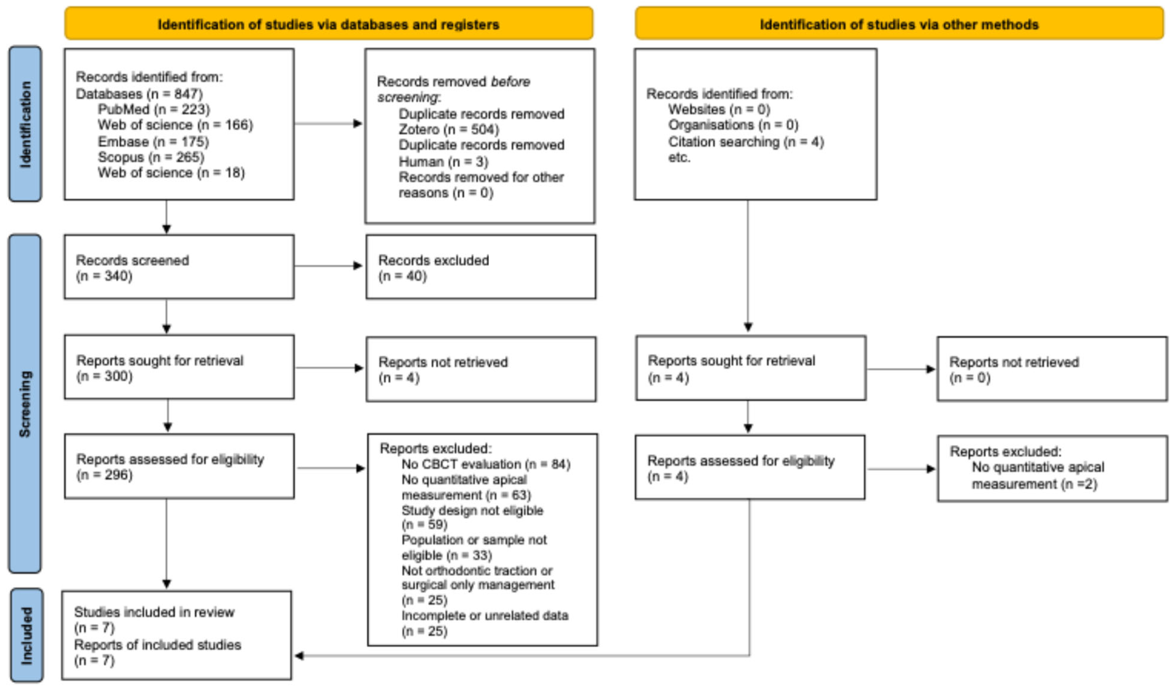
After completion of the selection process, seven studies met the inclusion criteria and were included in the qualitative synthesis (Table 1). An extended version of Table S2 is available in Supplementary Materials (Table S2). The study selection process is illustrated in Figure 2.
Table 1.
Characteristics of the studies included in the qualitative synthesis.
Figure 2.
Flowchart illustrating the selection process of included studies.
3.2. Characteristics of the Included Studies
Retrospective observational designs predominated (n = 5), alongside one prospective observational study and one prospective quasi-experimental study.
The samples originated from university hospitals and private clinics and included patients with buccal, palatal or bicortical impactions. Most studies employed pre-treatment CBCT (T0) to characterize the three-dimensional position of the canine, assessing variables such as initial apical location, angulation and distances to various reference planes.
Only two studies [42,44] performed direct measurements of apical displacement using CBCT–CBCT or CBCT–STL superimpositions. The remaining five studies [38,39,40,41,43] provided indirect evidence based on angular changes, positional parameters, or treatment-related variables, without directly measuring the linear displacement of the root apex. These types of outcomes represent different levels of evidential strength and should therefore be interpreted separately.
Reported orthodontic traction times ranged from 6.8 to 11.3 months; however, some studies provided only mean values without subgroup-specific analysis. In particular, Sosars et al. [40] reported only a global mean traction time without stratification according to canine position, which may limit the interpretation of its findings in relation to specific clinical scenarios.
However, the heterogeneity in measurement protocols and reference systems limited direct comparability across studies. This variability in CBCT acquisition protocols, segmentation methods, and three-dimensional reference systems further limits the comparability of results and should be considered when interpreting the findings.
3.3. Methodological Quality
Assessment using the JBI tools indicated an overall moderate methodological quality among the included studies (range: 6/11 to 9/11 for cohort studies; 7/9 for the quasi-experimental study). Results of JBI assessment are included in Table 2. Key strengths included the consistent use of CBCT and the application of reproducible three-dimensional measurement methods.
Table 2.
Methodological quality assessment of included studies according to the Joanna Briggs Institute [JBI] critical appraisal tools.
The primary methodological concerns were related to the predominance of retrospective designs, limited control of potential confounding variables, incomplete reporting of participant attrition, and the absence of randomization in the quasi-experimental study.
Across studies, uncertainty was most frequently observed in domains related to confounding management and follow-up adequacy, which may limit causal inference regarding the relationship between initial apical position and traction outcomes. Overall, while the included evidence provides clinically relevant insights, the moderate methodological quality warrants cautious interpretation of the reported associations.
3.4. Main Findings
3.4.1. Three-Dimensional Apical Displacement
The studies that directly measured root movement [42,44] reported apical displacement velocities ranging from 0.29 to 0.84 mm/month, depending on the measurement method and traction protocol applied. Taken together, these results may suggest predominantly bodily movement, with relatively stable trajectories, especially in palatally impacted canines.
3.4.2. Influence of the Initial Position on Traction Duration and Movement Behavior
Five studies [38,39,40,41,43] agreed that the initial position of the canine influences both the duration and complexity of traction. Palatal and bicortical impactions were associated with longer treatment times, whereas more mesially positioned canines or those with greater angular deviations demonstrated significant increases in traction duration.
3.4.3. Angular Parameters as Predictors
In CBCT-based studies [39,40,43], root and crown inclination angles directly associated with traction time and the magnitude of three-dimensional movement. Canines with greater inclinations towards the midline were associated with prolonged traction duration.
3.4.4. Root Changes Associated with Traction
Longitudinal studies [41,43] reported that root surface alterations were predominantly observed in the cervical third, particularly in buccally impacted canines. In contrast, the apical region demonstrated limited structural modification throughout orthodontic traction, indicating relative apical stability under controlled biomechanical conditions.
3.4.5. Comparison of Anchorage and Traction Methods
The quasi-experimental study [42] found that anchorage systems using TADs and TPAs produced comparable apical displacements, with no significant differences in movement velocity or trajectory. Additionally, one study [44] showed that the use of aligners, combined with an initial CBCT and serial STL models, allowed apical movement to be monitored without additional CBCT scans, maintaining consistent agreement with the digital treatment plan.
Taken together, the included studies suggest that the initial apical position and three-dimensional orientation of the impacted canine may be associated with differences in the magnitude and direction of movement during orthodontic traction, as well as with treatment duration. However, this interpretation is mainly based on indirect evidence, as only two studies directly quantified apical displacement. Therefore, these findings should be interpreted with caution, and the relative weight of direct and indirect evidence must be considered when drawing conclusions.
4. Discussion
This systematic review aimed to evaluate how the initial apical position of impacted maxillary canines influences both the distance and the biomechanical behavior of tooth movement during orthodontic traction, based on three-dimensional measurements obtained from CBCT imaging. Although the amount of direct evidence available is limited, the findings should be interpreted with caution, as most of the available data are derived from indirect parameters rather than direct measurements of apical displacement.
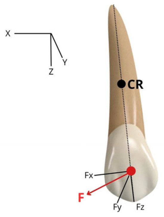
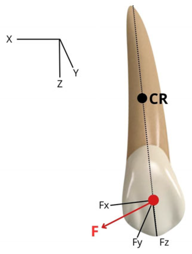
The spatial position of the root apex may modify the line of action of orthodontic forces relative to the center of resistance (Figure 3). These biomechanical interpretations are theoretical in nature and are presented to facilitate understanding of possible movement patterns, rather than as evidence derived from the included studies.
Figure 3.
Three-dimensional biomechanical representation of orthodontic traction applied to an impacted maxillary canine. The center of resistance (CR) is located approximately at the middle third of the root along the tooth’s longitudinal axis. The orthodontic traction vector (F), applied on the buccal surface of the crown, can be resolved into its mesiodistal (Fx), buccopalatal (Fy), and vertical (Fz) components within a three-dimensional coordinate system (X, Y, Z). This figure is a schematic representation intended to facilitate the interpretation of biomechanical principles discussed in the included studies. It does not represent original data but rather a conceptual model based on the interpretation of the reviewed evidence.
When the apex is positioned palatally or bicortically, traction may require complex three-dimensional displacement and may explain longer traction durations described in several studies.
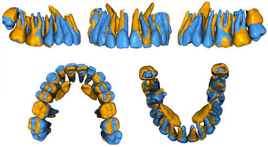
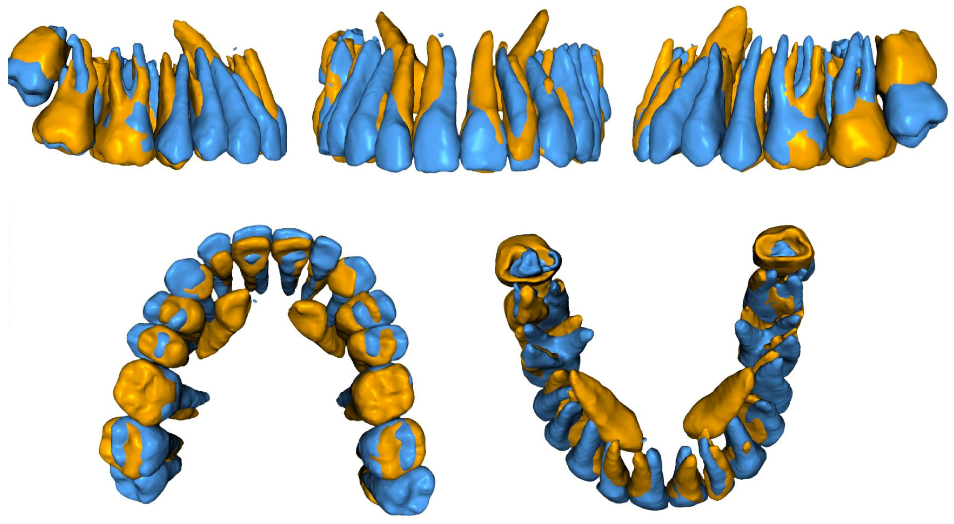
Conversely, canines with more favorable apical orientation may respond with more predictable bodily movement and shorter displacement paths (Figure 4).
Figure 4.
Three-dimensional superimposition of impacted maxillary canines (13 and 23) between the initial (T0, orange) and final (T1, blue) treatment stages. Segmented dental models derived from CBCT were superimposed using Blue Sky Plan® software (Version 5.0.24). Color coding (orange for T0 and blue for T1) highlights spatial differences. Upper images show lateral and frontal views, while lower images display occlusal and three-dimensional perspectives of the maxillary arch, allowing visualization of the three-dimensional displacement of the impacted canines during treatment. The images correspond to a representative clinical case and are provided for illustrative purposes.
This figure does not originate from the studies included in the systematic review but is intended to facilitate the understanding of three-dimensional movement patterns described in the literature, including changes in angulation, displacement trajectory, and spatial orientation. No quantitative analysis was derived from this figure.
Two studies directly assessed three-dimensional apical displacement [42,44]. One study [42] documented monthly apical movement velocities ranging from 0.44 to 0.84 mm/month. The other [44] reported mean apical velocities of 0.29 mm/month using CBCT–CBCT superimposition and 0.38 mm/month using CBCT–STL superimposition. Taken together, these values may suggest relatively stable and predominantly body-driven movement patterns; however, this interpretation is based on a limited number of studies directly assessing apical displacement. This pattern is consistent with classic biomechanical models, which describe controlled trajectories when the force is directed toward the tooth’s center of resistance [22]. However, the scarcity of studies that quantify apical displacement using three-dimensional linear measurements limits the possibility of establishing precise quantitative relationships between initial apical position and actual displacement magnitude. Consequently, interpretation of the precise biomechanical impact of apical positioning should be approached with caution.
Most of the included studies contributed indirect information regarding apical behavior by analyzing parameters such as root angulation, the three-dimensional localization of the tooth, and the duration of active traction [38,39,40,41,43]. Consistent with previous investigations, palatal and bicortical impactions may be associated with increased mechanical difficulty and longer treatment durations [2]. Similarly, canines positioned more mesially or those presenting greater angular deviations were linked to prolonged traction, reinforcing the concept that apical location and root axis orientation condition case complexity and influence the biological response to orthodontic movement [6,33,45,46]. Therefore, these findings should not be interpreted as direct evidence of apical displacement.
However, it is important to acknowledge that most of the available evidence is indirect and does not directly quantify apical displacement. Parameters such as root angulation, crown position, and traction duration are frequently used as surrogate indicators of tooth movement; however, they do not necessarily reflect true three-dimensional displacement of the root apex.
Changes in angular measurements may represent tipping rather than bodily movement, and prolonged traction duration may be influenced by multiple factors unrelated to apical position, including anchorage control, force magnitude, and patient-specific biological variability. Therefore, caution is required when interpreting these parameters as proxies for apical displacement.
The reliance on indirect measurements represents a significant methodological limitation in the current literature and may partially explain the inconsistency of findings across studies. Consequently, conclusions regarding the influence of initial apical position on true root movement should be considered with caution until more studies employing direct three-dimensional superimposition techniques are available.
Angular parameters evaluated three-dimensionally in several studies [39,40,43] were positively associated with traction duration. This suggests that root axis inclination not only reflects the spatial position of the impacted canine but also serves as a clinical indicator of treatment complexity and biomechanical predictability [7,23,33,46].
Regarding root changes, the longitudinal studies selected [41,43] showed that alterations were primarily concentrated in the cervical thirds, with minimal apical involvement. This pattern supports the hypothesis that apical tissues are subjected to lower mechanical stress during traction compared with cervical regions, a clinically relevant consideration when assessing the risk of root resorption [3,47].
With respect to anchorage methods, a quasi-experimental study reported that TADs and TPAs produced comparable apical displacements [42], suggesting that anchorage type may have a smaller influence on apical trajectory than traditionally assumed, allowing selection to be based on clinical rather than mechanical considerations. This observation is congruent with existing literature on three-dimensional control using skeletal anchorage [30,48]. Likewise, the use of aligners combined with an initial CBCT and serial STL scans proved effective for monitoring movement without additional radiographic exposure [44], representing an approach aligned with contemporary digital orthodontics [49].
Although the findings show general consistency, several relevant limitations must be acknowledged. Methodological heterogeneity across studies in terms of three-dimensional measurement techniques, reference planes and traction protocols limits comparability. Additionally, most investigations relied on indirect parameters, with only two studies reporting linear measurements of apical displacement, restricting the capacity to quantify the relationship between initial apical position and the distance required for alignment. The retrospective design of many studies and insufficient control of confounding factors further contribute to potential bias.
Nonetheless, this review provides a rigorous synthesis of current knowledge, following PRISMA guidelines and including only studies employing CBCT, a modality that has demonstrated improved diagnostic accuracy in the assessment of impacted canines and is considered the standard for three-dimensional characterization [50].
From a clinical perspective, the results suggest the potential relevance of incorporating a detailed three-dimensional evaluation of the initial apical position into traction planning. Early identification of unfavorable apical positions allows clinicians to anticipate more complex trajectories, refine force vectors and establish realistic expectations regarding treatment duration—critical elements for predictable planning. To advance the understanding of apical displacement, future research should focus on prospective studies using standardized and comparable three-dimensional measurement protocols.
Overall, the available evidence suggests that the initial apical position may be associated with differences in biomechanical behavior of impacted canines during traction and may affect the distance required to achieve alignment. However, the lack of studies with direct measurement of apical displacement remains the principal gap in the literature and a priority for future investigations.
An important source of variability across the studies included was the presence of significant methodological and clinical heterogeneity. This heterogeneity arose from multiple sources, including differences in CBCT acquisition parameters (voxel size, field of view), variation in three-dimensional reference systems and measurement protocols, diversity in orthodontic traction mechanics and anchorage strategies, and inconsistency in outcome reporting (direct apical displacement versus indirect parameters such as angular changes or treatment duration).
Additionally, differences in sample characteristics, including type of impaction [palatal, buccal, or bicortical] and initial spatial position of the canine, further contributed to variability in reported findings. This heterogeneity limits direct comparability between studies and was the main reason why quantitative synthesis was not feasible.
Recent advances in imaging analysis have incorporated radiomics and artificial intelligence-based approaches to extract quantitative data from medical images, improving diagnostic and predictive capabilities in different medical fields. Although these techniques have been mainly applied in other areas such as oncology and MRI-based analysis, they highlight the potential of advanced image processing methods to enhance the understanding of complex biological structures and treatment outcomes. In this context, future research in orthodontics could benefit from the integration of these advanced methodologies [51,52].
4.1. Limitations
This review presents several limitations. Only two studies directly quantified the three-dimensional apical displacement, whereas the remaining studies relied on indirect measurements, limiting the ability to establish precise quantitative relationships. A notable heterogeneity was observed in 3D measurement methods, reference planes and traction protocols, making comparisons between studies difficult. Moreover, the predominance of retrospective designs and the insufficient control of confounding factors increase the risk of bias. Finally, the relatively small sample sizes of several studies reduce the generalizability of the findings.
4.2. Practical Implications
The findings of this systematic review suggest the potential clinical relevance of incorporating a comprehensive three-dimensional CBCT-based assessment of the initial apical position of impacted maxillary canines into treatment planning. Identification of unfavorable apical configurations—such as palatal or bicortical impactions and pronounced mesial angulation—may assist clinicians in anticipating potential increases in biomechanical complexity and traction duration, as well as the need for careful force control. However, these considerations should be interpreted with caution, as only a limited number of studies directly quantified apical displacement, and most of the available evidence is based on indirect parameters.
Parameters including root angulation and initial mesial displacement may serve as practical indicators of mechanical difficulty, potentially supporting refinement of traction vectors and anchorage planning. The comparable apical displacement patterns observed between different anchorage systems, including TADs and TPAs, suggest that biomechanical control of root movement may depend more on force direction and treatment planning than on the specific anchorage device selected.
Furthermore, integration of baseline CBCT imaging with sequential three-dimensional digital models may represent a promising low-radiation strategy for monitoring root movement; however, further prospective validation is required. Overall, detailed three-dimensional evaluation of the apical position may enhance treatment predictability, support individualized biomechanical strategies, and facilitate clearer communication with patients regarding anticipated treatment duration and complexity.
4.3. Recommendations for Future Research
Prospective studies employing standardized three-dimensional methods to measure apical movement, including CBCT–CBCT or CBCT–STL superimpositions, are needed. Harmonization of reference systems and biomechanical protocols is recommended to facilitate meaningful comparisons across studies. Research involving larger samples and adequate control of confounding factors would improve the quality of the available evidence. Additionally, future investigations should quantitatively explore the relationship between initial apical position, three-dimensional movement trajectory and long-term clinical outcomes.
5. Conclusions
The initial apical position of the impacted maxillary canine may be associated with differences in its biomechanical behavior during orthodontic traction. Unfavorable apical positions, including palatal or bicortical impactions and teeth with marked inclinations towards the midline, are also likely to be associated with more complex traction paths and longer treatment durations.
However, only two studies directly assessed three-dimensional apical displacement, meaning that the exact quantitative relationship between the initial apical position and the true distance of movement cannot yet be determined with precision. Nevertheless, the available indirect findings suggest that pre-treatment three-dimensional evaluation may support treatment planning and contribute to a more comprehensive understanding of root displacement patterns.
Prospective studies using standardized three-dimensional measurement protocols are needed to quantify apical displacement reproducibly and to establish more accurate clinical guidelines for the orthodontic traction of impacted maxillary canines.
Supplementary Materials
The following supporting information can be downloaded at: https://www.mdpi.com/article/10.3390/app16073541/s1, Table S1: Search strategy for each database; Table S2: Characteristics of the studies included in the qualitative synthesis; Table S3: Detailed methodological quality assessment of included studies according to the JBI critical appraisal tools; Table S4: Detailed methodological quality assessment of the quasi-experimental study according to the JBI checklist.
Author Contributions
Conceptualization, N.F.G. and C.S.F.; methodology, N.F.G. and C.S.F.; formal analysis, N.F.G.; investigation, N.F.G. and C.S.F.; data curation, N.F.G. and C.S.F.; writing—original draft preparation, N.F.G.; writing—review and editing, N.F.G., C.S.F., A.M.I., N.S. and T.C.; supervision, C.S.F. and T.C. All authors have read and agreed to the published version of the manuscript.
Funding
This research received no external funding.
Institutional Review Board Statement
Not applicable.
Informed Consent Statement
Not applicable.
Data Availability Statement
No new data were created or analyzed in this study. Data sharing is not applicable to this article.
Acknowledgments
The authors have reviewed and edited the output and take full responsibility for the content of this publication.
Conflicts of Interest
The authors declare no conflicts of interest.
Abbreviations
The following abbreviations are used in this manuscript:
| PRISMA | Preferred Reporting Items for Systematic Reviews and Meta-Analyses |
| PROSPERO | International Prospective Register of Systematic Reviews |
| PICOS | Population, Intervention, Comparison, Outcome, Study design |
| JBI | Joanna Briggs Institute |
| ROC | Receiver Operating Characteristic |
| CBCT | Cone-Beam Computed Tomography |
| STL | Standard Tessellation Language |
| T0 | Baseline [pre-treatment time point] |
| TADs | Temporary Anchorage Devices |
| TPA | Transpalatal Arch |
| CBCT–CBCT | Superimposition between two CBCT scans |
| CBCT–STL | Superimposition between CBCT imaging and digital STL model |
References
- Ng, W.L.; Cunningham, A.; Pandis, N.; Bister, D.; Seehra, J. Impacted maxillary canine: Assessment of prevalence, severity and location of root resorption on maxillary incisors: A retrospective CBCT study. Int. Orthod. 2024, 22, 100890. [Google Scholar] [CrossRef] [Scilit] [PubMed]
- Ericson, S.; Kurol, J. Early treatment of palatally erupting maxillary canines by extraction of the primary canines. Eur. J. Orthod. 1988, 10, 283–295. [Google Scholar] [CrossRef] [Scilit]
- Bishara, S.E.; Ortho, D. Impacted maxillary canines: A review. Am. J. Orthod. Dentofac. Orthop. 1992, 101, 159–171. [Google Scholar] [CrossRef] [Scilit]
- Iacob, A.M.; Escobedo Martínez, M.F.; Olay García, S.; Junquera Olay, S.; Junquera Gutiérrez, L.M. Two-Dimensional Radiographic Diagnosis of Maxillary Canine Impactions. Dent. J. 2024, 12, 360. [Google Scholar] [CrossRef] [Scilit]
- Elangovan, B.; Pottipalli Sathyanarayana, H.; Padmanabhan, S. Effectiveness of various interceptive treatments on palatally displaced canine–A systematic review. Int. Orthod. 2019, 17, 634–642. [Google Scholar] [CrossRef] [Scilit]
- Schubert, M.; Baumert, U. Kritische Analyse von Weg und Zeit bei der Eckzahneinordnung in den Zahnbogen. J. Orofac. Orthop. 2009, 70, 200–212. [Google Scholar] [CrossRef] [Scilit]
- Kocyigit, S.; Oz, A.A.; Bas, B.; Arici, N.; Karahan, S. Are age and radiographic features effective on orthodontic alignment of palatally impacted maxillary canines? A retrospective study. Eur. Oral Res. 2019, 53, 132–136. [Google Scholar] [CrossRef] [Scilit]
- Stoustrup, P.; Videbæk, A.; Wenzel, A.; Matzen, L.H. Will supplemental cone beam computed tomography change the treatment plan of impacted maxillary canines based on 2D radiography? A prospective clinical study. Eur. J. Orthod. 2024, 46, cjad062. [Google Scholar] [CrossRef] [Scilit]
- Chung, H.K.; Pan, P.; Gallerano, R.L.; English, J.D. A novel 3D classification system for canine impactions—The KPG index. Int. J. Med. Robot. Comput. Assist. Surg. 2009, 5, 291–296. [Google Scholar] [CrossRef] [Scilit]
- Díaz-Reissner, C.; Pistilli, E.; Caje, R.; Maldonado, C.; Jolay, E.; Ferreira-Gaona, M. Prognosis of impacted canines according to their position in the upper maxilla using different radiographic analyses. Rev. Cient. Odontol. 2022, 10, e096. [Google Scholar] [CrossRef] [Scilit]
- Ericson, S.; Kurol, J. Radiographic assessment of maxillary canine eruption in children with clinical signs of eruption disturbance. Eur. J. Orthod. 1986, 8, 133–140. [Google Scholar] [CrossRef] [Scilit] [PubMed]
- Lindauer, S.J.; Rubenstein, L.K.; Hang, W.M.; Andersen, W.C.; Isaacson, R.J. Canine impaction identified early with panoramic radiographs. J. Am. Dent. Assoc. 1992, 123, 91–97. [Google Scholar] [CrossRef] [Scilit] [PubMed]
- Warford, J.H.; Grandhi, R.K.; Tira, D.E. Prediction of maxillary canine impaction using sectors and angular measurement. Am. J. Orthod. Dentofac. Orthop. 2003, 124, 651–655. [Google Scholar] [CrossRef] [Scilit]
- Farha, P.; Nguyen, M.; Karanth, D.; Dolce, C.; Arqub, S.A. Orthodontic Localization of Impacted Canines: Review of the Cutting-edge Evidence in Diagnosis and Treatment Planning Based on 3D CBCT Images. Turk. J. Orthod. 2023, 36, 261–269. [Google Scholar] [CrossRef] [Scilit]
- Walker, L.; Enciso, R.; Mah, J. Three-dimensional localization of maxillary canines with cone-beam computed tomography. Am. J. Orthod. Dentofac. 2005, 128, 418–423. [Google Scholar] [CrossRef] [Scilit] [PubMed]
- Alqerban, A.; Jacobs, R.; Fieuws, S.; Willems, G. Comparison of two cone beam computed tomographic systems versus panoramic imaging for localization of impacted maxillary canines and detection of root resorption. Eur. J. Orthod. 2011, 33, 93–102. [Google Scholar] [CrossRef] [Scilit] [PubMed]
- Scarfe, W.C.; Farman, A.G. What is Cone-Beam CT and How Does it Work? Dent. Clin. N. Am. 2008, 52, 707–730. [Google Scholar] [CrossRef] [Scilit]
- Jeon, S.Y.; Lee, N.Y.; Lee, S.H. Three-dimensional evaluation of impacted maxillary canines using cone beam computed tomography and panoramic radiographs. J. Korean Acad. Pediatr. Dent. 2013, 40, 106–117. [Google Scholar] [CrossRef] [Scilit]
- Fink, D.F.; Smith, R.J.; Louis, S. The duration of orthodontic treatment. Am. J. Orthod. Dentofac. Orthop. 1992, 102, 45–51. [Google Scholar] [CrossRef] [Scilit]
- San Martín, D.E.; English, J.D.; Kau, C.H.; Gallerano, R.L.; McGrory, K.R.; Salas, A.M.; Bouquot, J.; Xia, J.J. The KPG index-a novel 3D classification system for maxillary canine impactions. Tex. Dent. J. 2012, 129, 265–274. [Google Scholar]
- Papageorgiou, S.N.; Keilig, L.; Hasan, I.; Jäger, A.; Bourauel, C. Effect of material variation on the biomechanical behaviour of orthodontic fixed appliances: A finite element analysis. Eur. J. Orthod. 2016, 38, 300–307. [Google Scholar] [CrossRef] [Scilit] [PubMed]
- Becker, A. The Orthodontic Treatment of Impacted Teeth, 3rd ed.; Wiley–Blackwell: Hoboken, NJ, USA, 2012. [Google Scholar]
- Vasović, D.; Pajević, T.; Vučić, L.; Glišić, B.; Juloski, J. Factors influencing treatment duration of impacted maxillary canines. Angle Orthod. 2025, 95, 266–273. [Google Scholar] [CrossRef] [Scilit] [PubMed]
- Cernochova, P.; Cernoch, C.; Klimo Kanovska, K.; Tkadlec, E.; Izakovicova Holla, L. Treatment options for impacted maxillary canines and occurrence of ankylotic and resorptive processes: A 20-year retrospective study. BMC Oral Health 2024, 24, 877. [Google Scholar] [CrossRef] [Scilit]
- Keener, D.J.; de Oliveira Ruellas, A.C.; Aliaga-Del Castillo, A.; Arriola-Guillén, L.E.; Bianchi, J.; Oh, H.; Gurgel, M.L.; Benavides, E.; Soki, F.; Rodríguez-Cárdenas, Y.A.; et al. Three-dimensional decision support system for treatment of canine impaction. Am. J. Orthod. Dentofac. Orthop. 2023, 164, 491–504. [Google Scholar] [CrossRef] [Scilit] [PubMed]
- Mousa, M.R.; Hajeer, M.Y.; Burhan, A.S.; Heshmeh, O. The Effectiveness of Conventional and Accelerated Methods of Orthodontic Traction and Alignment of Palatally Impacted Canines in Terms of Treatment Time, Velocity of Tooth Movement, Periodontal, and Patient-Reported Outcomes: A Systematic Review. Cureus 2022, 14, e24888. [Google Scholar] [CrossRef] [Scilit]
- Tabchi, Y.; Oualalou, Y.; Zaoui, F.; Halimi, A.; Benyahia, H. Three-dimensional orthodontic anchorage management of impacted maxillary canines: A systematic review. APOS Trends Orthod. 2023, 13, 113–122. [Google Scholar] [CrossRef] [Scilit]
- Palone, M.; Gasperoni, I.; Cremonini, F.; Maino, G.B.; Paoletto, E.; Pramstraller, M.; Lombardo, L. Multipurpose miniscrew-anchored palatal appliance combined with a fixed multibracket appliance to correct a Class II Division 2 malocclusion with maxillary constriction and impacted upper canine in a 13-year-old girl: A case report. Int. Orthod. 2025, 23, 100984. [Google Scholar] [CrossRef] [Scilit]
- Lee, J.Y.; Choi, Y.J.; Choi, S.H.; Chung, C.J.; Yu, H.S.; Kim, K.H. Labially impacted maxillary canines after the closed eruption technique and orthodontic traction: A split-mouth comparison of periodontal recession. J. Periodontol. 2019, 90, 35–43. [Google Scholar] [CrossRef] [Scilit]
- Nakandakari, C.; Gonçalves, J.R.; Cassano, D.S.; Raveli, T.B.; Bianchi, J.; Raveli, D.B. Orthodontic Traction of Impacted Canine Using Cantilever. Case Rep. Dent. 2016, 2016, 4386464. [Google Scholar] [CrossRef] [Scilit]
- Cruz, R.M. Orthodontic traction of impacted canines: Concepts and clinical application. Dental Press J. Orthod. 2019, 24, 74–87. [Google Scholar] [CrossRef] [Scilit]
- Singh, S.; Parihar, A.V.; Chaturvedi, T.P.; Shukla, N. A case series of orthodontic traction of maxillary impacted canine. Natl. J. Maxillofac. Surg. 2022, 13, 147–152. [Google Scholar] [CrossRef] [Scilit]
- de-la-Rosa-Gay, C.; Valmaseda-Castellón, E.; Figueiredo, R.; Hernández-Mangas, A.; Camps-Font, O. Prediction of traction time for the alignment of impacted maxillary canines. BMC Oral Health 2025, 26, 123. [Google Scholar] [CrossRef] [Scilit]
- Singh, K.; Reddy, C.M.; Joshi, D.; Jain, S. Treatment of Maxillary Impacted Canine using Ballista Spring and Orthodontic Wire Traction. Int. J. Clin. Pediatr. Dent. 2017, 10, 313–317. [Google Scholar] [CrossRef] [Scilit]
- Plaisance, F.; Kerbrat, J.B.; Goudot, P. Three-dimensional reconstructions of CT scans and cone beam CT of impacted maxillary canines in order to locate areas of ankylosis before orthodontic traction. Int. Orthod. 2017, 15, 575–587. [Google Scholar] [CrossRef] [Scilit]
- Page, M.J.; McKenzie, J.E.; Bossuyt, P.M.; Boutron, I.; Hoffmann, T.C.; Mulrow, C.D.; Shamseer, L.; Tetzlaff, J.M.; Akl, E.A.; Brennan, S.E.; et al. The PRISMA 2020 statement: An updated guideline for reporting systematic reviews. BMJ 2021, 372, n71. [Google Scholar] [CrossRef] [Scilit]
- Joanna Briggs Institute. JBI Manual for Evidence Synthesis. Available online: https://synthesismanual.jbi.global (accessed on 8 December 2025).
- Arriola-Guillén, L.E.; Aliaga-Del Castillo, A.; Ruíz-Mora, G.A.; Rodríguez-Cárdenas, Y.A.; Dias-Da Silveira, H.L. Influence of maxillary canine impaction characteristics and factors associated with orthodontic treatment on the duration of active orthodontic traction. Am. J. Orthod. Dentofac. Orthop. 2019, 156, 391–400. [Google Scholar] [CrossRef] [Scilit] [PubMed]
- Shin, H.; Park, M.; Chae, J.M.; Lee, J.; Lim, H.J.; Kim, B.C. Factors affecting forced eruption duration of impacted and labially displaced canines. Am. J. Orthod. Dentofac. Orthop. 2019, 156, 808–817. [Google Scholar] [CrossRef] [Scilit] [PubMed]
- Sosars, P.; Jakobsone, G.; Neimane, L.; Mukans, M. Comparative analysis of panoramic radiography and cone-beam computed tomography in treatment planning of palatally displaced canines. Am. J. Orthod. Dentofac. Orthop. 2020, 157, 719–727. [Google Scholar] [CrossRef] [Scilit]
- Rodríguez-Cárdenas, Y.A.; Arriola-Guillén, L.E.; Ruíz-Mora, G.A.; Aliaga-Del Castillo, A.; Boessio-Vizzotto, M.; Dias-Da Silveira, H.L. Root changes in buccal versus palatal maxillary impacted canines of adults: A longitudinal and retrospective 3-dimensional study before and after orthodontic traction. Int. Orthod. 2020, 18, 490–502. [Google Scholar] [CrossRef] [Scilit]
- Migliorati, M.; Cevidanes, L.; Sinfonico, G.; Drago, S.; Dalessandri, D.; Isola, G.; Biavati, A.S. Three dimensional movement analysis of maxillary impacted canine using TADs: A pilot study. Head Face Med. 2021, 17, 1. [Google Scholar] [CrossRef] [Scilit] [PubMed]
- Rodríguez-Cárdenas, Y.A.; Arriola-Guillén, L.E.; Aliaga-Del Castillo, A.; Ruíz-Mora, G.A.; Janson, G.; Cevidanes, L.; Ruellas, A.C.; Yatabe, M.; Dias-Da Silveira, H.L. Three-dimensional changes in root angulation of buccal versus palatal maxillary impacted canines after orthodontic traction: A retrospective before and after study. Int. Orthod. 2021, 19, 216–227. [Google Scholar] [CrossRef] [Scilit]
- Pinho, T.; Carvalho, J.P. Three-Dimensional Comparison of CBCT and Intraoral Scans for Assessing Orthodontic Traction of Impacted Canines with Clear Aligners. Dent. J. 2025, 13, 286. [Google Scholar] [CrossRef] [Scilit]
- Mesotten, K.; Naert, I.; van Steenberghe, D.; Willems, G. Bilaterally impacted maxillary canines and multiple missing teeth: A challenging adult case. Orthod. Craniofac. Res. 2005, 8, 29–40. [Google Scholar] [CrossRef] [Scilit]
- Crescini, A.; Nieri, M.; Buti, J.; Baccetti, T.; Prato, G.P.P. Orthodontic and periodontal outcomes of treated impacted maxillary canines: An appraisal of prognostic factors. Angle Orthod. 2007, 77, 571–577. [Google Scholar] [CrossRef] [Scilit]
- Bianco, E.; Mirabelli, L.; Basilicata, M.; Bruno, G.; De Stefani, A.; Du, L.; Maddalone, M. Cone Beam Computed Tomography [CBCT] Aid in the Management of Apical Root Resorption of Impacted Maxillary Canines and Physiologically Erupted Maxillary Canines after Orthodontic Treatment. Appl. Sci. 2024, 14, 886. [Google Scholar] [CrossRef] [Scilit]
- Thebault, B.; Dutertre, E. Disimpaction of maxillary canines using temporary bone anchorage and cantilever springs. Int. Orthod. 2015, 13, 61–80. [Google Scholar] [CrossRef] [Scilit]
- Abdinian, M.; Yaghini, J.; Jazi, L. Comparison of intraoral digital radiography and cone-beam computed tomography in the measurement of periodontal bone defects. Dent. Med. Probl. 2020, 57, 269–273. [Google Scholar] [CrossRef] [Scilit] [PubMed]
- Arriola-Guillén, L.E.; Ruíz-Mora, G.A.; Rodríguez-Cárdenas, Y.A.; Aliaga-Del Castillo, A.; Boessio-Vizzotto, M.; Dias-Da Silveira, H.L. Influence of impacted maxillary canine orthodontic traction complexity on root resorption of incisors: A retrospective longitudinal study. Am. J. Orthod. Dentofac. Orthop. 2019, 155, 28–39. [Google Scholar] [CrossRef] [Scilit]
- Zhang, Q.; Du, H.; Ma, X.; Liu, S.; Kong, X.; Hu, M.; Shi, J.; Tang, Y.; Liu, S.; Meng, X.; et al. Mucin 18-targeted humanized monoclonal antibody immune positron emission tomography imaging and patient-derived tumor xenograft visualization. VIEW 2024, 5, 20240055. [Google Scholar] [CrossRef] [Scilit]
- Wang, J.; Tang, W.; Zhu, J.; Cui, J.; Chen, Y.; Gu, M.; Xu, H.; Zhan, M.; Chen, Q.; Xu, B. Predicting the pathological subdiagnosis of benign prostatic hyperplasia with MRI radiomics: A noninvasive approach. VIEW 2025, 6, 20240092. [Google Scholar] [CrossRef] [Scilit]

Disclaimer/Publisher’s Note: The statements, opinions and data contained in all publications are solely those of the individual author(s) and contributor(s) and not of MDPI and/or the editor(s). MDPI and/or the editor(s) disclaim responsibility for any injury to people or property resulting from any ideas, methods, instructions or products referred to in the content. |
© 2026 by the authors. Licensee MDPI, Basel, Switzerland. This article is an open access article distributed under the terms and conditions of the Creative Commons Attribution (CC BY) license.