


Abstract
Exopolysaccharides secreted by Geobacter play a pivotal role in mediating extracellular electron transfer (EET), biofilm formation, and environmental adaptability. However, existing methods for extracting and purifying Geobacter exopolysaccharides often suffer from low yield, structural damage, or contamination by intracellular components, limiting in-depth research on the structure and function of exopolysaccharides. This paper aimed to optimize the extraction and purification protocols for Geobacter exopolysaccharides. Three crude extraction methods (EDTA, high-speed centrifugation, and heating) for exopolysaccharides were evaluated, and the EDTA method was selected as the optimal crude extraction strategy, balancing exopolysaccharides yield (22.3 μg/mL) and cell viability (90.1%), outperforming high-speed centrifugation (lower yield) and heating (severe cell lysis). Purification was optimized using a two-step process: for deproteinization, the Sevag method was optimized to four cycles, removing 75% of proteins with minimal exopolysaccharides loss. Ethanol precipitation was optimized to 75–90% concentration and 24 h incubation, yielding 19.6–20.9 μg/mL of purified exopolysaccharides while eliminating soluble impurities. This optimized protocol ensures high-quality exopolysaccharides isolation with minimal cell lysis and reduced risk of structural disruption, providing a reliable foundation for investigating the roles of Geobacter exopolysaccharides in EET and environmental applications.
1. Introduction
The genus Geobacter has emerged as a pivotal player in microbial biogeochemistry, which is renowned for its exceptional capacity to mediate extracellular electron transfer (EET) with insoluble metal oxides and electrodes [1,2]. These Gram-negative, anaerobic bacteria are ubiquitously distributed in soil, aquatic sediments, and groundwater, where they drive critical biogeochemical cycles by reducing ferric iron, manganese, and other oxidized minerals [3]. Their metabolic versatility has positioned Geobacter as a cornerstone in environmental bioremediation particularly for the treatment of contaminated sites laden with heavy metals (e.g., uranium, chromium) and recalcitrant organic pollutants (e.g., aromatic hydrocarbons) [4,5]. Moreover, Geobacter-based microbial fuel cells (MFCs) have demonstrated promise in converting organic matter into electricity, highlighting their dual role in pollution mitigation and sustainable energy production [6]. The metabolic versatility of Geobacter underscores the importance of understanding its extracellular components, particularly extracellular polymeric substances (EPSs), which mediate critical interactions in these applications. Related studies have also confirmed that bacteria can regulate metabolic pathways and their EPS structures, thereby affecting the biofilm formation and microbial functions [7].
EPS, a complex matrix of exopolysaccharides, redox-active molecules, nucleic acids, and lipids secreted by Geobacter, are central to its EET processes. These substances form a protective microenvironment around cells and facilitate electron transfer between the bacterial membrane and external electron acceptors [8]. In particular, EPSs enable the formation of conductive biofilms, which enhance electron transport efficiency by establishing physical connections between cells and solid substrates [9,10]. Moreover, emerging evidence suggests that exopolysaccharides serve not only as structural scaffolds but also as active participants in stress response and pollutant transformation. For instance, electrode potential significantly influences the EPS secretion and protein secondary structure in Geobacter-dominated electroactive biofilms with optimal potentials promoting α-helix structures that enhance electron transfer and maintain Geobacter abundance above 86% under tetracycline stress [11]. Proteins such as c-type cytochrome (e.g., OmcS, OmcZ) embedded in EPS matrices have been identified as key electron carriers that enhance long-range electron transport within biofilms [12]. However, recent studies emphasize that exopolysaccharides, as the most abundant component of EPSs (comprising 40–95% of the matrix), play an underappreciated role in modulating EET dynamics [13,14].
Exopolysaccharides are important substances that ensure the structural and functional integrity of biofilms. They contribute to microbial adhesion, biofilm stability and protection from environmental stress [15]. The diversity of exopolysaccharides structures includes changes in monosaccharide composition, glycosidic bonds, and molecular weight, which directly affect the viscosity, surface charge, and biofilm structure of EPS and may be crucial for electron transfer kinetics [16]. Exopolysaccharides are not merely passive components of the EPS matrix but active regulators of Geobacter extracellular physiology. Studies in Geobacter sulfurreducens have shown that genetically engineered strains deficient in polysaccharide biosynthesis exhibit impaired biofilm formation and reduced electron transfer rates to ferric iron oxides [17]. Further research has found that exopolysaccharides act as anchor points for cytochrome proteins during the EET process of Geobacter, facilitating long-distance electron transfer [18]. Despite these critical functions, the structural diversity and molecular mechanisms of exopolysaccharides in Geobacter remain underexplored, which is partly due to technical challenges in isolating intact exopolysaccharides [19].
The extraction and purification of Geobacter exopolysaccharides have traditionally relied on protocols adapted from other microbial exopolysaccharides, such as centrifugation, ethanol precipitation and column chromatography [20]. However, due to the biological heterogeneity among distinct bacterial species, these methods often result in low recovery rates and damage to the structural integrity, adversely affecting both the yield and purity of exopolysaccharides [21]. Furthermore, the absence of standardized protocols specifically tailored to Geobacter exacerbates inconsistencies in exopolysaccharides yield quantification across studies. Addressing these gaps necessitates the development of optimized strain-specific methodologies that ensure high purity and yield while preserving exopolysaccharides architecture, thereby enabling deeper mechanistic insights into their roles in Geobacter physiology and environmental interplay. The ultimate goal of this paper was to establish a reliable extraction and purification protocol for Geobacter exopolysaccharides, free from associated proteins, thereby enabling future investigations into the independent role of the polysaccharides backbone in EET separate from the contributions of EPS-embedded cytochromes and other redox-active proteins.
2. Materials and Methods
2.1. Bacterial Cultivation and Biofilm Formation
Geobacter sulfurreducens strain PCA (DSM 12127) was purchased from the German Collection of Microorganisms and Cell Cultures (DSMZ) (Braunschweig, Germany), and it was used as a representative species of the genus Geobacter. The strain was routinely cultured in NBAF medium containing 15 mM acetate as the electron donor and 40 mM fumarate as the electron acceptor. Due to the anaerobic nature of Geobacter species, the NBAF medium was sparged with an N2:CO2 (80:20) gas mixture prior to use to maintain an anaerobic environment [14].
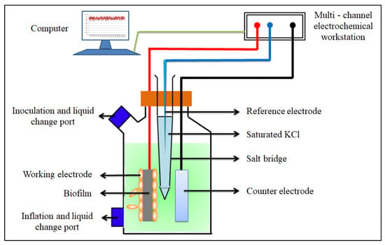
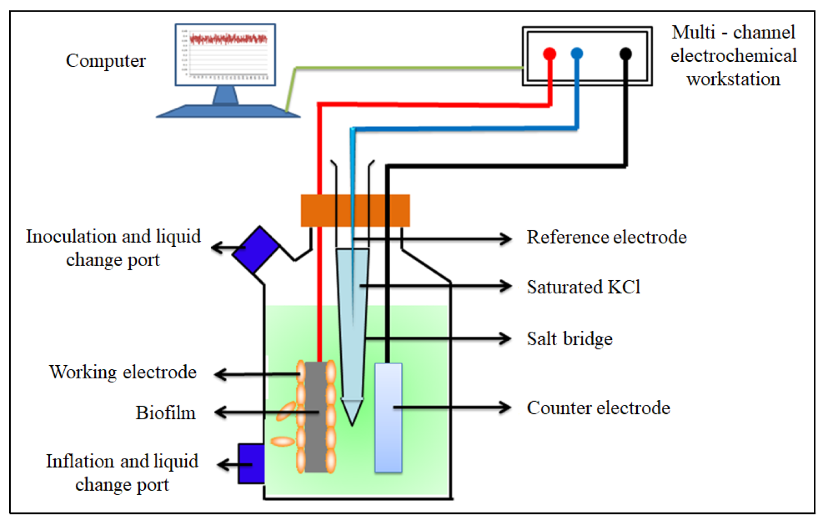
Given that exopolysaccharides are significantly more abundant in biofilms than in planktonic states, a bioelectrochemical system (BES) was constructed in this paper to promote biofilm enrichment on the anode [22]. As shown in Figure 1, the BES employed a three-electrode configuration: the working and counter electrodes consisted of polished graphite plates (30 × 15 × 5 mm), while a saturated calomel electrode (SCE, +0.2415 V vs. SHE) served as the reference electrode. The reactors were connected to a multi-channel electrochemical workstation (CHI1010C, CH Instruments, Shanghai, China) and maintained at 30 °C. The working electrode potential was set at +0.2 V (vs. SCE), and current data were recorded every 100 s using the CHI1010C. The reactors were filled with an electrolyte pre-saturated with an 80% N2–20% CO2 gas mixture. To accelerate start-up, the first cycle employed an electrolyte containing 15 mM acetate and 40 mM fumarate, while subsequent cycles used an electrolyte with only 15 mM acetate (no fumarate). When the electrochemical workstation showed a stable current, it indicated that the biofilm had matured. In this experiment, the current of BESs reached a stable state after running for 10 days.
Figure 1.
Schematic overview of BESs. The reactor consisted of a three-electrode configuration: a polished graphite plate as the working electrode, a similar graphite plate as the counter-electrode, and a saturated calomel electrode as the reference electrode. The current data are recorded by a multi-channel electrochemical workstation, and a stable current indicates that the biofilm has reached a mature state.
2.2. Extraction of Crude Exopolysaccharides
Mature biofilms were scraped and suspended in 0.9% NaCl solution. Three commonly used methods were selected for the crude extraction of exopolysaccharides from biofilm solutions: namely, EDTA, high-speed centrifugation and heating [23].
EDTA method: An equal volume of 2% (w/v) Na2-EDTA solution was added to 1 mL of biofilm solution, thoroughly mixed, and incubated at 4 °C for 3 h. The mixture was then centrifuged at 5000× g for 20 min at 4 °C to collect the supernatant.
High-speed centrifugation method: One milliliter of biofilm solution was centrifuged at 10,000× g for 30 min at 4 °C, and the supernatant was collected.
Heating method: One milliliter of biofilm solution was heated in a 60 °C constant-temperature water bath for 30 min, which was followed by centrifugation at 5000× g for 20 min at 4 °C to collect the supernatant.
The supernatants obtained from the above methods were filtered through a polyethersulfone membrane (0.22 μm) to remove bacterial cells and particulate impurities, and then the concentration of exopolysaccharides was determined by the anthrone method [24].
2.3. Cell Lysis Assessment
To evaluate the degree of cell lysis caused by the exopolysaccharides extraction process, cell viability was observed using confocal laser scanning microscope (CLSM, LSM 800, Carl Zeiss, Jena, Germany). For CLSM imaging, cells were stained using the LIVE/DEAD BacLight Bacterial Viability kit (Thermo Fisher Scientific, Waltham, MA, USA) in the dark for 15 min. The CLSM images were imported into Image-Pro Plus 6.0 to calculate the viability of cells under different extraction protocols, and the viability was expressed as a percentage of viable/total cells.
2.4. Purification of the Crude Exopolysaccharides
The crude exopolysaccharides were mixed with a large amount of proteins and various impurities. We purified the exopolysaccharides using a two-step approach: the Sevag method and ethanol precipitation method. To determine the optimal purification conditions for Geobacter exopolysaccharides, we systematically optimized the parameters of both methods.
The Sevag method aims to remove proteins [25]. The Sevag reagent was prepared by mixing chloroform and n-butanol at a volume ratio of 4:1, which was then added to the crude exopolysaccharides solution at a volume ratio of 1:4. After vigorous shaking for 30 min, the mixture was centrifuged at 5000× g for 10 min to induce layering: the upper aqueous layer (polysaccharides layer), the lower organic solvent layer, and denatured proteins as white substances at the interface. The aqueous layer was separated, and the above steps were repeated multiple times until the interface became clear. The protein concentration was detected using the BCA assay kit (Thermo Fisher Scientific).
The purpose of the ethanol precipitation method is used to remove residual proteins and various impurities [26]. An appropriate amount of ethanol was added to the exopolysaccharides solution from the previous step and mixed thoroughly to achieve ethanol concentrations of 70%, 75%, 80%, 85%, and 90% in the mixture. The mixtures were incubated at 4 °C for 6, 12, 24, 36, and 48 h, respectively, which was followed by centrifugation at 5000× g for 10 min to separate the phases. The supernatant was decanted, and the pellet was resuspended in deionized water to obtain the purified exopolysaccharides solution.
2.5. Statistical Analysis
All experiments were performed in triplicate, and data are expressed as mean ± standard deviation (SD). Statistical comparisons were analyzed using Student’s t-test in SPSS (version 16.0) with statistical significance defined as p < 0.05.
3. Results and Discussion
3.1. Extraction of Crude Exopolysaccharides and Cellular Viability
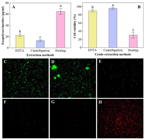
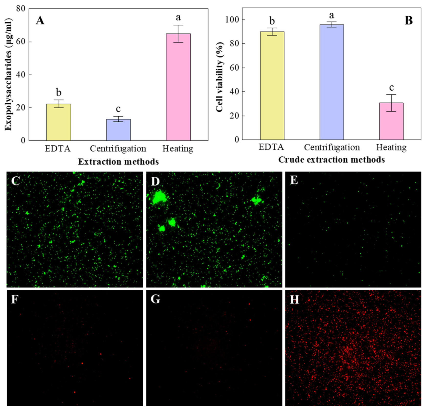
The crude exopolysaccharides extraction and cell damage effects of the EDTA method, high-speed centrifugation method, and heating method were compared. Although the EPS-free cells were not used for subsequent experiments in this paper, cell viability was monitored as a quality control parameter to validate the mildness of each extraction method and to ensure that the recovered exopolysaccharides were primarily of extracellular origin and free from significant contamination by intracellular components such as intracellular polysaccharides (e.g., cell wall-associated glycopolymers, cytoplasmic glycogen), cytoplasmic proteins, nucleic acids, and other soluble intracellular metabolites [23]. Among the three protocols in this paper, the heating method yielded higher amounts of crude exopolysaccharides (64.9 μg/mL) than EDTA (22.3 μg/mL) or high-speed centrifugation (13.1 μg/mL) methods (Figure 2A). The heating method had the highest extraction efficiency, but its damage to cell integrity should be considered. According to the results of cell viability staining, the heating method caused massive cell lysis with the cell viability being only 30.8% (Figure 2B,E,H). This led to the massive release of intracellular contents (including intracellular polysaccharides), thereby resulting in a false result of high extraction efficiency of exopolysaccharides. The high-speed centrifugation method is a purely physical-based approach for separating exopolysaccharides. It minimizes damage to cellular structural integrity while preserving the natural structure, conformation, and bioactivity (e.g., immunomodulatory activity, antioxidant capacity) of the exopolysaccharides [27]. As shown in Figure 2B,D,G, high-speed centrifugation is the least destructive method to cells, yielding the highest cell viability after treatment (96.2%). The EDTA extraction of exopolysaccharides is a mild chemical method based on chelation, which avoids hydrolysis or modification that may be caused by the use of strong acids, strong alkalis, high temperatures, or organic solvents [28]. Compared with the heating method, the EDTA method better protected the cell structure with the cell viability reaching as high as 90.1% after treatment (Figure 2B,C,F). Relative to high-speed centrifugation, EDTA-treated cells exhibited 6.3% lower viability but yielded 70.2% more exopolysaccharides (Figure 2A,B). Considering that we need to maximize exopolysaccharides extraction while preserving the integrity of the cells, the EDTA method is therefore more advantageous than both high-speed centrifugation and the heat method.
Figure 2.
The efficiency of crude exopolysaccharides extraction by different methods (A) and their damage to cell viability (B). CLSM images of cell viability staining under different treatments with red fluorescence representing dead cells and green fluorescence representing live cells: EDTA method (C,F), high-speed centrifugation method (D,G) and heating method (E,H). Different letters (a–c) indicate significant differences between treatments (p < 0.05).
It is important to note that LIVE/DEAD staining reflects membrane integrity rather than directly quantifying intracellular component leakage. While high cell viability (e.g., 90.1% for the EDTA method) suggests minimal cell disruption, it does not completely rule out the possibility of the partial leakage of intracellular polysaccharides or other soluble components. Previous studies have shown that EDTA-based extraction, due to its mild chelating mechanism, selectively releases extracellular polymers with minimal contamination by intracellular contents, as confirmed by the absence of typical intracellular markers such as glucose-6-phosphate dehydrogenase activity or DNA content in the extracts [23,28]. Nonetheless, the future optimization of Geobacter EPS extraction would benefit from the direct quantification of intracellular markers (e.g., enzyme activities, nucleic acid content) to more precisely assess contamination levels.
3.2. Optimization Methods for Exopolysaccharides Purification
In preliminary experiments, the EDTA method was employed for the crude extraction of exopolysaccharides. However, the extracts contained substantial proteins and impurities that could not be effectively removed by any single purification technique. Consequently, a two-step purification protocol was devised to address this challenge, namely the Sevag method and the ethanol precipitation method.
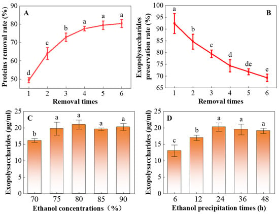
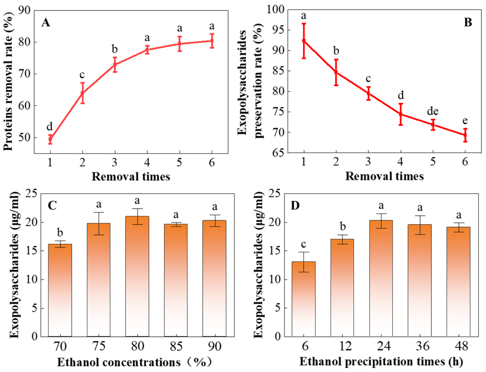
Due to the presence of significant residual proteins in the crudely extracted exopolysaccharides solution, the primary objective of the initial purification step was targeted proteins removal. The Sevag method, a classical approach for protein removal from polysaccharide preparations, was selected for this purpose. Its principle involves utilizing the Sevag reagent to denature and precipitate proteins, which was followed by phase separation via centrifugation or gravitational settling to isolate the proteinaceous layer from the polysaccharides solution. The advantage of this method lies in its mild conditions, which cause minimal damage to the exopolysaccharides structure. However, it is limited by relatively low efficiency, necessitating multiple repetitions to achieve satisfactory proteins removal [29]. As depicted in Figure 3A, the Sevag method could only remove 49.4% of proteins in the first treatment. With an increase in the number of treatments, the proteins removal rate improved significantly, reaching a plateau from the 4th treatment onwards, and ultimately achieving 80.4% by the 6th treatment. It is noteworthy that repeated applications of the Sevag method led to a gradual loss of exopolysaccharides: the exopolysaccharides preservation rate was 92.3% after the first treatment, then decreased progressively, and was only 69.3% by the 6th treatment (Figure 3B).
Figure 3.
The effect of Sevag method on protein removal rate (A) and exopolysaccharides preservation rate (B) under different removal times. The effect of different ethanol concentrations (C) and ethanol precipitation times (D) on exopolysaccharides purity. Different letters (a–e) indicate significant differences between treatments (p < 0.05).
The observed trade-off between protein removal efficiency and exopolysaccharides recovery is a well-recognized challenge in polysaccharide purification [30]. The progressive increase in protein removal with successive Sevag treatments reflects the cumulative denaturation and phase separation of proteins initially entrapped within the polysaccharide matrix. However, the diminishing returns beyond four cycles (from 75% to 80% protein removal between the 4th and 6th treatments) suggest that the majority of accessible proteins are removed within the first four treatments, while further cycles primarily cause exopolysaccharides loss without substantial additional purification. This pattern is consistent with previous reports showing that three to five Sevag treatments typically achieve optimal deproteinization for bacterial exopolysaccharides [31]. Importantly, the preservation rate of exopolysaccharides after four treatments remained above 70%, indicating that the structural integrity and yield of the target polysaccharides were maintained to an acceptable degree. Based on these considerations, four treatment cycles were selected as the optimal condition for Geobacter exopolysaccharides purification using the Sevag method, balancing the need for effective protein removal (approximately 75%) against the risk of excessive exopolysaccharides loss (approximately 30%). Under circumstances where sample quantity is limited or downstream applications require high polysaccharide yields, reducing the number of Sevag treatments to three may be considered albeit with a corresponding decrease in deproteinization efficiency.
According to the propensity for polysaccharides solubility to decrease precipitously in specific solvents under changing environmental conditions, the addition of precipitants (e.g., organic solvents or salts) facilitates polysaccharides precipitation from solution [26]. Therefore, the second step of the purification protocol employed the ethanol precipitation method. This method exploits ethanol to induce polysaccharides molecular aggregation and precipitation, thereby separating polysaccharides from soluble impurities such as water-soluble proteins, nucleic acids, and inorganic salts. To determine the optimal precipitation conditions, varying ethanol concentrations and precipitation times were investigated. Results indicate that employing 70% ethanol yielded the lowest exopolysaccharides content (16.2 μg/mL). Significantly enhanced yields (19.6–20.9 μg/mL) were consistently achieved with ethanol concentrations of 75%, 80%, 85%, and 90% despite the absence of significant differences between yields within this higher concentration group (Figure 3C). Although a range of 70–80% ethanol is commonly employed in the literature, the substantial heterogeneity in polysaccharides solubility necessitates that the optimal ethanol concentration for precipitation be empirically determined for each specific polysaccharide [32]. Precipitation time significantly influenced yield with maximum exopolysaccharides content attained after 24 h (20.3 μg/mL). While slight reductions occurred at 36 h and 48 h, these levels did not differ significantly from the 24 h peak. As a result, the efficient ethanol precipitation-based purification of Geobacter exopolysaccharides requires an ethanol concentration ≥ 75% and a precipitation time ≥ 24 h.
The optimal ethanol concentration range identified in this paper (≥75%) is broadly consistent with previous reports on microbial exopolysaccharides purification, although some variation exists depending on polysaccharide structural characteristics [33]. Xu et al. demonstrated that polysaccharides from different sources require individualized optimization with optimal concentrations ranging from 60% to 85% depending on molecular weight and structural characteristics. Specifically, higher molecular weight polysaccharides tend to precipitate at lower ethanol concentrations, while lower molecular weight or highly branched polysaccharides require higher ethanol concentrations (up to 85–90%) for efficient recovery [32]. Our finding that ethanol concentrations of 75–90% all yielded similarly high exopolysaccharides recovery suggests that the Geobacter exopolysaccharides in this paper possess intermediate solubility characteristics, precipitating efficiently across a broad concentration range. The optimal precipitation time of 24 h identified in this paper is consistent with standard protocols reported in the literature, which typically recommend overnight (12–24 h) incubation at 4 °C to maximize polysaccharide recovery while minimizing the co-precipitation of low-molecular-weight impurities [34]. In summary, our optimized ethanol precipitation conditions (≥75% ethanol, 24 h incubation) align with studies reporting higher optimal concentrations for structurally complex or charged polysaccharides, and they fall within the broader range of 60–85% documented in the literature. This contextualization demonstrates that our results are consistent with established principles of polysaccharide solubility while highlighting the need for empirical optimization for each specific exopolysaccharide.
4. Conclusions
This paper systematically optimized the extraction and purification protocols for Geobacter exopolysaccharides, addressing key challenges of yield, structural integrity, and purity. Among the three crude extraction methods evaluated, the EDTA method emerged as the most effective, as it balanced exopolysaccharides yield (22.3 μg/mL) with minimal cell lysis (90.1% viability), avoiding the overestimation of exopolysaccharides content caused by intracellular component release (heating method) and low yield limitations (high-speed centrifugation).
For purification, the two-step protocol (Sevag method followed by ethanol precipitation) was validated to efficiently remove impurities while preserving exopolysaccharides structure. The Sevag method, optimized to 4 cycles, achieved 75% proteins removal with acceptable exopolysaccharides loss (≤30%), overcoming the trade-off between deproteinization efficiency and exopolysaccharides retention. Ethanol precipitation under 75–90% concentration and 24 h incubation further enhanced purity by separating soluble impurities (e.g., water-soluble proteins, nucleic acids), yielding 19.6–20.9 μg/mL of purified exopolysaccharides. It should be noted that the assessment of purification efficacy in this paper was based on indirect parameters: protein removal efficiency, exopolysaccharides recovery rate, and the elimination of soluble impurities via ethanol precipitation. The direct quantification of absolute purity (e.g., carbohydrate content relative to dry weight, or residual protein/nucleic acid content) was not performed. Future studies aiming to characterize exopolysaccharide structure and function should incorporate such direct purity measurements to ensure the reliability of downstream analyses.
This optimized protocol provides a reliable reference for isolating high-quality exopolysaccharides from Geobacter, minimizing the risk of structural disruption, which is critical for studying their roles in EET, biofilm conductivity, and environmental interactions. Future studies can build on this method to explore the structural diversity of Geobacter exopolysaccharides and their specific functional motifs in electron transfer. Additionally, the protocol may be adapted for exopolysaccharides extraction and purification from other electroactive bacteria, facilitating comparative analyses of extracellular matrix functions in microbial electrochemical systems. Overall, this paper advances methodological standards for exopolysaccharides research of Geobacter, laying the groundwork for understanding the mechanistic links between exopolysaccharides structure and microbial ecological functions, with implications for bioremediation and sustainable energy technologies.
Author Contributions
Conceptualization, Z.Z.; methodology, Z.Z.; software, Y.S.; formal analysis, Y.S.; resources, Z.Z.; data curation, Y.S.; writing—original draft preparation, Y.S.; writing—review and editing, Z.Z.; supervision, Z.Z.; project administration, Z.Z. All authors have read and agreed to the published version of the manuscript.
Funding
This research was funded by The National Natural Science Foundation of China (No. 42407177) and the Surface Projects of Natural Science Foundation of the Higher Education Institutions of Jiangsu Province (No. 23KJB610008).
Institutional Review Board Statement
Not applicable.
Informed Consent Statement
Not applicable.
Data Availability Statement
The original contributions presented in this paper are included in the article. Further inquiries can be directed to the corresponding author.
Acknowledgments
We thank the reviewers and editors for their helpful comments regarding the manuscript.
Conflicts of Interest
The authors declare no conflicts of interest.
References
- Tremblay, P.; Aklujkar, M.; Leang, C.; Nevin, K.P.; Lovley, D. A genetic system for Geobacter metallireducens: Role of the flagellin and pilin in the reduction of Fe(III) oxide. Environ. Microbiol. Rep. 2012, 4, 82–88. [Google Scholar] [CrossRef] [Scilit]
- Sun, D.; Wang, A.; Cheng, S.; Yates, M.; Logan, B.E. Geobacter anodireducens sp. nov., an exoelectrogenic microbe in bioelectrochemical systems. Int. J. Syst. Evol. Micr. 2014, 64, 3485–3491. [Google Scholar] [CrossRef] [Scilit] [PubMed]
- Gregory, K.B.; Lovley, D.R. Remediation and recovery of uranium from contaminated subsurface environments with electrodes. Environ. Sci. Technol. 2005, 39, 8943–8947. [Google Scholar] [CrossRef] [Scilit] [PubMed]
- Williams, K.H.; Long, P.E.; Davis, J.A.; Wilkins, M.J.; N’Guessan, A.L.; Steefel, C.; Yang, L.; Newcomer, D.; Spane, F.A.; Kerkhof, L.J.; et al. Acetate availability and its influence on sustainable bioremediation of uranium-contaminated groundwater. Geomicrobiol. J. 2011, 28, 519–539. [Google Scholar] [CrossRef] [Scilit]
- Zhang, T.; Tremblay, P.; Chaurasia, A.K.; Smith, J.A.; Bain, T.S.; Lovley, D.R. Anaerobic benzene oxidation via phenol in Geobacter metallireducens. Appl. Environ. Microb. 2013, 79, 7800–7806. [Google Scholar] [CrossRef] [Scilit]
- Ivase, T.J.P.; Nyakuma, B.B.; Oladokun, O.; Abu, P.T.; Hassan, M.N. Review of the principal mechanisms, prospects, and challenges of bioelectrochemical systems. Environ. Prog. Sustain. 2020, 39, 13298. [Google Scholar] [CrossRef] [Scilit]
- Wang, Y.; Zhang, R.; Mathivanan, K.; Zhang, Y.; Yang, L.; Guan, F.; Duan, J. Proteomics and EPS compositional analysis reveals Desulfovibrio bisertensis SY-1 induced corrosion on Q235 steel by biofilm formation. Materials 2024, 17, 5060. [Google Scholar] [CrossRef] [Scilit]
- Xiao, Y.; Zhang, E.; Zhang, J.; Dai, Y.; Yang, Z.; Christensen, H.; Ulstrup, J.; Zhao, F. Extracellular polymeric substances are transient media for microbial extracellular electron transfer. Sci. Adv. 2017, 3, e1700623. [Google Scholar] [CrossRef] [Scilit]
- Stöckl, M.; Teubner, N.C.; Holtmann, D.; Mangold, K.; Sand, W. Extracellular polymeric substances from Geobacter sulfurreducens biofilms in microbial fuel cells. ACS Appl. Mater. Inter. 2019, 11, 8961–8968. [Google Scholar] [CrossRef] [Scilit]
- Yang, G.; Xia, X.; Nie, W.; Qin, B.; Hou, T.; Lin, A.; Yao, S.; Zhuang, L. Bidirectional extracellular electron transfer pathways of Geobacter sulfurreducens biofilms: Molecular insights into extracellular polymeric substances. Environ. Res. 2024, 245, 118038. [Google Scholar] [CrossRef] [Scilit]
- Li, F.; Zhou, L.; Wang, S.; Wu, Y.; Li, T.; Sun, S.; Wang, J. Electroactive biofilms alter the EPS structure and metabolic pathways to sense potential and tetracycline. J. Hazard. Mater. 2024, 488, 137414. [Google Scholar] [CrossRef] [Scilit]
- Rollefson, J.B.; Stephen, C.S.; Tien, M.; Bond, D.R. Identification of an extracellular polysaccharide network essential for cytochrome anchoring and biofilm formation in Geobacter sulfurreducens. J. Bacteriol. 2011, 193, 1023–1033. [Google Scholar] [CrossRef] [Scilit]
- Huang, L.; Puma, G.L. Physiological response of electroactive bacteria via secretion of extracellular polymeric substances in microbial electrochemical processes: A review. Curr. Opin. Electrochem. 2022, 36, 101168. [Google Scholar] [CrossRef] [Scilit]
- Zhuang, Z.; Yang, G.; Mai, Q.; Guo, J.; Liu, X.; Zhuang, L. Physiological potential of extracellular polysaccharide in promoting Geobacter biofilm formation and extracellular electron transfer. Sci. Total Environ. 2020, 741, 140365. [Google Scholar] [CrossRef] [Scilit] [PubMed]
- Flemming, H.; Wingender, J. The biofilm matrix. Nat. Rev. Microbiol. 2010, 8, 623–633. [Google Scholar] [CrossRef] [Scilit] [PubMed]
- Kumar, A.S.; Mody, K.; Jha, B. Bacterial exopolysaccharides—A perception. J. Basic Microb. 2007, 47, 103–117. [Google Scholar] [CrossRef] [Scilit]
- Rollefson, J.B.; Levar, C.E.; Bond, D.R. Identification of genes involved in biofilm formation and respiration via mini-himar transposon mutagenesis of Geobacter sulfurreducens. J. Bacteriol. 2009, 191, 4207–4217. [Google Scholar] [CrossRef] [Scilit]
- Zhuang, Z.; Yang, G.; Zhuang, L. Exopolysaccharides matrix affects the process of extracellular electron transfer in electroactive biofilm. Sci. Total Environ. 2022, 806, 150713. [Google Scholar] [CrossRef] [Scilit]
- Flemming, H.C.; Hullebusch, E.D.V.; Little, B.J.; Neu, T.R.; Nielsen, P.H.; Seviour, T.; Stoodley, P.; Wingender, J.; Wuertz, S. Microbial extracellular polymeric substances in the environment, technology and medicine. Nat. Rev. Microbiol. 2025, 23, 87–105. [Google Scholar] [CrossRef] [Scilit]
- Zhou, Y.; Cui, X.; Wu, B.; Wang, Z.; Liu, Y.; Ren, T.; Xia, S.; Rittmann, B.E. Microalgal extracellular polymeric substances (EPS) and their roles in cultivation, biomass harvesting, and bioproducts extraction. Bioresour. Technol. 2024, 406, 131054. [Google Scholar] [CrossRef] [Scilit]
- More, V.S.; Ebinesar, A.; Prakruthi, A.; Praveen, P.; Fasim, A.; Rao, A.; Zameer, F.; Anantharaju, K.S.; More, S. Isolation and purification of microbial exopolysaccharides and their industrial application. In Microbial Polymers: Applications and Ecological Perspectives; Springer: Singapore, 2021; pp. 69–86. [Google Scholar] [CrossRef] [Scilit]
- Glaven, S.M. Bioelectrochemical systems and synthetic biology: More power, more products. Microb. Biotechnol. 2019, 12, 819–823. [Google Scholar] [CrossRef] [Scilit]
- Yang, G.; Lin, J.; Zeng, E.Y.; Zhuang, L. Extraction and characterization of stratified extracellular polymeric substances in Geobacter biofilms. Bioresour. Technol. 2019, 276, 119–126. [Google Scholar] [CrossRef] [Scilit] [PubMed]
- Turula, V.T., Jr.; Gore, T.; Singh, S.; Arumugham, R. Automation of the anthrone assay for carbohydrate concentration determinations. Anal. Chem. 2010, 82, 1786–1792. [Google Scholar] [CrossRef] [Scilit] [PubMed]
- Sun, Y.; Gong, G.; Guo, Y.; Wang, Z.; Song, S.; Zhu, B.; Zhao, L.; Jiang, J. Purification, structural features and immunostimulatory activity of novel polysaccharides from Caulerpa lentillifera. Int. J. Biol. Macromol. 2018, 108, 314–323. [Google Scholar] [CrossRef] [Scilit] [PubMed]
- Hu, X.; Goff, H.D. Fractionation of polysaccharides by gradient non-solvent precipitation: A review. Trends Food Sci. Technol. 2018, 81, 108–115. [Google Scholar] [CrossRef] [Scilit]
- Xie, J.; Jin, M.; Morris, G.A.; Zha, X.; Chen, H.; Yi, Y.; Li, J.; Wang, Z.; Gao, J.; Nie, S.; et al. Advances on bioactive polysaccharides from medicinal plants. Crit. Rev. Food Sci. 2016, 56, S60–S84. [Google Scholar] [CrossRef] [Scilit]
- Castro, L.; Zhang, R.; Muñoz, J.A.; González, F.; Blázquez, M.L.; Sand, W.; Ballester, A. Characterization of exopolymeric substances (EPS) produced by Aeromonas hydrophila under reducing conditions. Biofouling 2014, 30, 501–511. [Google Scholar] [CrossRef] [Scilit]
- Huang, G.; Chen, F.; Yang, W.; Huang, H. Preparation, deproteinization and comparison of bioactive polysaccharides. Trends Food Sci. Technol. 2021, 109, 564–568. [Google Scholar] [CrossRef] [Scilit]
- Zhong, W.; Yang, C.; Zhang, Y.; Yang, D. The prebiotic properties of polysaccharides obtained by differentiated deproteinization methods from Flos Sophorae Immaturus on Lactobacillus fermentum. Front. Microbiol. 2022, 13, 1007267. [Google Scholar] [CrossRef] [Scilit]
- Li, X.; Zhao, R.; Zhou, H.; Wu, D. Deproteinization of polysaccharide from the stigma maydis by sevag method. Adv. Mater. Res. 2011, 340, 416–420. [Google Scholar] [CrossRef] [Scilit]
- Xu, J.; Yue, R.; Liu, J.; Ho, H.; Yi, T.; Chen, H.; Han, Q. Structural diversity requires individual optimization of ethanol concentration in polysaccharide precipitation. Int. J. Biol. Macromol. 2014, 67, 205–209. [Google Scholar] [CrossRef] [Scilit]
- Ziadi, M.; Bouzaiene, T.; Hir, S.M’.; Zaafouri, K.; Mokhtar, F.; Hamdi, M.; Boisset-Helbert, C. Evaluation of the efficiency of ethanol precipitation and ultrafiltration on the purification and characteristics of exopolysaccharides produced by three lactic acid bacteria. Biomed Res. Int. 2018, 2018, 1896240. [Google Scholar] [CrossRef] [Scilit]
- Xu, M.; Ren, J.; Jiang, Z.; Zhou, S.; Wang, E.; Li, H.; Wu, W.; Zhang, X.; Wang, J.; Jiao, L. Structural characterization and immunostimulant activities of polysaccharides fractionated by gradient ethanol precipitation method from Panax ginseng C. A. Meyer. Front. Pharmacol. 2024, 15, 1388206. [Google Scholar] [CrossRef] [Scilit]
Disclaimer/Publisher’s Note: The statements, opinions and data contained in all publications are solely those of the individual author(s) and contributor(s) and not of MDPI and/or the editor(s). MDPI and/or the editor(s) disclaim responsibility for any injury to people or property resulting from any ideas, methods, instructions or products referred to in the content. |
© 2026 by the authors. Licensee MDPI, Basel, Switzerland. This article is an open access article distributed under the terms and conditions of the Creative Commons Attribution (CC BY) license.