


Abstract
Trigeminal neuralgia and orofacial pain often require effective local anesthesia with minimal side effects. Puerarin (PUE), a major bioactive flavonoid derived from Pueraria lobata, has shown potential analgesic properties. This study aimed to investigate the inhibitory effects of local PUE administration on the excitability of wide-dynamic-range (WDR) neurons in the spinal trigeminal nucleus caudalis (SpVc) and to compare its potency with the conventional local anesthetic lidocaine. Extracellular single-unit recordings were performed on SpVc WDR neurons in anesthetized rats. PUE (1 and 10 mM) or lidocaine (37 mM; 1%) was administered subcutaneously into the peripheral receptive field. Neuronal responses to graded non-noxious and noxious mechanical stimuli were quantified before and after drug application. Local administration of PUE significantly suppressed the mean firing frequency of SpVc WDR neurons in a dose-dependent and reversible manner. The inhibitory effect peaked at 10 min post-injection and recovered within 30 min. Notably, 10 mM PUE exerted an inhibitory magnitude (68.7 ± 6.4%) comparable to that of 37 mM lidocaine (58.1 ± 4.3%), indicating that PUE possesses approximately four-fold the inhibitory potency of lidocaine on a molar basis. The suppressive effect was consistent across both non-noxious and noxious stimulus intensities. These findings provide the first in vivo evidence that PUE effectively attenuates trigeminal nociceptive transmission, likely via the modulation of voltage-gated sodium channels and acid-sensing ionic channels at peripheral nerve terminals. As a natural dietary constituent with high potency and a low risk of systemic side effects, PUR represents a promising candidate for complementary and alternative medicine in the management of orofacial pain, such as temporomandibular disorders and trigeminal neuralgia.
1. Introduction
Nociceptive transmission from the orofacial area involves small-diameter fibers (Aδ and C) that relay through the Trigeminal ganglion (TG) to the spinal trigeminal nucleus (SpV) and upper cervical cord. While the SpV comprises the subnuclei oralis, interpolaris, and caudalis, the SpVc/C1–2 complex is particularly essential for processing injury-induced pain [1,2,3]. This complex is characterized by nociceptive-specific (NS) neurons, which respond selectively to noxious stimuli for precise spatial encoding, and Wide-Dynamic-Range (WDR) neurons. Notably, WDR neurons exhibit graded responses to both innocuous and noxious stimuli; their tendency to increase firing in proportion to stimulus intensity makes them key drivers of pathological pain conditions like allodynia and hyperalgesia [2,3].
Complementary and Alternative Medicine (CAM), encompassing acupuncture and phytotherapy, is a common recourse for pain relief, particularly when traditional Western interventions yield insufficient results [4,5,6,7] or intolerable adverse effects [8,9]. Mounting evidence underscores the systemic benefits of dietary constituents in cardiovascular, neurological, and oncological contexts [10,11]. Recent pharmacological insights have elucidated how specific dietary supplements can modulate neuronal excitability. For example, genistein has been shown to reduce SpVc neuronal firing via the blockade of voltage-gated sodium (Nav) channels in peripheral TG terminals [12]. Likewise, our group previously identified that chlorogenic acid attenuates SpVc activity, a process likely driven by the activation of voltage-gated potassium (Kv) channels and the modulation of Acid-Sensing ion Channels (ASICs) [13]. While conventional local anesthetics are indispensable, their potential for systemic absorption poses risks of severe central nervous and cardiovascular toxicity [14]. Therefore, dietary-derived polyphenolic compounds represent promising candidates for the development of safer analgesics, offering a favorable therapeutic profile for managing orofacial pain conditions such as trigeminal neuralgia and temporomandibular disorders.
Puerarin (PUE), the major isoflavonoid extracted from Pueraria lobata, possesses notable pharmacological properties [15,16]. Previous in vitro assays have established its ability to suppress Nav currents in cardiomyocytes, thereby preventing arrhythmias [17]. In sensory neurons, PUE exhibits a dose-dependent inhibition of Nav currents in the dorsal root ganglion (DRG), with a potency comparable to lidocaine [18]. Notably, PUE shows a higher affinity for tetrodotoxin-resistant (TTX-R) Nav channels, specifically Nav1.8, which are pivotal in neuropathic pain signaling [19]. Additionally, its capacity to inhibit ASIC currents suggests that PUE modulates both stimulus transduction and action potential propagation [20]. Despite these mechanistic insights, the acute in vivo impact of PUE on trigeminal nociceptive transmission has yet to be elucidated. The present study, therefore, evaluated whether local PUE administration attenuates WDR neuron excitability in the SpVc. Our results demonstrate that peripheral PUE application effectively suppresses SpVc neuronal activity, likely by targeting TTX-R Nav and ASICs in trigeminal terminals. These findings suggest that PUE could serve as a safe, local analgesic alternative within the framework of CAM.
2. Materials and Methods
All experimental procedures described in this study were conducted with the approval of the Animal Use and Care Committee of Azabu University. The protocols strictly adhered to the ethical guidelines established by the International Association for the Study of Pain (IASP) to ensure the humane treatment of animals [21]. Throughout the study, rigorous efforts were implemented to minimize both the total number of animals utilized and any potential distress or discomfort during the experimental sessions. Furthermore, the study was designed and reported in accordance with the ARRIVE guidelines.
2.1. Extracellular Single Unit Recording of WDR Neuronal Activity in the SpVc
Extracellular single-unit recordings were obtained from 12 adult male Wistar rats (body weight: 210–260 g). Initial sedation was induced with 3% isoflurane, followed by general anesthesia using a triple-drug mixture (0.3 mg/kg medetomidine, 4.0 mg/kg midazolam, and 5.0 mg/kg butorphanol). Anesthesia was maintained via a jugular vein cannula with a continuous infusion of the same mixture at 0.25–0.45 mL/kg/h, adjusted according to the surgical depth. The adequacy of anesthesia was monitored throughout the recording sessions by confirming the absence of a withdrawal reflex to noxious paw pinching. Core body temperature was strictly maintained at 37.0 ± 0.5 °C using a homeothermic heating blanket. To prevent peripheral sensitization and ensure animal welfare, 2% lidocaine was applied to all wound margins throughout the surgical and recording procedures. Following anesthetic induction, the rats were secured in a stereotaxic frame. A dorsal midline incision was made to reflect the neck muscles, and the medullary brainstem was exposed by carefully incising the atlanto-occipital ligament and the underlying dura mater. Tungsten microelectrodes (impedance: 3–5 MΩ) were advanced into the ipsilateral SpVc using a motorized micromanipulator (SM-11 and MO-10; Narishige, Tokyo, Japan) in 10 μm increments. Electrode placement was guided by the stereotaxic coordinates from the rat brain atlas of Paxinos and Watson [22]. Extracellular signals were amplified (DAM80; World Precision Instruments, Sarasota, FL, USA), filtered (band-pass: 0.3–10 kHz), and monitored using an oscilloscope (SS-7672; Iwatsu, Tokyo, Japan). Data were digitized and recorded for offline analysis using PowerLab and Chart software (version 5; ADI Instruments, Oxford, UK), consistent with previously described protocols [12].
2.2. Experimental Protocols for Electrophysiological Recording
2.2.1. Neuronal Identification and Characterization
SpVc WDR units were identified by mechanical stimulation of the ipsilateral whisker pad. To prevent peripheral mechanoreceptor sensitization, a soft paintbrush was initially used to localize the receptive field. Subsequently, single-unit activity was characterized using a series of von Frey filaments, including non-noxious (2, 4, 6, and 10 g) and noxious (15, 26, and 60 g) intensities. Each stimulus was applied for 5 s with a 5 s inter-stimulus interval [12]. Neurons were classified as WDR based on their ability to exhibit graded firing frequencies in proportion to the mechanical stimulus intensity. Once a WDR neuron was identified, its mechanical threshold and receptive field size were determined. Receptive fields were mapped using von Frey hairs and documented on a life-sized schematic of the rat face [12].
2.2.2. Data Quantification and Drug Administration
Neuronal discharges were quantified by subtracting the mean spontaneous background activity, measured over a 2–5 min period, from the evoked response. Based on their established role in orofacial hyperalgesia and referred pain [2], WDR neurons were the exclusive focus of this study, and NS neurons were not analyzed. Post-stimulus histograms (bin width = 100 ms) were constructed for each stimulus. The effects of subcutaneous Puerarin (PUE; Sigma-Aldrich, Milano, Italy; 0.02 mL; 1 and 10 mM) and 1% lidocaine (37 mM; 0.05 mL) were evaluated. PUE was initially dissolved in dimethyl sulfoxide (DMSO) as a 10 mM stock solution and stored at −20 °C, then diluted with saline to the target concentrations immediately prior to use. Drugs were administered via a Hamilton microsyringe, and neuronal responses were recorded at 5, 10, 20, 30, and 40 min post-injection to capture the peak effect and recovery phase. Recording sites within the SpVc were confirmed using micromanipulator coordinates (distance from the obex, midline, and dorsal surface) in reference to the rat brain atlas [22].
2.3. Data Analysis
All data are presented as the mean ± standard error of the mean (SEM). Statistical significance was evaluated using one-way repeated-measures analysis of variance (ANOVA). When the ANOVA indicated significant differences, the Tukey–Kramer or Dunnett’s test was applied as a post hoc analysis to perform multiple comparisons of the electrophysiological data. All statistical procedures were conducted using Excel Statcel 4 software. For all tests, a two-tailed p-value of p < 0.05% was defined as the threshold for statistical significance. All illustrations were prepared with Adobe Photoshop (version 2024, Adobe Inc., San Jose, CA, USA).
3. Results
3.1. General Properties of SpVc WDR Neurons Innervating the Orofacial Skin
In the present study, extracellular single-unit recordings were obtained from a total of 12 neurons within the SpVc. Of these, seven neurons were utilized to evaluate the effects of subcutaneous PUE administration, while the remaining five served as a comparative group for the effects of the Na+ channel blocker, lidocaine.
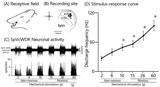
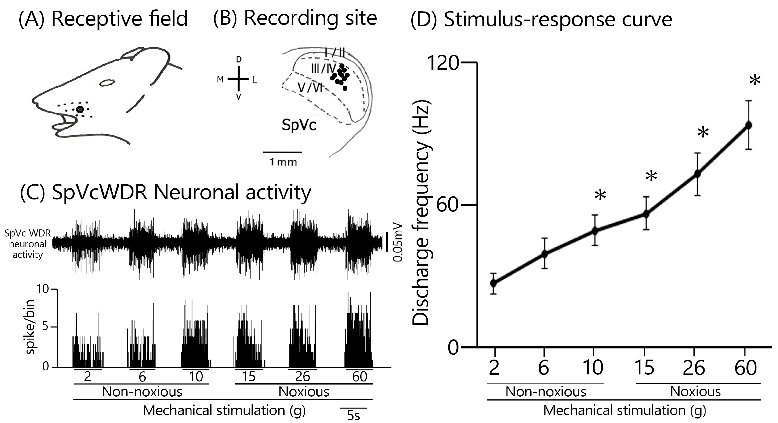
As illustrated in Figure 1A, the receptive fields of the recorded SpVc neurons were localized to the whisker pad and exhibited responsiveness to both non-noxious and noxious mechanical stimuli, consistent with previous reports [12]. The anatomical distribution of the recording sites was primarily concentrated within the maxillary branch area in laminas III–IV of the SpVc (Figure 1B). A representative histological confirmation of a recording site is shown in the inset of Figure 1B. No significant spatial differences in recording locations were observed between the PUE- and lidocaine-treated groups.
Figure 1.
Electrophysiological and topographical profiles of wide-dynamic-range (WDR) neurons in the spinal trigeminal nucleus caudalis (SpVc) under mechanical stimulation. (A) Representative receptive field (marked in black) localized to the ipsilateral facial vibrissal region. (B) Anatomical mapping of recorded SpVc WDR neurons sensitive to both innocuous and noxious mechanical inputs (n = 12). The inset displays a histological section verifying a representative recording site within the SpVc. D, dorsal; V, ventral; M, medial; L, lateral. (C) Typical extracellular waveforms (top) and corresponding post-stimulus time histograms (bottom) of an individual SpVc WDR neuron. Neuronal activity was elicited by graded mechanical stimuli, ranging from non-noxious (4, 6, and 10 g) to noxious intensities (15, 26, and 60 g), applied to the orofacial receptive field. (D) Stimulus–response relationship depicting the mean discharge frequency across varying mechanical intensities (n = 12). Data are expressed as mean ± SEM. * p < 0.05 denotes statistical significance compared to the baseline response at 2 g.
Typical examples of SpVc WDR neuronal firing patterns are presented in Figure 1C. These neurons demonstrated a characteristic increase in firing frequency that correlated positively with the intensity of mechanical stimulation applied to the most sensitive region of their receptive fields. Spontaneous activity was observed in 2 of the 12 recorded neurons. The relationship between mechanical stimulus intensity and the mean discharge frequency (stimulus–response curve) for these WDR units is depicted in Figure 1D. The mean threshold for mechanical stimulation-induced spikes was 1.9 ± 0.6 g. Based on these physiological properties, all recorded units were categorized as WDR neurons [12].
3.2. Impact of Peripheral PUE Administration on SpVc WDR Neuronal Responses to Mechanical Stimuli
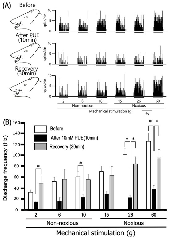
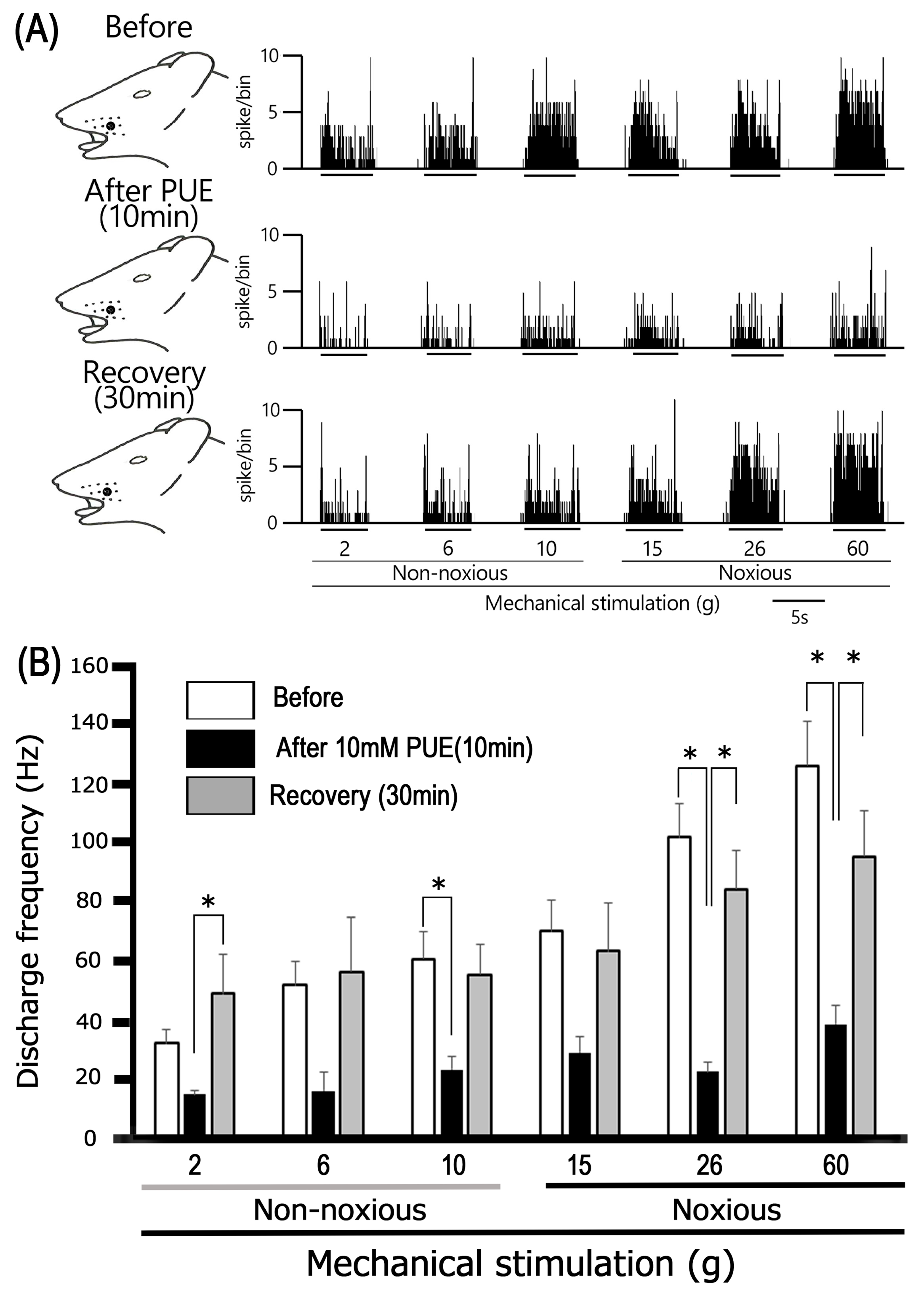
The suppressive effect of local subcutaneous PUE (10 mM) on the excitability of SpVc WDR neurons is exemplified in Figure 2A. Following the injection of 10 mM PUE into the receptive field center, neuronal firing in response to innocuous mechanical stimulation (4–10 g) was significantly attenuated at the 10 min interval. This inhibitory action was transient, as discharge rates reverted to pre-injection baseline levels by the 30 min mark. Notably, PUE administration did not significantly alter the mechanical activation threshold. The consolidated data for non-noxious stimulation are presented in Figure 2B. Mean firing rates elicited by innocuous stimuli showed a significant reduction post-PUE injection relative to controls (p < 0.05). Baseline recovery was consistently observed by 30 min. Furthermore, no discernible changes in spontaneous firing frequency were induced by PUE. As illustrated by the representative traces in Figure 2A, subcutaneous PUE also dampened SpVc WDR neuronal responses to noxious mechanical stimuli (15–60 g). These inhibitory effects reached their peak at 10 min post-administration and resolved within 30 min. Quantitative assessment (Figure 2B) verified a significant decrease in mean discharge frequency during noxious stimulation following PUE treatment (p < 0.05, n = 4). Conversely, local vehicle (DMSO) administration exerted no significant influence on either spontaneous or evoked activity (n = 3), consistent with previous reports [12].
Figure 2.
Inhibitory effects of local puerarin (PUE) administration on mechanical stimulus-evoked responses in SpVc WDR neurons. (A) Representative extracellular traces and post-stimulus histograms demonstrating SpVc WDR neuronal activity elicited by non-noxious (4, 6, and 10 g) and noxious mechanical stimulation (15, 26, and 60 g). Data are shown for baseline (pre-injection), and at 10 and 30 min following the subcutaneous delivery of 10 mM PUE into the peripheral receptive field. (B) Temporal profile of the inhibitory effect of local PUE on the mean discharge frequency across the range of stimulus intensities. The plot highlights a transient attenuation of neuronal firing, characterized by a peak reduction at 10 min and a return to baseline by 30 min post-administration. Data are expressed as mean ± SEM (n = 4). * p < 0.05 denotes a significant difference compared to the pre-injection baseline; * p < 0.05 indicates significant recovery at 30 min relative to the 10 min peak effect.
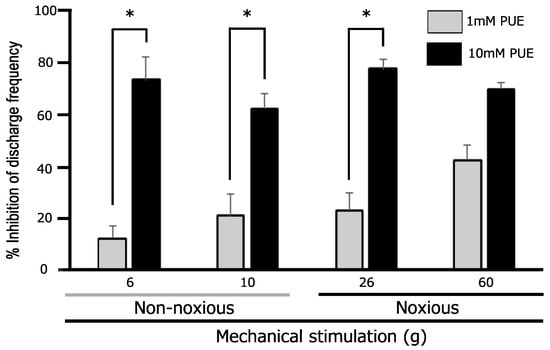
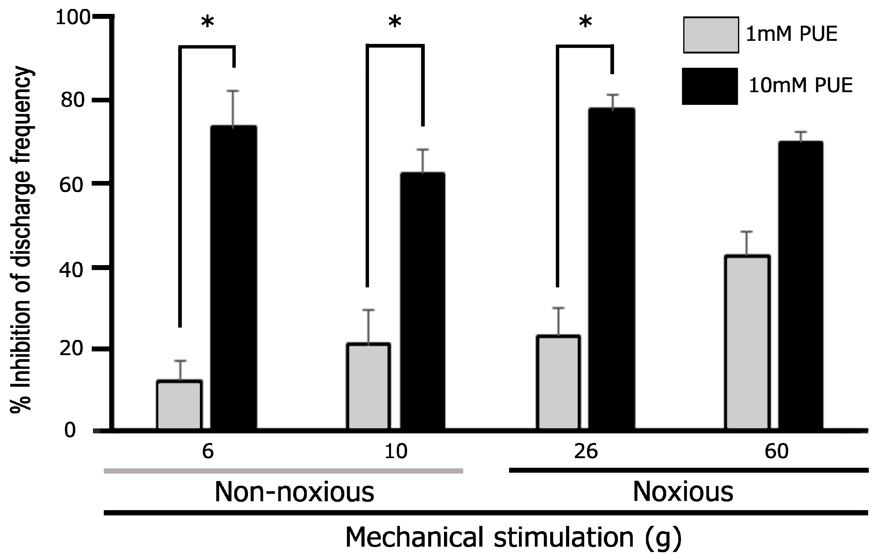
PUE administration resulted in a significant dose-dependent suppression (ranging from 1 to 10 mM) of SpVc WDR neuronal firing evoked by non-noxious mechanical stimulation (Figure 3). Specifically, a significant reduction in discharge frequency was observed when comparing the 0.1 mM dose to the 10 mM dose (p < 0.05%). Similarly, PUE exhibited a significant dose-dependent inhibitory effect on noxious mechanical stimulation-evoked responses within the 1–10 mM range (Figure 3). For these noxious stimuli, post hoc analysis revealed that both 1 mM and 10 mM concentrations significantly suppressed firing rates compared to the 0.1 mM dose (p < 0.05%).
Figure 3.
Concentration-dependent suppression of mechanical stimulus-evoked SpVc WDR neuronal activity by puerarin (PUE). The line graphs depict the mean discharge frequency of SpVc WDR neurons elicited by non-noxious, noxious stimuli across different concentrations of PUE (1 and 10 mM). PUE administration induced a significant, dose-dependent inhibition of neuronal responses across all mechanical stimulus intensities. Data are expressed as mean ± SEM (n = 4). * p < 0.05 denotes a significant difference between the 10 mM and 1 mM PUE treatment groups.
3.3. Comparative Analysis of PUE-Induced Inhibition on Noxious vs. Non-Noxious Mechanical Responses
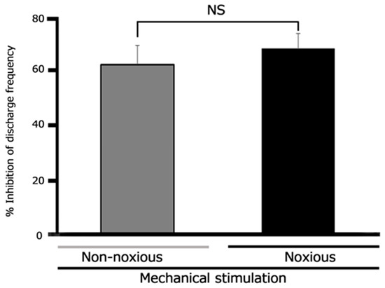
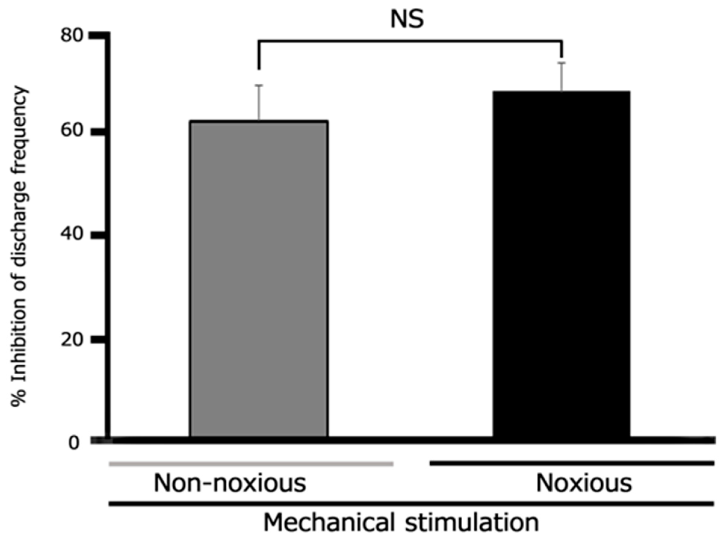
The inhibitory efficacy of subcutaneous PUE (10 mM) was evaluated by comparing its impact on responses elicited by non-noxious versus noxious mechanical stimuli. As depicted in Figure 4, the percentage reduction in mean discharge frequency following PUE administration showed no significant disparity between the two stimulus categories. These data suggest that PUE exerts a consistent inhibitory influence on SpVc WDR neuronal excitability, irrespective of the noxious or innocuous nature of the mechanical input.
Figure 4.
Comparative inhibitory efficacy of 10 mM puerarin (PUE) on non-noxious versus noxious mechanical stimulus-evoked responses. The bar graph illustrates the mean reduction in SpVc WDR neuronal discharge frequency following the administration of 10 mM PUE. The magnitude of inhibition did not differ significantly between responses elicited by innocuous and noxious stimulation, demonstrating that PUE exerts a comparable suppressive effect regardless of stimulus intensity. Data are expressed as mean ± SEM (n = 4). NS, not significant.
3.4. Comparative Effects of Lidocaine on Mechanical Stimulus-Evoked SpVc WDR Neuronal Responses
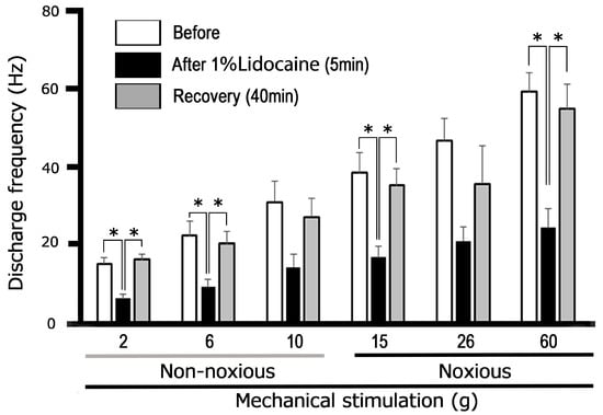
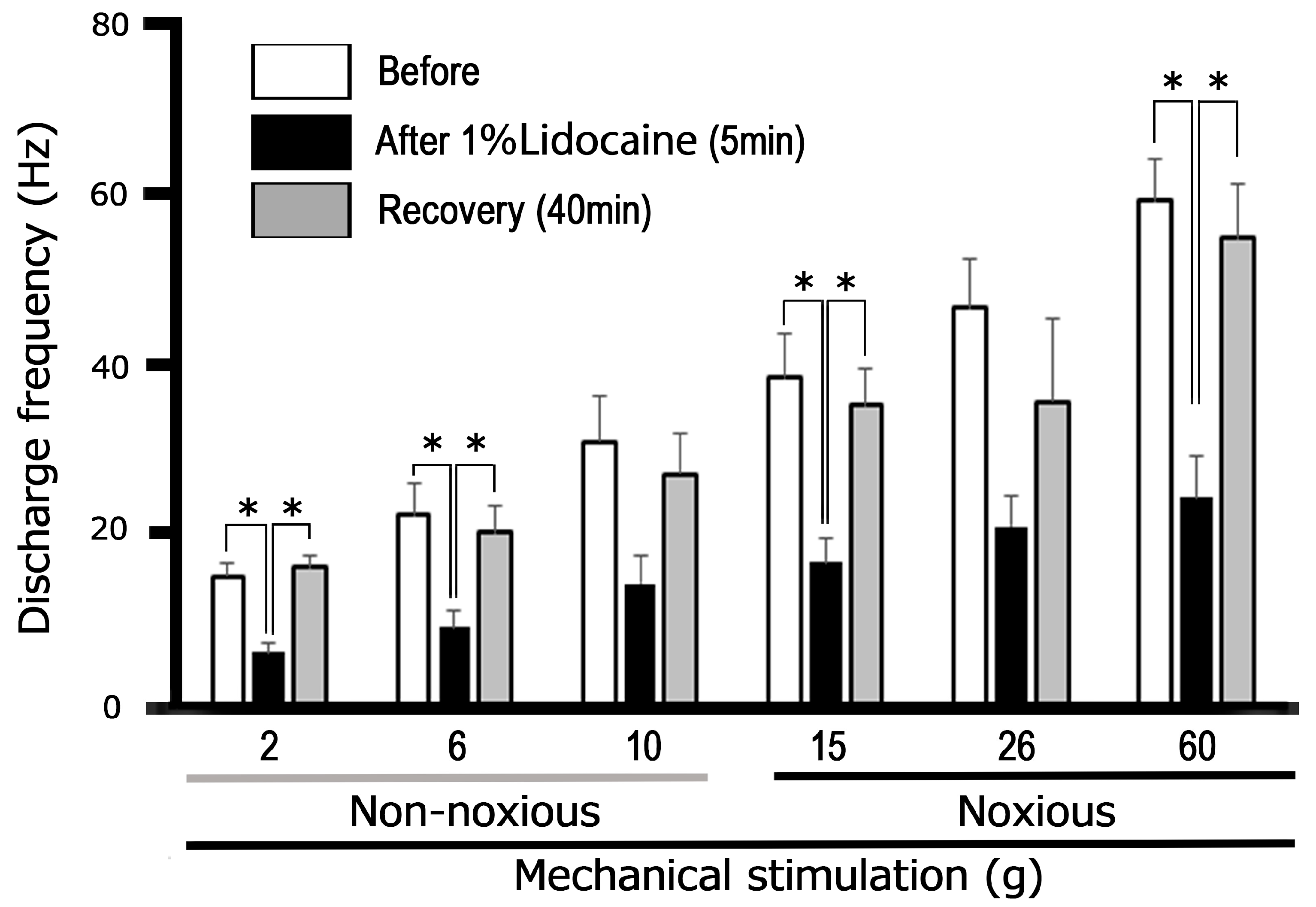
Finally, the inhibitory efficacy of PUE was benchmarked against that of lidocaine, a conventional local anesthetic. The impact of 1% lidocaine on mechanical stimulation-evoked activity is summarized in Figure 5. Following administration, a significant attenuation of mean firing rates for both innocuous and noxious stimuli was observed within 5 min (p < 0.05, n = 5). Notably, the duration of lidocaine-induced suppression was more extended than that of PUE; neuronal activity remained significantly depressed and returned to baseline levels only after approximately 40 min (p < 0.05; Figure 5).
Figure 5.
Inhibitory effects of local lidocaine administration on mechanical stimulation-evoked SpVc WDR neuronal excitability. The graphs illustrate the time-course of the effects of subcutaneous 1% lidocaine (37 mM) on the mean firing frequency of SpVc WDR neurons in response to non-noxious and noxious mechanical stimuli. Lidocaine administration significantly suppressed neuronal activity, with recovery occurring within approximately 40 min. Data are presented as mean± SEM (n = 5). * p < 0.05% indicates a significant difference when comparing the 5 min post-administration response to baseline (before); * p < 0.05% indicates a significant difference when comparing the 5 min post-administration response to the recovery point (40 min).
3.5. Comparative Potency of PUE and Lidocaine on Mechanical Stimulus-Evoked SpVc WDR Activity
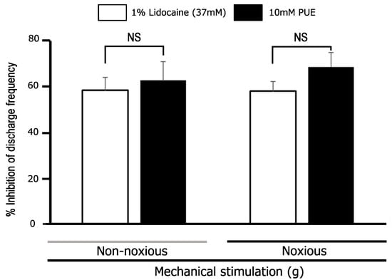
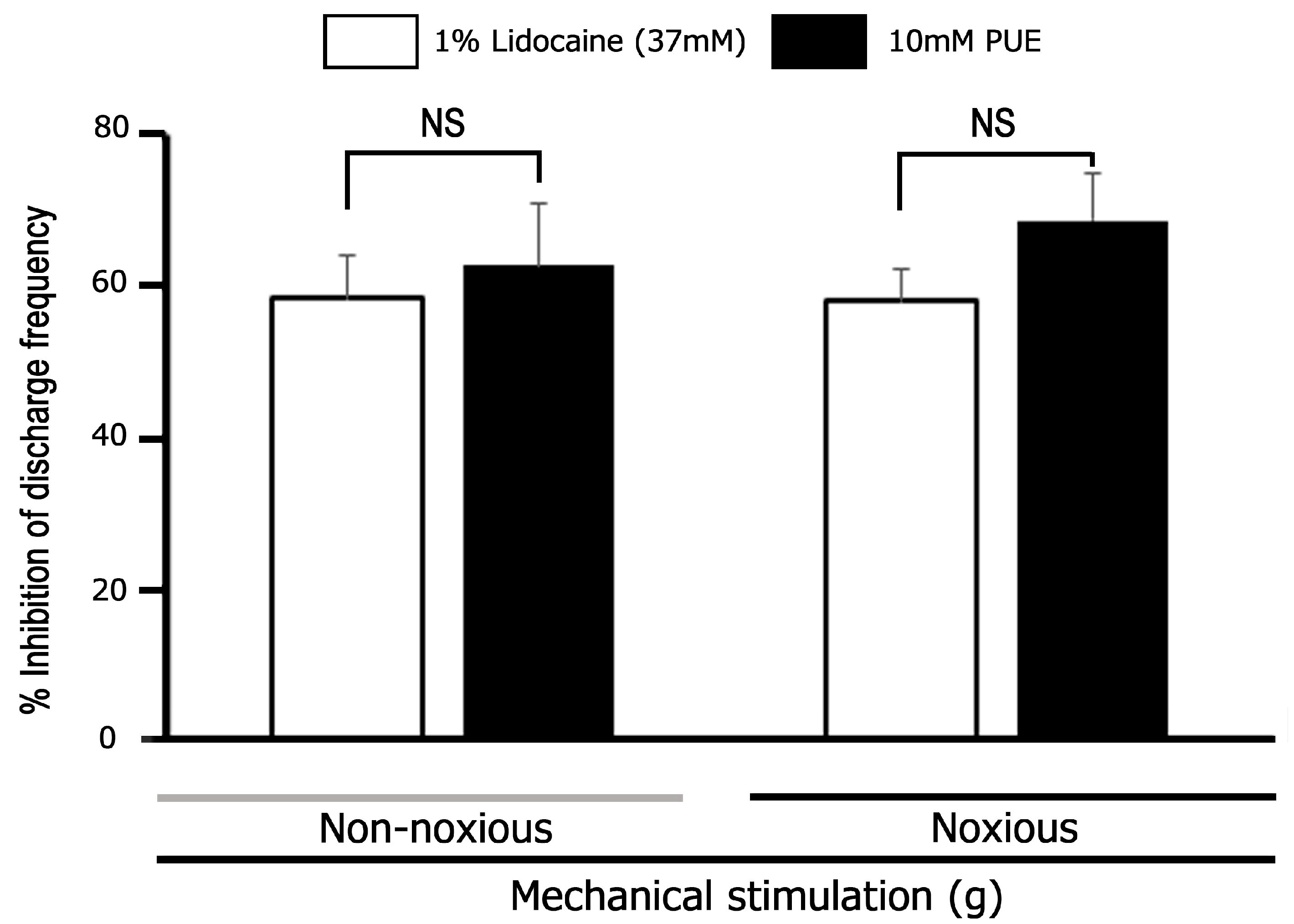
The inhibitory potency of 10 mM PUE on trigeminal nociceptive transmission was found to be comparable to that of 37 mM lidocaine. Quantitative analysis revealed that both agents achieved equivalent levels of mean inhibition, as illustrated in Figure 6.
Figure 6.
Comparative inhibitory potency of 10 mM Puerarin (PUE) and 1% lidocaine on SpVc WDR neuronal excitability. The bar graph compares the mean magnitude of inhibition in discharge frequency induced by PUE (10 mM) versus lidocaine (1% or 37 mM). The data demonstrate that the inhibitory effect of 10 mM PUE (n = 5) is equivalent to that of 37 mM lidocaine (n = 5), with no significant statistical difference observed between the two treatments. Data are presented as mean ± SEM. NS, not significant.
4. Discussion
4.1. Suppression of SpVc WDR Neuronal Excitability by Peripheral PUE Administration
This study primarily aimed to determine whether local PUE administration modulates the excitability of SpVc WDR neurons under physiological conditions, specifically in response to both noxious and innocuous mechanical stimuli. Our core findings demonstrate that: (i) peripheral injection of PUE (1–10 mM) induces a dose-dependent reduction in the mean discharge frequency of SpVc WDR neurons; (ii) this inhibitory action is reversible, with firing rates recovering to baseline within 30 min; and (iii) the vehicle (DMSO) lacks any significant effect on evoked neuronal activity. While in vitro evidence has established that 100 μM (0.1 mM) PUE significantly inhibits Nav currents in DRG neurons [18,19], the present in vivo study utilized a 10 mM concentration. This dose was selected based on the assumption that extracellular fluid dilution would yield a functional concentration of approximately 100 μM at the nociceptive terminals. To our knowledge, these results provide the inaugural in vivo evidence that local PUE application dampens the excitability of primary sensory afferents within the trigeminal system. This suppression is likely attributable to the blockade of Nav channels and ASICs residing on the peripheral terminals of TG neurons. Nevertheless, future investigations employing in vitro patch-clamp recordings from retrogradely labeled TG neurons innervating the whisker pad are necessary to definitively elucidate these underlying ionic mechanisms [23].
4.2. Peripheral Mechanisms Underlying the Suppression of SpVc Neuronal Excitability by PUE
Nociceptive signaling is fundamentally characterized by four sequential processes: (i) transduction, where external stimuli are converted at the peripheral terminal; (ii) generation, the initiation of action potentials; (iii) propagation, the conduction of potentials along the axon; and (iv) transmission, where the central terminal acts as the presynaptic element [2,24].
Nav channels, essential for the propagation of pain-related action potentials, are generally classified into two groups. Small-to-medium-sized nociceptive neurons selectively express TTX-R Nav channels, whereas large-to-medium-sized Aβ- and Aδ- sensory neurons primarily express TTX-sensitive (TTX-S) Nav channels [3,25]. Previous in vitro studies have demonstrated that PUR exerts a concentration-dependent inhibitory effect on Nav currents in DRG neurons, with an efficacy comparable to the local anesthetic lidocaine [18]. While PUE has been reported to show a stronger affinity for TTX-R Nav over TTX-S Nav currents in neuropathic pain models [19], our current in vivo data revealed a significant reduction in discharge frequency for both noxious and non-noxious stimuli. Interestingly, the mean inhibitory effect of PUE did not differ significantly between these stimulus types (62.8 ± 8.1% for non-noxious vs. 68.7 ± 6.4% for noxious). Given that PUE is known to suppress ASIC—a key candidate for mechanosensitive transduction—in hippocampal neurons [20], it is highly probable that peripheral PUE administration targets ASICs within the TG nerve terminals. By inhibiting both mechanosensitive transducer channels and voltage-gated Nav channels, PUR effectively attenuates generator potentials and subsequent action potential firing in both nociceptive and non-nociceptive TG neurons.
Furthermore, calcium-activated potassium (Kca) channels play a critical role in regulating neuronal excitability by linking intracellular Ca2+ concentrations to membrane potential [26,27,28,29]. An influx of Ca2+ triggers these channels, leading to K+ efflux and subsequent membrane hyperpolarization, which regulates action potential frequency and burst intervals [30]. Since PUE has been shown to act as a Kca channel opener in vascular smooth muscle cells [15], it may similarly activate Kca channels in sensory neurons, further dampening SpVc neuronal firing in response to mechanical stimulation. However, additional in vitro investigations are necessary to confirm the involvement of Kca channels in this specific analgesic pathway.
4.3. Functional Significance of PUE in the Modulation of Nociceptive SpVc Neuronal Activity
In recent years, there has been a burgeoning interest in CAM for the management of both acute and chronic pain [7,31,32,33]. Patients often seek CAM interventions when conventional Western pharmacological treatments prove ineffective or intolerable [6]. Notably, traditional local anesthetics can occasionally induce adverse effects on the central nervous and cardiovascular systems [14]. Consequently, the identification of bioactive dietary constituents that function as effective analgesics with minimal side effects represents a significant contribution to the field of CAM.
Lidocaine, a classic Nav channel blocker, remains the gold standard for local anesthesia. However, our recent in vivo studies have demonstrated that other dietary constituents, such as quercetin and (−)-epigallocatechin-3-gallate (EGCG), also effectively attenuate the excitability of trigeminal nociceptive primary neurons [34,35]. These studies revealed that: (i) quercetin and EGCG modulate generator potentials; (ii) they suppress Nav channels while potentiating Kv channels; and (iii) their inhibitory potency on nociceptive primary afferents follows the order: 10 mM Quercetin = 10 mM EGCG = 37 mM (1%) Lidocaine [34,35]. In the present study, we established that 10 mM PUE provides an inhibitory effect on SpVc WDR neuronal discharge frequency that is comparable to 37 mM lidocaine. This indicates that PUE possesses approximately four-fold the inhibitory potency of lidocaine on a molar basis. Taken together with our previous findings, the relative local analgesic potency for these compounds can be summarized as: PUE = Quercetin = EGCG (10 mM) > 1% Lidocaine (37 mM). In contrast to the broad action of lidocaine, the selective modulation of Nav (both TTX-R and TTX-S) and ASICs by PUE provides a more targeted approach to attenuating primary neuronal excitability. This specific pharmacological profile underscores PUE’s potential to overcome the clinical constraints associated with current local anesthetics, offering a refined and safer strategy for analgesia.
Furthermore, peripheral nerve damage caused by chemotherapy agents, such as paclitaxel, often leads to debilitating neuropathic pain [36,37]. Recent evidence suggests that local PUE administration dose-dependently attenuates paclitaxel-induced mechanical allodynia and thermal hyperalgesia by specifically inhibiting TTX-R Nav currents [19]. Our findings support the hypothesis that PUE preferentially blocks TTX-R Nav channels, thereby reducing nociceptive neuron excitability. This suggests a promising new clinical application for PUR in mitigating chemotherapy-induced peripheral neuropathy (CIPN), although further investigations are warranted to fully elucidate these therapeutic possibilities.
5. Conclusions
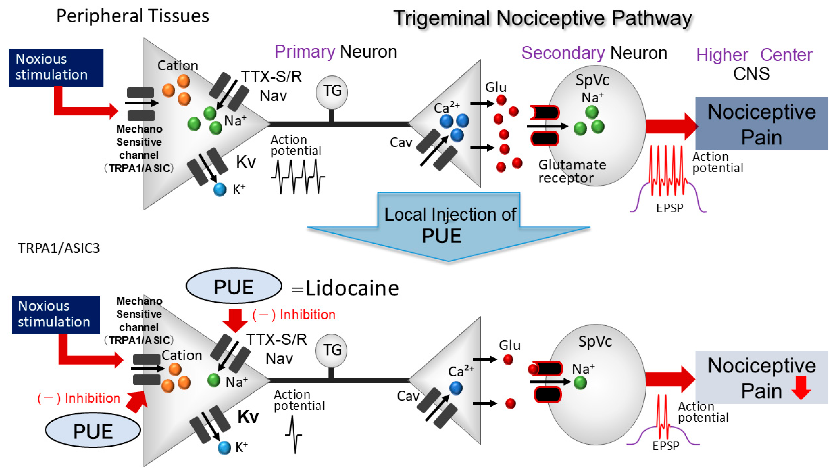
The present study provides the first in vivo evidence that local administration of PUE to peripheral receptive fields significantly attenuates the excitability of nociceptive second-order neurons within the SpVc. This inhibitory effect is likely mediated by the modulation of TTX-R/S Nav and ASICs at the peripheral terminals of TG neurons (Figure 7). While the specific cellular mechanisms in this in vivo model remain to be fully elucidated through further mechanistic studies, our findings underscore the pharmacological potential of PUE as a plant-derived analgesic. Given its favorable safety profile and low risk of systemic adverse effects, PUE represents a promising alternative or adjunct in the field of CAM. These results support the development of PUE-based therapies for managing clinical orofacial pain conditions, such as temporomandibular disorders and trigeminal neuralgia.
Figure 7.
Proposed mechanism underlying Puerarin (PUE)-mediated inhibition of SpVc WDR neuronal responses to mechanical nociception. Peripheral administration of PUE is hypothesized to suppress the initiation of both generator potentials and action potentials within nociceptive afferent terminals. This inhibitory action likely arises from the modulation of mechanosensitive and transducer ion channels, including acid-sensing ion channels (ASICs) and transient receptor potential ankyrin 1 (TRPA1), alongside the blockade of both tetrodotoxin-resistant (TTX-R) and tetrodotoxin-sensitive (TTX-S) voltage-gated sodium (Nav) channels. Notably, the pharmacological efficacy of PUE in attenuating these nociceptive signals is comparable to that of traditional Nav channel blockers, such as clinical local anesthetics.
Author Contributions
R.H.: conceptualization, writing—original draft, methodology, data curation, formal analysis, investigation. R.C.: conceptualization, writing—original draft, methodology, data curation, formal analysis, investigation. S.U.: conceptualization, writing—original draft, methodology, data curation, formal analysis, investigation. M.T.: conceptualization, funding acquisition, methodology, project administration, resources, supervision, validation, writing—original draft, writing—review and editing. All authors have read and agreed to the published version of the manuscript.
Funding
This study was supported by a Grant-in-Aid for Scientific Research (C) from the Japanese Society for the Production of Science (No. 22K10232).
Institutional Review Board Statement
All experiments reported herein were approved by the Animal Use and Care Committee of Azabu University on 8 February 2024 (No. 200529-3).
Informed Consent Statement
Not applicable.
Data Availability Statement
Data presented in this study are available on request from the corresponding author.
Conflicts of Interest
The authors declare no potential conflicts of interest with respect to the research, authorship, and/or publication of this article.
Abbreviations
The following abbreviations are used in this manuscript:
| TG | trigeminal ganglion |
| SpVc | spinal trigeminal nucleus caudalis |
| C1-C2 | upper cervical spinal cord |
| CAM | Complementary alternative medicine |
| Nav | voltage-gated Na+ |
| Kv | voltage-gated potassium |
| Kca | Ca2+-activated K+ |
| ASIC | acid-sensing Ion channel |
| PUE | Puerarin |
| TXX-R | tetrodotoxin-resistant |
| TTX-S | tetrodotoxin-sensitive |
| DMSO | dimethyl sulfoxide |
| EGCG | (−)-epigallocatechin-3-gallate |
| ANOVA | analysis of variance |
References
- Sessle, B.J. Peripheral and central mechanisms of orofacial pain and their clinical correlates. Minerva Anestesiol. 2005, 71, 117–136. [Google Scholar] [PubMed]
- Takeda, M.; Matsumoto, S.; Sessle, B.J.; Shinoda, M.; Iwata, K. Peripheral and central mechanisms of trigeminal neuropathic and inflammatory pain. J. Oral Biosci. 2011, 53, 318–329. [Google Scholar] [CrossRef]
- Iwata, K.; Takeda, M.; Oh, S.; Shinoda, M. Neurophysiology of Orofacial Pain. In Contemporary Oral Medicine; Farah, C.S., Balasubramaniam, R., McCullough, M.J., Eds.; Springer International Publishing: New York, NY, USA, 2017. [Google Scholar]
- Rao, J.K.; Mihaliak, K.; Kroenke, K.; Bradley, J.; Tierney, W.M.; Weinberger, M. Use of complementary therapies for arthritis among patients of rheumatologists. Ann. Intern. Med. 1999, 131, 409–416. [Google Scholar] [CrossRef] [Scilit] [PubMed]
- Konvicka, J.J.; Meyer, T.A.; McDavid, A.J.; Roberson, C.R. Complementary/alternative medicine use among chronic pain clinic patients. J. Perianesth. Nurs. 2008, 23, 17–23. [Google Scholar] [CrossRef] [Scilit]
- Rosenberg, E.I.; Genao, I.; Chen, I.; Mechaber, A.J.; Wood, J.A.; Faselis, C.J.; Kurz, J.; Menon, M.; O’Rorke, J.; Panda, M.; et al. Complementary and alternative medicine use by primary care patients with chronic pain. Pain Med. 2008, 9, 1065–1072. [Google Scholar] [CrossRef] [Scilit]
- Bauer, B.; Tilburt, C.; Sood, A.; Li, G.X.; Wang, S.H. Complementary and alternative medicine therapies for chronic pain. Chin. J. Integr. Med. 2016, 6, 403–411. [Google Scholar] [CrossRef] [Scilit] [PubMed]
- Shir, Y.; Raja, S.N.; Weissman, C.S.; Campbell, J.N.; Seltzer, Z. Consumption of soy diet before nerve injury preempts the development of neuropathic pain in rats. Anesthesiology 2001, 95, 1238–1244. [Google Scholar] [CrossRef] [Scilit] [PubMed]
- Ernst, E. Complementary medicine. Curr. Opin. Rheumatol. 2003, 15, 151–155. [Google Scholar] [CrossRef] [Scilit]
- Frémont, L. Biological effects of resveratrol. Life Sci. 2000, 66, 663–673. [Google Scholar] [CrossRef] [Scilit]
- Pervaiz, S. Resveratrol: From grapevines to mammalian biology. FASEB J. 2003, 17, 1975–1985. [Google Scholar] [CrossRef] [Scilit]
- Yamaguchi, M.; Kinouchi, R.; Morizumi, S.; Shmazu, Y.; Takeda, M. Local administration of genistein as a local anesthetic agent inhibits the trigeminal nociceptive neuronal activity in rats. Brain Res. Bull. 2021, 172, 120–128. [Google Scholar] [CrossRef] [Scilit]
- Kakita, K.; Tsubouchi, H.; Adachi, M.; Takehana, S.; Shimazu, Y.; Takeda, M. Local subcutaneous injection of chlorogenic acid inhibits the nociceptive trigeminal spinal nucleus caudalis neurons in rats. Neurosci. Res. 2018, 134, 49–55. [Google Scholar] [CrossRef] [Scilit]
- Miller, R.D. Chapter 26, Local Anesthetics. In Basic & Clinical Pharmacology, 6th ed.; Katzung, B.G., Ed.; Lange Medical Books/McGraw-Hill: New York, NY, USA, 1996; pp. 425–433. [Google Scholar]
- Gao, Q.; Yang, B.; Ye, Z.G.; Wang, J.; Bruce, I.C.; Xia, Q. Opening the calcium-activated potassium channel participates in the cardioprotective effect of puerarin. Eur. J. Pharmacol. 2007, 574, 179–184. [Google Scholar] [CrossRef] [Scilit]
- Yeung, D.K.Y.; Leung, S.W.S.; Xu, Y.C.; Vanhoutte, P.M.; Man, R.Y.K. Puerarin, an isoflavonoid derived from Radix puerariae, potentiates endothelium-independent relaxation via the cyclic AMP pathway in porcine coronary artery. Eur. J. Pharmacol. 2006, 552, 105–111. [Google Scholar] [CrossRef] [Scilit]
- Zhang, G.Q.; Hao, X.M.; Dai, D.Z.; Fu, Y.; Zhou, P.A.; Wu, C.H. Puerarin blocks Na+ current in rat ventricular myocytes. Acta Pharmacol. Sin. 2003, 24, 1212–1216. [Google Scholar] [PubMed]
- Ji, H.L.; Wang, T.Y. Puerarin inhibits tetrodotoxin-resistant sodium current in dorsal root ganglion neurons. Acta Pharmacol. Sin. 1996, 17, 115–118. [Google Scholar]
- Zhang, X.L.; Cao, X.Y.; Lai, R.C.; Xie, M.X.; Zeng, W.A. Puerarin Relieves Paclitaxel-Induced Neuropathic Pain: The Role of Nav1.8 β1 Subunit of Sensory Neurons. Front. Pharmacol. 2019, 9, 1510. [Google Scholar] [CrossRef] [Scilit]
- Gu, L.; Yang, Y.Y.; Sun, Y.; Zheng, X. Puerarin inhibits acid-sensing ion channels and protects against neuron death induced by acidosis. Planta Med. 2010, 76, 583–588. [Google Scholar] [CrossRef] [Scilit] [PubMed]
- Zimmermann, M. Ethical guidelines for investigations of experimental pain in conscious animals. Pain 1983, 16, 109–110. [Google Scholar] [CrossRef] [Scilit]
- Paxinos, G.; Watson, C. The Rat Brain in Stereotaxic Coordinates, 2nd ed.; Academic Press: New York, NY, USA, 1986. [Google Scholar]
- Nakagawa, K.; Takeda, M.; Tsuboi, Y.; Kondo, M.; Kitagawa, J.; Matsumoto, S.; Kobayashi, A.; Sessle, B.J.; Shinoda, M.; Iwata, K. Alternation of primary afferent activity following inferior alveolar nerve transection in rats. Mol. Pain 2010, 6, 9. [Google Scholar] [CrossRef] [Scilit] [PubMed]
- Harriott, A.M.; Gold, M.S. Contribution of primary afferent channels to neuropathic pain. Curr. Pain Headache Rep. 2009, 13, 197–207. [Google Scholar] [CrossRef] [Scilit]
- Akopian, A.N.; Sivilotti, L.; Wood, J.N. A tetrodotoxin-resistant voltage-gated sodium channel expressed by sensory neurons. Nature 1996, 379, 257–262. [Google Scholar] [CrossRef] [Scilit]
- Poolos, N.P.; Johnston, D. Calcium-activated potassium conductances contribute to action potential repolarization at the soma but not the dendrites of hippocampal CA1 pyramidal neurons. J. Neurosci. 1999, 19, 5205–5212. [Google Scholar] [CrossRef] [Scilit]
- Martinez-Pinna, J.; Davies, P.J.; McLachlan, E.M. Diversity of channels involved in Ca2+ activation of K+ channels during the prolonged AHP in guinea-pig sympathetic neurons. J. Neurophysiol. 2000, 84, 1346–1354. [Google Scholar] [CrossRef] [Scilit] [PubMed]
- Pedarzani, P.; Kulik, A.; Muller, M.; Ballanyi, K.; Stocker, M. Molecular determinants of Ca2+-dependent K+ channel function in rat dorsal vagal neurones. J. Physiol. 2000, 527, 283–290. [Google Scholar] [CrossRef] [Scilit] [PubMed]
- Zhang, X.F.; Gopalakrishnan, M.; Shieh, C.C. Modulation of action potential firing by iberiotoxin and NS1619 in rat dorsal root ganglion neurons. Neuroscience 2003, 122, 1003–1011. [Google Scholar] [CrossRef] [Scilit]
- Faber, E.S.; Sah, P. Calcium-activated potassium channels: Multiple contributions to neuronal function. Neuroscientist 2003, 9, 181–194. [Google Scholar] [CrossRef] [Scilit]
- Cady, R.J.; Hirst, J.J.; Durham, P.L. Dietary grape seed polyphenols repress neuron and glia activation in trigeminal ganglion and trigeminal nucleus caudalis. Mol. Pain 2010, 6, 1744–8069. [Google Scholar] [CrossRef] [Scilit]
- Boadas-Vaello, P.; Vela, J.M.; Verdú, E. New pharmacological approaches using polyphenols on the physiopathology of neuropathic pain. Curr. Drug Targets 2017, 18, 160–173. [Google Scholar] [CrossRef] [Scilit] [PubMed]
- Cornelison, L.E.; Chelliboina, N.; Woodman, S.E.; Durham, P.L. Dietary supplementation with grape seed extract prevents development of trigeminal sensitization and inhibits pain signaling in a preclinical chronic temporomandibular disorder model. J. Oral Pathol. Med. 2020, 49, 514–521. [Google Scholar] [CrossRef] [Scilit]
- Toyota, R.; Itou, H.; Sashide, Y.; Takeda, M. Suppression of the excitability of rat nociceptive primary sensory neurons following local administration of the phytochemical quercetin. J. Pain 2023, 24, 540–549. [Google Scholar] [CrossRef] [Scilit] [PubMed]
- Utugi, S.; Chida, R.; Yamaguchi, S.; Sashide, Y.; Takeda, M. Local Administration of (−)-Epigallocatechin-3-Gallate as a Local Anesthetic Agent Inhibits the Excitability of Rat Nociceptive Primary Sensory Neurons. Cells 2025, 14, 52. [Google Scholar] [CrossRef] [Scilit] [PubMed]
- Cavaletti, G.; Cavalletti, E.; Oggioni, N.; Sottani, C.; Minoia, C.; D’Incalci, M.; Zucchetti, M.; Marmiroli, P.; Tredici, G. Distribution of paclitaxel within the nervous system of the rat after repeated intravenous administration. Neurotoxicology 2000, 21, 389–393. [Google Scholar] [CrossRef] [Scilit]
- Dougherty, P.M.; Cata, J.P.; Cordella, J.V.; Burton, A.; Weng, H.R. Taxol-induced sensory disturbance is characterized by preferential impairment of myelinated fiber function in cancer patients. Pain 2004, 109, 132–142. [Google Scholar] [CrossRef] [Scilit] [PubMed]
Disclaimer/Publisher’s Note: The statements, opinions and data contained in all publications are solely those of the individual author(s) and contributor(s) and not of MDPI and/or the editor(s). MDPI and/or the editor(s) disclaim responsibility for any injury to people or property resulting from any ideas, methods, instructions or products referred to in the content. |
© 2026 by the authors. Licensee MDPI, Basel, Switzerland. This article is an open access article distributed under the terms and conditions of the Creative Commons Attribution (CC BY) license.