


Abstract
Oral mucositis is an inflammatory and ulcerative disease that affects patients undergoing cancer treatment, namely chemotherapy and radiotherapy, significantly compromising their quality of life. Despite its high incidence and clinical impact, current therapeutic options remain limited, reinforcing the need for new treatment approaches. In this context, the Instituto Português de Oncologia do Porto FG, EPE (IPO-Porto) developed an innovative mouthwash containing dexamethasone sodium phosphate (DSP), ascorbic acid, and sodium hyaluronate. In this work the physicochemical stability of this formulation was evaluated to ensure its quality and safety as compounded medicine. Exploratory studies were performed under accelerated aging conditions (40 ± 2 °C), followed by stability studies under real-time conditions (20 ± 2 °C) and refrigeration conditions (4 ± 2 °C), as well as in-use stability studies simulating daily patient handling. At each sampling time, physicochemical characterization was carried out (pH, color, odor, and precipitate formation) as well as the DSP quantification using high-performance liquid chromatography. The acceptable range of 90.0–110.0%, compared to the initial concentration of DSP, was used as the primary stability criterion. Results showed that the formulation remained stable at high temperatures (40 °C), on short exposures times (≤6 h), and for 30 days at 4 °C in closed amber glass bottles. In-use stability studies performed during the 7 days after opening (considering three daily applications) showed that the DSP concentration remained above 90%. The results obtained enabled us to establish a definition of storage conditions and beyond-use recommendations, since they showed that the prepared formulation is stable when stored under refrigeration. Further studies are currently being developed with a view to its clinical use as a compounded mouthwash in a hospital setting for the treatment of oral mucositis.
1. Introduction
Cancers are one of the most devastating classes of human pathologies, leading to millions of deaths each year around the globe. Although significant advances have been made in the fight against this group of diseases, cancer remains a key public health concern and a tremendous burden on European Union (EU) societies. In 2021, cancer was the second leading cause of death in the EU, with 1.1 million deaths [1]. Estimated cancer incidence in Portugal, while around the EU average, is projected to increase 20% by 2040, with the country currently leading EU rates for stomach and pediatric cancers [2].
Despite the emergence of advanced therapies such as immunotherapy and gene therapy, traditional treatments—surgery, radiotherapy, chemotherapy, and hormonal therapy—remain the most commonly adopted due to their cost-effectiveness and widespread use. Chemotherapy (CT) is one of the most widely used conventional approaches to cancer treatment. It targets key cell cycle phases to induce cell death. Radiotherapy (RT) is a highly effective cancer treatment that uses high-energy particles, such as X-rays, gamma rays, and protons, to destroy cancer cells by damaging their DNA [3]. However, chemotherapy and radiotherapy can be associated with adverse effects that are typically managed in the outpatient setting [4,5,6,7].
Oral mucositis (OM) is a common and feared adverse effect in patients with cancer who undergo anticancer treatment. The incidence and severity of OM depend on treatment regimen, patient-related factors, and the oral hygiene protocol followed, among others [5]. It is estimated that OM affects around 40% to 80% of patients undergoing CT and 59.4% to 100% of patients with head and neck cancer who are treated with RT [8,9]. The real frequency of OM may be underestimated, since its reporting is influenced by the heterogeneity of clinical studies and the diversity of diagnostic criteria adopted [4]. Mucositis presents as erythema, edema, or ulceration and may be associated with symptoms ranging from mild burning sensations to extensive, painful ulcers that adversely affect patients’ quality of life and impair basic oral functions, including speech, saliva swallowing, and eating [10,11]. In the most severe cases, severe pain and the inability to eat orally may require the use of parenteral nutrition [12]. In addition, mechanical factors such as sharp teeth, bruxism, food debris, foreign substances, and microorganisms can aggravate the condition [4]. It should be noted that the breakdown of the oral mucosa barrier, often caused by these factors, increases the risk of local or systemic infections, prolongs hospitalizations, and can interrupt cancer treatment [11].
The severity of OM is commonly graded using two primary standardized systems. The World Health Organization (WHO) scale is the most widely used in clinical research, as it combines objective clinical findings with functional outcomes, like the patient’s ability to eat. In routine clinical practice, the Common Terminology Criteria for Adverse Events (CTCAE) developed by the United States National Cancer Institute is frequently employed, which contemplate the patient symptoms, oral intake capacity, and the need for treatment measures [10,12]. These tools help healthcare providers determine the severity of mouth sores and guide nutritional and pain management.
The management of OM remains a clinical challenge due to its complex biological nature, and no universally effective treatment is currently available [10,13]. Existing therapeutic approaches primarily focus on symptom relief and the reduction in associated complications [13]. According to the clinical guidelines of the Multinational Association of Supportive Care in Cancer and the International Society of Oral Oncology (MASCC/ISOO), the implementation of oral hygiene protocols is widely recognized as a preventive strategy in OM management, as they promote oral health and improve patient comfort. These measures include mechanical cleaning (toothbrushing, flossing, etc.), the use of mouthwashes to reduce bacterial accumulation, and maintaining oral hydration and lubrication using moisturizing agents. While there are no strict guidelines on specific mouthwash formulations, experts generally recommend using bland rinses and moisturizers, such as saline or sodium bicarbonate solutions [14]. Palifermin, a recombinant keratinocyte growth factor, is another preventive option. It protects the oral lining, reduces cell death, and has been shown to lower the risk and severity of OM, particularly in patients receiving high-dose chemotherapy or total body irradiation before stem cell transplantation [15]. Other non-pharmacological interventions, such as photobiomodulation (low-level laser therapy) and oral cryotherapy, have shown efficacy in preventing and reducing OM severity, although their use is limited to specific clinical settings [14].
Pain control is one of the main therapeutic priorities in OM management, as it aims to relieve symptoms of already established lesions. Among the topical pharmacological strategies recommended by MASCC/ISOO for pain management are mouthwashes containing analgesics such as 0.2% topical morphine or 0.5% doxepin. Other topical agents, such as sucralfate, form a protective layer over the oral mucosa, but MASCC/ISOO guidelines do not recommend their use for the prevention or treatment of OM in patients undergoing CT or RT, as there is insufficient evidence of efficacy [16]. In more severe cases, systemic medications may be required, including opioids or even transdermal fentanyl [13,16]. Even so, both topical and systemic options only act on symptomatic control and do not act on the underlying cause of the inflammation and do not prevent the lesion from progressing.
For this reason, pharmacological strategies that target the pathophysiological mechanisms of OM have been explored. Among these are anti-inflammatory agents, investigated for their ability to modulate the characteristic inflammatory response of OM [17]. Topical anti-inflammatory mouthwashes such as benzydamine, which has anti-inflammatory, analgesic, anesthetic, and antimicrobial properties, have been recommended for the prevention of OM [10]. However, their applicability is limited in patients receiving higher doses of RT or concomitant CT, making the use of this drug unfeasible [12,16].
Corticosteroids have shown efficacy in reducing the incidence and severity of OM due to their anti-inflammatory and immunosuppressive properties [17,18]. Dexamethasone sodium phosphate (DSP) is a potent synthetic glucocorticosteroid widely used as an anti-inflammatory and immunosuppressive agent and is included in the WHO’s list of essential medicines due to its safety and therapeutic effectiveness [19,20]. Clinical studies have shown that the use of DSP mouthwash can significantly reduce the incidence and severity of CT-induced OM [17].
Given the multifactorial nature of OM, controlling inflammation alone may not be sufficient. Therefore, it is important to develop mouthwashes that combine the proven anti-inflammatory efficacy of DSP with other components capable of offering additional benefits, such as protecting the mucosa, exerting an antioxidant action, and improving patient comfort. This integrated approach could represent an important advance over the options currently available.
In this sense, the combination of hyaluronic acid, ascorbic acid, and DSP in the same formulation may potentiate the anti-inflammatory effect and accelerate tissue repair. Regarding safety, both hyaluronic acid and topical dexamethasone exhibit excellent safety profiles in clinical studies, with no reports of serious adverse events at the doses used [21,22,23,24]. Hyaluronic acid has demonstrated efficacy in reducing the severity of oral mucositis by decreasing oxidative stress, cellular apoptosis, and the expression of inflammatory cytokines. It exhibits mucoadhesive properties and promotes wound healing, while also playing a role in the modulation of inflammation and tissue regeneration [25,26,27,28,29]. In addition, by imparting viscosity to aqueous solutions, hyaluronic acid increases the retention time of dexamethasone on the buccal mucosa and may help maintain moisture on affected tissues, providing a soothing effect that can potentially improve patient comfort and adherence during treatment. Ascorbic acid is a well-known antioxidant agent, capable of protecting the mucosa and preventing oxidative processes.
Given the limitations of currently available treatments, Instituto Português de Oncologia do Porto FG, EPE (IPO-Porto) developed an innovative compounded mouthwash containing DSP, ascorbic acid (AA), and sodium hyaluronate (SH), with the aim of improving the management of oral mucositis induced by anticancer therapies in in- and outpatients. Pharmaceutical compounding corresponds to the preparation of medicines to meet the specific needs of patients, which cannot be met by the commercially available medicines provided by the pharmaceutical industry. These formulations are prepared in hospital or community pharmacies following predefined operational protocols and subjected to quality control. To define a beyond-use date, it is essential to assess the stability of the formulation to ensure that its quality and, consequently, its efficacy and safety are maintained throughout the entire period of use [30]. Currently, there is no systematic data on the physicochemical stability of the combination of DSP, HA, and AA, which represents a critical barrier to the safe development, storage, and use of this type of formulation in clinical practice. Therefore, in addition to ensuring the strength and quality control of the compounded preparation, this study aimed to evaluate the compatibility of its chemical constituents and the physicochemical stability of the mouthwash under different storage and in-use conditions in order to estimate its beyond-use date and define optimal storage conditions.
2. Materials and Methods
2.1. Reagents and Samples
2.1.1. Chemicals
All chemicals used were of pharmaceutical grade. Dexamethasone sodium phosphate (DSP) and sodium hyaluronate (SH) were supplied by Fagron (Barcelona, Spain) and ascorbic acid (AA) by Acofarma (Barcelona, Spain). Chemicals used in the chromatographic analysis were of analytical reagent grade and used without any further purification. HPLC-grade acetonitrile, methanol, and tetrahydrofuran (THF) were obtained from Carlo Erba Reagents (Val de Reuil, France) and Fisher Scientific (Porto Salvo, Portugal). Phosphoric acid was obtained from Sigma-Aldrich (Algés, Portugal) and sodium hydroxide from Riedel-de Haën™ (Seelze, Germany). Water from a Direct Q® system (Merck Millipore, Molsheim, France), with a specific conductance less than 0.1 µS·cm−1 was used throughout.
2.1.2. Composition of the Mouthwash Formulation
The composition of the oral mouthwash is described in Table 1. In addition to the anti-inflammatory and immunosuppressive corticosteroid (DSP), the solution includes a protector of oral mucosa that accelerates wound healing (SH) and an antioxidant (AA), which preserves cells against reactive oxygen species (ROS) and favors wound healing. The amount of each of the excipients used was defined according to the usual levels in pharmaceutical formulations [31]. The formulation was prepared using magnetic stirring at 20 °C until complete dissolution (approximately 30 min).
Table 1.
Composition (% (w/v)) and function of the constituents of the mouthwash formulation.
2.2. Study of the Physicochemical Stability of the Oral Mouthwash Formulation
2.2.1. Sample Preparation and Storage Conditions
Three batches of the mouthwash formulation were prepared, at the IPO-Porto pharmaceutical services, poured into 500 mL type III amber glass bottles and sealed with a screw-on cap. The flasks were stored at a pre-determined temperature until analysis. For each batch, one bottle was reserved exclusively for analyses at the initial time point (time 0), serving as a reference for stability studies. The remaining bottles were stored under different storage conditions and kept closed until the moment of analysis: 4 ± 2 °C (refrigerator), 20 ± 2 °C (incubator), and 40 ± 2 °C (incubator). These conditions were selected according to guidelines for stability studies of compounding medicines, namely the USP Compounding recommendations [37], to simulate different conservation scenarios.
The initial assays (time 0) were carried out immediately after sample preparation. Samples stored at 4 °C and 20 °C were analyzed after 30 days of storage and the results obtained were compared with the results from the initial analysis (time 0). After 30 days of storage at 4 °C, an in-use stability study was performed. Over the following 7 days, three 10 mL aliquots of the mouthwash formulation (corresponding to the maximum recommended dose for treatment) were withdrawn daily from each flask, simulating the conditions of use in a hospital setting (in-use stability). At the end of the 7 days, the remaining solution in the bottle was analyzed. The samples stored at 40 °C were analyzed immediately after preparation and also after 6 h, 24 h, 48 h, and 168 h (7 days) of storage at this temperature.
Additionally, in order to understand the influence of each excipient (AA and SH) on the stability of DSP (Active Pharmaceutical Ingredient; API) under accelerated conditions, stability exploratory studies were performed at 40 ± 2 °C. For this purpose, three solutions were prepared, at the same concentrations of the mouthwash formulation, using ultrapure water as the solvent: DSP (control), and two binary mixtures (DSP plus AA, and DSP plus SH). The prepared solutions were stored at 40 °C and analyzed over time.
2.2.2. Physicochemical Stability Evaluation
For each bottle of mouthwash formulation, kept under different storage conditions, the pH, appearance (including precipitate formation), color, odor, and DSP content were determined (with the frequency described in the previous section). Prior to HPLC analysis, all samples were filtered through 0.45 µm syringe filters.
For the purposes of this study, the formulation was considered stable if DSP content remained within ≥90% of its initial concentration, consistent with USP guidelines (90–110% of the initial value) [38].
Chromatographic Conditions Used for DSP Assay
The analytical quantification of DSP was carried out by HPLC according to the procedure described in the European Pharmacopeia 11.0 [39]. For this purpose, a Shimadzu (Kyoto, Japan) LC-20AD chromatographic system was used, equipped with a DGU-20A5 degasser, an SPD-M2OA photodiode array detector, and a CTO-10AS VP column oven. Chromatographic separation was achieved using an EC 250/4 Nucleosil® 100-5 C18 column (250 mm × 4 mm, 5 µm) from Macherey-Nagel (Düren, Germany), thermostated at 25 °C. Isocratic elution was performed using a mobile phase consisting of a mixture of 520 mL ultrapure water, 2 mL phosphoric acid (pH adjusted to 2.6), 36 mL THF, and 364 mL methanol and was delivered at a flow rate of 1.0 mL/min. The mobile phase was previously degassed and filtered through a nylon membrane filter (47 mm, 0.45 µm) from MontaMil® (Knebel, Denmark). Detection was performed at the wavelength of 242 nm, and the sample injection volume was 20 μL. Data acquisition was carried out using the Shimadzu LC Solution software (Shimadzu, Kyoto, Japan; https://www.shimadzu.com).
2.3. Method Validation and System Suitability
The International Council for Harmonization (ICH) [40] and United States Pharmacopeia (USP) [41] criteria were followed in the validation of the developed HPLC method for the quantification of DSP. Parameters examined included linearity, precision, specificity, sensitivity, detection limit (LD), quantification limit (LQ), and system suitability.
Initially, to evaluate the chromatographic profile of each compound present in the mouthwash, standard solutions of each substance (DSP at 100 µg/mL, SH at 1000 µg/mL and AA at 1000 µg/mL) were analyzed in triplicate.
Linearity was assessed using three independent calibration curves for DSP standards. A 600 µg/mL DSP stock solution was prepared by dissolving the appropriate amount of the compound in 25.0 mL of ultrapure water. The working standard solutions were prepared by dilution of the aforementioned solution, in ultrapure water: 50, 75, 100, 125, and 150 µg/mL. All standard solutions were filtered through 0.45 µm syringe filters and analyzed in triplicate. Calibration curves were obtained by plotting the chromatographic peak areas against the concentration of the DSP working standard solutions. The data obtained were subjected to regression analysis using the least-squares method.
Repeatability (intra-day precision) and intermediate precision (inter-day precision) were used to assess the analytical method’s precision. The method’s intra- and inter-day precision was examined through the analysis of six independent replicates of a 100 µg/mL DSP sample solution. The precision of the analytical method was expressed as the coefficient of variation in the measurements. The LD and LQ were obtained from the standard deviation of the analyte response and the slope of the calibration curve, following the criteria established in the ICH Q2(R1) guideline [40].
Specificity is often demonstrated using spiking experiments, where the pure substance is spiked with impurities and components of the matrix (e.g., excipients, preservatives, antioxidants, etc.) and appropriate resolution is demonstrated [40]. The peak area of a DSP standard solution (100 µg/mL) was compared with the data obtained for a mixture containing 100 µg/mL of DSP, 1000 µg/mL SH, and 1000 µg/mL AA to ensure that SH and AA did not affect the measurement.
System suitability test was carried out to confirm that the chromatographic system was appropriate for the analysis, by injecting three replicates of a standard solution containing DSP, AA, and SH (DSP 100 µg/mL, SH 1000 µg/mL and AA 1000 µg/mL). Identification of system suitability parameters was performed for the retention time, peak resolution, peak area, and tailing factor.
3. Results
3.1. Chromatographic Profile of DSP, AA, and SH
The chromatogram obtained for a 100 µg/mL DSP standard solution showed, at a wavelength of 242 nm, a single peak at 15.90 min (CV = 1.16%), with an average peak area of 3,115,493 a.u. (CV = 1.30%), demonstrating good repeatability according to the generally accepted criterion (CV ≤ 2%) [42].
Injection of a 1000 µg/mL SH solution did not produce a well-defined chromatographic peak at 242 nm. The spectral analysis of the SH solution revealed a maximum absorbance at 205 nm, which is consistent with the literature [34,43]. At 242 nm, SH showed a small peak with a mean retention time of 1.19 min (CV = 1.38%) and an average area of 24,068 a.u. (CV = 0.99%). Injection of a 1000 µg/mL AA standard solution produced a well-defined peak with a mean retention time of 2.24 min (CV = 0.42%) and an average area of 50,188,492 a.u. (CV = 0.63%).
3.2. System Suitability
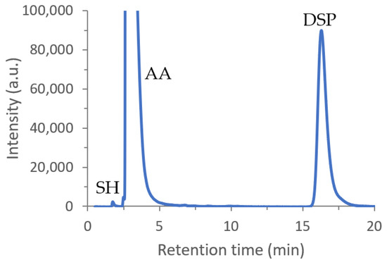
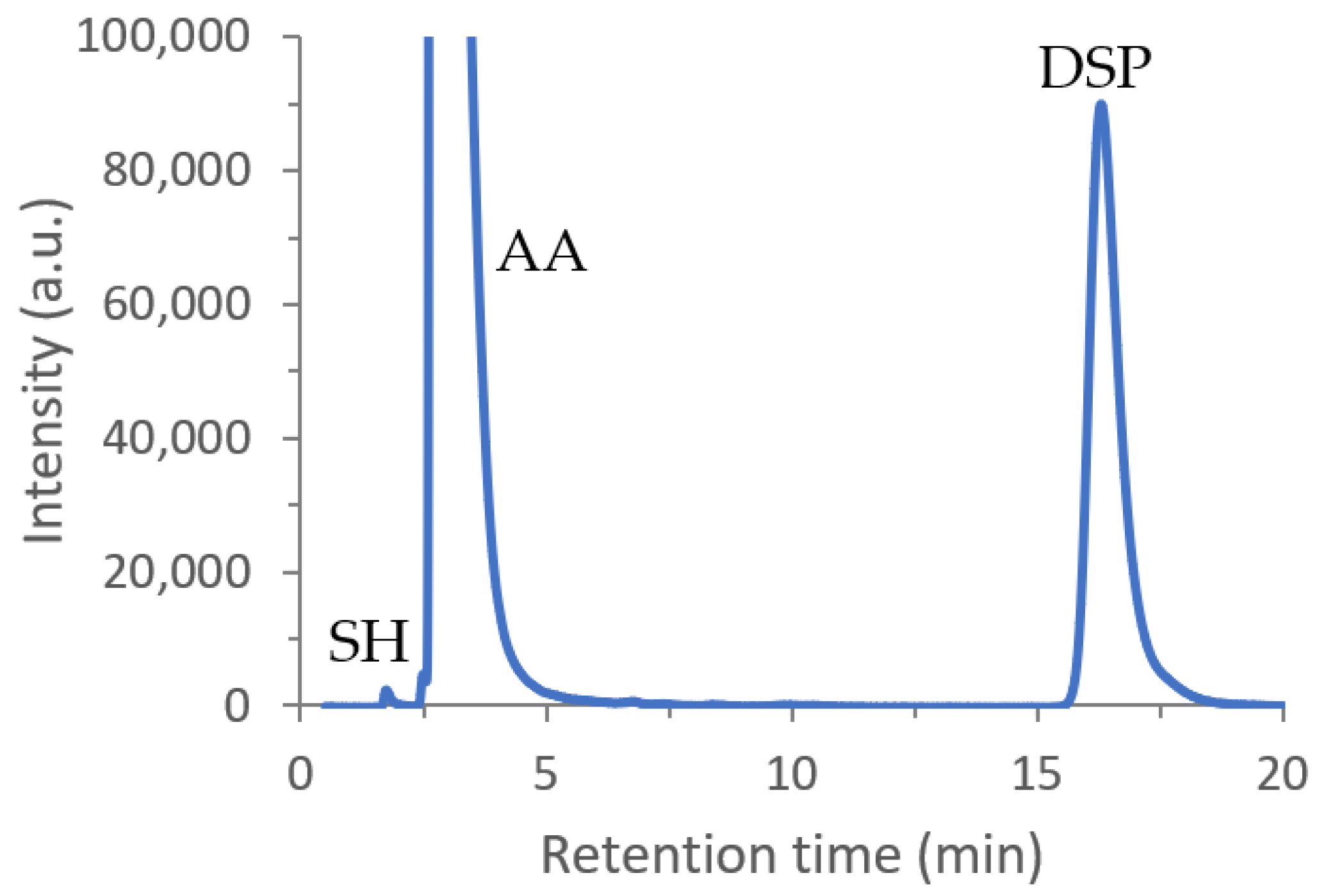
The chromatographic profile of the DSP, SH, and AA mixture (Section 2.3) showed three well-resolved peaks (Figure 1).
Figure 1.
Chromatogram obtained for the assay of DSP (100 µg/mL) in the presence of SH (1000 µg/mL) and AA (1000 µg/mL).
The average retention times and peak areas of SH, AA, and DSP are presented in Table 2 and Table 3, respectively. Good repeatability was observed, with CVs below 2% for both retention times and peak areas [42].
Table 2.
Average retention times (in minutes) obtained for the mixture of DSP, SH, and AA.
Table 3.
Average areas (a.u.) obtained for chromatographic peaks of the mixture of DSP, SH, and AA.
Based on the average retention time values, selectivity factors between the compounds were calculated. The selectivity factor of AA relative to SH was 1.85, while that of DSP relative to AA was 6.92. Since both values are greater than unity, no peak overlap was observed, confirming an efficient separation of the substances, which is essential for reliable quantification of DSP in the presence of these excipients [44].
The chromatographic resolution between DSP and the nearest eluting peak (AA) was consistently high, with an average value of approximately 9.55 (CV = 0.86%), largely exceeding the minimum acceptance criterion of 1.5 recommended by the USP [41]. Additionally, the tailing factor for DSP was 1.93 (CV = 1.32%), which is below the USP limit of 2.0 [41], confirming that the peak exhibited acceptable symmetry.
3.3. Method Validation
3.3.1. Linearity
A good linearity between the chromatographic response (peak area) and the concentration of the analyte was exhibited over the range of concentrations tested (50–150 μg/mL). The average values obtained from three independent calibration curves were 31,300 ± 1090 for the slope and −101,314 ± 164,376 for the y-intercept, with a good correlation coefficient of 0.998 [45]. Calibration parameters are presented in Table 4.
Table 4.
Calibration parameters and sensitivity obtained for the HPLC method.
3.3.2. Precision
The precision, evaluated as the repeatability of the method, was studied by calculating the CV for six determinations of a 100 μg/mL sample of DSP performed on the same day and under the same experimental conditions. The obtained CV value was 0.95%. The CV values for repeatability study were found to be lower than 1%, which indicates that the proposed method is repeatable [42]. The intermediate precision was assessed by analyzing the 100 μg/mL sample of DSP on three different days (inter-day). F-test, on the data found, confirmed the homogeneity of variances (p = 0.384), and Student’s t-test showed no significant difference between mean concentrations (t = −1.316, p = 0.259), demonstrating consistent results between days.
3.3.3. Specificity
The mean DSP peak area obtained for the mixture of DSP, SH, and AA (3,108,618 a.u.) was statistically compared with that found for the DSP standard solution (3,115,493 a.u., Section 3.1). The F-test confirmed the homogeneity of variances (p = 0.605, p > 0.05), allowing the assumption of equal variances in Student’s t-test, which yielded t = 0.245 and p = 0.819 (p > 0.05), indicating no significant difference between the mean concentrations.
3.3.4. Sensitivity
The sensitivity was evaluated through the determination of LD and LQ. Based on the parameters of the calibration curve (Table 4), the LD was 7.15 µg/mL and the LQ was 21.66 µg/mL. These results indicate that the proposed method possesses adequate sensitivity for the quantification of DSP within the required concentration range, as the LQ is significantly smaller than lowest calibration concentration (50 µg/mL) [46].
3.4. Mouthwash Stability Study
3.4.1. Accelerated Conditions (40 ± 2 °C)
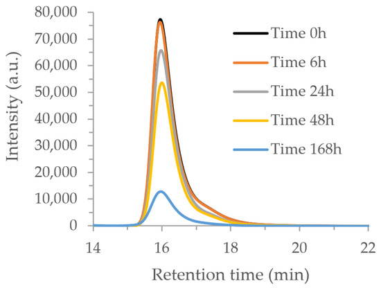
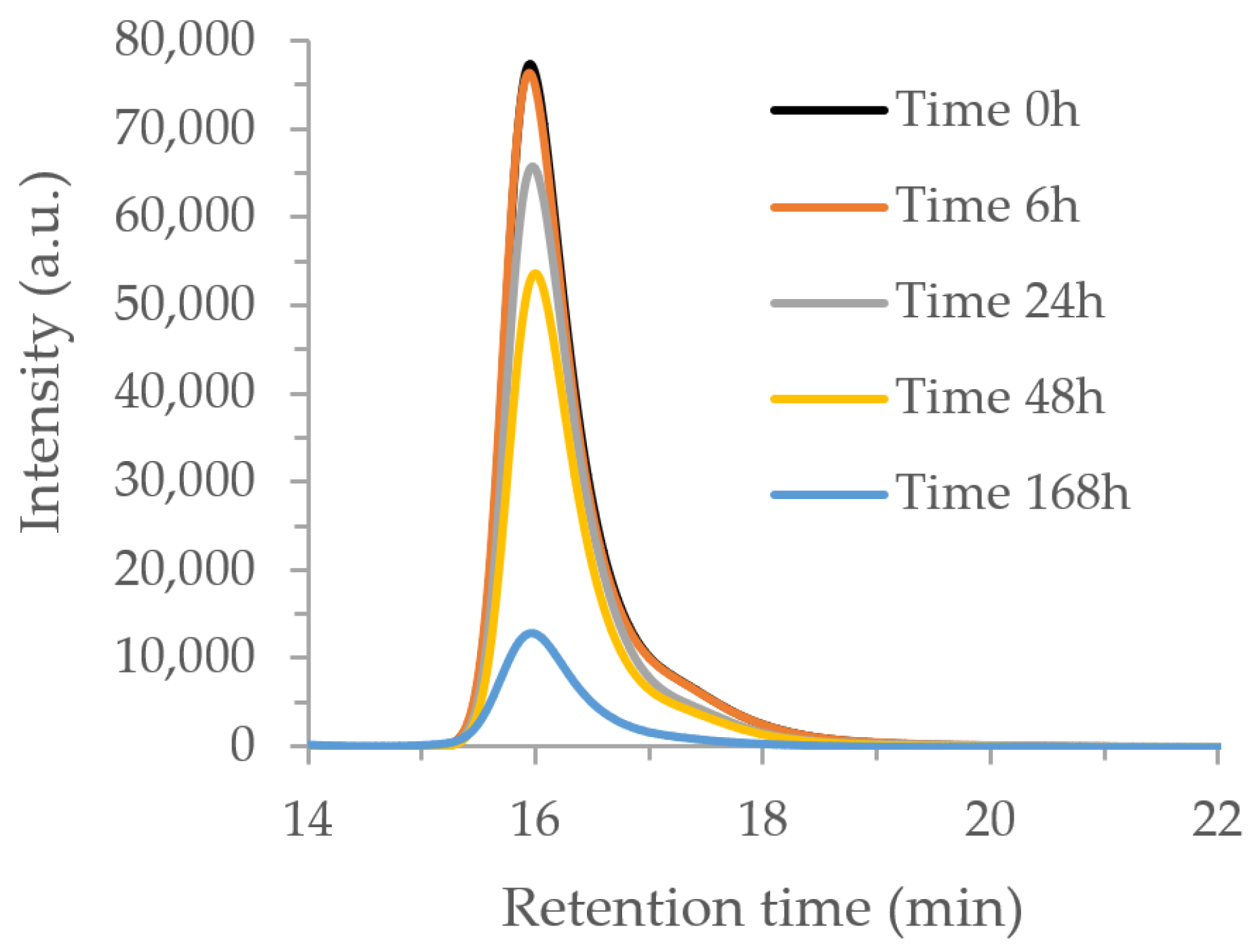
Samples were analyzed by HPLC to quantify the DSP content at time zero, which served as the reference for the study. After being stored in an oven at 40 ± 2 °C, to assess the impact of elevated temperatures on the mouthwash, aliquots of the samples were removed, and the DSP content was assayed. The chromatograms obtained are shown in Figure 2.
Figure 2.
Chromatograms obtained over time for analysis of batch 1 of the mouthwash formulation, after storage at 40 °C.
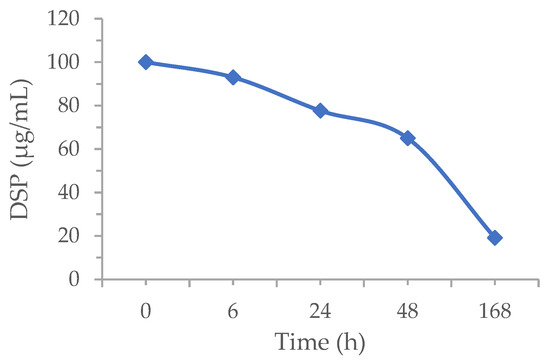
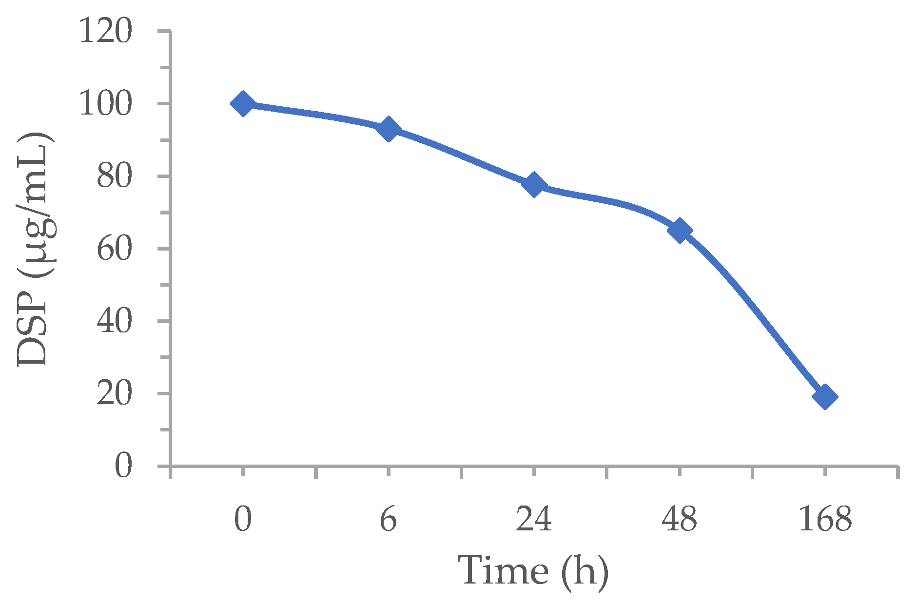
As can be seen, a decrease in DSP concentration is observed over the storage time at a temperature of 40 °C. The decrease becomes more noticeable after 6 h of storage, and after 7 days (168 h) the DSP content drops to approximately 20% compared to the initial concentration of the corticosteroid (Figure 3). Despite this decrease, no new peaks were visible during the chromatographic run.
Figure 3.
Decrease in DSP concentration over the storage time during the accelerated aging study.
In addition to studying the DSP content over time, an analysis of other important physicochemical parameters for characterizing the mouthwash formulation was also performed. Throughout the storage period at 40 °C, there was a slight decrease in pH, and a change in the color and turbidity of the samples was visually observed due to the presence of a suspended solid (Table 5).
Table 5.
Physicochemical stability of mouthwash formulation stored at 40 °C.
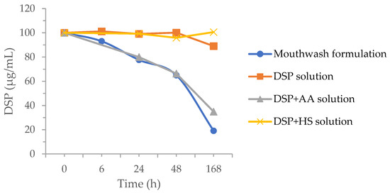
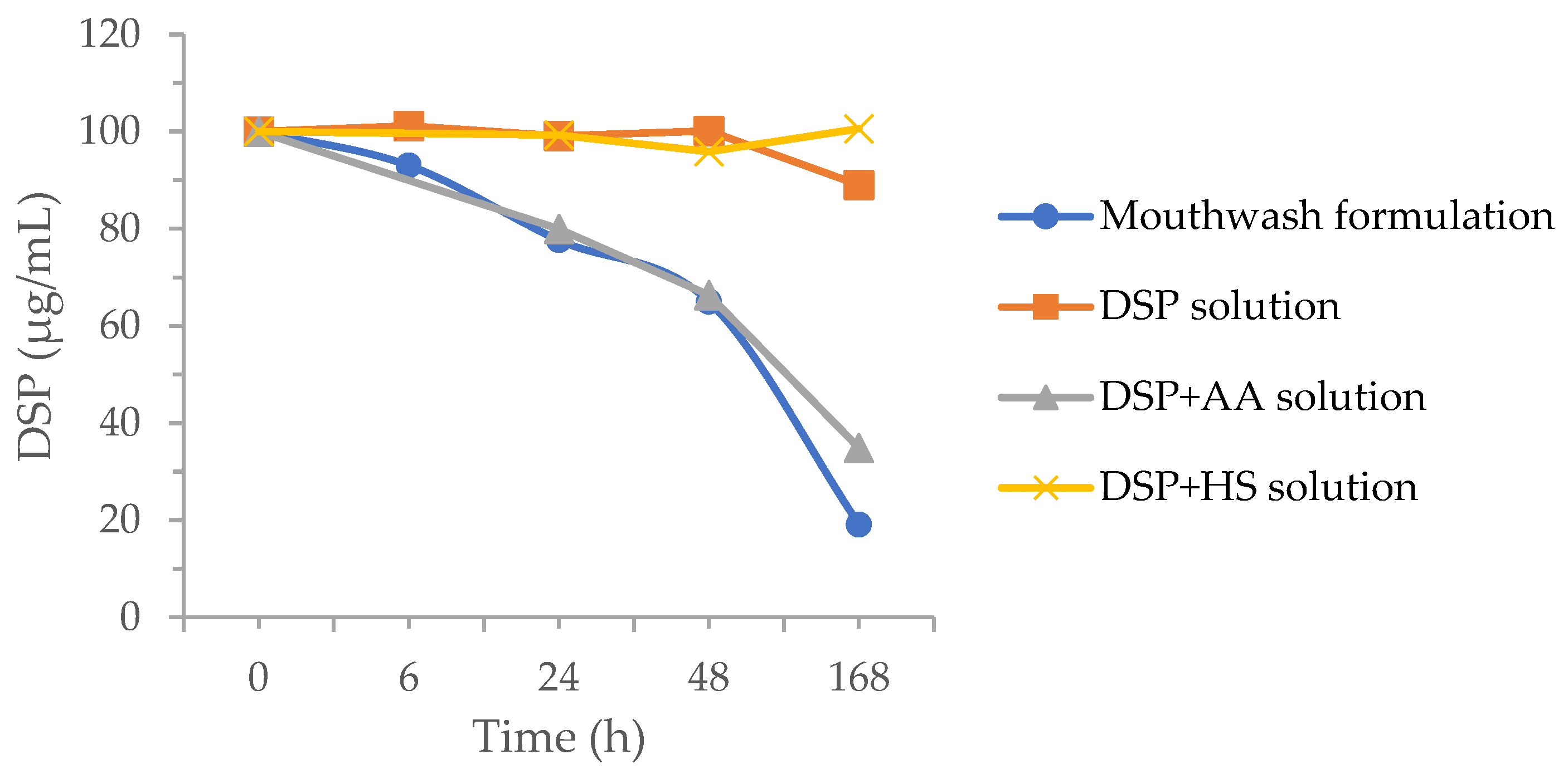
To understand the influence of each excipient (AA and SH) on DSP stability, under these temperature conditions (40 ± 2 °C), exploratory studies were carried out comparing the results obtained for the samples with those achieved using composed binary mixtures containing the same concentrations found in the mouthwash formulation (Figure 4).
Figure 4.
Evolution of DSP concentration (µg/mL) over time, obtained for different solutions (mouthwash formulation, DSP, DSP+AA, and DSP+SH) stored at 40 °C.
The sample containing only DSP showed a slight decrease in concentration after 168 h of storage at 40 °C, with an approximate loss of 10% compared to time zero. Similar results were reported by Watson et al. [47], who found that DSP remained stable at 37 °C for 192 h without significant loss of concentration. Likewise, for the DSP+SH mixture, there is no significant variation in the corticosteroid concentration after 7 days preserved under the described conditions, indicating that the presence of this excipient does not directly compromise DSP stability. Conversely, both the mouthwash formulation and the DSP+AA binary mixture exhibited a pronounced decrease in corticosteroid concentration shortly after 6 h of storage at 40 °C, which is consistent with the results described above. Although AA is widely used in dermatological products due to its diverse biological activities, as it possesses high antioxidant power capable of neutralizing oxidative stress triggered by various factors, its stability is the main drawback limiting its use in pharmaceutical formulations (see Section 4) [48].
3.4.2. Stability Study After Storage at 20 °C and 4 °C
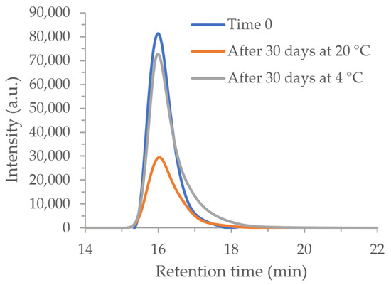
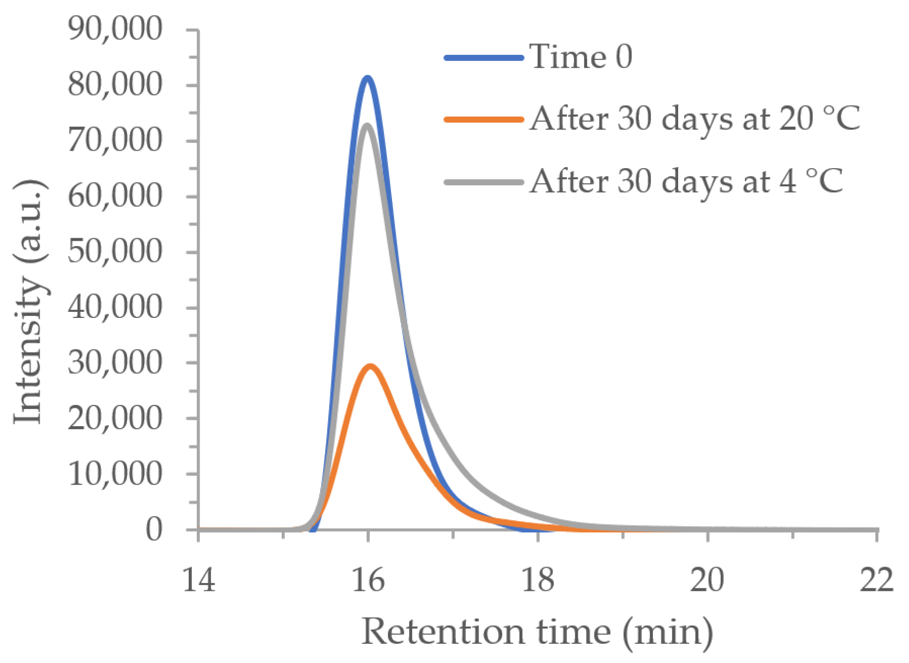
To evaluate chemical stability under storage conditions, the DSP content present in the mouthwash formulation was quantified after the sealed bottles had been kept for 30 days at 20 °C and 4 °C. At 20 °C, the DSP content showed a significant decrease, with concentration falling to approximately 50% of the initial value (Figure 5). In contrast, at 4 °C, DSP concentrations remained above 90% of the initial value, demonstrating good stability under the tested refrigeration conditions (Figure 5).
Figure 5.
Chromatograms obtained for batch 2 of the mouthwash formulation under conditions simulating pre-use storage. DSP contents were measured after 30 days in sealed bottles kept at 20 °C and 4 °C.
Regarding physicochemical parameters, after 30 days storage, no significant changes were observed in the mouthwash formulation stored at 4 °C (Table 6). However, in bottles stored at 20 °C, the presence of suspended particles was detected, which may indicate that the stability of the formulation is compromised. These results are consistent with the data previously presented for the DSP assay at this temperature.
Table 6.
Physicochemical stability of mouthwash formulation (batch 2) at time zero and after 30 days of storage at 20 °C and 4 °C.
3.4.3. In-Use Stability Study
After evaluating the stability of the mouthwash formulation in closed bottles at 4 °C, a simulation of use was carried out to reproduce outpatient handling conditions and assess the impact of daily opening (Section 2.2.1) on the formulation’s stability [49].
This assay is particularly relevant for oxidizable drugs, as daily opening of the container increases oxygen exposure, which may promote degradation processes. DSP has been described as susceptible to degradation under oxidative conditions, as reported by Buga et al. [32].
The simulation of use is an important parameter in the stability protocol once opening the bottle and removing a sample can lead to chemical loss, and the formation of degradation products due to the exposure to atmospheric oxygen, and environmental contaminants.
After storing the flasks for 30 days at 4 °C, three 10 mL aliquots (corresponding to the maximum recommended dose for treatment) were taken daily from the sample over the following 7 days. At the end of the 7 days, the DSP content was determined in the remaining solution that stayed in the flask.
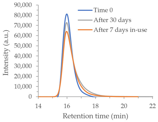
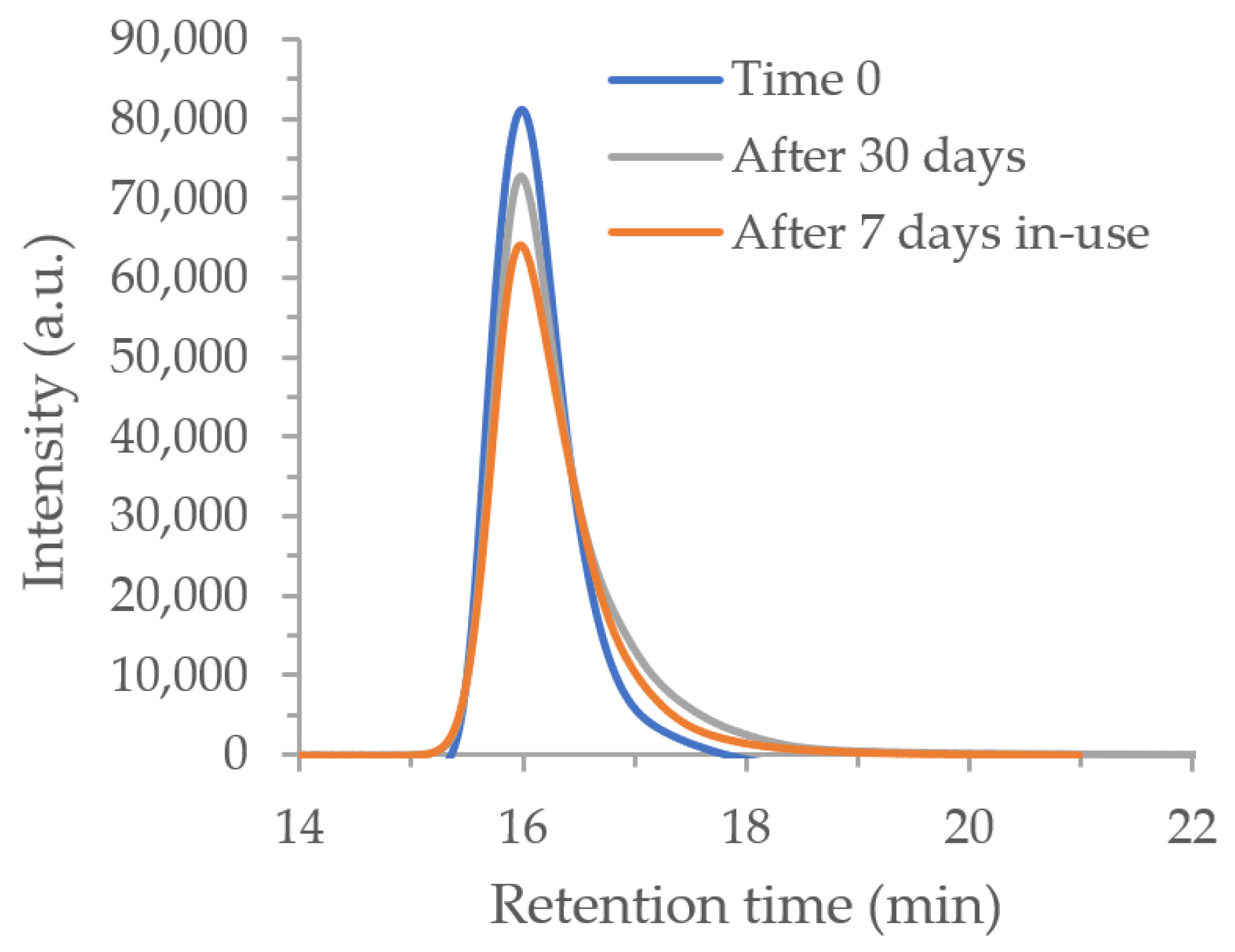
Comparative analysis of the chromatograms obtained after 7 days of the in-use study (Figure 6) showed that the sample maintained a DSP content greater than 90%. In fact, after 30 days of storage, followed by 7 days of in-use study, the DSP content obtained for the mouthwash formulation kept at 4 °C corresponded to 90.43% of the initial value.
Figure 6.
Representative chromatograms of mouthwash formulation (batch 2) after 7 days of in-use study.
Similar results were obtained for physicochemical stability after the in-use study, as there were no significant changes in any of the parameters evaluated (Table 7).
Table 7.
Physicochemical stability of mouthwash formulation after 7 days of in-use study.
4. Discussion
Overall, the analytical results for DSP degradation were consistent across the three evaluated batches, with coefficients of variation below 1% in studies conducted at 4 °C.
The physicochemical stability of the mouthwash formulation was assessed under different storage conditions according to USP stability criteria [38]. The mouthwash formulation showed low thermal stability at elevated temperature, highlighting the need to define optimal storage conditions. According to USP [38], a preparation is stable if the active pharmaceutical ingredient remains within 90–110% of the initial value at any time during the period of use. In this study, DSP concentration dropped below 90% of its initial value after 24 h at 40 °C and decreased further to approximately 20% after one week, which will inevitably impact the therapeutic efficacy of mouthwash. Additionally, the observed decrease in pH, along with changes in color and turbidity after 168 h, corroborates the DSP quantification data. Actually, the relatively low pH of the formulation may promote, over time, acid-catalyzed hydrolysis of the phosphate ester group of DSP, generating dexamethasone which, being practically insoluble in water, may cause the turbidity observed in the formulations.
However, if this study is considered solely in terms of short-term exposure of the mouthwash samples to this temperature (40 °C), the conclusion is necessarily different. In fact, it is unlikely that such storage conditions would exceed 6 h and, in that case, it can be concluded that exposure of the mouthwash formulation to temperatures up to 40 °C for a short period (e.g., transporting the mouthwash formulation by car to the patient’s home on hot days) does not have a significant impact on the stability of the DSP.
Solutions containing only DSP and DSP plus SH remained stable after 168 h at 40 °C, with DSP content remaining above 90%, indicating that SH does not directly compromise DSP stability. In contrast, both the mouthwash formulation and the DSP+AA mixture showed a progressive decrease in DSP concentration over time, following a similar pattern. This suggests that DSP degradation is primarily driven by the presence of AA. In fact, the main challenge in developing products containing AA is its high instability and reactivity. AA is extremely sensitive to light, pH, temperature, and oxidation, which can affect its degradation, often accompanied by a change in the color of the final product [50]. Moreover, studies have shown that, although AA is a potent antioxidant, it may exhibit pro-oxidant behavior depending on concentration and environmental conditions, particularly in aqueous solutions and at elevated temperatures. Under such conditions, AA degradation has been associated with the formation of ROS, which may promote oxidative degradation of other formulation components [51,52].
DSP contains functional groups that are susceptible to oxidative degradation and acid- or base-catalyzed hydrolysis [32]. Therefore, the generation of ROS during AA degradation may contribute to DSP degradation, as already reported for oxidative stress conditions by Buga et al. [32]. Additionally, the acidic nature of AA in solution could facilitate acid-catalyzed hydrolysis of the phosphate ester group of DSP, contributing to the observed decrease in DSP content. These findings support the hypothesis that AA degradation in solution may contribute to DSP degradation in the mouthwash formulation.
Physicochemical observations further support this conclusion. A change in pH was observed only in the DSP+AA solution, further reinforcing the hypothesis that AA plays a key role in the instability of the formulation. Moreover, when comparing the physicochemical stability results of the DSP, DSP+AA, and DSP+SH solutions with those obtained for the formulation stored at 40 °C, more pronounced physical changes were observed in the mouthwash formulation after 168 h, particularly the presence of insoluble particles. This suggests that the combination of AA and SH may exert a synergistic effect in reducing the stability of the solution. To date, no studies have directly evaluated the interaction between these two compounds. However, according to Šoltés et al. [53], SH is susceptible to oxidative degradation by ROS. Therefore, since AA can act as a source of such species [51], it is plausible that it enhances SH degradation, contributing to the overall decrease in the formulation’s stability.
When considering storage at lower temperatures, the stability profile of the formulation differed notably. After 30 days in closed bottles at 4 °C, the formulation remained stable, since it retained more than 90% of its initial DSP content after 30 days of storage [38]. In contrast, samples stored at 20 °C showed DSP levels well below 90%, indicating that the formulation cannot be considered stable under these conditions. Changes in physicochemical parameters, such as the appearance of suspended particles, were also observed only in the 20 °C samples, suggesting the onset of chemical and/or microbiological degradation. This appearance may be indicative of fungal growth, although microbiological confirmation is required, especially since the formulation contains no preservatives.
Based on the closed-bottle stability results, the in-use stability study was performed only for samples stored at 4 °C, as this was the only condition under which the formulation remained stable. Under refrigerated conditions, the formulation maintained more than 90% of its initial DSP content over the 7-day in-use period, with no significant changes in pH or appearance. These results provide practical guidance for hospital compounding of the mouthwash, as they support a preliminary beyond-use period of up to 30 days for closed bottles, and up to 7 days after opening, provided that refrigerated storage is maintained. Maintaining the formulation under refrigerated conditions and adhering to defined beyond-use periods helps ensure physicochemical stability, while preserving appearance, pH, and palatability, factors that can influence patient adherence and acceptance during treatment.
It is worth noting that this work inevitably presents certain limitations that should be taken into account in future applications of this formulation. Although the contribution of AA to DSP degradation is evident, its mechanism of action has not been fully elucidated, and therefore remains speculative. The presence of AA may lower the formulation pH, thereby promoting DSP hydrolysis, enhance the formation of ROS that may contribute to DSP degradation, or act through a combination of these mechanisms. As the results indicate that AA adversely affects formulation stability, alternative antioxidants with greater chemical stability may be evaluated in future studies to improve formulation stability. This study was limited to the evaluation of physicochemical stability of the mouthwash formulation. Further studies incorporating microbiological assessments and complementary analyses may provide more comprehensive information regarding product quality and patient safety.
5. Conclusions
This work aimed to address a therapeutic gap in medicines prepared for cancer patients and made it possible to assemble a significant set of previously unavailable data on the physicochemical stability of a hospital mouthwash formulation intended for the treatment of oral mucositis. In addition to highlighting the crucial role of certain excipients in drug stability, this study demonstrated the importance of evaluating not only the chemical component but also the physicochemical parameters that determine the final quality and acceptability of the formulation.
The results found indicate that the mouthwash formulation remains stable when stored at 4 °C in properly closed bottles. Based on these physicochemical data, a preliminary beyond-use period of up to 30 days is suggested, with a recommended use within 7 days after opening.
As the results indicate that AA plays a significant role in formulation instability, future studies should investigate its replacement with a chemically more stable antioxidant. In parallel, given the potential susceptibility of the formulation to microbiological degradation, the relevance and impact of incorporating a preservative should be evaluated to ensure microbiological stability.
In addition, palatability represents an important aspect of the formulation, as alterations may affect patient acceptability even when physicochemical stability is maintained. Further clinical studies are needed to evaluate the efficacy, safety, and patient acceptability of the mouthwash formulation.
Author Contributions
Conceptualization, J.G., I.F.A. and S.P.; methodology, M.N., E.M.P.J.G. and J.G.; validation, E.M.P.J.G., J.G. and I.F.A.; investigation, M.N.; resources, S.P.; data curation, M.N., E.M.P.J.G. and J.G.; writing—original draft preparation, M.N., E.M.P.J.G. and J.G.; writing—review and editing, M.N., E.M.P.J.G., S.P., I.F.A. and J.G.; supervision, E.M.P.J.G., I.F.A. and J.G. All authors have read and agreed to the published version of the manuscript.
Funding
The authors thank the Portuguese Foundation for Science and Technology (FCT, Portugal) for the financial support to CIQUP (Project UIDB/00081/2025), and IMS-Institute of Molecular Sciences (LA/P/0056/2020).
Institutional Review Board Statement
Not applicable.
Informed Consent Statement
Not applicable.
Data Availability Statement
The original contributions presented in this study are included in the article. Further inquiries can be directed to the corresponding author.
Acknowledgments
The authors thank Carla Carlos and Márcia Batista for their valuable contributions to this work.
Conflicts of Interest
The authors declare no conflicts of interest.
Abbreviations
The following abbreviations are used in this manuscript:
| CT | Chemotherapy |
| DSP | Dexamethasone Sodium Phosphate |
| CV | Coefficient of variation |
| EU | European Union |
| HPLC | High-Performance Liquid Chromatography |
| LD | Detection Limit |
| LQ | Quantification Limit |
| MASCC/ISOO | Multinational Association of Supportive Care in Cancer and International Society of Oral Oncology |
| OM | Oral Mucositis |
| ROS | Reactive oxygen species |
| RT | Radiotherapy |
| SH | Sodium Hyaluronate |
| THF | Tetrahydrofuran |
| WHO | World Health Organization |
References
- Eurostat Cancer Statistics. Available online: https://ec.europa.eu/eurostat/statistics-explained/index.php?title=Cancer_statistics (accessed on 9 December 2025).
- OECD. EU Country Cancer Profile: Portugal 2025; OECD Publishing: Paris, France, 2025. [Google Scholar]
- Zafar, A.; Khatoon, S.; Khan, M.J.; Abu, J.; Naeem, A. Advancements and Limitations in Traditional Anti-Cancer Therapies: A Comprehensive Review of Surgery, Chemotherapy, Radiation Therapy, and Hormonal Therapy. Discov. Oncol. 2025, 16, 607. [Google Scholar] [CrossRef] [Scilit]
- Shetty, S.S.; Maruthi, M.; Dhara, V.; de Arruda, J.A.A.; Abreu, L.G.; Mesquita, R.A.; Teixeira, A.L.; Silva, T.A.; Merchant, Y. Oral Mucositis: Current Knowledge and Future Directions. Disease-a-Month 2022, 68, 101300. [Google Scholar] [CrossRef] [Scilit] [PubMed]
- Al-Rudayni, A.H.M.; Gopinath, D.; Maharajan, M.K.; Menon, R.K. Impact of Oral Mucositis on Quality of Life in Patients Undergoing Oncological Treatment: A Systematic Review. Transl. Cancer Res. 2020, 9, 3126–3134. [Google Scholar] [CrossRef] [Scilit] [PubMed]
- Mercadante, S.; Aielli, F.; Adile, C.; Ferrera, P.; Valle, A.; Fusco, F.; Caruselli, A.; Cartoni, C.; Massimo, P.; Masedu, F.; et al. Prevalence of Oral Mucositis, Dry Mouth, and Dysphagia in Advanced Cancer Patients. Support. Care Cancer 2015, 23, 3249–3255. [Google Scholar] [CrossRef] [Scilit] [PubMed]
- Brown, T.J.; Gupta, A. Management of Cancer Therapy–Associated Oral Mucositis. JCO Oncol. Pract. 2020, 16, 103–109. [Google Scholar] [CrossRef] [Scilit]
- Elad, S.; Yarom, N.; Zadik, Y.; Kuten-Shorrer, M.; Sonis, S.T. The Broadening Scope of Oral Mucositis and Oral Ulcerative Mucosal Toxicities of Anticancer Therapies. CA Cancer J. Clin. 2022, 72, 57–77. [Google Scholar] [CrossRef] [Scilit]
- da Cruz Campos, M.I.; Neiva Campos, C.; Monteiro Aarestrup, F.; Vieira Aarestrup, B.J. Oral Mucositis in Cancer Treatment: Natural History, Prevention and Treatment. Mol. Clin. Oncol. 2014, 2, 337–340. [Google Scholar] [CrossRef] [Scilit]
- Chaveli-Lopez, B.; Bagan-Sebastian, J. V Treatment of Oral Mucositis Due to Chemotherapy. J. Clin. Exp. Dent. 2016, 8, 201–209. [Google Scholar] [CrossRef] [Scilit]
- Wu, Y.; Shi, W.; Li, C.; Liu, X.; Jiang, Y.; Qiu, Y.; Chen, Q.; Luo, X. Managing Strategies of Chemotherapy and Radiotherapy-Induced Oral Mucositis. Cancer Treat. Rev. 2025, 133, 102883. [Google Scholar] [CrossRef] [Scilit]
- Bell, A.; Kasi, A. Oral Mucositis. In StatPearls; StatPearls Publishing: Treasure Island, FL, USA, 2023. [Google Scholar]
- Eilers, J.; Million, R. Prevention and Management of Oral Mucositis in Patients With Cancer. Semin. Oncol. Nurs. 2007, 23, 201–212. [Google Scholar] [CrossRef] [Scilit]
- Elad, S.; Cheng, K.K.F.; Lalla, R.V.; Yarom, N.; Hong, C.; Logan, R.M.; Bowen, J.; Gibson, R.; Saunders, D.P.; Zadik, Y.; et al. MASCC/ISOO Clinical Practice Guidelines for the Management of Mucositis Secondary to Cancer Therapy. Cancer 2020, 126, 4423–4431. [Google Scholar] [CrossRef] [Scilit]
- Isozaki, A.B.; Brant, J.M. Clinical Updates in Mucositis-Related Symptom Management. Semin. Oncol. Nurs. 2022, 38, 151252. [Google Scholar] [CrossRef] [Scilit] [PubMed]
- Lalla, R.V.; Brennan, M.T.; Gordon, S.M.; Sonis, S.T.; Rosenthal, D.I.; Keefe, D.M. Oral Mucositis Due to High-Dose Chemotherapy and/or Head and Neck Radiation Therapy. J. Natl. Cancer Inst. Monogr. 2019, 2019, lgz011. [Google Scholar] [CrossRef] [Scilit] [PubMed]
- Thornton, C.P.; Li, M.; Budhathoki, C.; Yeh, C.H.; Ruble, K. Anti-Inflammatory Mouthwashes for the Prevention of Oral Mucositis in Cancer Therapy: An Integrative Review and Meta-Analysis. Support. Care Cancer 2022, 30, 7205–7218. [Google Scholar] [CrossRef] [Scilit] [PubMed]
- Ramamoorthy, S.; Cidlowski, J.A. Corticosteroids. Rheum. Dis. Clin. N. Am. 2016, 42, 15–31. [Google Scholar] [CrossRef] [Scilit]
- Synaridou, M.S.; Andriotis, E.G.; Zacharis, C.K.; Fatouros, D.G.; Markopoulou, C.K. Solid Dosage Forms of Dexamethasone Sodium Phosphate Intended for Pediatric Use: Formulation and Stability Studies. Pharmaceutics 2020, 12, 354. [Google Scholar] [CrossRef] [Scilit]
- Duarah, S.; Sharma, M.; Wen, J. Rapid and Simultaneous Determination of Dexamethasone and Dexamethasone Sodium Phosphate Using HPLC-UV: Application in Microneedle-Assisted Skin Permeation and Deposition Studies. J. Chromatogr. B 2021, 1170, 122609. [Google Scholar] [CrossRef] [Scilit]
- Shahrabi, M.; Solduzian, M.; Babaie, M.H.; Mousavi, S.A.; Goodarzi, N.; Ravari, N.S.; Sadeghi, K. The Effects of a Combination Oral Spray (Mucosamin®) for the Prevention of Oral Mucositis in Pediatric Patients Undergoing Hematopoietic Stem Cell Transplantation: A Double Blind Randomized Clinical Trial. Support. Care Cancer 2022, 30, 7963–7972. [Google Scholar] [CrossRef] [Scilit]
- Lodolo, M.; Thanasuwat, B.; Veluppillai, P.; Bassani, G.; Villa, A. Dexamethasone Solution and Dexamethasone in MucoloxTM for the Treatment of Oral Inflammatory Ulcerative Diseases: A Phase II Randomized Clinical Trial. J. Oral Pathol. Med. 2023, 52, 860–866. [Google Scholar] [CrossRef] [Scilit]
- Yang, Z.; Li, M.; Xiao, L.; Yi, Z.; Zhao, M.; Ma, S. Hyaluronic Acid versus Dexamethasone for the Treatment of Recurrent Aphthous Stomatitis in Children: Efficacy and Safety Analysis. Braz. J. Med. Biol. Res. 2020, 53, e9886. [Google Scholar] [CrossRef] [Scilit]
- Wong, S.P.; Tan, S.M.; Lee, C.-S.; Law, K.B.; Lim, Y.A.L.; Rajasuriar, R. Prospective Longitudinal Analysis of Clinical and Immunological Risk Factors Associated with Oral and Gastrointestinal Mucositis Following Autologous Stem Cell Transplant in Adults. Support. Care Cancer 2023, 31, 494. [Google Scholar] [CrossRef] [Scilit]
- Zhang, Y.; Ma, R.; You, C.; Leng, X.; Wang, D.; Deng, S.; He, B.; Guo, Z.; Guan, Z.; Lei, H.; et al. Hyaluronic Acid Modified Oral Drug Delivery System with Mucoadhesiveness and Macrophage-Targeting for Colitis Treatment. Carbohydr. Polym. 2023, 313, 120884. [Google Scholar] [CrossRef] [Scilit]
- dos Santos, R.W.; Martins, A.F.L.; Sousa-Neto, S.S.; Morais, M.O.; de Oliveira Moreira, V.H.L.; Arantes, D.A.C.; Oton-Leite, A.F.; Freitas, N.M.A.; Leles, C.R.; Mendonça, E.F. Effects of L-Glutamine and Hyaluronic Acid Associated with Photobiomodulation on Oral Mucositis: A Randomized Clinical Trial. Support. Care Cancer 2025, 33, 430. [Google Scholar] [CrossRef] [Scilit] [PubMed]
- Colella, G.; Boschetti, C.E.; Farina, F.; Colella, M.L.; Vicidomini, A.; Cirillo, N. Mucosamin Spray for the Prevention of Oral Mucositis in Cancer Patients Receiving Chemotherapy: A Retrospective Cohort Study. J. Oral Pathol. Med. 2025, 54, 397–400. [Google Scholar] [CrossRef] [Scilit] [PubMed]
- Cirillo, N.; Vicidomini, A.; McCullough, M.; Gambardella, A.; Hassona, Y.; Prime, S.S.; Colella, G. A Hyaluronic Acid-Based Compound Inhibits Fibroblast Senescence Induced by Oxidative Stress In Vitro and Prevents Oral Mucositis In Vivo. J. Cell. Physiol. 2015, 230, 1421–1429. [Google Scholar] [CrossRef] [Scilit] [PubMed]
- Mohammed, A.I.; Celentano, A.; Paolini, R.; Low, J.T.; Silke, J.; O’ Reilly, L.A.; McCullough, M.; Cirillo, N. High Molecular Weight Hyaluronic Acid Drastically Reduces Chemotherapy-Induced Mucositis and Apoptotic Cell Death. Cell Death Dis. 2023, 14, 453. [Google Scholar] [CrossRef] [Scilit]
- Yoshioka, S.; Stella, V.J. Stability of Drugs and Dosage Forms; Springer: Berlin/Heidelberg, Germany, 2002. [Google Scholar]
- Rowe, R.C.; Sheskey, P.J.; Weller, P.J. Handbook of Pharmaceutical Excipients; Pharmaceutical Press: London, UK, 2006; Volume 6. [Google Scholar]
- Buga, I.; Uzoma, J.I.; Reindel, K.; Rashid, K.; Diep, T.; McCartan, P.; Zhao, F. Physical and Chemical Stability of Dexamethasone Sodium Phosphate in Intravenous Admixtures Used to Prevent Chemotherapy-Induced Nausea and Vomiting. Hosp. Pharm. 2021, 56, 215–220. [Google Scholar] [CrossRef] [Scilit]
- Carmo, F.J.R.; Salote, E.P.Z.; Valente, A.J.M.; Ribeiro, A.C.F.; Nicolau, P.M.G.; Fangaia, S.I.G. Diffusion of Sodium Hyaluronate in Artificial Saliva to Optimize Its Topical Application. Molecules 2025, 30, 2140. [Google Scholar] [CrossRef] [Scilit]
- Boltia, S.A.; Ibrahim, M.; Ibrahim, M.M.; Ramadan, N.K. Development and Validation of a RP-HPLC Method for the Simultaneous Determination of Silver Sulfadiazine and Sodium Hyaluronate in the Presence of Methyl and Propyl Paraben in a Pharmaceutical Cream for Burns. J. Chromatogr. Sci. 2024, 62, 878–885. [Google Scholar] [CrossRef] [Scilit]
- Mitić, S.S.; Kostić, D.A.; Nasković, D.C.; Mitic, M.N. Rapid and Reliable HPLC Method for the Determination of Vitamin C in Pharmaceutical Samples. Trop. J. Pharm. Res. 2011, 10, 105–111. [Google Scholar] [CrossRef] [Scilit]
- Chaitanya, N.C.; Muthukrishnan, A.; Purushotham Rao, K.; Reshma Priyanka, D.; Ujwala, P.; Abhijeeth, H.; Kovur, A.; Nikhil Kumar, A. Oral Mucositis Severity Assessment by Supplementation of High Dose Ascorbic Acid During Chemo and/or Radiotherapy of Oro-Pharyngeal Cancers—A Pilot Project. Indian J. Pharm. Educ. Res. 2018, 52, 532–539. [Google Scholar] [CrossRef] [Scilit]
- The United States Pharmacopeial Convention. United States Pharmacopeia (USP) General Chapter ⟨795⟩ Pharmaceutical Compounding—Nonsterile Preparations. In United States Pharmacopeia and National Formulary (USP–NF); The United States Pharmacopeial Convention: Rockville, MD, USA, 2022. [Google Scholar]
- Allen, L.V.; Bassani, G.S.; Elder, E.J.; Parr, A.F. Strength and Stability Testing for Compounded Preparations. US Pharmacop. 2014, 78, 164. [Google Scholar]
- European Directorate for the Quality of Medicines & HealthCare. Dexamethasone Sodium Phosphate. In European Pharmacopoeia; Council of Europe: Strasbourg, France, 2021; pp. 2496–2498. [Google Scholar]
- Borman, P.; Elder, D. Q2(R1) Validation of Analytical Procedures. In ICH Quality Guidelines; Wiley: Hoboken, NJ, USA, 2017; pp. 127–166. [Google Scholar]
- United States Pharmacopeial Convention. The United States Pharmacopeia: USP 30; The National Formulary: NF 25; United States Pharmacopeial Convention: Rockville, MD, USA, 2006. [Google Scholar]
- Dong, M.; Huynh-Ba, K.; Wong, A. Validation of Stability-Indicating HPLC Methods for Pharmaceuticals: Overview, Methodologies, and Case Studies. LCGC N. Am. 2020, 38, 606–611. [Google Scholar]
- Ruckmani, K.; Shaikh, S.Z.; Khalil, P.; Muneera, M.S.; Thusleem, O.A. Determination of Sodium Hyaluronate in Pharmaceutical Formulations by HPLC–UV. J. Pharm. Anal. 2013, 3, 324–329. [Google Scholar] [CrossRef] [Scilit]
- Recommendations, I. Nomenclature for Chromatography. Pure Appl. Chem. 1993, 65, 819–872. [Google Scholar] [CrossRef] [Scilit]
- Van Loco, J.; Elskens, M.; Croux, C.; Beernaert, H. Linearity of Calibration Curves: Use and Misuse of the Correlation Coefficient. Accredit. Qual. Assur. 2002, 7, 281–285. [Google Scholar] [CrossRef] [Scilit]
- International Conference on Harmonisation. ICH Q2(R1)—Validation of Analytical Procedures: Text and Methodology; International Conference on Harmonisation: Geneva, Switzerland, 2005. [Google Scholar]
- Watson, D.G.; Lin, M.; Morton, A.; Cable, C.G.; McArthur, D.A. Compatibility and Stability of Dexamethasone Sodium Phosphate and Ketamine Hydrochloride Subcutaneous Infusions in Polypropylene Syringes. J. Pain Symptom Manag. 2005, 30, 80–86. [Google Scholar] [CrossRef] [Scilit]
- Caritá, A.C.; Resende de Azevedo, J.; Vinícius Buri, M.; Bolzinger, M.-A.; Chevalier, Y.; Riske, K.A.; Ricci Leonardi, G. Stabilization of Vitamin C in Emulsions of Liquid Crystalline Structures. Int. J. Pharm. 2021, 592, 120092. [Google Scholar] [CrossRef] [Scilit]
- Committee for Proprietary Medicinal Products. Note for Guidance on In-Use Stability Testing of Human Medicinal Products; Committee for Proprietary Medicinal Products: Amsterdam, The Netherlands, 2001. [Google Scholar]
- Shelke, O.; Susarla, K.P.C.; Bankar, M. Understand the Stabilization Engineering of Ascorbic Acid, Mapping the Scheme for Stabilization, and Advancement. AAPS PharmSciTech 2024, 25, 159. [Google Scholar] [CrossRef] [Scilit]
- Yin, X.; Chen, K.; Cheng, H.; Chen, X.; Feng, S.; Song, Y.; Liang, L. Chemical Stability of Ascorbic Acid Integrated into Commercial Products: A Review on Bioactivity and Delivery Technology. Antioxidants 2022, 11, 153. [Google Scholar] [CrossRef] [Scilit]
- Yuan, J.-P.; Chen, F. Degradation of Ascorbic Acid in Aqueous Solution. J. Agric. Food Chem. 1998, 46, 5078–5082. [Google Scholar] [CrossRef] [Scilit]
- Šoltés, L.; Kogan, G.; Stankovská, M.; Mendichi, R.; Schiller, J.; Gemeiner, P. Degradation of High-Molar-Mass Hyaluronan and Characterization of Fragments. Biomacromolecules 2007, 8, 2697–2705. [Google Scholar] [CrossRef] [Scilit] [PubMed]

Disclaimer/Publisher’s Note: The statements, opinions and data contained in all publications are solely those of the individual author(s) and contributor(s) and not of MDPI and/or the editor(s). MDPI and/or the editor(s) disclaim responsibility for any injury to people or property resulting from any ideas, methods, instructions or products referred to in the content. |
© 2026 by the authors. Licensee MDPI, Basel, Switzerland. This article is an open access article distributed under the terms and conditions of the Creative Commons Attribution (CC BY) license.