


Abstract
Historic gardens represent complex systems in which vegetation, artistic and architectural elements all require equal protection. This becomes particularly challenging when considering the dual role of vegetation between bioprotection and biodeterioration. This study addresses the conservation issues associated with the modern sculptural group of the Niobids, located in the historic garden of Villa Medici. The group presents critical issues related to the materials used in its creation (i.e., white-cement- and epoxy-resin-based composites) and the limited literature documenting the effectiveness of cleaning methods for this type of artwork. To address this knowledge gap and provide a basis for a low-intensity maintenance plan, investigations were carried out to identify the different components of the biopatinas, as well as higher vegetation in direct contact with the statues. In this context, the efficacy of a protocol based on water micronisation for biopatina removal was also assessed. Clamydomonas spp. was a frequent phototrophic element, along with Candelariella species, Xanthoria parietina, Flavoplaca flavocitrina, and Verrucaria nigrescens. The cultivable fungal fraction evidenced the substantial presence of Parengyodontium album and melanised strains belonging to the genera Chaetocapnodium, Exophiala, Scolecobasidium and Coniosporium. Vegetation cover was characterised by the frequent presence of Grimmia pulvinata on the sculptures and Acanthus mollis in their surroundings. These findings raise significant conservation concerns, as the observed taxa suggest an advanced stage of ecological succession driving pedogenesis. The water micronisation protocol produced noteworthy results, highlighting its potential for treating new materials and as a maintenance technology within planned conservation. Indeed, regular monitoring and low-intensity preventive actions can prevent the need for late restorative interventions and, above all, transform emergency management from the rule into the exception.
1. Introduction
Historic gardens represent complex systems that integrate vegetative, architectural, and sculptural elements [1]. Conservation management requires a balance between the protection of artistic heritage and the maintenance of the plant ecosystem respecting the garden’s historical vegetation design. The preservation of historic and artistic artefacts within historic parks is particularly challenging due to the ambivalent role of vegetation. On the one hand, vegetation promotes biological colonisation. Indeed, shading, which slows evaporative processes, tends to prolong the period during which surfaces remain moist, thereby favouring the deposition of common pollutants such as sulphur dioxide, enhancing soiling phenomena, and making materials more susceptible to biological attack increasing their bioreceptivity [2,3,4]. Furthermore, vegetation may support the growth and spread of biofilms by providing them with airborne nutrients, typically scarce on rocky substrates, such as pollen, honeydew, fruit residues, or other organic matter. Vegetation may also promote the exchange and transfer of the plant-associated microbiome onto built heritage surfaces, particularly those with stress-tolerant traits [5], and potentially increase the risk of root damage [6]. Nevertheless, the protective role of vegetation should not be overlooked. As has already been demonstrated at archaeological sites and in urban areas [7,8], vegetation can shield exposed surfaces by acting as a buffer against extreme weather conditions, preventing the rapid and substantial fluctuations to which outdoor heritage materials are exposed and thereby reducing the risk of physical deterioration. Additionally, vegetation is not merely a decorative or static element; it represents a dynamic system of living organisms that themselves constitute a form of natural heritage. Historic gardens serve as crucial biodiversity niches within urban environments, functioning as living time capsules whose ecological and cultural values must be preserved [9].
The need for careful assessment of conservation issues relating to both built structures and the vegetative component is established in the Historic Gardens Charter, commonly known as the Florence Charter, drafted by the International Committee on Historic Gardens and Sites [10]. Article 1 defines a historic garden as “an architectural and horticultural composition which, from the historical or artistic point of view, is of public interest. As such, it is to be considered a monument”. Other forms of historic greenery deserve equal consideration such as cemeteries, urban green areas (e.g., avenues and boulevards, parks, and promenades) and public green spaces characterized in the ICOMOS-IFLA Document on historic urban parks as “an essential and inalienable part of traditions and plans of many towns and settlements” [11,12].
Cultural heritage refers to the legacy of tangible items and their intangible attributes that are inherited from the past by a group or society and conserved for future generations due to their artistic, cultural, or historic value [13]. For this reason, the need to preserve and conserve an artefact is not necessarily linked to the intrinsic value of the materials employed in its creation, nor to its dimensions. The cultural, historical, documentary and symbolic significance of an object may, in fact, exist independently of the rarity or economic value of the materials from which it is made. The sculpture in the modern era has been marked by relevant innovations in materials and styles, reflecting broader artistic movements and technological advancements. The traditional materials such as marble wood and metals have been complemented or replaced by diverse new media, including plastics, synthetic resins, industrial polymers, concrete, rubber and composite materials [14]. In this context, the Niobid statues located in the gardens of Villa Medici, Rome, replicas of the ancient Roman sculptural group originally belonging to Ferdinando de’ Medici’s collection, represent a notable example. The replicas were produced in 1976 by the sculptor Michel Bourbon using moulding techniques and an innovative composite material consisting of marble powder and epoxy resin [15]. The expansion of artistic materials introduces new conservation challenges, as many modern materials undergo complex physical and chemical degradation processes that often differ from those affecting traditional sculptural media and, in many cases, remain poorly understood, particularly in their biological components [16]. In outdoor environments, where the application of indirect methods is often limited or negligible, direct approaches remain the commonest strategy to control biodeterioration.
Despite the green transition following the Biocidal Products Regulation (EU) No 528/2012, which has led to the development of numerous alternative protocols, including plant-derived extracts, low environmental impact substances, either applied alone or delivered through nanoparticles [17,18], and physical methods [19], effective protocols specifically addressing the conservation of new artistic materials remain lacking. The suitability of protocols based on traditional biocides for these materials has not been systematically assessed, and potential interactions with the constituent components have not been adequately investigated.
Considering the relevance of historical gardens in which artworks are preserved, the adoption of effective and environmentally sustainable methods is of primary importance as an integral component of a planned conservation strategy. Such a management approach moves beyond the conventional model of emergency intervention by incorporating systematic monitoring, preventive measures, regular low-impact maintenance, and the timely planning of conservation treatments. In other words, it represents a model of continuous care.
This study was undertaken as part of a major maintenance programme aimed at improving the overall appearance and legibility of the sculptural assemblage. Due to the nature of the biological colonisation on and around the sculptural group, a scientific investigation was required to provide a robust basis for the subsequent maintenance plan. The aims of this work were: (i) to characterise the biological colonisation affecting the sculptural group; (ii) to assess the efficacy of a cleaning protocol based on micronised water nebulisation; and (iii) to establish a low-intensity maintenance plan.
2. Materials and Methods
2.1. Study Site
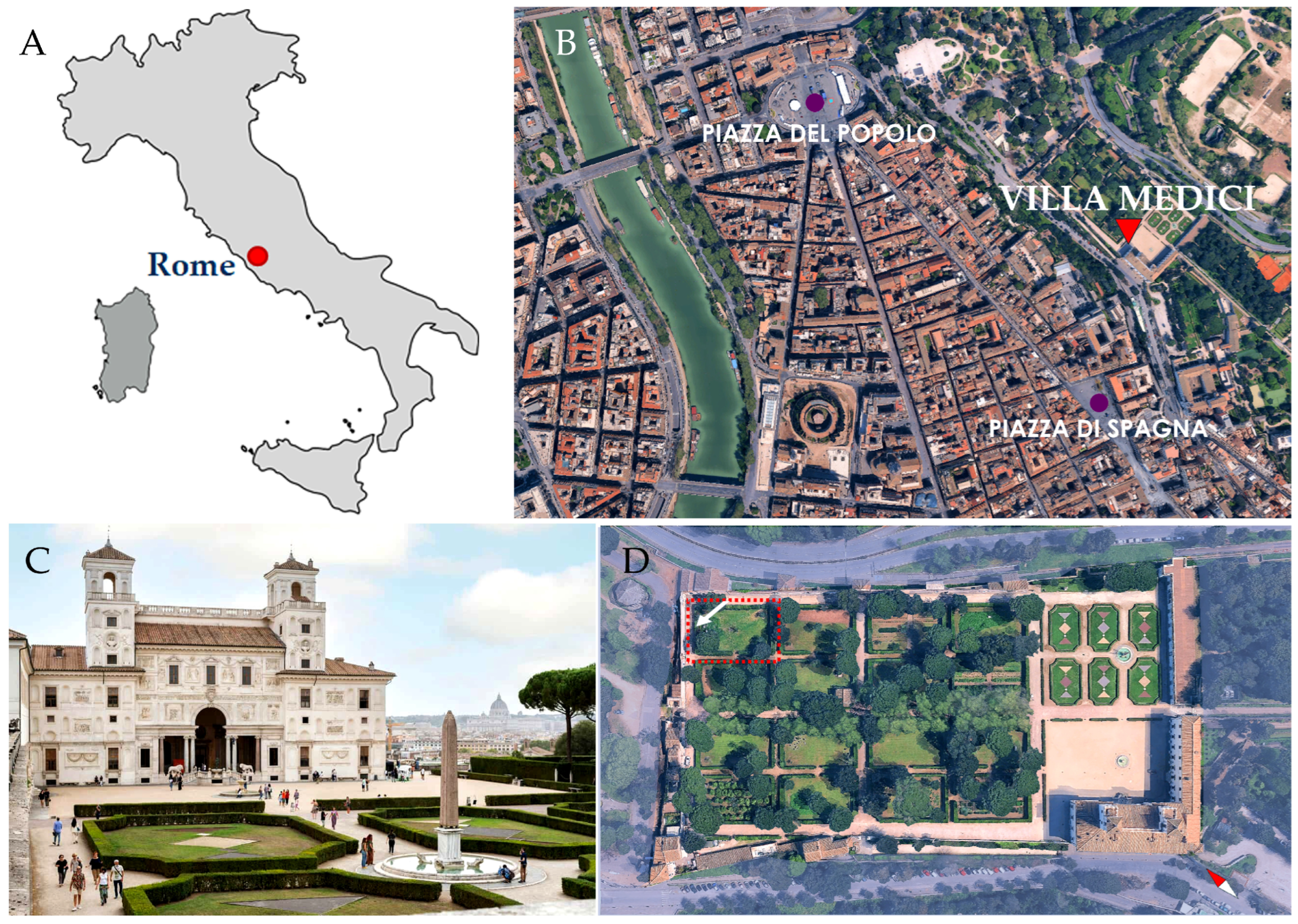
The Villa Medici stands on the Pincio hill, north of the centre of Rome (Figure 1), an area occupied by villas and gardens since antiquity. In 1576, Cardinal Ferdinando de’ Medici (1549–1609) acquired a small villa, and commissioned the architect Bartolomeo Ammannati to transform it into a luxurious residence. The Villa and its gardens were designed to house the cardinal’s art collection. The finest ancient bas-reliefs, from the Della Valle-Capranica collection, were incorporated into the east façade, whilst statues and sarcophagi were displayed in the garden and quadrangles. The major part of this famous collection was transferred to Florence at the end of the 18th century.
Since 1803, the Villa Medici has been the seat of the French Academy in Rome, founded in 1666 by King Louis XIV to accommodate painters and sculptors sent to perfect their craft in the Eternal City. During the 19th century, the institution and the Villa came to epitomise the artistic academy and academic art, and the works of French artists joined those remaining from the Medici collections. In the 1960’s, Director Balthus commissioned copies of missing antiquities, including the Niobids group. The Villa’s gardens therefore display a collection of sculptures spanning from antiquity to the 20th century. Established in 2012, the «Mission Patrimoine» catalogues, studies, preserves, restores and promotes the Academy’s heritage and collections. Within this framework, major restoration projects have been undertaken. At the same time, the Academy is gradually implementing a programme of collection monitoring and preventive conservation aimed at limiting deterioration, carrying out regular maintenance and reducing the frequency of major interventions. The restoration of the Niobides group provides an opportunity to extend this programme to works exhibited outdoors.
The climate of the city and surrounding areas is classified as Mediterranean (Köppen classification: Csa) with rainfall concentrated in autumn and winter, dry summers, and average annual rainfall of about 800 mm [20]. The dominant wind directions in Rome are from the NE in autumn and winter, while in spring they are predominantly from the SW, and in summer they are mostly oriented along the SW–NE axis [21]. These ventilation patterns result from sea-breeze regimes originating from the Tyrrhenian Sea, as well as slope flows generated by the mountain chains surrounding the city [21].
Figure 1.
Position and views of the study site. (A) Location of Rome; (B) aerial view of Villa Medici within its urban context; (C) main building of Villa Medici; (D) aerial view of Villa Medici and the portion of the historic garden open to visitors. In red, “Le carré des Niobides”, the white arrow indicates the side in which the sculptures are located.
2.2. The Historical Garden and Vegetation Context
Covering an area of almost seven hectares and inspired by Tuscan gardens, the historic gardens of Villa Medici retain their overall structure through clearly differentiated areas. Following a prolonged period of neglect, it regained its dignity and function as a representative space serving the prestigious French institution. Restoration works, begun in the early nineteenth century, encompassed the entire garden. Throughout these interventions, the Directors of the Academy demonstrated considerable respect for its original character, preserving many of the features and plant elements that had historically defined its landscape.
The historic garden of Villa Medici comprises three principal areas: the parterre and sixteen adjoining quadrangles, the wood, and the braccheria. In front of the main building, the parterre extends along the villa’s central axis and features six geometric flowerbeds edged with clipped Buxus sempervirens L. hedges. The adjoining quadrangles are enclosed by tall hedges dominated by Laurus nobilis L., creating a distinctly labyrinthine appearance. Other hedge species include Buxus sempervirens L., Pistacia lentiscus L., Phillyrea angustifolia L., Phillyrea latifolia L., Myrtus communis L. and Rhamnus alaternus L. subsp. alaternus. The quadrangles also contain trees such as Quercus ilex L., Ulmus minor Mill. and Pinus pinea L. [22].
The woods, historically used for bird hunting under Ferdinando de’ Medici, are characterised by mature holm oaks (Quercus ilex L.) and some phyllireas. The braccheria, dominated by Pinus pinea L. and isolated boxwoods, now houses the greenhouse and some living quarters for Villa Medici fellows and Academy students [22].
The garden also contains a citrus collection reflecting the Medici family’s longstanding interest in citrus cultivation. Distributed throughout the garden, except in the wood, are more than 350 trees representing nine species and approximately fifty varieties, including bitter oranges, lemons and citrons [22].
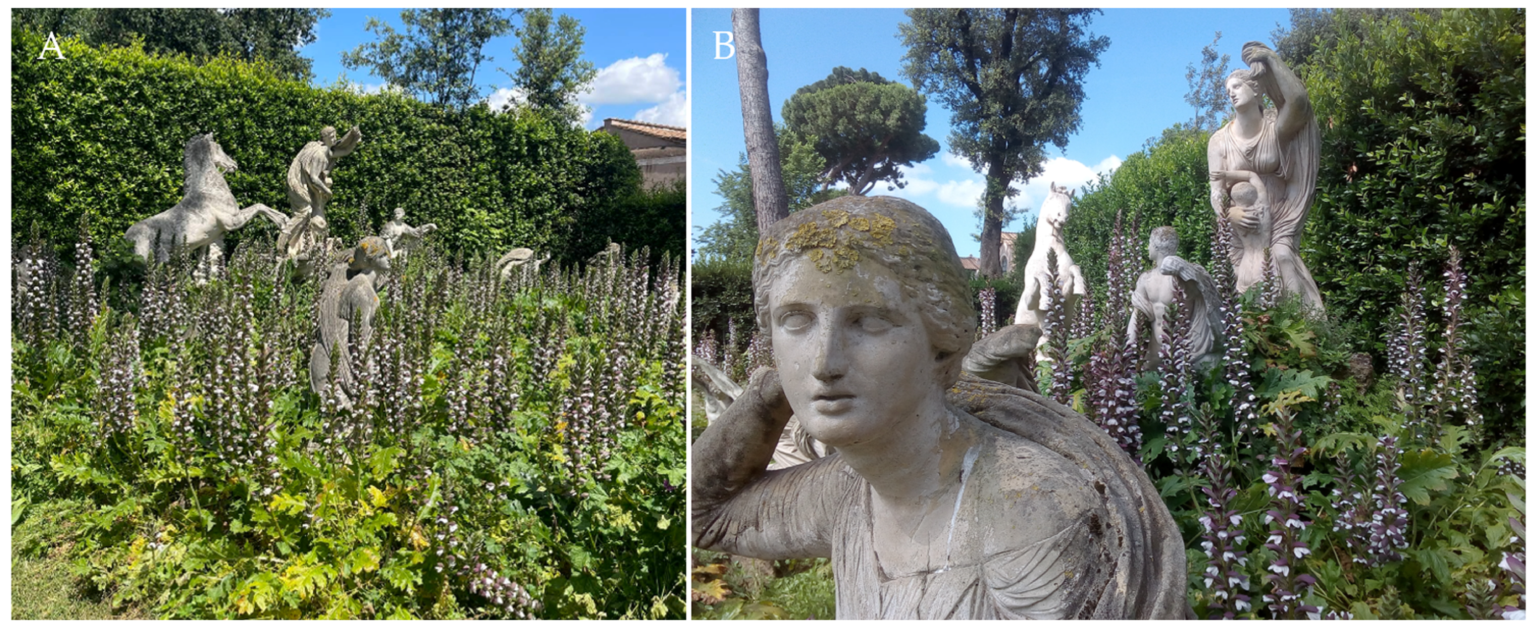
At the study site, known as “Le Carré des Niobides”, the vegetation actively contributes to the representation of the myth, unifying, framing and providing a backdrop to the sculptures. The tall laurel hedge and two pines (Pinus pinea L.) flanking the sculptural group reinforce its visual integration within the garden. Acanthus mollis L., the dominant plant, was used to harmonise gaps and visual dissonances within the group. The acanthus is one of the most common perennials found in Renaissance and Mannerist gardens [23]. Even if it is considered a basic decorative species of the garden, it is necessary to underline its habit of growth. Acanthus can become an invasive species because of its robust vegetative spread and its ability to thrive in varied ruderal and shaded habitats [24]. This opportunistic expansion can happen when there are no direct competitors or in environments characterized by high levels of anthropogenic disturbance, where its high propagule pressure facilitates rapid colonization [25]. As shown in Figure 2 and Figure S1, this species is particularly problematic, as its dense and recurrent growth obstructs views of the statuary and, in the case of No. 11, completely conceals it (Figure 2A). This species has a HI (Hazard Index) equal to 4 [26], and the potential deteriogenic impact of the population is rated as 24 (on a scale from 0–60) because its frequency higher than 76% [27]. The presence of other species such as Parietaria judaica L. (HI 5) and Silene latifolia Poir. (HI 3), was quite limited (Figure S1), and a single young Ficus carica L. close to the Horse (No. 2) was found. The latter is attributed an HI of 10.
The intervention, aimed at preserving the stability of the statue while simultaneously improving the visibility of the sculptural group, involved vegetation management measures primarily targeting a reduction in the acanthus population. This work was carried out in parallel with the reorganisation of the plinths and, through minor excavation works, enabled the removal of part of the rhizome system (Figure S1). About the 60% of the acanthus has been removed (Figure S1). Similarly, the small fig plant was removed. No phytosanitary products were used.
Figure 2.
Carré des Niobides. (A) General view of the sculptural group, and (B) a detail of the acanthus cover.
2.3. Artworks in Study
The Niobid sculptural group was installed in the north-eastern corner of the historic garden of Villa Medici between 1975 and 1976, when the sculptor Michel Bourbon, then Director of the Academy’s Sculpture Department, obtained permission to reproduce the works in the Uffizi Gallery by means of “direct casting”. This work formed part of a decade-long programme of restoration initiated in 1961 by Balthus (Balthasar Klossowski de Rola, 1908–2001), then Director of the Academy, with the aim of restoring the villa to its sixteenth-century character.
The examination of the internal accounts [15] makes it possible to identify the materials employed by Michel Bourbon in the production of the Niobids. Of the thirteen statues, a clear distinction can be made between the constituent materials: eight were produced using an organic resin-based binder, namely epoxy resin, while the remaining five were made using an inorganic mortar consisting of white cement-based concrete.
In the former case, the material comprises Araldite LY 560 epoxy resin with HY 560 hardener (Ciba–Geigy Ltd., Basel, Switzerland), mixed with white Carrara marble chips and limestone powder. This mixture was used for Niobid statues No. 1, 5, 6, 7, 10, 11, 12 and 13, as well as for the right arm of the Niobid No. 4 (Table 1). All the sculptures originally featured a surface finish consisting of a dark patina it is hypothesized that these patinas were applied as transparent glazes containing organic colorants derived from plant infusions, treatment intended to mitigate the stark whiteness of the marble-dust-filled mortars. This intervention was intended to achieve greater degree of chromatic uniformity and harmony among the different materials comprising the polymaterial sculptural group, in keeping with the artist’s aesthetic intentions [28].
Most of this original coloration has since been lost; however, rare and scant traces of the original finish remain in areas that have been better protected from the direct action of atmospheric agents.
The casts were installed on bases made of red brick and concrete, with a layer of insulation on top, consisting of a sheet of bituminous membrane, on which each individual statue was placed. The ground surface consisted of a naturalistic arrangement of tufa rocks of different sizes, assembled “dry” and placed around the bases to hide them from view. For statues No. 1, 2, 3 and 4, the bases were raised above ground level to different heights; in these cases, tufa rocks were also used to create a natural appearance. Several irrigation nozzles are distributed throughout the study area, concealed among the rocks and vegetation.
This complex underwent major restoration work in 2009, during which a huge number of missing elements were reconstructed, and extensive cleaning was carried out. The present study builds upon this intervention within the framework of an extraordinary maintenance plan, as a continuation of the conservation and restoration work previously undertaken.
The works were carried out between May and November 2025. In view of the high summer temperatures and frequent autumn rainfall, a protective cover was installed to provide suitable working conditions for the conservators and prevent thermal stress to the artworks. A single-pitch canopy, supported by a prefabricated metal-frame structure, was designed to provide protection while maintaining adequate ventilation. The structure comprised timber beams supporting transparent corrugated fibreglass sheeting, over which a white PVC tarpaulin (640 g/m2) was secured. Its thickness, considering the site’s natural light conditions, provided adequate shading without compromising visibility for the conservators.
Table 1.
Statues under study and their main features. Dimensions are reported as Length × Width × Height (L × W × H) in cm.
2.4. Biological Component Identification
Owing to the unique nature of these artefacts, we aimed to provide the broadest possible spectrum the organisms composing biological patinas.
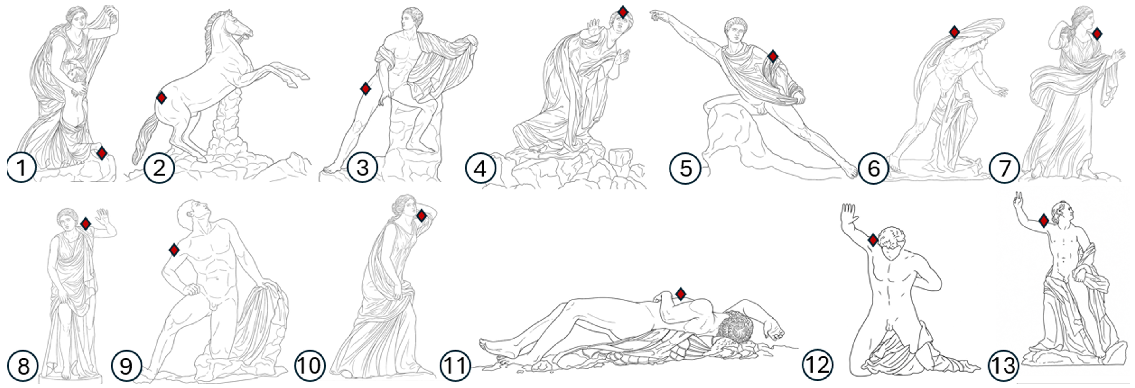
Prior to treatment, the biopatinas were documented for each statue assessing the distribution of lichen and moss components. To characterise the microbial component, small samples were collected from each statue from the most structured biopatinas (Figure 3), subject to scaffolding accessibility. Samples were collected using sterile cotton swabs, transported in a refrigerated container, and processed upon arrival at the laboratory. Each sample was suspended in a sterile centrifuge tube containing 5 mL of sterile saline solution (0.9% NaCl). Aliquots of 400 μL were inoculated into a flask containing 50 mL of BlueGreen-11 freshwater medium (BG11; Sigma-Aldrich, Darmstadt, Germany) for the cultivation of cyanobacteria and containing 50 mL of Bold’s Basal Medium (BBM; Sigma-Aldrich, Darmstadt, Germany) for microalgae, following the procedures outlined in NORMAL 9/88 [29]. The remaining suspension was serially diluted and plated onto Dichloran Rose Bengal Chloramphenicol Agar (DRBC; VWR International GmbH, Darmstadt, Germany) for fungal isolation with attention to black fungi as representative of lithobiontic communities [30], and other dominant fungi. Plates were then incubated at room temperature (18 ± 1 °C) and inspected daily for 2–3 weeks. To improve the isolation yields of the slow-growing black fungi, after the first run of plating, the remaining suspension was stored at low temperature according to the long cold incubation method [31]. After six months, inoculation on agarised medium and subsequent incubation were carried out as above. All morphologically distinct black colonies were transferred onto malt agar (MA, malt extract 30 g/L, agar 15 g/L) and processed for molecular identification.
Cyanobacterial and algal cultures were incubated at room temperature (20 ± 2 °C) for eight weeks under cool-white, fluorescent illumination (Osram Dulux L 36W/840 Lumilux, 2900 lm; Munich, Germany) using a 12 h light/12 h dark photoperiod at room temperature (20 ± 2 °C).
At the end of the fieldwork, a small fragment of the plastic tarpaulin was collected for further analysis to have indirect evidence of the surrounding environment. The sample was further divided in two, with the former part used for direct observation by low-magnification optical microscope and the other for microbiological analyses.
Figure 3.
Drawings of the studied replica sculptures and their respective sampling points (red diamonds) in sequential numbering assigned to the sculptures in this study. The statues are not shown to scale relative to one another. The actual dimensions of the reproductions are given in Table 1.
2.5. Molecular and Morphological Identification
Genomic DNA was extracted from pure fungal colonies using a Nucleospin Plant kit (Macherey-Nagel, Düren, Germany) using the protocol optimised for fungi. The molecular identification of isolated fungal strains was performed through sequencing of the ITS barcode region. PCR reactions were carried out in a final volume of 25 μL using BioMix (VWR), supplemented with 5 pmol of each primer (ITS4/ITS5: 5′-TCCTCCGCTTATTGATATGC-3′/5′-GGAAGTAAAAGTCGTAACAAGG-3′) and approximately 30 ng of template DNA. Amplification was performed using a MyCycler™ Thermal Cycler (Bio-Rad Laboratories, Munich, Germany), as previously described [6]. Sequencing was conducted by Macrogen (Milan, Italy), and the resulting electropherograms were manually inspected and assembled using Chromas Pro 1.41 software (Technelysium, Southport, QLD, Australia). Sequence similarity matching was performed using the BLASTn against the GenBank database, excluding sequences originating from uncultured or environmental samples, and prioritising sequences from type strains.
The morphological identification of phototrophs was performed using the analytic keys of Guiry and Guiry [32]. While lichens were identified using identification keys in ITALIC 8.0 [33], and mosses using the Italian moss guide [34].
2.6. Idreta Cleaning Protocol
The decision to adopt this new physical cleaning tool was motivated not only by the need to pursue a more environmentally sustainable and low-impact approach, but also by the poor performance of the protocols previously tested for the control of biological patinas. These protocols included both physical and chemical methods, as well as conventional and innovative products. All the protocols tested produced negligible results, while others were excluded from the outset because of potential solvent-related interference (Figures S2–S4).
The Idreta S.W.S. (Soft Wash System; Arbizzano-Santa Maria, VR, Italy), an innovative, controlled water-cleaning technology developed by IdroEletrika to provide precise and gentle nebulisation for conservation and restoration purposes, was used in two or three runs separated by a pause of 5–10 days according to the openings of the site. In detail, the micronized washing at controlled pressure was performed using warm water (at 38 °C). The instrument, equipped by a circular fan jet nozzle orifice Ø of 1 mm, was set to 140 bar pressure jet, flow rate 1 L/min, and heating power 2.6 W. The micro-nebulised spray was applied perpendicular to the surface, at a distance of approximately 10 cm, over small sections no wider than 20–25 cm at a time, using a slow and steady back-and-forth movement along a straight line.
Following the completion of the cleaning operations and based on the results of preliminary tests, a water-based nanotechnological water-repellent protective treatment was selected. Nano Silo W® (CTS S.r.l., Altavilla Vicentina, Italy) was applied by brush in a single application. On the resin statues, to attenuate the alteration and yellowing and to achieve aesthetic harmonisation of the sculptural group, the same protective treatment was premixed with white titanium pigment at 10% (w/w) and applied consistently. The titanium white pigment was also supplied by CTS S.r.l.
The recorded images were processed using their luminosity histograms using Adobe Photoshop (version 7.0, Adobe™, San Jose, CA, USA), displaying brightness values derived from the combined RGB channels, ranging from 0 (black) to 255 (white). To minimise the effects of variations in field conditions before and after the treatment, the post-treatment images selected for analysis were those taken in the early morning, from the shaded side of the sculptures. Similarly, the resin sculptures were excluded from this assessment.
3. Results
3.1. Biopatinas Distribution and Characterization
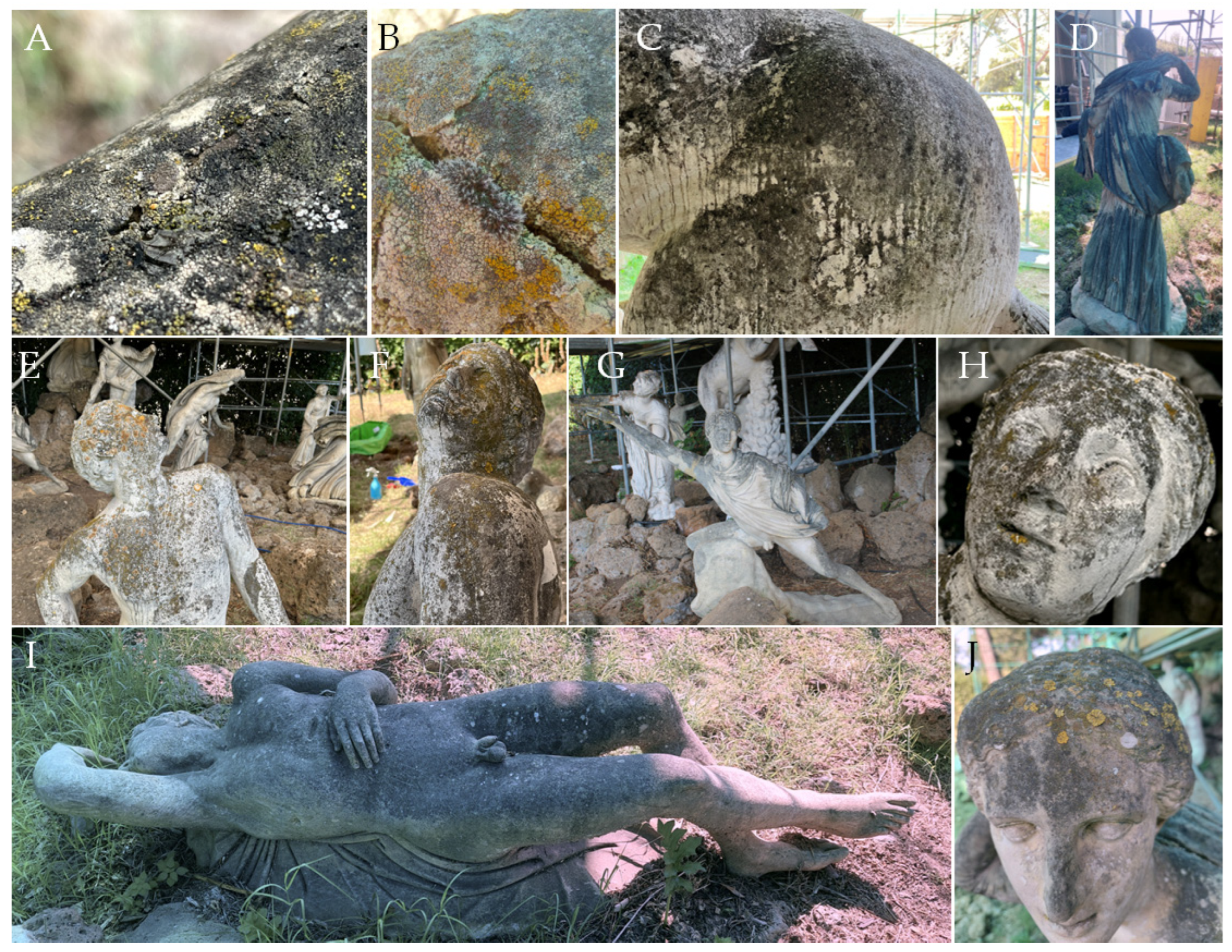
The distribution of biopatinas generally followed the pattern of surface colonisation, with a preference for horizontal or gently inclined areas where water and deposited material can accumulate and favour microbial growth. As shown in Figure 4, the biopatinas appear thick, dark, and structurally complex, comprising lichens, fungi, cyanobacteria, and algae, as well as mosses.
Figure 4.
Examples of the distribution and appearance of biopatinas: (A,B) detail of different lichen- dominated biopatinas on sculpture No. 12 and No. 7, with (A) water droplet perched on the colonised surface, and (B) moss growth within an area of discontinuity. (C) Dark, irregular phototroph-dominated patina showing localized dripping lines on sculpture No. 2; (D–J) sculptures exhibiting variable degrees of surface colonisation, distribution, and disfiguring effects, including dark deposits and staining, alongside scattered whitish and yellow-orange patches consistent with lichen growth. (D) Back of sculpture No. 7 (E,F) sculpture No. 9; (G) sculpture No. 5; (H) face of the sculpture No. 4; (I) extensive and uniform dark discolouration on sculpture No. 11; (J) head of sculpture No. 7.
Cyanobacterial communities associated with the sculptures comprised a diverse assemblage of taxa, including both filamentous and unicellular forms, with filamentous taxa being particularly frequent. To avoid misclassifications driven by cryptic morphology and phenotypic plasticity, a conservative genus-level identification was maintained. Among them were found representatives of Aphanothece, Calothrix, Chroococcus, Phormidium, Pseudanabaena, Scytonema, and Synechococcus. The algal component was represented by a range of morphologically diverse taxa, including flagellated forms such as Chlamydomonas and Haematococcus, together with non-flagellated genera including, for example, Apatococcus, Chlorella, Chlorococcum, Cosmarium, Oocystis, and Scenedesmus (Table 2).
The lichen taxa recorded encompassed predominantly crustose species, including Candelariella species, Flavoplaca flavocitrina, Lecanora campestris, Polyozosia dispersa, Protoparmeliopsis muralis, Variospora aurantia and Verrucaria nigrescens, together with foliose taxa such as Parmelia saxatilis, Physcia adscendens and Xanthoria parietina (Table 2).
Regarding the fungal component (Table 3), several black fungi were detected, including Aureobasidium, Exophiala and other unidentified taxa within the Chaetothyriales. Scolecobasidium, Vermiconidia and Knufia isolates were obtained only after prolonged cold incubation, highlighting their oligotrophic nature. Chaetocapnodium polonicum was the most frequently isolated melanised fungus. Notably, Parengyodontium album was particularly frequent across the statuary group and, in terms of abundance on individual sculptures, was dominant on some pieces, including sculptures No. 2 and 3. The obtained sequences (deposited under accession numbers QB081791–QB081809), together with their closest matches and percentage identities, are reported in Table S1.
Mosses occurred in small tufts on horizontal surfaces or around surface discontinuities, namely joints and small cracks. They appeared as rounded, grey-green, cushion-like pulvini, with ovate-lanceolate phylloids bearing a robust costa extending to the apex and usually continuing into a conspicuous, long, hyaline hair-point. All these diagnostic features are consistent with Grimmia pulvinata.
Table 2.
Presence of mosses, lichens, cyanobacteria, and algae on the studied sculptures.
Table 3.
Presence of fungi on the studied sculptures. The best BLASTn matches are reported in Table S1.
The analyses performed on the tarpaulin made it possible to qualitatively and quantitatively assess the particulate deposits, namely, the material that could potentially be deposited on the statues. The deposit on the protective tarpaulin appeared thick and extremely dark. The adherent material, approximately 0.5 mm thick, was strongly attached in its deeper layers but less firmly adhered at the surface, where material could be readily removed by rubbing. As expected, direct observation under a stereomicroscope revealed a substantial presence of pollen grains, as well as seeds and arthropod remains (e.g., complete exoskeletons, legs and wings) (Figure S5).
Microscopic observation also revealed large groups of fungal spores, the most readily identifiable of which belonged to the Alternaria group. Pollen grains and vegetal trichomes were likewise frequently observed.
The cultures of the material collected from the protective plastic covering revealed a marked predominance of yeasts and yeast-like fungi over filamentous forms. The isolates showed the closest matches with type strains of Rhodotorula mucilaginosa (99.18%), Naganishia vaughanmartiniae (98.65%), Naganisha albida (99.82%), and Aureobasidium melanogenum (99.81–100%). Among the yeast-like colonies, Aureobasidium were numerically less abundant, as they were recognisable on aged plates by darkened colonies. The filamentous component was limited to some isolates belonging to Nectriaceae. The sequences obtained (deposited under accession numbers QB081810–QB081814), together with their closest matches and percentage identities, are reported in Table S1.
3.2. Cleaning Protocol Outcomes
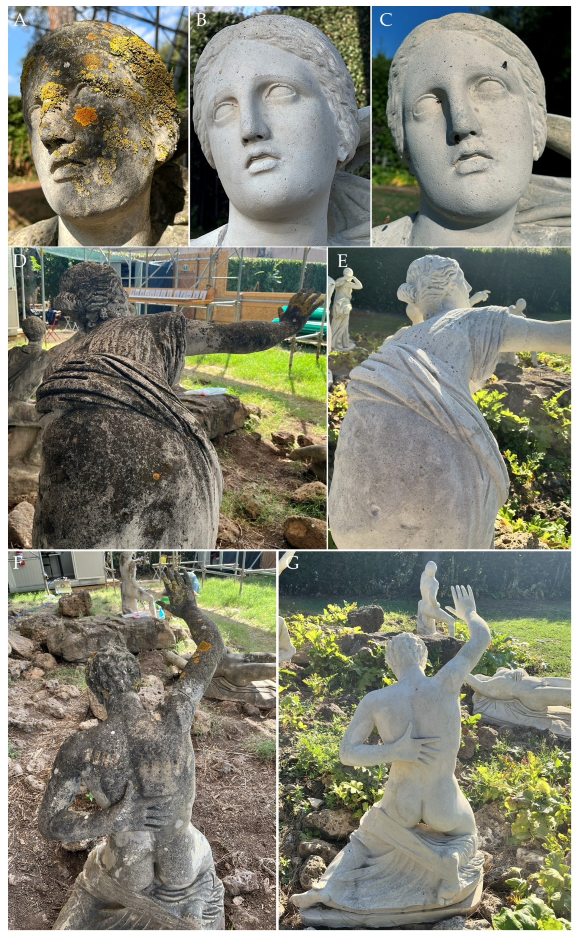
In general, following the first cleaning cycle using micronised water, the biopatinas were completely removed from approximately 90% of the treated surfaces. The removal of thicker and more persistent deposits required further treatment cycles, interspersed with periods of complete drying. The biopatinas on Niobid No. 11 proved particularly tenacious; for example, two additional treatment cycles were required in those areas where residual biomass was still visibly present.
After cleaning, the first notable difference among the statues of the Niobids group was their colour. The statues produced using cement-based mortar retain a white appearance, while those made with epoxy resin-bound mortar have undergone marked yellowing as consequence of the direct exposure to ultraviolet radiation (Figure S5A,B).
The removal of the biogenic patinas revealed more clearly the evidence of a general process of erosion affecting the sculptural surfaces of both the cement-based statues and those made from epoxy resin. In some cases, this manifested as a loss of definition of the features, as observed on statue No. 9 (Figure S5C); in others, as surface roughness detectable by touch, or as the formation of a network of superficial microcracks (crazing) in the epoxy resin statues, as observed on the mantle of Niobid No. 6. Another phenomenon observed was progressive pore coarsening. In this case, it is not possible to determine whether the phenomenon originated during casting, resulted from biogenic activity, or was caused by a combination of both factors. In any event, the phenomenon should be monitored, particularly in areas where it is more pronounced, such as on the back of Niobid No. 4 (Figure 5F) and the left hand of Niobid No. 9 (Figure S5E, both of which are cement-based).
The image analysis highlighted the visible differences between the images acquired before and after treatment. In some areas, small spotted remains of residual biopatina are appreciable upon close observation but, as reported in Table S2, lightness values after cleaning increased by between 20% (in partially colonised areas) and 205% (in fully colonised areas). These data should nevertheless be considered an indicative underestimation of the actual treatment efficacy, given the conservative measurement approach adopted.
Based on the evidence obtained and on the thickness of the material deposited on the protective tarpaulin, it was agreed that monitoring should be carried out at six-month intervals, in line with the schedule adopted during the intervention and eventually implemented. It would be particularly useful for these monitoring phases to coincide with the end of the conifer pollination period and the end of the autumn rainy season, respectively.
Figure 5.
Examples of cleaning outcomes: before (A,D,F), and after treatment (B,C,E,G). (A–C) Sculpture No. 10: (A) 17 July 2025; (B) 13 February 2026; (C) 7 July 2026. (D,E) Sculpture No. 4: (D) 17 July 2025, (E) 7 July 2026. (F,G) Sculpture No. 12 before and after cleaning: (F) 17 July 2025, (G) 7 July 2026.
4. Discussion
In recent years, we have witnessed the effects of anthropogenic impact, with the worrying loss of animal and plant species every year and the dramatic effects of climate change characterised, among others, by heatwaves and extreme rainfall events. The global economic crisis does the rest, eroding purchasing power, creating uncertainty in the markets, and ultimately limiting investment. In this context, allocating resources to culture and nature, particularly to historic gardens where cultural identity and environmental resilience intersect, is not only commendable but also essential to their preservation. Planned conservation could play a key role in ensuring long-term protection and sustainable management of cultural and natural heritage. This approach is not new because it is based on fundamental concepts conceived by Brandi and Urbani, according to whom, preserving heritage does not mean freezing it, but managing its transition across generations [35,36,37]. This form of management is in contrast with an approach in which conservation interventions, such as restoration, are carried out as a reactive “event” only after damage has occurred, often under emergency conditions [36,38,39].
In this view, for example, Della Torre redefines planned conservation as an ongoing, operational strategy designed to minimize risk, manage decay proactively, and maximize the social and economic benefits of historic structures by applying low-intensity continuous care as a part of a long-term process [36].
Villa Medici, as the seat of the Académie de France, has adopted this approach, and the cleaning protocol presented here forms part of this broader framework of management and continuous care. However, in order to develop implementation protocols within a planned conservation and continuous care strategy, it was first necessary to gather information on the biogenic component and to identify, through testing, a low-impact method that was both effective and compatible with the conservation objectives and specific needs of the site.
Indeed, the major challenge is related to the materials used. On the one hand, some statues can, to a certain extent, be assimilated to other construction materials; on the other hand, the biodeterioration of epoxy resins remains a poorly investigated topic. Only a few studies have addressed this issue, including investigations on materials used in restoration [16,40,41,42], as well as a sculpture in Devin (Bratislava, Slovakia) and an epoxy-resin kiosk in Belgrade [43,44]. From a biological perspective, the biopatinas proved to be quite complex, both qualitatively and quantitatively. The identification of phototrophs with a flagellate phase, together with taxa frequently reported in fountains and Nymphaea [45,46], such as species of the genus Chlamydomonas, detected on 61% of the sculptures, and Chlorella, could suggest that the persistent availability of water in the gardens, associated with regular irrigation, may be an important factor in shaping the observed biotic communities. Among the cyanobacteria, Phormidium was the most frequently recorded genus. Cyanobacterial filamentous taxa in terrestrial environments play a critical role in the formation of stable biofilms on exposed stone surfaces through the development of dense networks and the production of extracellular polysaccharides, which form a robust matrix that provides protection against desiccation and extreme temperature fluctuations [47]. Despite the valuable ecological insights already obtained, future work will aim to combine morphological observations with molecular analyses to achieve a more precise resolution of the biodiversity and ecology of the phototrophic species involved.
Lichens are known to have a particular affinity for the substrates they colonise. On stone objects, pH is a primary factor in the selection of lichen flora [48,49]. Beyond substrate chemistry, uncommon materials can also be colonised, such as glass, metal, leather, pottery, plastic, rubber and other anthropogenic materials [50]. Epoxy resins have been used extensively across industrial and conservation sectors due to their robust bonding strength, chemical resistance, and structural versatility [51]. Their hydrophobic surfaces may initially provide a barrier to biological colonisation; however, prolonged environmental exposure may promote photo-oxidative degradation of the polymer matrix, potentially increasing its bioreceptivity over time. In the present case, however, we did not observe a clear difference in the species recorded on the two materials. The finding of species known as calcicolous also on epoxy resin statues needs to be further investigated. Indeed, in this study, no specific measurements were undertaken to determine the extent to which the incorporation of flakes and dust of marble within the epoxy binder may modify the physicochemical properties of the resin, or to establish whether these mineral components become exposed at the surface as the material undergoes degradation. Another possibility could be related to the development of a sufficiently thick biopatina, which could partially mask or override the influence of the underlying substrate. Beyond the substrate question, it is relevant the presence of species typical of disturbed /anthropic environments, such as Xanthoria parietina, which occurred on eleven out thirteen Niobids (84.6%).
Compared with fungal communities reported in other studies of monuments in the Mediterranean area, the cultivable fraction identified in this study exhibited several distinctive features, while also confirming some previously reported findings.
Exposed stone surfaces, like other extreme environments and underexplored niches, harbour several unknown taxa [52]. This study aligns with this finding recording at least ten isolates sharing less than 97% sequence identity with type strains of Exophiala bergeri, Veronaea botryosa, Tiankengomelania guangxiense (order Chaetothyriales) and Coniosporium apollinis (Dothideomycetes incertae sedis). Moreover, this study confirms the efficacy of long cold incubation in improving the isolation of slow-growing oligotrophic black fungi [31], as Scolecobasidium and Coniosporium species were isolated only using this method.
Unlike other studies focusing on fungal diversity on outdoor monuments in the Mediterranean region, the present study revealed a marked prevalence of Parengyodontium album. This species has previously been reported from several historical sites and materials, including caves, wall paintings and museums, and is frequently associated with environments affected by salt efflorescence [53,54]. To the best of our knowledge, the present study is the first to report the occurrence of P. album on sculptures preserved in an outdoor environment. Given its broad distribution and association with soil, the detection of P. album is not unexpected. Rather, the main interest lies in its associations with other ubiquitous fungi, both across different substrates and within individual artefacts, as well as in its metabolic capabilities. This species has recently attracted considerable attention owing to its demonstrated ability to degrade polyethylene, one of the most widely used plastic polymers, and to mineralise it to CO2. These findings could suggest a broader biodegradative potential towards synthetic materials and artificial resins [41,55]. It may therefore be worthwhile to investigate whether its distribution is random or instead reflects a preference for particular substrates; epoxy resins, for instance, may represent materials susceptible to biological degradation. Unfortunately, although our dataset derives from samples collected from the most heavily colonised areas, the available evidence does not allow us to advance any robust hypothesis. Future monitoring will provide an opportunity to determine whether a genuine and statistically significant affinity for a particular substrate exists.
The genus Chaetocapnodium was similarly frequent. Notably, C. polonicum, the species most closely matching our isolates, has been associated with resinous substrates [56], suggesting possible transfer from nearby pines to the artefacts. This exchange has also been documented for Aureobasidium. Notably, A. pullulans and A. melanogenum are common wood-discolouring and plant-associated microorganisms, pioneer colonisers of marble, and inhabitants of extreme, oligotrophic environments [52,57,58,59]. In this study, Aureobasidium species were detected both on the protective plastic tarpaulin, which collected material falling from the trees above, and on the statues. The yeasts Naganishia globosa and N. vaughanmartiniae were likewise detected on both the tarpaulin and the statues, raising the possibility of their involvement in biofilm formation and consolidation on monuments, a potential role that has often been overlooked.
The composition of the biopatinas, as well as the material on which they develop, influences their degree of adhesion to the substrate and may therefore affect the efficacy of removal treatments. This became evident from the early stages of the study, when other cleaning methods, including laser cleaning, conventional biocidal treatments, and essential-oil-based treatments with different carriers, were considered for testing. Although concerns regarding the use of a novel cleaning system are understandable, given the known effects of water runoff and the potentially detrimental effects of high-pressure water jets, in which water impacts the surface with considerable force, these considerations do not necessarily apply to systems that use pressure to atomise water into a fine spray, thereby reducing its mechanical impact on the substrate.
In conventional pressure washing, droplets possess sufficient mass and velocity to generate substantial local stresses upon impact, including impact pressures, shear stresses and micro-abrasive effects, thereby promoting the disaggregation and removal of loosely bound material. To a first approximation, the effectiveness of this mechanical action is related to the momentum and kinetic energy transferred at the point of impact.
In contrast to conventional high-pressure water jets, the proposed system uses pressure to atomise the water into a finely dispersed spray, substantially reducing the mechanical impact exerted on the substrate and allowing the treatment to be applied with minimal mechanical stress to the surface. This allowed the treatment action to be carefully modulated, with optimal results achieved after successive treatment cycles, each stopped when no further improvement was observed and followed by complete drying. The observation that simple drying and the reapplication of the same settings restored the effectiveness of biopatina removal prompted investigation into the possible reasons underlying this renewed effectiveness. In this light, we can suppose that this aspect may have both a biological and physical explanation. From a biological perspective, the weakening of the biofilm increases the susceptibility of organisms to external agents. In summer, elevated temperatures, UV radiation in exposed areas, and drought can further impair the vitality of the colonising microorganisms that compose the relict biofilm. In the meantime, the drying cycles, through the contraction of their extracellular polymeric substance (EPS) matrix, generate internal stresses that weaken the biofilm’s attachment to the stone substrate and making it more susceptible to detachment upon subsequent rewetting. Conversely, drying is a necessary step during the cleaning process because, once the material’s surface is fully and deeply saturated, this creates a superficial barrier, a sort of water cushion, that prevents the penetration of micronised water [60]. While the same water film could, at the micro- and nanoscale, form capillary bridges, generating capillary forces that promote adhesion and surface tension forces that retain particles within the aqueous film [61]. Taken together, these findings indirectly support the gentle and respectful nature of the IDRETA cleaning process towards sculptural surfaces. The efficacy of the treatment is well-established; however, as the treatment was not combined with any biocidal treatment, rapid recolonisation can be expected based on the prevailing environmental conditions and previous studies [62,63]. This fact does not constitute a concern, as a slight patina is acceptable both in terms of naturalistic integration and because of the monitoring and low-intensity conservation plan (e.g., dusting removal, soft wash) aimed at preventing deep and extensive colonisation. Given the pilot nature of this investigation, we do not rule out the possibility of refining the established protocol based on emerging empirical evidence during the initial monitoring phases. Such modifications may include shortening the evaluation intervals (e.g., from six to four months) as well as reapplying or adjusting the formulation of the protective agent to effectively delay biological recolonisation.
For wider application of the IDRETA water atomisation tool, certain logistical, operational prerequisites are essential and other may have positively influenced the cleaning process and its repeatability. Indeed, water and electricity connections were essential, while the shade cover required under current Italian legislation for worker safety may also have facilitated treatment by allowing the sculptural surfaces to be wetted and dried without excessive thermal stress during the height of summer. Moreover, considering the wider application of this method, once the structural integrity of the material has been verified, it could be used either alone, as in the present case, or in combination with other treatments, where preliminary biofilm removal could reduce the amount of product required and/or treatment time.
5. Conclusions
In cultural heritage conservation, new methods are needed to control biopatinas, improving both protocol efficacy and environmental sustainability, particularly for materials lacking standardised protocols.
The unconventional materials used to create the Niobids sculptures, together with their close proximity to vegetation, may account for the distinctive fungal composition of the biopatinas, characterised by the prevalence of Parengyodontium album and Chaetocapnodium polonicum.
Micronised water cleaning proved effective for biopatina removal, warranting consideration at the design stage not only as a tool within “continuous care” protocols, but also as a complementary treatment to weaken biopatinas, potentially enhancing the efficacy of other control methods and/or reducing contact times. Its use of water without additional chemicals makes it as a genuinely “green” and environmentally sustainable approach to biopatina control.
More interestingly, this study has shown that environmental and economic sustainability can be achieved by adopting a different intervention paradigm, where conservation interventions are based on monitoring and low-intensity preventive measures instead of reactive interventions undertaken only after damage has occurred, often under emergency conditions.
Supplementary Materials
The following supporting information can be downloaded at https://www.mdpi.com/article/10.3390/app162211216/s1. Figure S1. Site map showing the distribution of major species found and the irrigation system; Figure S2. Example of results achieved on statue No. 2 (The Horse) using Infinito 100W laser (El.En. SpA); Figure S3. Examples of tests performed on Niobid No. 11 (the Dying Niobid) using traditional biocides; Figure S4. Examples of tests performed on Niobid No. 11 (the Dying Niobid) using BioTersus 2%; Figure S5. Material adhering to the plastic-coated tarpaulin; Figure S6. Early outcomes of cleaning; Figure S7. Example of the image analysis performed on Niobid No. 4 to measure differences in lightness using Adobe Photoshop 7.0; Table S1. Isolated strains and their best BLASTn (software version 2.17.0.) matches; Table S2. Result of image analysis performed on Niobids No. 4 using Adobe Photoshop 7.0 histograms.
Author Contributions
Conceptualization, E.R., A.G. and D.I.; methodology, E.R. and D.I.; software, D.I.; investigation, D.I., E.R., G.F.M. and E.D.; writing—original draft preparation, D.I. and E.R.; writing—review and editing, D.I., E.R., E.D., G.F.M. and A.G.; visualization, D.I., E.R. and G.F.M. All authors have read and agreed to the published version of the manuscript.
Funding
This research received no external research funding, but it represents the professional activities of specialists engaged by the Académie de France à Rome, Villa Médicis to address the conservation issue presented here through an environmentally sustainable approach. The APC was funded by Daniela Isola.
Institutional Review Board Statement
Not applicable.
Informed Consent Statement
Not applicable.
Data Availability Statement
The original contributions presented in the study are included in the article/Supplementary Material, further inquiries can be directed to the corresponding author.
Acknowledgments
The authors thank Pierre-Antoine Gatier, Architecte en chef des monuments historiques, and architects Françoise Laurent and Yannick Legal for their collaborative support. Valentina Peri Proto, Ilaria Lupini, and Chiara Sabatini are acknowledged for their unvaluable help in decay mapping and photographic documentation.
Conflicts of Interest
The authors declare no conflicts of interest.
References
- Olivadese, M.; Dindo, M.L. Historic and contemporary gardens: A humanistic approach to evaluate their role in enhancing cultural, natural and social heritage. Land 2022, 11, 2214. [Google Scholar] [CrossRef] [Scilit]
- Guillitte, O. Bioreceptivity: A new concept for building ecology studies. Sci. Total Environ. 1995, 167, 215–220. [Google Scholar] [CrossRef] [Scilit]
- Miller, A.Z.; Sanmartín, P.; Pereira-Pardo, L.; Dionísio, A.; Sáiz-Jiménez, C.; Macedo, M.F.; Prieto, B. Bioreceptivity of building stones: A review. Sci. Total Environ. 2012, 426, 1–12. [Google Scholar] [CrossRef] [Scilit] [PubMed]
- Sanmartín, P.; Miller, A.Z.; Prieto, B.; Viles, H.A. Revisiting and reanalysing the concept of bioreceptivity 25 years on. Sci. Total Environ. 2021, 770, 145314. [Google Scholar] [CrossRef] [Scilit] [PubMed]
- Markovskaja, S.; Kačergius, A.; Davydenko, K.; Fraser, S. First record of Neocatenulostroma germanicum on pines in Lithuania and Ukraine and its co-occurrence with Dothistroma spp. and other pathogens. For. Pathol. 2016, 46, 522–533. [Google Scholar] [CrossRef] [Scilit]
- Isola, D.; Bartoli, F.; Langone, S.; Ceschin, S.; Zucconi, L.; Caneva, G. Plant DNA barcode as a tool for root identification in hypogea: The Case of the Etruscan Tombs of Tarquinia (Central Italy). Plants 2021, 10, 1138. [Google Scholar] [CrossRef] [Scilit] [PubMed]
- Caneva, G.; Langone, S.; Bartoli, F.; Cecchini, A.; Meneghini, C. Vegetation cover and tumuli’s shape as affecting factors of microclimate and biodeterioration risk for the conservation of Etruscan tombs (Tarquinia, Italy). Sustainability 2021, 13, 3393. [Google Scholar] [CrossRef] [Scilit]
- Marando, F.; Salvatori, E.; Sebastiani, A.; Fusaro, L.; Manes, F. Regulating ecosystem services and green infrastructure: Assessment of urban heat island effect mitigation in the municipality of Rome, Italy. Ecol. Modell. 2019, 392, 92–102. [Google Scholar] [CrossRef] [Scilit]
- Bartoli, F.; Pontrandolfi, M.A.; Hosseini, Z.; Caneva, G. A Floristic Analysis and Reconstruction for the Renewal of Botanical Memory: The “Experimental Agricultural Garden of Villa Santa Maria in Potenza” (Southern Italy) as a Model. J. Zool. Bot. Gard. 2026, 7, 12. [Google Scholar] [CrossRef] [Scilit]
- ICOMOS-IFLA. Historic Gardens—The Florence Charter. 1981. Available online: https://www.icomos.org/images/DOCUMENTS/Charters/gardens_e.pdf (accessed on 7 August 2026).
- ICOMOS-IFLA. Document on Historic Urban Parks. 2017. Available online: https://www.icomos.org/images/DOCUMENTS/General_Assemblies/19th_Delhi_2017/Working_Documents-First_Batch-August_2017/GA2017_6-3-2_HistoricUrbanPublicParks_EN_final20170730.pdf (accessed on 9 August 2026).
- Jagiełło, M. Do we need a new Florence Charter? The importance of authenticity for the maintenance of historic gardens and other historic greenery layouts in the context of source research (past) and taking into account the implementation of the sustainable development idea (future). Sustainability 2021, 13, 4900. [Google Scholar] [CrossRef] [Scilit]
- ICOMOS. Carta del Restauro, the First International Congress of Architects and Technicians of Historic Monuments, Athens, 1931. Available online: https://www.icomos.org/en/167-the-athens-charter-for-the-restoration-of-historic-monuments (accessed on 9 August 2026).
- Velayutham, A.; Raman, K. Sculpture in the modern era: Innovations in materials and styles. ShodhKosh J. Vis. Perform. Arts. 2024, 5, 148–162. [Google Scholar] [CrossRef] [Scilit]
- Archives de l’Académie de France à Rome [AFR]. Faldone C 595. 1975.
- Cappitelli, F.; Principi, P.; Sorlini, C. Biodeterioration of modern materials in contemporary collections: Can biotechnology help? Trends Biotechnol. 2006, 24, 350–354. [Google Scholar] [CrossRef] [Scilit] [PubMed]
- Isola, D.; Capobianco, G.; Tovazzi, V.; Pelosi, C.; Trotta, O.; Serranti, S.; Zucconi, L.; Spizzichino, V. Biopatinas on peperino stone: Three eco-friendly methods for their control and multi-technique approach to evaluate their efficacy. Microorganisms 2025, 13, 375. [Google Scholar] [CrossRef] [Scilit] [PubMed]
- Moosavy, M.H.; de la Guardia, M.; Mokhtarzadeh, A.; Khatibi, S.A.; Hosseinzadeh, N.; Hajipour, N. Green synthesis, characterization, and biological evaluation of gold and silver nanoparticles using Mentha spicata essential oil. Sci. Rep. 2023, 13, 7230. [Google Scholar] [CrossRef] [Scilit] [PubMed]
- Favero-Longo, S.E.; Matteucci, E.; Pinna, D.; Ruggiero, M.G.; Riminesi, C. Efficacy of the environmentally sustainable microwave heating compared to biocide applications in the devitalization of phototrophic communities colonizing rock engravings of Valle Camonica, UNESCO world heritage site, Italy. Int. Biodeterior. Biodegrad. 2021, 165, 105327. [Google Scholar] [CrossRef] [Scilit]
- Fratarcangeli, C.; Fanelli, G.; Testolin, R.; Buffi, F.; Travaglini, A. Floristic changes of vascular flora in the city of Rome through grid-cell census over 23 years. Urban Ecosyst. 2022, 25, 1851–1864. [Google Scholar] [CrossRef] [Scilit]
- Pelliccioni, A.; Monti, P.; Gariazzo, C.; Leuzzi, G. Some characteristics of the urban boundary layer above Rome, Italy, and applicability of Monin–Obukhov similarity. Environ. Fluid Mech. 2012, 12, 405–428. [Google Scholar] [CrossRef] [Scilit]
- Fernández Medina, G. Piano di Gestione di Villa Medici 2023/2027; Villa Médicis, Académie de France à Rome: Rome, France, 2023. [Google Scholar]
- Vonešová, V.; Vacek, O.; Vaněk, J. Restoration of a Rudolfine Mannerist historical castle garden. Hortic. Sci. 2018, 45, 101. [Google Scholar] [CrossRef] [Scilit]
- Tan, K.; Panitsa, M.; Kofinas, G. The genus Acanthus (Acanthaceae) in Greece. Phytol. Balc. 2023, 29, 87–96. [Google Scholar] [CrossRef] [Scilit]
- Lazzaro, L.; Bolpagni, R.; Buffa, G.; Gentili, R.; Lonati, M.; Stinca, A.; Acosta, A.T.R.; Adorni, M.; Aleffi, M.; Allegrezza, M.; et al. Impact of invasive alien plants on native plant communities and Natura 2000 habitats: State of the art, gap analysis and perspectives in Italy. J. Environ. Manag. 2020, 274, 111140. [Google Scholar] [CrossRef] [Scilit] [PubMed]
- Signorini, M.A. L’indice di pericolosità: Un contributo del botanico al controllo della vegetazione infestante nelle aree monumentali. Inform. Bot. Ital. 1996, 28, 7–14. [Google Scholar]
- Motti, R.; Bonanomi, G.; Stinca, A. Biodeteriogens at a southern Italian heritage site: Analysis and management of vascular flora on the walls of Villa Rufolo. Int. Biodeterior. Biodegrad. 2021, 162, 105252. [Google Scholar] [CrossRef] [Scilit]
- Ricchi, E. Balthus e i “décors Balthus” a Villa Medici. L’opera dell’artista e gli interventi del restauratore Angelo Arnolfo Crucianelli. Studiolo 2015, 12, 310–321. [Google Scholar] [CrossRef] [Scilit]
- NORMAL 9/88 Microflora Autotrofa ed Eterotrofa: Tecniche di Isolamento in Coltura; CNR-ICR: Roma, Italy, 1990.
- Toreno, G.; Zucconi, L.; Caneva, G.; Meloni, P.; Isola, D. Recolonization dynamics of marble monuments after cleaning treatments: A nine-year follow-up study. Sci. Total Environ. 2024, 912, 169350. [Google Scholar] [CrossRef] [Scilit] [PubMed]
- Isola, D.; Scano, A.; Orrù, G.; Prenafeta-Boldú, F.X.; Zucconi, L. Hydrocarbon-contaminated sites: Is there something more than Exophiala xenobiotica? New insights into black fungal diversity using the long cold incubation method. J. Fungi 2021, 7, 817. [Google Scholar] [CrossRef] [Scilit] [PubMed]
- Guiry, M.D.; Guiry, G.M. AlgaeBase Version 4.2. World-Wide Electronic Publication; National University of Ireland: Maynooth, Ireland, 2007; Available online: http://www.algaebase.org.
- Martellos, S.; Conti, M.; Nimis, P.L. Aggregation of Italian Lichen Data in ITALIC7.0. J. Fungi 2023, 9, 556. [Google Scholar] [CrossRef] [Scilit] [PubMed]
- Cortini Pedrotti, C. Flora dei Muschi D’italia; Antonio Delfino Editore: Rome, Italy, 2001; p. 832. [Google Scholar]
- Clini, P.; Mariotti, C.; Angeloni, R.; Muñoz Cádiz, J. Architectural heritage digital representations for conservation strategies. Int. Arch. Photogramm. Remote Sens. Spat. Inf. Sci. 2024, 48, 111–118. [Google Scholar] [CrossRef] [Scilit]
- Della Torre, S. Italian perspective on the planned preventive conservation of architectural heritage. Front. Archit. Res. 2021, 10, 108–116. [Google Scholar] [CrossRef] [Scilit]
- Petraroia, P. Conservazione programmata: Nascita, rifiuti, adozioni (memorie 1976–2005). In La Conservazione Preventive e Programmata: Una Strategia per il Futuro. Premesse, Esiti e Prospettive Degli Interventi di Fondazione Cariplo Sul Territorio; Moioli, R., Ed.; Nardini: Milan, Italy, 2023; pp. 11–38. [Google Scholar]
- Ladiana, D.; Di Sivo, M. Programmed conservation of historical and architectural heritage. tools for optimising a process based on knowledge and information. Int. J. Des. Nat. Ecodyn. 2019, 14, 229–240. [Google Scholar] [CrossRef] [Scilit]
- Fregonese, L.; Rosina, E.; Adami, A.; Bottacchi, M.C.; Romoli, E.; Lattanzi, D. Monitoring as strategy for planned conservation: The case of Sant’Andrea in Mantova (Mantua). Appl. Geomat. 2018, 10, 441–451. [Google Scholar] [CrossRef] [Scilit]
- Cappitelli, F.; Zanardini, E.; Sorlini, C. The biodeterioration of synthetic resins used in conservation. Macromol. Biosci. 2004, 4, 399–406. [Google Scholar] [CrossRef] [Scilit] [PubMed]
- Cappitelli, F.; Sorlini, C. Microorganisms attack synthetic polymers in items representing our cultural heritage. Appl. Environ. Microbiol. 2008, 74, 564–569. [Google Scholar] [CrossRef] [Scilit] [PubMed]
- Sun, M.; Zhang, F.; Huang, X.; Han, Y.; Jiang, N.; Cui, B.; Guo, Q.; Kong, M.; Song, L.; Pan, J. Analysis of microbial community in the archaeological ruins of Liangzhu city and study on protective materials. Front. Microbiol. 2020, 11, 684. [Google Scholar] [CrossRef] [Scilit] [PubMed]
- Pangallo, D.; Bučková, M.; Kraková, L.; Puškárová, A.; Šaková, N.; Grivalský, T.; Chovanová, K.; Zemánková, M. Biodeterioration of epoxy resin: A microbial survey through culture-independent and culture-dependent approaches. Environ. Microbiol. 2015, 17, 462–479. [Google Scholar] [CrossRef] [Scilit] [PubMed]
- Unkovic, N.; (University of Belgrade, Belgrade, Serbia). Personal communication, 2026.
- Bolívar-Galiano, F.; Abad-Ruiz, C.; Sánchez-Castillo, P.; Toscano, M.; Romero-Noguera, J. Frequent microalgae in the fountains of the Alhambra and Generalife: Identification and creation of a culture collection. Appl. Sci. 2020, 10, 6603. [Google Scholar] [CrossRef] [Scilit]
- Bolivar-Galiano, F.; Cuzman, O.A.; Abad-Ruiz, C.; Sánchez-Castillo, P. Facing phototrophic microorganisms that colonize artistic fountains and other wet stone surfaces: Identification keys. Appl. Sci. 2021, 11, 8787. [Google Scholar] [CrossRef] [Scilit]
- Zammit, G. Phototrophic biofilm communities and adaptation to growth on ancient archaeological surfaces. Ann. Microbiol. 2019, 69, 1047–1058. [Google Scholar] [CrossRef] [Scilit]
- Lisci, M.; Monte, M.; Pacini, E. Lichens and higher plants on stone: A review. Int. Biodeterior. Biodegrad. 2003, 51, 1–17. [Google Scholar] [CrossRef] [Scilit]
- Piervittori, R. Lichens and the biodeterioration of stonework: The Italian experience. In Biodeterioration of Stone Surfaces: Lichens and Biofilms as Weathering Agents of Rocks and Cultural Heritage; St. Clair, L.L., Seaward, M.R.D., Eds.; Springer: Dordrecht, The Netherlands, 2004; pp. 45–68. [Google Scholar]
- De Vries, B. An oddity: Lichens growing on an old & weathered baby shoe. Blue Jay 2006, 64, 69–71. [Google Scholar] [CrossRef] [Scilit]
- Tang, Z.; Liu, X.; Feng, X. Research progress on epoxy resins in cultural heritage conservation. Polymers 2005, 17, 1747. [Google Scholar] [CrossRef] [Scilit] [PubMed]
- Isola, D.; Selbmann, L.; Meloni, P.; Maracci, E.; Onofri, S.; Zucconi, L. Detrimental rock black fungi and biocides: A study on the Monumental Cemetery of Cagliari. In Science and Technology for the Conservation of Cultural Heritage; Rogerio-Candelera, M.A., Lazzari, M., Cano, E., Eds.; CRC Press: Leiden, The Netherlands, 2013; pp. 83–86. [Google Scholar]
- Leplat, J.; François, A.; Bousta, F. Parengyodontium album, a frequently reported fungal species in the cultural heritage environment. Fun. Biol. Rev. 2020, 34, 126–135. [Google Scholar] [CrossRef] [Scilit]
- Bartoli, F.; Casanova Municchia, A.; Tescari, M.; Ciccone, I.; Rosati, P.; Lazzara, A.; Catrambone, M. Preventive Diagnosis of Biological Colonization and Salt-Related Decay on the Frescoes of the “Oratorio dell’Annunziata”(Riofreddo, Latium, Italy) to Improve Conservation Plans. Appl. Sci. 2025, 15, 8762. [Google Scholar] [CrossRef] [Scilit]
- Vaksmaa, A.; Vielfaure, H.; Polerecky, L.; Kienhuis, M.V.M.; Van Der Meer, M.T.J.; Pflüger, T.; Egger, M.; Niemann, H. Biodegradation of polyethylene by the marine fungus Parengyodontium album. Sci. Total Environ. 2024, 934, 172819. [Google Scholar] [CrossRef] [Scilit] [PubMed]
- Czachura, P.; Janik, P.; Piątek, M. Chaetocapnodium magnum and Chaetocapnodium polonicum from conifer resins disclose an unknown lifestyle in the Capnodiales (Dothideomycetes). MycoKeys 2025, 119, 315. [Google Scholar] [CrossRef] [Scilit] [PubMed]
- Gadd, G.M.; Fomina, M.; Pinzari, F. Fungal biodeterioration and preservation of cultural heritage, artwork, and historical artifacts: Extremophily and adaptation. Microbiol. Mol. Biol. Rev. 2024, 88, e00200-22. [Google Scholar] [CrossRef] [Scilit] [PubMed]
- Czachura, P.; Piątek, M. The genus Aureobasidium from sooty mould communities in Poland, including A. epipinicola sp. nov. and A. insectorum, new to Europe. Plant Pathol. 2025, 74, 1593–1611. [Google Scholar] [CrossRef] [Scilit]
- Isola, D.; Lee, H.J.; Chung, Y.J.; Zucconi, L.; Pelosi, C. Once upon a time, there was a piece of wood: Present knowledge and future perspectives in fungal deterioration of wooden cultural heritage in terrestrial ecosystems and diagnostic tools. J. Fungi 2024, 10, 366. [Google Scholar] [CrossRef] [Scilit] [PubMed]
- Fujisawa, K.; Yamagata, T.; Fujisawa, N. Damping effect on impact pressure from liquid droplet impingement on wet wall. Ann. Nucl. Energy 2018, 121, 260–268. [Google Scholar] [CrossRef] [Scilit]
- Van Honschoten, J.W.; Brunets, N.; Tas, N.R. Capillarity at the nanoscale. Chem. Soc. Rev. 2010, 39, 1096–1114. [Google Scholar] [CrossRef] [Scilit] [PubMed]
- Hallmann, C.; Wedekind, W.; Hause-Reitner, D.; Hoppert, M. Cryptogam covers on sepulchral monuments and re-colonization of a marble surface after cleaning. Environ. Earth Sci. 2013, 69, 1149–1160. [Google Scholar] [CrossRef] [Scilit]
- Pinna, D. Microbial recolonization of artificial and natural stone artworks after cleaning and coating treatments. J. Cult. Herit. 2023, 61, 217–228. [Google Scholar] [CrossRef] [Scilit]
Disclaimer/Publisher’s Note: The statements, opinions and data contained in all publications are solely those of the individual author(s) and contributor(s) and not of MDPI and/or the editor(s). MDPI and/or the editor(s) disclaim responsibility for any injury to people or property resulting from any ideas, methods, instructions or products referred to in the content. |
© 2026 by the authors. Licensee MDPI, Basel, Switzerland. This article is an open access article distributed under the terms and conditions of the Creative Commons Attribution (CC BY) license.