


Abstract
Coronary artery bypass grafting (CABG) remains an important surgical treatment for patients with complex coronary artery disease; however, its long-term success depends substantially on graft patency. This narrative review examines the structural, cellular, and molecular mechanisms potentially contributing to vascular conduit adaptation, remodeling, and graft durability following CABG, with particular emphasis on the internal thoracic artery and saphenous vein. The review discusses endothelial homeostasis, KLF2/KLF4–eNOS–NO signaling, oxidative stress, inflammatory activation, vascular smooth muscle cell phenotypic switching, and extracellular matrix remodeling. Particular attention is given to signaling pathways involving NF-κB, PDGF-BB, TGF-β, Akt, RhoA/ROCK, MAPK, and JAK2/STAT3, as well as matrix metalloproteinases and regulatory non-coding RNAs. Comparative analyses of human vascular tissues and isolated cells have identified molecular and cellular differences that may contribute to the distinct responses of vascular conduits to arterial hemodynamic conditions. However, direct molecular evidence from human CABG grafts remains limited, and much of the available mechanistic knowledge is derived from analyses of harvested human vascular tissues, isolated cell models, ex vivo arterialization experiments, and animal studies. Consequently, these findings should not be interpreted as direct evidence that specific molecular pathways determine long-term graft patency in patients undergoing CABG. Moreover, conduit-specific molecular evidence remains substantially more extensive for the internal thoracic artery and saphenous vein than for other arterial conduits. A better understanding of these mechanisms, supported by prospective studies integrating molecular findings with clinical and imaging outcomes, may facilitate the identification of clinically relevant biomarkers and potential therapeutic targets to improve graft patency.
1. Introduction
Coronary artery bypass grafting (CABG) remains one of the most effective and widely performed methods of surgical myocardial revascularization and is considered the treatment of choice for patients with complex multivessel coronary artery disease (CAD) or left main coronary artery stenosis. CABG is particularly important in patients with high-risk clinical features, including severely reduced left ventricular ejection fraction (≤30%), diabetes mellitus, unstable angina, or proximal left anterior descending artery stenosis. CABG remains a cornerstone of surgical revascularization [1]. Its long-term clinical effectiveness depends not only on the completeness of revascularization and perioperative outcomes but also on the durability of the conduits used to restore myocardial perfusion. Although the internal thoracic artery (ITA) and radial artery (RA) generally demonstrate more favorable long-term performance than the saphenous vein graft (SVG), venous conduits remain widely used because of their availability and versatility. SVG failure remains a clinically important problem, contributing to recurrent myocardial ischemia, repeat revascularization, and adverse cardiovascular outcomes [1,2].
The differences in long-term performance between arterial and venous conduits cannot be explained solely by surgical technique or the anatomical characteristics of the target coronary artery. They also reflect differences in vascular wall architecture, endothelial function, inflammatory responses, vascular smooth muscle cell (VSMC) phenotype, and extracellular matrix (ECM) remodeling. Moreover, the abrupt transition of a vein from a low-pressure venous environment to the arterial circulation exposes the graft to substantial mechanical and biological stress, triggering adaptive responses that may ultimately contribute to intimal hyperplasia and accelerated graft degeneration. In contrast, arterial conduits exhibit intrinsic vasoprotective properties, partly mediated by the release of vasoactive factors such as nitric oxide (NO) and prostacyclin, which help maintain endothelial function and may attenuate atherosclerotic progression [3,4]. These characteristics are believed to contribute to the superior long-term outcomes observed after CABG compared with percutaneous coronary intervention and optimal medical therapy [5].
A comprehensive understanding of these processes requires integration of molecular and cellular evidence with clinical studies evaluating graft patency and long-term outcomes. However, clinical endpoints are influenced by patient characteristics, pharmacological treatment, target vessel anatomy, conduit selection, and surgical expertise, which complicates the interpretation of associations between molecular pathways and graft failure [6,7]. This review examines the structural and molecular determinants of vascular conduit adaptation and long-term patency after CABG, with particular emphasis on endothelial homeostasis, inflammatory signaling, VSMC and endothelial cell phenotypic modulation, and ECM remodeling. The clinical evidence supporting differences in conduit performance is considered alongside the methodological limitations that must be addressed when translating mechanistic findings into predictive biomarkers and therapeutic strategies.
2. Evidence-Based Assessment of Graft Patency and Long-Term Outcomes After CABG
2.1. Definitions of Graft Failure and Patency Assessment
Assessment of vascular conduit performance after CABG requires a clear distinction between anatomical graft failure and clinically relevant adverse outcomes. Graft failure may be defined by complete occlusion or a prespecified degree of luminal stenosis, although the threshold used varies among studies [8,9,10]. Hess et al. during the PREVENT IV trial defined angiographic SVG failure as at least 75% stenosis or occlusion detected at follow-up angiography 12 to 18 months after CABG or earlier angiography performed for clinical indications. At 12–18 months after surgery, failure affected 42.8% of patients and 25.2% of individual vein grafts, illustrating the importance of specifying whether outcomes are reported at the patient or graft level [9]. These denominators are not interchangeable: one patient may have multiple grafts, and failure of one conduit does not necessarily indicate failure of the entire revascularization strategy. Graft failure may be defined as complete occlusion or a prespecified degree of luminal stenosis, although the criteria used vary across studies and no universally accepted definition exists [8]. As highlighted by Gaudino et al., the incidence of graft failure varies for the different types of conduits. At 5 years, failure rates are 17.5% for the SVG, 2.3% and 13.5% for the left and right internal thoracic artery (LITA and RITA), respectively, and 9.4% for the RA [11].
Invasive coronary angiography remains an established reference method for assessing graft anatomy and luminal patency [11]. Coronary computed tomography angiography (CCTA) provides a non-invasive alternative for evaluating graft patency and can be particularly useful during follow-up when anatomical assessment is clinically indicated [12]. However, the choice of imaging modality, timing of assessment, stenosis threshold, and indication for testing may substantially influence reported failure rates. Protocol-mandated angiography detects asymptomatic graft disease, whereas clinically driven imaging preferentially identifies patients with recurrent symptoms or suspected ischemia. Consequently, studies employing these different approaches may yield results that are not directly comparable [13].
Anatomical patency should also be distinguished from clinical endpoints, including all-cause mortality, myocardial infarction, repeat revascularization, and composite outcomes such as major adverse cardiac and cerebrovascular events (MACCE). These endpoints reflect different aspects of treatment effectiveness. Graft failure may remain clinically silent, while adverse outcomes can occur despite patent grafts because of progression of native coronary disease, failure of another conduit, or non-coronary cardiovascular events. Conversely, an occluded graft may not result in a major clinical event if collateral circulation or other patent grafts preserve myocardial perfusion [13]. Therefore, anatomical and clinical outcomes should be interpreted jointly but not treated as equivalent measures of graft performance.
2.2. Evidence from Observational Studies and Randomized Trials
Observational studies have frequently reported better long-term outcomes with multiple arterial grafting (MAG) than with single arterial grafting (SAG). However, these findings should be interpreted cautiously because conduit selection is influenced by patient characteristics, comorbidities, coronary anatomy, and surgical expertise. Randomized trials provide a more robust framework for comparing conduit strategies, although their findings differ depending on the grafts examined and whether the primary endpoint is anatomical patency or a clinical outcome.
The Arterial Revascularization Trial (ART) compared bilateral internal thoracic arteries (BITA) grafting with single internal thoracic artery (SITA) grafting in 3102 patients. At 10 years, all-cause mortality did not differ significantly between the groups (20.3% vs. 21.2%), although event rates were numerically lower in the BITA group [14,15]. Thus, ART did not demonstrate a statistically significant survival advantage for BITA grafting at the 10-year follow-up.
Evidence supporting the use of the RA as an alternative to the SV is provided by the Radial Artery Patency and Clinical Outcomes (RAPCO) trial and the Radial Artery Versus Saphenous Vein Patency (RSVP) trial. RAPCO evaluated conduit outcomes in two age-defined cohorts. Among older patients, the 10-year patency rate was 85% for RA grafts and 71% for SV grafts, while estimated survival was 72.6% and 65.2%, respectively. Among younger patients, RA patency was 89%, compared with 80% for free RITA grafts; estimated survival was 90.9% and 83.7%, respectively [16]. These findings support the long-term performance of the RA, although the survival estimates should be interpreted in the context of the trial populations and comparisons.
The RSVP trial, which included 142 patients undergoing CABG, also demonstrated superior patency with RA compared with SV grafts at five years (98.3% vs. 86.4%; p = 0.04). RA grafts also exhibited less luminal narrowing (10% vs. 23%; p = 0.01) [17]. These results provide anatomical evidence in favor of the RA as a second conduit.
Consistent with these findings, the Radial Artery Database International Alliance (RADIAL) individual-patient-data analysis included 1036 patients and reported that RA grafting was associated with a significantly lower incidence of the composite outcome of death, myocardial infarction, or repeat revascularization over 10 years than SV grafting. The analysis also found a lower incidence of the combined endpoint of death or myocardial infarction [18]. Collectively, RAPCO, RSVP, and RADIAL support the use of the RA over the SV in appropriately selected patients, although these comparisons do not directly establish the superiority of BITA grafting over a single arterial conduit.
The Randomized Comparison of the Outcome of Single versus Multiple Arterial Grafts (ROMA) trial was designed to address this broader question by comparing SAG with MAG in a planned population of more than 4000 patients. Its primary endpoint is a composite of death, stroke, myocardial infarction, or repeat revascularization. As the trial is ongoing and results are awaited, its findings are not yet available to resolve the uncertainty surrounding the clinical benefits of MAG [19].
Overall, the randomized evidence distinguishes the demonstrated advantages of RA grafting over SV grafting from the less conclusive evidence for a survival benefit of BITA grafting over SITA. These findings also highlight the importance of distinguishing anatomical graft patency from patient-level clinical outcomes. Although patency is an important determinant of long-term graft performance, it should not be interpreted as a direct surrogate for survival or freedom from major adverse cardiovascular events in every clinical context. Table 1 provides an overview of completed and ongoing randomized trials evaluating arterial and venous grafting strategies in coronary artery bypass surgery, highlighting differences in study design, graft patency, and clinical outcomes.
Table 1.
Completed and ongoing randomized trials comparing arterial and venous grafts in coronary artery bypass grafting.
2.3. Patient-, Treatment-, and Procedure-Related Confounders
The association between conduit choice and long-term outcome is modified by patient characteristics and treatment-related factors. These variables must be considered when comparing clinical studies and when interpreting the potential prognostic relevance of molecular markers.
Sex. Women have historically been underrepresented in CABG trials and have been less likely than men to receive multiarterial grafting. A Society of Thoracic Surgeons database analysis of more than 1.2 million patients found lower adjusted odds of multiarterial grafting in women than in men [20]. Other researchers [21] investigated the impact of sex on long-term survival among patients with multivessel CAD undergoing CABG. In this propensity score (PS)-matched study, no significant association was observed between sex and long-term survival [21]. The authors further demonstrated that the use of MAG, as opposed to a single arterial conduit, was associated with significantly improved long-term survival. These findings have prompted further investigations into the influence of sex on outcomes following CABG. Recent pooled analyses of randomized controlled trials have found that women experience less favorable early and long-term outcomes than men after CABG [22,23]. In particular, Gaudino et al. reported a significantly higher adjusted incidence of MACCE among women during the first five years after surgery [23]. Consistent with these observations, subsequent studies demonstrated that the use of MAG is associated with better outcomes among low-risk patients of both sexes, with risk thresholds differing between men and women, suggesting that women present a worse preoperative risk profile compared with men [24]. Furthermore, Dimitrova et al. showed superior 15-year survival in women receiving the RA as a second conduit compared with those undergoing SAG [25]. A recurring finding across numerous studies is that the choice of graft conduit appears to play a pivotal role in determining postoperative outcomes.
Diabetes mellitus. Diabetes is associated with systemic vascular dysfunction and may alter endothelial responses, inflammatory signaling, and vascular remodeling [26]. Observational studies have reported favorable long-term outcomes with MAG in patients with diabetes, including a propensity score-matched analysis of 24,944 patients in which MAG was associated with lower long-term mortality than SAG (hazard ratio (HR), 0.75; 95% CI, 0.68–0.83) [27]. Nevertheless, selection effects and residual confounding remain possible, and diabetes may also modify the balance between potential long-term benefit and perioperative complications. Its presence should therefore be incorporated into clinical risk adjustment and into mechanistic studies of graft biology.
Obesity and chronic kidney disease. Obesity may affect the risk–benefit balance of MAG, particularly when BITA harvesting is considered. A 2023 systematic review and meta-analysis found that BITA grafting was associated with a higher risk of sternal wound complications in patients with obesity than SITA grafting, although the effect of BITA on long-term mortality in this subgroup remained uncertain [28]. The risk of wound complications should therefore be considered alongside the potential long-term benefits of arterial revascularization. Chronic kidney disease (CKD) may also influence conduit selection and long-term outcome assessment. The 2021 ACC/AHA/SCAI coronary revascularization guidelines recommend avoiding RA harvesting in patients with CKD who have a high likelihood of progression to hemodialysis, in order to preserve the vessel for potential future dialysis access [3]. In addition, CKD may increase competing risks of morbidity and mortality, potentially limiting the time available to benefit from improved graft durability. Consequently, subgroup findings should be interpreted in the context of baseline risk, competing events, conduit-selection criteria, and the precision of effect estimates rather than generalized across all patients.
Surgical technique and operator experience. Conduit harvesting, handling, distension, storage, anastomotic technique, completeness of revascularization, and the use of on-pump or off-pump surgery can influence graft performance. Therefore, one of the important strategies aimed at reducing the risk of postoperative complications is the optimization of CABG procedures. An extensively investigated approach aimed at reducing the incidence of costly postoperative surgical complications involves the implementation of quality-of-care measures designed to improve patient outcomes. CABG has been the subject of numerous studies evaluating the relationship between quality of care, procedural volume, and clinical outcomes. These studies have demonstrated that consistent adherence to established quality standards is associated with lower mortality rates and improved clinical outcomes, irrespective of hospital or surgeon procedural volume [29,30]. Furthermore, it is well recognized that the implementation of standardized patient management protocols in both cardiac surgery as a whole [31] and CABG specifically [29] significantly contributes to improving the quality of improved healthcare delivery while simultaneously reducing healthcare costs.
Conduit selection and arterial revascularization strategy. Graft patency is a major determinant of long-term outcomes after CABG, and the choice of conduit represents an important treatment-related factor influencing both graft durability and patient survival. ITA grafts demonstrate excellent long-term performance, with reported 10-year patency rates exceeding 90% [14]. In contrast, SVGs, despite their widespread use due to their availability, ease of harvesting, and technical versatility [32,33,34], exhibit substantially lower durability, with failure rates reaching approximately 40–50% within 10 years of surgery [33,35,36]. Evidence comparing different revascularization strategies generally favors the use of multiple arterial conduits, although the magnitude of the benefit may depend on the specific strategy and patient population. Aboul-Hassan et al. reported a significant long-term survival advantage associated with MAG compared with SAG supplemented by SVGs, irrespective of sex [21]. However, a subsequent comparison of total arterial grafting (TAG) with MAG incorporating SVGs found no significant differences in early or long-term clinical outcomes, suggesting that the selective use of venous conduits within a multiarterial strategy may achieve outcomes comparable to those of exclusively arterial revascularization [37].
The superiority of arterial over venous conduits with respect to long-term graft durability and patient survival has been consistently documented [38,39,40]. In a comprehensive review, Aboul-Hassan et al. [38] demonstrated that the use of a second SVG was associated with higher 30-day mortality, less favorable long-term survival, and a significantly increased risk of 30-day MACCE compared with implantation of a second arterial conduit, irrespective of the arterial conduit selected. In particular, BITA and the RA were associated with more favorable outcomes than SVGs [38]. Other investigations have shown that the absence of significant differences in major postoperative complications within the first 30 days after surgery suggests that the influence of unmeasured or hidden confounding factors is likely to be minimal [39]. In contrast, Chikwe et al. reported that the predominantly employed single-arterial strategy supplemented with SVGs was associated with a significantly higher risk of all-cause mortality, myocardial infarction, and repeat revascularization than multiarterial CABG. Furthermore, venous conduits were associated with lower freedom from reintervention compared with arterial grafts. Based on these findings, the authors advocated broader implementation of multiarterial revascularization, particularly among younger patients expected to derive the greatest long-term benefit from this surgical strategy [39]. These observations were further supported by a network meta-analysis of randomized controlled trials evaluating clinical outcomes according to the type of second conduit used for CABG [40]. Gaudino et al. demonstrated that arterial grafts conferred a significant long-term clinical advantage over SVGs [40]. Likewise, another network meta-analysis comparing the RITA, RA, and right gastroepiploic artery (RGEA) with SVGs demonstrated superior angiographic patency of arterial conduits when used as the second graft during CABG [41]. Similar conclusions were reached by Sabik et al. in their comparison of MAG and SAG using SVG [5]. Although propensity score-matched analyses revealed no statistically significant differences in operative mortality between the two strategies, longitudinal analyses involving more than one million patients undergoing isolated CABG consistently demonstrated superior survival following MAG throughout the entire follow-up period, including the early (0–1 year; HR, 0.58), intermediate (1–5 years; HR, 0.62), and late (5–12 years; HR, 0.58) phases of observation. Notably, the only subgroup in which the use of SVGs was associated with superior survival comprised patients with severe obesity (body mass index ≥ 40 kg/m2) [5].
2.4. Implications for Interpreting Long-Term Graft Patency
The clinical literature provides convincing evidence that conduit choice influences long-term outcomes after CABG, although the strength of the evidence varies according to the comparison and endpoint considered. Observational studies remain valuable for examining rare outcomes, extended follow-up periods, and clinically important subgroups, but their findings must be interpreted in light of treatment selection and the potential for residual confounding. These methodological considerations are directly relevant to studies investigating molecular determinants of graft durability. Molecular measurements obtained from conduit tissue before implantation may reflect the baseline vascular phenotype, whereas measurements obtained after implantation may capture the response to surgical injury, altered arterial pressure and shear stress, inflammation, and vascular remodeling. These processes evolve over time and contribute differently to graft failure: early failure is predominantly associated with thrombosis and technical factors, intermediate failure with intimal hyperplasia, and late failure with accelerated atherosclerotic degeneration [42,43]. Consequently, the biological significance of a molecular finding depends on the timing of sample collection and the endpoint being investigated, whether early thrombosis, intimal hyperplasia, late atherosclerotic disease, anatomical graft occlusion, or a patient-level clinical event. A molecular factor associated with graft remodeling should therefore not automatically be regarded as a validated predictor of long-term patency.
The translational value of candidate biomarkers will depend on prospective validation against clearly defined anatomical and clinical endpoints, with appropriate adjustment for patient characteristics, pharmacological therapy, target-vessel anatomy, conduit selection, and surgical technique. Such studies should distinguish analytical and clinical validation and establish whether candidate markers provide prognostic information beyond established clinical variables [44]. Where feasible, both patient-level and graft-level outcomes should be reported, with appropriate consideration of the statistical dependence between multiple grafts within the same patient. Furthermore, the development and evaluation of multivariable prediction models should follow established principles of transparent reporting, internal validation, and independent external validation [45]. These requirements are essential for distinguishing exploratory mechanistic associations from independently validated prognostic models.
Integrating molecular profiling with robust clinical phenotyping may ultimately improve the identification of grafts at increased risk of failure and support the development of targeted interventions. At present, however, molecular pathways should primarily be regarded as mechanistic determinants or candidate biomarkers unless their prognostic performance has been demonstrated in appropriately designed clinical studies. In particular, evidence of biological plausibility or an association with vascular remodeling is insufficient, on its own, to establish the ability of a molecular marker to predict long-term graft patency.
3. Differences Between Vessels as a Basis for Different Molecular Responses
The patient’s own blood vessels are used as grafts in CABG surgery. The most commonly used arterial conduits in CABG are the ITA and RA, whereas RGEA and inferior epigastric artery (IEA) are used less frequently. These conduits may be considered in selected patients, particularly when additional arterial grafts are required to achieve multiarterial revascularization. However, their use remains limited because harvesting is technically more demanding, clinical experience is less extensive, and long-term outcome data are less robust than for the ITA and RA. The most commonly used venous aorto-coronary bypass graft is the saphenous vein (SV) [46]. Arterial grafts are considered more durable than venous grafts, with the highest long-term patency observed with the ITA [47]. Structural and biomechanical differences between arterial and venous grafts determine their differing molecular responses following implantation during CABG surgery.
3.1. Differences in the Structure of the Vascular Wall
The wall of all blood vessels consists of three layers: tunica intima, tunica media, and tunica adventitia. Depending on the type of vessel and its location, these layers exhibit significant structural differences. These differences also apply to the vessels used in CABG procedures. Since the 1990s, the histological differences between grafts used in CABG have been systematically analyzed. The studies carried out mainly involved comparative histological examinations of the ITA with other arteries, including the coronary artery, RA, IEA, and RGEA (29–31) [48,49,50].
Although the ITA is classified as a medium-caliber artery, it exhibits characteristics of a musculo-elastic artery, including an increased content of elastic fibers in the tunica media, which distinguishes it from typical muscular arteries such as the coronary arteries, the RA, the IEA, and the RGEA. The presence of numerous elastic fibers is one of the advantages of the ITA, as it makes it less susceptible to pathological changes, such as intimal hyperplasia and the development of atherosclerosis, compared with other arterial vessels used as grafts in CABG surgery [51]. In CABG procedures, the LITA is used more frequently than the RITA or BITA; however, both vessels—the LITA and the RITA—exhibit equivalent histological and morphometric characteristics. The study by Kinoshita et al. found no significant differences in intima–media thickness or basic morphometric parameters of the vessel wall between the groups [52]. Furthermore, research by Borović et al. has shown that the middle layer of the ITA consists of two parts: an inner and an outer layer. The inner layer is muscular, with muscle cells arranged in a circular pattern, and elastic fibers are present sporadically. The outer layer, in turn, resembles elastic arteries in structure, containing both elastic laminae and smooth muscle cells, arranged in a spiral pattern. Researchers have also demonstrated that the number of elastic laminae decreases along the length of the ITA and that there is individual variation in the number of elastic laminae [53].
The RA is characterized by a thick tunica media rich in smooth muscle cells; however, its pronounced vasoreactivity predisposes the vessel to vasospasm. The RA is not structurally uniform along its entire length, as morphological differences exist between individual segments. The proximal segment (brachial end) of the RA, characterized by a more classical arterial structure, has a thinner tunica intima, whilst the distal segment (carpal end) has a thickened tunica intima. Conversely, the tunica media in the proximal segment is thinner than in the distal segment. These observations have prompted discussion regarding optimal graft orientation, although routine reversal of the RA is not universally adopted, i.e., the proximal end of the RA is anastomosed to the coronary artery and the distal end to the aorta. This means that in surgical practice, these differences are taken into account when choosing the graft orientation, although the optimal strategy remains a matter of discussion [54]. Differences in the structure of the vascular wall determine not only the graft’s adaptability, but also its reactivity, including its tendency to contract. The example of RA shows that structural heterogeneity exists even within a single graft, indicating that local hemodynamic conditions play a key role in shaping the vessel wall’s properties.
The RGEA and IEA, which are also classified as muscular arteries, are used much less frequently in CABG procedures. Morphometric analyses showed that both RGEA and IEA are characterized by a predominance of smooth muscle cells in the tunica media and a low content of elastic fibers, distinguishing them from the myo-elastic ITA. A study by van Son et al. found that the tunica media of the RGEA is distinctly muscular, with only sporadic elastic fibers, whereas the IEA exhibits a similar vascular wall organization but features intimal hyperplasia more commonly, particularly in the proximal segments. In addition, the thickness of the tunica media in both vessels decreases distally, suggesting some structural variability [55]. In contrast, other studies indicate that the RGEA wall has a relatively homogeneous structure along its length, with similar thicknesses of the tunica intima and tunica media and a predominance of smooth muscle cells in the tunica media [56].
Due to the well-developed medial layer, rich in smooth muscle cells, perioperative spasm may occur in arterial grafts, posing a significant clinical problem. The main contributing mechanisms include metabolic pathways that regulate intracellular calcium levels and endothelial dysfunction. To prevent graft vasospasm, patients are often treated with vasodilators [57].
The SV is a vascular graft frequently used in CABG procedures because it is easily accessible. In comparative microscopic studies of blood vessels, conducted using standard histological stains (e.g., hematoxylin and eosin), it is observed that, compared to the ITA, the SV has a relatively thicker tunica intima with a well-developed subendothelial layer, which is not observed in the ITA [58]. In contrast, the study by Golbasi et al. focused on comparing the proximal and distal segments of the SV. No significant morphological differences were found between the proximal and distal segments, either in terms of vessel diameter or the thickness of the tunica intima and tunica media. This indicates a relatively homogeneous structure of the SV wall. Although the ratio of VSMCs to the extracellular matrix (SMC/ECM) was slightly higher in the distal segments, this difference was not statistically significant [59].
The structural diversity of grafts forms the basis for their differing adaptation to the hemodynamic conditions of coronary circulation and determines the activation of vascular wall remodeling processes and the associated molecular mechanisms.
3.2. Adaptation to a High-Pressure Environment
Vascular grafts used in CABG procedures are subjected to different hemodynamic conditions characteristic of the high-pressure system. Blood vessels programmed to perform a specific function must adapt to the conditions of coronary circulation, which may differ significantly from their original environment. These changes include, among others, an increase in blood pressure, variable flow patterns, and the effect of shear stress induced by blood flow on endothelial cells [60,61]. Under physiological conditions, the endothelium is constantly exposed to mechanical forces, such as shear stress, tensile stress, and hydrostatic pressure, which determine its function and the maintenance of vascular homeostasis. These stresses play a key role in vascular remodeling processes involving changes in both the structure and composition of blood vessels. Vascular remodeling can be physiological, as in the processes of angiogenesis or wound healing, but it can also be pathological, leading to the development of conditions such as restenosis, atherosclerosis, or aneurysms. Mechanical stresses can activate signaling pathways in endothelial and smooth muscle cells, regulating their proliferation, migration, and the balance between ECM synthesis and degradation. These processes directly influence the composition of the ECM, wall thickness, and vessel diameter. Prolonged exposure to abnormal or fluctuating hemodynamic conditions can lead to adverse remodeling of the vascular wall, resulting in, among other things, neointimal hyperplasia, increased arterial stiffness, and the development of atherosclerotic lesions [61]. This means that in response to changes in hemodynamic parameters, the vascular wall undergoes adaptive remodeling, affecting the endothelium, the tunica media, and the ECM. All of this allows the vessel’s mechanical properties to adapt to the increased load.
A physiological characteristic of arteries is their adaptation to transporting blood under high pressure. This is due to the structure of the vascular wall discussed earlier, which includes a well-developed tunica media rich in smooth muscle cells and elastic fibers. Because they combine the characteristics of elastic and muscular arteries, mixed-type arteries adapt better to hemodynamic changes [53]. These characteristics enable adjustment of the graft wall tension and compensation for pressure changes. Among arterial grafts, the ITA stands out due to its high content of elastic fibers, which may facilitate adaptation to arterial hemodynamics and could contribute to the superior long-term patency observed in ITA grafts [62,63,64]. Although intimal hyperplasia may occur in the ITA, progression to clinically significant atherosclerotic lesions appears substantially less frequent than in SVGs, suggesting differences in the mechanisms regulating vascular remodeling in this vessel [52].
3.3. Arterialization of SV and Its Consequences
The development of SV insufficiency is associated with pathophysiological processes, including thrombosis, technical graft failure, intimal hyperplasia and atherosclerosis. The first of these processes most often occurs between the first week and one month after CABG; the second usually develops between the first month and the first year; and atherosclerotic changes appear after one year has elapsed.
Technical factors—including graft trauma during harvesting and undetected graft pathology, as well as external factors such as the patient’s hypercoagulability—are considered causes of early graft failure. During vessel harvesting or storage, the delicate vessel wall may be damaged, resulting in endothelial denudation and injury to VSMCs. Endothelial injury often triggers subsequent processes, including ECM remodeling and activation of the coagulation cascade [54].
Surgical harvesting techniques may significantly influence the biological properties and long-term patency of bypass conduits. In particular, no-touch SV harvesting has been associated with superior long-term graft performance compared with conventional vein harvesting. In a randomized trial with 16-year follow-up, no-touch SVGs demonstrated patency rates comparable to those of the LITA [65]. More recently, analysis of over 1500 consecutive patients confirmed significantly better long-term patency of no-touch vein grafts compared with conventionally harvested SVGs [66]. Several mechanisms have been proposed to explain these results. No-touch harvesting preserves the integrity and density of the vasa vasorum within the vessel wall, potentially maintaining transmural blood supply and reducing ischemic injury to the graft [67]. Preservation of perivascular adipose tissue (PVAT) may exert local vasoprotective effects through the release of bioactive mediators and the maintenance of vascular homeostasis. Furthermore, preserved PVAT has been identified as an important source of NO, a key regulator of vascular tone that may inhibit vasospasm, platelet aggregation, and neointimal hyperplasia [68]. Experimental studies have suggested that preservation of endothelial integrity, vasa vasorum, and PVAT attenuates harvesting-induced vascular injury, limits neointimal hyperplasia, and promotes more favorable adaptation of the vein graft to the arterial circulation. Although these biological mechanisms provide a rationale for the no-touch harvesting technique, its clinical benefits require a balanced interpretation. Randomized trials and meta-analyses have demonstrated improvements in SV graft patency in some patient populations, while other trials have not confirmed a corresponding reduction in major clinical events. Furthermore, no-touch harvesting has been associated with an increased incidence of leg wound complications. Thus, although preservation of perivascular structures represents a clinically investigated strategy for limiting graft injury, improved angiographic patency should not be equated with a consistently demonstrated improvement in patient-level outcomes.
The adaptation of the SV to the arterial environment is governed by two key hemodynamic principles: the Hagen–Poiseuille relationship and Laplace’s law. Exposure to arterial blood flow markedly increases shear stress acting on the endothelial surface. To accommodate these new conditions and maintain efficient blood transport, the graft undergoes outward remodeling, resulting in an increase in luminal diameter and a reduction in flow resistance, as predicted by the Hagen–Poiseuille relationship. Simultaneously, the substantially higher intraluminal pressure in the arterial circulation increases circumferential wall tension according to Laplace’s law [69]. Increased blood flow and shear stress require outward remodeling of the venous wall, whereas increased intraluminal pressure and circumferential wall stress promote wall thickening. This process, known as arterialization, is associated with intimal hyperplasia in the SV. The consequences of arterialization within a few months after CABG include mild luminal narrowing and activation of coagulation- and proliferation-related pathways, leading to neointimal formation on a layer of platelets and fibrin within the vessel wall. The intima gradually thickens as cells from the tunica media migrate into the tunica intima, accompanied by increased ECM production. As a result of these processes, intimal hyperplasia develops. Although initially adaptive, this response may cause significant narrowing of the vessel lumen. Intimal hyperplasia also facilitates the development of atherosclerosis within the graft wall, ultimately contributing to SV graft failure. Compared with coronary artery atherosclerosis, venous graft atherosclerosis develops more rapidly and is characterized by more diffuse and concentric atherosclerotic plaques, resulting in a greater susceptibility to rupture, thrombus formation, and graft occlusion [42,43,70]. The progression of these changes is a major contributor to the limited longevity of venous grafts and the need for revascularization in patients who have undergone CABG. The degree of vascular wall remodeling can also be assessed using the intima–media thickness index, which is an important prognostic indicator of cardiovascular events and graft patency [54]. Experimental studies by Berard et al. conducted ex vivo demonstrated that changes in the hemodynamic environment are a major driver of SV remodeling and arterialization following implantation into the arterial circulation [71].
Changes related to SV arterialization are reflected in angiographic results obtained from patients at a specific time after CABG surgery. In a pooled analysis by Guadino et al., venous graft failure occurred in 19.7% of grafts over approximately 1 year after surgery [72]. According to a study by Hess et al., 12–18 months after surgery, 42.8% of patients had at least one failed vein graft, while 25.2% of the evaluated vein grafts had failed [9]. In a long-term follow-up study spanning 6–7 years after CABG surgery, Janiec et al. reported graft failure in 43.2% of patients who underwent the no-touch technique and 53.6% of patients who underwent conventional surgery [73]. Furthermore, in the angiographic study by Cho et al., which assessed long-term graft patency using follow-up angiography at 1 and 5 years after CABG, the patency rates were 82.4% and 80.2%, respectively. At the same time, an increase in the percentage of grafts with stenosis was observed, indicating progressive vascular wall degeneration despite relatively high patency [74]. It is estimated that approximately 10 years after CABG, the patency rate of SV grafts is 40–50% [10]. Although SV grafts have a shorter lifespan compared to arterial grafts, in individual cases they may remain patent even more than 25–30 years after CABG, although most of them still show signs of progressive graft disease [75]. Despite the shorter longevity of venous grafts, advances in surgical techniques, such as no-touch harvesting, the use of composite grafts based on in situ ITA, and external SV stenting, suggest that preserving vascular wall integrity and modifying the hemodynamic environment may promote favorable venous remodeling and improve long-term patency [76].
3.4. Activation of Molecular Pathways After Graft Implantation
Structural differences among vascular conduits influence their adaptation to the coronary hemodynamic environment after CABG. These relationships are summarized in Figure 1. Differences in graft patency are likely influenced by conduit structure, harvesting technique, and distinct molecular mechanisms. Conduit-specific molecular responses may contribute to differences in vascular remodeling and long-term graft patency. However, their independent contribution to clinical outcomes remains incompletely defined, and graft performance should be interpreted in the context of vascular structure, hemodynamic conditions, harvesting technique, and patient- and procedure-related factors.
Figure 1.
Histological characteristics of vascular grafts predominantly used in coronary artery bypass grafting (CABG) and their adaptation to the coronary hemodynamic environment. Following implantation, arterial and venous grafts are exposed to increased pressure, shear stress, cyclic stretch, and disturbed flow patterns, resulting in distinct remodeling responses that contribute to differences in long-term graft patency. Abbreviations: CABG—coronary artery bypass grafting; ITA—internal thoracic artery; RA—radial artery; SV—saphenous vein. Created in BioRender, accessed on 10 October 2024.
Although several types of arterial conduits are available for CABG, the ITA remains the gold-standard arterial graft, whereas the SV continues to be the most frequently used complementary conduit. Consequently, most mechanistic and molecular studies investigating graft remodeling, adaptation, and failure have focused on the ITA and SV, which represent the most extensively studied conduit types. Accordingly, the following sections primarily discuss evidence derived from studies of the ITA and SV. Nevertheless, available evidence regarding other arterial conduits, particularly the RA, is discussed where relevant.
4. Molecular Factors Regulating Endothelial Function and Vascular Homeostasis
4.1. Molecular Basis of the Protective Function of the Endothelium
In endothelial cells, laminar, atheroprotective blood flow induces the expression of Krüppel-like factor-2 (KLF2) and Krüppel-like factor-4 (KLF4), which contribute to maintaining the proper endothelial phenotype [77,78,79]. One of the mechanisms involved in the regulation of KLF2 in response to flow-induced shear stress involves AMP-activated protein kinase (AMPK). It is responsible for the phosphorylation of both myocyte enhancer factor 2 (MEF2) and extracellular signal-regulated kinase 5 (ERK5), thereby increasing KLF2 expression [80]. KLF2 can also be regulated by microRNA miR-92a. This mechanism involves a reduction in miR-92a levels in response to atheroprotective flow, accompanied by increased KLF2 expression and upregulation of its target genes, including endothelial nitric oxide synthase (eNOS) [78]. The second factor is KLF4, whose expression is regulated, among other mechanisms, by atheroprotective shear stress through Mitogen-Activated Protein Kinase Kinase-5- (MEK5-) and MEF2-dependent pathways [79]. The aforementioned factors play an important role in the regulation of cellular processes, including the development of inflammatory responses [78].
Both KLF2 and KLF4 regulate eNOS expression [79,81,82,83]. Experimental studies using human umbilical vein endothelial cells (HUVECs) demonstrated that laminar flow increases KLF2 expression, while KLF2 overexpression enhances eNOS expression and enzymatic activity. However, these findings originate from an in vitro endothelial cell model and have not been directly validated in human CABG grafts [84]. Although eNOS catalyzes NO synthesis, its expression level alone does not determine NO bioavailability, which also depends on enzyme coupling and the cellular redox environment [81,85,86,87]. NO contributes to vascular homeostasis by promoting cGMP (cyclic guanosine monophosphate)-dependent vasodilation, inhibiting platelet activation and aggregation, and limiting endothelial inflammatory activation and leukocyte adhesion [82,85,87,88].
In contrast to atheroprotective flow, disturbed blood flow is associated with the loss of the protective endothelial phenotype, alterations in KLF2- and KLF4-dependent signaling and increased oxidative stress [77,89,90]. These mechanisms provide a potential explanation for endothelial dysfunction. However, general studies of blood vessels do not allow the direct determination of the extent to which these mechanisms contribute to dysfunction in individual types of grafts used in CABG.
4.2. Regulation of Nitric Oxide Bioavailability and Shear Stress Response
At the molecular level, NO bioavailability depends on the balance between eNOS-mediated synthesis and oxidative inactivation by reactive oxygen species (ROS). For this reaction, the cofactor—tetrahydrobiopterin (BH4) is necessary. BH4 is synthesized from guanosine triphosphate (GTP) by GTP cyclohydrolase I (GTPCH) and regenerated from 7,8-dihydrobiopterin (BH2) by dihydrofolate reductase. In cases of low BH4 bioavailability relative to eNOS or BH2, oxygen activation is “uncoupled” from L-arginine oxidation, and eNOS generates superoxide (O2−) instead of NO. eNOS-derived superoxide reacts with NO to form peroxynitrite (ONOO−), which quickly oxidizes BH4 and promotes NOS uncoupling [91]. Endothelial NO production is regulated by both hemodynamic and biochemical stimuli. Shear stress and cyclic vascular wall strain activate mechanosensitive signaling pathways, including caveolin-1-associated regulation, integrin-mediated signaling, and downstream kinase activation, which modulate eNOS activity [92,93,94,95,96]. In parallel, biochemical agonists, including bradykinin and acetylcholine, can stimulate calcium-dependent eNOS activation through calmodulin and associated phosphorylation pathways [97,98,99,100,101].
In addition to oxidative inactivation, NO bioavailability may be reduced by other mechanisms, including hemolysis-associated release of free hemoglobin and accumulation of endogenous eNOS inhibitors such as asymmetric dimethylarginine (ADMA) [102,103]. These mechanisms may further compromise endothelial function, although their specific contribution to CABG graft failure remains insufficiently established. Restoring NO bioavailability represents a potential therapeutic strategy for limiting endothelial dysfunction and pathological remodeling in vein grafts. Experimental studies using human saphenous vein segments have demonstrated that eNOS gene transfer can enhance NO production, improve endothelium-dependent relaxation, and reduce platelet adhesion and vascular smooth muscle cell outgrowth [104,105]. Furthermore, local perivascular delivery of NO donors has been shown to attenuate neointimal hyperplasia in experimental vein graft models [106]. However, increasing eNOS expression or activity alone may be insufficient under conditions of oxidative stress and BH4 depletion, when eNOS uncoupling can promote superoxide generation rather than NO production [107,108]. Strategies aimed at preserving eNOS coupling, maintaining BH4 availability, or limiting oxidative NO inactivation therefore warrant further investigation. Nevertheless, the therapeutic enhancement of NO signaling requires careful consideration of potential adverse effects, particularly systemic hypotension, altered platelet function, and the development of tolerance or endothelial dysfunction associated with prolonged exposure to certain NO donors [109,110]. Importantly, although experimental findings support the biological feasibility of NO-targeted interventions, their clinical efficacy, optimal delivery methods, and long-term safety in human CABG vein grafts remain insufficiently established. The principal molecular mechanisms regulating endothelial phenotype in response to physiological laminar flow and disturbed hemodynamic conditions are schematically illustrated in Figure 2.
Figure 2.
Molecular pathways regulating endothelial phenotype under physiological and disturbed flow conditions. Physiological laminar flow promotes activation of KLF2/KLF4 signaling, leading to increased eNOS expression and enhanced nitric oxide (NO) bioavailability. NO-mediated signaling supports vasodilation and maintains anti-inflammatory and antithrombotic endothelial phenotypes. In contrast, disturbed flow and arterialization-related stress promote oxidative stress, tetrahydrobiopterin (BH4) oxidation, eNOS uncoupling, and reduced NO bioavailability. Under these conditions, uncoupled eNOS generates superoxide (O2•−) rather than NO, while superoxide reacts with NO to form peroxynitrite (ONOO−). Peroxynitrite further oxidizes BH4, creating a feed-forward cycle that exacerbates eNOS uncoupling and contributes to endothelial dysfunction, resulting in a pro-inflammatory and pro-thrombotic endothelial phenotype. Blue arrows indicate physiological laminar flow, whereas red wavy arrows indicate disturbed flow conditions. Created in BioRender, accessed on 10 October 2024.
4.3. Differences in Gene Expression
Comparisons of gene expression profiles have revealed molecular differences between the blood vessels used as grafts in CABG, particularly between the SV and the ITA. A study of human VSMCs isolated from both vessel types found that 1075 genes were expressed at higher levels and 1399 at lower levels in SV cells than in ITA cells. Changes exceeding a twofold difference in expression were observed for 406 and 424 genes [111]. Differences were also identified in an analysis of the vascular transcriptome. Among 49,308 transcripts analyzed, 1170 showed differential expression between SV and ITA. Principal component analysis (PCA) of genes associated with skeletal development and morphogenesis distinguished the expression profiles of the two vessel types [112].
Differences in gene expression were also observed in samples from patients who experienced early graft occlusion. SV and ITA samples from patients with three-vessel CAD and a history of early graft occlusion showed 383 genes with higher expression and 301 with lower expression in ITA than in SV samples [112].
In an ex vivo perfusion model using porcine ITA, RA, and SV, differences in the expression of COL3A1, SERPINE1, FN1, and TGFB1 were identified before perfusion. After exposure to perfusion conditions designed to mimic coronary circulation, adverse remodeling of the vessel wall was observed in the RA and SV, but not in the ITA. Some changes in the expression of genes associated with remodeling, including COL3A1, FN1, MMP-2, and TIMP1, returned towards baseline after seven days. However, changes in the expression of SERPINE1, TGFB1, and VCAM1 persisted [113]. Complementary evidence has been obtained from studies of human endothelial cells. In a co-culture model involving human coronary artery endothelial cells (HCAECs) and either human ITA cells (HITAECs) or human saphenous vein endothelial cells (HSaVECs), differences were observed in the expression of genes associated with inflammatory responses and endothelial-to-mesenchymal transition. The expression of CXCL8, ICAM1, and SELE was higher in HITAECs than in HSaVECs. Differences were also observed in the expression of SNAI2, TWIST1, CDH2 and CDH5 [114]. A study of human SV segments exposed to arterial conditions ex vivo identified 413 differentially expressed genes, of which 372 showed increased expression, and 41 showed decreased expression. These changes affected pathways associated with inflammatory responses, leukocyte activation, adhesion signaling, and tissue remodeling [115]. Another ex vivo model, based on seven-day cultures of human SV segments collected during CABG, demonstrated neointimal formation accompanied by changes in transcriptomic and proteomic profiles. Spatial analysis revealed changes in markers of the contractile phenotype of smooth muscle cells. Changes in genes associated with ECM remodeling were also observed in regions containing fibroblasts [116]. Importantly, transcriptional alterations have also been identified directly in stenosed human coronary vein grafts. Samples of stenosed coronary bypass grafts collected during redo surgery were compared with unused SV segments from the same patients. Increased expression of several genes, including c-JUN, CDK10, HSP70, FN1, ERBB3 and MYC, was observed. Increased c-MYC expression was confirmed in smooth muscle cells within the media and neointima in all five stenosed grafts analyzed [117]. Molecular changes associated with graft remodeling also involve regulatory RNA. Increased miR-21 expression was observed following vein graft implantation in a porcine model and was subsequently confirmed in mouse and human models. In the porcine model, elevated miR-21 levels persisted for 28 days after implantation. Inhibition of this factor reduced neointimal formation in both a mouse vein graft model and ex vivo cultures of human SV segments [118]. Patient clinical characteristics may also influence gene expression profiles.
In a study comparing SV samples from patients with type 2 diabetes and those without diabetes, 84 genes associated with the ECM were analyzed. At least a threefold difference in expression between the groups was observed for 25 genes: 20 showed increased expression, and 5 showed decreased expression. Patients with diabetes also had higher concentrations of MMP-2 and MMP-9 proteins and lower concentrations of TIMP-2 and TIMP-3 [119].
Differences in gene expression have also been identified in PVAT, which surrounds blood vessels but does not form part of the vessel wall. In samples collected from patients undergoing CABG, adipose tissue surrounding the SV and ITA showed less fibrosis and lower expression of inflammatory markers than adipose tissue surrounding the coronary arteries and aorta. Higher adiponectin expression was also observed in SV-associated PVAT. In addition, the gene expression profiles associated with adipocyte development differed between these tissues [120].
4.4. Differences in the Expression of Proteins Associated with Endothelial Integrity in the ITA, SV, Etc
Compared with the SV, the ITA exhibits greater NO production, which may contribute to its antithrombotic and antiatherogenic properties. However, the extent to which this difference independently determines long-term graft patency remains uncertain [121]. Both the ITA and RGEA have been reported to exhibit higher baseline prostacyclin production than the SV, whereas an inducible increase in prostacyclin release was observed specifically in the ITA [122]. Given its vasodilatory and antiplatelet properties, this response may contribute to the antithrombotic profile of the ITA, although its direct relationship with long-term graft patency remains uncertain [81].
Heparan sulfate represents a major glycosaminoglycan component of the ITA, whereas venous tissue contains relatively higher proportions of dermatan sulfate [123]. Through its interactions with antithrombin, heparan sulfate may contribute to local antithrombotic protection [124]. However, whether differences in glycosaminoglycan composition directly influence long-term CABG graft patency remains to be established.
Vascular endothelial cadherin forms intercellular adherens junctions that structurally seal adjacent endothelial cells. Arteries and veins differ in their baseline permeability [125]. Under resting conditions in vivo, vascular endothelial cadherin is constitutively phosphorylated on tyrosine residues (specifically Y658 and Y685) in the endothelium of veins, which destabilizes endothelial adherens junctions. This may place the SV at a biological disadvantage following exposure to arterial pressure and flow conditions. Conversely, vascular endothelial cadherin remains unphosphorylated in arteries, maintaining stable cell-to-cell junctions [126].
ICAM-1 and VCAM-1 serve as critical cellular gatekeepers regulating leukocyte attachment and transmigration during inflammation [127]. While SV exhibits an abundance of VCAM-1- and ICAM-1-positive cells, the ITA displays minimal expression. This elevated expression in vein grafts suggests a greater propensity for leukocyte recruitment, vascular inflammation, and subsequent graft remodeling [128].
Thrombomodulin, an integral membrane protein, is expressed on vascular endothelial cells, where its primary function is to mediate endothelial thromboresistance. When bound to thrombin, it drastically increases the activation rate of protein C and thrombin-activatable fibrinolysis inhibitor, providing anticoagulant, anti-inflammatory, and vasoprotective effects. Thrombomodulin functions differently in veins and arteries due to variations in local hemodynamic forces, such as shear stress and cyclic strain, which regulate its expression [129]. When an SV is placed in an arterial environment (e.g., in heart bypass surgery), the high-pressure, high-speed blood flow triggers physical stress on the inner cell lining, downregulating protective, anti-clotting thrombomodulin [130]. Moreover, Muluk et al. observed that exposing vein grafts to coronary arterial hemodynamics elevates Tissue Factor, also known as CD142 expression, potentially contributing to early graft thrombosis [131].
Together, thrombomodulin downregulation and Tissue Factor upregulation may shift vein grafts toward a more pro-thrombotic phenotype following arterialization, increasing their susceptibility to early thrombosis and long-term graft failure.
The key molecular determinants associated with endothelial function, vascular homeostasis, and long-term graft patency are summarized in Table 2.
Table 2.
Key molecular determinants associated with long-term vascular graft patency were identified in studies of conduits used for coronary artery bypass grafting (CABG).
4.5. The Significance of These Mechanisms for Graft Resistance to Thrombosis and Inflammation
The antithrombotic properties of the endothelium are particularly important in vascular grafts, where surgical manipulation and implantation may compromise endothelial integrity [89,139].
Beyond its role in eNOS regulation, KLF2 contributes to the antithrombotic endothelial phenotype by modulating thrombomodulin and plasminogen activator inhibitor-1 (PAI-1) expression [140]. Although early thrombomodulin downregulation has been observed following vein graft implantation [141], direct evidence linking KLF2 activity to differences in long-term patency among CABG conduit types remains limited. In vein grafts, endothelial dysfunction and reduced NO bioavailability have been associated with thrombosis, intimal hyperplasia, and atherosclerosis [142].
Endothelial injury during vein harvesting, preparation, and subsequent exposure to arterial hemodynamic conditions may initiate inflammatory signaling and promote leukocyte recruitment through increased expression of adhesion molecules. These processes may contribute to intimal hyperplasia and progressive graft remodeling [139,143,144]. KLF2 and KLF4 may modulate endothelial inflammatory responses through the regulation of inflammatory mediators and adhesion molecules [139,143]. However, these findings do not directly establish that reduced KLF2 or KLF4 activity is the main cause of inflammation in human CABG grafts. Among the transcriptional differences identified between SV and ITA, PLAT expression may be particularly relevant to graft thromboresistance [111]. Consistent with these mechanisms, ex vivo exposure of human SV segments to arterial conditions has been associated with transcriptional changes involving inflammatory signaling, leukocyte activation, and adhesion pathways [115].
4.6. Study Limitations and Lack of Direct Correlations with Long-Term Patency
Parolari et al. concluded that the direct link between molecular markers and graft patency remains weak, limited, and insufficient for definitive clinical prediction. Although hemostatic markers, such as fibrinogen and beta-thromboglobulin, are heavily involved in perioperative bleeding risk, they do not reliably predict major health outcomes or long-term bypass patency. Furthermore, most clinical studies assessing the correlation between genetic polymorphisms—such as the Factor V Leiden mutation—and graft patency have failed to demonstrate definitive links, often due to small sample sizes or a lack of statistical power during long-term follow-up. Consequently, identifying a reliable panel of predictive biomarkers for graft patency remains in an early developmental phase, necessitating larger, multi-center studies [145].
Although circulating biomarkers, including cell-free DNA, have shown potential for perioperative risk assessment in patients undergoing CABG, their incremental prognostic value beyond established clinical and procedural risk factors remains incompletely established [146,147]. At the same time, the increasing availability of transcriptomic, genomic, proteomic and bioinformatic approaches provides new opportunities to integrate molecular data with clinical and surgical characteristics. Recent studies applying integrated bioinformatic analyses, genome-wide approaches and spatial transcriptomics to CABG-related processes demonstrate the potential of such strategies to identify molecular pathways and candidate targets associated with graft adaptation and disease [115,148].
Despite their therapeutic potential, gene-based interventions for vascular graft modification remain challenging to translate into clinical practice because of unresolved molecular, surgical, and regulatory considerations [149].
A substantial proportion of current research relies on in vitro models that cannot fully replicate the complex biomechanical and hemodynamic conditions of native blood vessels. Additionally, removing smooth muscle cells from their natural environment induces an artificial phenotypic transition that distorts their physiological behavior. Consequently, methodological inconsistencies leave the precise physiological relevance of smooth muscle cell responses to shear stress largely unclear [150].
Animal studies present additional translational limitations. Variability in species, age, sex, implantation site, and immune responses, together with differences in experimental protocols, may substantially influence graft-related outcomes [151].
Other emerging aspects of vascular biology, including mechanically induced cellular memory and adventitial lymphatic drainage, may also influence vascular adaptation and remodeling. However, their specific contribution to long-term patency of autologous CABG conduits remains insufficiently characterized, limiting the possibility of drawing conduit-specific mechanistic conclusions. Overall, the current evidence remains insufficient to establish direct causal relationships between individual molecular pathways and long-term graft patency in human CABG populations. Future studies should prioritize standardized human tissue sampling, prospective clinical validation, and the integration of molecular findings with angiographic and patient-level outcomes [115,150,151,152].
5. Molecular Mechanisms of the Inflammatory Response in Vascular Grafts
The adaptation of venous conduits to the arterial environment is an inherent consequence of their use in CABG. The inflammatory response is one of the key mechanisms determining this process. In vein grafts, inflammation results from a combination of several factors. It begins during harvesting and preparation of the conduit, while exposure to the arterial environment after implantation contributes to its persistence.
Vein graft disease progresses through a sequence of temporally distinct but interconnected phases. It begins with early thrombosis, followed by early inflammation and neointimal hyperplasia, and ultimately culminates in late accelerated atherosclerosis. This progression is accompanied by changes in both the populations of activated cells and the spatial distribution of their activity—from platelet–leukocyte interactions at the luminal surface to inflammatory and remodeling processes involving the intima, media, adventitia, and PVAT [43,153].
5.1. Activation and Persistence of the Inflammatory Response in Vein Grafts
Harvesting and ex vivo storage of the SV cause mechanical injury to the vessel wall. This directly results in the loss of a proportion of endothelial cells, disruption of intercellular junction integrity, and activation of endothelial cells that remain within the vessel wall, thereby initiating inflammatory mechanisms. In human SV, conventional harvesting has been shown to increase the expression of tumor necrosis factor-α (TNF-α), interleukin-1 β, IL-1β, E-selectin, and ICAM-1 and to activate transcription factor NF-κB [154]. High-pressure distension of the vein, which is used to overcome vasospasm during preparation, is associated with pressure-dependent increases in the expression of Toll-like receptors TLR2 and TLR4 and the adhesion molecules VCAM-1 and ICAM-1 [155]. Thus, the inflammatory response appears to begin even before the SV is implanted into the arterial circulation.
Early thrombosis develops within the first hours after graft implantation and is closely associated with the initial injury to endothelial cells. Disruption of endothelial integrity exposes the subendothelial compartment, thereby promoting platelet adhesion and fibrin accumulation. Histopathological analysis of early lesions in human vein grafts demonstrated loss of the endothelial lining, accumulation of blood cellular elements within the intima, and thrombus formation [156]. Platelet activation at sites of endothelial injury is also crucial for leukocyte recruitment. Activated platelets release mediators such as platelet-derived growth factor (PDGF), transforming growth factor-β (TGF-β), and the interleukins IL-1 and IL-6, thereby linking the thrombotic response with subsequent inflammation and vascular remodeling [153]. This relationship was supported by Tseng et al., who observed extensive platelet adhesion as early as 1 h after implantation in a murine vein graft model. Platelet inhibition or depletion reduced both leukocyte recruitment and subsequent neointimal hyperplasia, indicating that thrombosis and inflammation are closely interconnected [157].
Neutrophils are among the cells detected during the early response to vein graft arterialization. In a canine model of autologous vein grafting, low-flow grafts examined 6 h after implantation showed extensive subendothelial and transmural neutrophil accumulation accompanied by marked endothelial injury [158]. In a murine model, both neutrophils and monocytes/macrophages were detected at early time points after graft implantation, concomitantly with endothelial expression of E-selectin and VCAM-1 [159]. Similar observations have been reported in human vascular conduits, in which neutrophil accumulation was detected in both the SV and ITA [160].
Veins are physiologically adapted to a low-pressure environment. During CABG, they are implanted into a hemodynamic microenvironment characterized by high pressure and increased wall shear stress (WSS). This change stimulates the activation of endothelial inflammatory pathways. Ex vivo and in vitro studies have demonstrated that arterial levels of WSS activate the transcription factor NF-κB within 30 min, accompanied by increased production of the chemokine CCL2/MCP-1 (monocyte chemoattractant protein-1) and enhanced monocyte adhesion to the endothelial layer. Importantly, inhibition of NF-κB effectively abolishes these effects, supporting a role for NF-κB in regulating inflammatory responses under experimental graft conditions [133]. In vitro studies by Girão-Silva et al. showed that high stretch increases the expression of adhesion molecules, including VCAM-1 and E-selectin, and reduces NO bioavailability [161]. Animal studies have also demonstrated a marked increase in the expression of the pro-inflammatory cytokine IL-1β during the early phase after implantation [162].
Recent spatial transcriptomic analysis demonstrated that exposure of human SV segments to ex vivo arterial hemodynamics rapidly activates inflammatory and remodeling-associated transcriptional programs, including NF-κB, TNF, MAPK, and PI3K/Akt signaling pathways. These findings suggest that arterial haemodynamic loading alone is sufficient to trigger early inflammatory responses and vascular remodeling in the human SV [115]. However, these findings reflect acute transcriptional responses in an ex vivo model involving only four patients. The system did not reproduce the recruitment of circulating platelets and leukocytes, and changes in transcript abundance do not necessarily indicate corresponding alterations in protein expression or cellular function. Therefore, the results identify early, spatially localized responses to arterial hemodynamics but do not establish their contribution to long-term graft patency or clinical vein graft disease.
SV harvesting damages the adventitia and the vasa vasorum, resulting in ischemia and hypoxia within the vessel wall [163]. Together with the altered hemodynamic environment, these changes induce oxidative stress, as reflected by the generation of ROS [164]. ROS derived from sources including NADPH oxidases and dysfunctional mitochondria enhance NF-κB activation, reduce NO bioavailability, and intensify the inflammatory response by promoting the expression of adhesion molecules [139,144]. Furthermore, treatment with the antioxidant N-acetylcysteine (NAC) decreases the expression of pro-inflammatory adhesion molecules, indirectly supporting the contribution of ROS to the inflammatory response [161]. This creates a positive feedback loop in which vascular injury stimulates ROS production; ROS then amplify inflammatory signaling, and recruited inflammatory cells generate additional ROS. A comparative study of arterial and venous grafts demonstrated greater oxidative stress in SV [165].
Mechanical injury, transient ischemia associated with vessel harvesting, and oxidative stress lead to the release of damage-associated molecular patterns (DAMPs). These signaling molecules can activate pattern-recognition receptors such as TLR4 and thereby initiate an inflammatory cascade [166]. TLR4 is expressed in both freshly harvested SV and vein grafts implanted into the arterial circulation in mice. Moreover, TLR4 deficiency results in reduced graft-wall thickening [132].
A pro-inflammatory phenotype also accompanies the progression of vein graft disease. Affected vein grafts exhibit markedly higher expression of IL-1β, IL-6, IL-8, and TNF-α than atherosclerotic coronary arteries [167]. This finding suggests that, as remodeling progresses, vein grafts develop a particularly intense inflammatory response that may influence long-term outcomes after CABG.
5.2. Molecular Regulation of Endothelial–Inflammatory Cell Interactions
Under physiological conditions, the vascular endothelium limits the adhesion of immune cells and their migration into the vessel wall. This is achieved through the tightly regulated expression of selectins, chemokines, and adhesion molecules belonging to the immunoglobulin superfamily. The combined action of endothelial-activating factors induces a cellular response that initiates inflammation [139,144]. Activated endothelial cells express E-selectin and P-selectin, as well as platelet endothelial cell adhesion molecule-1 (PECAM-1), VCAM-1, and ICAM-1. These molecules initiate and regulate the sequence of events that ultimately results in leukocyte migration across the endothelial barrier [168,169]. E-selectin and P-selectin mediate leukocyte capture and rolling, whereas ICAM-1 and VCAM-1 support integrin-dependent firm adhesion, and PECAM-1 contributes to subsequent transendothelial migration [170]. Activation of individual components of this cascade has also been demonstrated in vein grafts. Hinokiyama et al. reported increased expression of ICAM-1, VCAM-1, and E-selectin [154]. In a murine vein graft model, Zou et al. showed that ICAM-1 deficiency significantly reduced neointimal hyperplasia, strongly supporting the contribution of inflammation to graft disease [171].
The chemokine MCP-1 and its receptor CCR2 also play an important role. MCP-1 is a key mediator of monocyte recruitment and is produced by endothelial cells as well as fibroblasts, leukocytes, and VSMCs. MCP-1 expression increases markedly within the first hours after implantation, reaches its maximum during the first week, and then declines; nevertheless, elevated levels remain detectable for up to eight weeks after surgery. Importantly, chemokine expression correlates with macrophage infiltration [172]. Animal studies support a causal role for this pathway in vascular remodeling. MCP-1 blockade reduces neointima formation [173]. Fu et al. further demonstrated that the absence of MCP-1 in both graft-intrinsic and graft-extrinsic cells, as well as the absence of CCR2 in cells outside the graft wall, inhibits neointima formation [174]. Similarly, CCR2 silencing in hypercholesterolemic mice attenuates the progression of vein graft disease [175].
Endothelial–inflammatory cell interactions may also be regulated by mediators that do not act directly on immune cells but instead stimulate the endothelial expression of chemotactic and adhesion molecules. TNF-α appears to be particularly relevant in vein grafts. Its effects depend on the receptor involved and the responding cell type. In endothelial cells, TNF-α stimulates the production of adhesion molecules. Zhang et al. demonstrated that the absence of TNFR1 reduces the expression of ICAM-1, VCAM-1, and MCP-1 and limits neointimal growth [176]. Several years later, the same group examined the consequences of TNFR2 deficiency. In this setting, VCAM-1 expression increased, endothelial apoptosis was enhanced, and intimal thickening was aggravated, indicating opposing functions of the two TNF receptors [177].
5.3. Contribution of the Adventitia and Perivascular Tissue to Vein Graft Inflammation
The inflammatory response associated with CABG is not limited to endothelial activation and leukocyte influx from the vascular lumen. The adventitia and surrounding PVAT also contribute to the regulation of inflammation and remodeling from the outer aspect of the vessel wall. Mechanical injury to these compartments during SV harvesting may result in local hypoxia and alter inflammatory signaling and communication between the layers of the vessel wall.
Owing to the presence of adipocytes, fibroblasts, microvascular endothelial cells, and immune cells, PVAT may affect the graft through the release of adipokines, cytokines, and mediators regulating vascular tone. However, the nature of these effects depends on the anatomical location and metabolic phenotype of the adipose tissue.
In a murine SV graft model, local activation of innate immune responses in perivascular tissues intensified and prolonged inflammation within the graft wall. This finding supports an “outside-in” mechanism through which processes initiated in the perivascular compartment affect the deeper layers of the vessel wall [178]. Conversely, Mikami et al., using human tissue collected from patients undergoing CABG, demonstrated that PVAT surrounding the SV exhibited less fibrosis, fewer infiltrating CD11c-positive macrophages, higher adiponectin expression, and lower expression of pro-inflammatory cytokines than PVAT surrounding the coronary arteries and aorta. This phenotype may be consistent with biological features associated with improved graft adaptation [120].
The protective significance of the perivascular tissue for graft patency is also supported by studies of the no-touch harvesting technique, in which the vein is removed with a pedicle of surrounding tissue and is not subjected to high-pressure distension. Studies of human vein grafts harvested using this technique have shown that, in addition to providing mechanical support, limiting adventitial injury, and preserving the vasa vasorum, preservation of PVAT may contribute to improved graft patency, partly through maintenance of local NO bioavailability [68].
5.4. Role of Chronic Inflammation in the Progression of Vessel-Wall Remodeling
Chronic inflammation plays an important role in vessel-wall remodeling after CABG. During an appropriate adaptive response, inflammation should gradually resolve as a functional endothelial layer is restored. In vein grafts, however, persistent recruitment of inflammatory cells and continued production of cytokines and chemokines promote pathological changes within the graft. This leads to neointimal hyperplasia, fibrosis, and late graft atherosclerosis, although the individual stages of vein graft disease are not strictly separated in time and partially overlap [179].
Macrophages become the predominant inflammatory cells in the graft within 72 h of implantation, and their abundance remains elevated for a prolonged period. These cells produce pro-inflammatory mediators and growth factors, including PDGF, TGF-β, FGF, and VEGF, which stimulate VSMC migration and proliferation, thereby linking chronic inflammation to the development of neointimal hyperplasia [180]. Their importance in graft-wall remodeling has been confirmed in a rat model, in which macrophage depletion inhibited intimal hyperplasia, whereas restoration of the macrophage population reinitiated this process [181]. The observations of Stark et al. provide further evidence of their involvement: increased MCP-1 expression was accompanied by macrophage infiltration of the vessel wall for up to eight weeks after surgery [172]. Macrophages therefore appear to be not merely markers of an ongoing inflammatory response but active regulators of graft remodeling.
Following the early thrombotic phase and acute inflammatory response, neointimal hyperplasia begins to develop. This phase is generally considered to occur between approximately 1 month and 1 year after CABG, although its temporal boundaries cannot be precisely defined. Intimal thickening develops in response to increased arterial pressure and is driven predominantly by the accumulation of VSMCs [70]. Persistent inflammation also affects the smooth muscle cells of the vessel wall. These cells lose their contractile phenotype and acquire a synthetic phenotype characterized by enhanced proliferative and migratory capacity. This transition is additionally regulated by pro-inflammatory mediators and growth factors produced by macrophages and activated vascular cells, including PDGF and TGF-β, indicating that VSMC phenotypic switching is closely influenced by the local inflammatory microenvironment [180].
Persistent activation of pro-inflammatory pathways also affects ECM remodeling. Arterialization of vein grafts increases the expression and activity of matrix metalloproteinases (MMPs). Sustained elevations of MMP-2, MMP-9, and membrane type 1 MMP (MT1-MMP) have been observed together with reduced levels of tissue inhibitor of metalloproteinases-2 (TIMP-2). These changes are associated with the influx of inflammatory cells, particularly polymorphonuclear neutrophils, the activity of TNF-α, and activation of extracellular signal-regulated kinases 1 and 2 (ERK1/2) [182]. Thus, ECM proteolysis and the resulting vessel-wall remodeling are at least partly regulated by inflammatory processes. They also promote VSMC migration and neointimal formation.
Ref. [176] Progressive neointimal thickening during vein graft remodeling creates an environment that promotes the subsequent development of atherosclerosis. Persistent endothelial dysfunction, increased vessel-wall permeability, and the chronic presence of macrophages facilitate lipoprotein entry and retention. Lipid-laden macrophages develop into foam cells, followed by the formation of a necrotic core, neovascularization, intraplaque hemorrhage, cholesterol crystals, and calcification [183,184]. Atherosclerotic lesions in vein grafts are often more diffuse and concentric, have a less well-developed fibrous cap, and show a greater propensity for rupture and thrombosis than lesions in native coronary arteries [184].
Vein graft atherosclerosis develops predominantly within pre-existing neointimal hyperplasia and becomes a major cause of late graft failure after CABG [70]. Chronic inflammation therefore provides a mechanistic link between early graft injury, neointimal hyperplasia, and late atherosclerosis. The ultimate pattern of remodeling is determined by multiple factors, including the intensity of the inflammatory response, the capacity for its resolution, restoration of functional endothelium, and control of macrophage, VSMC, and fibroblast activation. Consequently, persistent inflammation promotes progressive graft stenosis and may ultimately lead to graft occlusion.
5.5. Differences Between Arterial Grafts (ITA and RA) and Venous Grafts (SV) in the Control of Inflammation
Arterial and venous grafts differ substantially in their long-term durability. SVGs are markedly more susceptible to chronic inflammation. In SV, the combined effects of tissue injury and exposure to a new hemodynamic environment promote NF-κB activation, the expression of cytokines, chemokines, and adhesion molecules, and monocyte recruitment [133,154,155,164]. The ITA and RA, by contrast, are physiologically exposed to high pressure and pulsatile blood flow. Consequently, their endothelial and smooth muscle cells exhibit different responses to mechanical loading and have distinct baseline molecular profiles, although an inflammatory response is not entirely absent [185].
The most robustly documented difference between arterial and venous conduits concerns the eNOS/NO system. NO has broad anti-inflammatory activity: it limits NF-κB activation, reduces VCAM-1 and ICAM-1 expression, and inhibits leukocyte adhesion. In a study of conduits obtained from 23 patients undergoing CABG, eNOS was detected in the intima of the ITA, RA, and SV. However, in arterial conduits—particularly the ITA—eNOS was also expressed by medial smooth muscle cells, indicating that arterial walls possess a broader cellular source of NO [121]. Nguyen et al. reported higher natriuretic peptide receptor (NPR) expression and a greater cGMP-dependent response in the ITA and RA than in the SV. Because natriuretic peptide signaling exerts anti-inflammatory and vasoprotective effects, these findings may indirectly indicate a greater anti-inflammatory capacity of arterial conduits [186].
The propensity to generate oxidative stress represents another important difference between arterial and venous grafts. Venous conduits show a greater tendency to produce ROS. The resulting decrease in NO bioavailability and activation of redox-sensitive pro-inflammatory transcription factors such as NF-κB promote adhesion molecule expression and monocyte recruitment. Because its wall is adapted to high-pressure conditions and exhibits greater eNOS activity, the ITA may be less susceptible to oxidative stress and its inflammatory consequences [121,165,185].
Comparative studies nevertheless indicate that both SV and ITA can initiate inflammatory responses. Following surgical preparation, activated endothelium in both types of conduit supports neutrophil adhesion [160]. Similarly, mechanical stretching of human ITA and SV induces C-reactive protein expression and NF-κB activation [187]. Thus, the advantage of the ITA over the SV in CABG is unlikely to result from a complete absence of inflammatory activation. Instead, it may reflect less severe initial injury and more favorable vasoprotective and antioxidant mechanisms.
An important consideration is the influence of the harvesting technique on the maintenance of the inflammatory phenotype of the SV. Long-term randomized studies indicate that SV grafts harvested using the no-touch technique, which preserves the adventitia, vasa vasorum, and PVAT, achieve patency rates comparable to those of ITA grafts [65]. This finding suggests that the susceptibility of the SV to inflammation is not determined exclusively by its intrinsic biological properties but is also influenced by perioperative factors.
Because direct comparative evidence remains limited, it cannot be concluded unequivocally that the superior performance of arterial grafts is attributable to a lower inflammatory potential. Their greater durability most likely results from the interaction of several factors, including a more favorable endothelial phenotype, lower oxidative stress, differences in vessel-wall structure, and less extensive injury during harvesting and preparation. Nevertheless, the sequence of events extending from endothelial injury and platelet activation through leukocyte recruitment and neointimal hyperplasia to atherosclerotic plaque formation appears to be more characteristic of vein grafts.
Another important consideration in the inflammatory response of vascular grafts is the pharmacotherapy administered before and after CABG. Statins, renin–angiotensin system inhibitors, β-blockers, and antiplatelet agents may influence endothelial function, inflammatory cell activity, and the VSMC phenotype, thereby modifying vascular remodeling. Preoperative statin therapy has been associated with attenuation of the systemic inflammatory response, whereas postoperative statin treatment may limit the progression of vein graft disease [188]. In addition to its central role in blood-pressure regulation, the renin–angiotensin system contributes to pathological cardiovascular remodeling. Pharmacological modulation of this system can inhibit atherosclerosis and cardiovascular remodeling. Experimental models support the involvement of renin–angiotensin signaling in neointimal hyperplasia; however, a direct beneficial effect of renin–angiotensin system inhibitors on neointimal hyperplasia in human vein grafts has not yet been conclusively demonstrated [189].
Antiplatelet agents primarily affect the early thrombotic phase; however, by reducing platelet activation and platelet–leukocyte interactions, they may also modify the inflammatory response. In a randomized trial, Mannacio et al. demonstrated that dual antiplatelet therapy with aspirin and clopidogrel reduced the rate of vein graft occlusion compared with aspirin alone. This clinical benefit cannot, however, be attributed exclusively to an anti-inflammatory mechanism. β-blockers are commonly administered after CABG to prevent postoperative complications, but their direct effects on graft inflammation and vascular-wall remodeling remain insufficiently investigated [190]. The principal molecular mediators involved in vascular inflammation, along with their associations with long-term graft patency, are summarized in Table 2.
6. Regulation of Vascular Smooth Muscle Cell Phenotype in Vascular Grafts
6.1. Contractile vs. Synthetic VSMC Phenotype—Biological Relevance
VSMCs form the middle layer of blood vessel walls. Their role primarily centers on their contractile function, through which they regulate vessel diameter and tone, blood pressure, and blood flow to tissues. VSMCs are characterized by a high degree of cellular plasticity, which allows them to radically alter their phenotype in response to environmental stimuli and extracellular signals. Phenotypic plasticity is crucial for vascular growth and remodeling; however, it may also contribute to cardiovascular diseases. Pathologies associated with phenotypically modulated VSMCs include atherosclerosis, vascular calcification, intimal hyperplasia and restenosis, post-transplant vasculopathy, aneurysms, hypertension, and abnormal tumor vasculation [191]. The phenotypic modulation of VSMCs is particularly relevant in vascular grafts used for CABG, where surgical injury and abrupt exposure to altered hemodynamic conditions trigger extensive vascular remodeling.
The contractile phenotype reflects the normal state of blood vessels and is associated with high expression of contractile proteins, namely: myosin heavy chain 11 (MYH11), actin alpha 2, smooth muscle (ACTA2), transgelin (TAGLN, also known as SM22α), calponin 1 (CNN1), smoothelin (SMTN), leiomodin 1 (LMOD1), Potassium Calcium-Activated Channel Subfamily M Regulatory Beta Subunit 1 (KCNMB1) and Potassium Voltage-Gated Channel Subfamily A Member 5 (Kv1.5 also known as KCNA5), and the intermediate filament proteins synemin (SYNM) and myocardin (MYOCD). Among these, MYH11, ACTA2, TAGLN, CNN1, and SMTN are considered the most characteristic markers of the differentiated contractile VSMC phenotype. In response to mechanical stimuli or environmental changes, VSMCs are activated and transition from a contractile phenotype to a synthetic phenotype—a process known as dedifferentiation—which is associated with reduced expression of contractile proteins. In addition, cell morphology changes, and there is increased synthesis of ECM proteins, as well as proliferation, migration, and expression of selected genes, such as Collagen Type I Alpha 1 Chain (COL1A1), Myosin Heavy Chain 10 (MYH10), Secreted Phosphoprotein 1/Osteopontin (SPP1), Potassium Voltage-Gated Channel Subfamily A Member 3 (KCNA3), KLF4, and Lectin, Galactoside-Binding Soluble 3 (LGALS3). Accumulating evidence indicates that VSMCs may adopt multiple intermediate phenotypic states rather than undergoing a simple binary transition between contractile and synthetic phenotypes [192].
A major pathological process associated with VSMC phenotypic switching is atherosclerotic plaque formation. Atherosclerotic plaque development is driven by chronic vascular inflammation, endothelial dysfunction, lipid accumulation and VSMC phenotypic modulation, ultimately leading to progressive narrowing of the vessel lumen. VSMCs are not merely structural components of the vascular wall; they also contribute to the development of atherosclerotic changes, restenosis, and vascular wall remodeling. During the development of an atherosclerotic plaque, under the influence of growth factors and pro-inflammatory cytokines, VSMCs proliferate rapidly and synthesize large amounts of ECM, leading to the formation and expansion of the neointima. Furthermore, in response to PDGF and other cytokines, VSMCs migrate to the intima, contributing to thickening of the vessel wall and narrowing of the lumen. Phenotypic plasticity of VSMCs is observed through the loss of classic smooth muscle markers and the acquisition of characteristics of macrophages or even mesenchymal cells [193,194,195,196]. Similar phenotypic transitions have also been described during vein graft remodeling. Following implantation into the arterial circulation, VSMCs rapidly downregulate contractile markers and acquire a proliferative and migratory phenotype, promoting ECM deposition, neointimal hyperplasia, and progressive graft failure. Recent evidence further suggests that phenotypically modulated VSMCs may adopt pro-inflammatory features that contribute to chronic vascular inflammation within the graft wall [197,198].
Recent studies have shown that the phenotypic modulation of VSMCs during atherosclerosis is far more complex than a simple transition between contractile and synthetic states. VSMCs can adopt alternative phenotypes, including those resembling foam cells, macrophages, mesenchymal stem cells, and osteochondrogenic cells [199]. For instance, the transition of VSMCs into cells with an inflammatory, macrophage-like phenotype may promote the progression and destabilization of an atherosclerotic plaque. Conversely, a shift toward a synthetic phenotype characterized by increased production of ECM components may support plaque stabilization by thickening the fibrous cap. Cells derived from VSMCs via phenotypic modulation participate in regulating numerous processes that determine plaque stability, including the formation and rupture of the fibrous cap, lipid retention, inflammatory responses, calcification, and ECM remodeling. It remains unclear, however, whether VSMC phenotypic modulation is primarily protective or if it contributes to the progression of atherosclerosis, as its effects may depend on the local microenvironment and the predominant cellular phenotype. Studies employing lineage tracing and single-cell RNA sequencing (scRNA-seq) have played a pivotal role in identifying these phenomena. Such research has demonstrated that VSMCs can differentiate into a variety of specialized phenotypic states, revealing a degree of plasticity and heterogeneity far greater than previously assumed. Furthermore, lineage-tracing studies have shown that cells originating from VSMCs can lose their classic smooth muscle markers [199,200]. Meanwhile, studies by Pan et al., based on single-cell genomics, have demonstrated that VSMCs, by passing through an intermediate state during atherosclerosis, can differentiate into macrophage-like cells and fibrochondrocytes, as well as revert to an SMC phenotype [201].
The phenomenon of heterogeneity is not limited to atherosclerosis; it may also be particularly significant in vascular graft remodeling. Research by Sun et al., based on single-cell transcriptomic analysis of SV, has revealed substantial cellular heterogeneity. Within the EC population, in particular, several distinct subpopulations were identified, characterized by varying gene expression profiles and potentially different biological functions. Furthermore, it has been shown that VSMCs do not constitute a homogeneous population, but rather exhibit diverse transcriptional profiles [202]. Meanwhile, single-cell analyses conducted on human arteries and veins (brachial arteries, basilic veins, and cephalic veins) have revealed significant differences in the cellular composition and transcriptional programs of cells involved in vascular remodeling. Compared with arteries, veins exhibited a higher proportion of macrophages, fibroblasts, and modified smooth muscle cells, indicating distinct remodeling programs within the vessel wall [203]. Despite the growing number of studies utilizing single-cell technologies, data directly comparing the vessels most commonly used as conduits in CABG, specifically the SV and the ITA, remain limited. Although the phenotypic plasticity of VSMCs has been extensively described in the context of atherosclerosis, restenosis, hypertension, and vascular remodeling following injury [192], increasing attention is being paid to its importance in vascular transplantation. Numerous studies indicate that the transition of smooth muscle cells from a contractile to a synthetic phenotype is a key process underlying intimal hyperplasia and late venous graft failure. Therefore, understanding the molecular mechanisms governing VSMC phenotypic switching is essential for identifying pathways that may determine long-term graft patency following CABG. A schematic overview of the factors driving VSMC phenotypic switching and the resulting pathological changes within vascular grafts is presented in Figure 3.
Figure 3.
Phenotypic switching of vascular smooth muscle cells (VSMCs) during vascular graft remodeling, particularly in saphenous vein grafts exposed to the arterial environment after CABG. Following graft implantation, exposure to altered hemodynamic conditions, surgical injury, oxidative stress, inflammatory cytokines, and growth factors promotes the transition of VSMCs from a contractile to a synthetic phenotype. This process is characterized by reduced expression of contractile markers (MYH11, ACTA2, TAGLN, CNN1, SMTN) and increased expression of genes associated with proliferation, migration, extracellular matrix synthesis, and inflammation (KLF4, SPP1, COL1A1, MYH10, LGALS3). Phenotypic modulation of VSMCs promotes migration and proliferation of VSMCs, extracellular matrix deposition, and neointimal hyperplasia, leading to graft stenosis and reduced long-term graft patency after CABG. Thin solid arrows indicate pathways supported by direct or indirect evidence from vein graft remodeling studies, including investigations using human vascular grafts. Thin dashed arrows indicate biologically plausible or context-dependent mechanisms supported primarily by experimental studies, for which direct evidence in human CABG conduits remains limited. Abbreviations: EC—endothelial cell; VSMC—vascular smooth muscle cell; ECM—extracellular matrix; PDGF—platelet-derived growth factor; TGF-β—transforming growth factor beta. Created in BioRender, accessed on 10 October 2024.
6.2. Molecular Drivers of Phenotypic Switching
Exposure of vascular grafts, particularly SVGs, to the arterial environment activates a complex network of molecular signals that regulate VSMC phenotype and vascular remodeling. Phenotypic switching is initiated by endothelial injury, altered hemodynamic forces, and local inflammatory responses, and is subsequently amplified by growth factors and signaling pathways that regulate proliferation, migration, and ECM synthesis. Among the most important regulators of VSMC phenotypic switching are growth factors and cytokines (including platelet-derived growth factor BB (PDGF-BB), fibroblast growth factors (FGF), TGF-β, insulin-like growth factor 1 (IGF-1), angiotensin II (Ang II), signaling pathways (PI3K/Akt/mTOR, Hippo, ERK1/2, and JAK2/STAT3) and transcriptional regulators (including serum response factor (SRF), myocardin (MYOCD), and KLF4), epigenetic regulators associated with DNA methylation and demethylation (including TET2), histone deacetylases (HDACs), and bromodomain and extraterminal domain (BET) proteins of the BET family (e.g., BRD4) and non-coding RNAs (including miR-21, miR-221, miR-145) [192,198]. Collectively, these molecular mechanisms determine whether VSMCs maintain a differentiated contractile phenotype or undergo phenotypic modulation toward synthetic, proliferative, and pro-remodeling states that contribute to intimal hyperplasia and graft failure. The clinical relevance of these molecular regulators has been demonstrated in studies evaluating the impact of graft harvesting techniques on VSMC phenotype and vein graft remodeling. Importantly, accumulating evidence indicates that the harvesting technique for the SV affects VSMC phenotype and subsequent graft remodeling. In the PATENT study, markers of vessel wall injury and VSMC activation were compared between SVGs harvested using the no-touch technique, which preserved the perivascular tissue pedicle, and those harvested using the conventional technique. In addition to structural alterations, such as loss of intimal corrugation and a greater lumen-to-wall thickness ratio in conventionally harvested grafts, significant molecular differences were also observed. Specifically, the conventional harvesting group exhibited increased expression of KLF4, a key regulator of the phenotypic switch of VSMCs from a contractile to a synthetic state, together with reduced expression of SRF, MYOCD, and miR-145, all of which are involved in maintaining the differentiated, contractile VSMC phenotype. These findings suggest that the no-touch technique helps preserve the physiological VSMC phenotype, thereby potentially limiting adverse graft remodeling and the development of pathological changes that contribute to graft failure [204]. It should be emphasized that only some of the identified regulators of VSMC phenotypic switching have been studied in the context of vascular grafts. Therefore, the remainder of this chapter focuses on molecular mechanisms that are particularly relevant to vascular conduits used in CABG.
6.2.1. Growth Factors Regulating VSMC Phenotype
The most important growth factors involved in regulating the VSMC phenotype include PDGF-BB, TGF-β, and FGFs. These factors play a significant role in maintaining the balance between the contractile and synthetic phenotypes of VSMCs [205].
PDGF-BB is considered one of the main factors inducing the transition of VSMCs from a contractile phenotype to a synthetic phenotype. In studies conducted on rat VSMCs, PDGF-BB stimulation resulted in a decrease in the expression of key markers associated with the contractile phenotype, namely: Myosin Heavy Chain 11 (MYH11), α-smooth muscle actin (α-SMA, also known as ACTA2), calponin 1 (CNN1), transgelin (SM22α/TAGLN), and phosphorylated myosin light chain 2 (p-MLC2). In addition, the changes in gene expression were accompanied by increased cell proliferation and migration, characteristic of the synthetic phenotype. Furthermore, the transition to the synthetic phenotype was also associated with a reprogramming of cellular metabolism, including increased expression of pyruvate kinase M2 (PKM2) and enhanced glycolysis [205]. These findings indicate that phenotypic switching is accompanied not only by transcriptional reprogramming but also by profound metabolic adaptation. PDGF-BB not only regulates the phenotypic modulation of VSMCs but also promotes cell migration in association with the platelet-derived growth factor receptor β (PDGFR-β) and enhanced activation of the RhoA/ROCK signaling pathway [134]. In contrast, VSMCs isolated from the ITA exhibit lower proliferation in response to PDGF-BB than cells derived from the SV. However, differences in receptor expression were relatively small and did not account for the large differences in proliferation observed. Importantly, these findings suggest the existence of intrinsic biological differences between arterial and venous VSMCs that may contribute to the superior long-term patency of arterial grafts. The authors suggest that VSMCs are important in the pathogenesis of bypass graft disease and influence long-term patency rates, and that lower VSMC proliferation in the ITA may reduce susceptibility to atherosclerotic remodeling and intimal hyperplasia [135]. Further evidence supporting the role of PDGF-BB in graft remodeling comes from studies conducted on cultured human bypass conduit segments. Huang et al. demonstrated that PDGF-BB promoted intimal hyperplasia, VSMC accumulation, and vascular wall thickening in both SV and ITA specimens maintained in organ culture. Notably, these effects were more pronounced in SV segments, indicating a greater susceptibility of venous conduits to PDGF-mediated remodeling [206]. Importantly, PDGF-BB signaling represents one of the few pathways supported by evidence obtained directly from human ITA and SV specimens, underscoring its relevance to conduit remodeling beyond observations derived solely from experimental animal models or cultured cells. Although its precise contribution to long-term graft patency remains difficult to quantify, available data support its involvement in the early stages of intimal hyperplasia, VSMC phenotypic modulation, and graft remodeling. TGF-β has effects that are largely opposite to those of PDGF-BB with respect to VSMC differentiation. Unlike PDGF-BB, it promotes maintenance of the contractile phenotype by increasing the expression of VSMC differentiation markers, including MYH11, ACTA2, CNN1, and SM22α/TAGLN [205]. Experimental studies on human aortic smooth muscle cells have demonstrated that TGF-β signaling, particularly through the ALK5/Smad pathway, plays a central role in maintaining the differentiated phenotype of VSMCs and in regulating vascular homeostasis. However, the biological effects of TGF-β are highly context-dependent. Under pathological conditions, excessive or dysregulated TGF-β signaling may promote ECM deposition, stimulate vascular remodeling, and contribute to neointimal hyperplasia through interactions with additional signaling pathways, including ERK/MAPK [207]. Most current knowledge of the role of TGF-β signaling derives from studies of vascular injury, neointimal formation, and VSMC biology rather than from investigations conducted directly in human CABG conduits. Therefore, the relevance of these findings to graft remodeling after CABG should be interpreted with caution.
FGF signaling represents another important regulator of VSMC phenotype. Evidence indicates that FGF signaling acts in functional opposition to TGF-β by suppressing TGF-β-dependent differentiation programs and promoting the transition of VSMCs toward a proliferative and migratory phenotype. Pei-Yu Chen and colleagues demonstrated that activation of FGF signaling induces phenotypic switching of VSMCs, whereas inhibition of FGF signaling restores the contractile phenotype and reduces neointimal formation and vascular remodeling in vivo. These findings suggest that the balance between FGFs and TGF-β signaling is a key determinant of VSMC phenotypic state and may influence the progression of graft remodeling after CABG. Nevertheless, the role of FGF signaling in vascular graft remodeling has been investigated less extensively than that of PDGF-BB and TGF-β [208]. In contrast to PDGF-BB signaling, which has been investigated directly in human ITA and SV tissues, evidence supporting the role of FGF signaling in CABG conduit remodeling is derived mainly from experimental studies of VSMC phenotypic modulation and vascular injury. Consequently, its specific contribution to human graft remodeling remains incompletely characterized.
6.2.2. Intracellular Signaling Pathways
The effects of growth factors on VSMCs are mediated through numerous signaling pathways that regulate cell survival, proliferation, migration, and differentiation. Among the most important mechanisms involved in VSMC phenotypic switching are the PI3K/Akt, RhoA/ROCK, and MAPK pathways [192,209].
Protein kinase B (Akt) is a central downstream effector of PI3K signaling that regulates VSMC survival, proliferation, migration, and phenotypic modulation. Its activation is regulated by a multistep process involving phosphoinositide 3-kinase (PI3K) [210]. In VSMCs, the PI3K/Akt pathway is one of the major regulators of cell survival and proliferation. Akt activation promotes VSMC growth and survival by inhibiting apoptotic processes; therefore, abnormalities in this pathway can significantly affect vascular wall remodeling.
Frischknecht et al. further demonstrated that ITA-derived VSMCs show greater susceptibility to apoptosis, which was associated with reduced Akt activity, as reflected by lower levels of phosphorylated Akt and GSK3β despite comparable total Akt protein expression. These findings suggest that reduced Akt pathway activity promotes apoptotic elimination of VSMCs and limits their excessive accumulation within the vessel wall. Such a mechanism may contribute to the greater resistance of arterial grafts to intimal hyperplasia [135].
In addition to the PI3K/Akt pathway, the RhoA/ROCK pathway plays an important role in regulating cytoskeletal organization and VSMC migration. The activation of RhoA by ROCK proteins (Rho-dependent kinases) regulates the formation of actin stress fibers, which are essential for the mechanical movement of cells. In addition to regulating cytoskeletal organization and migration, RhoA/ROCK signaling contributes to VSMC phenotypic switching and neointimal formation. Compared with ITA cells, SV cells exhibit stronger activation of the PDGFR-β–RhoA/ROCK pathway in response to PDGF-BB stimulation, which may promote neointimal development [134].
The MAPK pathway also plays a significant role in regulating VSMC behavior; it is extensively involved in atherosclerotic processes, the inflammatory response, endothelial cell function, and vascular wall remodeling. In one of the first studies comparing the properties of VSMCs derived from the ITA and the SV, Yang et al. demonstrated that SV cells exhibit greater proliferative capacity in response to PDGF-BB stimulation than IMA cells. Although MAPK activation occurred in both cell types, an increase in MAPK protein levels was observed only in VSMCs derived from the SV. In addition, PDGF-BB caused a greater reduction in the expression of the cell cycle inhibitor p27^Kip1^ in venous cells than in arterial cells. These results suggest that differences in MAPK pathway regulation may be partly responsible for the limited proliferative response of VSMCs in the ITA, which may contribute to conduit-specific differences in VSMC proliferative responses [211]. Although the involvement of MAPK signaling in VSMC proliferation and phenotypic modulation is well established, its specific contribution to graft remodeling and long-term conduit patency requires further investigation.
Taken together, the PI3K/Akt, RhoA/ROCK, and MAPK pathways provide important mechanistic insights into the molecular basis of conduit-specific differences between ITA- and SV-derived VSMCs. Nevertheless, current evidence is based primarily on studies of isolated human vascular cells rather than on direct analyses of graft outcomes. Therefore, although these pathways are strongly associated with processes involved in vascular remodeling, their causal contribution to graft remodeling and long-term graft patency has not yet been fully established.
6.2.3. Transcriptional and Epigenetic Regulators
In addition to growth factors and intracellular signaling pathways, transcription factors and epigenetic mechanisms play key roles in regulating VSMC phenotype by controlling the expression of genes associated with cell proliferation, migration, and differentiation.
RNA-binding protein 24 (RBM24) has recently emerged as a potential regulator of VSMC phenotypic switching through its effects on mRNA stability. With regard to VSMCs, Zhang et al. found that RBM24 stabilizes JAK2 mRNA, promoting its translation and activating the JAK2-STAT3-KLF4 signaling pathway, which is responsible for the phenotypic switch from a contractile to a synthetic phenotype. The role of RBM24 has been confirmed in mouse carotid artery injury models and in human VSMCs, as higher levels of this protein were observed in damaged arteries and in cells stimulated by growth factors. In contrast, knocking out the RBM24 gene in mice attenuated injury-induced vascular remodeling and reduced neointimal formation, while its overexpression accelerated adverse vascular remodeling [212].
Among the regulators of key cellular processes, particularly for VSMCs, miR-21 may play a significant role. In a study by Alshanwani et al. on VSMCs isolated from the SV, it was shown that miR-21 overexpression leads to a shift in the cell phenotype toward a synthetic phenotype, resulting in morphological changes and increased proliferation. There is also an increase in the expression of genes associated with vascular wall remodeling, such as COL1A1 and MMP1. In addition, the researchers observed a decrease in the expression of RECK, an inhibitor of ECM metalloproteinases, which may promote vascular remodeling. The authors demonstrated that miR-21 expression is associated with the aforementioned growth factor PDGF-BB and signaling pathways (PI3K/Akt and ERK1/2). These studies suggest that the PDGF-BB–miR-21 axis may play a significant role in the development of intimal hyperplasia and venous graft failure [137]. Because intimal hyperplasia is a major cause of late SVG failure, miR-21 has been proposed as a potential therapeutic target for modulating vein graft remodeling.
An example of a potent epigenetic regulator is suppressor of variegation 3–9 homolog 1 (SUV39H1). It has been shown that PDGF stimulation of cells increases SUV39H1 expression, which in turn promotes KLF4 transcription and VSMC dedifferentiation. This mechanism is associated with the regulation of histone H3 lysine 9 trimethylation (H3K9me3) and with the interaction between SUV39H1 and the histone demethylase KDM4A, which leads to changes in chromatin organization. In human coronary artery SMCs, downregulation of SUV39H1 increased expression of genes characteristic of the contractile phenotype, restored contractile cell morphology, and reduced cell proliferation and migration. These results indicate that SUV39H1 plays a significant role in regulating VSMC plasticity by controlling chromatin accessibility and coordinating epigenetic changes, including histone methylation-dependent epigenetic remodeling [213].
Studies of gene expression profiles provide additional information on the molecular differences between the vessels used in CABG. Zhang et al., in their analysis of VSMs derived from SV and ITA, demonstrated distinct expression patterns. The expression of genes that promote VSMC migration, as well as those associated with the ECM, varies by vessel type. In SV, the expression of genes such as COL4A4, COL11A1, FN1, TNC, THBS, FBLN, MMP3, MMP9, TIMP3, WNT5A, and SGCD was higher, while that of COL14A1, ELN, and PLAT was lower than in ITA. In addition, PLAT expression was lower in the tunica media of the SV segments than in the ITA [111]. In our previous transcriptomic analysis of ITA and SV samples, we identified numerous differences in the expression of genes associated with muscle cell proliferation, the regulation of VSMC proliferation, and function. Among the genes with increased expression, particular attention was paid to ACTN2, NR4A3, and KCNA5, while genes such as P2RX1, KCNH2, and DES showed decreased expression [214]. Although there is considerable evidence suggesting greater phenotypic stability of VSMCs in ITA, the direct link between specific molecular regulators and long-term graft patency remains limited and requires further study.
Consequently, phenotypic switching of VSMCs should be viewed as the result of an integrated regulatory network rather than the action of a single pathway. The balance among these signaling mechanisms may ultimately determine the extent of vascular remodeling, intimal hyperplasia, and long-term graft patency following CABG.
An important issue concerning the transcriptional and epigenetic regulators described above is the strength of the evidence supporting their significance. Although differences in the expression of numerous genes related to VSMC function have been demonstrated in ITA and SV grafts, the data regarding RBM24, miR-21, SUV39H1, and the JAK2/STAT3 signaling pathway are derived primarily from cellular and animal studies. Therefore, these mechanisms should currently be considered biologically plausible regulators of vascular remodeling, rather than clinically confirmed determinants of long-term graft patency. Further research, utilizing human vascular grafts and clinical data, is essential to determine their true significance.
The key molecular determinants regulating VSMC phenotype, function, and their contribution to long-term graft patency are summarized in Table 2.
6.2.4. Potential Effects of Pharmacological Therapy on VSMC Behavior
In addition to endogenous molecular regulators, VSMC phenotype may also be influenced by pharmacological therapies used in cardiovascular diseases. Several cardiovascular drugs have been reported to modulate VSMC proliferation, migration, inflammatory signaling, and phenotypic switching through pathways overlapping with those discussed above. Among lipid-lowering agents, atorvastatin (belonging to the statins) appears particularly relevant. In rat VSMC cultures, Du et al. demonstrated that atorvastatin attenuated Ang II-induced phenotypic switching through epigenetic regulation of contractile proteins, including α-SMA, SM-MHC, and SM22α, while also modulating PDGF-BB-induced phenotypic transformation via the Akt/FOXO4 signaling axis [215].
These findings are supported by the study of Chen et al., who showed that atorvastatin inhibits PDGF-BB-induced VSMC phenotypic modulation, suppresses proliferation and migration, preserves the expression of contractile markers, and attenuates activation of the PI3K/Akt signaling pathway. Collectively, these observations suggest that statin therapy may influence vascular remodeling not only through lipid lowering but also through direct effects on VSMC phenotypic regulation [216]. However, these findings are derived primarily from cellular and experimental models, and their direct relevance to vascular graft remodeling and long-term graft patency in humans remains to be established.
In addition to lipid-lowering therapy, modulation of the renin–angiotensin system may also influence VSMC phenotypic plasticity. Experimental studies on mice have shown that angiotensin II (Ang II) promotes VSMC proliferation, migration, and transition toward a synthetic phenotype through activation of multiple signaling pathways, including MAPK, PI3K/Akt, and ROS-dependent mechanisms. Ang II also contributes to vascular remodeling by stimulating the expression of inflammatory mediators and growth factors involved in neointimal formation. Consequently, pharmacological inhibition of the renin–angiotensin system using angiotensin-converting enzyme inhibitors or angiotensin receptor blockers may attenuate these responses and help preserve a more differentiated VSMC phenotype [217,218]. However, similar to the studies evaluating statins, most available evidence originates from experimental models, and the direct impact of these mechanisms on vascular graft remodeling and long-term graft patency after CABG remains insufficiently defined.
β-adrenergic signaling can also influence vascular remodeling by affecting the behavior of VSMCs. In an experimental study using rat aortic VSMCs, nebivolol—a β-adrenergic receptor antagonist with additional β3-adrenergic receptor agonist activity—was found to modulate VSMC proliferation and migration induced by basic FGF. The effects varied depending on the concentration of nebivolol: low concentrations enhanced VSMC proliferation, whereas higher concentrations inhibited it. In contrast, all tested concentrations of nebivolol inhibited VSMC migration. These findings suggest that β-adrenergic signaling may influence pathways involved in VSMC proliferation and migration, potentially affecting vascular remodeling processes [219]. However, these observations were derived from experimental cell culture models and have not been directly evaluated in the context of vascular graft remodeling or CABG.
7. Molecular Determinants of Extracellular Matrix Remodeling
ECM is an essential component of the vascular wall, serving not only a supportive function but also actively participating in the regulation of endothelial cells and VSMC behavior. Imbalances between the synthesis and degradation of ECM components can result in vascular wall remodeling, the development of intimal hyperplasia, and progressive graft failure.
As mentioned earlier, one of the principal mechanisms underlying late venous graft failure is neointimal hyperplasia that develops in response to damage to the vessel wall, hemodynamic changes, and VSMC migration. However, this process involves not only the accumulation of smooth muscle cells but also increased deposition of ECM components, such as collagens and proteoglycans. Excessive accumulation of the ECM promotes thickening of the intima and leads to gradual narrowing of the graft lumen [179]. These processes are particularly relevant in SVGs, where excessive ECM deposition contributes to progressive luminal narrowing following arterialization.
ECM remodeling is regulated primarily by MMPs and their inhibitor—TIMPs. The balance between the activity of these molecules determines the proper adaptation of grafts, particularly venous grafts, to the arterial environment, while a disruption of this balance can lead to pathological remodeling of the vessel wall and graft failure [220].
An important factor influencing ECM remodeling is the hemodynamic force acting upon the graft wall. Berard et al. compared the morphology and molecular profile of SV maintained under low-pressure (7 mm Hg) conditions with those exposed to arterial shear stress and high pressure (70 mm Hg). The authors demonstrated in an ex vivo study that exposure to shear stress and elevated pressure leads to the development of neointima. Furthermore, exposure to arterial shear stress increased the expression and activity of MMP-2, MMP-9, and the tissue inhibitor of metalloproteinases -1 (TIMP-1), at both the transcript and protein levels. This effect was further exacerbated by high pressure, which was accompanied by increased expression of the protein TIMP-2. The expression of PAI-1 (plasminogen activator inhibitor-1) increased under the influence of shear stress, though it was independent of pressure levels. Additionally, arterial conditions led to the loss of venous phenotypic characteristics, as evidenced by decreased expression of the venous marker Eph-B4. These findings suggest that biomechanical stimuli may contribute to neointima formation and regulate the expression of mediators involved in ECM remodeling, potentially influencing the adaptation of venous grafts following arterialization [71]. Integrins are an important element linking biomechanical stimuli to ECM remodeling. These receptors mediate interactions between the cell and the ECM and function as mechanosensors, enabling the transmission of mechanical forces into the cell. In in vitro studies using human umbilical vein endothelial cells (HUVEC) subjected to shear stress, Jalali et al. demonstrated that the cellular response to mechanical stimuli depends on the interaction between integrins and specific ECM components. These findings suggest that integrins may participate in mechanotransduction pathways linking hemodynamic stimuli to signaling mechanisms involved in vascular cell behavior and ECM remodeling [221]. However, direct evidence linking integrin-dependent mechanotransduction to ECM remodeling and the long-term patency of grafts used in CABG remains limited. Furthermore, there is a lack of studies directly comparing the significance of these mechanisms across different types of grafts used, such as the ITA, RA, and SV.
Matrix stiffness represents an additional regulator of extracellular matrix remodeling. Changes in matrix stiffness have been shown to influence ECM turnover by modulating the expression of matrix-related proteins and proteolytic enzymes. Tian et al. investigated the effects of matrix stiffness on the phenotype and functions of VSMCs and demonstrated that softer matrices were associated with increased MMP-2 expression, whereas increased stiffness promoted activation of TGF-β/Smad signaling, a key pathway regulating collagen, fibronectin, and proteoglycan synthesis. These findings suggest that alterations in the mechanical properties of the vascular wall may actively contribute to ECM remodeling by influencing the balance between matrix degradation and matrix deposition. Although these observations were not obtained directly in CABG conduits, they indicate that matrix stiffness may represent an additional factor governing graft adaptation and pathological remodeling following implantation [222].
Among the MMPs, MMP-2 may be of particular importance, since in the study by Dummler et al., exposure of SV to arterial conditions caused a marked increase in its expression after just a few days of arterialization, suggesting that MMP-2 may be involved in the early molecular response associated with vein graft adaptation and remodeling [223]. Similarly, Zhang et al. reported that MMP-2 and MMP-9 degrade the ECM, which allows cells to migrate from the tunica media to the tunica intima of a blood vessel. The degradation of ECM components by MMPs may facilitate VSMC migration and has been associated with the acquisition of a synthetic phenotype, creating a positive feedback loop between matrix remodeling and neointimal development [198]. The miR-21–RECK pathway has also been implicated in ECM remodeling, primarily through modulation of metalloproteinase activity [137].
MMP activity remains closely linked to the action of transforming growth factor β (TGF-β), one of the main regulators of ECM homeostasis. Consistent with its role as a regulator of VSMC phenotype, TGF-β also acts as a major regulator of ECM homeostasis. TGF-β stimulates the synthesis of collagen, proteoglycans, and fibronectin and influences the expression of protease inhibitors, leading to the accumulation of ECM components. Excessive activation of this pathway may promote vascular wall fibrosis, intimal hyperplasia, and progressive narrowing of the graft lumen [224].
Components of the ECM, particularly proteoglycans, can contribute to vascular remodeling by retaining lipoproteins and regulating cellular behavior. Scott et al. demonstrated significantly greater synthesis of subendothelial proteoglycans in SV than in ITA, suggesting substantial differences in the ECM composition of these blood vessels. Furthermore, TGF-β has been identified as an important regulator of proteoglycan production, underscoring its pivotal role in graft remodeling. These findings suggest that differences in ECM composition between the SV and the ITA are consistent with the greater susceptibility of SVG to remodeling processes observed clinically [225].
Recent spatial transcriptomic analyses have demonstrated that ECM remodeling in SV is highly heterogeneous and differs between individual layers of the vessel wall. Kamada et al., in their analysis of various SV types: unused SV (de novo), non-stenotic portion of the graft (non-stenosed), and stenosed vein graft (stenosed), examined the intima, media, and adventitia separately. They showed that graft remodeling is region-specific and does not occur uniformly throughout the vessel wall. In both non-stenotic and stenotic grafts, elevated levels of the large proteoglycans versican (VCAN) and aggrecan (ACAN) were observed within the intima; this aligns with their established roles in neointima formation and early responses to arterialization. Decorin (DCN), by contrast, was expressed primarily in the adventitia, confirming its involvement in collagen fibrillogenesis and the modulation of profibrotic signaling. Furthermore, a trend toward collagen-dominated remodeling was observed in stenotic grafts. Stenotic grafts also exhibited increased MAPK pathway activity and greater macrophage accumulation, suggesting a close interplay between inflammation and ECM remodeling during graft failure. Although COL1A1 gene expression was higher in these grafts than in veins prior to implantation, it did not differ significantly from the levels found in non-stenotic grafts. Additionally, increased TIMP1 expression was observed in the intima of non-stenotic grafts, suggesting that this protein may serve a protective role by helping maintain the balance of ECM turnover [226].
In addition to the classic components of the ECM, recent studies have also highlighted the contribution of basement membrane proteins to graft remodeling. One example is nidogen-2, which has been reported to support the maintenance of the contractile VSMC phenotype and has been associated with reduced neointimal formation through regulation of Jagged1-Notch3 signaling [227].
Available data indicate that ECM remodeling in vascular grafts is a dynamic process regulated by interactions among multiple factors, including growth factors, non-coding RNAs, and basement membrane components. In turn, a disruption of this balance contributes to the development of intimal hyperplasia and subsequent graft failure. Despite growing evidence linking ECM remodeling to graft adaptation and failure, the precise molecular pathways involved remain incompletely understood and warrant further investigation.
8. Clinical Implications and Future Perspectives
The growing understanding of the molecular mechanisms governing vascular graft adaptation has substantial clinical implications for CABG. Although surgical factors play a crucial role in graft occlusion, as demonstrated by studies reporting superior outcomes with the no-touch vein harvesting technique compared with conventional harvesting methods [228], current evidence indicates that long-term graft patency is determined not only by surgical technique and conduit selection but also by the intrinsic biological properties of the graft and its molecular response to arterialization [229]. Consequently, molecular profiling of vascular conduits before implantation may emerge as a valuable tool for predicting long-term graft durability and identifying patients at increased risk of graft failure.
Several molecular markers have shown promise as potential predictors of graft performance. Transcriptomic analyses of the ITA and the SV demonstrated that increased RUNX2 gene expression may reflect a molecular phenotype associated with enhanced susceptibility to osteogenic and vascular remodeling [58]. Runx2 (runt-related transcription factor 2) is a key regulator of VSMC osteogenic transdifferentiation in response to elevated phosphate levels. This phenotypic transition plays a central role in the development of arterial medial calcification (AMC), a process implicated in vascular dysfunction [230]. Furthermore, transcriptomic profiling revealed significantly higher levels of RUNX2 expression in SVG, suggesting that venous conduits may be more susceptible to osteogenic remodeling and subsequent calcific degeneration than arterial grafts [58].
Among the molecular markers investigated to date, MMPs appear to possess the greatest translational potential. MMPs are extracellular proteolytic enzymes that play a pivotal role in ECM degradation, a process essential for facilitating the migration of venous VSMCs from the tunica media to the tunica intima through the internal elastic lamina, which represents a key event in the development of neointima [231]. Increased baseline expression of MMP-2 in SVG prior to implantation, accompanied by reduced expression of the tissue inhibitors of metalloproteinases (TIMPs), particularly TIMP-2 and TIMP-3, has been associated with subsequent development of vein graft disease and unfavorable long-term outcomes following CABG [232]. Furthermore, other investigators have demonstrated that smoking markedly increases the expression of native MMP-2 and MMP-9, while simultaneously downregulating TIMP-1 and TIMP-2 expression in SVG [233]. The authors indicate that these alterations in MMP gene expression appear to persist even after long-term smoking cessation, suggesting that smoking exerts sustained adverse effects on vascular biology and long-term graft performance [233]. Collectively, these findings indicate that preoperative assessment of MMP-2/TIMP balance in SV segments may represent a promising biomarker for identifying conduits at increased risk of late graft failure.
Among the molecular pathways involved in graft biology, eNOS activity, NO bioavailability, and oxidative stress have attracted considerable attention because of their central role in maintaining endothelial homeostasis [234,235,236]. Webb et al. compared the relative expression of neuronal nitric oxide synthase (nNOS) in ITA and the SV [235]. Western blot analysis, the authors demonstrated that nNOS protein expression was comparable in both vascular conduits [235]. Given the pivotal role of endothelial dysfunction in graft failure, several investigators have explored pharmacological strategies aimed at enhancing eNOS activity. In particular, the effects of the eNOS enhancer AVE3085 on vascular conduits used for CABG have been extensively investigated [237,238]. Xuan et al. evaluated the protective effects of AVE3085 against endothelial dysfunction induced by asymmetric dimethylarginine, an endogenous inhibitor of eNOS. Their findings demonstrated that AVE3085 increased eNOS expression and significantly improved endothelium-dependent vasorelaxation impaired by asymmetric dimethylarginine [237]. Similarly, Hou et al. investigated the protective effects of AVE3085 on ITA segments obtained from patients undergoing CABG and exposed to homocysteine (Hcy)-induced endothelial injury [238]. The authors showed that elevated Hcy concentrations reduced eNOS protein expression and impaired endothelial function, whereas treatment with AVE3085 largely reversed these detrimental effects [238]. Collectively, these experimental findings support the potential of AVE3085-mediated eNOS enhancement to preserve endothelial function in coronary bypass conduits. However, evidence of improved endothelial responses in isolated vascular tissue does not establish that this intervention improves long-term graft patency following CABG, and further in vivo and clinical validation is required. Alternative approaches aimed at preserving vein graft integrity have also yielded promising experimental results. In an elegant rabbit model, Dai et al. demonstrated that perivascular application of a double-network hydrogel external stent (DNHES) around vein grafts significantly attenuated the inflammatory response following carotid artery bypass grafting [239]. Moreover, DNHES effectively limited adverse vascular remodeling and appeared to represent a safe and promising strategy for the prevention of vein graft failure [239]. Experimental and translational studies consistently demonstrate that reduced eNOS activity and impaired NO bioavailability promote endothelial dysfunction, platelet activation, leukocyte adhesion, VSMC proliferation, and ECM remodeling, thereby facilitating intimal hyperplasia and accelerated vein graft disease. Conversely, preservation of endothelial integrity and maintenance of physiological NO signaling are considered key determinants of successful vein graft adaptation following arterialization. Future studies integrating endothelial biomarkers with transcriptomic, proteomic, and clinical data may improve prediction of graft durability. Rather than relying on a single molecular marker, multimarker approaches reflecting endothelial function, oxidative balance, inflammation, and ECM remodeling are likely to provide greater predictive accuracy for long-term graft performance.
MicroRNAs (miRNAs) have recently emerged as promising biomarkers and potential therapeutic targets for improving risk stratification and clinical outcomes following CABG [7]. These short endogenous non-coding RNA molecules, typically comprising 19–22 nucleotides, regulate gene expression at the post-transcriptional level by binding to complementary sequences within the 3′ untranslated region (3′UTR) of target messenger RNAs, thereby suppressing protein translation or promoting mRNA degradation [240]. The expression profile of numerous miRNAs is profoundly influenced by biological processes that accompany CABG, including inflammation, apoptosis, and ischemia–reperfusion injury [241,242,243]. Consequently, circulating miRNAs have attracted considerable interest as sensitive indicators of perioperative myocardial injury. In this context, Yao et al. demonstrated that circulating miR-499 exhibited greater sensitivity and specificity than cardiac troponin I (cTnI) for the detection of perioperative myocardial infarction (PMI) in patients undergoing cardiac surgery, highlighting its potential as an early diagnostic biomarker [244]. Beyond their diagnostic value, several miRNAs have been implicated in cardioprotective mechanisms. Lusha et al. reported that overexpression of miR-144 significantly attenuated myocardial ischemia–reperfusion injury and reduced cardiomyocyte apoptosis in a rat model of myocardial ischemia [245]. Comparable protective effects were subsequently confirmed in H9C2 cardiomyoblasts exposed to hypoxia–reoxygenation, suggesting that miR-144 may represent a promising target for limiting ischemia-induced myocardial damage [245]. In addition to their role as biomarkers, increasing attention has been directed toward the therapeutic potential of miRNA-based interventions for the prevention of vein graft failure. In particular, miRNA mimics have emerged as a novel strategy for modulating the molecular pathways responsible for pathological vascular remodeling. Rodor et al. identified seven miRNAs with potent antiproliferative activity across VSMCs derived from multiple vascular beds [246]. Exogenous delivery of these miRNA mimics effectively suppressed VSMC proliferation and demonstrated therapeutic efficacy in both in vitro and ex vivo models, supporting their potential application as a targeted approach to prevent neointimal hyperplasia and improve long-term vein graft patency following CABG [246].
Future research will likely focus on integrating multi-omics technologies, including transcriptomics, proteomics, metabolomics, epigenomics, and single-cell sequencing, to establish comprehensive molecular signatures of vascular graft adaptation. Combined with advances in artificial intelligence and machine-learning algorithms, these approaches may enable individualized prediction of graft durability and facilitate precision conduit selection tailored to patient-specific biological characteristics. Ultimately, incorporation of molecular profiling into clinical decision-making could complement traditional anatomical and surgical considerations, allowing a more personalized approach to CABG and improving long-term clinical outcomes.
Therapeutic Strategies Targeting Vascular Graft Remodeling: Experimental Evidence and Clinical Translation
The identification of molecular pathways involved in vascular graft remodeling has stimulated the investigation of targeted interventions aimed at preserving endothelial function and limiting pathological vascular remodeling. Experimental approaches include pharmacological modulation of the eNOS–NO signaling pathway and inhibition of miR-21-dependent neointimal formation [237]. In parallel, clinically evaluated strategies include no-touch SV harvesting and ex vivo molecular interventions targeting VSMC proliferation, although their clinical effectiveness has varied substantially [247].
Among the clinically investigated approaches, no-touch SV harvesting and external graft support represent strategies aimed at modifying the biological and mechanical environment of venous conduits. No-touch harvesting preserves perivascular tissue and the vasa vasorum, potentially limiting endothelial injury and maintaining local vasoprotective mechanisms [248]. Randomized clinical evidence supports improved graft patency with this technique, as demonstrated in the PATENCY trial, which reported significantly lower vein graft occlusion rates at three years compared with conventional harvesting (5.7% vs. 9.0%) [247]. However, the clinical benefits of no-touch harvesting have not been consistent across randomized trials. In the SWEDEGRAFT trial, no statistically significant reduction in the primary composite endpoint, which included graft failure assessed by coronary CCTA, vein graft intervention, or death, was observed. Moreover, no-touch harvesting was associated with a higher incidence of leg wound complications. The recently published seven-year follow-up also failed to demonstrate a significant reduction in the composite of all-cause mortality, myocardial infarction, or repeat revascularization, although graft patency was not reassessed at this time point [249,250].
External stenting represents a complementary mechanical approach intended to attenuate pathological remodeling following vein graft arterialization. In a multi-center randomized trial, external stenting significantly reduced intimal hyperplasia area and thickness at two years, although overall graft patency did not differ significantly between stented and non-stented conduits [251]. Thus, although both approaches have undergone randomized clinical evaluation, their effects on longer-term graft patency and patient-level clinical outcomes require further investigation.
At the molecular level, pharmacological enhancement of eNOS signaling has been investigated as a potential strategy for preserving vascular homeostasis. Experimental studies involving the eNOS enhancer AVE3085 demonstrated increased eNOS expression and improved endothelium-dependent vasorelaxation in isolated human ITA segments exposed to endothelial dysfunction induced by asymmetric dimethylarginine or homocysteine [237,238]. These findings provide experimental evidence supporting the endothelial protective potential of AVE3085. However, they do not establish improved graft patency following CABG, and further validation in clinically relevant in vivo models and human studies is required.
Targeting inflammatory signaling represents another potential therapeutic approach. Experimental studies using human SVG exposed to arterial shear stress have demonstrated that NF-κB activation contributes to increased CCL2/MCP-1 expression and monocyte adhesion. Importantly, pharmacological inhibition of NF-κB using BAY11-7085 attenuated these inflammatory responses in both in vitro and ex vivo models [133]. These findings provide experimental evidence supporting the potential of NF-κB inhibition to limit early inflammatory responses associated with vein graft arterialization. However, whether this approach can prevent neointimal hyperplasia and improve long-term graft patency in patients undergoing CABG remains to be established.
Strategies targeting VSMC proliferation and phenotypic switching have also attracted considerable attention. Among these, inhibition of miR-21 has provided particularly relevant experimental evidence. McDonald et al. demonstrated that genetic deletion of miR-21 attenuated neointimal formation in a mouse vein graft model, while pharmacological inhibition of miR-21 reduced neointimal thickening in human SVG maintained ex vivo [118]. These findings support the feasibility of targeting miR-21 to modulate pathological vein graft remodeling. Nevertheless, translation into clinical practice will require further investigation of local delivery strategies, tissue specificity, duration of therapeutic effects, and safety.
TGF-β-dependent signaling represents another potential therapeutic target for modulating vascular graft remodeling. Experimental studies have demonstrated that sustained TGF-β/CTGF signaling is associated with progressive ECM accumulation and fibrotic neointimal expansion in vein grafts [252]. More direct evidence supporting therapeutic modulation of this pathway was provided by Cooley et al., who demonstrated that TGF-β neutralization or suppression of Smad2/3 signaling attenuated endothelial-to-mesenchymal transition and neointimal formation in experimental vein graft models [253]. However, the effects of TGF-β inhibition are not uniform across experimental settings. In a rabbit vein graft model, systemic administration of a TGF-β-neutralizing antibody restored endothelial thrombomodulin expression and reduced local thrombus formation without significantly affecting neointimal hyperplasia [254]. These findings highlight the context-dependent effects of TGF-β signaling and the challenges associated with its therapeutic modulation. Although experimental results support TGF-β signaling as a potential target, its inhibition has not been demonstrated to improve long-term graft patency in patients undergoing CABG.
The challenges associated with translating molecular interventions into clinical practice are illustrated by the PREVENT IV randomized trial. In this study, ex vivo treatment of saphenous vein grafts with edifoligide, an E2F transcription factor decoy designed to inhibit VSMC proliferation, failed to reduce angiographically assessed graft failure at 12–18 months after CABG [255]. This negative result highlights the importance of evaluating molecular interventions against clinically relevant outcomes rather than relying exclusively on experimental changes in cellular signaling or neointimal formation.
Collectively, these findings highlight substantial differences in the translational maturity of strategies aimed at improving vascular graft durability. While surgical techniques such as no-touch harvesting and mechanical approaches involving external graft support have been evaluated in randomized clinical trials, most molecular interventions remain confined to experimental animal models or isolated human vascular tissue. Moreover, the failure of edifoligide to improve graft outcomes in the PREVENT IV trial illustrates the limitations of extrapolating mechanistic findings to clinical efficacy. Future investigations should therefore prioritize conduit-specific validation, safe and localized delivery of molecular therapies, and prospective evaluation of graft patency and patient-level outcomes using standardized clinical and imaging endpoints.
9. Literature Search Strategy and Limitations
9.1. Literature Search Strategy
This narrative review focuses on the structural, cellular, and molecular factors associated with vascular remodeling and long-term graft patency following CABG. Source materials were identified through searches of the PubMed, Scopus, and Web of Science databases. The literature search included English-language publications published between 1985 and 2026 and used combinations of keywords related to CABG, arterial and venous grafts, vascular remodeling, endothelial dysfunction, inflammation, phenotypic switching of VSMCs, ECM remodeling, graft patency, and molecular biomarkers associated with long-term graft durability.
Additional relevant publications were identified through manual screening of the reference lists of selected articles. Studies were selected for their relevance to understanding the biological mechanisms that influence vascular remodeling and long-term graft patency. Priority was given to original research articles, clinical studies, randomized controlled trials, meta-analyses, and relevant review articles. Publications not directly related to CABG, vascular remodeling, or graft patency were excluded.
9.2. Limitations of the Review
Several limitations should be considered when interpreting the evidence discussed in this review. The first limitation stems from the review’s narrative nature and the selection process for the cited studies; despite the use of defined search strategies, there remains an inherent risk of selection bias. Furthermore, the available literature is highly heterogeneous, varying in terms of study design, experimental models, patient populations, and the types of blood vessels examined—all of which significantly complicate comparative analysis.
Another important limitation is that many mechanistic findings do not directly reflect the biological processes occurring in human vascular grafts following CABG. Much of the existing research relies on in vitro experiments, ex vivo studies, or animal models. Conversely, studies involving human vascular tissue used in CABG are limited by factors such as small sample sizes, variations in tissue-harvesting procedures, and differences in graft-preparation techniques.
An additional limitation that warrants emphasis is that the long-term patency of vascular grafts results from complex interactions between the vessels’ biological properties and the patient’s individual predispositions. Many factors—related to both individual patient characteristics (such as age, sex, and comorbidities such as diabetes, obesity, or renal disease) and the surgical procedure itself—may significantly correlate with vascular remodeling, which in turn affects the long-term patency of bypass grafts.
For this reason, the relationships between observed molecular changes and the long-term patency of bypass grafts should be interpreted with caution. The direct translation of findings from basic research into long-term clinical outcomes for patients undergoing CABG remains limited and requires further investigation within well-characterized patient populations.
10. Conclusions
Long-term graft patency remains a major determinant of clinical outcomes following CABG. Current evidence indicates that the superior long-term performance of arterial conduits, particularly the ITA, compared with SVGs, reflects not only differences in structural and biomechanical properties but also distinct cellular and molecular responses to the coronary circulation environment.
The evidence reviewed in this article indicates that endothelial function, inflammatory signaling, VSMC phenotype, and ECM homeostasis form interconnected regulatory networks involved in graft adaptation and remodeling. Arterial conduits exhibit molecular features associated with preservation of endothelial homeostasis, including enhanced KLF2/KLF4-eNOS signaling, greater NO bioavailability, increased thrombomodulin expression, and mechanisms limiting oxidative stress. In contrast, vein graft adaptation to the arterial environment is accompanied by endothelial dysfunction, altered NO signaling, oxidative and inflammatory responses, and activation of pathways involving NF-κB, TLR4, MCP-1, ICAM-1, and VCAM-1.
VSMCs represent another major cellular component of graft remodeling. Evidence from experimental and conduit-specific studies indicates that signaling pathways involving PDGF-BB/PDGFRβ, RhoA/ROCK, Akt, MAPK, TGF-β, and JAK2/STAT3 can regulate the transition between contractile and synthetic VSMC phenotypes. Activation of these pathways may promote VSMC proliferation and migration, ECM production, and neointimal formation, thereby contributing to pathological vascular remodeling. Emerging evidence also implicates epigenetic mechanisms and non-coding RNAs, including miR-21 and its interaction with the RECK/MMP axis, in the regulation of VSMC behavior and ECM remodeling.
ECM remodeling constitutes an additional component of graft adaptation and failure. The balance between MMPs, their endogenous inhibitors, and profibrotic signaling, particularly TGF-β, contributes to the regulation of matrix turnover. Dysregulation of these processes may favor excessive matrix deposition, degradation of structural components, and progression from adaptive remodeling toward intimal hyperplasia and graft failure.
Importantly, comparative molecular studies of arterial and venous conduits have identified differences in gene and protein expression that may contribute to their distinct responses to the coronary circulation. More recent transcriptomic, genomic, and bioinformatic approaches, including spatial transcriptomics, provide further opportunities to characterize these differences at greater molecular and cellular resolution and to integrate molecular profiles with clinical and surgical characteristics.
Future research should prioritize the validation of conduit-specific molecular signatures in well-characterized human CABG cohorts, with particular emphasis on their association with angiographically confirmed graft patency and long-term clinical outcomes. Prospective studies integrating molecular profiling with patient characteristics, surgical techniques, and pharmacological treatment are needed to determine whether alterations in KLF2/KLF4–eNOS signaling, inflammatory mediators, VSMC regulatory pathways, and MMP/TIMP balance have predictive value for graft failure. In parallel, mechanistic studies using human vascular tissue and experimentally controlled arterialization models should evaluate whether targeted modulation of these pathways can reduce endothelial dysfunction, neointimal hyperplasia, and pathological ECM remodeling. Such approaches may help distinguish molecular associations from causal mechanisms and identify candidates suitable for further translational development.
Overall, the available evidence supports the hypothesis that conduit-specific structural, cellular, and molecular properties contribute to differences in graft adaptation and remodeling. Nevertheless, the evidence varies substantially in its direct relevance to human CABG, and many proposed mechanisms are supported primarily by isolated-cell experiments, ex vivo arterialization models, or animal studies. Consequently, the causal contribution of individual molecular pathways to long-term graft patency remains insufficiently established. Future research should integrate molecular profiling with prospective graft-level imaging and clinical outcomes to determine which pathways and molecular signatures have independent prognostic or therapeutic relevance.
Author Contributions
Conceptualization, S.K. and A.M.; writing—original draft preparation, S.K., M.J.N., B.C.-G., P.S.-K., K.D., and A.M.; writing—review and editing, A.M. and S.K.; visualization, S.K.; supervision, A.M.; funding acquisition, A.M. All authors have read and agreed to the published version of the manuscript.
Funding
The article processing charges (APC) were covered by the Department of Histology and Embryology, Poznan University of Medical Sciences (PUMS), under internal funding number JAK0000132.
Data Availability Statement
No new data were created or analyzed in this study. Data sharing is not applicable to this article.
Acknowledgments
Monika Świerczewska (Department of Histology and Embryology, Poznan University of Medical Sciences) contributed to preparing Figure 1, Figure 2 and Figure 3 with the use of BioRender software accessed on 10 October 2024 (https://www.biorender.com/). During the preparation of this narrative review, the authors used a large language model to improve the linguistic quality, clarity, and readability of the manuscript. The AI-assisted tool was not used to independently select scientific literature, critically evaluate or interpret published evidence, or generate references. All AI-assisted content was carefully reviewed, verified against the original scientific publications, and edited by the authors, who take full responsibility for the accuracy, scientific integrity, and final content of the manuscript.
Conflicts of Interest
The authors declare no conflicts of interest. The funders had no role in the selection of literature, critical evaluation or interpretation of the published evidence, preparation of the manuscript, or decision to submit the manuscript for publication.
Abbreviations
The following abbreviations are used in this manuscript:
| CABG | Coronary Artery Bypass Grafting |
| CAD | Coronary Artery Disease |
| ITA | Internal thoracic artery |
| RITA | Right Internal Thoracic Artery |
| LITA | Left Internal Thoracic Artery |
| BITA | Bilateral Internal Thoracic Arteries |
| SV | Saphenous Vein |
| SVG | Saphenous Vein Graft |
| RA | Radial Artery |
| RGEA | Right Gastroepiploic Artery |
| IEA | Inferior Epigastric Artery |
| CCTA | Computed tomography angiography |
| MAG | Multiple Arterial Grafting |
| MACCE | Major Adverse Cardiac and Cerebrovascular Event |
| ART | Arterial Revascularization Trial |
| RAPCO | Radial Artery Patency and Clinical Outcomes |
| RSVP | Radial Artery Versus Saphenous Vein Patency |
| RADIAL | Radial Artery Database International Alliance |
| ROMA | Randomized Comparison of the Outcome of Single versus Multiple Arterial Grafts |
| CKD | Chronic kidney disease |
| HR | Hazard ratio |
| VSMCs | Vascular Smooth Muscle Cells |
| PVAT | Perivascular adipose tissue |
| ECM | Extracellular Matrix |
| TNF-α | Tumor necrosis factor-α |
| KLF2 | Krüppel-Like Factor 2 |
| KLF4 | Krüppel-Like Factor 4 |
| AMPK | AMP-Activated Protein Kinase |
| eNOS | Endothelial Nitric Oxide Synthase |
| NO | Nitric Oxide |
| cGMP | Cyclic Guanosine Monophosphate |
| ROS | Reactive Oxygen Species |
| ICAM-1 | Intercellular Adhesion Molecule 1 |
| VCAM-1 | Vascular Cell Adhesion Molecule 1 |
| MMP | Matrix Metalloproteinase |
| TIMP | Tissue Inhibitor of Metalloproteinases |
| TGF-β | Transforming Growth Factor Beta |
| PDGF | Platelet-Derived Growth Factor |
| FGF | Fibroblast Growth Factor |
| IGF-1 | Insulin-Like Growth Factor 1 |
| TLR4 | Toll-Like Receptor 4 |
| NF-κB | Nuclear Factor kappa-light-chain-enhancer of activated B cells |
| MCP-1 | Monocyte Chemoattractant Protein-1 |
| MAPK | Mitogen-Activated Protein Kinase |
References
- Rexius, H.; Brandrup-Wognsen, G.; Ekroth, R.; Odén, A. Does Coronary Artery Bypass Surgery Improve Survival? Scand. Cardiovasc. J. 2012, 46, 269–277. [Google Scholar] [CrossRef] [Scilit] [PubMed]
- Sabik, J.F.; Bakaeen, F.G.; Ruel, M.; Moon, M.R.; Malaisrie, S.C.; Calhoon, J.H.; Girardi, L.N.; Guyton, R. The American Association for Thoracic Surgery and The Society of Thoracic Surgeons Reasoning for Not Endorsing the 2021 ACC/AHA/SCAI Coronary Revascularization Guidelines. Ann. Thorac. Surg. 2022, 113, 1065–1068. [Google Scholar] [CrossRef] [Scilit] [PubMed]
- Writing Committee Members; Lawton, J.S.; Tamis-Holland, J.E.; Bangalore, S.; Bates, E.R.; Beckie, T.M.; Bischoff, J.M.; Bittl, J.A.; Cohen, M.G.; DiMaio, J.M.; et al. 2021 ACC/AHA/SCAI Guideline for Coronary Artery Revascularization: Executive Summary: A Report of the American College of Cardiology/American Heart Association Joint Committee on Clinical Practice Guidelines. J. Am. Coll. Cardiol. 2022, 79, 197–215. [Google Scholar] [CrossRef] [Scilit] [PubMed]
- Ruel, M.; Chikwe, J. Coronary Artery Bypass Grafting: Past and Future. Circulation 2024, 150, 1067–1069. [Google Scholar] [CrossRef] [Scilit] [PubMed]
- Sabik, J.F.; Mehaffey, J.H.; Badhwar, V.; Ruel, M.; Myers, P.O.; Sandner, S.; Bakaeen, F.; Puskas, J.; Taggart, D.; Schwann, T.; et al. Multiarterial vs Single-Arterial Coronary Surgery: 10-Year Follow-up of 1 Million Patients. Ann. Thorac. Surg. 2024, 117, 780–788. [Google Scholar] [CrossRef] [Scilit] [PubMed]
- Jan, A.; Hayat, M.K.; Khan, M.A.A.; Ullah, R. Trends in Per-Operative Parameters and Postoperative Complications Associated with Coronary Artery Bypass Graft Surgery (CABG); A Four-Year Retrospective Study. Pak. J. Med. Sci. 2021, 37, 1734–1739. [Google Scholar] [CrossRef] [Scilit] [PubMed]
- Błażejowska, E.; Urbanowicz, T.; Gąsecka, A.; Olasińska-Wiśniewska, A.; Jaguszewski, M.J.; Targoński, R.; Szarpak, Ł.; Filipiak, K.J.; Perek, B.; Jemielity, M. Diagnostic and Prognostic Value of miRNAs after Coronary Artery Bypass Grafting: A Review. Biology 2021, 10, 1350. [Google Scholar] [CrossRef] [Scilit] [PubMed]
- Buxton, B.F.; Hayward, P.A.R.; Newcomb, A.E.; Moten, S.; Seevanayagam, S.; Gordon, I. Choice of Conduits for Coronary Artery Bypass Grafting: Craft or Science? Eur. J. Cardiothorac. Surg. 2009, 35, 658–670. [Google Scholar] [CrossRef] [Scilit] [PubMed]
- Hess, C.N.; Lopes, R.D.; Gibson, C.M.; Hager, R.; Wojdyla, D.M.; Englum, B.R.; Mack, M.J.; Califf, R.M.; Kouchoukos, N.T.; Peterson, E.D.; et al. Saphenous Vein Graft Failure after Coronary Artery Bypass Surgery Insights from PREVENT IV. Circulation 2014, 130, 1445–1451. [Google Scholar] [CrossRef] [Scilit] [PubMed]
- Goldman, S.; Zadina, K.; Moritz, T.; Ovitt, T.; Sethi, G.; Copeland, J.G.; Thottapurathu, L.; Krasnicka, B.; Ellis, N.; Anderson, R.J.; et al. Long-Term Patency of Saphenous Vein and Left Internal Mammary Artery Grafts after Coronary Artery Bypass Surgery: Results from a Department of Veterans Affairs Cooperative Study. J. Am. Coll. Cardiol. 2004, 44, 2149–2156. [Google Scholar] [CrossRef] [Scilit] [PubMed]
- Gaudino, M.; Di Franco, A.; Bhatt, D.L.; Alexander, J.H.; Abbate, A.; Azzalini, L.; Sandner, S.; Sharma, G.; Rao, S.V.; Crea, F.; et al. The Association between Coronary Graft Patency and Clinical Status in Patients with Coronary Artery Disease. Eur. Heart J. 2021, 42, 1433–1441. [Google Scholar] [CrossRef] [Scilit] [PubMed]
- Narula, J.; Chandrashekhar, Y.; Ahmadi, A.; Abbara, S.; Berman, D.S.; Blankstein, R.; Leipsic, J.; Newby, D.; Nicol, E.D.; Nieman, K.; et al. SCCT 2021 Expert Consensus Document on Coronary Computed Tomographic Angiography: A Report of the Society of Cardiovascular Computed Tomography. J. Cardiovasc. Comput. Tomogr. 2020, 15, 192. [Google Scholar] [CrossRef] [Scilit] [PubMed]
- Vrints, C.; Andreotti, F.; Koskinas, K.C.; Rossello, X.; Adamo, M.; Ainslie, J.; Banning, A.P.; Budaj, A.; Buechel, R.R.; Chiariello, G.A.; et al. 2024 ESC Guidelines for the Management of Chronic Coronary Syndromes. Eur. Heart J. 2024, 45, 3415–3537. [Google Scholar] [CrossRef] [Scilit] [PubMed]
- Taggart, D.P.; Benedetto, U.; Gerry, S.; Altman, D.G.; Gray, A.M.; Lees, B.; Gaudino, M.; Zamvar, V.; Bochenek, A.; Buxton, B.; et al. Bilateral versus Single Internal-Thoracic-Artery Grafts at 10 Years. N. Engl. J. Med. 2019, 380, 437–446. [Google Scholar] [CrossRef] [Scilit] [PubMed]
- Flather, M.; Dimagli, A.; Benedetto, U.; Lees, B.; Gray, A.; Gerry, S.; Naik, A.; Cook, J.; Gaudino, M.; Little, M.; et al. Bilateral versus Single Internal Thoracic Coronary Artery Bypass Grafting: The ART RCT. Effic. Mech. Eval. 2023, 10, 1–20. [Google Scholar] [CrossRef] [Scilit] [PubMed]
- Buxton, B.F.; Hayward, P.A.; Raman, J.; Moten, S.C.; Rosalion, A.; Gordon, I.; Seevanayagam, S.; Matalanis, G.; Benedetto, U.; Gaudino, M.; et al. Long-Term Results of the RAPCO Trials. Circulation 2020, 142, 1330–1338. [Google Scholar] [CrossRef] [Scilit] [PubMed]
- Collins, P.; Webb, C.M.; Chong, C.F.; Moat, N.E. Radial Artery versus Saphenous Vein Patency Randomized Trial: Five-Year Angiographic Follow-Up. Circulation 2008, 117, 2859–2864. [Google Scholar] [CrossRef] [Scilit] [PubMed]
- Gaudino, M.; Benedetto, U.; Fremes, S.; Ballman, K.; Biondi-Zoccai, G.; Sedrakyan, A.; Nasso, G.; Raman, J.; Buxton, B.; Hayward, P.A.; et al. Association of Radial Artery Graft vs Saphenous Vein Graft With Long-Term Cardiovascular Outcomes Among Patients Undergoing Coronary Artery Bypass Grafting: A Systematic Review and Meta-Analysis. JAMA 2020, 324, 179–187. [Google Scholar] [CrossRef] [Scilit] [PubMed]
- Study Details|NCT03217006|Randomization of Single vs Multiple Arterial Grafts|ClinicalTrials.Gov. Available online: https://clinicaltrials.gov/study/NCT03217006 (accessed on 23 October 2026).
- Jawitz, O.K.; Lawton, J.S.; Thibault, D.; O’Brien, S.; Higgins, R.S.D.; Schena, S.; Vemulapalli, S.; Thomas, K.L.; Zwischenberger, B.A. Sex Differences in Coronary Artery Bypass Grafting Techniques: A Society of Thoracic Surgeons Database Analysis. Ann. Thorac. Surg. 2022, 113, 1979–1988. [Google Scholar] [CrossRef] [Scilit] [PubMed]
- Aboul-Hassan, S.S.; Hirnle, G.; Perek, B.; Jemielity, M.; Hirnle, T.; Brykczynski, M.; Deja, M.; Rogowski, J.; Cisowski, M.; Krejca, M.; et al. Single versus Multiple Arterial Coronary Artery Bypass Grafting in Men and Women: Results from Polish National Registry of Cardiac Surgery Procedures. Int. J. Surg. 2024, 110, 2234–2242. [Google Scholar] [CrossRef] [Scilit] [PubMed]
- Gupta, S.; Lui, B.; Ma, X.; Walline, M.; Ivascu, N.S.; White, R.S. Sex Differences in Outcomes After Coronary Artery Bypass Grafting. J. Cardiothorac. Vasc. Anesth. 2020, 34, 3259–3266. [Google Scholar] [CrossRef] [Scilit] [PubMed]
- Gaudino, M.; Di Franco, A.; Alexander, J.H.; Bakaeen, F.; Egorova, N.; Kurlansky, P.; Boening, A.; Chikwe, J.; Demetres, M.; Devereaux, P.J.; et al. Sex Differences in Outcomes after Coronary Artery Bypass Grafting: A Pooled Analysis of Individual Patient Data. Eur. Heart J. 2021, 43, 18–28. [Google Scholar] [CrossRef] [Scilit] [PubMed]
- Gaudino, M.; Samadashvili, Z.; Hameed, I.; Chikwe, J.; Girardi, L.N.; Hannan, E.L. Differences in Long-Term Outcomes After Coronary Artery Bypass Grafting Using Single vs Multiple Arterial Grafts and the Association With Sex. JAMA Cardiol. 2021, 6, 401–409. [Google Scholar] [CrossRef] [Scilit] [PubMed]
- Dimitrova, K.R.; Hoffman, D.M.; Geller, C.M.; Ko, W.; Lucido, D.J.; Dincheva, G.R.; Tranbaugh, R.F. Radial Artery Grafting in Women Improves 15-Year Survival. J. Thorac. Cardiovasc. Surg. 2013, 146, 1467–1473. [Google Scholar] [CrossRef] [Scilit] [PubMed]
- Xue, C.; Chen, K.; Gao, Z.; Bao, T.; Dong, L.S.; Zhao, L.; Tong, X.; Li, X. Common Mechanisms Underlying Diabetic Vascular Complications: Focus on the Interaction of Metabolic Disorders, Immuno-Inflammation, and Endothelial Dysfunction. Cell Commun. Signal. 2023, 21, 298. [Google Scholar] [CrossRef] [Scilit] [PubMed]
- Alsaleh, D.; Sun, E.; Alzahrani, A.; Itagaki, S.; Puskas, J.; Chikwe, J.; Egorova, N. Multiple Arterial versus Single Arterial Grafting in Patients with Diabetes Undergoing Coronary Artery Bypass Surgery. JTCVS Open 2023, 13, 119–135. [Google Scholar] [CrossRef] [Scilit] [PubMed]
- Stefil, M.; Dixon, M.; Benedetto, U.; Gaudino, M.; Lees, B.; Gray, A.; Gerry, S.; Taggart, D.; Flather, M. Coronary Artery Bypass Grafting Using Bilateral Internal Thoracic Arteries in Patients with Diabetes and Obesity: A Systematic Review and Meta-Analysis. Int. J. Cardiol. Heart Vasc. 2023, 47, 101235. [Google Scholar] [CrossRef] [Scilit] [PubMed]
- Auerbach, A.D.; Hilton, J.F.; Maselli, J.; Pekow, P.S.; Rothberg, M.B.; Lindenauer, P.K. Shop for Quality or Volume? Volume, Quality, and Outcomes of Coronary Artery Bypass Surgery. Ann. Intern. Med. 2009, 150, 696–704. [Google Scholar] [CrossRef] [Scilit] [PubMed]
- Auerbach, A.D.; Hilton, J.F.; Maselli, J.; Pekow, P.S.; Rothberg, M.B.; Lindenauer, P.K. Case Volume, Quality of Care, and Care Efficiency in Coronary Artery Bypass Surgery. Arch. Intern. Med. 2010, 170, 1202–1208. [Google Scholar] [CrossRef] [Scilit] [PubMed]
- Shake, J.G.; Pronovost, P.J.; Whitman, G.J.R. Cardiac Surgical ICU Care: Eliminating “Preventable” Complications. J. Card. Surg. 2013, 28, 406–413. [Google Scholar] [CrossRef] [Scilit] [PubMed]
- Back, L.; Ladwiniec, A. Saphenous Vein Graft Failure: Current Challenges and a Review of the Contemporary Percutaneous Options for Management. J. Clin. Med. 2023, 12, 7118. [Google Scholar] [CrossRef] [Scilit] [PubMed]
- Caliskan, E.; de Souza, D.R.; Böning, A.; Liakopoulos, O.J.; Choi, Y.H.; Pepper, J.; Gibson, C.M.; Perrault, L.P.; Wolf, R.K.; Kim, K.B.; et al. Saphenous Vein Grafts in Contemporary Coronary Artery Bypass Graft Surgery. Nat. Rev. Cardiol. 2019, 17, 155–169. [Google Scholar] [CrossRef] [Scilit] [PubMed]
- Aldea, G.S.; Bakaeen, F.G.; Pal, J.; Fremes, S.; Head, S.J.; Sabik, J.; Rosengart, T.; Kappetein, A.P.; Thourani, V.H.; Firestone, S.; et al. The Society of Thoracic Surgeons Clinical Practice Guidelines on Arterial Conduits for Coronary Artery Bypass Grafting. Ann. Thorac. Surg. 2016, 101, 801–809. [Google Scholar] [CrossRef] [Scilit] [PubMed]
- Royse, A.; Pamment, W.; Pawanis, Z.; Clarke-Errey, S.; Eccleston, D.; Ajani, A.; Wilson, W.; Canty, D.; Royse, C. Patency of Conduits in Patients Who Received Internal Mammary Artery, Radial Artery and Saphenous Vein Grafts. BMC Cardiovasc. Disord. 2020, 20, 148. [Google Scholar] [CrossRef] [Scilit] [PubMed]
- Hwang, H.Y.; Lee, Y.; Sohn, S.H.; Choi, J.W.; Kim, K.B. Equivalent 10-Year Angiographic and Long-Term Clinical Outcomes with Saphenous Vein Composite Grafts and Arterial Composite Grafts. J. Thorac. Cardiovasc. Surg. 2021, 162, 1535–1543.e4. [Google Scholar] [CrossRef] [Scilit] [PubMed]
- Aboul-Hassan, S.S.; Stankowski, T.; Perek, B.; Olasinska-Wisniewska, A.; Urbanowicz, T.; Jemielity, M.; Moskal, L.; Majchrowicz, W.; Pompeu Sá, M.; Awad, A.K.; et al. Coronary Artery Bypass Surgery: Comparing the Long-Term Impact of Total Arterial Grafting With Multiarterial Plus Saphenous Vein Grafting. J. Surg. Res. 2023, 290, 276–284. [Google Scholar] [CrossRef] [Scilit] [PubMed]
- Aboul-Hassan, S.S.; Marczak, J.; Stankowski, T.; Moskal, L.; Stanislawski, R.; Perek, B.; Cichon, R. Impact of Second Arterial Conduit on Outcomes Following Coronary Bypass Grafting. Thorac. Cardiovasc. Surg. 2023, 71, 434–440. [Google Scholar] [CrossRef] [Scilit] [PubMed]
- Chikwe, J.; Sun, E.; Hannan, E.L.; Itagaki, S.; Lee, T.; Adams, D.H.; Egorova, N.N. Outcomes of Second Arterial Conduits in Patients Undergoing Multivessel Coronary Artery Bypass Graft Surgery. J. Am. Coll. Cardiol. 2019, 74, 2238–2248. [Google Scholar] [CrossRef] [Scilit] [PubMed]
- Gaudino, M.; Lorusso, R.; Rahouma, M.; Abouarab, A.; Tam, D.Y.; Spadaccio, C.; Saint-Hilary, G.; Leonard, J.; Iannaccone, M.; D’Ascenzo, F.; et al. Radial Artery Versus Right Internal Thoracic Artery Versus Saphenous Vein as the Second Conduit for Coronary Artery Bypass Surgery: A Network Meta-Analysis of Clinical Outcomes. J. Am. Heart Assoc. 2019, 8, e010839. [Google Scholar] [CrossRef] [Scilit] [PubMed]
- Benedetto, U.; Raja, S.G.; Albanese, A.; Amrani, M.; Biondi-Zoccai, G.; Frati, G. Searching for the Second Best Graft for Coronary Artery Bypass Surgery: A Network Meta-Analysis of Randomized Controlled Trials. Eur. J. Cardiothorac. Surg. 2015, 47, 59–65. [Google Scholar] [CrossRef] [Scilit] [PubMed]
- Xenogiannis, I.; Zenati, M.; Bhatt, D.L.; Rao, S.V.; Rodés-Cabau, J.; Goldman, S.; Shunk, K.A.; Mavromatis, K.; Banerjee, S.; Alaswad, K.; et al. Saphenous Vein Graft Failure: From Pathophysiology to Prevention and Treatment Strategies. Circulation 2021, 144, 728–745. [Google Scholar] [CrossRef] [Scilit] [PubMed]
- Guida, G.A.; Angelini, G.D. Pathophysiology and Mechanisms of Saphenous Vein Graft Failure. Braz. J. Cardiovasc. Surg. 2022, 37, 32–37. [Google Scholar] [CrossRef] [Scilit] [PubMed]
- Pepe, M.S.; Feng, Z.; Janes, H.; Bossuyt, P.M.; Potter, J.D. Pivotal Evaluation of the Accuracy of a Biomarker Used for Classification or Prediction: Standards for Study Design. JNCI J. Natl. Cancer Inst. 2008, 100, 1432–1438. [Google Scholar] [CrossRef] [Scilit] [PubMed]
- Collins, G.S.; Reitsma, J.B.; Altman, D.G.; Moons, K.G.M. Transparent Reporting of a Multivariable Prediction Model for Individual Prognosis or Diagnosis (TRIPOD): The TRIPOD Statement. BMJ 2015, 350, g7594. [Google Scholar] [CrossRef] [Scilit] [PubMed]
- Gaudino, M.; Bakaeen, F.G.; Sandner, S.; Aldea, G.S.; Arai, H.; Chikwe, J.; Firestone, S.; Fremes, S.E.; Gomes, W.J.; Bong-Kim, K.; et al. Expert Systematic Review on the Choice of Conduits for Coronary Artery Bypass Grafting: Endorsed by the European Association for Cardio-Thoracic Surgery (EACTS) and The Society of Thoracic Surgeons (STS). Ann. Thorac. Surg. 2023, 116, 659–674. [Google Scholar] [CrossRef] [Scilit] [PubMed]
- Baikoussis, N.G.; Papakonstantinou, N.A.; Apostolakis, E. Radial Artery as Graft for Coronary Artery Bypass Surgery: Advantages and Disadvantages for Its Usage Focused on Structural and Biological Characteristics. J. Cardiol. 2014, 63, 321–328. [Google Scholar] [CrossRef] [Scilit] [PubMed]
- Acar, C.; Jebara, V.A.; Portoghèse, M.; Fontaliran, F.; Dervanian, P.; Chachques, J.C.; Meininger, V.; Carpentier, A. Comparative Anatomy and Histology of the Radial Artery and the Internal Thoracic Artery. Implication for Coronary Artery Bypass. Surg. Radiol. Anat. 1991, 13, 283–288. [Google Scholar] [CrossRef] [Scilit] [PubMed]
- Malhotra, R.; Singh Bedi, H.; Bazaz, S.; Jain, S.; Trehan, N. Morphometric Analysis of the Right Gastroepiploic Artery and the Internal Mammary Artery. Ann. Thorac. Surg. 1996, 61, 124–127. [Google Scholar] [CrossRef] [Scilit] [PubMed]
- Ruengsakulrach, P.; Sinclair, R.; Komeda, M.; Raman, J.; Gordon, I.; Buxton, B. Comparative Histopathology of Radial Artery versus Internal Thoracic Artery and Risk Factors for Development of Intimal Hyperplasia and Atherosclerosis. Circulation 1999, 100, II-144. [Google Scholar] [CrossRef] [Scilit] [PubMed]
- Reddy, S.; Kumar, P.; Prasad, K. Histomorphometric and Sympathetic Innervation of the Human Internal Thoracic Artery. Clinics 2011, 66, 131–136. [Google Scholar] [CrossRef] [Scilit] [PubMed]
- Kinoshita, T.; Asai, T.; Suzuki, T.; Van phung, D. Histomorphology of Right versus Left Internal Thoracic Artery and Risk Factors for Intimal Hyperplasia. Eur. J. Cardiothorac. Surg. 2014, 45, 726–731. [Google Scholar] [CrossRef] [Scilit] [PubMed]
- Borović, M.L.; Borović, S.; Perić, M.; Vuković, P.; Marinković, J.; Todorović, V.; Radak, D.; Lačković, V. The Internal Thoracic Artery as a Transitional Type of Artery: A Morphological and Morphometric Study. Histol. Histopathol. 2010, 25, 561–576. [Google Scholar] [CrossRef] [Scilit] [PubMed]
- Mir Mohammad Sadeghi, P.; Mir Mohammad Sadeghi, M.; Derakhshan, M.; Mir Mohammad Sadeghi, A.; kazemian, A.H.; Abbasivand, A. To Reverse or Not to Reverse the Radial Artery in Coronary Artery Bypass Graft Surgery? Histopathologic Concerns and Media Thickness. Heliyon 2023, 9, e20873. [Google Scholar] [CrossRef] [Scilit] [PubMed]
- Van Son, J.A.M.; Smedts, F.M.; Yang, C.Q.; Mravunac, M.; Falk, V.; Mohr, F.W.; He, G.W. Morphometric Study of the Right Gastroepiploic and Inferior Epigastric Arteries. Ann. Thorac. Surg. 1997, 63, 709–715. [Google Scholar] [CrossRef] [Scilit] [PubMed]
- Tavilla, G.; van Son, J.A.M.; Verhagen, A.F.; Smedts, F. Retrogastric versus Antegastric Routing and Histology of the Right Gastroepiploic Artery. Ann. Thorac. Surg. 1992, 53, 1057–1061. [Google Scholar] [CrossRef] [Scilit] [PubMed]
- He, G.W.; Taggart, D.P. Spasm in Arterial Grafts in Coronary Artery Bypass Grafting Surgery. Ann. Thorac. Surg. 2016, 101, 1222–1229. [Google Scholar] [CrossRef] [Scilit] [PubMed]
- Nawrocki, M.J.; Perek, B.; Sujka-Kordowska, P.; Konwerska, A.; Kałużna, S.; Zawierucha, P.; Bruska, M.; Zabel, M.; Jemielity, M.; Nowicki, M.; et al. Differences in Expression of Genes Involved in Bone Development and Morphogenesis in the Walls of Internal Thoracic Artery and Saphenous Vein Conduits May Provide Markers Useful for Evaluation Graft Patency. Int. J. Mol. Sci. 2019, 20, 4890. [Google Scholar] [CrossRef] [Scilit] [PubMed]
- Golbasi, I.; Tasatargil, A.; Aksoy, N.H.; Sadan, G.; Karasu, E.; Turkay, C.; Bayezid, O. A Functional and Histopathological Comparison of Proximal and Distal Saphenous Vein Contractility and Morphology. Tex. Heart Inst. J. 2005, 32, 287. [Google Scholar] [PubMed]
- Ghista, D.N.; Kabinejadian, F. Coronary Artery Bypass Grafting Hemodynamics and Anastomosis Design: A Biomedical Engineering Review. Biomed. Eng. Online 2013, 12, 129. [Google Scholar] [CrossRef] [Scilit] [PubMed]
- Katoh, K. Effects of Mechanical Stress on Endothelial Cells In Situ and In Vitro. Int. J. Mol. Sci. 2023, 24, 16518. [Google Scholar] [CrossRef] [Scilit] [PubMed]
- Isomura, T.; Hirota, M.; Yoshida, M.; Yamagishi, S.; Sumi, K.; Yoshida, S. The Growth Potential and Patency of Free Right Internal Thoracic Arteries Verified by Computed Tomography Angiography. Ann. Thorac. Surg. 2021, 112, 1990–1996. [Google Scholar] [CrossRef] [Scilit] [PubMed]
- Ohno, T. The Magnitude of the Survival Benefit of Internal Thoracic Artery Grafting: Absolute Risk Reduction. JTCVS Open 2022, 9, 106–111. [Google Scholar] [CrossRef] [Scilit] [PubMed]
- Raza, S.; Blackstone, E.H.; Bakaeen, F.G.; Ravichandren, K.; Tappuni, B.; Ahmad, M.A.; Ahmad, F.A.; Houghtaling, P.L.; Sabik, J.F. Long-Term Patency of Individual Segments of Different Internal Thoracic Artery Graft Configurations. Ann. Thorac. Surg. 2019, 107, 740–746. [Google Scholar] [CrossRef] [Scilit] [PubMed]
- Samano, N.; Geijer, H.; Liden, M.; Fremes, S.; Bodin, L.; Souza, D. The No-Touch Saphenous Vein for Coronary Artery Bypass Grafting Maintains a Patency, after 16 Years, Comparable to the Left Internal Thoracic Artery: A Randomized Trial. J. Thorac. Cardiovasc. Surg. 2015, 150, 880–888. [Google Scholar] [CrossRef] [Scilit] [PubMed]
- Ferrari, G.; Loayza, R.; Azari, A.; Geijer, H.; Cao, Y.; Carlsson, R.; Bojö, L.; Samano, N.; Souza, D. Superior Long-Term Patency of No-Touch Vein Graft Compared to Conventional Vein Grafts in over 1500 Consecutive Patients. J. Cardiothorac. Surg. 2024, 19, 570. [Google Scholar] [CrossRef] [Scilit] [PubMed]
- Dreifaldt, M.; Souza, D.S.R.; Loesch, A.; Muddle, J.R.; Karlsson, M.G.; Filbey, D.; Bodin, L.; Norgren, L.; Dashwood, M.R. The “No-Touch” Harvesting Technique for Vein Grafts in Coronary Artery Bypass Surgery Preserves an Intact Vasa Vasorum. J. Thorac. Cardiovasc. Surg. 2011, 141, 145–150. [Google Scholar] [CrossRef] [Scilit] [PubMed]
- Saito, T.; Kurazumi, H.; Suzuki, R.; Matsunaga, K.; Tsubone, S.; Lv, B.; Kobayashi, S.; Nagase, T.; Mizoguchi, T.; Samura, M.; et al. Perivascular Adipose Tissue Is a Major Source of Nitric Oxide in Saphenous Vein Grafts Harvested via the No-Touch Technique. J. Am. Heart Assoc. 2022, 11, e020637. [Google Scholar] [CrossRef] [Scilit] [PubMed]
- Lu, D.Y.; Chen, E.Y.; Wong, D.J.; Yamamoto, K.; Protack, C.D.; Williams, W.T.; Assi, R.; Hall, M.R.; Sadaghianloo, N.; Dardik, A. Vein Graft Adaptation and Fistula Maturation in the Arterial Environment. J. Surg. Res. 2014, 188, 162–173. [Google Scholar] [CrossRef] [Scilit] [PubMed]
- Harskamp, R.E.; Lopes, R.D.; Baisden, C.E.; De Winter, R.J.; Alexander, J.H. Saphenous Vein Graft Failure after Coronary Artery Bypass Surgery: Pathophysiology, Management, and Future Directions. Ann. Surg. 2013, 257, 824–833. [Google Scholar] [CrossRef] [Scilit] [PubMed]
- Berard, X.; Déglise, S.; Alonso, F.; Saucy, F.; Meda, P.; Bordenave, L.; Corpataux, J.M.; Haefliger, J.A. Role of Hemodynamic Forces in the Ex Vivo Arterialization of Human Saphenous Veins. J. Vasc. Surg. 2013, 57, 1371–1382. [Google Scholar] [CrossRef] [Scilit] [PubMed]
- Gaudino, M.; Sandner, S.; An, K.R.; Dimagli, A.; Di Franco, A.; Audisio, K.; Harik, L.; Perezgrovas-Olaria, R.; Soletti, G.; Fremes, S.E.; et al. Graft Failure After Coronary Artery Bypass Grafting and Its Association With Patient Characteristics and Clinical Events: A Pooled Individual Patient Data Analysis of Clinical Trials With Imaging Follow-Up. Circulation 2023, 148, 1305–1315. [Google Scholar] [CrossRef] [Scilit] [PubMed]
- Janiec, M.; Friberg, Ö.; Thelin, S. Long-Term Clinical Outcomes after Coronary Artery Bypass Grafting with Pedicled Saphenous Vein Grafts. J. Cardiothorac. Surg. 2018, 13, 122. [Google Scholar] [CrossRef] [Scilit] [PubMed]
- Cho, K.R.; Kim, J.S.; Choi, J.S.; Kim, K.B. Serial Angiographic Follow-up of Grafts One Year and Five Years after Coronary Artery Bypass Surgery. Eur. J. Cardiothorac. Surg. 2006, 29, 511–516. [Google Scholar] [CrossRef] [Scilit] [PubMed]
- Furukawa, H.; Yamane, N.; Honda, T.; Yamasawa, T.; Kanaoka, Y.; Tanemoto, K. Angiographic Appearance of Patent Saphenous Vein Grafts More than 25 Years after Coronary Artery Bypass Grafting. Gen. Thorac. Cardiovasc. Surg. 2020, 68, 177–180. [Google Scholar] [CrossRef] [Scilit] [PubMed]
- Kim, K.B.; Hwang, H.Y.; de Souza, D.S.R.; Taggart, D.P. Saphenous Vein: Advances. Indian J. Thorac. Cardiovasc. Surg. 2018, 34, 251–257. [Google Scholar] [CrossRef] [Scilit] [PubMed]
- Fang, Y.; Wu, D.; Birukov, K.G. Mechanosensing and Mechanoregulation of Endothelial Cell Functions. Compr. Physiol. 2019, 9, 873–904. [Google Scholar] [CrossRef] [Scilit] [PubMed]
- Gao, J.; Pan, H.; Guo, X.; Huang, Y.; Luo, J.Y. Endothelial Krüppel-like Factor 2/4: Regulation and Function in Cardiovascular Diseases. Cell. Signal. 2025, 130, 111699. [Google Scholar] [CrossRef] [Scilit] [PubMed]
- Villarreal, G.; Zhang, Y.; Larman, H.B.; Gracia-Sancho, J.; Koo, A.; García-Cardeña, G. Defining the Regulation of KLF4 Expression and Its Downstream Transcriptional Targets in Vascular Endothelial Cells. Biochem. Biophys. Res. Commun. 2010, 391, 984–989. [Google Scholar] [CrossRef] [Scilit] [PubMed]
- Young, A.; Wu, W.; Sun, W.; Larman, H.B.; Wang, N.; Li, Y.S.; Shyy, J.Y.; Chien, S.; García-Cardeña, G. Flow Activation of AMP-Activated Protein Kinase in Vascular Endothelium Leads to Krüppel-like Factor 2 Expression. Arterioscler. Thromb. Vasc. Biol. 2009, 29, 1902–1908. [Google Scholar] [CrossRef] [Scilit] [PubMed]
- Dabravolski, S.A.; Sukhorukov, V.N.; Kalmykov, V.A.; Grechko, A.V.; Shakhpazyan, N.K.; Orekhov, A.N. The Role of KLF2 in the Regulation of Atherosclerosis Development and Potential Use of KLF2-Targeted Therapy. Biomedicines 2022, 10, 254. [Google Scholar] [CrossRef] [Scilit] [PubMed]
- Fan, Y.; Lu, H.; Liang, W.; Hu, W.; Zhang, J.; Chen, Y.E. Krüppel-like Factors and Vascular Wall Homeostasis. J. Mol. Cell Biol. 2017, 9, 352–363. [Google Scholar] [CrossRef] [Scilit] [PubMed]
- Wu, W.; Xiao, H.; Laguna-Fernandez, A.; Villarreal, G., Jr.; Wang, K.-C.; Geary, G.G.; Zhang, Y.; Wang, W.-C.; Huang, H.-D.; Zhou, J.; et al. Flow-Dependent Regulation of Kruppel-Like Factor 2 Is Mediated by MicroRNA-92a. Circulation 2011, 124, 633–641. [Google Scholar] [CrossRef] [Scilit] [PubMed]
- SenBanerjee, S.; Lin, Z.; Atkins, G.B.; Greif, D.M.; Rao, R.M.; Kumar, A.; Feinberg, M.W.; Chen, Z.; Simon, D.I.; Luscinskas, F.W.; et al. KLF2 Is a Novel Transcriptional Regulator of Endothelial Proinflammatory Activation. J. Exp. Med. 2004, 199, 1305–1315. [Google Scholar] [CrossRef] [Scilit] [PubMed]
- Förstermann, U.; Sessa, W.C. Nitric Oxide Synthases: Regulation and Function. Eur. Heart J. 2012, 33, 829–837. [Google Scholar] [CrossRef] [Scilit] [PubMed]
- Janaszak-Jasiecka, A.; Płoska, A.; Wierońska, J.M.; Dobrucki, L.W.; Kalinowski, L. Endothelial Dysfunction Due to eNOS Uncoupling: Molecular Mechanisms as Potential Therapeutic Targets. Cell. Mol. Biol. Lett. 2023, 28, 21. [Google Scholar] [CrossRef] [Scilit] [PubMed]
- Loscalzo, J.; Jin, R.C. Vascular Nitric Oxide: Formation and Function. J. Blood Med. 2010, 2010, 147. [Google Scholar] [CrossRef] [Scilit] [PubMed]
- Cyr, A.R.; Huckaby, L.V.; Shiva, S.S.; Zuckerbraun, B.S. Nitric Oxide and Endothelial Dysfunction. Crit. Care Clin. 2020, 36, 307–321. [Google Scholar] [CrossRef] [Scilit] [PubMed]
- Cahill, P.A.; Redmond, E.M. Vascular Endothelium—Gatekeeper of Vessel Health. Atherosclerosis 2016, 248, 97–109. [Google Scholar] [CrossRef] [Scilit] [PubMed]
- Hsieh, H.J.; Liu, C.A.; Huang, B.; Tseng, A.H.; Wang, D.L. Shear-Induced Endothelial Mechanotransduction: The Interplay between Reactive Oxygen Species (ROS) and Nitric Oxide (NO) and the Pathophysiological Implications. J. Biomed. Sci. 2014, 21, 3. [Google Scholar] [CrossRef] [Scilit] [PubMed]
- Alkaitis, M.S.; Crabtree, M.J. Recoupling the Cardiac Nitric Oxide Synthases: Tetrahydrobiopterin Synthesis and Recycling. Curr. Heart Fail. Rep. 2012, 9, 200–210. [Google Scholar] [CrossRef] [Scilit] [PubMed]
- Chen, W.H.; Chen, C.H.; Hsu, M.C.; Chang, R.W.; Wang, C.H.; Lee, T.S. Advances in the Molecular Mechanisms of Statins in Regulating Endothelial Nitric Oxide Bioavailability: Interlocking Biology between eNOS Activity and L-Arginine Metabolism. Biomed. Pharmacother. 2024, 171, 116192. [Google Scholar] [CrossRef] [Scilit] [PubMed]
- Fleming, I. Molecular Mechanisms Underlying the Activation of eNOS. Pflug. Arch. 2010, 459, 793–806. [Google Scholar] [CrossRef] [Scilit] [PubMed]
- Benarroch, E. What Are the Functions of Caveolins and Their Role in Neurologic Disorders? Neurology 2025, 104, e213341. [Google Scholar] [CrossRef] [Scilit] [PubMed]
- Katoh, K. Integrin and Its Associated Proteins as a Mediator for Mechano-Signal Transduction. Biomolecules 2025, 15, 166. [Google Scholar] [CrossRef] [Scilit] [PubMed]
- Liu, S.; Premont, R.T.; Rockey, D.C. Endothelial Nitric-Oxide Synthase (eNOS) Is Activated through G-Protein-Coupled Receptor Kinase-Interacting Protein 1 (GIT1) Tyrosine Phosphorylation and Src Protein. J. Biol. Chem. 2014, 289, 18163–18174. [Google Scholar] [CrossRef] [Scilit] [PubMed]
- Boo, Y.C.; Hwang, J.; Sykes, M.; Michell, B.J.; Kemp, B.E.; Lum, H.; Jo, H. Shear Stress Stimulates Phosphorylation of eNOS at Ser(635) by a Protein Kinase A-Dependent Mechanism. Am. J. Physiol. Heart Circ. Physiol. 2002, 283, H1819–H1828. [Google Scholar] [CrossRef] [Scilit] [PubMed]
- Busse, R.; Fleming, I. Pulsatile Stretch and Shear Stress: Physical Stimuli Determining the Production of Endothelium-Derived Relaxing Factors. J. Vasc. Res. 1998, 35, 73–84. [Google Scholar] [CrossRef] [Scilit] [PubMed]
- Davies, M.G.; Hagen, P.O. Reprinted Article “Pathophysiology of Vein Graft Failure: A Review”. Eur. J. Vasc. Endovasc. Surg. 2011, 42, S19–S29. [Google Scholar] [CrossRef] [Scilit] [PubMed]
- Gonçalves Da Silva, C.; Specht, A.; Wegiel, B.; Ferran, C.; Kaczmarek, E. Mechanism of Purinergic Activation of Endothelial Nitric Oxide Synthase in Endothelial Cells. Circulation 2009, 119, 871–879. [Google Scholar] [CrossRef] [Scilit] [PubMed]
- Schneider, J.C.; El Kebir, D.; Chéreau, C.; Lanone, S.; Huang, X.L.; De Buys Roessingh, A.S.; Mercier, J.C.; Dall’Ava-Santucci, J.; Dinh-Xuan, A.T. Involvement of Ca2+/Calmodulin-Dependent Protein Kinase II in Endothelial NO Production and Endothelium-Dependent Relaxation. Am. J. Physiol. Heart Circ. Physiol. 2003, 284, H2311–H2319. [Google Scholar] [CrossRef] [Scilit] [PubMed]
- Vermeulen Windsant, I.C.; de Wit, N.C.J.; Sertorio, J.T.C.; van Bijnen, A.A.; Ganushchak, Y.M.; Heijmans, J.H.; Tanus-Santos, J.E.; Jacobs, M.J.; Maessen, J.G.; Buurman, W.A. Hemolysis during Cardiac Surgery Is Associated with Increased Intravascular Nitric Oxide Consumption and Perioperative Kidney and Intestinal Tissue Damage. Front. Physiol. 2014, 5, 104571. [Google Scholar] [CrossRef] [Scilit] [PubMed]
- Antoniades, C.; Shirodaria, C.; Leeson, P.; Antonopoulos, A.; Warrick, N.; Van-Assche, T.; Cunnington, C.; Tousoulis, D.; Pillai, R.; Ratnatunga, C.; et al. Association of Plasma Asymmetrical Dimethylarginine (ADMA) with Elevated Vascular Superoxide Production and Endothelial Nitric Oxide Synthase Uncoupling: Implications for Endothelial Function in Human Atherosclerosis. Eur. Heart J. 2009, 30, 1142–1150. [Google Scholar] [CrossRef] [Scilit] [PubMed]
- Cable, D.G.; Pompili, V.J.; O’Brien, T.; Schaff, H.V. Recombinant Gene Transfer of Endothelial Nitric Oxide Synthase Augments Coronary Artery Relaxations during Hypoxia. Circulation 1999, 100, II-339. [Google Scholar] [CrossRef] [Scilit]
- Tanner, F.C.; Largiadèr, T.; Greutert, H.; Yang, Z.; Lüscher, T.F. Nitric Oxide Synthase Gene Transfer Inhibits Biological Features of Bypass Graft Disease in the Human Saphenous Vein. J. Thorac. Cardiovasc. Surg. 2004, 127, 20–26. [Google Scholar] [CrossRef] [Scilit] [PubMed]
- Chaux, A.; Ruan, X.M.; Fishbein, M.C.; Ouyang, Y.; Kaul, S.; Pass, J.A.; Matloff, J.M. Perivascular Delivery of a Nitric Oxide Donor Inhibits Neointimal Hyperplasia in Vein Grafts Implanted in the Arterial Circulation. J. Thorac. Cardiovasc. Surg. 1998, 115, 604–614. [Google Scholar] [CrossRef] [Scilit] [PubMed]
- Bevers, L.M.; Braam, B.; Post, J.A.; Van Zonneveld, A.J.; Rabelink, T.J.; Koomans, H.A.; Verhaar, M.C.; Joles, J.A. Tetrahydrobiopterin, but Not L-Arginine, Decreases NO Synthase Uncoupling in Cells Expressing High Levels of Endothelial NO Synthase. Hypertension 2006, 47, 87–94. [Google Scholar] [CrossRef] [Scilit] [PubMed]
- Schmidt, K.; Kolesnik, B.; Gorren, A.C.F.; Werner, E.R.; Mayer, B. Cell Type-Specific Recycling of Tetrahydrobiopterin by Dihydrofolate Reductase Explains Differential Effects of 7,8-Dihydrobiopterin on Endothelial Nitric Oxide Synthase Uncoupling. Biochem. Pharmacol. 2014, 90, 246–253. [Google Scholar] [CrossRef] [Scilit] [PubMed]
- Iachini Bellisarii, F.; Radico, F.; Muscente, F.; Horowitz, J.; De Caterina, R. Nitrates and Other Nitric Oxide Donors in Cardiology: Current Positioning and Perspectives. Cardiovasc. Drugs Ther. 2012, 26, 55–69. [Google Scholar] [CrossRef] [Scilit] [PubMed]
- Bagdy, G.; Riba, P.; Kecskeméti, V.; Chase, D.; Juhász, G. Headache-Type Adverse Effects of NO Donors: Vasodilation and Beyond. Br. J. Pharmacol. 2010, 160, 20–35. [Google Scholar] [CrossRef] [Scilit] [PubMed]
- Zhu, T.-X.; Lan, B.; Meng, L.-Y.; Yang, Y.-L.; Li, R.-X.; Li, E.-M.; Zheng, S.-Y.; Xu, L.-Y. ECM-Related Gene Expression Profile in Vascular Smooth Muscle Cells from Human Saphenous Vein and Internal Thoracic Artery. J. Cardiothorac. Surg. 2013, 8, 155. [Google Scholar] [CrossRef] [Scilit] [PubMed]
- Podemska-Jedrzejczak, Z.; Malinska, A.; Sujka-Kordowska, P.; Nowicki, M.; Puslecki, M.; Jemielity, M.; Perek, B. Vascular Restenosis in Coronary Artery Bypass Grafting Might Be Associated with VEGF-C/VEGFR-3 Signaling Pathway. Heart Vessel. 2018, 33, 1106–1120. [Google Scholar] [CrossRef] [Scilit] [PubMed]
- Prim, D.A.; Menon, V.; Hasanian, S.; Carter, L.; Shazly, T.; Potts, J.D.; Eberth, J.F. Perfusion Tissue Culture Initiates Differential Remodeling of Internal Thoracic Arteries, Radial Arteries, and Saphenous Veins. J. Vasc. Res. 2018, 55, 255–267. [Google Scholar] [CrossRef] [Scilit] [PubMed]
- Shishkova, D.; Markova, V.; Sinitsky, M.; Tsepokina, A.; Frolov, A.; Zagorodnikov, N.; Bogdanov, L.; Kutikhin, A. Co-Culture of Primary Human Coronary Artery and Internal Thoracic Artery Endothelial Cells Results in Mutually Beneficial Paracrine Interactions. Int. J. Mol. Sci. 2020, 21, 8032. [Google Scholar] [CrossRef] [Scilit] [PubMed]
- McQueen, L.W.; Ladak, S.S.; Layton, G.R.; Wozniak, M.; Solomon, C.; El-Dean, Z.; Murphy, G.J.; Zakkar, M. Spatial Transcriptomic Profiling of Human Saphenous Vein Exposed to Ex Vivo Arterial Haemodynamics—Implications for Coronary Artery Bypass Graft Patency and Vein Graft Disease. Int. J. Mol. Sci. 2024, 25, 10368. [Google Scholar] [CrossRef] [Scilit] [PubMed]
- Kim, D.S.; Goo, B.; Shi, H.; Dong, S.; Weintraub, D.S.; Coffey, P.; Veerapaneni, P.; Chouhaita, R.; Guduru, M.; Cyriac, N.; et al. Comprehensive Transcriptomics and Proteomics Analysis of Neointima Formation in Human Saphenous Vein: Implications for Bypass Graft Disease. Am. J. Physiol. Heart Circ. Physiol. 2026, 330, H620–H641. [Google Scholar] [CrossRef] [Scilit] [PubMed]
- Hilker, M.; Längin, T.; Hake, U.; Schmid, F.X.; Kuroczynski, W.; Lehr, H.A.; Oelert, H.; Buerke, M. Gene Expression Profiling of Human Stenotic Aorto-Coronary Bypass Grafts by cDNA Array Analysis. Eur. J. Cardiothorac. Surg. 2003, 23, 620–625. [Google Scholar] [CrossRef] [Scilit] [PubMed]
- McDonald, R.A.; White, K.M.; Wu, J.; Cooley, B.C.; Robertson, K.E.; Halliday, C.A.; McClure, J.D.; Francis, S.; Lu, R.; Kennedy, S.; et al. miRNA-21 Is Dysregulated in Response to Vein Grafting in Multiple Models and Genetic Ablation in Mice Attenuates Neointima Formation. Eur. Heart J. 2013, 34, 1636–1643. [Google Scholar] [CrossRef] [Scilit] [PubMed]
- Sun, Y.; Ding, W.; Wei, Q.; Sheng, W.C. Disorder Gene Expression of Extracellular Matrix and Adhesion Molecules in Saphenous Vein Conduits of Diabetic Patients. Interact. Cardiovasc. Thorac. Surg. 2012, 14, 279–282. [Google Scholar] [CrossRef] [Scilit] [PubMed]
- Mikami, T.; Furuhashi, M.; Sakai, A.; Numaguchi, R.; Harada, R.; Naraoka, S.; Kamada, T.; Higashiura, Y.; Tanaka, M.; Ohori, S.; et al. Antiatherosclerotic Phenotype of Perivascular Adipose Tissue Surrounding the Saphenous Vein in Coronary Artery Bypass Grafting. J. Am. Heart Assoc. 2021, 10, e018905. [Google Scholar] [CrossRef] [Scilit] [PubMed]
- Gaudino, M.; Toesca, A.; Maggiano, N.; Pragliola, C.; Possati, G. Localization of Nitric Oxide Synthase Type III in the Internal Thoracic and Radial Arteries and the Great Saphenous Vein: A Comparative Immunohistochemical Study. J. Thorac. Cardiovasc. Surg. 2003, 125, 1510–1515. [Google Scholar] [CrossRef] [PubMed]
- Bonatti, J.; Dichtl, W.; Artner Dworzak, E.; Antretter, H.; Unger, F.; Puschendorf, B.; Dapunt, O.E. Stimulated Prostacyclin Release by Conduits Used for Coronary Artery Bypass Grafting. Thorac. Cardiovasc. Surg. 1998, 46, 59–62. [Google Scholar] [CrossRef] [Scilit] [PubMed]
- Sisto, M.T.; Ylä-Herttuala, M.S.; Luoma, M.J.; Riekkinen, M.H.; Nikkari, M.T. Biochemical Composition of Human Internal Mammary Artery and Saphenous Vein. J. Vasc. Surg. 1990, 11, 418–422. [Google Scholar] [CrossRef] [Scilit] [PubMed]
- Weitz, J.I. Heparan Sulfate: Antithrombotic or Not? J. Clin. Investig. 2003, 111, 952–954. [Google Scholar] [CrossRef] [PubMed]
- Orsenigo, F.; Giampietro, C.; Ferrari, A.; Corada, M.; Galaup, A.; Sigismund, S.; Ristagno, G.; Maddaluno, L.; Koh, G.Y.; Franco, D.; et al. Phosphorylation of VE-Cadherin Is Modulated by Haemodynamic Forces and Contributes to the Regulation of Vascular Permeability in Vivo. Nat. Commun. 2012, 3, 1208. [Google Scholar] [CrossRef] [Scilit] [PubMed]
- Nan, W.; He, Y.; Wang, S.; Zhang, Y. Molecular Mechanism of VE-Cadherin in Regulating Endothelial Cell Behaviour during Angiogenesis. Front. Physiol. 2023, 14, 1234104. [Google Scholar] [CrossRef] [Scilit] [PubMed]
- Singh, V.; Kaur, R.; Kumari, P.; Pasricha, C.; Singh, R. ICAM-1 and VCAM-1: Gatekeepers in Various Inflammatory and Cardiovascular Disorders. Clin. Chim. Acta 2023, 548, 117487. [Google Scholar] [CrossRef] [Scilit] [PubMed]
- Garnizone, M.; Vartina, E.; Pilmane, M. Morphologic Comparison of Blood Vessels Used for Coronary Artery Bypass Graft Surgery. Folia Morphol. 2022, 81, 584–593. [Google Scholar] [CrossRef] [Scilit] [PubMed]
- Martin, F.A.; Murphy, R.P.; Cummins, P.M. Thrombomodulin and the Vascular Endothelium: Insights into Functional, Regulatory, and Therapeutic Aspects. Am. J. Physiol. Heart Circ. Physiol. 2013, 304, H1585–H1597. [Google Scholar] [CrossRef] [Scilit] [PubMed]
- Gosling, M.; Golledge, J.; Turner, R.J.; Powell, J.T. Arterial Flow Conditions Downregulate Thrombomodulin on Saphenous Vein Endothelium. Circulation 1999, 99, 1047–1053. [Google Scholar] [CrossRef] [Scilit] [PubMed]
- Muluk, S.C.; Vorp, D.A.; Severyn, D.A.; Gleixner, S.; Johnson, P.C.; Webster, M.W. Enhancement of Tissue Factor Expression by Vein Segments Exposed to Coronary Arterial Hemodynamics. J. Vasc. Surg. 1998, 27, 521–527. [Google Scholar] [CrossRef] [Scilit] [PubMed]
- Karper, J.C.; De Vries, M.R.; Van Den Brand, B.T.; Hoefer, I.E.; Fischer, J.W.; Jukema, J.W.; Niessen, H.W.M.; Quax, P.H.A. Toll-like Receptor 4 Is Involved in Human and Mouse Vein Graft Remodeling, and Local Gene Silencing Reduces Vein Graft Disease in Hypercholesterolemic APOE*3Leiden Mice. Arterioscler. Thromb. Vasc. Biol. 2011, 31, 1033–1040. [Google Scholar] [CrossRef] [Scilit] [PubMed]
- Ward, A.O.; Angelini, G.D.; Caputo, M.; Evans, P.C.; Johnson, J.L.; Suleiman, M.S.; Tulloh, R.M.; George, S.J.; Zakkar, M. NF-κB Inhibition Prevents Acute Shear Stress-Induced Inflammation in the Saphenous Vein Graft Endothelium. Sci. Rep. 2020, 10, 15133. [Google Scholar] [CrossRef] [Scilit] [PubMed]
- Weiss, S.; Frischknecht, K.; Greutert, H.; Payeli, S.; Steffel, J.; Lüscher, T.F.; Carrel, T.P.; Tanner, F.C. Different Migration of Vascular Smooth Muscle Cells from Human Coronary Artery Bypass Vessels. Role of Rho/ROCK Pathway. J. Vasc. Res. 2007, 44, 149–156. [Google Scholar] [CrossRef] [Scilit] [PubMed]
- Frischknecht, K.; Greutert, H.; Weisshaupt, C.; Kaspar, M.; Yang, Z.; Lüscher, T.F.; Carrel, T.P.; Tanner, F.C. Different Vascular Smooth Muscle Cell Apoptosis in the Human Internal Mammary Artery and the Saphenous Vein. Implications for Bypass Graft Disease. J. Vasc. Res. 2006, 43, 338–346. [Google Scholar] [CrossRef] [Scilit]
- Jiang, L.; Gao, H.; Gu, T.; Shi, E. Tenascin-C Drives Inflammatory VSMC Phenotypic Switching Through NF-κB Signaling in Saphenous Vein Graft Restenosis. Int. J. Mol. Sci. 2026, 27, 5516. [Google Scholar] [CrossRef] [Scilit] [PubMed]
- Alshanwani, A.R.; Riches-Suman, K.; O’Regan, D.J.; Wood, I.C.; Turner, N.A.; Porter, K.E. MicroRNA-21 Drives the Switch to a Synthetic Phenotype in Human Saphenous Vein Smooth Muscle Cells. IUBMB Life 2018, 70, 649–657. [Google Scholar] [CrossRef] [Scilit] [PubMed]
- Qu, Q.; Bing, W.; Meng, X.; Xi, J.; Bai, X.; Liu, Q.; Guo, Y.; Zhao, X.; Bi, Y. Upregulation of miR-126-3p Promotes Human Saphenous Vein Endothelial Cell Proliferation in Vitro and Prevents Vein Graft Neointimal Formation Ex Vivo and in Vivo. Oncotarget 2017, 8, 106790–106806. [Google Scholar] [CrossRef] [Scilit] [PubMed]
- Ladak, S.S.; McQueen, L.W.; Layton, G.R.; Aujla, H.; Adebayo, A.; Zakkar, M. The Role of Endothelial Cells in the Onset, Development and Modulation of Vein Graft Disease. Cells 2022, 11, 3066. [Google Scholar] [CrossRef] [Scilit] [PubMed]
- Lin, Z.; Kumar, A.; SenBanerjee, S.; Staniszewski, K.; Parmar, K.; Vaughan, D.E.; Gimbrone, M.A.; Balasubramanian, V.; García-Cardeña, G.; Jain, M.K. Kruppel-like Factor 2 (KLF2) Regulates Endothelial Thrombotic Function. Circ. Res. 2005, 96, e48–e57. [Google Scholar] [CrossRef] [Scilit] [PubMed]
- Kim, A.Y.; Walinsky, P.L.; Kolodgie, F.D.; Bian, C.; Sperry, J.L.; Deming, C.B.; Peck, E.A.; Shake, J.G.; Ang, G.B.; Sohn, R.H.; et al. Early Loss of Thrombomodulin Expression Impairs Vein Graft Thromboresistance: Implications for Vein Graft Failure. Circ. Res. 2002, 90, 205–212. [Google Scholar] [CrossRef] [Scilit] [PubMed]
- Dell’Aquila, M.; Prapas, S.; Falco, G.; Abdalla, S.; Tejada, B.; Challagalla, M.; Condello, I.; Newman, J.; Jarral, O.; Pupovac, S.; et al. Saphenous Vein Graft and Nitric Oxide: Strategies to Prevent Graft Failure and Enhance Patency in Coronary Artery Bypass Grafting. Front. Cardiovasc. Med. 2026, 12, 1745260. [Google Scholar] [CrossRef] [Scilit] [PubMed]
- Sweet, D.R.; Fan, L.; Hsieh, P.N.; Jain, M.K. Krüppel-Like Factors in Vascular Inflammation: Mechanistic Insights and Therapeutic Potential. Front. Cardiovasc. Med. 2018, 5, 6. [Google Scholar] [CrossRef] [Scilit] [PubMed]
- Ward, A.O.; Caputo, M.; Angelini, G.D.; George, S.J.; Zakkar, M. Activation and Inflammation of the Venous Endothelium in Vein Graft Disease. Atherosclerosis 2017, 265, 266–274. [Google Scholar] [CrossRef] [Scilit] [PubMed]
- Parolari, A.; Poggio, P.; Myasoedova, V.; Songia, P.; Bonalumi, G.; Pilozzi, A.; Pacini, D.; Alamanni, F.; Tremoli, E. Biomarkers in Coronary Artery Bypass Surgery: Ready for Prime Time and Outcome Prediction? Front. Cardiovasc. Med. 2016, 2, 39. [Google Scholar] [CrossRef] [Scilit] [PubMed]
- Ishay, S.Y.; Abu-Tailakh, M.; Raichel, L.; Hershenhoren, T.F.; Matsa, M.; Lev-Ran, O.; Gideon, S.; Douvdevani, A. A Prospective Cohort Study of Dynamic Cell-Free DNA Elevation during Cardiac Surgery with Cardiopulmonary Bypass. PLoS ONE 2022, 17, e0276443. [Google Scholar] [CrossRef] [Scilit] [PubMed]
- Kumcu, S.; Kalbisağde, E.O.; Toprak, B. Prognostic Value of Dynamic Hematologic Immune-Inflammatory Indices in Patients Undergoing Isolated Coronary Artery Bypass Grafting. J. Clin. Med. 2026, 15, 7064. [Google Scholar] [CrossRef] [Scilit] [PubMed]
- Kertai, M.D.; Li, Y.J.; Li, Y.W.; Ji, Y.; Alexander, J.; Newman, M.F.; Smith, P.K.; Joseph, D.; Mathew, J.P.; Podgoreanu, M.V. Genome-Wide Association Study of Perioperative Myocardial Infarction after Coronary Artery Bypass Surgery. BMJ Open 2015, 5, e006920. [Google Scholar] [CrossRef] [Scilit] [PubMed]
- Thakare, V.S.; Sontakke, N.G.; Wasnik, P.; Kanyal, D. Recent Advances in Coronary Artery Bypass Grafting Techniques and Outcomes: A Narrative Review. Cureus 2023, 15, e45511. [Google Scholar] [CrossRef] [Scilit] [PubMed]
- McQueen, L.W.; Ladak, S.S.; Zakkar, M. Acute Shear Stress and Vein Graft Disease. Int. J. Biochem. Cell Biol. 2022, 144, 106173. [Google Scholar] [CrossRef] [Scilit] [PubMed]
- Koch, S.E.; de Kort, B.J.; Holshuijsen, N.; Brouwer, H.F.M.; van der Valk, D.C.; Dankers, P.Y.W.; van Luijk, J.A.K.R.; Hooijmans, C.R.; de Vries, R.B.M.; Bouten, C.V.C.; et al. Animal Studies for the Evaluation of in Situ Tissue-Engineered Vascular Grafts—A Systematic Review, Evidence Map, and Meta-Analysis. npj Regen. Med. 2022, 7, 17. [Google Scholar] [CrossRef] [Scilit] [PubMed]
- Sarikaya, S.; Aydin, E.; Ozen, Y.; Ozer, T.; Kirali, K.; Rabus, M.B. The Role of Genetics in Coronary Artery Bypass Surgery Patients under 30 Years of Age. Cardiovasc. J. Afr. 2017, 28, 77–80. [Google Scholar] [CrossRef] [Scilit] [PubMed]
- Kim, F.Y.; Marhefka, G.; Ruggiero, N.J.; Adams, S.; Whellan, D.J. Saphenous Vein Graft Disease: Review of Pathophysiology, Prevention, and Treatment. Cardiol. Rev. 2013, 21, 101–109. [Google Scholar] [PubMed]
- Hinokiyama, K.; Valen, G.; Tokuno, S.; Vedin, J.B.; Vaage, J. Vein Graft Harvesting Induces Inflammation and Impairs Vessel Reactivity. Ann. Thorac. Surg. 2006, 82, 1458–1464. [Google Scholar] [CrossRef] [Scilit] [PubMed]
- Khaleel, M.S.; Dorheim, T.A.; Duryee, M.J.; Durbin, H.E.; Bussey, W.D.; Garvin, R.P.; Klassen, L.W.; Thiele, G.M.; Anderson, D.R. High-Pressure Distention of the Saphenous Vein during Preparation Results in Increased Markers of Inflammation: A Potential Mechanism for Graft Failure. Ann. Thorac. Surg. 2012, 93, 552–558. [Google Scholar] [CrossRef] [Scilit] [PubMed]
- Sasaki, Y.; Suehiro, S.; Becker, A.E.; Kinoshita, H.; Ueda, M. Role of Endothelial Cell Denudation and Smooth Muscle Cell Dedifferentiation in Neointimal Formation of Human Vein Grafts after Coronary Artery Bypass Grafting: Therapeutic Implications. Heart 2000, 83, 69–75. [Google Scholar] [CrossRef] [Scilit] [PubMed]
- Tseng, C.N.; Chang, Y.T.; Lengquist, M.; Kronqvist, M.; Hedin, U.; Eriksson, E.E. Platelet Adhesion on Endothelium Early after Vein Grafting Mediates Leukocyte Recruitment and Intimal Hyperplasia in a Murine Model. Thromb. Haemost. 2015, 113, 813–825. [Google Scholar] [CrossRef] [Scilit] [PubMed]
- Pearce, J.E.; Dujovny, M.; Ho, K.L.; Shrontz, C.; Ausman, J.I.; Berman, S.K.; Diaz, F.G. Acute Inflammation and Endothelial Injury in Vein Grafts. Neurosurgery 1985, 17, 626–634. [Google Scholar] [CrossRef] [Scilit] [PubMed]
- Kwei, S.; Stavrakis, G.; Takahas, M.; Taylor, G.; Folkman, M.J.; Gimbrone, M.A.; García-Cardeña, G. Early Adaptive Responses of the Vascular Wall during Venous Arterialization in Mice. Am. J. Pathol. 2004, 164, 81–89. [Google Scholar] [CrossRef] [Scilit] [PubMed]
- Schlitt, A.; Pruefer, D.; Buerke, U.; Russ, M.; Dahm, M.; Oelert, H.; Werdan, K.; Buerke, M. Neutrophil Adherence to Activated Saphenous Vein and Mammary Endothelium after Graft Preparation. Ann. Thorac. Surg. 2006, 81, 1262–1268. [Google Scholar] [CrossRef] [Scilit] [PubMed]
- Girão-Silva, T.; Fonseca-Alaniz, M.H.; Ribeiro-Silva, J.C.; Lee, J.; Patil, N.P.; Dallan, L.A.; Baker, A.B.; Harmsen, M.C.; Krieger, J.E.; Miyakawa, A.A. High Stretch Induces Endothelial Dysfunction Accompanied by Oxidative Stress and Actin Remodeling in Human Saphenous Vein Endothelial Cells. Sci. Rep. 2021, 11, 13493. [Google Scholar] [CrossRef] [Scilit] [PubMed]
- Jiang, Z.; Berceli, S.A.; Pfahnl, C.L.; Wu, L.; Goldman, D.; Tao, M.; Kagayama, M.; Matsukawa, A.; Ozaki, C.K. Wall Shear Modulation of Cytokines in Early Vein Grafts. J. Vasc. Surg. 2004, 40, 345–350. [Google Scholar] [CrossRef] [Scilit] [PubMed]
- Dashwood, M.R.; Anand, R.; Loesch, A.; Souza, D.S.R. Hypothesis: A Potential Role for the Vasa Vasorum in the Maintenance of Vein Graft Patency. Angiology 2004, 55, 385–395. [Google Scholar] [CrossRef] [Scilit] [PubMed]
- Osgood, M.J.; Hocking, K.M.; Voskresensky, I.V.; Li, F.D.; Komalavilas, P.; Cheung-Flynn, J.; Brophy, C.M. Surgical Vein Graft Preparation Promotes Cellular Dysfunction, Oxidative Stress, and Intimal Hyperplasia in Human Saphenous Vein. J. Vasc. Surg. 2014, 60, 202–211. [Google Scholar] [CrossRef] [Scilit] [PubMed]
- Shi, Y.; Patel, S.; Davenpeck, K.L.; Niculescu, R.; Rodriguez, E.; Magno, M.G.; Ormont, M.L.; Mannion, J.D.; Zalewski, A. Oxidative Stress and Lipid Retention in Vascular Grafts: Comparison between Venous and Arterial Conduits. Circulation 2001, 103, 2408–2413. [Google Scholar] [CrossRef] [Scilit] [PubMed]
- de Vries, M.R.; Quax, P.H.A. Inflammation in Vein Graft Disease. Front. Cardiovasc. Med. 2018, 5, 3. [Google Scholar] [CrossRef] [Scilit] [PubMed]
- Christiansen, J.F.; Hartwig, D.; Bechtel, J.F.M.; Klüter, H.; Sievers, H.H.; Schönbeck, U.; Bartels, C. Diseased Vein Grafts Express Elevated Inflammatory Cytokine Levels Compared with Atherosclerotic Coronary Arteries. Ann. Thorac. Surg. 2004, 77, 1575–1579. [Google Scholar] [CrossRef] [Scilit] [PubMed]
- Berk, B.C.; Abe, J.I.; Min, W.; Surapisitchat, J.; Yan, C. Endothelial Atheroprotective and Anti-Inflammatory Mechanisms. Ann. N. Y. Acad. Sci. 2001, 947, 93–111. [Google Scholar] [CrossRef] [Scilit] [PubMed]
- Bonetti, P.O.; Lerman, L.O.; Lerman, A. Endothelial Dysfunction: A Marker of Atherosclerotic Risk. Arterioscler. Thromb. Vasc. Biol. 2003, 23, 168–175. [Google Scholar] [CrossRef] [Scilit] [PubMed]
- Ley, K.; Laudanna, C.; Cybulsky, M.I.; Nourshargh, S. Getting to the Site of Inflammation: The Leukocyte Adhesion Cascade Updated. Nat. Rev. Immunol. 2007, 7, 678–689. [Google Scholar] [CrossRef] [Scilit] [PubMed]
- Zou, Y.; Hu, Y.; Mayr, M.; Dietrich, H.; Wick, G.; Xu, Q. Reduced Neointima Hyperplasia of Vein Bypass Grafts in Intercellular Adhesion Molecule-1-Deficient Mice. Circ. Res. 2000, 86, 434–440. [Google Scholar] [CrossRef] [Scilit] [PubMed]
- Stark, V.K.; Hoch, J.R.; Warner, T.F.; Hullett, D.A. Monocyte Chemotactic Protein-1 Expression Is Associated with the Development of Vein Graft Intimal Hyperplasia. Arterioscler. Thromb. Vasc. Biol. 1997, 17, 1614–1621. [Google Scholar] [CrossRef] [Scilit] [PubMed]
- Tatewaki, H.; Egashira, K.; Kimura, S.; Nishida, T.; Morita, S.; Tominaga, R. Blockade of Monocyte Chemoattractant Protein-1 by Adenoviral Gene Transfer Inhibits Experimental Vein Graft Neointimal Formation. J. Vasc. Surg. 2007, 45, 1236–1243. [Google Scholar] [CrossRef] [Scilit] [PubMed]
- Fu, C.; Yu, P.; Tao, M.; Gupta, T.; Moldawer, L.L.; Berceli, S.A.; Jiang, Z. Monocyte Chemoattractant Protein-1/CCR2 Axis Promotes Vein Graft Neointimal Hyperplasia through Its Signaling in Graft-Extrinsic Cell Populations. Arterioscler. Thromb. Vasc. Biol. 2012, 32, 2418–2426. [Google Scholar] [CrossRef] [Scilit] [PubMed]
- Eefting, D.; Bot, I.; de Vries, M.R.; Schepers, A.; van Bockel, J.H.; Van Berkel, T.J.C.; Biessen, E.A.L.; Quax, P.H.A. Local Lentiviral Short Hairpin RNA Silencing of CCR2 Inhibits Vein Graft Thickening in Hypercholesterolemic Apolipoprotein E3-Leiden Mice. J. Vasc. Surg. 2009, 50, 152–160. [Google Scholar] [CrossRef] [Scilit] [PubMed]
- Zhang, L.; Peppel, K.; Brian, L.; Chien, L.; Freedman, N.J. Vein Graft Neointimal Hyperplasia Is Exacerbated by Tumor Necrosis Factor Receptor-1 Signaling in Graft-Intrinsic Cells. Arterioscler. Thromb. Vasc. Biol. 2004, 24, 2277–2283. [Google Scholar] [CrossRef] [Scilit] [PubMed]
- Zhang, L.; Sivashanmugam, P.; Wu, J.H.; Brian, L.; Exum, S.T.; Freedman, N.J.; Peppel, K. Tumor Necrosis Factor Receptor-2 Signaling Attenuates Vein Graft Neointima Formation by Promoting Endothelial Recovery. Arterioscler. Thromb. Vasc. Biol. 2008, 28, 284–289. [Google Scholar] [CrossRef] [Scilit] [PubMed]
- Nguyen, B.T.; Yu, P.; Tao, M.; Hao, S.; Jiang, T.; Ozaki, C.K. Perivascular Innate Immune Events Modulate Early Murine Vein Graft Adaptations. J. Vasc. Surg. 2013, 57, 486–492.e2. [Google Scholar] [CrossRef] [Scilit] [PubMed]
- De Vries, M.R.; Simons, K.H.; Jukema, J.W.; Braun, J.; Quax, P.H.A. Vein Graft Failure: From Pathophysiology to Clinical Outcomes. Nat. Rev. Cardiol. 2016, 13, 451–470. [Google Scholar] [CrossRef] [Scilit] [PubMed]
- Lazovic, D.M.; Cvetkovic, D.; Kocica, M.K.; Nesic, S.; Ivanisevic, D.; Aleksic, V.; Kocica, M.J.; Klac, J.; Grujic, D.; Jovicic, V.; et al. Molecular Mechanisms of Intimal Hyperplasia in Saphenous Vein Grafts After Coronary Artery Bypass Grafting. Cells 2026, 15, 1520. [Google Scholar] [CrossRef] [Scilit] [PubMed]
- Hoch, J.R.; Stark, V.K.; Van Rooijen, N.; Kim, J.L.; Nutt, M.P.; Warner, T.F. Macrophage Depletion Alters Vein Graft Intimal Hyperplasia. Surgery 1999, 126, 428–437. [Google Scholar] [CrossRef]
- Sharony, R.; Pintucci, G.; Saunders, P.C.; Grossi, E.A.; Baumann, F.G.; Galloway, A.C.; Mignatti, P. Matrix Metalloproteinase Expression in Vein Grafts: Role of Inflammatory Mediators and Extracellular Signal-Regulated Kinases-1 and -2. Am. J. Physiol. Heart Circ. Physiol. 2006, 290, H1651–H1659. [Google Scholar] [CrossRef] [Scilit] [PubMed]
- Lardenoye, J.H.P.; De Vries, M.R.; Löwik, C.W.G.M.; Xu, Q.; Dhore, C.R.; Cleutjens, J.P.M.; Van Hinsbergh, V.W.M.; Van Bockel, J.H.; Quax, P.H.A. Accelerated Atherosclerosis and Calcification in Vein Grafts: A Study in APOE*3 Leiden Transgenic Mice. Circ. Res. 2002, 91, 577–584. [Google Scholar] [CrossRef] [Scilit] [PubMed]
- Yahagi, K.; Kolodgie, F.D.; Otsuka, F.; Finn, A.V.; Davis, H.R.; Joner, M.; Virmani, R. Pathophysiology of Native Coronary, Vein Graft, and in-Stent Atherosclerosis. Nat. Rev. Cardiol. 2016, 13, 79–98. [Google Scholar] [CrossRef] [Scilit] [PubMed]
- Steger, C.M.; Hartmann, A.; Rieker, R.J. Molecular Differences between Arterial and Venous Grafts in the First Year after Coronary Artery Bypass Grafting. Histochem. Cell Biol. 2020, 154, 405–419. [Google Scholar] [CrossRef] [Scilit] [PubMed]
- Nguyen, H.G.; Korach, A.; Collura, C.; Eskenazi, B.R.; Vita, J.A.; Shapira, O.M. Differential Effects of Natriuretic Peptides on Arterial and Venous Coronary Artery Bypass Conduits. Ann. Thorac. Surg. 2009, 87, 748–756. [Google Scholar] [CrossRef] [Scilit] [PubMed]
- Huang, G.; Luo, C.; Gu, X.; Wu, Z.; Wang, Z.; Du, Z.; Hu, C.; Tang, L. Mechanical Strain Induces Expression of C-Reactive Protein in Human Blood Vessels. J. Pharmacol. Exp. Ther. 2009, 330, 206–211. [Google Scholar] [CrossRef] [Scilit] [PubMed]
- Kulik, A.; Ruel, M. Statins and Coronary Artery Bypass Graft Surgery: Preoperative and Postoperative Efficacy and Safety. Expert Opin. Drug Saf. 2009, 8, 559–571. [Google Scholar] [CrossRef] [Scilit] [PubMed]
- Osgood, M.J.; Harrison, D.G.; Sexton, K.W.; Hocking, K.M.; Voskresensky, I.V.; Komalavilas, P.; Cheung-Flynn, J.; Guzman, R.J.; Brophy, C.M. Role of the Renin-Angiotensin System in the Pathogenesis of Intimal Hyperplasia: Therapeutic Potential for Prevention of Vein Graft Failure? Ann. Vasc. Surg. 2012, 26, 1130–1144. [Google Scholar] [CrossRef] [Scilit] [PubMed]
- Mannacio, V.A.; Di Tommaso, L.; Antignan, A.; De Amicis, V.; Vosa, C. Aspirin plus Clopidogrel for Optimal Platelet Inhibition Following Off-Pump Coronary Artery Bypass Surgery: Results from the CRYSSA (Prevention of Coronary arteRY bypaSS Occlusion after off-Pump Procedures) Randomised Study. Heart 2012, 98, 1710–1715. [Google Scholar] [CrossRef] [Scilit] [PubMed]
- Frismantiene, A.; Philippova, M.; Erne, P.; Resink, T.J. Smooth Muscle Cell-Driven Vascular Diseases and Molecular Mechanisms of VSMC Plasticity. Cell. Signal. 2018, 52, 48–64. [Google Scholar] [CrossRef] [Scilit] [PubMed]
- Chakraborty, R.; Chatterjee, P.; Dave, J.M.; Ostriker, A.C.; Greif, D.M.; Rzucidlo, E.M.; Martin, K.A. Targeting Smooth Muscle Cell Phenotypic Switching in Vascular Disease. JVS. Vasc. Sci. 2021, 2, 79–94. [Google Scholar] [CrossRef] [Scilit] [PubMed]
- Owens, G.K.; Kumar, M.S.; Wamhoff, B.R. Molecular Regulation of Vascular Smooth Muscle Cell Differentiation in Development and Disease. Physiol. Rev. 2004, 84, 767–801. [Google Scholar] [CrossRef] [Scilit] [PubMed]
- Grootaert, M.O.J.; Bennett, M.R. Vascular Smooth Muscle Cells in Atherosclerosis: Time for a Re-Assessment. Cardiovasc. Res. 2021, 117, 2326–2339. [Google Scholar] [CrossRef] [Scilit] [PubMed]
- Durham, A.L.; Speer, M.Y.; Scatena, M.; Giachelli, C.M.; Shanahan, C.M. Role of Smooth Muscle Cells in Vascular Calcification: Implications in Atherosclerosis and Arterial Stiffness. Cardiovasc. Res. 2018, 114, 590–600. [Google Scholar] [CrossRef] [Scilit] [PubMed]
- Cao, G.; Xuan, X.; Hu, J.; Zhang, R.; Jin, H.; Dong, H. How Vascular Smooth Muscle Cell Phenotype Switching Contributes to Vascular Disease. Cell Commun. Signal. 2022, 20, 180. [Google Scholar] [CrossRef] [Scilit] [PubMed]
- Klein, B.; Destephens, A.; Dumeny, L.; Hu, Q.; He, Y.; O’Malley, K.; Jiang, Z.; Tran-Son-Tay, R.; Berceli, S. Hemodynamic Influence on Smooth Muscle Cell Kinetics and Phenotype During Early Vein Graft Adaptation. Ann. Biomed. Eng. 2017, 45, 644–655. [Google Scholar] [CrossRef] [Scilit] [PubMed]
- Zhang, D.; Cao, Y.; Liu, D.; Zhang, J.; Guo, Y. The Etiology and Molecular Mechanism Underlying Smooth Muscle Phenotype Switching in Intimal Hyperplasia of Vein Graft and the Regulatory Role of microRNAs. Front. Cardiovasc. Med. 2022, 9, 935054. [Google Scholar] [CrossRef] [Scilit] [PubMed]
- Basatemur, G.L.; Jørgensen, H.F.; Clarke, M.C.H.; Bennett, M.R.; Mallat, Z. Vascular Smooth Muscle Cells in Atherosclerosis. Nat. Rev. Cardiol. 2019, 16, 727–744. [Google Scholar] [CrossRef] [Scilit] [PubMed]
- Zhang, F.; Guo, X.; Xia, Y.; Mao, L. An Update on the Phenotypic Switching of Vascular Smooth Muscle Cells in the Pathogenesis of Atherosclerosis. Cell. Mol. Life Sci. 2021, 79, 6. [Google Scholar] [CrossRef] [Scilit] [PubMed]
- Pan, H.; Xue, C.; Auerbach, B.J.; Fan, J.; Bashore, A.C.; Cui, J.; Yang, D.Y.; Trignano, S.B.; Liu, W.; Shi, J.; et al. Single-Cell Genomics Reveals a Novel Cell State During Smooth Muscle Cell Phenotypic Switching and Potential Therapeutic Targets for Atherosclerosis in Mouse and Human. Circulation 2020, 142, 2060–2075. [Google Scholar] [CrossRef] [Scilit] [PubMed]
- Sun, Y.; Hu, X.; Zhang, K.; Rao, M.; Yin, P.; Dong, R. A Single-Cell Survey of Cellular Heterogeneity in Human Great Saphenous Veins. Cells 2022, 11, 2711. [Google Scholar] [CrossRef] [Scilit] [PubMed]
- Rojas, M.G.; Pereira-Simon, S.; Zigmond, Z.M.; Varona Santos, J.; Perla, M.; Santos Falcon, N.; Stoyell-Conti, F.F.; Salama, A.; Yang, X.; Long, X.; et al. Single-Cell Analyses Offer Insights into the Different Remodeling Programs of Arteries and Veins. Cells 2024, 13, 793. [Google Scholar] [CrossRef] [Scilit] [PubMed]
- Verma, S.; Lovren, F.; Pan, Y.; Yanagawa, B.; Deb, S.; Karkhanis, R.; Quan, A.; Teoh, H.; Feder-Elituv, R.; Moussa, F.; et al. Pedicled No-Touch Saphenous Vein Graft Harvest Limits Vascular Smooth Muscle Cell Activation: The PATENT Saphenous Vein Graft Study. Eur. J. Cardiothorac. Surg. 2014, 45, 717–725. [Google Scholar] [CrossRef] [Scilit] [PubMed]
- Jia, Y.; Mao, C.; Ma, Z.; Huang, J.; Li, W.; Ma, X.; Zhang, S.; Li, M.; Yu, F.; Sun, Y.; et al. PHB2 Maintains the Contractile Phenotype of VSMCs by Counteracting PKM2 Splicing. Circ. Res. 2022, 131, 807–824. [Google Scholar] [CrossRef] [Scilit] [PubMed]
- Huang, B.; Dreyer, T.; Heidt, M.; Yu, J.C.M.; Philipp, M.; Hehrlein, F.W.; Katz, N.; Al-Fakhri, N. Insulin and Local Growth Factor PDGF Induce Intimal Hyperplasia in Bypass Graft Culture Models of Saphenous Vein and Internal Mammary Artery. Eur. J. Cardiothorac. Surg. 2002, 21, 1002–1008. [Google Scholar] [CrossRef] [Scilit] [PubMed]
- Tang, Y.; Yang, X.; Friesel, R.E.; Vary, C.P.H.; Liaw, L. Mechanisms of TGF-β-Induced Differentiation in Human Vascular Smooth Muscle Cells. J. Vasc. Res. 2011, 48, 485–494. [Google Scholar] [CrossRef] [Scilit] [PubMed]
- Chen, P.Y.; Qin, L.; Li, G.; Tellides, G.; Simons, M. Fibroblast Growth Factor (FGF) Signaling Regulates Transforming Growth Factor Beta (TGFβ)-Dependent Smooth Muscle Cell Phenotype Modulation. Sci. Rep. 2016, 6, 33407. [Google Scholar] [CrossRef] [Scilit] [PubMed]
- Wang, X.; Liu, R.; Liu, D. The Role of the MAPK Signaling Pathway in Cardiovascular Disease: Pathophysiological Mechanisms and Clinical Therapy. Int. J. Mol. Sci. 2025, 26, 2667. [Google Scholar] [CrossRef] [Scilit] [PubMed]
- Hemmings, B.A.; Restuccia, D.F. PI3K-PKB/Akt Pathway. Cold Spring Harb. Perspect. Biol. 2012, 4, a011189. [Google Scholar] [CrossRef] [Scilit] [PubMed]
- Yang, Z.; Oemar, B.S.; Carrel, T.; Kipfer, B.; Julmy, F.; Lüscher, T.F. Different Proliferative Properties of Smooth Muscle Cells of Human Arterial and Venous Bypass Vessels: Role of PDGF Receptors, Mitogen-Activated Protein Kinase, and Cyclin-Dependent Kinase Inhibitors. Circulation 1998, 97, 181–187. [Google Scholar] [CrossRef] [Scilit] [PubMed]
- Zhang, H.; Liu, J.; Sun, D.; Liu, Y.; Sun, W.; Wang, S.; Xu, X. RBM24 Regulates Phenotypic Switching of Smooth Muscle Cell in Vascular Remodeling by Stabilizing JAK2 mRNA. Cardiovasc. Res. 2025, 121, 2791–2808. [Google Scholar] [CrossRef] [Scilit] [PubMed]
- Chatterjee, P.; Chakraborty, R.; Sizer, A.J.; O’Brien, B.J.; Xu, P.; Hwa, J.M.; Xie, Y.; Yan, Q.; Hwa, J.; Martin, K.A. SUV39H1 Regulates KLF4 and Chromatin Remodeling in Smooth Muscle Cell Phenotypic Plasticity. Arterioscler. Thromb. Vasc. Biol. 2025, 45, 2015–2033. [Google Scholar] [CrossRef] [Scilit] [PubMed]
- Kałuzna, S.; Nawrocki, M.J.; Jopek, K.; Hutchings, G.; Perek, B.; Jemielity, M.; Kempisty, B.; Malińska, A.; Mozdziak, P.; Nowicki, M. In Search of Markers Useful for Evaluation of Graft Patency-Molecular Analysis of “muscle System Process” for Internal Thoracic Artery and Saphenous Vein Conduits. Med. J. Cell Biol. 2020, 8, 12–23. [Google Scholar] [CrossRef] [Scilit]
- Du, X.; Sun, Z.; Cao, Z.; Zhou, X.; Wang, D.; Wang, K.; Li, X.; Zuo, G. Atorvastatin Regulates Vascular Smooth Muscle Cell Phenotypic Transformation by Epigenetically Modulating Contractile Proteins and Mediating Akt/FOXO4 Axis. Mol. Med. Rep. 2022, 25, 167. [Google Scholar] [CrossRef] [Scilit] [PubMed]
- Chen, S.; Liu, B.; Kong, D.; Li, S.; Li, C.; Wang, H.; Sun, Y. Atorvastatin Calcium Inhibits Phenotypic Modulation of PDGF-BB-Induced VSMCs via down-Regulation the Akt Signaling Pathway. PLoS ONE 2015, 10, e0122577. [Google Scholar] [CrossRef] [Scilit] [PubMed]
- Zhang, F.; Hu, Y.; Xu, Q.; Ye, S. Different Effects of Angiotensin II and Angiotensin-(1-7) on Vascular Smooth Muscle Cell Proliferation and Migration. PLoS ONE 2010, 5, e12323. [Google Scholar] [CrossRef] [Scilit] [PubMed]
- Wang, Y.; Zhang, X.; Gao, L.; Li, J.; Chen, W.; Chi, J.; Zhang, X.; Fu, Y.; Zhao, M.; Liu, N.; et al. Cortistatin Exerts Antiproliferation and Antimigration Effects in Vascular Smooth Muscle Cells Stimulated by Ang II through Suppressing ERK1/2, P38 MAPK, JNK and ERK5 Signaling Pathways. Ann. Transl. Med. 2019, 7, 561. [Google Scholar] [CrossRef] [Scilit] [PubMed]
- Seemann, E.; Beeler, T.; Alfarra, M.; Cosio, M.; Chan, C.; Grant, P.; Chang, Y. Mechanisms of Nebivolol-Mediated Effects on bFGF-Induced Vascular Smooth Muscle Cell Proliferation and Migration. Curr. Res. Pharmacol. Drug Discov. 2025, 8, 100214. [Google Scholar] [CrossRef] [Scilit] [PubMed]
- Berceli, S.A.; Jiang, Z.; Klingman, N.V.; Pfahnl, C.L.; Abouhamze, Z.S.; Frase, C.D.; Schultz, G.S.; Ozaki, C.K. Differential Expression and Activity of Matrix Metalloproteinases during Flow-Modulated Vein Graft Remodeling. J. Vasc. Surg. 2004, 39, 1084–1090. [Google Scholar] [CrossRef] [Scilit] [PubMed]
- Jalali, S.; Del Pozo, M.A.; Den Chen, K.; Miao, H.; Li, Y.S.; Schwartz, M.A.; Shyy, J.Y.J.; Chien, S. Integrin-Mediated Mechanotransduction Requires Its Dynamic Interaction with Specific Extracellular Matrix (ECM) Ligands. Proc. Natl. Acad. Sci. USA 2001, 98, 1042–1046. [Google Scholar] [CrossRef] [PubMed]
- Tian, B.; Ding, X.; Song, Y.; Chen, W.; Liang, J.; Yang, L.; Fan, Y.; Li, S.; Zhou, Y. Matrix Stiffness Regulates SMC Functions via TGF-β Signaling Pathway. Biomaterials 2019, 221, 119407. [Google Scholar] [CrossRef] [Scilit] [PubMed]
- Dummler, S.; Eichhorn, S.; Tesche, C.; Schreiber, U.; Voss, B.; Deutsch, M.A.; Hauner, H.; Lahm, H.; Lange, R.; Krane, M. Pulsatile Ex Vivo Perfusion of Human Saphenous Vein Grafts under Controlled Pressure Conditions Increases MMP-2 Expression. Biomed. Eng. Online 2011, 10, 62. [Google Scholar] [CrossRef] [Scilit] [PubMed]
- He, C.; Ye, P.; Zhang, X.; Esmaeili, E.; Li, Y.; Lü, P.; Cai, C. The Role of TGF-β Signaling in Saphenous Vein Graft Failure after Peripheral Arterial Disease Bypass Surgery. Int. J. Mol. Sci. 2023, 24, 10381. [Google Scholar] [CrossRef] [Scilit] [PubMed]
- Scott, L.; Kerr, A.; Haydock, D.; Merrilees, M. Subendothelial Proteoglycan Synthesis and Transforming Growth Factor Beta Distribution Correlate with Susceptibility to Atherosclerosis. J. Vasc. Res. 1997, 34, 365–377. [Google Scholar] [CrossRef] [Scilit] [PubMed]
- Kamada, K.; Niu, H.; Kikuchi, S.; Azuma, N.; Tang, G.L. Spatial Transcriptomics Reveals Region-Specific Remodeling in Vein Grafts After Peripheral Arterial Bypass. JVS. Vasc. Sci. 2026, 7, 100457. [Google Scholar] [CrossRef] [Scilit]
- Mao, C.; Ma, Z.; Jia, Y.; Li, W.; Xie, N.; Zhao, G.; Ma, B.; Yu, F.; Sun, J.; Zhou, Y.; et al. Nidogen-2 Maintains the Contractile Phenotype of Vascular Smooth Muscle Cells and Prevents Neointima Formation via Bridging Jagged1-Notch3 Signaling. Circulation 2021, 144, 1244–1261. [Google Scholar] [CrossRef] [Scilit] [PubMed]
- Tian, M.; Wang, X.; Sun, H.; Feng, W.; Song, Y.; Lu, F.; Wang, L.; Wang, Y.; Xu, B.; Wang, H.; et al. No-Touch Versus Conventional Vein Harvesting Techniques at 12 Months After Coronary Artery Bypass Grafting Surgery: Multicenter Randomized, Controlled Trial. Circulation 2021, 144, 1120–1129. [Google Scholar] [CrossRef] [Scilit] [PubMed]
- Abusnina, W.; Al Qaraghuli, A.K.; Chitturi, K.R.; Chaturvedi, A.; Verma, B.R.; Galo, J.; Case, B.C.; Ben-Dor, I.; Hashim, H.D.; Mintz, G.S.; et al. Saphenous Vein Graft Longevity: Novel Approaches. Cardiovasc. Revascularization Med. 2026, 88, 84–91. [Google Scholar] [CrossRef] [Scilit] [PubMed]
- Kendrick, J.; Chonchol, M. The Role of Phosphorus in the Development and Progression of Vascular Calcification. Am. J. Kidney Dis. 2011, 58, 826–834. [Google Scholar] [CrossRef] [Scilit] [PubMed]
- Muto, A.; Model, L.; Ziegler, K.; Eghbalieh, S.D.D.; Dardik, A. Mechanisms of Vein Graft Adaptation to the Arterial Circulation: Insights into the Neointimal Algorithm and Management Strategies. Circ. J. 2010, 74, 1501–1512. [Google Scholar] [CrossRef] [Scilit] [PubMed]
- Perek, B.; Malinska, A.; Misterski, M.; Ostalska-Nowicka, D.; Zabel, M.; Perek, A.; Nowicki, M. Preexisting High Expression of Matrix Metalloproteinase-2 in Tunica Media of Saphenous Vein Conduits Is Associated with Unfavorable Long-Term Outcomes after Coronary Artery Bypass Grafting. BioMed Res. Int. 2013, 2013, 730721. [Google Scholar] [CrossRef] [Scilit] [PubMed]
- Yongxin, S.; Wenjun, D.; Qiang, W.; Yunqing, S.; Liming, Z.; Chunsheng, W. Heavy Smoking before Coronary Surgical Procedures Affects the Native Matrix Metalloproteinase-2 and Matrix Metalloproteinase-9 Gene Expression in Saphenous Vein Conduits. Ann. Thorac. Surg. 2013, 95, 55–61. [Google Scholar] [CrossRef] [Scilit] [PubMed]
- Brown, S.D.; Sanchez-Esteban, S.; Clark, L.; Miteva, K.; Krilis, G.; Forsythe, R.O.; Nicklin, S.A.; Carmichael, S.; Berry, C.; Al-Attar, N.; et al. Vein Graft Failure: Pathophysiology, Detection, Prevention and Emerging Therapeutic Strategies. Pharmacol. Rev. 2026, 78, 100139. [Google Scholar] [CrossRef] [Scilit] [PubMed]
- Webb, G.D.; Lim, L.H.; Oh, V.M.S.; El Oakley, R.; Lee, C.N.; Wong, P.S.; Aye, W.M.M.; Chan, E.S.Y.; Moore, P.K. Expression of Neuronal Nitric Oxide Synthase in the Internal Thoracic Artery and Saphenous Vein. J. Thorac. Cardiovasc. Surg. 2006, 132, 1131–1136. [Google Scholar] [CrossRef] [Scilit] [PubMed]
- Gharibeh, L.; Ferrari, G.; Ouimet, M.; Grau, J.B. Conduits’ Biology Regulates the Outcomes of Coronary Artery Bypass Grafting. JACC Basic Transl. Sci. 2021, 6, 388–396. [Google Scholar] [CrossRef] [Scilit] [PubMed]
- Xuan, C.; Chang, F.J.; Liu, X.C.; Bai, X.Y.; Liao, X.L.; He, G.W.; Ou, J.S. Endothelial Nitric Oxide Synthase Enhancer for Protection of Endothelial Function from Asymmetric Dimethylarginine-Induced Injury in Human Internal Thoracic Artery. J. Thorac. Cardiovasc. Surg. 2012, 144, 697–703. [Google Scholar] [CrossRef] [Scilit] [PubMed]
- Hou, H.T.; Wang, J.; Zhang, X.; Wang, Z.Q.; Chen, T.N.; Zhang, J.L.; Yang, Q.; He, G.W. Endothelial Nitric Oxide Synthase Enhancer AVE3085 Reverses Endothelial Dysfunction Induced by Homocysteine in Human Internal Mammary Arteries. Nitric Oxide 2018, 81, 21–27. [Google Scholar] [CrossRef] [Scilit] [PubMed]
- Dai, L.; Yu, W.; Yu, Y. New Strategy of Using Double-Network Hydrogel Extravascular Stent for Preventing Venous Graft Restenosis after Coronary Artery Bypass Grafting. Perfusion 2023, 38, 1240–1249. [Google Scholar] [CrossRef] [Scilit] [PubMed]
- Vimalraj, S.; Selvamurugan, N. MicroRNAs: Synthesis, Gene Regulation and Osteoblast Differentiation. Curr. Issues Mol. Biol. 2013, 15, 7–18. [Google Scholar] [CrossRef] [Scilit] [PubMed]
- Sonkoly, E.; Pivarcsi, A. microRNAs in Inflammation. Int. Rev. Immunol. 2009, 28, 535–561. [Google Scholar] [CrossRef] [Scilit] [PubMed]
- Shirjang, S.; Mansoori, B.; Asghari, S.; Duijf, P.H.G.; Mohammadi, A.; Gjerstorff, M.; Baradaran, B. MicroRNAs in Cancer Cell Death Pathways: Apoptosis and Necroptosis. Free Radic. Biol. Med. 2019, 139, 1–15. [Google Scholar] [CrossRef] [Scilit] [PubMed]
- Ye, Y.; Perez-Polo, J.R.; Qian, J.; Birnbaum, Y. The Role of microRNA in Modulating Myocardial Ischemia-Reperfusion Injury. Physiol. Genom. 2011, 43, 534–542. [Google Scholar] [CrossRef] [Scilit] [PubMed]
- Yao, Y.; Du, J.; Cao, X.; Wang, Y.; Huang, Y.; Hu, S.; Zheng, Z. Plasma Levels of microRNA-499 Provide an Early Indication of Perioperative Myocardial Infarction in Coronary Artery Bypass Graft Patients. PLoS ONE 2014, 9, e104618. [Google Scholar] [CrossRef] [Scilit] [PubMed]
- Lusha, E.; Jiang, H.; Lu, Z. MicroRNA-144 Attenuates Cardiac Ischemia/Reperfusion Injury by Targeting FOXO1. Exp. Ther. Med. 2019, 17, 2152–2160. [Google Scholar] [CrossRef] [Scilit] [PubMed]
- Rodor, J.; Klimi, E.; Brown, S.D.; Krilis, G.; Braga, L.; Ring, N.A.R.; Ballantyne, M.D.; Kesidou, D.; Nguyen Dinh Cat, A.; Miscianinov, V.; et al. Functional Screening Identifies miRNAs with a Novel Function Inhibiting Vascular Smooth Muscle Cell Proliferation. Mol. Ther. 2025, 33, 615–630. [Google Scholar] [CrossRef] [Scilit] [PubMed]
- Tian, M.; Wang, X.; Feng, W.; Wang, H.; Liu, S.; Liu, Z.; Chen, Y.; Miao, Q.; Su, P.; Li, X.; et al. No-Touch versus Conventional Vein in Coronary Artery Bypass Grafting: Three Year Follow-up of Multicentre Randomised PATENCY Trial. BMJ 2025, 389, e082883. [Google Scholar] [CrossRef] [Scilit] [PubMed]
- Dreifaldt, M.; Mannion, J.D.; Geijer, H.; Lidén, M.; Bodin, L.; Souza, D. The No-Touch Saphenous Vein Is an Excellent Alternative Conduit to the Radial Artery 8 Years after Coronary Artery Bypass Grafting: A Randomized Trial. J. Thorac. Cardiovasc. Surg. 2021, 161, 624–630. [Google Scholar] [CrossRef] [Scilit] [PubMed]
- Thelin, S.; Modrau, I.S.; Duvernoy, O.; Daln, M.; Dreifaldt, M.; Ericsson, A.; Friberg, Ö.; Holmgren, A.; Hostrup Nielsen, P.; Hultkvist, H.; et al. No-Touch Vein Grafts in Coronary Artery Bypass Surgery: A Registry-Based Randomized Clinical Trial. Eur. Heart J. 2025, 46, 1720–1729. [Google Scholar] [CrossRef] [Scilit] [PubMed]
- Unosson, H.; Dalén, M.; Jensevik Eriksson, K.; Hultkvist, H.; James, S.; Jeppsson, A.; Kimose, H.H.; Modrau, I.S.; Ragnarsson, S.; Sartipy, U.; et al. Risk Factors for Leg Wound Infection after Conventional and No-Touch Vein Harvesting: Insights from a Multicentre Randomised Trial. Scand. Cardiovasc. J. 2026, 60, 2730710. [Google Scholar] [CrossRef] [Scilit] [PubMed]
- Taggart, D.P.; Gavrilov, Y.; Krasopoulos, G.; Rajakaruna, C.; Zacharias, J.; De Silva, R.; Channon, K.M.; Gehrig, T.; Donovan, T.J.; Friedrich, I.; et al. External Stenting and Disease Progression in Saphenous Vein Grafts Two Years after Coronary Artery Bypass Grafting: A Multicenter Randomized Trial. J. Thorac. Cardiovasc. Surg. 2022, 164, 1532–1541.e2. [Google Scholar] [CrossRef] [Scilit] [PubMed]
- Jiang, Z.; Tao, M.; Omalley, K.A.; Wang, D.; Ozaki, C.K.; Berceli, S.A. Established Neointimal Hyperplasia in Vein Grafts Expands via TGF-Beta-Mediated Progressive Fibrosis. Am. J. Physiol. Heart Circ. Physiol. 2009, 297, H1200–H1207. [Google Scholar] [CrossRef] [Scilit] [PubMed]
- Cooley, B.C.; Nevado, J.; Mellad, J.; Yang, D.; St. Hilaire, C.; Negro, A.; Fang, F.; Chen, G.; San, H.; Walts, A.D.; et al. TGF-β Signaling Mediates Endothelial-to-Mesenchymal Transition (EndMT) during Vein Graft Remodeling. Sci. Transl. Med. 2014, 6, 227ra34. [Google Scholar] [CrossRef] [Scilit] [PubMed]
- Kapur, N.K.; Bian, C.; Lin, E.; Deming, C.B.; Sperry, J.L.; Hansen, B.S.; Kakouros, N.; Rade, J.J. Inhibition of Transforming Growth Factor-β Restores Endothelial Thromboresistance in Vein Grafts. J. Vasc. Surg. 2011, 54, 1117–1123.e1. [Google Scholar] [CrossRef] [Scilit] [PubMed]
- Alexander, J.H.; Hafley, G.; Harrington, R.A.; Peterson, E.D.; Ferguson, T.B.; Lorenz, T.J.; Goyal, A.; Gibson, M.; Mack, M.J.; Gennevois, D.; et al. Efficacy and Safety of Edifoligide, an E2F Transcription Factor Decoy, for Prevention of Vein Graft Failure Following Coronary Artery Bypass Graft Surgery: PREVENT IV: A Randomized Controlled Trial. JAMA 2005, 294, 2446–2454. [Google Scholar] [CrossRef] [Scilit] [PubMed]
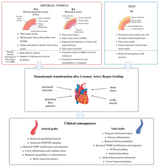
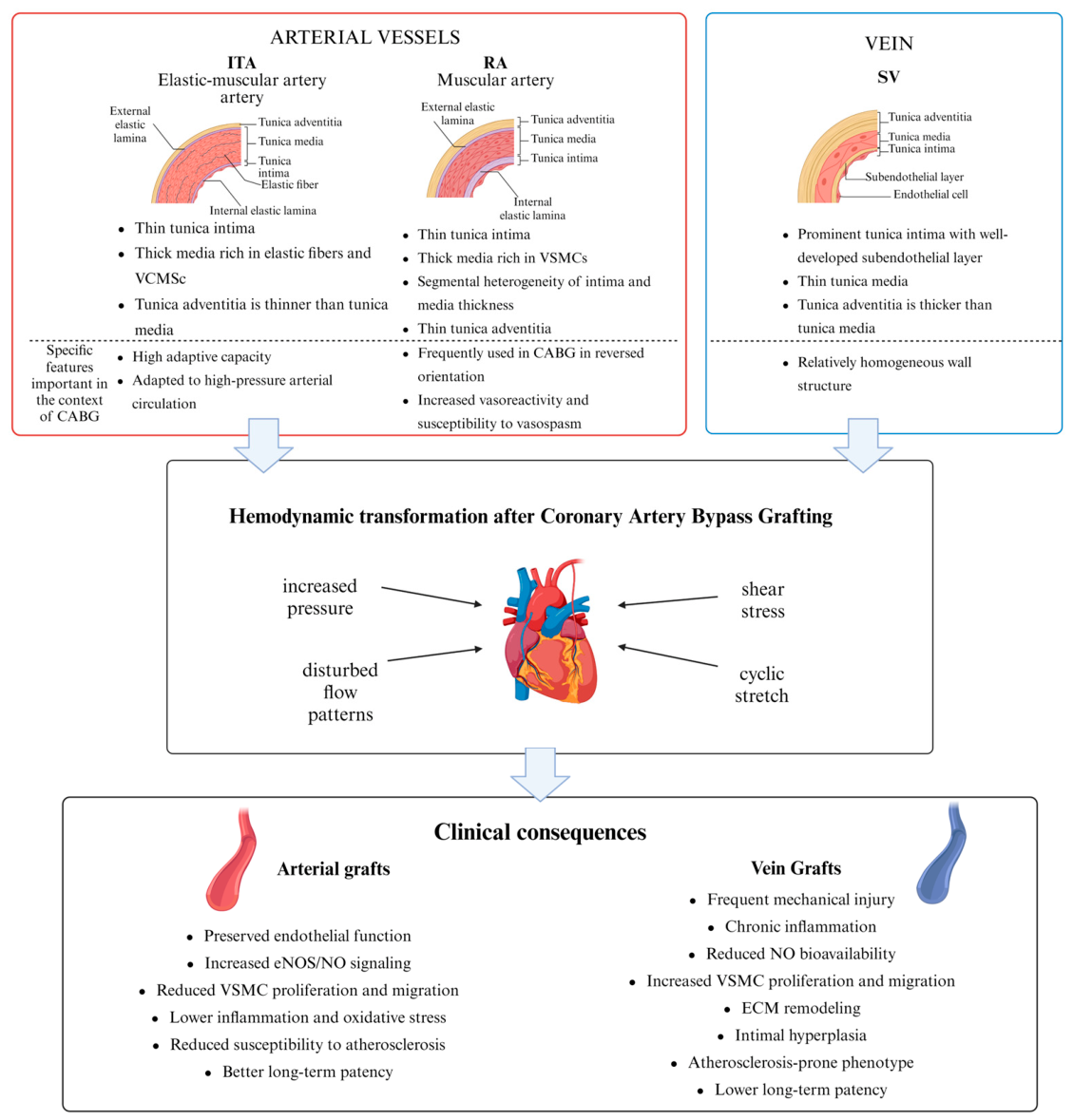
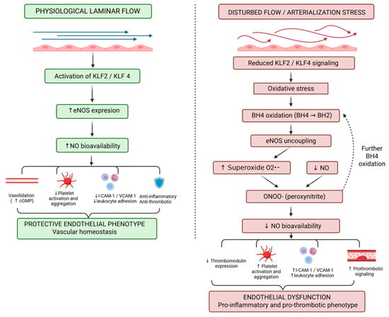
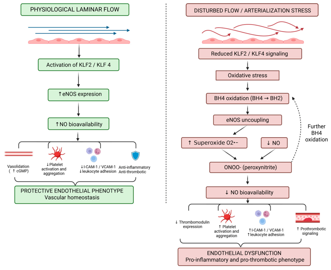
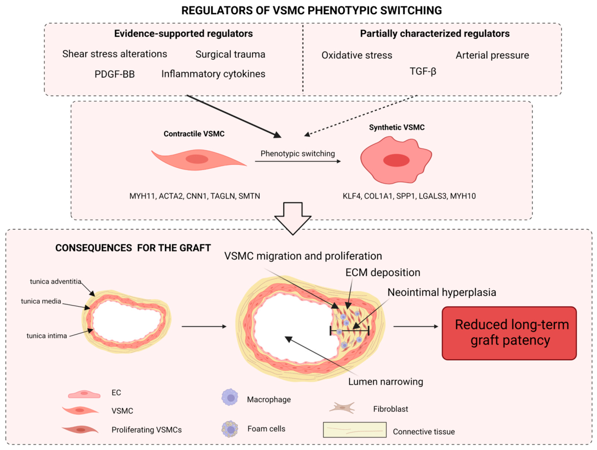

Disclaimer/Publisher’s Note: The statements, opinions and data contained in all publications are solely those of the individual author(s) and contributor(s) and not of MDPI and/or the editor(s). MDPI and/or the editor(s) disclaim responsibility for any injury to people or property resulting from any ideas, methods, instructions or products referred to in the content. |
© 2026 by the authors. Licensee MDPI, Basel, Switzerland. This article is an open access article distributed under the terms and conditions of the Creative Commons Attribution (CC BY) license.