


Featured Application
Mechanical characterization of biological particles and the design of ultrasound-responsive imaging agents and selectively actuated systems.
Abstract
Acoustic excitation provides a physical means of interrogating and modifying biological structures, yet the relationship between resonance, energy absorption and irreversible damage remains incompletely established. This narrative review connects early proposals for resonant viral inactivation with later advances in viral mechanics, cell disruption, microbubble dynamics and genetically encoded acoustic nanostructures. A unifying framework is developed around eigenfrequencies, coupling to an incident field, damping, nonlinear response and failure thresholds. Particular emphasis is placed on distinguishing measured structural resonances from frequency-dependent biological effects that arise from cavitation, heating or exposure geometry. Recent observations of single-virus vibrations and ultrasound-mediated disruption of enveloped viruses are assessed alongside oncotripsy and gas-vesicle imaging and mechanotherapy. Worked examples illustrate size-dependent frequency scales, resonance linewidths, pulse buildup, bubble oscillations and the practical limits of spectral selectivity. Detection and disruption are treated as distinct operating regimes of a coupled mechanical system rather than interchangeable evidence of a single mechanism. The most robust applications combine calibrated acoustic fields with independent mechanical measurements and clear biological endpoints. Future progress will require linking a measured mode to absorbed energy, structural failure and target-specific loss of function under realistic environmental conditions.
1. Historical Development and Scope
1.1. From a Viral Inactivation Hypothesis to a Broader Mechanical Question
The possibility of damaging a small biological structure by exciting one of its mechanical modes is attractive because the interaction need not depend exclusively on a biochemical receptor. In 2000, Babincová, Sourivong, and Babinec proposed resonant absorption of ultrasound energy as a route to the destruction of human immunodeficiency virus (HIV) and other viruses [1]. The proposal used an object-size and sound-speed estimate to place the relevant frequency in the GHz range. It was a hypothesis, not an experimental demonstration of viral inactivation. A later letter extended the concept to nanomachines [2]. These contributions identified a physical question that can now be formulated more precisely: when does a biological or engineered structure possess an accessible mode that couples strongly enough to an external field to produce a useful signal or irreversible change?
Subsequent work developed several parts of this question. Ford estimated the normal-mode frequencies of nearly spherical viral particles using liquid-drop and elastic-sphere models [3]. Saviot and colleagues corrected aspects of the elastic-sphere calculation and emphasized treatment of environmental damping [4]. More recently, experimental studies have interrogated mechanical properties of individual biological entities, while engineered gas structures have made ultrasound both a molecular imaging tool and an actuator. These developments justify revisiting the early hypothesis within a framework that separates the existence of a mode, its excitation, its detection, and the mechanism of damage.
The original structural analogy also requires revision. The mature HIV-1 capsid is a fullerene-like cone assembled from capsid-protein hexamers and pentamers, rather than a regular icosahedral shell [5]. Its envelope, capsid, genome, and associated proteins form distinct mechanical components. Symmetry can classify modes and restrict which modes a particular probe detects, but it does not imply that a complex particle has only one or a few mechanical degrees of freedom. A review grounded in current structural knowledge should therefore treat the early size estimate as motivation for more realistic models.
1.2. The Contribution of the Present Review
The specific gap addressed here is the separation between literature on biological vibrational modes, literature on acoustic imaging agents, and literature on ultrasound bioeffects. A recent review considers viral inactivation by vibrational resonance, including ultrasound and microwave excitation [6]. We extend that discussion across viruses, bacteria, mammalian cells, microbubbles, and gas vesicles, using the same questions about excitation, dissipation, readout, and failure. The contribution is a comparative interpretation and an experimental decision framework, rather than a new resonance law or an exhaustive catalogue. In particular, the review explains why optical evidence of a mode, a detectable nonlinear echo, and a biological damage maximum cannot be substituted for one another. It also identifies which measurements would connect them in the same biological environment.
The term acoustic resonance refers here to enhanced mechanical response associated with a mode of the target or a coupled target–environment system. Resonant absorption is narrower: it requires enhanced irreversible energy transfer into dissipative channels. Resonant scattering, which produces a detectable outgoing field, can occur without substantial absorption in the target. Selective disruption requires a further condition: the target reaches a failure or functional-inactivation threshold before neighboring structures do. The three concepts are related but experimentally distinct.
The resulting insight is that mechanical selectivity depends jointly on spectral separation, mode-specific coupling, environmental damping, and failure thresholds. The revised synthesis connects these quantities to a graded evidence framework, quantitative exposure summaries, a comparison of measurement methods, and application-specific validation steps. An acoustically actuated agent can be useful when its mechanism is nonlinear buckling, cavitation, or phase conversion, even if the unmodified biological target has no uniquely addressable resonance. This distinction links the detection and intervention branches of the review without treating their evidence as interchangeable.
Recent work has expanded the field along several experimentally distinct directions. AFM comparisons now resolve variant-dependent virion geometry and deformation, while time-resolved bubble imaging links local fluid motion to membrane transport [7,8]. Engineered acoustic reporters have progressed from a single contrast channel to multiplexed gene-expression imaging and real-time volumetric acquisition [9,10]. These advances strengthen the case for treating biological mechanics, acoustic transduction, and functional outcomes together. They also expose an asymmetry in the evidence: increasingly sophisticated measurements and engineered actuators are available, whereas direct frequency-selective destruction of an unmodified biological particle still requires a matched demonstration of its mode spectrum and failure response.
1.3. Review Methodology and Scope of the Literature Update
This article is a critical narrative review, not a systematic review or meta-analysis. The revision used targeted web-assisted searches of PubMed-indexed records, PubMed Central articles, publisher platforms, and institutional author manuscripts, followed by checking the existing bibliography and relevant cited primary studies. This was not an exhaustive native search of PubMed, Scopus, or Web of Science, and no such database coverage is claimed. The targeted update was completed on 27 October 2026. It emphasized work published from 2000 to that date, while retaining earlier foundational studies of elasticity, bubble dynamics, capsid mechanics, and sonochemistry when needed to explain the mechanisms.
Search terms combined the object terms virus, capsid, bacterium, cell, microbubble, gas vesicle, or droplet with acoustic resonance, ultrasound, mechanical spectroscopy, vibration, nonlinear imaging, sonoporation, oncotripsy, sonochemistry, or sonodynamic effects. Additional topic searches addressed ultrasound localization microscopy, blood-brain barrier opening, and histotripsy. Title- and DOI-based searches were used to verify key papers and extract exposure conditions. Searches were iteratively refined by mechanism and application; they were not a prespecified Boolean database protocol.
Priority was given to peer-reviewed primary experiments with an identifiable biological system, defined excitation or measurement method, and a relevant physical or biological endpoint. Mechanistic models and historical hypotheses were retained when they clarified a testable assumption and were explicitly distinguished from experiments. Clinical reports were included as translational comparators. Studies with null or ambiguous resonance findings were retained when they informed coupling, damping, or alternative explanations. Duplicate accounts, promotional claims without traceable primary evidence, and studies without a relevant acoustic or mechanical connection were excluded. English-language availability and access to methods constrained assessment; these limitations may introduce language, accessibility, and publication bias.
For quantitative comparisons, the extracted fields were target dimensions and preparation, excitation route, frequency, pressure convention, pulse and total exposure duration, measured resonance parameters, biological endpoint, and interpretation limits. Missing or non-comparable quantities were not reconstructed from nominal instrument settings. Citation selection was checked qualitatively for over-reliance on a single group or region, and primary contributions from different research communities were incorporated according to relevance. Country was not an eligibility criterion, and the review does not claim a geographically exhaustive or bibliometrically representative sample. Heterogeneous exposures and endpoints were synthesized narratively rather than pooled.
2. Physical Foundations of Acoustic Resonance
2.1. Elastic Modes and Characteristic Frequency Scales
The free vibrations of an elastic sphere provide a classical starting point [11]. A homogeneous solid supports spheroidal modes, containing radial and tangential displacement, and torsional modes, containing shear displacement without volume change. A hollow shell, liquid-filled capsule, rod, or supported particle has a different spectrum. In a biological system, membrane tension, shell thickness, internal pressure, anisotropy, and surrounding fluid can be as important as diameter.
For a particular elastic mode, dimensional analysis gives a characteristic scaling:
Here, fn is the mode frequency, L is a specified characteristic dimension, veff is a relevant elastic-wave speed, and Cn is a dimensionless coefficient determined by geometry, boundary conditions, and mode identity. For a solid, transverse and longitudinal sound speeds depend on the shear and bulk elastic moduli and density. Replacing all these quantities with the sound speed of the surrounding water is generally unjustified. For soft structures, bending- or tension-dominated modes can have scaling laws different from Equation (1).
Example 1.
Size alone does not identify a viral resonance. With L = 100 nm, veff = 1000 m/s, and an assumed Cn = 1, Equation (1) gives fn = 10 GHz. For L = 1 µm and 10 µm, the same assumptions give 1 GHz and 100 MHz, respectively. These are order-of-magnitude estimates, not mode assignments. A factor of ten uncertainty in effective wave speed or modal coefficient produces a factor of ten uncertainty in frequency. A proposed MHz resonance of a nanoscale particle therefore needs a specific compliant mode or coupled-system model; it cannot be obtained by simply relabeling the compact-particle GHz estimate.
2.2. A Forced Damped Mode
A single mode can be represented locally by a driven damped oscillator:
The generalized coordinate q may describe radial displacement, shell deformation, or another modal amplitude. The effective modal mass is meff, the undamped angular frequency is ω0, the viscous damping rate is γ, and the projected force amplitude is F0. The steady-state displacement amplitude is:
This expression separates natural frequency from driving frequency and makes damping explicit. For weak damping, the quality factor is Q = ω0/(2γ), and the approximate linewidth is Δf = f0/Q. The amplitude resonance is slightly shifted below the undamped natural frequency; this shift and the linewidth become consequential at low Q. A strongly damped mode may not produce a well-resolved displacement peak even though the mathematical system retains characteristic relaxation times.
For a damping force 2meff γ dq/dt, the cycle-averaged power dissipated by this idealized mode is:
Pdiss = meffγω2A2
Equation (4) highlights a distinction between displacement and absorption. A large displacement does not uniquely identify where energy is dissipated. The observed damping can include losses inside the particle, viscous losses in the surrounding liquid, and acoustic radiation. Only the component dissipated in the mechanically relevant target necessarily contributes directly to target heating or damage. Radiation damping instead transfers energy back into the acoustic field.
2.3. Environmental Damping and Coupling
An embedded particle must satisfy mechanical boundary conditions with its environment. Fluid inertia changes the effective modal mass, while viscosity and acoustic radiation contribute to damping. Interfacial layers and imperfect contact can modify these effects. Saviot and Murray showed that long-lived vibrational modes of embedded nanoparticles cannot be inferred solely from a simple longitudinal acoustic-impedance comparison [12]. Environmental damping requires a mode-dependent calculation or measurement.
The force in Equation (2) is a generalized force: the incident stress must be projected onto the mode shape. A symmetry-allowed mode can still couple weakly if opposite regions experience forces that largely cancel. A nearly uniform pressure couples naturally to a volumetric breathing mode but does not efficiently excite every shear or bending mode. Particle orientation, proximity to a wall, the spatial profile of a near field, and the presence of a mechanically attached agent can change the projection.
A cell model must distinguish compressional, shear, membrane, and cytoskeletal responses. A low-frequency indentation modulus cannot be inserted into a high-frequency mode calculation without a constitutive model linking the two measurements.
2.4. Quality Factor and Finite Pulses
For a lightly damped mode, its freely decaying amplitude varies approximately as exp(−γt). The amplitude ringdown time and resonance bandwidth are related by:
The same damping scale controls resonant amplitude buildup under a suddenly applied sinusoidal force. For weak damping and exact resonance, an approximate envelope is Ass[1 − exp(−t/τA)]. A short pulse may end before the steady-state enhancement develops. Its frequency spectrum is also broadened: for a rectangular burst of duration T, the first zeros around the carrier are separated from it by approximately 1/T. Other pulse envelopes have different spectral widths and sidelobes. Spectral selectivity therefore depends on the waveform as well as the target.
Example 2.
Pulse duration as a constraint. For f0 = 1 MHz and Q = 100, τA = 31.83 µs. A 10 µs resonant burst reaches about 27% of the steady-state envelope, whereas a 100 µs burst reaches about 96%, under this idealized model. Increasing pulse length can improve buildup but can also change heating, cavitation, and biological repair. The pulse duration must therefore be reported independently of nominal intensity.
2.5. Conditions for Spectral Selectivity
For two targets driven with comparable normalized coupling, a useful first approximation is the near-resonance response relative to the resonant response:
The approximation assumes weak damping and small fractional detuning δ = (f − f0)/f0. A practical selectivity condition is that the difference between target and off-target frequencies exceed their linewidths and the effective excitation bandwidth. This condition is not sufficient if an off-target structure has stronger coupling or a lower failure threshold.
Example 3.
A nominal frequency difference may provide little protection. At 10% detuning, Equation (6) predicts relative amplitudes of 0.45 for Q = 10 and 0.05 for Q = 100. Real populations also have distributions of size, stiffness, and orientation, broadening the aggregate response. A narrow individual resonance does not guarantee a narrow population response. The illustrative curves in Figure 1 therefore represent single-mode responses rather than biological classification accuracy.
Figure 1.
Illustrative single-mode calculations. (a) Near-resonance amplitude normalized to its resonant value for quality factors Q = 10 and 100, using Equation (6). (b) Approximate resonant buildup envelope at 1 MHz and Q = 100; markers denote 10 and 100 µs bursts. (c) Size-dependent frequency scale from Equation (1), assuming an effective wave speed of 1000 m/s and modal coefficient of 1. The curves are calculated examples, not measurements or biological efficacy data.
2.6. Propagation and the Distinction Between Local Excitation and Deep Delivery
The frequency accessible at a transducer is not necessarily the frequency delivered effectively at the target. High-frequency attenuation in aqueous media can restrict GHz acoustic interactions to short paths. A GHz ultrasonic sensor operating at 1.55 GHz illustrates the value of a highly localized interaction volume rather than deep penetration [13]. Near-surface sensing, microfluidic measurements, and local optical generation may exploit such confinement. They should not be presented as proof that externally generated GHz ultrasound can treat a deep viral reservoir.
For pressure-amplitude attenuation coefficient αp, a simple traveling wave satisfies p(z) = p(0) exp(−αp z), while its intensity decays as exp(−2αp z). An intensity attenuation coefficient uses a different convention. Reporting the convention prevents a factor-of-two error when estimating penetration or heating. Biological tissue additionally introduces heterogeneous absorption, interfaces, aberration, and scattering; extrapolating a water measurement to an organ is insufficient.
3. Viruses and Capsids
3.1. Mechanical Architecture and Realistic Models
Viral particles range from approximately spherical protein shells to filamentous structures and enveloped particles with mechanically distinct internal components. Atomic force microscopy (AFM) established that bacteriophage capsids behave as mechanically resilient nanoshells, providing experimentally accessible stiffness and deformation information [14]. These quasistatic measurements constrain models but do not directly measure a GHz eigenfrequency or its linewidth. Genome loading, hydration, and maturation can alter the mechanical response, so empty capsids and infectious virions should not automatically be treated as equivalent targets.
The thin-shell perspective adds a structural variable that cannot be captured by diameter alone. Lidmar, Mirny, and Nelson related the faceting of icosahedral shells to the competition between in-plane stretching and bending elasticity [15]. Their calculation concerns equilibrium shape and buckling, rather than an experimentally measured absorption line, but it explains why shells with similar radii can have different deformation pathways. In complementary AFM experiments on cowpea chlorotic mottle virus, Michel and colleagues found that RNA-filled capsids resisted indentation more strongly than empty capsids and that a capsid-protein mutation changed stiffness [16]. Genome loading and protein contacts should therefore enter a resonance model as independently constrained properties, rather than being absorbed into an arbitrary effective sound speed.
Continuum models offer a tractable route to frequencies and mode shapes, but they can obscure heterogeneous contacts between proteins. Dykeman and Sankey introduced an atomistic approach to low-frequency mechanical modes of viral capsids [17]. Such calculations can identify collective motions inaccessible to a homogeneous-sphere description. The challenge is to connect the predicted modes to a realistic driving stress and dissipative environment, rather than treating a free-particle spectrum as a complete inactivation model.
Explicit-solvent simulations provide another level of description. Perilla and Schulten simulated an empty HIV-1 capsid in a system containing more than 64 million atoms, analyzing its vibrational and acoustic behavior together with electrostatics and solvent effects [18]. Such calculations retain the asymmetric capsid architecture and spatially heterogeneous protein interactions. Their value for ultrasound research is to connect molecular structure to candidate collective coordinates. However, an equilibrium fluctuation or calculated mode is not a calibrated acoustic transfer function: predicting an exposure threshold additionally requires the incident stress, dissipation, loading history, and an appropriate failure criterion.
3.2. Measurements of Viral Mechanical Response
Brillouin scattering has been used to determine acoustic properties of satellite tobacco mosaic virus crystals [19]. The hydration dependence and the distinction between crystal acoustics and an isolated-particle mode are important. An acoustic velocity inferred from a viral crystal can inform material parameters, but a crystal phonon is not automatically a confined mode of an individual freely suspended virion.
Sirotkin and colleagues compared large viruses with similarly sized polymer colloids using structural and low-frequency scattering measurements [20]. Their study underscores that applying a generic nanoparticle description to a hydrated biological object requires experimental scrutiny. Internal composition and interaction with water can make collective dynamics differ from those of a simple homogeneous particle. The absence of a clear particle-associated spectral feature in a particular configuration should be retained as evidence about coupling and damping rather than omitted from a resonance-focused account.
Low-frequency Raman measurements of bacteriophage M13 reported a spectral feature near 8.5 cm−1 [21]. This is an optical Raman shift associated with a proposed vibrational mode, not an ultrasound transducer frequency. Conversion gives approximately 255 GHz, demonstrating why spectroscopic units must be translated carefully when comparing excitation methods. Background subtraction, solvent contributions, and mode assignment remain essential parts of such measurements.
In 2025, Zhang and colleagues tracked single-virus vibrational dynamics using ultrafast spectroscopy and reported coherent oscillations lasting several nanoseconds under ambient conditions [22]. This is an important step toward direct characterization of individual viral motions. However, the observed lifetime, optical coupling, particle support conditions, and pseudovirus identity must be retained when interpreting the result. The experiment establishes a measurable vibrational response in its measurement configuration; it does not by itself establish resonant inactivation by propagating ultrasound in physiological fluid.
Measurements on enveloped virions illustrate why compliance should not be equated with fragility. Kiss and colleagues observed a mobile spike layer and substantial recovery after mechanical deformation of native SARS-CoV-2 particles [23]. More recent AFM comparisons of chemically fixed wild-type, alpha, and delta variants identified differences in apparent geometry and deformation under matched preparation conditions [7]. The latter study used a vesicle-based geometric analysis, and its descriptors remain conditional on capture, fixation, and surface contact. Together, these results support heterogeneous, state-dependent mechanical models; they do not justify assigning a universal rupture strain or resonance frequency to all SARS-CoV-2 particles.
3.3. Related Excitation Routes
Optical and microwave excitation provide complementary tests of mechanical hypotheses. Tsen and colleagues reported M13 inactivation with visible femtosecond pulses and proposed coherent vibrational excitation through impulsive stimulated Raman scattering [24]. The incident energy is optical, and alternative photophysical or structural pathways require assessment before attributing inactivation exclusively to an acoustic mode.
Yang and colleagues investigated microwave coupling to confined acoustic vibrations of influenza A H3N2 particles, reporting frequency-dependent inactivation near the proposed dipolar-mode resonance [25]. Their work explicitly connects its motivation to the early ultrasound hypothesis. Nonetheless, electromagnetic coupling depends on charge distributions and polarization as well as mechanics. A microwave absorption peak cannot simply be transferred to the design of an acoustic transducer. These studies are best used to compare excitation routes and formulate tests of mode-specific energy transfer.
3.4. Recent Ultrasound-Mediated Viral Disruption
Veras and colleagues reported ultrasound-induced structural alterations in influenza A H1N1 and SARS-CoV-2, together with reduced SARS-CoV-2 infectivity in cell-based experiments [26]. The study examined MHz exposures and used microscopy and particle characterization; a 7.5 MHz, 5 min exposure was used for key structural analyses. The authors interpreted the effects as resonance-mediated destabilization and reported temperature and pH controls. These findings support a mechanical-disruption hypothesis, while causal assignment to a specific eigenmode still requires an independently measured resonance and a calibrated frequency-response comparison. MHz operating frequency alone cannot exclude cavitation.
A separate preliminary study by Alhasawi and colleagues used 25 MHz ultrasound and reported changes in SARS-CoV-2 RT-qPCR measurements [27]. Its molecular endpoint should be distinguished from direct infectivity measurements. RNA loss, amplification inhibition, sampling variability, and loss of infectious capacity are different possibilities. The result motivates additional characterization but should not be described as a demonstrated clinical antiviral treatment or a definitive measurement of viral resonance.
These reports make it particularly important to specify which mechanical component could respond at MHz frequencies. For a nanoscale particle, compliant envelope motions, collective behavior, interactions with boundaries, or indirect fluid-mediated stresses are different possibilities from a GHz compact-particle breathing mode. The distinction is a testable modeling problem. A mechanistic interpretation should predict not only an optimal frequency but also a measurable dependence on particle dimensions, mechanical properties, or medium conditions.
A specific theoretical route to lower-frequency viral deformation was developed by Wierzbicki and colleagues, who modeled a coronavirus envelope coupled to discrete spike-like beams and masses [28]. Their modal and nonlinear transient calculations distinguished local spike motion from global shell deformation and predicted large strains and buckling under selected harmonic loads. This is a more explicit mechanical hypothesis than a compact-sphere size estimate. Nevertheless, the predicted response depends on the assumed envelope and spike properties, surrounding-fluid treatment, and imposed pressure. Agreement between a treatment frequency and a model frequency would become persuasive only if those parameters and the corresponding mode were independently constrained for the exposed virions. Table 1 summarizes representative studies across excitation and measurement routes and identifies the limits of their mechanistic interpretation.
Table 1.
Representative studies and the limits of their mechanistic interpretation. Optical, electromagnetic, and acoustic excitation routes are kept distinct.
3.5. Biological Endpoints and the Meaning of Destruction
Physical fragmentation is not required for viral inactivation, and visible deformation does not prove that every particle has lost infectivity. A mechanically altered envelope or capsid may impair entry, uncoating, or genome delivery while leaving RNA detectable. Conversely, an RT-qPCR signal can decrease without identifying which viral function was affected. Plaque assays or other validated infectivity assays should accompany molecular and structural measurements where feasible.
For a proposed selective antiviral application, the exposure must be tested against relevant host cells under the same acoustic conditions. A viable-cell count immediately after insonation does not capture all delayed functional effects. Furthermore, inactivation of extracellular virions does not establish elimination of infected cells or latent reservoirs. The original HIV concept is therefore most appropriately evaluated as a physical hypothesis about susceptible particles, with the biological compartment and achievable exposure stated explicitly.
Mechanical failure can also accumulate without a single catastrophic load. Ortega-Esteban and colleagues followed individual adenoviruses under repeated AFM loading and observed progressive vertex loss, shell disruption, and core exposure [32]. This establishes a fatigue-related disassembly pathway under contact loading, rather than an acoustic resonance. In HIV capsid-protein lattices, Domínguez-Zotes and colleagues further showed that capsid-binding antiviral compounds altered elasticity, strength, and fatigue resistance in different ways [33]. These observations suggest that chemical state and maturation can shift susceptibility to repeated mechanical stress. Testing such shifts acoustically would require matched drug-only controls and separate measurements of resonance, loading, and infectivity.
4. Cells and Cellular Structures
4.1. Cell Mechanics and the Difficulty of a Unique Cellular Resonance
A mammalian cell is a heterogeneous viscoelastic structure containing a membrane, cytoskeleton, cytoplasm, nucleus, and organelles. Its mechanical response depends on cell-cycle stage, adhesion, osmotic conditions, and active remodeling. A cell in suspension, a spread cell attached to a substrate, and a cell embedded in extracellular matrix are different mechanical systems. A spectrum computed for one should not be assumed to describe the others.
The relevant material properties are dynamic. A soft response measured by slow indentation may coexist with a much stiffer high-frequency compressional response. In addition, cellular damage is governed by biological repair as well as mechanical loading. A membrane pore can reseal, whereas cytoskeletal or nuclear damage may evolve after the pulse. Selectivity must consequently be assessed using both mechanical characterization and suitable delayed functional endpoints.
Recent optical micromechanics clarifies what a high-frequency cellular measurement actually represents. Schlüßler and colleagues combined Brillouin microscopy, optical diffraction tomography, and fluorescence to determine refractive index, density, and longitudinal mechanical properties of identified subcellular compartments [34]. Steelman and colleagues combined quantitative phase imaging with Brillouin spectroscopy to obtain multiple biophysical variables from individual cells [35]. These approaches reduce the ambiguity of interpreting a frequency shift as a stiffness change. Brillouin measurements probe a high-frequency longitudinal response, whereas indentation and many cell-deformation assays predominantly probe slower, geometrically different mechanical processes; their moduli should not be substituted for one another without a constitutive model.
The distinction remains relevant in the latest work. Vovard and colleagues varied cell hydration by osmotic compression and analyzed both Brillouin shift and linewidth in nuclei [36]. Their comparison with a poroelastic description showed that frequency shifts were strongly influenced by water content, while linewidth analysis helped identify contributions from the biopolymer network. For selective acoustic intervention, this means that a proposed mechanical fingerprint should include hydration and loss information as well as a peak frequency. A spectral difference between cell populations cannot automatically be interpreted as a difference in their low-frequency elastic stiffness or resistance to membrane failure.
4.2. Oncotripsy as a Testable Mechanical-Selectivity Hypothesis
Heyden and Ortiz introduced oncotripsy through models of harmonic excitation that predicted differences between healthy and cancerous cell responses [37]. Their analysis included cellular compartments and explored the possibility of exploiting spectral differences to induce lysis selectively. Subsequent work investigated the influence of viscoelasticity [38]. These studies show how a mechanical-selectivity hypothesis can be translated into a quantitative model, while also revealing its dependence on constitutive assumptions and boundary conditions.
Mittelstein and colleagues experimentally demonstrated selective disruption of several cancer-cell models in suspension with low-intensity pulsed ultrasound at 0.5–0.67 MHz and pulse durations exceeding 20 ms [29]. Healthy immune cells and red blood cells were comparatively spared under the tested conditions. Mechanistic experiments implicated acoustic standing waves and cell-seeded cavitation. The findings therefore support selective ultrasound bioeffects, but do not establish that an isolated malignant-cell eigenmode is the sole cause. Acoustic geometry and bubble nucleation must be considered alongside cellular mechanics.
Schibber and colleagues developed a dynamical model incorporating cytoskeletal damage and repair to interpret oncotripsy-related behavior [39]. Such a model shifts attention from instantaneous rupture to accumulated fatigue. Its predictions should be tested against pulse spacing, delayed viability, and structural recovery, rather than only against total exposure duration. Agreement with an observed death curve does not uniquely identify the microscopic failure mechanism.
4.3. Bacteria and Mechanical Spectroscopy
Bacterial envelopes provide a comparatively well-defined structural target, although turgor, hydration, and wall architecture still vary. Gil-Santos and colleagues measured single-bacterium vibrational modes through coupling to an optomechanical disk resonator, obtaining information on eigenfrequency and mechanical loss [30]. This constitutes mechanical spectroscopy of a coupled sensor–bacterium system. The support and hydration state are part of the measurement, and an eigenfrequency measured in that configuration should not be treated as the free-suspension value without a coupling model.
Earlier bacterial shell models explicitly included cytoplasmic and surrounding-fluid viscosity [40]. They provide a reminder that bacterial resonance is a coupled elastic and hydrodynamic problem. Before proposing selective bacterial disruption, modeled frequencies and quality factors need validation in the same hydration and support conditions as the intended exposure.
Malvar and colleagues used multimode nanomechanical resonators to extract mass and stiffness information from nanoparticles and intact bacteria [41]. This provides a complementary route to identifying mechanical properties. It also illustrates an important distinction: a sensor-frequency shift caused by an attached particle is not automatically a resonance of the particle itself. Coupled-mode analysis or multiple independent observables are needed to separate added mass, stiffness, and intrinsic analyte dynamics.
4.4. Membrane Permeabilization and Irreversible Failure
Microbubble-mediated sonoporation provides direct evidence that an acoustically actuated agent can permeabilize a nearby cell without exciting an intrinsic cellular mode [42,43]. The relevant variables are bubble dynamics, proximity, and membrane recovery. Pore size and extracellular calcium influence resealing [44], so increased transport should be evaluated together with delayed viability and the intended functional endpoint.
Transport can involve both transient pores and endocytosis, with the balance depending on cargo size and acoustic pressure [45,46]. F-actin disruption and endothelial gap formation add a paracellular route [47]. Consequently, total fluorescence uptake alone does not identify membrane rupture: cytosolic delivery, vesicular localization, barrier recovery, and cell survival should be distinguished.
Direct imaging has tightened the connection between bubble dynamics and these endpoints. Helfield and colleagues correlated microsecond bubble oscillations with subsequent endothelial permeability and identified a shear-stress-related threshold dependent on exposure conditions [48]. In 2025, Cattaneo and colleagues visualized stable cyclic microjets generated by bubble shape instabilities and linked them to membrane puncture and uptake at pressures below 100 kPa in their experimental configuration [8]. This result distinguishes cyclic jetting from the familiar jet accompanying a violent inertial collapse. Low incident pressure therefore does not, by itself, exclude strongly localized fluid-mediated membrane stresses.
4.5. From Suspensions to Tissues
The most demanding extension is from isolated cells to tissue. Mechanical coupling to extracellular matrix and neighboring cells changes deformation, while attenuation and standing-wave structure alter the delivered field. Cell-line comparisons also do not resolve interpatient heterogeneity or differences between malignant cells within a tumor. A useful translational sequence begins with calibrated suspensions, proceeds to attached cells and three-dimensional models, and then tests tissue-level selectivity with spatially resolved exposure and damage measurements.
A direct test of the mechanical environment was provided by van den Broek and colleagues, who compared endothelial cells on a rigid polymer support with cells on a soft collagen scaffold [49]. Despite similar measured bubble dynamics, the relationship between bubble excursion and sonoporation differed markedly between supports. This experimentally demonstrates that bubble motion alone is insufficient to predict cellular outcome. Culture substrate, tissue constraint, and cell adhesion belong in the definition of the exposed mechanical system, and apparent differences between cell types should be checked against these environmental factors.
Mechanical targeting may ultimately complement molecular targeting rather than replace it. A ligand-bound bubble or acoustically active nanostructure can concentrate mechanical effects near a selected cell, while spectral or pulse tuning controls its response. This combined approach should be evaluated by comparing the agent alone, ultrasound alone, untargeted agent plus ultrasound, and targeted agent plus ultrasound under matched conditions.
Translation of an agent-mediated interaction is already being explored through blood-brain barrier opening. Mainprize and colleagues demonstrated the feasibility of transient opening with noninvasive MR-guided focused ultrasound and intravenously administered microbubbles in a small glioma study [50]. Sonabend and colleagues subsequently investigated repeated opening using an implanted ultrasound device together with albumin-bound paclitaxel in recurrent glioblastoma [51]. These early clinical studies concern localized barrier modulation and drug delivery; they do not establish intrinsic cancer-cell resonance or selective resonant lysis. They nevertheless provide a concrete translational comparison: the relevant outcomes include delivered drug concentration, barrier recovery, neurological tolerability, and reproducibility over repeated exposures.
A separate branch of cellular acoustic actuation operates through mechanotransduction. Yoo and colleagues linked excitation of cultured cortical neurons to calcium-permeable mechanosensitive channels and downstream electrical amplification [52]. Duque and colleagues showed that expression of human TRPA1 could confer ultrasound responsiveness to mammalian cells and enable sonogenetic manipulation [53]. These studies extend acoustic control beyond permeabilization or destruction. Their specificity is associated with molecular sensing and cellular signaling, and should not be described as a measured resonance of the entire cell. Pharmacological, genetic, and electrophysiological controls provide ways to distinguish this pathway from bubble-mediated transport or irreversible mechanical injury.
5. Microbubbles and Engineered Acoustic Structures
5.1. Gas Bubbles as a Reference System
Gas microbubbles provide a useful reference because their compressibility gives strong coupling to an acoustic pressure field. For a free spherical bubble, neglecting surface tension, viscosity, and thermal and radiation corrections, the Minnaert frequency is [54]:
R0 is the equilibrium radius, P0 the ambient pressure, ρl the liquid density, and κ the gas polytropic exponent. The relation is qualitatively different from the use of a solid sound speed in Equation (1). Bubble stiffness originates mainly from gas compression, while the surrounding liquid supplies much of the inertia. The free-bubble result should not be applied unmodified to coated microbubbles or nanoscale gas structures.
Example 4.
Free-bubble size and frequency. With P0 = 101.33 kPa, ρl = 1000 kg/m3, and an assumed κ = 1.40, R0 = 1 µm gives f0 = 3.28 MHz; R0 = 2 µm gives 1.64 MHz. The omitted corrections become important for small bubbles, and the actual effective κ can depend on thermal exchange and frequency. This example establishes a baseline for comparing a measured coated-bubble spectrum, not a prediction of a commercial contrast agent’s operating frequency.
5.2. Nonlinear Oscillations and Shell Mechanics
The Rayleigh–Plesset model balances gas pressure, surface tension, liquid inertia, and viscosity [55]. Compressible extensions such as the Keller–Miksis equation include additional radiation and finite-sound-speed effects [56]. These models require a gas law, equilibrium state, and driving waveform; an incompressible model is insufficient for reliable shock-pressure predictions during violent collapse. Here, their role is to identify which restoring and loss terms must be constrained experimentally.
An encapsulating shell changes both restoring forces and damping. Marmottant and colleagues introduced a model accounting for lipid-shell buckling, elastic expansion, and rupture [57]. Its piecewise shell response explains why coated bubbles can be strongly nonlinear and why their response can depend on driving pressure. A spectral peak measured at low pressure need not remain fixed at the pressure used for delivery or disruption.
Single-bubble experiments provide unusually direct tests of a resonance model. Van der Meer and colleagues swept the driving frequency while recording individual microbubbles with ultrafast optical imaging, reconstructing resonance curves and separating shell-related restoring and dissipative contributions [58]. Overvelde and colleagues then measured the pressure dependence of these curves and observed classes of strongly nonlinear response associated with the phospholipid coating [59]. These measurements demonstrate why a bubble’s resonance should be reported together with pressure, shell state, and radius. An ensemble attenuation maximum can conceal substantial variation between individual agents, even before interaction with cells or a vessel wall is introduced.
Shell rupture and gas loss can also change the object during exposure. Therefore, the observed response may be history dependent: an initially intact resonator can transform into a different bubble or disappear. Population polydispersity broadens the spectrum further. Experiments designed to test resonance should characterize bubble size distributions before and after insonation and report whether measurements concern stable oscillation, destruction, or a mixture.
Recent polymer-shell studies show how these variables can be engineered rather than merely measured. Hard-shelled poly(n-butyl cyanoacrylate) microbubbles produced sonoporation and sonopermeation in vitro even in a compression-dominated response regime [60]. A subsequent study varied poly(alkyl cyanoacrylate) composition to tune shell stiffness by more than an order of magnitude while maintaining a narrow micrometer-scale size distribution and improving imaging and delivery performance [61]. These findings challenge a design strategy based only on maximizing radial expansion. Shell mechanics, loading capacity, acoustic response, and the intended biological endpoint must be optimized together, with the useful exposure window established for each formulation.
5.3. Gas Vesicles and the Connection Between Detection and Actuation
Gas vesicles are gas-filled protein nanostructures with a mechanically supported shell. Shapiro and colleagues demonstrated their use as ultrasonic molecular reporters [62]. Their detection despite nanoscale dimensions illustrates that contrast depends on material response as well as physical size. A nanoscale scatterer can produce useful signals as part of a population without being spatially resolved as an individual object.
Molecular engineering of gas vesicles changed their mechanical and acoustic properties and enabled additional imaging and targeting functions [63]. Their nonlinear response has been exploited by specialized imaging sequences [64,65]. These structures should not be described simply as free nanobubbles: shell deformation, buckling, and pressure-induced collapse are central to their acoustic behavior. The term resonance should be used only when an associated mode and its frequency response have actually been established.
Structural work has made gas-vesicle mechanics more tangible. Huber and colleagues resolved the GvpA shell by cryo-electron microscopy, revealing a corrugated helical architecture, a gas-permeable wall, and a water-repelling interior [66]. Dutka and colleagues used cryo-electron tomography of Anabaena gas vesicles to resolve the GvpA arrangement and the reinforcing GvpC cage [67]. These studies provide molecular constraints for models of buckling and collapse. They explain why changing protein composition or shell geometry can alter acoustic behavior, while also emphasizing that the appropriate reference is a reinforced protein shell surrounding gas, rather than a free bubble with an unmodified Minnaert resonance.
Acoustic reporter genes extended gas-vesicle contrast to engineered microorganisms in mammalian hosts [68], and later to gene expression in mammalian cells [69]. The biological specificity in these systems comes substantially from genetic expression of the acoustic structure. It does not require that an unmodified cell have a unique resonance distinguishable from every other cell.
Improvements in reporter performance arise through distinct routes: genomic mining identified strong and sustained reporters [70], drug-selectable expression simplified production of ultrasound-visible human cells [71], and directed evolution improved nonlinear scattering [72]. Their comparison should separate expression and assembly efficiency from the acoustic response per structure; a stronger image need not imply a stronger intrinsic resonance.
Genetic engineering also modifies vesicle size [73]. Sub-80 nm biconical structures have shown detectable in vivo contrast, tumor access, and cavitation nucleation in experimental models [74]. These findings connect particle design to delivery and actuation, while leaving vascular accessibility and spatial imaging resolution as separate constraints.
Reporter expression can itself alter the monitored biology. Sequential expression of assembly factors and shell protein reduced the growth and viability burden associated with gas-vesicle production in bacteria [75]. Longitudinal studies therefore need expression-stability and viability controls before treating acoustic signal as a direct measure of cell number.
Bar-Zion and colleagues demonstrated gas-vesicle-enabled mechanotherapy, including cell disruption and effects in an animal model [31]. The reported mechanism involves ultrasound-triggered conversion into cavitating bubbles. This supplies an experimentally supported link between engineered nanostructures and mechanical intervention, but it is mechanistically distinct from directly breaking a virion through its intrinsic elastic resonance.
Recent acoustic and optical characterization provides a more detailed account of the transition to cavitation. Measurements at 0.5 and 1.6 MHz combined passive acoustic mapping with ultrafast imaging and suggested that coalescence of gas released by vesicles formed larger bubbles responsible for much of the detected activity [76]. Broadband and harmonic emissions differed with frequency, and activity diminished over subsequent pulses as the initial nuclei were consumed or transformed. Consequently, the therapeutic actuator can evolve during the first burst. A low-pressure gas-vesicle contrast measurement cannot be assumed to describe the size, spectrum, or persistence of the bubbles that subsequently deliver mechanical stress.
5.4. Phase-Changing Droplets and Designed Capsules
Acoustic droplet vaporization provides another route from a small precursor to a strong acoustic response. Kripfgans and colleagues showed ultrasound-induced vaporization of perfluorocarbon droplets for diagnostic and therapeutic applications [77]. Vaporization is a phase transition with a threshold, not necessarily resonant rupture of the original droplet. The resulting gas bubble may subsequently oscillate or cavitate, making a sequence of mechanisms possible within one exposure.
Droplet composition and nucleation physics introduce additional design variables. Sheeran and colleagues showed that metastable decafluorobutane droplets could be activated at lower acoustic exposures than less volatile perfluorocarbon formulations [78]. Shpak and colleagues demonstrated that nonlinear propagation and superharmonic focusing within droplets could explain localized nucleation and a frequency-dependent vaporization threshold in their system [79]. Thus, an activation optimum can arise from waveform distortion and internal focusing, rather than a resonance of the original liquid particle. Droplet size, temperature, formulation, and delivered pressure spectrum are necessary for interpreting such thresholds.
Designed capsules and vesicles could, in principle, combine low-pressure detection with high-pressure opening or disassembly. The design problem is to separate these operating regimes while retaining mechanical stability during transport. Radius, shell thickness, elasticity, defects, and internal pressure become tunable variables. This is an engineering opportunity rather than evidence that all capsules can be tuned to a sharply selective frequency. A successful design requires measurements of leakage, signal stability, opening threshold, and the fate of the shell and payload.
Proof-of-concept delivery studies illustrate how detection and intervention can be combined without claiming intrinsic target resonance. Rapoport and colleagues integrated perfluorocarbon-containing drug carriers, ultrasound contrast, and ultrasound-enhanced doxorubicin delivery in tumor models [80]. Airan and colleagues used focused ultrasound to release propofol from nanoemulsions and modulate neural activity in rats [81]. The downstream effect in the latter system was pharmacological, enabled by spatially gated release. These examples motivate a modular interpretation: carrier localization, acoustic activation, payload release, and biological response are separate stages, each requiring its own efficiency measurement and control. Table 2 compares the structural classes discussed above and distinguishes demonstrated acoustic responses from proposed routes to selective actuation.
Table 2.
Distinct structural classes and their acoustic operating mechanisms. Possible design routes are distinguished from established examples.
5.5. Quantitative Comparison Across Biological and Engineered Targets
Table 3 brings together representative measurements from Section 3, Section 4 and Section 5. It separates the applied carrier frequency from an independently measured mode frequency and distinguishes pulse length from total exposure. The entries are protocol-specific examples, not recommended treatment settings or a pooled comparison of efficacy. Optical and contact measurements have no directly interchangeable incident ultrasound pressure.
6. Detection and Mechanical Characterization
6.1. What Is Being Measured
Detection can mean locating an acoustically active population, identifying a particle type, measuring a mechanical parameter, or observing a change in biological state. These objectives require different evidence. A strong echo demonstrates acoustic contrast; it does not identify a mode. A frequency-dependent extinction signal combines scattering and absorption, and can also contain resonances of the sample chamber. An optical pump–probe signal can reveal motion while relying on an excitation mechanism different from insonation.
Ideally, a resonance measurement includes frequency, linewidth, phase behavior, and a mode assignment. The dependence on size or stiffness provides an additional test. Measuring both amplitude and phase reduces ambiguity compared with identifying a single intensity maximum. In a coupled sensor, the sensor, attachment, and target should be modeled together; otherwise an instrument resonance may be mislabeled as an intrinsic target property.
6.2. Scattering from Subwavelength Structures
For a particle of radius a insonated at frequency f in a medium with sound speed c, the size parameter is:
When ka is much smaller than unity, a nonresonant particle is in the long-wavelength regime and its scattering is usually weak. Strong compressibility or a suitable resonance can enhance the response, but the effect is constrained by coupling and losses. Strong contrast from gas-containing agents should not be assumed to imply comparable contrast from compact protein particles of the same radius.
Example 5.
Nanoscale contrast does not imply nanoscale image resolution. With a = 50 nm, f = 10 MHz, and c = 1500 m/s, ka = 0.0021 and the wavelength is 150 µm. Detecting a population of such structures is therefore different from resolving adjacent 100 nm particles. At 1 GHz, the wavelength is 1.50 µm and ka = 0.21, but attenuation and instrumentation impose additional constraints. Resolution, detection sensitivity, and target size are separate quantities.
6.3. Nonlinear Signals and Biological Specificity
Nonlinear detection can suppress a largely linear background. Amplitude modulation compares responses to pulses with different amplitudes, while pulse inversion combines echoes from opposite-polarity excitations. Both approaches require knowledge of the nonlinear response of the agent and the medium. Gas-vesicle studies established pressure-dependent backscattering and nonlinear imaging approaches [64]. X-wave amplitude modulation was developed to reduce background associated with nonlinear acoustic propagation [65].
An observed harmonic is not unique evidence for intrinsic structural resonance: coated-bubble dynamics, shell buckling, tissue propagation, and electronics can generate harmonics. Specificity comes from appropriate controls, agent design, and signal processing. A genetically encoded reporter or molecularly targeted agent supplies a biological association that the acoustic spectrum alone may lack.
Biochemical responsiveness supplies specificity beyond particle localization. Engineered gas-vesicle biosensors convert protease activity into changes in nonlinear ultrasound response through modification of the shell-associated protein [82]. More recently, probiotic biosensors coupled sensing of thiosulfate and tetrathionate to gas-vesicle expression and enabled ultrasound imaging of an inflammation model in the mouse gastrointestinal tract [83]. The first strategy changes the mechanics of existing structures; the second changes their biological production. Their signal kinetics, amplification, and recovery differ accordingly. Neither should be interpreted as detecting a unique intrinsic acoustic resonance of an inflammatory molecule or diseased tissue.
Pressure-induced collapse can provide another contrast dimension. Comparing signals before and after a controlled destructive pulse can separate responsive structures from persistent background. However, this consumes or modifies the reporter, potentially preventing repeated monitoring. Collapse thresholds must be interpreted as mechanical failure characteristics, not automatically as resonance frequencies. Low-pressure interrogation and destructive confirmation should be reported as distinct stages.
Multiplexed reporters add a further discrimination axis. Engineered gas-vesicle variants with different pressure-response profiles enabled two-channel ultrasound imaging of bacterial populations and states, including in vivo demonstrations [9]. Here, separation is achieved by how the structures respond to acoustic pressure, rather than by assigning each population a narrow resonance frequency. This approach should be distinguished from spectral selectivity in Equation (6). Its performance depends on the overlap of pressure-response curves, local attenuation, reporter expression, and the imaging sequence; calibration in mixtures is needed before assigning changes in channel intensity to changes in cell abundance.
6.4. Mechanical Fingerprints and Inverse Problems
A set of modes, linewidths, and their environmental dependence can provide a richer fingerprint than one peak. In principle, it may distinguish changes in mass, shell stiffness, hydration, or internal loading. Yet the inverse problem is often nonunique: several combinations of size and modulus may predict the same frequency. Independent microscopy, mass information, or multiple mode measurements can reduce this ambiguity, as demonstrated by multimode mechanical sensing approaches [41].
A classifier based on spectral fingerprints should be validated on independent samples and relevant confounders. Differences in preparation, humidity, medium viscosity, or particle concentration can otherwise dominate the apparent biological signature. For longitudinal monitoring, repeatability and the stability of the reporter may matter more than the nominal sharpness of a single resonance.
Mechanical contrast can also be produced by combining different driving and sensing modalities. Magneto-gas vesicles couple magnetic nanoparticles to acoustically visible protein structures, allowing magnetically driven motion to be detected by ultrasound and related to surrounding tissue mechanics [84]. The study demonstrated measurements in three-dimensional materials and in vivo fibrosis models. This provides a route to interrogating the environment of a reporter rather than merely its presence. Inverse analysis must still distinguish probe coupling and distribution from tissue viscoelasticity: the measurement concerns the driven probe-tissue system, not an isolated particle eigenmode.
6.5. An Experimental Sequence for Establishing a Useful Signal
A practical sequence begins with a reference target of known dimensions in a defined medium. The field and receiving system are calibrated separately, then target concentration, driving frequency, and pressure are varied. Blank-medium and chamber controls identify background resonances. An independent method constrains the geometry and mechanics. Finally, blinded mixtures test whether a signal remains identifiable in a realistic matrix.
Detection becomes mechanistically informative when a change in the acoustic signal can be linked to a defined structural or functional change. For example, a frequency shift may indicate stiffness alteration, whereas signal loss may reflect collapse, clearance, aggregation, or movement out of the sampled volume. Treating all signal loss as destruction would confound these possibilities.
Acquisition geometry is also advancing. The 2026 Takoyaki ultrasound study used multiplexed matrix-array transmission to obtain volumetric nonlinear images of gas-vesicle agents and reporter-expressing cells [10]. Demonstrations included a mouse glioblastoma model and tracking contrast transport in brain ventricles. This addresses a practical limitation of mechanically assembling a volume from successive two-dimensional scans, where motion and timing can confound apparent spatial changes. It improves the readout of acoustically active agents without converting contrast detection into direct spectroscopy of the host cell. Volumetric localization and a validated mechanical spectrum remain complementary observables.
6.6. Choosing a Detection and Mechanical Characterization Method
The method should be selected according to the intended observable. AFM constrains contact stiffness and failure; Brillouin microscopy maps high-frequency longitudinal response; time-resolved optical spectroscopy and coupled resonators can resolve particle-associated modes. Pulse-echo and nonlinear ultrasound provide spatial readout over larger fields, but a contrast signal alone does not determine a target eigenfrequency. Table 4 compares the demonstrated frequency domains, sensitivity observables, spatial scales, and biological constraints. Instrumental detection limits are not interchangeable: force noise, displacement noise, spectral linewidth uncertainty, and a concentration detection threshold quantify different tasks.
Table 4.
Comparison of representative characterization and detection methods. Frequency entries describe the cited implementations or response domains, not universal instrument specifications.
Ultrasound localization microscopy adds a distinct detection route: repeated localization of spatially separable microbubble echoes reconstructs microvascular structure and flow. Errico and colleagues demonstrated rodent cerebral microvessel imaging at depths exceeding 10 mm with acquisitions lasting tens of seconds [85]. The improvement arises from localization and accumulation of events; it does not imply that the imaging wavelength has become nanometric or that the host tissue resonance has been identified. Localization precision, reconstructed vessel resolution, sampling density, and acquisition duration should all be reported.
For diagnostic evaluation, sensitivity should be stated as a measurable limit at a defined false-positive rate, concentration, depth, and acquisition time. Specificity requires relevant background structures and blinded samples, rather than a comparison with buffer alone. A mechanical classifier should be tested on independent specimens with controlled hydration and preparation. For repeated monitoring, the interrogation pressure must preserve the reporter or the expected depletion must be modeled. These requirements translate the physical readout into an assessable detection application.
7. Disruption and Mechanistic Attribution
7.1. Different Routes to Irreversible Change
Ultrasound can produce irreversible change through direct deformation, cyclic fatigue, bubble-mediated mechanical stress, heating, or sonochemical reactions. These pathways can act jointly. Resonance can enhance the response of a target or a cavitation nucleus without specifying the final damage pathway. Attribution should therefore identify both the acoustically responsive structure and the proximate cause of loss of function; bubble resonance followed by radical-mediated injury is distinct from direct resonant rupture of a virion.
The endpoint should match the application. For a virus, loss of infectivity may be sufficient even without fragmentation. For a delivery capsule, opening without destruction of the payload is desirable. For a cell, the relevant endpoint may be loss of clonogenic capacity rather than immediate membrane permeability. A frequency that optimizes one endpoint need not optimize another.
Histotripsy provides a useful clinical comparison for irreversible mechanical disruption. The first-in-human THERESA feasibility study evaluated focused-ultrasound treatment of liver tumors by generating a localized cavitation-mediated tissue effect [86]. Its relevance here is the use of a physically confined mechanical intervention with imaging-based assessment, rather than intrinsic resonance matching to malignant cells. Early feasibility results should not be generalized to a proven biological frequency-selectivity mechanism or long-term comparative efficacy. This distinction preserves the contribution of established cavitation-based approaches while defining the additional evidence required for direct resonant disruption of a biological microstructure.
7.2. Strain Thresholds and Accumulated Fatigue
A characteristic modal strain scales as displacement divided by target dimension. Failure also depends on stress concentration, defects, and interfacial constraints, so a single displacement-to-size ratio is not a universal rupture criterion.
Fatigue depends on repeated loading and recovery. The number of carrier cycles within one pulse is N = fT, and total exposure depends additionally on pulse repetition frequency and pulse count. In a 20 ms burst at 0.50 MHz, N = 10,000. That large cycle count can motivate fatigue models, but it does not prove that the target experiences the carrier-frequency strain at the assumed amplitude. The local deformation and interburst repair must be established independently.
In a threshold formulation, the minimum pressure required for damage is governed by the mechanical transfer function and the failure displacement:
Here Hqp relates local pressure to modal displacement and qfail is an effective failure threshold. For a target and an off-target structure, a pressure window exists only if the target threshold is lower under the same exposure conditions. The formulation emphasizes that different coupling or fragility can provide selectivity even when spectra overlap. Conversely, distinct resonance frequencies provide little protection if collateral effects arise through surrounding-fluid heating or cavitation. Figure 2 illustrates how the frequency dependence of mechanical response and different failure thresholds may define a target-only pressure window under explicitly assumed conditions.
Figure 2.
Illustrative response and failure thresholds. (a) Single-mode responses with Q = 30 and frequencies in the ratio 1:1.12. (b) Inverse-response pressure thresholds, assuming equal normalized coupling and an off-target failure displacement 1.40 times that of the target. Shading marks a possible target-only window. All quantities are normalized, and the assumed parameters do not represent measured therapeutic limits.
7.3. Cavitation and Field Geometry
Cavitation must be assessed experimentally rather than inferred only from the carrier frequency. Its probability depends on pressure, nuclei, pulse length, dissolved gas, interfaces, and the medium. Acoustic emissions, high-speed observation where feasible, and controlled changes in gas content provide complementary evidence. Degassing can change both bubble activity and biological behavior, so a single degassed-versus-untreated comparison is not sufficient by itself.
Standing waves can redistribute cells and agents and create spatially varying exposure. A frequency sweep changes the wavelength and may change chamber resonances, reflection patterns, and focal amplitude. An efficacy maximum can consequently arise from the apparatus. Calibration should be performed in the actual exposure geometry, with frequency-dependent pressure normalization. Varying chamber dimensions provides an especially useful test: an apparatus peak should move with the chamber, whereas an intrinsic target mode should follow the target’s mechanical properties.
7.4. Heating and Dose Conventions
For a sinusoidal plane traveling wave, the average intensity equals the squared peak pressure divided by twice the acoustic impedance of the medium. This conversion assumes a known local field; it cannot replace field mapping in strongly standing or focused waves.
Duty cycle, pulse-average and temporal-average intensity, and spatial peak must be distinguished. Electrical settings and nominal source intensity are not measurements of pressure or absorbed power at the target. Thermal effects require their own measurements and controls.
Temperature should be recorded with adequate temporal and spatial resolution, and a matched thermal control should test whether heating alone reproduces the endpoint. A stable bulk temperature constrains average heating but may not resolve microscopic or transient effects. Temperature, cavitation, and direct deformation measurements together are more informative than any single exclusion argument.
7.5. Sonochemical Effects and Mixed Mechanisms
Cavitation can produce reactive species that damage membranes, proteins, and nucleic acids alongside mechanical and thermal effects. Spin-trapping electron-spin-resonance measurements demonstrated radical production in aqueous solutions exposed to diagnostic ultrasound [87]. In a sensitizer-containing cell system, Umemura and colleagues used scavenger and solvent-substitution experiments to support a role for singlet oxygen in hematoporphyrin-enhanced ultrasonic damage [88]. This is evidence for a sonochemical contribution in that system, not a universal mechanism for every sonodynamic formulation or ultrasound exposure.
A useful mechanistic experiment combines acoustic-emission monitoring with appropriate chemical assays, temperature measurements, and biological endpoints. Depending on the proposed pathway, controls can include oxygen variation, selective quenchers, sensitizer-only and ultrasound-only arms, and orthogonal measurements of reactive species or oxidative injury. Scavengers may alter cell physiology or bubble dynamics, while fluorescent probes can respond to multiple oxidants; a change in one assay is not by itself a complete causal test. Persistence of damage under a stable bulk temperature does not exclude radical chemistry.
Sonochemical pathways also change the interpretation of selectivity. Localization of a sensitizer, differential antioxidant defenses, and spatially confined cavitation can create a selective biological outcome without a distinct target eigenfrequency. Conversely, a mechanically resonant actuator may initiate both stress and chemistry. The appropriate conclusion can therefore be mixed-mechanism damage with evidence for a resonance contribution. The relative contributions should be bounded by controls and uncertainty, rather than forcing every observation into a single exclusive category.
7.6. Graded Evidence for Resonance-Mediated Disruption
Evidence for resonance involvement should be assessed cumulatively and in relation to the strength of the claim. A reproducible frequency-dependent bioeffect is an observation requiring explanation. A target-associated spectrum or a constrained mechanical model provides supportive evidence. Confidence increases when calibrated frequency sweeps, phase or linewidth information, appropriate detuning controls, and functional assays converge. Independent changes in size, stiffness, or environment that shift both the mode and the effect in the predicted direction provide particularly strong causal leverage, but not every experiment must implement every test.
The framework in Table 5 is a guide to graded confidence, not a pass/fail checklist or a ranking of therapeutic usefulness. Direct mode measurements may be inaccessible in vivo; validated surrogate measurements and complementary perturbations can still support a qualified inference. The strongest claim of intrinsic resonance-mediated selective disruption requires a convincing connection between target mechanics, delivered excitation, and differential functional loss, together with assessment of plausible competing pathways. Replication across geometries and independent methods strengthens that inference, while mixed mechanisms should be acknowledged explicitly.
Table 5.
Graded confidence in mechanistic attribution. The examples are complementary evidence routes, not individually mandatory criteria or a universal ranking of clinical utility.
8. Evidence Gaps and Future Experimental Priorities
8.1. The Missing Links Between a Mode and a Biological Outcome
The central unresolved issue is not whether small biological structures can vibrate. Mechanical models and measurements establish that they can. The harder task is demonstrating sufficient mode-specific energy transfer in a biologically relevant medium, followed by a reproducible and selective functional consequence. The existence of a narrow mode, the observation of ultrasound damage, and successful imaging are three separate findings until they are connected within the same system.
For viruses, measurements should bridge the gap between optical or contact spectroscopy and insonation in suspension. For cells, constitutive data should cover the dynamic range relevant to the exposure and distinguish suspended from attached or tissue-embedded states. For engineered agents, the important quantities include stability, nonlinear response, collapse or opening thresholds, and the localization of mechanical effects. A common vocabulary for these observables would make comparisons more reliable.
8.2. A Staged Experimental Program
The first stage should use particles with well-defined geometry and independently characterized mechanical properties. Calibrated field measurements and a frequency sweep establish whether target-associated response can be separated from the chamber. A model predicts changes under controlled variation of size, shell thickness, or medium properties. Measuring these changes tests causality more rigorously than selecting one empirically favorable frequency.
The second stage adds biological endpoints and competing mechanisms. For viral particles, morphology and infectivity should be assessed on matched samples. For cells, membrane integrity, delayed survival, and functional recovery should be distinguished. Cavitation emissions and temperature are measured concurrently where possible. Pressure, pulse duration, repetition frequency, and total time are varied independently rather than collapsed into one nominal dose.
The third stage tests realistic matrices and spatial delivery. Protein-rich media, extracellular matrix, heterogeneous tissue, and flowing systems can change both the target and the field. Selectivity should be quantified across target and off-target populations under matched exposure. Tests in three-dimensional cultures or tissue preparations provide a more demanding assessment than isolated-cell suspensions. Preclinical translation should follow only when the responsible mechanism and achievable target exposure are reasonably established.
8.3. Combining Acoustic Detection with Controlled Intervention
A useful future system would first interrogate the target at a nondestructive exposure, estimate a mechanical parameter or verify reporter localization, and then deliver a separately defined actuation pulse. Its monitoring signal would assess the response during or after intervention. This strategy makes detection an input to control rather than merely an image acquired before treatment.
For engineered agents, a detectable signal could confirm local presence before payload release or mechanical disruption. For intrinsic biological targets, a spectrum could guide waveform selection only after the association between spectrum and susceptibility is validated. A closed-loop controller must also account for reporter depletion and changing target mechanics. Once a shell fails or a particle fragments, the initial resonance model may no longer describe the system.
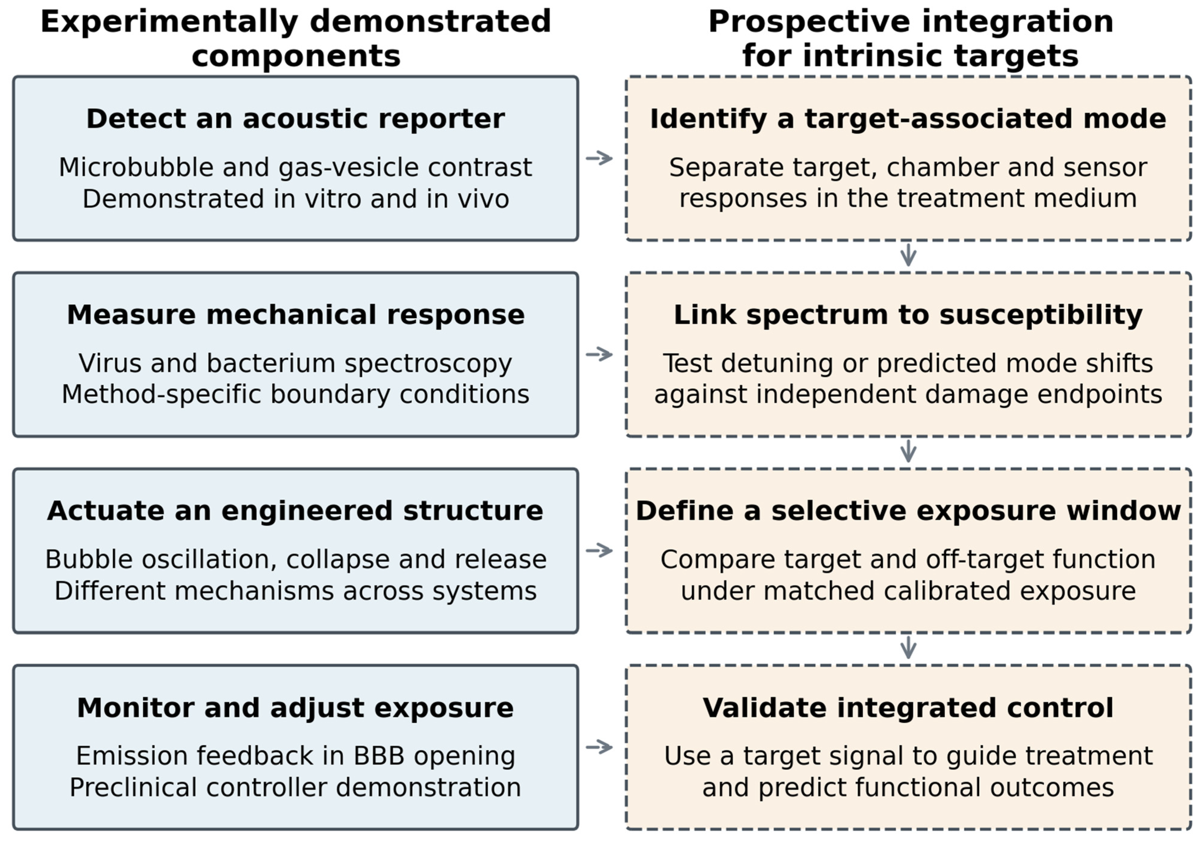
Feedback based on the actuator response already has an experimental precedent. O’Reilly and Hynynen developed an acoustic-emissions controller for focused-ultrasound blood-brain barrier opening, increasing pressure until ultraharmonic emissions appeared and then reducing it to a selected fraction of that level [89]. This illustrates how an observed mechanical response can guide exposure rather than relying solely on a preset source amplitude. Extending such control to a nanoscale target would require a detectable target-associated signal, a response metric linked to the intended endpoint, and allowance for changes in reporter number or actuator state during exposure. Figure 3 distinguishes experimentally demonstrated components from the proposed integration needed for intrinsic mode-guided selective disruption.
Figure 3.
Evidence status of the detection-to-intervention workflow. Solid boxes identify components demonstrated in specific systems: reporter detection [62,68,69], single-entity mechanical spectroscopy [22,30], engineered-agent actuation [31,77], and acoustic-emission feedback for blood-brain barrier opening [89]. Dashed boxes identify the proposed integration needed for intrinsic mode-guided selective disruption. Individual components were demonstrated in different models and are not evidence that the complete workflow has been validated for an unmodified virus or cancer cell. The arrows express a proposed relationship, not an established causal chain.
Published reporter measurements also illustrate how detection and destructive confirmation can be separated experimentally. Shapiro et al. [62] found that Halo-vesicle harmonic contrast relative to a linear polystyrene reference was improved by factors of 3.7 and 4.6 at the second and third harmonics compared with processing at the 6 MHz transmit frequency and also pre/post-collapse subtraction improved Halo- and Ana-vesicle contrast relative to polystyrene by factors of 10 and 22 after an 8.6 MHz, 650 kPa peak-pressure scan.
8.4. Reproducibility and Reporting Priorities
Comparable studies need complete descriptions of the target and exposure. The medium, temperature, size distribution, concentration, and sample geometry should accompany transducer characteristics and calibrated acoustic variables. Reported pressure must identify its location and whether it is peak positive, peak negative, or another quantity. Pulse waveform, duty cycle, uncertainty, and calibration procedure should be available. A negative result at one frequency can be informative only when the delivered field and target spectrum are sufficiently characterized.
Mechanistic conclusions should be matched to evidence. We recommend wording such as predicted mode, measured vibrational response, ultrasound-associated inactivation, or resonance-mediated disruption according to what was actually established. This prevents a useful observation from becoming an unsupported clinical or physical claim. Sharing field maps, waveforms, analysis procedures, and appropriately documented negative controls would facilitate independent comparison. Table 6 consolidates the minimum information needed to report target properties, exposure conditions, measurements, controls, and biological outcomes reproducibly.
Table 6.
Minimum reporting information for reproducible mechanical characterization and disruption studies.
8.5. Application-Specific Development Pathways
The next experiment should be chosen by application rather than by a universal frequency-matching goal. Table 7 separates near-term characterization and engineered-agent applications from the less mature claim of intrinsic target-selective disruption. Each pathway specifies a meaningful endpoint, the central unresolved issue, and a decision point for progression. A useful detection method can advance without demonstrating destruction, while a therapeutic method must justify the delivered exposure and relevant off-target effects independently.
Table 7.
Application-specific validation priorities. The development milestones are proposed by this review; they are not established regulatory thresholds.
8.6. Clinical Translation and Remaining Challenges
Clinical maturity differs sharply between mechanisms. Contrast microbubbles provide an established example of acoustic detection, while microbubble-assisted blood-brain barrier opening has reached early clinical studies [50,51]. These studies assess feasibility, drug access, reversibility, and tolerability; they do not demonstrate selective rupture through a malignant-cell eigenmode. For gas-vesicle reporters and mechanotherapy, the studies considered here provide preclinical evidence [31,68,69,70,71,72,73,74,75,76]. Their translation must address reproducible manufacture or expression, biodistribution, immune responses, persistence, and the distinction between a nondestructive imaging exposure and an activating exposure.
Histotripsy offers a more advanced comparator for mechanical tissue destruction. Beyond the THERESA feasibility report [86], the #HOPE4LIVER pivotal study reported technical success in 42 of 44 evaluable tumors (95%) and procedure-related major complications in 3 of 44 participants (7%) within 30 days [90]. These denominators refer to different endpoints; the study treated 49 tumors in 44 participants. Its single-arm design establishes feasibility and performance under the study conditions, not comparative superiority or intrinsic resonance selectivity.
The one-year #HOPE4LIVER update reported 47 patients and 52 tumors, with local control estimates of 63.4% using the primary assessment and 90% using a post hoc assessment; six serious adverse device-related effects occurred within 30 days [91]. The discrepancy in imaging interpretation is material and both estimates are retained. This follow-up is not an independent replication of the pivotal program. It illustrates why technical ablation success, durable control, adverse events, and the rules for image interpretation should be reported separately.
Across applications, attenuation, acoustic access, motion, and heterogeneous tissue alter the exposure available at the target. A measured GHz particle mode does not resolve the challenge of delivering sufficient energy through a clinically relevant path. Agent-based systems additionally need batch consistency and reproducible activation thresholds. Clinical development should specify the intended indication, realistic comparator, local exposure uncertainty, safety margin, and durable functional outcome. For intrinsic biological targets, independent validation of the proposed mode–damage link remains a prerequisite for a confident mechanistic claim, while progression of useful mixed-mechanism technologies can be judged on their own measured performance.
8.7. Conclusions
The reviewed evidence supports three distinct findings. Biological particles possess measurable mechanical responses, but frequencies obtained with optical or contact probes do not automatically establish efficient excitation by an incident ultrasound field. Engineered gas structures provide the strongest experimental bridge between acoustic detection and controlled actuation: single-bubble spectroscopy directly tests resonance and damping, whereas gas-vesicle imaging and mechanotherapy exploit shell nonlinearity, collapse, and cavitation. Direct selective disruption of unmodified viruses or cells through an independently assigned intrinsic mode remains less securely established under physiologically relevant exposure conditions.
The most compelling mechanistic evidence combines direct measurements of an actuator or target response with functional outcomes and appropriate controls. Bubble-mediated membrane transport, cavitation-mediated tissue disruption, and reporter buckling or collapse are supported by complementary experiments; sonochemical injury can contribute alongside these pathways. A frequency-dependent effect alone does not distinguish them. Evidence should therefore be graded according to the inference supported, with uncertainty and mixed mechanisms stated rather than excluded by definition.
Development of selective acoustic manipulation should connect a validated target or agent signal to calibrated local exposure, quantify the relevant coupling and loss processes, and demonstrate an operating window with matched off-target functional tests. Independent perturbations and detuning controls are valuable routes to causal attribution, while realistic matrices, delayed outcomes, and reproducibility determine practical utility. Clinical barrier-opening and histotripsy studies show that controlled acoustic intervention can translate, but their success should not be used as proof of intrinsic biological resonance selectivity. The priority is to connect measurement, mechanism, and application-specific benefit within the same validated system.
Author Contributions
Conceptualization, methodology, validation, formal analysis, N.A., P.B., M.B. and Š.D.; Writing—original draft preparation, N.A.; Writing—review and editing, M.B., P.B. and Š.D. All authors have read and agreed to the published version of the manuscript.
Funding
This research was funded by the Slovak Grant Agency VEGA project No. 1/0572/26.
Institutional Review Board Statement
Not applicable.
Informed Consent Statement
Not applicable.
Data Availability Statement
No new experimental data were generated in this review. The quantitative summaries are attributed to the cited primary publications.
Conflicts of Interest
The authors declare no conflict of interest.
References
- Babincová, M.; Sourivong, P.; Babinec, P. Resonant absorption of ultrasound energy as a method of HIV destruction. Med. Hypotheses 2000, 55, 450–451. [Google Scholar] [CrossRef] [Scilit]
- Babinec, P.; Babincová, M. Non-invasive method for selective in vivo destruction of nanomachines by resonant absorption of ultrasound energy. Med. Hypotheses 2006, 66, 1249–1250. [Google Scholar] [CrossRef] [Scilit] [PubMed]
- Ford, L.H. Estimate of the vibrational frequencies of spherical virus particles. Phys. Rev. E 2003, 67, 051924. [Google Scholar] [CrossRef] [Scilit] [PubMed]
- Saviot, L.; Murray, D.B.; Mermet, A.; Duval, E. Comment on “Estimate of the vibrational frequencies of spherical virus particles”. Phys. Rev. E 2004, 69, 023901. [Google Scholar] [CrossRef] [Scilit]
- Zhao, G.; Perilla, J.R.; Yufenyuy, E.L.; Meng, X.; Chen, B.; Ning, J.; Ahn, J.; Gronenborn, A.M.; Schulten, K.; Aiken, C.; et al. Mature HIV-1 capsid structure by cryo-electron microscopy and all-atom molecular dynamics. Nature 2013, 497, 643–646. [Google Scholar] [CrossRef] [Scilit]
- Sadraeian, M.; Kabakova, I.; Zhou, J.; Jin, D. Virus inactivation by matching the vibrational resonance. Appl. Phys. Rev. 2024, 11, 021324. [Google Scholar] [CrossRef] [Scilit]
- Sziklai, D.; Budavári, B.; Kiss, B.; Herényi, L.; Kis, Z.; Pályi, B.; Kellermayer, M. Unveiling the Structural and Mechanical Diversity of SARS-CoV-2 Variants Using Atomic Force Microscopy. ACS Appl. Mater. Interfaces 2026, 18, 32325–32338. [Google Scholar] [CrossRef] [Scilit] [PubMed]
- Cattaneo, M.; Guerriero, G.; Shakya, G.; Krattiger, L.A.; Paganella, L.G.; Narciso, M.L.; Supponen, O. Cyclic jetting enables microbubble-mediated drug delivery. Nat. Phys. 2025, 21, 590–598. [Google Scholar] [CrossRef] [Scilit] [PubMed]
- Nyström, N.N.; Jin, Z.; Bennett, M.E.; Zhang, R.; Swift, M.B.; Shapiro, M.G. Multiplexed ultrasound imaging of gene expression. Nat. Methods 2025, 22, 2594–2600. [Google Scholar] [CrossRef] [Scilit] [PubMed]
- Lee, S.; Wu, D.; Malounda, D.; Rabut, C.; Shapiro, M.G. Real-time volumetric imaging of cells and molecules in deep tissues with Takoyaki ultrasound. Nat. Commun. 2026, 17, 7281. [Google Scholar] [CrossRef] [Scilit]
- Lamb, H. On the Vibrations of an Elastic Sphere. Proc. Lond Math. Soc. 1881, s1-13, 189–212. [Google Scholar] [CrossRef] [Scilit]
- Saviot, L.; Murray, D.B. Long Lived Acoustic Vibrational Modes of an Embedded Nanoparticle. Phys. Rev. Lett. 2004, 93, 055506. [Google Scholar] [CrossRef] [Scilit] [PubMed]
- Balasubramanian, P.S.; Lal, A. GHz ultrasonic sensor for ionic content with high sensitivity and localization. iScience 2023, 26, 106907. [Google Scholar] [CrossRef] [Scilit]
- Ivanovska, I.L.; de Pablo, P.J.; Ibarra, B.; Sgalari, G.; MacKintosh, F.C.; Carrascosa, J.L.; Schmidt, C.F.; Wuite, G.J.L. Bacteriophage capsids: Tough nanoshells with complex elastic properties. Proc. Natl. Acad. Sci. USA 2004, 101, 7600–7605. [Google Scholar] [CrossRef] [Scilit]
- Lidmar, J.; Mirny, L.; Nelson, D.R. Virus shapes and buckling transitions in spherical shells. Phys. Rev. E 2003, 68, 051910. [Google Scholar] [CrossRef] [Scilit]
- Michel, J.P.; Ivanovska, I.L.; Gibbons, M.M.; Klug, W.S.; Knobler, C.M.; Wuite, G.J.L.; Schmidt, C.F. Nanoindentation studies of full and empty viral capsids and the effects of capsid protein mutations on elasticity and strength. Proc. Natl. Acad. Sci. USA 2006, 103, 6184–6189. [Google Scholar] [CrossRef] [Scilit] [PubMed]
- Dykeman, E.C.; Sankey, O.F. Low Frequency Mechanical Modes of Viral Capsids: An Atomistic Approach. Phys. Rev. Lett. 2008, 100, 028101. [Google Scholar] [CrossRef] [Scilit] [PubMed]
- Perilla, J.R.; Schulten, K. Physical properties of the HIV-1 capsid from all-atom molecular dynamics simulations. Nat. Commun. 2017, 8, 15959. [Google Scholar] [CrossRef] [Scilit]
- Stephanidis, B.; Adichtchev, S.; Gouet, P.; McPherson, A.; Mermet, A. Elastic Properties of Viruses. Biophys. J. 2007, 93, 1354–1359. [Google Scholar] [CrossRef] [Scilit][Green Version]
- Sirotkin, S.; Mermet, A.; Bergoin, M.; Ward, V.; Van Etten, J.L. Viruses as nanoparticles: Structure versus collective dynamics. Phys. Rev. E 2014, 90, 022718. [Google Scholar] [CrossRef] [Scilit] [PubMed]
- Tsen, K.T.; Dykeman, E.C.; Sankey, O.F.; Tsen, S.W.D.; Lin, N.T.; Kiang, J.G. Probing the low-frequency vibrational modes of viruses with Raman scattering—Bacteriophage M13 in water. J. Biomed. Opt. 2007, 12, 024009. [Google Scholar] [CrossRef] [Scilit] [PubMed]
- Zhang, Y.; Wu, R.; Shahjahan, M.; Yang, C.; Pyeon, D.; Harel, E. Nanoscopic acoustic vibrational dynamics of a single virus captured by ultrafast spectroscopy. Proc. Natl. Acad. Sci. USA 2025, 122, e2420428122. [Google Scholar] [CrossRef] [Scilit]
- Kiss, B.; Kis, Z.; Pályi, B.; Kellermayer, M.S.Z. Topography, Spike Dynamics, and Nanomechanics of Individual Native SARS-CoV-2 Virions. Nano Lett. 2021, 21, 2675–2680. [Google Scholar] [CrossRef] [Scilit]
- Tsen, K.; Tsen, S.W.D.; Chang, C.L.; Hung, C.F.; Wu, T.C.; Kiang, J.G. Inactivation of viruses by coherent excitations with a low power visible femtosecond laser. Virol. J. 2007, 4, 50. [Google Scholar] [CrossRef] [Scilit] [PubMed]
- Yang, S.C.; Lin, H.C.; Liu, T.M.; Lu, J.T.; Hung, W.T.; Huang, Y.R.; Tsai, Y.C.; Kao, C.L.; Chen, S.Y.; Sun, C.K. Efficient Structure Resonance Energy Transfer from Microwaves to Confined Acoustic Vibrations in Viruses. Sci. Rep. 2015, 5, 18030. [Google Scholar] [CrossRef] [Scilit] [PubMed]
- Veras, F.P.; Nakamura, G.; Pereira-da-Silva, M.A.; Ruiz, G.C.M.; Constantino, C.J.L.; Martins, R.; Arruda, E.; Cunha, F.Q.; Bruno, O.M. Ultrasound effectively destabilizes and disrupts the structural integrity of enveloped respiratory viruses. Sci. Rep. 2026, 16, 8612. [Google Scholar] [CrossRef] [Scilit]
- Alhasawi, A.; Alassaf, F.; Hassan, A. Destruction/Inactivation of SARS-CoV-2 Virus Using Ultrasound Excitation: A Preliminary Study. Viruses 2026, 18, 152. [Google Scholar] [CrossRef] [Scilit]
- Wierzbicki, T.; Li, W.; Liu, Y.; Zhu, J. Effect of receptors on the resonant and transient harmonic vibrations of Coronavirus. J. Mech. Phys. Solids 2021, 150, 104369. [Google Scholar] [CrossRef] [Scilit] [PubMed]
- Mittelstein, D.R.; Ye, J.; Schibber, E.F.; Roychoudhury, A.; Martinez, L.T.; Fekrazad, M.H.; Ortiz, M.; Lee, P.P.; Shapiro, M.G.; Gharib, M. Selective ablation of cancer cells with low intensity pulsed ultrasound. Appl. Phys. Lett. 2020, 116, 013701. [Google Scholar] [CrossRef] [Scilit]
- Gil-Santos, E.; Ruz, J.J.; Malvar, O.; Favero, I.; Lemaître, A.; Kosaka, P.M.; García-López, S.; Calleja, M.; Tamayo, J. Optomechanical detection of vibration modes of a single bacterium. Nat. Nanotechnol. 2020, 15, 469–474. [Google Scholar] [CrossRef] [Scilit] [PubMed]
- Bar-Zion, A.; Nourmahnad, A.; Mittelstein, D.R.; Shivaei, S.; Yoo, S.; Buss, M.T.; Hurt, R.C.; Malounda, D.; Abedi, M.H.; Lee-Gosselin, A.; et al. Acoustically triggered mechanotherapy using genetically encoded gas vesicles. Nat. Nanotechnol. 2021, 16, 1403–1412. [Google Scholar] [CrossRef] [Scilit] [PubMed]
- Ortega-Esteban, A.; Pérez-Berná, A.J.; Menéndez-Conejero, R.; Flint, S.J.; San Martín, C.; de Pablo, P.J. Monitoring dynamics of human adenovirus disassembly induced by mechanical fatigue. Sci. Rep. 2013, 3, 1434. [Google Scholar] [CrossRef] [Scilit] [PubMed]
- Domínguez-Zotes, S.; Valbuena, A.; Mateu, M.G. Antiviral compounds modulate elasticity, strength and material fatigue of a virus capsid framework. Biophys. J. 2022, 121, 919–931. [Google Scholar] [CrossRef] [Scilit] [PubMed]
- Schlüßler, R.; Kim, K.; Nötzel, M.; Taubenberger, A.; Abuhattum, S.; Beck, T.; Müller, P.; Maharana, S.; Cojoc, G.; Girardo, S.; et al. Correlative all-optical quantification of mass density and mechanics of subcellular compartments with fluorescence specificity. eLife 2022, 11, e68490. [Google Scholar] [CrossRef] [Scilit] [PubMed]
- Steelman, Z.A.; Coker, Z.N.; Sedelnikova, A.; Keppler, M.A.; Kiester, A.S.; Troyanova-Wood, M.A.; Ibey, B.L.; Bixler, J.N. Comprehensive single-shot biophysical cytometry using simultaneous quantitative phase imaging and Brillouin spectroscopy. Sci. Rep. 2022, 12, 18285. [Google Scholar] [CrossRef] [Scilit]
- Vovard, L.; Viel, A.; Bastien, E.; Goutier, L.A.; Jardiné, G.; Margueritat, J.; Monnier, S.; Dehoux, T. Separating Water Content from Network Dynamics in Cell Nuclei with Brillouin Microscopy. Phys. Rev. Lett. 2026, 136, 128401. [Google Scholar] [CrossRef] [Scilit] [PubMed]
- Heyden, S.; Ortiz, M. Oncotripsy: Targeting cancer cells selectively via resonant harmonic excitation. J. Mech. Phys. Solids 2016, 92, 164–175. [Google Scholar] [CrossRef] [Scilit]
- Heyden, S.; Ortiz, M. Investigation of the influence of viscoelasticity on oncotripsy. Comput. Methods Appl. Mech. Eng. 2017, 314, 314–322. [Google Scholar] [CrossRef] [Scilit]
- Schibber, E.F.; Mittelstein, D.R.; Gharib, M.; Shapiro, M.G.; Lee, P.P.; Ortiz, M. A dynamical model of oncotripsy by mechanical cell fatigue: Selective cancer cell ablation by low-intensity pulsed ultrasound. Proc. R. Soc. A Math. Phys. Eng. Sci. 2020, 476, 20190692. [Google Scholar] [CrossRef] [Scilit] [PubMed]
- Zinin, P.V.; Allen, J.S.; Levin, V.M. Mechanical resonances of bacteria cells. Phys. Rev. E 2005, 72, 061907. [Google Scholar] [CrossRef] [Scilit]
- Malvar, O.; Ruz, J.J.; Kosaka, P.M.; Domínguez, C.M.; Gil-Santos, E.; Calleja, M.; Tamayo, J. Mass and stiffness spectrometry of nanoparticles and whole intact bacteria by multimode nanomechanical resonators. Nat. Commun. 2016, 7, 13452. [Google Scholar] [CrossRef] [Scilit] [PubMed]
- Kudo, N.; Okada, K.; Yamamoto, K. Sonoporation by Single-Shot Pulsed Ultrasound with Microbubbles Adjacent to Cells. Biophys. J. 2009, 96, 4866–4876. [Google Scholar] [CrossRef] [Scilit] [PubMed]
- Moosavi Nejad, S.; Hosseini, S.H.R.; Akiyama, H.; Tachibana, K. Optical observation of cell sonoporation with low intensity ultrasound. Biochem. Biophys. Res. Commun. 2011, 413, 218–223. [Google Scholar] [CrossRef] [Scilit] [PubMed]
- Hu, Y.; Wan, J.M.F.; Yu, A.C.H. Membrane Perforation and Recovery Dynamics in Microbubble-Mediated Sonoporation. Ultrasound Med. Biol. 2013, 39, 2393–2405. [Google Scholar] [CrossRef] [Scilit] [PubMed]
- Meijering, B.D.M.; Juffermans, L.J.M.; van Wamel, A.; Henning, R.H.; Zuhorn, I.S.; Emmer, M.; Versteilen, A.M.G.; Paulus, W.J.; van Gilst, W.H.; Kooiman, K.; et al. Ultrasound and Microbubble-Targeted Delivery of Macromolecules Is Regulated by Induction of Endocytosis and Pore Formation. Circ. Res. 2009, 104, 679–687. [Google Scholar] [CrossRef] [Scilit] [PubMed]
- De Cock, I.; Zagato, E.; Braeckmans, K.; Luan, Y.; de Jong, N.; De Smedt, S.C.; Lentacker, I. Ultrasound and microbubble mediated drug delivery: Acoustic pressure as determinant for uptake via membrane pores or endocytosis. J. Control. Release 2015, 197, 20–28. [Google Scholar] [CrossRef] [Scilit]
- Meijlink, B.; van der Kooij, H.R.; Wang, Y.; Li, H.; Huveneers, S.; Kooiman, K. Ultrasound-activated microbubbles mediate F-actin disruptions and endothelial gap formation during sonoporation. J. Control. Release 2024, 376, 1176–1189. [Google Scholar] [CrossRef] [Scilit] [PubMed]
- Helfield, B.; Chen, X.; Watkins, S.C.; Villanueva, F.S. Biophysical insight into mechanisms of sonoporation. Proc. Natl. Acad. Sci. USA 2016, 113, 9983–9988. [Google Scholar] [CrossRef] [Scilit] [PubMed]
- van den Broek, M.R.P.; Klassen, R.; Versluis, M.; Yu, A.C.H.; Segers, T. Role of Cell Culture Scaffold Stiffness on Sonoporation Efficiency. Ultrasound Med. Biol. 2026, 52, 336–345. [Google Scholar] [CrossRef] [Scilit] [PubMed]
- Mainprize, T.; Lipsman, N.; Huang, Y.; Meng, Y.; Bethune, A.; Ironside, S.; Heyn, C.; Alkins, R.; Trudeau, M.; Sahgal, A.; et al. Blood-Brain Barrier Opening in Primary Brain Tumors with Non-invasive MR-Guided Focused Ultrasound: A Clinical Safety and Feasibility Study. Sci. Rep. 2019, 9, 321. [Google Scholar] [CrossRef] [Scilit] [PubMed]
- Sonabend, A.M.; Gould, A.; Amidei, C.; Ward, R.; Schmidt, K.A.; Zhang, D.Y.; Gomez, C.; Bebawy, J.F.; Liu, B.P.; Bouchoux, G.; et al. Repeated blood–brain barrier opening with an implantable ultrasound device for delivery of albumin-bound paclitaxel in patients with recurrent glioblastoma: A phase 1 trial. Lancet Oncol. 2023, 24, 509–522. [Google Scholar] [CrossRef] [Scilit] [PubMed]
- Yoo, S.; Mittelstein, D.R.; Hurt, R.C.; Lacroix, J.; Shapiro, M.G. Focused ultrasound excites cortical neurons via mechanosensitive calcium accumulation and ion channel amplification. Nat. Commun. 2022, 13, 493. [Google Scholar] [CrossRef] [Scilit] [PubMed]
- Duque, M.; Lee-Kubli, C.A.; Tufail, Y.; Magaram, U.; Patel, J.; Chakraborty, A.; Mendoza Lopez, J.; Edsinger, E.; Vasan, A.; Shiao, R.; et al. Sonogenetic control of mammalian cells using exogenous Transient Receptor Potential A1 channels. Nat. Commun. 2022, 13, 600. [Google Scholar] [CrossRef] [Scilit] [PubMed]
- Minnaert, M. On musical air-bubbles and the sounds of running water. Lond. Edinb. Dublin Philos. Mag. J. Sci. 1933, 16, 235–248. [Google Scholar] [CrossRef] [Scilit]
- Plesset, M.S. The Dynamics of Cavitation Bubbles. J. Appl. Mech. 1949, 16, 277–282. [Google Scholar] [CrossRef] [Scilit]
- Keller, J.B.; Miksis, M. Bubble oscillations of large amplitude. J. Acoust. Soc. Am. 1980, 68, 628–633. [Google Scholar] [CrossRef] [Scilit]
- Marmottant, P.; van der Meer, S.; Emmer, M.; Versluis, M.; de Jong, N.; Hilgenfeldt, S.; Lohse, D. A model for large amplitude oscillations of coated bubbles accounting for buckling and rupture. J. Acoust. Soc. Am. 2005, 118, 3499–3505. [Google Scholar] [CrossRef] [Scilit]
- van der Meer, S.M.; Dollet, B.; Voormolen, M.M.; Chin, C.T.; Bouakaz, A.; de Jong, N.; Versluis, M.; Lohse, D. Microbubble spectroscopy of ultrasound contrast agents. J. Acoust. Soc. Am. 2007, 121, 648–656. [Google Scholar] [CrossRef] [Scilit] [PubMed]
- Overvelde, M.; Garbin, V.; Sijl, J.; Dollet, B.; de Jong, N.; Lohse, D.; Versluis, M. Nonlinear Shell Behavior of Phospholipid-Coated Microbubbles. Ultrasound Med. Biol. 2010, 36, 2080–2092. [Google Scholar] [CrossRef] [Scilit] [PubMed]
- Blöck, J.; Li, H.; Collado-Lara, G.; Kooiman, K.; Rix, A.; Chen, J.; Hark, C.; Radermacher, H.; Porte, C.; Kiessling, F. The Compression-Dominated Ultrasound Response of Poly(n-butyl cyanoacrylate) Hard-Shelled Microbubbles Induces Significant Sonoporation and Sonopermeation Effects In Vitro. ACS Appl. Bio Mater. 2025, 8, 1240–1250. [Google Scholar] [CrossRef] [Scilit] [PubMed]
- Barmin, R.A.; Moosavifar, M.; Rama, E.; Blöck, J.; Rix, A.; Petrovskii, V.S.; Gumerov, R.A.; Köhler, J.; Pohl, M.; Bastard, C.; et al. Microbubble Shell Stiffness Engineering Enhances Ultrasound Imaging, Drug Delivery, and Sonoporation. Adv. Mater. 2026, 38, e07655. [Google Scholar] [CrossRef] [Scilit]
- Shapiro, M.G.; Goodwill, P.W.; Neogy, A.; Yin, M.; Foster, F.S.; Schaffer, D.V.; Conolly, S.M. Biogenic gas nanostructures as ultrasonic molecular reporters. Nat. Nanotechnol. 2014, 9, 311–316. [Google Scholar] [CrossRef] [Scilit]
- Lakshmanan, A.; Farhadi, A.; Nety, S.P.; Lee-Gosselin, A.; Bourdeau, R.W.; Maresca, D.; Shapiro, M.G. Molecular Engineering of Acoustic Protein Nanostructures. ACS Nano 2016, 10, 7314–7322. [Google Scholar] [CrossRef] [Scilit] [PubMed]
- Maresca, D.; Lakshmanan, A.; Lee-Gosselin, A.; Melis, J.M.; Ni, Y.L.; Bourdeau, R.W.; Kochmann, D.M.; Shapiro, M.G. Nonlinear ultrasound imaging of nanoscale acoustic biomolecules. Appl. Phys. Lett. 2017, 110, 073704. [Google Scholar] [CrossRef] [Scilit] [PubMed]
- Maresca, D.; Sawyer, D.P.; Renaud, G.; Lee-Gosselin, A.; Shapiro, M.G. Nonlinear X-Wave Ultrasound Imaging of Acoustic Biomolecules. Phys. Rev. X 2018, 8, 041002. [Google Scholar] [CrossRef] [Scilit] [PubMed]
- Huber, S.T.; Terwiel, D.; Evers, W.H.; Maresca, D.; Jakobi, A.J. Cryo-EM structure of gas vesicles for buoyancy-controlled motility. Cell 2023, 186, 975–986.e13. [Google Scholar] [CrossRef] [Scilit] [PubMed]
- Dutka, P.; Metskas, L.A.; Hurt, R.C.; Salahshoor, H.; Wang, T.Y.; Malounda, D.; Lu, G.J.; Chou, T.F.; Shapiro, M.G.; Jensen, G.J. Structure of Anabaena flos-aquae gas vesicles revealed by cryo-ET. Structure 2023, 31, 518–528.e6. [Google Scholar] [CrossRef] [Scilit]
- Bourdeau, R.W.; Lee-Gosselin, A.; Lakshmanan, A.; Farhadi, A.; Kumar, S.R.; Nety, S.P.; Shapiro, M.G. Acoustic reporter genes for noninvasive imaging of microorganisms in mammalian hosts. Nature 2018, 553, 86–90. [Google Scholar] [CrossRef] [Scilit] [PubMed]
- Farhadi, A.; Ho, G.H.; Sawyer, D.P.; Bourdeau, R.W.; Shapiro, M.G. Ultrasound imaging of gene expression in mammalian cells. Science 2019, 365, 1469–1475. [Google Scholar] [CrossRef] [Scilit] [PubMed]
- Hurt, R.C.; Buss, M.T.; Duan, M.; Wong, K.; You, M.Y.; Sawyer, D.P.; Swift, M.B.; Dutka, P.; Barturen-Larrea, P.; Mittelstein, D.R.; et al. Genomically mined acoustic reporter genes for real-time in vivo monitoring of tumors and tumor-homing bacteria. Nat. Biotechnol. 2023, 41, 919–931. [Google Scholar] [CrossRef] [Scilit] [PubMed]
- Howells, A.R.; Welch, P.J.; Kim, J.; Forest, C.R.; Shi, C.; Lian, X.L. A drug-selectable acoustic reporter gene system for human cell ultrasound imaging. Bioeng. Transl. Med. 2024, 9, e10584. [Google Scholar] [CrossRef] [Scilit] [PubMed]
- Hurt, R.C.; Jin, Z.; Soufi, M.; Wong, K.K.; Sawyer, D.P.; Shen, H.K.; Dutka, P.; Deshpande, R.; Zhang, R.; Mittelstein, D.R.; et al. Directed Evolution of Acoustic Reporter Genes Using High-Throughput Acoustic Screening. ACS Synth. Biol. 2024, 13, 2215–2226. [Google Scholar] [CrossRef] [Scilit] [PubMed]
- Fu, M.; Wang, Y.; Wang, J.; Hao, Y.; Zeng, F.; Zhang, Z.; Du, J.; Long, H.; Yan, F. Genetic Modulation of Biosynthetic Gas Vesicles for Ultrasound Imaging. Small 2024, 20, 2310008. [Google Scholar] [CrossRef] [Scilit] [PubMed]
- Ling, B.; Gungoren, B.; Yao, Y.; Dutka, P.; Vassallo, R.; Nayak, R.; Smith, C.A.B.; Lee, J.; Swift, M.B.; Shapiro, M.G. Truly Tiny Acoustic Biomolecules for Ultrasound Imaging and Therapy. Adv. Mater. 2024, 36, 2307106. [Google Scholar] [CrossRef] [Scilit] [PubMed]
- Li, Z.; Ho, C.Y.; Barr, D.E.; Jeong, S.; Ma, L.; Lu, G.J. Temporal gene regulation enables controlled expression of gas vesicles and preserves bacterial viability. Nat. Commun. 2026, 17, 953. [Google Scholar] [CrossRef] [Scilit]
- Smith, C.A.B.; Bar-Zion, A.; Wu, Q.; Malounda, D.; Bau, L.; Stride, E.; Shapiro, M.G.; Coussios, C.C. Ultrafast optical and passive acoustic mapping characterization of nanoscale cavitation nuclei based on gas vesicle proteins. AIP Adv. 2025, 15, 025016. [Google Scholar] [CrossRef] [Scilit] [PubMed]
- Kripfgans, O.D.; Fowlkes, J.B.; Miller, D.L.; Eldevik, O.P.; Carson, P.L. Acoustic droplet vaporization for therapeutic and diagnostic applications. Ultrasound Med. Biol. 2000, 26, 1177–1189. [Google Scholar] [CrossRef] [Scilit] [PubMed]
- Sheeran, P.S.; Wong, V.P.; Luois, S.; McFarland, R.J.; Ross, W.D.; Feingold, S.; Matsunaga, T.O.; Dayton, P.A. Decafluorobutane as a Phase-Change Contrast Agent for Low-Energy Extravascular Ultrasonic Imaging. Ultrasound Med. Biol. 2011, 37, 1518–1530. [Google Scholar] [CrossRef] [Scilit]
- Shpak, O.; Verweij, M.; Vos, H.J.; de Jong, N.; Lohse, D.; Versluis, M. Acoustic droplet vaporization is initiated by superharmonic focusing. Proc. Natl. Acad. Sci. USA 2014, 111, 1697–1702. [Google Scholar] [CrossRef] [Scilit]
- Rapoport, N.; Gao, Z.; Kennedy, A. Multifunctional Nanoparticles for Combining Ultrasonic Tumor Imaging and Targeted Chemotherapy. JNCI J. Natl. Cancer Inst. 2007, 99, 1095–1106. [Google Scholar] [CrossRef] [Scilit] [PubMed]
- Airan, R.D.; Meyer, R.A.; Ellens, N.P.K.; Rhodes, K.R.; Farahani, K.; Pomper, M.G.; Kadam, S.D.; Green, J.J. Noninvasive Targeted Transcranial Neuromodulation via Focused Ultrasound Gated Drug Release from Nanoemulsions. Nano Lett. 2017, 17, 652–659. [Google Scholar] [CrossRef] [Scilit] [PubMed]
- Lakshmanan, A.; Jin, Z.; Nety, S.P.; Sawyer, D.P.; Lee-Gosselin, A.; Malounda, D.; Swift, M.B.; Maresca, D.; Shapiro, M.G. Acoustic biosensors for ultrasound imaging of enzyme activity. Nat. Chem. Biol. 2020, 16, 988–996. [Google Scholar] [CrossRef] [Scilit] [PubMed]
- Buss, M.T.; Zhu, L.; Kwon, J.H.; Tabor, J.J.; Shapiro, M.G. Probiotic acoustic biosensors for noninvasive imaging of gut inflammation. Nat. Commun. 2025, 16, 7931. [Google Scholar] [CrossRef] [Scilit] [PubMed]
- Kim, W.S.; Min, S.; Kim, S.K.; Kang, S.; An, S.; Criado-Hidalgo, E.; Davis, H.; Bar-Zion, A.; Malounda, D.; Kim, Y.H.; et al. Magneto-acoustic protein nanostructures for non-invasive imaging of tissue mechanics in vivo. Nat. Mater. 2024, 23, 290–300. [Google Scholar] [CrossRef] [Scilit] [PubMed]
- Errico, C.; Pierre, J.; Pezet, S.; Desailly, Y.; Lenkei, Z.; Couture, O.; Tanter, M. Ultrafast ultrasound localization microscopy for deep super-resolution vascular imaging. Nature 2015, 527, 499–502. [Google Scholar] [CrossRef] [Scilit] [PubMed]
- Vidal-Jove, J.; Serres, X.; Vlaisavljevich, E.; Cannata, J.; Duryea, A.; Miller, R.; Merino, X.; Velat, M.; Kam, Y.; Bolduan, R.; et al. First-in-man histotripsy of hepatic tumors: The THERESA trial, a feasibility study. Int. J. Hyperth. 2022, 39, 1115–1123. [Google Scholar] [CrossRef] [Scilit]
- Christman, C.L.; Carmichael, A.J.; Mossoba, M.M.; Riesz, P. Evidence for free radicals produced in aqueous solutions by diagnostic ultrasound. Ultrasonics 1987, 25, 31–34. [Google Scholar] [CrossRef] [Scilit] [PubMed]
- Umemura, S.; Yumita, N.; Nishigaki, R.; Umemura, K. Mechanism of cell damage by ultrasound in combination with hematoporphyrin. Jpn. J. Cancer Res. 1990, 81, 962–966. [Google Scholar] [CrossRef] [Scilit] [PubMed]
- O’Reilly, M.A.; Hynynen, K. Blood-Brain Barrier: Real-time Feedback-controlled Focused Ultrasound Disruption by Using an Acoustic Emissions–based Controller. Radiology 2012, 263, 96–106. [Google Scholar] [CrossRef] [Scilit]
- Mendiratta-Lala, M.; Wiggermann, P.; Pech, M.; Serres-Creixams, X.; White, S.B.; Davis, C.; Ahmed, O.; Parikh, N.D.; Planert, M.; Thormann, M.; et al. The #HOPE4LIVER Single-Arm Pivotal Trial for Histotripsy of Primary and Metastatic Liver Tumors. Radiology 2024, 312, e233051. [Google Scholar] [CrossRef] [Scilit] [PubMed]
- Ziemlewicz, T.J.; Critchfield, J.J.; Mendiratta-Lala, M.; Wiggermann, P.; Pech, M.; Serres-Créixams, X.; Lubner, M.; Wah, T.M.; Littler, P.; Davis, C.R.; et al. The #HOPE4LIVER Single-arm Pivotal Trial for Histotripsy of Primary and Metastatic Liver Tumors: One-year Update of Clinical Outcomes. Ann. Surg. 2025, 282, 908–916. [Google Scholar] [CrossRef] [Scilit]
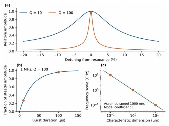
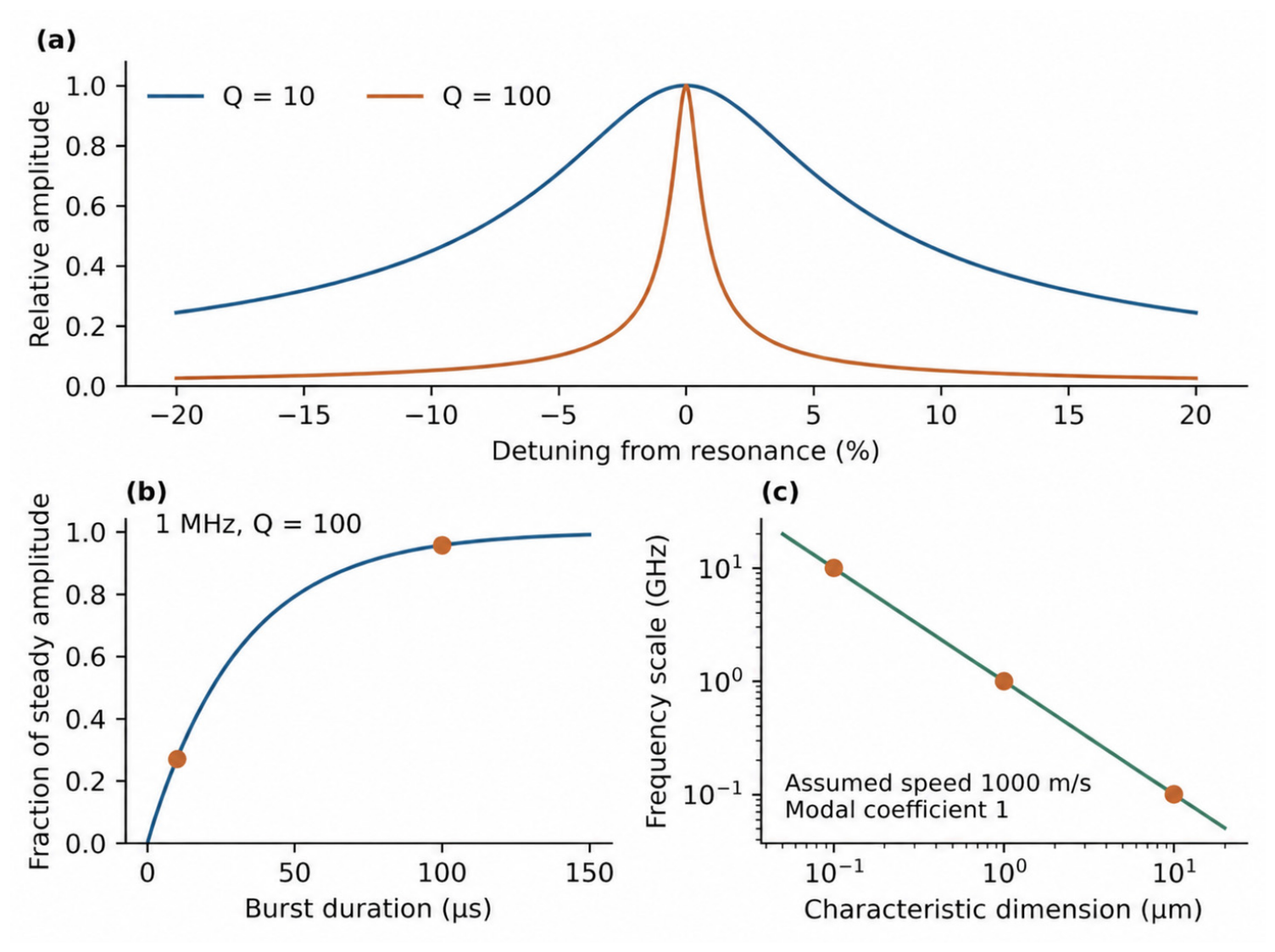
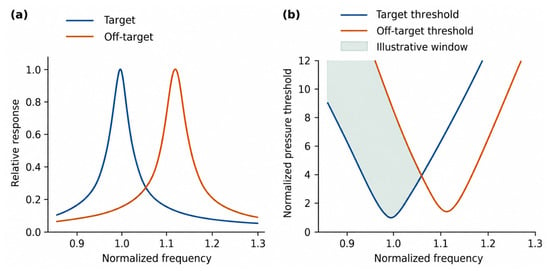
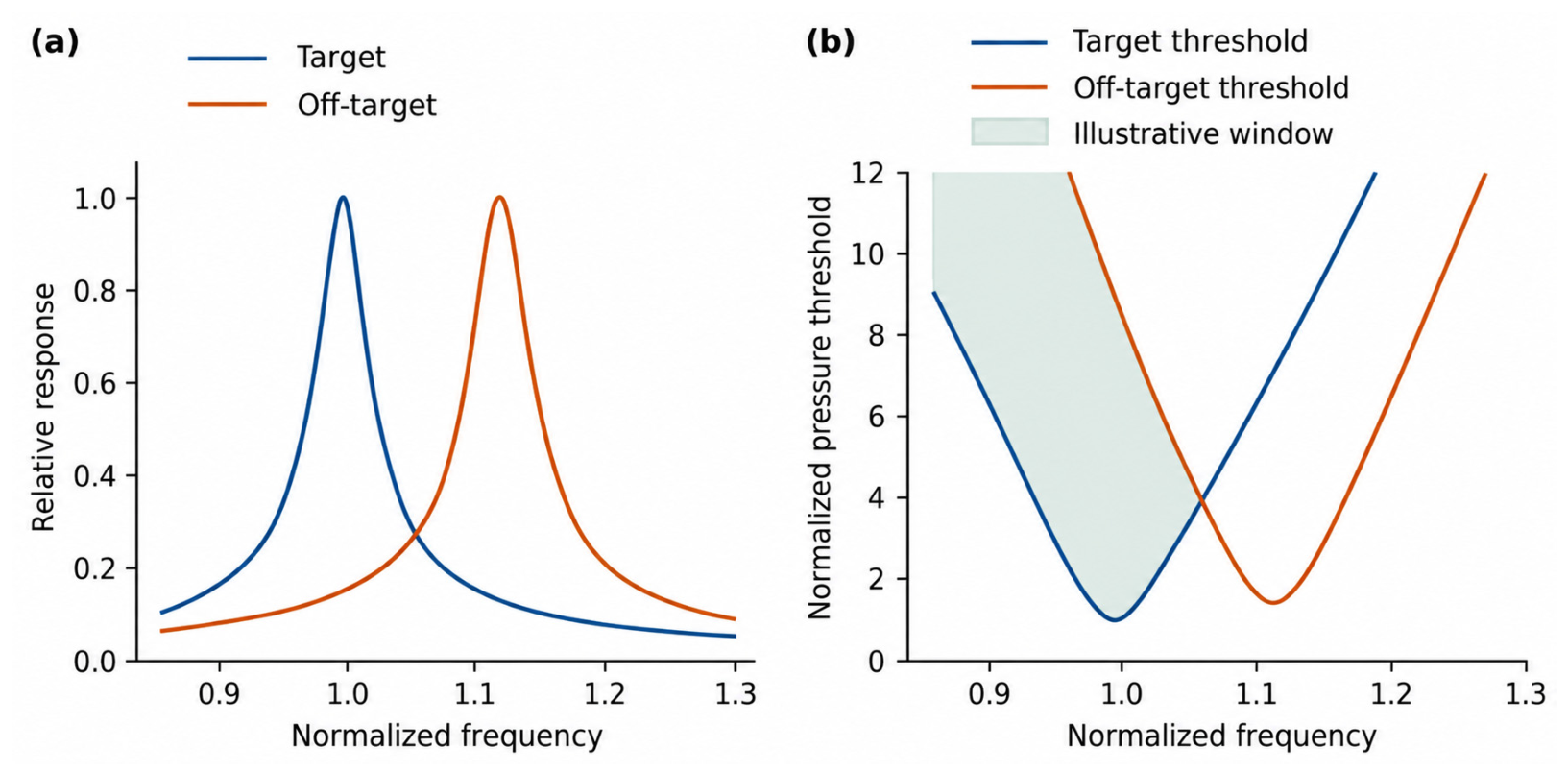

Disclaimer/Publisher’s Note: The statements, opinions and data contained in all publications are solely those of the individual author(s) and contributor(s) and not of MDPI and/or the editor(s). MDPI and/or the editor(s) disclaim responsibility for any injury to people or property resulting from any ideas, methods, instructions or products referred to in the content. |
© 2026 by the authors. Licensee MDPI, Basel, Switzerland. This article is an open access article distributed under the terms and conditions of the Creative Commons Attribution (CC BY) license.