


Abstract
Background: Sepsis-associated encephalopathy (SAE) is among the most frequent neurological complications of sepsis, contributing independently to mortality, prolonged mechanical ventilation and intensive care unit stay and long-term cognitive disability. The gut–brain axis has been proposed as a key mediator, through dysbiosis, intestinal barrier failure, bacterial translocation and neuroinflammation. This review synthesises the evidence on this axis in SAE and appraises whether nutritional and microbiome-directed strategies can be regarded as modulators of it, or should be framed as a testable hypothesis. Methods: A narrative review was conducted and reported according to the SANRA criteria. PubMed/MEDLINE, Scopus and Web of Science were searched from inception to September 2026 for preclinical and clinical studies on gut microbiota, intestinal permeability, neuroinflammation, blood–brain barrier dysfunction and nutritional or microbiome-directed strategies in sepsis. Results: Four communication pathways are described: neural (vagal), immune/humoral, metabolic (short-chain fatty acids, tryptophan-derived indoles, host kynurenines) and neuroendocrine. Evidence that nutritional or microbiome-directed interventions act on them is almost entirely preclinical and derives from rodent models of limited translational validity. Randomised trials of feeding route and dose, immunonutrients and probiotics were designed around mortality, infection and respiratory outcomes—endpoints of the gut–immune and gut–lung axes—and, apart from one negative cognitive follow-up in acute lung injury, none has reported SAE, delirium or cognition. This is an absence of evidence rather than heterogeneous evidence. Conversely, microbial composition and metabolites are emerging as candidate predictors of SAE and intensive care unit delirium. Conclusions: The gut–brain axis in SAE remains an unproven hypothesis. Nutrition should be regarded as a biologically plausible but untested modulator rather than an established therapy. Prospective studies with standardised neurological endpoints, and microbiota-based risk stratification, are required before any gut-directed intervention can be recommended for brain protection in sepsis.
1. Introduction
Sepsis is defined as a life-threatening organ dysfunction caused by a dysregulated host response to infection [1]. Among the organs affected, the brain is often the first to fail: sepsis-associated encephalopathy (SAE) is a diffuse cerebral dysfunction that occurs in the absence of direct central nervous system (CNS) infection, structural lesions or other identifiable causes of encephalopathy [2,3]. Clinically, SAE ranges from subtle alterations of attention and sickness behaviour to delirium and deep coma, and its severity correlates with short-term mortality [3,4]. Beyond the acute phase, sepsis survivors show a substantially increased risk of new, persistent cognitive impairment and functional disability [5,6], with neuroimaging and electrophysiological correlates such as hippocampal atrophy and electroencephalographic slowing [7].
Despite its clinical relevance, no specific therapy for SAE exists, and management remains limited to control of the infectious source, hemodynamic and metabolic support, minimisation of iatrogenic and deliriogenic factors, implementation of non-pharmacological delirium-prevention bundles and avoidance of neurotoxic drugs and modifiable risk factors [3,8,9]. Therefore, there is a strong rationale for identifying upstream mechanisms that are both pathophysiologically relevant and amenable to intervention at the bedside.
The gut has long been considered the “motor” of multiple organ dysfunction in critical illness [10], although, as discussed in Section 4.1, this remains a hypothesis rather than a demonstrated mechanism. In sepsis, intestinal hypoperfusion, epithelial apoptosis, loss of tight-junction integrity, impaired mucosal immunity and profound remodelling of the microbial community converge to produce what has recently been conceptualised as “gut failure” [11,12]. In parallel, research on the microbiota–gut–brain axis (MGBA) has shown that intestinal microbes and their metabolites regulate microglial maturation, blood–brain barrier (BBB) permeability, neurotransmission and behaviour [13]. The convergence of these two fields has generated the hypothesis that sepsis-induced gut dysfunction is not only a bystander but an active contributor to brain injury [14,15,16].
Nutrition is intimately linked to both sides of this axis. Luminal nutrients sustain enterocyte and colonocyte function, shape microbial ecology and determine the production of neuroactive metabolites, while the metabolic state induced by feeding (glycaemia, electrolyte balance, micronutrient availability) directly affects neuronal function [11,17]. Yet, nutritional guidelines for critically ill patients are designed around global outcomes such as mortality, infections and length of stay, and do not specifically address brain protection [18,19]; as detailed in Section 6.1, the outcomes these trials measured belong primarily to the gut–immune and gut–lung axes rather than to the gut–brain axis.
The aim of this narrative review is to summarise current knowledge on the gut–brain axis in SAE, to examine critically how nutritional strategies and microbiome-targeted therapies have been proposed to modulate this axis, to review the emerging use of microbiota-derived signatures for the prediction of SAE, and to identify knowledge gaps and research priorities. Attention is given to patients with sepsis of abdominal origin, in whom the gut is simultaneously the source of infection, the site of surgical injury and the main target of nutritional therapy; the limited population-specific evidence available for this group is stated explicitly throughout, and the clinical implications are presented as hypotheses in Section 9.2.
2. Methods
This narrative review was conducted and reported following the criteria of the Scale for the Assessment of Narrative Review Articles (SANRA) [20]. A literature search was performed in PubMed/MEDLINE, Scopus and Web of Science from inception to September 2026, using combinations of the following terms: “sepsis-associated encephalopathy”, “septic encephalopathy”, “sepsis-associated delirium”, “gut–brain axis”, “microbiota”, “dysbiosis”, “intestinal barrier”, “short-chain fatty acids”, “tryptophan”, “vagus nerve”, “enteral nutrition”, “parenteral nutrition”, “probiotics”, “synbiotics”, “fecal microbiota transplantation”, “selective digestive decontamination”, “hepatic encephalopathy”, “machine learning” and “prediction model”. The reference lists of relevant reviews and international guidelines were hand-searched, and ClinicalTrials.gov was queried for ongoing interventional studies.
To make the search reproducible, the complete Boolean strings run in each database, together with the date on which they were last executed, are reported in Appendix A. Eighty-three publications are cited in this review. Consistent with the narrative design and with SANRA, selection was purposive rather than exhaustive: records were screened for relevance against the eligibility criteria below by a single author, screening was not duplicated, no review protocol was registered or published, and no formal risk-of-bias assessment or quantitative synthesis was undertaken. For the same reason a PRISMA-style record flow (numbers of records identified, screened, excluded and assessed in full text) was not prospectively recorded and is therefore not reported; we have preferred to state this explicitly rather than reconstruct counts retrospectively. These methodological choices are those of a narrative, not a systematic, review, and are restated among the limitations in Section 10.
Articles published in English were considered eligible if they reported (i) clinical or epidemiological data on SAE or sepsis-associated delirium; (ii) mechanistic experimental data on the relationship between the gut, its microbiota or its metabolites and brain dysfunction in sepsis; (iii) studies evaluating microbial, metabolomic or clinical predictors of SAE or intensive care unit delirium; or (iv) randomized trials, meta-analyses or guidelines on nutritional or microbiome-directed interventions in sepsis and critical illness. Priority was given to original research articles, in particular to randomised controlled trials, international guidelines and experimental studies testing causality (e.g., microbiota transfer, gene or receptor inhibition, metabolite supplementation); review articles are cited only where a broad synthesis is required and not in support of specific experimental findings, mechanistic steps, animal models, biomarkers or trial results, for which the primary sources are now cited throughout. Case reports were considered only when no higher-level evidence was available. Given the heterogeneity of study designs, populations and outcomes, the evidence is presented descriptively, explicitly distinguishing preclinical from clinical data, and—within the preclinical literature—endotoxaemia models from polymicrobial sepsis models (Section 5.6).
During the preparation of this manuscript, the authors used a generative artificial intelligence tool (Claude Opus 5.5) to support literature retrieval, drafting and language editing. The authors have reviewed and edited the output and take full responsibility for the content of this publication.
3. Sepsis-Associated Encephalopathy: Clinical Spectrum and Pathophysiology
3.1. Definition, Diagnosis and Outcomes
SAE is a diagnosis of exclusion that requires ruling out CNS infection, structural brain lesions, drug-induced encephalopathy, non-convulsive status epilepticus and specific metabolic causes such as hepatic or uremic encephalopathy [2,3]. In clinical practice, SAE is usually operationalized as a Glasgow Coma Scale score below 15 or the presence of delirium detected with validated tools such as the Confusion Assessment Method for the Intensive Care Unit (CAM-ICU) [9,21]. Electroencephalography, neuroimaging and biomarkers of neuronal and glial injury may support the diagnosis and prognostication, but none is specific [3,4]. Magnetic resonance imaging may reveal ischaemic lesions and white matter abnormalities, particularly in patients with septic shock [22].
SAE affects up to 70% of septic patients depending on the definition and population, and even mild impairments in consciousness are independently associated with increased mortality [2,3]. In a large single-centre cohort of elderly septic patients, SAE occurred in 48.7% and was associated with a more than twofold higher mortality than sepsis without encephalopathy (28.8% vs. 12.6%) [23]. Beyond mortality, SAE is associated with a longer duration of mechanical ventilation and with prolonged intensive care unit and hospital length of stay [24], which is why these outcomes are mentioned in the Abstract. In survivors, severe sepsis is associated with a markedly increased risk of long-term cognitive impairment and functional disability [5], and longer duration of delirium during critical illness predicts worse global cognition at 3 and 12 months [6]. Importantly, several factors contributing to SAE are potentially modifiable, including acute kidney injury, dysglycemia, hypercapnia, hypernatremia and specific pathogens such as Staphylococcus aureus [8].
3.2. Core Pathophysiological Mechanisms
The pathophysiology of SAE is multifactorial and incompletely understood [4,16]. Systemic inflammation is transmitted to the brain through circumventricular organs lacking a complete BBB, through activation of cerebral endothelial cells, and through afferent vagal fibres that sense peripheral cytokines [2,25]. Endothelial activation and disruption of tight junctions increase BBB permeability, allowing inflammatory mediators, leukocytes and neurotoxic molecules to enter the brain parenchyma [4]. Microglia and astrocytes become activated and amplify neuroinflammation; in experimental sepsis, myeloid inflammation of the CNS may persist long after resolution of the infection, providing a plausible substrate for long-term cognitive decline [26].
Additional mechanisms include mitochondrial dysfunction and oxidative stress, alterations in cholinergic, dopaminergic, GABAergic and glutamatergic neurotransmission, impaired cerebral autoregulation and microcirculatory dysfunction, and accumulation of neurotoxic metabolites secondary to hepatic and renal dysfunction [15,16]. It should be emphasised that these mechanisms are not specific to signals arising from the gut: systemic inflammation of any origin, as well as dysfunction of the heart, liver, kidney and lung, can drive each of them. This point is returned to in Section 6.1, because it limits the inference that can be drawn when a gut-directed intervention improves neurological outcomes in animal models. As discussed below, virtually all these mechanisms can be influenced by signals originating from the gut.
4. The Gut in Sepsis: From Barrier Failure to Dysbiosis
4.1. Intestinal Barrier Failure and Translocation
The intestinal barrier comprises the mucus layer, the epithelial monolayer sealed by tight junctions, the gut-associated lymphoid tissue (GALT) and the commensal microbiota. In sepsis, splanchnic vasoconstriction and redistribution of blood flow expose the mucosa to hypoperfusion and ischaemia–reperfusion injury; epithelial apoptosis increases, proliferation decreases and tight-junction proteins are dysregulated [10,12]. The resulting hyperpermeability allows translocation of live bacteria, pathogen-associated molecular patterns (PAMPs) such as lipopolysaccharide (LPS), and damage-associated molecular patterns (DAMPs) into the mesenteric lymph and portal circulation, sustaining systemic inflammation [11,12]. Loss of barrier integrity can be probed clinically with circulating markers such as D-lactate—a stereoisomer produced almost exclusively by intestinal bacteria and poorly metabolised by human tissue—together with endotoxin and diamine oxidase; these are the gut-barrier read-outs used in ongoing studies of intensive care unit delirium [27,28]. The intestinal immune compartment is simultaneously activated and suppressed, which promotes both tissue damage and susceptibility to secondary infections [12].
The “gut as the motor of multiple organ dysfunction” remains a hypothesis rather than an established mechanism, and its history is instructive. In its original formulation, portal translocation of viable bacteria and endotoxin was proposed to drive systemic inflammation; this prediction was tested directly in humans and not confirmed. In twenty patients with major torso trauma in whom portal venous catheters were placed and portal blood sampled for five days, neither bacteraemia nor endotoxaemia was detected in portal blood, despite a high incidence of subsequent multiple organ failure [29]. Subsequent reformulations therefore shifted emphasis from viable bacteria in the portal vein to gut-derived mediators carried in the mesenteric lymph, and more recently to the microbiome and its metabolites as a whole [10,11,12]. Critics continue to point out that bacterial translocation is readily demonstrated in rodents but rarely documented in critically ill patients, and that much of the supporting human evidence is associative. Throughout this review, the gut is therefore treated as a biologically plausible and potentially modifiable contributor to organ dysfunction, not as a demonstrated causal “motor”.
Patients with sepsis of abdominal origin merit specific consideration already at this level, because in them the gut is simultaneously the infectious source, the site of surgical injury and the main target of therapy. Peritonitis and emergency laparotomy add direct serosal and mucosal injury to the hypoperfusion common to all septic states; postoperative ileus, bowel oedema, an open abdomen and intra-abdominal hypertension further impair barrier function and delay the delivery of luminal nutrients; and antimicrobial exposure is both broader in spectrum and longer in duration than in most other sources of sepsis [11,12,30]. Postoperative peritonitis is also characterised by a distinctive microbial ecology, with enterococcal and fungal overgrowth that reflects selection pressure on the gut community rather than the original inoculum [30]. Each of these features is a plausible amplifier of the mechanisms described in this section, but, as acknowledged in Section 9.2, none has been studied with neurological endpoints.
4.2. Sepsis-Induced Dysbiosis
Critical illness causes one of the most extreme perturbations of the human microbiome described to date. Within days of intensive care unit (ICU) admission, the gut microbiota loses diversity and commensal anaerobes (classically described as a loss of Firmicutes and Bacteroidetes), while potentially pathogenic taxa such as Enterobacteriaceae and Enterococcus expand [27,31]. We note explicitly that the Firmicutes-to-Bacteroidetes ratio is a contested summary metric: it aggregates phyla of great functional heterogeneity, is highly sensitive to sequencing and bioinformatic choices, and has repeatedly failed to reproduce across cohorts. It is used here only as shorthand for the loss of commensal anaerobes, and functional read-outs—depletion of short-chain fatty acids and of tryptophan-derived indoles—are preferred throughout. Broad-spectrum antibiotics, proton-pump inhibitors, opioids, catecholamines, altered motility and the absence of luminal nutrients all contribute to this “pathobiome” [11,27]. Gut-derived bacteria can also be found in distant organs, as shown by the enrichment of the lung microbiome with intestinal taxa in sepsis and acute respiratory distress syndrome [32]. A functional consequence of dysbiosis is the rapid and sustained depletion of faecal short-chain fatty acids (SCFAs), key microbial metabolites for colonocyte energy supply, barrier integrity and immune regulation [33,34]. These alterations persist beyond the acute phase and may shape the long-term trajectory of survivors [27].
Dysbiosis in the ICU is also prognostically informative, which is relevant to Section 8. In a prospective cohort of 301 medical ICU patients, faecal biodiversity at admission did not differ according to outcome, but domination of the gut community by Enterococcus (≥30% of 16S reads) and colonisation with vancomycin-resistant Enterococcus were each independently associated with subsequent death or culture-proven infection, and carriage of specific pathogens at admission predicted later infection with the same organism [35]. This establishes the principle that the admission gut community carries prognostic information in critical illness, even though the outcomes examined were infectious rather than neurological.
5. Gut–Brain Communication Pathways in Sepsis-Associated Encephalopathy
The MGBA is a bidirectional communication network that integrates neural, immune, endocrine and metabolic signals between the intestine and the CNS [13]. In SAE, each of these channels may convey injurious signals from the dysfunctional gut to the brain, and conversely, brain dysfunction may worsen gut function through autonomic and neuroendocrine efferents [14,36,37,38]. Four pathways—neural, immune/humoral, metabolic and neuroendocrine—are described below and summarised in Figure 1.
Figure 1.
Schematic representation of the gut–brain axis in sepsis-associated encephalopathy. Sepsis-induced gut barrier failure and dysbiosis generate neural, immune/humoral, metabolic and neuroendocrine signals that converge on the brain, promoting blood–brain barrier disruption, microglial activation and neurotransmitter imbalance. Nutritional and microbiome-targeted interventions are shown as hypothesised points of action, not as interventions of demonstrated neurological benefit. Abbreviations: BBB, blood–brain barrier; FMT, faecal microbiota transplantation; HPA, hypothalamic–pituitary–adrenal; LPS, lipopolysaccharide; SCFAs, short-chain fatty acids.
5.1. Neural Pathway: The Vagus Nerve and the Cholinergic Anti-Inflammatory Pathway
The vagus nerve is the main anatomical link between the gut and the brainstem. Vagal afferents detect luminal and mucosal signals, including cytokines, microbial products and gut hormones, and relay them to the nucleus tractus solitarius, from which they reach the hypothalamus, limbic system and cortex [13,14]. The efferent arm constitutes the “inflammatory reflex”, whereby acetylcholine released from vagal efferents acts on α7 nicotinic receptors of macrophages to inhibit cytokine release [25]; in experimental endotoxemia, vagus nerve stimulation attenuates the systemic inflammatory response [39]. In SAE, the vagus nerve appears to be a key mediator of the impact of the microbiota on the brain: in LPS-treated mice, faecal microbiota transplantation (FMT) from healthy donors improved spatial memory and electroencephalographic abnormalities, reduced cortical cytokines and microglial activation, and these benefits were abolished by vagotomy [40].
Two caveats must be stated explicitly. First, the vagal arm of the gut–brain axis in SAE rests substantially on this single study [40], and the model used was LPS endotoxaemia, not sepsis. Intraperitoneal endotoxin reproduces an acute, self-limited and monophasic cytokine surge in the absence of a replicating infectious focus, of a necrotic source, of surgical injury and of the antibiotic, vasopressor and sedative exposure that characterise clinical sepsis; it is a model of systemic inflammation rather than of sepsis, and Table 1 labels it as such. Second, vagotomy is not a selective tool: section of the vagus abolishes afferent as well as efferent signalling and alters gastric emptying, motility and splanchnic perfusion, so the loss of benefit after vagotomy is compatible with, but does not prove, a vagally mediated microbiota effect. Supportive but equally preliminary evidence comes from stimulation experiments: non-invasive high-frequency ultrasound stimulation of the cervical vagus in LPS-treated mice restored macrophage α7 nicotinic receptor expression, lowered plasma interleukin-1β and improved locomotor activity and survival [41]. Reduced cholinergic tone in the brain is also thought to contribute to delirium and cognitive impairment, linking the neural pathway to the neurotransmitter hypothesis of SAE [15,16].
Table 1.
Selected experimental studies directly linking the gut microbiota to sepsis-associated encephalopathy. LPS endotoxaemia is distinguished from polymicrobial CLP sepsis, and the behavioural or neurological read-out actually measured is reported for each study.
5.2. Immune and Humoral Pathway
Translocated PAMPs and DAMPs activate Toll-like receptors on cerebral endothelial cells and perivascular macrophages, inducing the release of cytokines, prostaglandins and nitric oxide and increasing BBB permeability [4,48]. Gram-negative LPS, Gram-positive cell wall components and fungal products have all been shown to activate glial cells and reduce the expression of tight-junction proteins such as occludin and claudin-5 [48]. Peripheral immune cells, including T lymphocytes primed in the gut-associated lymphoid tissue, may traffic to the brain and contribute to neuroinflammation [15,16]. An additional and more recently described route involves extracellular vesicles: in experimental sepsis, exosomes released by intestinal epithelial cells carried inflammatory signals to the brain and mediated interleukin-1β-dependent neuronal injury [42]. Notably, the gut microbiota itself regulates BBB integrity, since germ-free mice display increased BBB permeability and reduced expression of tight-junction proteins, which is reversed by colonisation with a normal microbiota or with SCFA-producing bacteria [49].
5.3. Metabolic Pathway: Microbial Metabolites
Microbial metabolites represent perhaps the most attractive link between the gut, nutrition and the brain, as their production depends directly on dietary substrates. SCFAs (acetate, propionate and butyrate) are produced by fermentation of dietary fibre, serve as the main energy source for colonocytes, enhance tight-junction assembly, promote regulatory T cell differentiation and exert anti-inflammatory effects through G-protein-coupled receptors and histone deacetylase inhibition [34]. SCFAs also cross the BBB and are required for the maturation and function of microglia [50]. In a murine model of caecal ligation and puncture (CLP), mice with mild SAE produced more butyric acid than mice with severe SAE, and supplementation with sodium butyrate reduced cerebral oxidative stress and improved survival through the GPR109A/Nrf2/HO-1 pathway [44]. Two further CLP studies support and refine this mechanism with original experimental data: exogenous SCFAs attenuated blood–brain barrier disruption, neuroinflammation and behavioural impairment [51], and pre-treatment with an acetate/propionate/butyrate mixture restored Morris water maze performance and lowered hippocampal interleukin-1β, interleukin-6 and tumour necrosis factor-α, an effect abolished by the GPR43 antagonist GLPG0974, identifying GPR43 as a necessary receptor [45]. Notably, in the latter study faecal acetate and propionate, rather than butyrate, were the species depleted, indicating that the individual SCFA responsible for protection is not yet settled. Given the rapid depletion of faecal SCFAs observed in critically ill patients [33], loss of these metabolites may represent a mechanistic bridge between dysbiosis, lack of enteral substrate and brain dysfunction.
Tryptophan metabolism provides a second key metabolic link. Commensal bacteria convert dietary tryptophan into indole derivatives that act as ligands of the aryl hydrocarbon receptor (AhR) on astrocytes and microglia, limiting CNS inflammation [52]. In CLP-induced sepsis, mice resistant to SAE were enriched in the microbial metabolite indole-3-propionic acid (IPA), which alleviated anxiety-like behaviour and memory impairment and inhibited microglial NLRP3 inflammasome activation in an AhR-dependent manner [36,43]. Similarly, microbiota-derived indole-3-acetic acid protected against SAE through microglial AhR signalling [47]. Conversely, during systemic inflammation, tryptophan is diverted toward the host kynurenine pathway, generating neuroactive metabolites; in critically ill patients, alterations of the kynurenine pathway have been associated with acute brain dysfunction, including delirium and coma [53]. Other microbiota-derived molecules, such as bile acids, trimethylamine N-oxide and neurotransmitter precursors (γ-aminobutyric acid, serotonin, dopamine), are also altered in sepsis and may contribute to brain dysfunction, although evidence in SAE remains preliminary [38,48].
5.4. Neuroendocrine Pathway
The hypothalamic–pituitary–adrenal (HPA) axis is the principal neuroendocrine component of the MGBA [13]. The microbiota modulates HPA reactivity, and in turn, glucocorticoids and catecholamines released during sepsis affect gut permeability, motility and microbial composition [14]. Central stress and sympathetic overactivation may therefore further impair intestinal function, establishing a vicious circle in which brain dysfunction perpetuates gut failure and vice versa [11,14].
5.5. Experimental Evidence Linking the Microbiota to SAE Susceptibility
Several experimental studies have moved beyond association to test causality (Table 1). The most compelling evidence comes from models in which the post-septic microbiota was transferred between animals: in CLP-induced sepsis, mice receiving faeces from SAE-susceptible animals developed more severe cortical inflammation than those receiving faeces from SAE-resistant animals [43], and microbiota transfer experiments identified butyrate production as a determinant of protection [44]. These data suggest that inter-individual variability in sepsis-induced dysbiosis may contribute to the heterogeneous neurological phenotype observed in patients. Probiotic supplementation produces concordant findings: in female mice subjected to CLP, Lacticaseibacillus rhamnosus GG given before and after surgery improved 21-day survival, reduced anxiety-like behaviour in the open field, restored novel-object recognition, preserved hippocampal CA1 and CA3 neurons and maintained BDNF expression and TrkB phosphorylation [46]. However, most studies used rodent models with limited translational validity, and human studies linking gut microbial signatures to SAE are still scarce; the extent of that limitation is examined in the following section.
5.6. Translational Validity of Rodent Models of SAE
Because Table 1 rests almost entirely on rodent work, the correspondence between these models and the clinical syndrome deserves to be stated explicitly. A systematic review of 226 studies found that both CLP and LPS injection reliably induce short- and long-term behavioural impairment, but that the two differ fundamentally: LPS endotoxaemia delivers a defined, non-replicating stimulus and produces a monophasic inflammatory response, whereas CLP creates a persistent polymicrobial focus with a necrotic source, and is therefore the closer analogue of clinical sepsis [54]. Neither model routinely includes the interventions that define the intensive care unit—antibiotics, source control, fluid resuscitation, vasopressors, sedation, mechanical ventilation and artificial nutrition—each of which independently shapes both the microbiome and the brain.
What can realistically be modelled in rodents is a restricted set of domains: level of arousal and sickness behaviour; locomotor activity (open field, SHIRPA); anxiety-like behaviour (open field centre time, elevated plus maze); hippocampus-dependent spatial learning and memory (Morris water maze, Y-maze); recognition memory (novel object recognition); electroencephalographic slowing; and the cellular substrate of neuroinflammation—microglial and astrocytic activation, cytokine expression, blood–brain barrier permeability and neuronal loss. These are the read-outs reported in every study in Table 1.
What cannot be reproduced is, unfortunately, much of what defines SAE at the bedside. Delirium is diagnosed in humans by inattention, disorganised thinking and an altered level of consciousness that fluctuates over hours; there is no validated rodent assay for inattention, for disorganised thought or for fluctuation, and the hypoactive–hyperactive distinction has no behavioural equivalent. Higher-order cognition—executive function, language, abstraction, verbal reasoning—is simply not available for testing in mice, yet it is precisely the domain impaired in sepsis survivors [5,6,7]. Further, the behavioural tests themselves are imperfect surrogates: the Morris water maze is a forced-swim task whose performance is confounded by the muscle weakness, deconditioning and reduced motivation that follow sepsis, so a longer escape latency may reflect myopathy rather than amnesia; the open field conflates anxiety with hypoactivity; and tests are generally performed in survivors days to weeks after the insult, i.e., in the recovery phase, whereas delirium is an acute-phase phenomenon. Mortality in CLP is high and non-random, so surviving animals represent a selected, more resilient population. Finally, most studies use young, genetically homogeneous male animals housed in specific-pathogen-free conditions, whereas SAE predominantly affects older, comorbid, microbially diverse humans; the probiotic study cited above [46] is a rare exception in using female animals. Rodent models should therefore be read as tools for dissecting mechanism—for which they are well suited, since they permit microbiota transfer, receptor blockade and metabolite supplementation—rather than as a scale model of human delirium.
5.7. Evidence from Human Studies
Human data supporting the gut–brain axis in SAE are indirect but broadly consistent with the experimental findings. Critically ill patients show early and profound loss of commensal taxa with overgrowth of potential pathogens [31], together with a rapid and sustained fall in faecal SCFAs [33]. In septic patients, faecal levels of the tryptophan-derived metabolite IPA are reduced, and lower levels are associated with worse clinical outcomes, while in experimental sepsis IPA enhances macrophage phagocytosis through AhR signalling [55]. Alterations of the host kynurenine pathway have been associated with fewer days alive without delirium or coma in critically ill patients [53]. Three small clinical studies have compared the gut community directly between septic patients with and without encephalopathy. In 20 patients with SAE and 20 septic controls, 16S rDNA sequencing showed reduced microbial diversity in SAE, accompanied by a distinct serum metabolomic profile [56]. In a more recent multi-omics study of 10 patients with SAE and 20 with sepsis alone, α- and β-diversity did not differ, but compositional shifts were observed together with a faecal microRNA signature and higher serum interleukin-1β [57]. Outside sepsis, the largest prospective dataset comes from 133 acutely ill hospitalised older adults, of whom 38 (29%) developed delirium: gut microbiota diversity and composition differed significantly between those who did and did not become delirious, and the association persisted irrespective of baseline nutritional status [58].
These human data must be interpreted with considerable caution, and three problems deserve to be named. First, confounding by indication is severe and largely unmeasured. Dysbiosis in the ICU is driven by broad-spectrum antibiotics, proton-pump inhibitors, opioids, catecholamines, enteral feeding interruptions and immobility [11,27,59]; every one of these is also independently associated with delirium, so an observed association between a microbial signature and SAE may reflect shared exposure rather than shared mechanism. Second, reverse causation cannot be excluded: SAE and its treatment alter autonomic tone, gastric emptying, motility and sedative and opioid requirements, each of which remodels the gut community, so the brain may be shaping the microbiome as readily as the reverse—the bidirectionality that is theoretically attractive (Section 5.4) is methodologically disabling in cross-sectional data. Third, sample sizes are very small (n = 10–20 per group in the SAE-specific studies), the designs are cross-sectional and single-centre, and no study has adjusted for antibiotic class and duration, which alone could account for the differences reported. Disentangling these will require longitudinal sampling from before the onset of encephalopathy, explicit adjustment for drug exposure, and ideally quasi-experimental designs such as randomisation of antibiotic duration or of microbiome-directed therapy with neurological endpoints.
However, no prospective human study has yet correlated gut microbial composition or microbial metabolites with the incidence, severity or long-term consequences of SAE using standardised neurological assessment together with objective markers of neuroaxonal injury, and this represents a major knowledge gap.
6. Nutrition as a Potential Modulator of the Gut–Brain Axis in SAE: Evidence and Hypothesis
Nutritional therapy is part of standard ICU care and, unlike most experimental interventions discussed above, is available for every patient. It must, however, be stated at the outset that no clinical trial has evaluated a nutritional strategy with SAE, delirium or cognition as a primary or secondary outcome in sepsis. The material presented in this section is therefore a rationale, not an evidence base, and the primary outcomes actually measured in each trial are reported first (Table 2), with possible effects on the brain discussed afterwards and explicitly as hypotheses.
Table 2.
Nutritional and microbiome-directed interventions: the primary outcome actually measured in the key trials, the relevant secondary outcomes and the axis they engage, and what is known about SAE or delirium.
6.1. Which Axis Do the Available Clinical Trials Actually Address?
Enteral nutrition preserves GALT mass and secretory IgA production, maintains mucosal blood flow and motility, and sustains a more physiological microbial ecology [11,75]. The outcomes measured in nutritional trials—nosocomial infection, ventilator-associated pneumonia, bacteraemia, gastrointestinal complications—are read-outs of the gut–immune axis: the capacity of luminal nutrients and commensal flora to maintain mucosal immunity and limit the passage of pathogens and PAMPs into mesenteric lymph and portal blood [69,70,71,75]. A second, partly independent route is the gut–lung axis: in sepsis and acute respiratory distress syndrome the lung microbiome becomes enriched with intestinal taxa [32], and gut-derived mediators carried in mesenteric lymph contribute to acute lung injury; this is the axis most plausibly engaged when a nutritional or probiotic intervention reduces ventilator-associated pneumonia [69,71]. The gut–brain axis shares the upstream events—barrier failure, dysbiosis, metabolite depletion—with both, but differs in its effectors (vagal afferents, cerebral endothelium, microglia, neurotransmitter systems) and, critically, in its outcome measures (delirium, cognition, electroencephalography, neuroaxonal injury markers).
Two consequences follow. First, a reduction in infections or in respiratory complications cannot be taken as evidence that a nutritional intervention has modulated the gut–brain axis; these are different endpoints on different axes. Second, in the animal studies of Section 5, neurological improvement may well have arisen secondarily, through attenuation of systemic inflammation and of concomitant cardiac, hepatic, renal and pulmonary dysfunction, rather than through direct gut-to-brain signalling. Only a minority of those studies included the manipulations—vagotomy [40], receptor antagonism [45], AhR-dependence experiments [43,47]—that begin to establish pathway specificity, and even those have the limitations set out in Section 5.1 and Section 5.6. We therefore present the following subsections as a summary of what the trials measured, followed by a clearly labelled hypothesis.
6.2. Route of Feeding
Enteral nutrition (EN) provides luminal substrates for enterocytes, stimulates mucosal blood flow and motility, preserves GALT mass and secretory IgA production, and maintains a more physiological microbial ecology [11,75]. In experimental models, the absence of enteral stimulation leads to mucosal atrophy and impaired mucosal immunity [75]. On this basis, international guidelines recommend EN over parenteral nutrition (PN) whenever the gastrointestinal tract is functional [18,19,76]. For clarity, and because different documents use different time windows, “early” enteral nutrition is defined throughout this review as initiation within 48 h of ICU admission, which is the window used in the ESICM clinical practice guidelines [76]; where the Surviving Sepsis Campaign is cited, its own wider window of 72 h is stated explicitly [77]. The Surviving Sepsis Campaign suggests early initiation of EN (within 72 h) in adults with sepsis or septic shock who can be fed enterally [77].
Primary outcome. In NUTRIREA-2, 2410 ventilated adults with shock were randomised to early isocaloric enteral versus parenteral nutrition; the primary outcome was 28-day mortality, which did not differ between groups. Secondary outcomes showed no reduction in ICU-acquired infections, and a significant excess of gastrointestinal complications in the enteral arm, including vomiting, diarrhoea, bowel ischaemia and acute colonic pseudo-obstruction [60]. No neurological or delirium endpoint was collected. This highlights that the potential benefit of enteral substrates on the gut–brain axis must be balanced against the risk of feeding a hypoperfused gut. Current ESICM recommendations suggest delaying EN in uncontrolled shock, overt bowel ischaemia, active gastrointestinal bleeding or abdominal compartment syndrome, and otherwise starting low-dose (trophic) EN and advancing it progressively [76].
6.3. Timing and Dose
The optimal caloric and protein dose during the acute phase of sepsis remains debated. Primary outcomes. In EPaNIC (n = 4640), the primary outcome was ICU length of stay: late initiation of parenteral nutrition shortened it and reduced the duration of mechanical ventilation and renal replacement therapy, with fewer new infections as a secondary outcome [61]. In NUTRIREA-3 (n = 3044 ventilated adults with shock), the co-primary outcomes were 90-day mortality and time to readiness for ICU discharge: mortality did not differ, while low-calorie, low-protein feeding shortened time to readiness for discharge and reduced vomiting, diarrhoea, bowel ischaemia and liver dysfunction [62]. Neither trial assessed delirium or cognition. Accordingly, current guidelines favour a progressive increase in energy delivery during the first days, avoiding early overfeeding [17,19].
One important exception must be acknowledged, and it tempers the enthusiasm of the preceding paragraphs. The EDEN trial randomised 1000 patients with acute lung injury to initial trophic versus full enteral feeding for up to six days; in a prospectively planned one-year follow-up of 525 survivors, there was no difference between arms in physical function, in survival or in any of multiple secondary outcomes at 6 and 12 months [63]. In a parallel ancillary study, 174 consecutive survivors underwent blinded neuropsychological assessment of executive function, language, memory, verbal reasoning and attention: performance was substantially below predicted values in both arms, but initial trophic versus full feeding had no effect on cognitive impairment status at either time point [78]. This is, to our knowledge, the only randomised nutritional trial in critical illness that has reported a formal cognitive endpoint, and it was unequivocally negative. The population was acute lung injury rather than sepsis, the intervention lasted six days, and the comparison was of caloric dose rather than of substrate composition or microbiome-directed therapy, so the result does not exclude an effect of other nutritional strategies. It does, however, demonstrate that cognitive endpoints are feasible in critical care nutrition trials, and it argues against assuming that feeding strategy carries measurable neurological benefit. From a gut–brain perspective, the concept of “trophic” feeding remains hypothetically attractive: small volumes of enteral nutrients may be sufficient to preserve mucosal integrity and microbial substrate while limiting the metabolic burden and the risk of intolerance. Whether this translates into less delirium or better long-term cognitive outcomes in sepsis has not been tested.
6.4. Glycaemic and Metabolic Control
Metabolic derangements are among the few modifiable contributors to SAE. In a large multicentre cohort, hyperglycaemia (>180 mg/dL [>10 mmol/L]), hypoglycaemia (<54 mg/dL [<3 mmol/L]), hypernatremia and hypercapnia were independently associated with SAE [8]; this is an observational association, and the cohort was not designed to test whether correcting these derangements prevents SAE. Nutritional therapy, especially PN and early full-dose feeding, is a major determinant of hyperglycaemia, while interruptions of feeding under insulin therapy expose patients to hypoglycaemia. Primary outcome. In NICE-SUGAR (n = 6104), the primary outcome was 90-day mortality: intensive glucose control (81–108 mg/dL [4.5–6.0 mmol/L]) increased mortality compared with a conventional target of ≤180 mg/dL [≤10 mmol/L], and severe hypoglycaemia was far more frequent [64]. Maintaining moderate glycaemic targets, avoiding glycaemic variability and hypoglycaemia, and correcting sodium and fluid imbalances are therefore reasonable brain-protective measures intimately linked to nutritional management [3,8]. All glucose values in this review are reported in mg/dL with the corresponding mmol/L value in square brackets, for consistency.
6.5. Micronutrients and Immunonutrients
Thiamine deficiency is common in septic shock and may cause or worsen encephalopathy through impaired cerebral oxidative metabolism. Primary outcome. In a randomised, double-blind, placebo-controlled pilot trial of 88 patients with septic shock, the primary outcome was the change in lactate at 24 h, which did not differ overall; a pre-specified subgroup of thiamine-deficient patients showed lower lactate and lower mortality over time [65]. Screening for deficiency and supplementation in patients at risk (e.g., alcohol use disorder, malnutrition, prolonged PN, large gastrointestinal losses) is therefore justified, although effects on SAE remain unproven. Other immunonutrients have yielded disappointing results: in the REDOXS trial (n = 1223 mechanically ventilated patients with multiorgan failure), the primary outcome was 28-day mortality, which showed a trend toward harm with glutamine, while in-hospital and 6-month mortality were significantly increased [66]; in the SISPCT trial (n = 1089), the primary outcome of 28-day mortality was unaffected by high-dose sodium selenite [67]. Omega-3 fatty acids have anti-inflammatory and pro-resolving properties and are included in some PN lipid emulsions, but evidence of benefit on clinically relevant outcomes in sepsis is inconsistent and none addresses neurological endpoints [17]. Overall, current guidelines do not support routine pharmacological doses of immunonutrients in sepsis [17,18,19].
6.6. Fermentable Fibre and Prebiotics
Dietary fibre is the main substrate for microbial SCFA production [34]. Since SCFAs support barrier integrity, microglial homeostasis and BBB function [49,50], and their faecal concentrations drop rapidly in critically ill patients [33], fibre-containing enteral formulas and prebiotics represent a biologically plausible strategy to restore this metabolic pathway. Fermentable soluble fibre is also suggested to manage diarrhoea in tube-fed critically ill patients [68]. However, no randomised trial has measured the effect of fibre-enriched enteral nutrition on faecal SCFA concentrations, on microbial composition or on any neurological outcome in sepsis, and fibre should be used with caution in patients with gut hypoperfusion or dysmotility.
6.7. Summary: A Hypothesis, Not an Established Therapy
Taken together, the trials summarised above establish what nutrition does for survival, infection and gastrointestinal tolerance in critical illness; they do not establish anything about the brain. The proposition that preserving the intestinal barrier and the SCFA- and indole-producing community during sepsis will reduce the incidence or severity of SAE is coherent with the mechanisms of Section 5, is supported by animal data of the quality discussed in Section 5.6, and is—at present—untested in humans. It should be treated as a hypothesis generating future trials, and as such it is carried forward to Section 9 and Section 11 rather than into any present-day recommendation.
7. Microbiome-Targeted Therapies
7.1. Probiotics and Synbiotics
Probiotics and synbiotics aim to restore commensal populations and their metabolic functions. In critically ill patients with severe systemic inflammatory response syndrome, synbiotic therapy was associated with preservation of the gut microbiota and fewer infectious complications [70]. Meta-analyses have suggested a reduction in overall infections and ventilator-associated pneumonia, but without an effect on mortality (corrected pooled risk ratio 1.02, 95% CI 0.85–1.22) and with substantial heterogeneity [71]. Importantly, the large PROSPECT trial showed that Lactobacillus rhamnosus GG did not reduce ventilator-associated pneumonia or other clinically important outcomes in mechanically ventilated patients [69]. Probiotics have shown neuroprotective effects in experimental SAE [15,46], but no clinical trial has evaluated their effect on delirium or cognition in septic patients. Safety concerns, including probiotic-related bacteraemia or fungemia in immunocompromised patients and in those with central venous catheters or impaired gut barrier, also warrant caution [15,27], and are particularly relevant in abdominal sepsis, where the barrier is breached by definition.
7.2. Faecal Microbiota Transplantation
FMT offers the most comprehensive restoration of the gut ecosystem and is the intervention with the strongest experimental support in SAE, where it attenuates neuroinflammation and cognitive impairment through vagal and metabolic mechanisms [40,43,44]. Clinical experience in sepsis is limited to case reports and small series; for instance, FMT successfully treated severe sepsis with refractory diarrhoea in a patient after vagotomy [72]. Interventional studies of FMT for ICU delirium have recently been registered [28], but donor selection, route of administration, timing relative to antibiotic exposure and safety in patients with a disrupted intestinal barrier remain major open issues [38,48].
7.3. Postbiotics and Metabolite Supplementation
The administration of defined microbial metabolites, such as butyrate or indole derivatives, may bypass the need for a functional microbial community and avoid the infectious risks of live organisms. Experimental data support protective effects of butyrate, IPA and indole-3-acetic acid in SAE [43,44,45,47,51]. These “postbiotic” strategies are appealing for precision medicine but have not yet been tested in humans with sepsis, and the identity of the protective SCFA species remains unsettled (Section 5.3).
7.4. Vagus Nerve Stimulation
Given the central role of the vagus nerve in both gut–brain signalling and the control of inflammation [25,39], vagus nerve stimulation has been proposed as a neuromodulatory therapy for SAE, and protective effects have been reported in experimental models [15,38,41]. Non-invasive transcutaneous or ultrasound auricular stimulation may facilitate clinical translation [41], but human data in sepsis are lacking.
7.5. Antibiotic Stewardship, Duration and Neurotoxicity
Antibiotics are lifesaving in sepsis but are also a major driver of dysbiosis [27]. Shortening the duration of therapy is, on current evidence, the most actionable gut-sparing intervention available at the bedside, and in abdominal sepsis it is supported by two randomised trials. In STOP-IT, 518 patients with complicated intra-abdominal infection and adequate source control were randomised to a fixed 4-day course or to therapy continued until two days after resolution of physiological abnormalities; the composite primary outcome of surgical-site infection, recurrent intra-abdominal infection or death was equivalent [74]. In DURAPOP, 249 critically ill patients with postoperative peritonitis were randomised on day 8 to stop or to continue to day 15; the 8-day arm had significantly more antibiotic-free days with equivalent 45-day mortality and no excess of multidrug-resistant organisms [30]. Neither trial measured delirium or cognition, but both establish that a substantial reduction in antimicrobial exposure—and therefore in its ecological impact on the gut community—is achievable without clinical cost in precisely the population that is the focus of this review.
Antibiotic neurotoxicity, moreover, extends well beyond cefepime. A systematic review of 391 reported cases identified three distinct clinical phenotypes of antibiotic-associated encephalopathy: encephalopathy with seizures or myoclonus arising within days, caused by penicillins and cephalosporins; encephalopathy with prominent psychosis arising within days, caused by quinolones, macrolides and procaine penicillin; and encephalopathy with cerebellar signs and MRI abnormalities emerging after weeks, caused by metronidazole [79]. Carbapenems—especially imipenem—and piperacillin–tazobactam are additional recognised causes, and all these agents are commonly used, frequently in combination and often at renally unadjusted doses, in exactly the patients at risk of SAE. Antibiotic-associated encephalopathy is therefore both a differential diagnosis for SAE and a reason to narrow and shorten therapy: in most classes, encephalopathy resolves within a median of about five days of discontinuation [79]. Timely de-escalation, adequate source control and shortest effective duration of therapy may therefore contribute to preserving the gut–brain axis, in addition to their established role in limiting antimicrobial resistance [77].
7.6. Selective Decontamination of the Digestive Tract
Selective decontamination of the digestive tract (SDD) and selective oropharyngeal decontamination (SOD) deserve separate mention, because they constitute the largest deliberate human experiment in manipulating the gut community in critical illness. In the SuDDICU cluster-crossover randomised trial, 5982 mechanically ventilated adults in 19 ICUs received SDD or standard care; in-hospital mortality within 90 days, the primary outcome, was 27.0% versus 29.1% (mean difference −1.7%, 95% CI −4.8% to 1.3%; OR 0.91, 95% CI 0.82–1.02; p = 0.12), i.e., not significantly reduced, although the confidence interval does not exclude a clinically important benefit [73].
What makes SDD directly relevant here is its ecological footprint. Molecular analysis of faecal samples from patients enrolled in the Dutch SDD/SOD trial showed that SDD, unlike SOD, did not spare the anaerobic community: Faecalibacterium prausnitzii—the predominant butyrate producer of the human colon—was significantly depleted, as were Eubacterium rectale and Roseburia spp., while enterococci rose by an order of magnitude [59]. SDD therefore suppresses precisely the taxa whose metabolite is proposed to be neuroprotective (Section 5.3). This is a cautionary observation of some importance: an intervention can reduce Gram-negative colonisation and infection—a gut–immune benefit—while simultaneously degrading the SCFA-producing capacity of the community. It illustrates why outcomes on one axis cannot be extrapolated to another, and why any future gut-directed strategy aimed at the brain will need to measure microbial function, not only microbial composition or infection rates.
7.7. Lessons from Hepatic Encephalopathy: The Existing Clinical Precedent
The clearest proof that an encephalopathy can be treated by acting on the gut comes not from sepsis but from liver disease, and it has been curiously absent from the SAE literature. In hepatic encephalopathy, the non-absorbable disaccharide lactulose acidifies the colonic lumen, traps ammonia as ammonium and alters the composition and metabolic output of the colonic flora, while the non-absorbable antibiotic rifaximin suppresses ammoniagenic bacteria. In a randomised, double-blind trial of 299 patients in remission from recurrent overt hepatic encephalopathy, rifaximin reduced the risk of a breakthrough episode by 58% over six months and reduced hepatic encephalopathy-related hospitalisation, with most patients receiving concomitant lactulose [80]; both agents are recommended in the joint AASLD/EASL practice guideline, lactulose as first-line therapy and rifaximin as add-on for secondary prophylaxis [81].
The analogy is instructive in both directions. On one hand, it demonstrates that gut-directed therapy can produce clinically meaningful neurological benefit, measured with a neurological endpoint, in a human encephalopathy—the design that is entirely missing from the sepsis literature. On the other hand, the differences are substantial and should temper enthusiasm. Hepatic encephalopathy has a single, measurable, well-characterised gut-derived toxin (ammonia) with a defined route of entry via portosystemic shunting, and interventions are given over months in ambulatory, haemodynamically stable patients. SAE has no identified equivalent toxin, no measurable target analogous to ammonia, an acute time course of hours to days, and occurs in patients with shock, ileus and absent luminal transit, in whom non-absorbable agents may not even be delivered to the colon. Hepatic encephalopathy therefore establishes the feasibility of the trial design rather than the transferability of the therapy, and its main lesson for SAE research is the value of identifying a single quantifiable gut-derived mediator before designing an intervention.
8. From Treatment to Prediction: Can the Gut Predict the Brain?
The preceding sections lead to an uncomfortable conclusion: no gut-directed therapy can currently be recommended for the prevention or treatment of SAE. Prediction, however, is a different question, and here the field is appreciably more advanced than intervention. We therefore devote a separate section to it, because risk stratification is both the more realistic near-term translational goal and the necessary precondition for an efficient interventional trial.
8.1. Microbial and Metabolite Signatures as Candidate Predictors
Four lines of evidence suggest that the gut carries prognostic information about the brain. First, the admission gut community predicts hard outcomes in critical illness: Enterococcus domination and vancomycin-resistant Enterococcus colonisation independently predicted death or infection in a prospective cohort of 301 ICU patients [35], establishing the principle even though the endpoints were not neurological. Second, SAE-specific case–control studies, though very small, consistently find a different community in patients with encephalopathy: reduced diversity with a distinct serum metabolome in 20 SAE versus 20 non-SAE patients [56], and compositional shifts with a faecal microRNA signature and higher serum interleukin-1β in 10 SAE versus 20 septic controls [57]. Third, in the largest prospective dataset on delirium (133 acutely ill older adults, 29% delirious), microbiota diversity and composition differed significantly between those who did and did not become delirious [58]. Fourth, and most attractive from a mechanistic standpoint, specific metabolites rather than taxa may carry the signal: faecal SCFAs fall rapidly and persistently in critical illness [33], faecal IPA is reduced in septic patients and lower levels track with worse outcomes [55], and host kynurenine pathway alterations are associated with fewer days alive without delirium or coma [53]. Metabolites have the advantage of being quantifiable, biologically interpretable and directly actionable through substrate provision or supplementation.
8.2. Machine-Learning Models and the Case for Adding Microbial Features
In parallel, several groups have developed machine-learning models for SAE using routinely collected clinical data. Nine algorithms trained on 6994 MIMIC-IV patients predicted 30-day mortality after SAE with areas under the curve of 0.80–0.83, with acute physiology score III, Glasgow Coma Scale and red cell distribution width carrying the greatest weight, and were deployed as an online calculator [24]. More recently, LASSO–Boruta feature selection with XGBoost, CatBoost, LightGBM, multilayer perceptron and support vector machine models, interpreted with SHapley Additive exPlanations, was applied to the prediction of SAE occurrence—rather than mortality—in 3156 elderly septic patients, in whom SAE incidence was 48.7% [23]. A stacking ensemble achieved an area under the curve of 0.807 internally but only 0.671 on external validation in the eICU database, a drop that is itself informative about the generalisability of these models.
The salient observation is that not one of these models includes a microbial or microbial-metabolite feature. They are built exclusively on physiology, laboratory values and comorbidity. This is the obvious point of convergence between the two literatures reviewed here: a prospective study enrolling septic patients at ICU admission, sampling stool or rectal swabs for 16S or shotgun sequencing and for targeted SCFA and tryptophan-metabolite quantification, adjudicating SAE with CAM-ICU and the Richmond Agitation–Sedation Scale, and testing whether microbial features add incremental discrimination over an established clinical model, would be feasible with existing technology and would answer a question that neither field can answer alone. Such a study should be reported according to TRIPOD, should pre-specify the comparator model, and must include external validation, given the performance drop observed above.
8.3. Anchoring Prediction to an Objective Measure of Brain Injury
A persistent obstacle is that SAE lacks an objective endpoint. Blood-based markers of neuroaxonal injury offer a partial solution. In a prospective pilot study of 20 patients with septic shock and 5 non-septic controls, plasma neurofilament light chain (NfL) rose over time in septic patients but remained stable in controls, with higher levels in those with brain dysfunction, and provided prognostic information on death and on functional and cognitive outcome in survivors [82]. NfL has since been proposed as the most promising candidate biomarker for SAE, with glial fibrillary acidic protein as a complementary marker of astroglial injury, although assay standardisation, cut-off definition and the confounding effects of renal failure and age remain unresolved [83]. Combining a microbial or metabolite signature with a quantitative marker of neuroaxonal injury would give future studies both an exposure and an outcome that are measurable, and would move the field beyond the categorical, observer-dependent diagnosis of delirium.
9. Clinical Implications: Toward a Gut-Oriented Approach to Brain Protection
9.1. What Can Reasonably Be Done Today
Although the evidence is largely experimental, the concepts reviewed here support an integrated, gut-oriented approach to brain protection in septic patients that relies entirely on existing, guideline-concordant measures. It is important to be clear that none of the following is recommended because of demonstrated benefit on the gut–brain axis; each is recommended on its own evidence, and the gut–brain rationale is an additional, speculative argument in its favour. These include early source control and hemodynamic optimisation to restore splanchnic perfusion [77]; early, low-dose EN (initiated within 48 h where not contraindicated, as defined in Section 6.2) advanced according to gastrointestinal tolerance, with PN reserved for patients in whom EN is contraindicated or insufficient after the first days [17,76]; moderate glycaemic targets (≤180 mg/dL [≤10 mmol/L]) avoiding hypoglycaemia and glycaemic variability [8,64]; correction of sodium, fluid and acid–base derangements [8]; screening and treatment of thiamine deficiency in high-risk patients [65]; antimicrobial stewardship with the shortest effective duration, for which randomised evidence now exists in intra-abdominal infection [30,74], and avoidance where possible of the antibiotic classes most often implicated in antibiotic-associated encephalopathy [79]; minimization of deliriogenic drugs and implementation of non-pharmacological delirium prevention bundles [9], which remain the only intervention of proven value for delirium in the ICU.
9.2. Abdominal Sepsis: A Hypothesis in Search of Data
Patients with abdominal sepsis deserve specific consideration. In these patients, the gut is the source of infection and is often injured by peritonitis and surgery; bowel oedema, ileus, open abdomen and intra-abdominal hypertension frequently delay EN, while exposure to broad-spectrum antibiotics is prolonged [11,12,30]. Such patients may plausibly experience more profound gut barrier failure and dysbiosis than patients with sepsis of other origin, but no claim of a higher risk of gut-mediated brain injury can be sustained on present data. No study has compared the incidence, severity or duration of SAE between abdominal and non-abdominal sources of sepsis; no study has compared microbial or metabolite profiles between these groups; and the trials cited in Section 6 and Section 7 that did enrol abdominal sepsis populations—STOP-IT and DURAPOP [30,74]—measured infectious and antimicrobial-exposure endpoints only. The proposition that abdominal sepsis is a high-risk phenotype for gut-mediated brain injury is a hypothesis, and we present it as such. It is, however, an eminently testable one, and the abdominal sepsis population has two features that make it an attractive setting for the studies proposed in Section 8: the timing of the gut insult is known precisely (the operation), and the gut is directly accessible for sampling and for the staged delivery of enteral substrate. The timing of EN after abdominal surgery, the role of trophic feeding in the open abdomen, and monitoring of gastrointestinal function (e.g., through validated scores or biomarkers of enterocyte injury, such as intestinal fatty acid-binding protein, citrulline or D-lactate) are relevant questions for future research in this population [12,76].
10. Strengths and Limitations
This review integrates mechanistic, clinical and nutritional evidence across disciplines that are usually examined separately, and it explicitly distinguishes experimental from clinical data, endotoxaemia models from polymicrobial sepsis models, and the gut–immune and gut–lung axes from the gut–brain axis. Nevertheless, several limitations should be acknowledged. First, as a narrative review, it did not follow a systematic search and selection protocol: no protocol was registered, screening was performed by a single author and not duplicated, and no risk-of-bias assessment or quantitative synthesis was undertaken. The search strategy, databases and eligibility criteria are nonetheless reported in Section 2 in the interest of transparency and reproducibility, and the reader should understand these as a description of what was done rather than as a systematic method; selection bias cannot be excluded. Second, most of the mechanistic evidence derives from rodent models, whose relevance to human sepsis is limited for the reasons set out in detail in Section 5.6, and the single most cited observation on the vagal pathway comes from one study in an endotoxaemia model [40]. Third, clinical studies on nutrition in sepsis did not assess neurological outcomes, so their relevance to SAE is inferred rather than demonstrated; the one trial that did assess cognition was in acute lung injury and was negative [63]. Fourth, the human microbiome data in SAE are few, small, cross-sectional and heavily confounded by antibiotics, proton-pump inhibitors, opioids and vasopressors, and reverse causation cannot be excluded (Section 5.7). Fifth, the “gut as the motor of multiple organ dysfunction” framework on which much of this literature rests remains contested (Section 4.1). Finally, the rapid growth of the field means that some recent experimental findings have not yet been independently replicated.
11. Knowledge Gaps and Future Directions
Several gaps currently limit the translation of gut–brain concepts to the care of patients with SAE. First, SAE lacks a consensus operational definition and standardised diagnostic criteria, which hampers comparisons across studies and the design of interventional trials [3]; anchoring the diagnosis to a quantitative marker of neuroaxonal injury such as NfL may partly circumvent this [82,83]. Second, most mechanistic data derive from rodent models of CLP or LPS injection, which only partially reproduce human sepsis and do not include standard ICU interventions such as antibiotics, sedation, vasopressors and nutritional support [14,37,54]; models incorporating antibiotic treatment, fluid resuscitation and aged or comorbid animals, and assays addressing attention rather than spatial memory alone, would materially improve translational validity. Third, human studies correlating gut microbial composition, faecal and circulating metabolites, and markers of barrier dysfunction with neurological phenotypes are scarce and mostly cross-sectional. Longitudinal multi-omics studies integrating microbiome, metabolome, biomarkers of brain injury (e.g., neurofilament light chain, glial fibrillary acidic protein) and long-term cognitive outcomes are needed [38,48,57,82,83], with sampling beginning before the onset of encephalopathy and with explicit adjustment for antibiotic class and duration, proton-pump inhibitors, opioids and vasopressors. Fourth, nutritional trials in critically ill patients have rarely included delirium or cognition as outcomes; incorporating these endpoints into future trials of feeding route, dose, fibre and micronutrient supplementation would be of great value, and the EDEN follow-up demonstrates that this is operationally feasible [63]. Fifth, microbiota-based prediction of SAE should be tested directly: no existing machine-learning model for SAE incorporates microbial or metabolite features [23,24], and whether they add incremental discrimination over clinical variables is an answerable question (Section 8.2). Sixth, microbiome-targeted interventions (probiotics, FMT, postbiotics, vagus nerve stimulation) should be tested in carefully selected populations, with rigorous safety monitoring and biologically informed stratification, for example according to baseline dysbiosis or metabolite deficiency [15,48]. Finally, antibiotic duration is the one gut-sparing intervention for which randomised infrastructure already exists [30,74]; embedding delirium and cognitive endpoints into future antimicrobial-duration trials would be a low-cost, high-yield way of testing the gut–brain hypothesis in humans.
12. Conclusions
SAE is a frequent and severe manifestation of sepsis with long-lasting consequences for survivors. A growing body of experimental evidence indicates that sepsis-induced gut barrier failure and dysbiosis may contribute to brain dysfunction through neural, immune, metabolic and neuroendocrine pathways, and that restoring the gut ecosystem or its metabolites can mitigate neuroinflammation and cognitive impairment in animal models, although in those models a contribution from the attenuation of systemic and non-cerebral organ dysfunction cannot be excluded. Nutrition represents the most immediately available tool with which this axis might be addressed, through the choice of route, timing and dose of feeding, glycaemic and electrolyte control, micronutrient repletion and provision of fermentable substrates. However, clinical evidence directly linking these interventions to neurological outcomes is absent rather than merely inconsistent, and nutrition must therefore be presented as a hypothesis to be tested rather than as a therapeutic or preventive strategy for SAE. In the shorter term, the more tractable translational opportunity may lie in prediction: microbial and metabolite signatures, combined with existing clinical machine-learning models and anchored to quantitative markers of neuroaxonal injury, could identify the patients in whom a gut-directed intervention would be worth testing. Future research should incorporate brain-related endpoints into nutritional and microbiome-directed trials, particularly in high-risk populations such as patients with abdominal sepsis, to determine whether protecting the gut can indeed protect the brain.
Author Contributions
Conceptualisation, M.F. and G.C.; methodology, F.C. and R.G.; writing—original draft preparation, M.F.; writing—review and editing, P.S. and V.P.; supervision, M.C.P. All authors have read and agreed to the published version of the manuscript.
Funding
This research received no external funding.
Institutional Review Board Statement
Not applicable.
Informed Consent Statement
Not applicable.
Data Availability Statement
No new data were created or analysed in this study. Data sharing is not applicable to this article.
Conflicts of Interest
The authors declare no conflicts of interest.
Abbreviations
AhR, aryl hydrocarbon receptor; ASPEN, American Society for Parenteral and Enteral Nutrition; BBB, blood–brain barrier; BDNF, brain-derived neurotrophic factor; CAM-ICU, Confusion Assessment Method for the Intensive Care Unit; CLP, caecal ligation and puncture; CNS, central nervous system; DAMPs, damage-associated molecular patterns; EN, enteral nutrition; ESICM, European Society of Intensive Care Medicine; ESPEN, European Society for Clinical Nutrition and Metabolism; FMT, faecal microbiota transplantation; GALT, gut-associated lymphoid tissue; GFAP, glial fibrillary acidic protein; HPA, hypothalamic–pituitary–adrenal; HE, hepatic encephalopathy; ICU, intensive care unit; IPA, indole-3-propionic acid; LPS, lipopolysaccharide; MGBA, microbiota–gut–brain axis; ML, machine learning; NfL, neurofilament light chain; PAMPs, pathogen-associated molecular patterns; PN, parenteral nutrition; SAE, sepsis-associated encephalopathy; SCFAs, short-chain fatty acids; SANRA, Scale for the Assessment of Narrative Review Articles; SDD, selective decontamination of the digestive tract; SHAP, SHapley Additive exPlanations; SOD, selective oropharyngeal decontamination; VNS, vagus nerve stimulation.
Appendix A. Search Strategy
The following Boolean strings were run in PubMed/MEDLINE (last run 20 October 2026) and adapted to the syntax of Scopus and Web of Science, which were used for cross-checking and for forward and backward citation tracking. No language limit other than English was applied; no date limit was applied.
A1. Syndrome: (“sepsis-associated encephalopathy”[tiab] OR “septic encephalopathy”[tiab] OR “sepsis-associated delirium”[tiab] OR (“sepsis”[MeSH] AND (“delirium”[MeSH] OR “cognitive dysfunction”[MeSH] OR “brain diseases”[MeSH]))).
A2. Gut–brain axis: (“gut-brain axis”[tiab] OR “microbiota-gut-brain”[tiab] OR “brain-gut axis”[tiab]) AND (“sepsis”[tiab] OR “critical illness”[tiab] OR “intensive care”[tiab]).
A3. Barrier and microbiome: (“intestinal barrier”[tiab] OR “intestinal permeability”[tiab] OR “bacterial translocation”[tiab] OR “dysbiosis”[tiab] OR “gut microbiota”[tiab] OR “microbiome”[tiab]) AND (“sepsis”[tiab] OR “septic shock”[tiab] OR “critical illness”[tiab]).
A4. Metabolites: (“short-chain fatty acid*”[tiab] OR “butyrate”[tiab] OR “tryptophan”[tiab] OR “indole-3-propionic”[tiab] OR “indole-3-acetic”[tiab] OR “kynurenine”[tiab] OR “aryl hydrocarbon receptor”[tiab]) AND (“sepsis”[tiab] OR “encephalopathy”[tiab] OR “microglia”[tiab]).
A5. Nutrition and microbiome-directed therapy: (“enteral nutrition”[MeSH] OR “parenteral nutrition”[MeSH] OR “probiotics”[MeSH] OR “synbiotics”[tiab] OR “prebiotics”[tiab] OR “fecal microbiota transplantation”[MeSH] OR “selective decontamination”[tiab] OR “glutamine”[tiab] OR “thiamine”[tiab] OR “dietary fiber”[MeSH]) AND (“sepsis”[tiab] OR “critical illness”[tiab] OR “intensive care units”[MeSH]).
A6. Prediction and biomarkers: (“machine learning”[tiab] OR “prediction model”[tiab] OR “risk prediction”[tiab] OR “neurofilament”[tiab] OR “glial fibrillary acidic protein”[tiab]) AND (“sepsis-associated encephalopathy”[tiab] OR “delirium”[tiab]).
A7. Vagus nerve: (“vagus nerve”[MeSH] OR “vagotomy”[tiab] OR “cholinergic anti-inflammatory”[tiab]) AND (“sepsis”[tiab] OR “endotoxemia”[tiab] OR “encephalopathy”[tiab]).
In addition, the reference lists of all included reviews and of the international guidelines cited were hand-searched, and ClinicalTrials.gov was queried for registered interventional studies using the terms “sepsis”, “delirium”, “microbiota” and “fecal microbiota transplantation”.
References
- Singer, M.; Deutschman, C.S.; Seymour, C.W.; Shankar-Hari, M.; Annane, D.; Bauer, M.; Bellomo, R.; Bernard, G.R.; Chiche, J.D.; Coopersmith, C.M.; et al. The Third International Consensus Definitions for Sepsis and Septic Shock (Sepsis-3). JAMA 2016, 315, 801–810. [Google Scholar] [CrossRef] [Scilit]
- Gofton, T.E.; Young, G.B. Sepsis-Associated Encephalopathy. Nat. Rev. Neurol. 2012, 8, 557–566. [Google Scholar] [CrossRef] [Scilit]
- Sonneville, R.; Benghanem, S.; Jeantin, L.; de Montmollin, E.; Doman, M.; Gaudemer, A.; Thy, M.; Timsit, J.F. The Spectrum of Sepsis-Associated Encephalopathy: A Clinical Perspective. Crit. Care 2023, 27, 386. [Google Scholar] [CrossRef] [Scilit]
- Mazeraud, A.; Righy, C.; Bouchereau, E.; Benghanem, S.; Bozza, F.A.; Sharshar, T. Septic-Associated Encephalopathy: A Comprehensive Review. Neurotherapeutics 2020, 17, 392–403. [Google Scholar] [CrossRef] [Scilit]
- Iwashyna, T.J.; Ely, E.W.; Smith, D.M.; Langa, K.M. Long-Term Cognitive Impairment and Functional Disability among Survivors of Severe Sepsis. JAMA 2010, 304, 1787–1794. [Google Scholar] [CrossRef] [Scilit]
- Pandharipande, P.P.; Girard, T.D.; Jackson, J.C.; Morandi, A.; Thompson, J.L.; Pun, B.T.; Brummel, N.E.; Hughes, C.G.; Vasilevskis, E.E.; Shintani, A.K.; et al. Long-Term Cognitive Impairment after Critical Illness. N. Engl. J. Med. 2013, 369, 1306–1316. [Google Scholar] [CrossRef] [Scilit]
- Semmler, A.; Widmann, C.N.; Okulla, T.; Urbach, H.; Kaiser, M.; Widman, G.; Mormann, F.; Weide, J.; Fliessbach, K.; Hoeft, A.; et al. Persistent Cognitive Impairment, Hippocampal Atrophy and EEG Changes in Sepsis Survivors. J. Neurol. Neurosurg. Psychiatry 2013, 84, 62–69. [Google Scholar] [CrossRef] [Scilit]
- Sonneville, R.; de Montmollin, E.; Poujade, J.; Garrouste-Orgeas, M.; Souweine, B.; Darmon, M.; Mariotte, E.; Argaud, L.; Barbier, F.; Goldgran-Toledano, D.; et al. Potentially Modifiable Factors Contributing to Sepsis-Associated Encephalopathy. Intensive Care Med. 2017, 43, 1075–1084. [Google Scholar] [CrossRef] [Scilit]
- Devlin, J.W.; Skrobik, Y.; Gélinas, C.; Needham, D.M.; Slooter, A.J.C.; Pandharipande, P.P.; Watson, P.L.; Weinhouse, G.L.; Nunnally, M.E.; Rochwerg, B.; et al. Clinical Practice Guidelines for the Prevention and Management of Pain, Agitation/Sedation, Delirium, Immobility, and Sleep Disruption in Adult Patients in the ICU. Crit. Care Med. 2018, 46, e825–e873. [Google Scholar] [CrossRef] [Scilit]
- Klingensmith, N.J.; Coopersmith, C.M. The Gut as the Motor of Multiple Organ Dysfunction in Critical Illness. Crit. Care Clin. 2016, 32, 203–212. [Google Scholar] [CrossRef] [Scilit]
- Corriero, A.; Gadaleta, R.M.; Puntillo, F.; Inchingolo, F.; Moschetta, A.; Brienza, N. The Central Role of the Gut in Intensive Care. Crit. Care 2022, 26, 379. [Google Scholar] [CrossRef] [Scilit]
- Soranno, D.E.; Coopersmith, C.M.; Brinkworth, J.F.; Factora, F.N.F.; Muntean, J.H.; Mythen, M.G.; Raphael, J.; Shaw, A.D.; Vachharajani, V.; Messer, J.S. A Review of Gut Failure as a Cause and Consequence of Critical Illness. Crit. Care 2025, 29, 91. [Google Scholar] [CrossRef] [Scilit]
- Cryan, J.F.; O’Riordan, K.J.; Cowan, C.S.M.; Sandhu, K.V.; Bastiaanssen, T.F.S.; Boehme, M.; Codagnone, M.G.; Cussotto, S.; Fulling, C.; Golubeva, A.V.; et al. The Microbiota-Gut-Brain Axis. Physiol. Rev. 2019, 99, 1877–2013. [Google Scholar] [CrossRef] [Scilit]
- Wang, X.; Wen, X.; Yuan, S.; Zhang, J. Gut-Brain Axis in the Pathogenesis of Sepsis-Associated Encephalopathy. Neurobiol. Dis. 2024, 195, 106499. [Google Scholar] [CrossRef] [Scilit]
- Barlow, B.; Ponnaluri, S.; Barlow, A.; Roth, W. Targeting the Gut Microbiome in the Management of Sepsis-Associated Encephalopathy. Front. Neurol. 2022, 13, 999035. [Google Scholar] [CrossRef] [Scilit]
- Zhang, J.; Chen, S.; Hu, X.; Huang, L.; Loh, P.; Yuan, X.; Liu, Z.; Lian, J.; Geng, L.; Chen, Z.; et al. The Role of the Peripheral System Dysfunction in the Pathogenesis of Sepsis-Associated Encephalopathy. Front. Microbiol. 2024, 15, 1337994. [Google Scholar] [CrossRef] [Scilit]
- Singer, P.; Blaser, A.R.; Berger, M.M.; Calder, P.C.; Casaer, M.; Hiesmayr, M.; Mayer, K.; Montejo-Gonzalez, J.C.; Pichard, C.; Preiser, J.C.; et al. ESPEN Practical and Partially Revised Guideline: Clinical Nutrition in the Intensive Care Unit. Clin. Nutr. 2023, 42, 1671–1689. [Google Scholar] [CrossRef] [Scilit]
- Singer, P.; Blaser, A.R.; Berger, M.M.; Alhazzani, W.; Calder, P.C.; Casaer, M.P.; Hiesmayr, M.; Mayer, K.; Montejo, J.C.; Pichard, C.; et al. ESPEN Guideline on Clinical Nutrition in the Intensive Care Unit. Clin. Nutr. 2019, 38, 48–79. [Google Scholar] [CrossRef] [Scilit]
- Compher, C.; Bingham, A.L.; McCall, M.; Patel, J.; Rice, T.W.; Braunschweig, C.; McKeever, L. Guidelines for the Provision of Nutrition Support Therapy in the Adult Critically Ill Patient: The American Society for Parenteral and Enteral Nutrition. J. Parenter. Enter. Nutr. 2022, 46, 12–41. [Google Scholar] [CrossRef] [Scilit]
- Baethge, C.; Goldbeck-Wood, S.; Mertens, S. SANRA—A Scale for the Quality Assessment of Narrative Review Articles. Res. Integr. Peer Rev. 2019, 4, 5. [Google Scholar] [CrossRef] [Scilit]
- Ely, E.W.; Inouye, S.K.; Bernard, G.R.; Gordon, S.; Francis, J.; May, L.; Truman, B.; Speroff, T.; Gautam, S.; Margolin, R.; et al. Delirium in Mechanically Ventilated Patients: Validity and Reliability of the Confusion Assessment Method for the Intensive Care Unit (CAM-ICU). JAMA 2001, 286, 2703–2710. [Google Scholar] [CrossRef] [Scilit]
- Sharshar, T.; Carlier, R.; Bernard, F.; Guidoux, C.; Brouland, J.P.; Nardi, O.; de la Grandmaison, G.L.; Aboab, J.; Gray, F.; Menon, D.; et al. Brain Lesions in Septic Shock: A Magnetic Resonance Imaging Study. Intensive Care Med. 2007, 33, 798–806. [Google Scholar] [CrossRef] [Scilit]
- Han, Y.; Xie, X.; Qiu, J.; Tang, Y.; Song, Z.; Li, W.; Wu, X. Early Prediction of Sepsis Associated Encephalopathy in Elderly ICU Patients Using Machine Learning Models: A Retrospective Study Based on the MIMIC-IV Database. Front. Cell. Infect. Microbiol. 2025, 15, 1545979. [Google Scholar] [CrossRef] [Scilit]
- Peng, L.; Peng, C.; Yang, F.; Wang, J.; Zuo, W.; Cheng, C.; Mao, Z.; Jin, Z.; Li, W. Machine Learning Approach for the Prediction of 30-Day Mortality in Patients with Sepsis-Associated Encephalopathy. BMC Med. Res. Methodol. 2022, 22, 183. [Google Scholar] [CrossRef] [Scilit]
- Tracey, K.J. The Inflammatory Reflex. Nature 2002, 420, 853–859. [Google Scholar] [CrossRef] [Scilit]
- Singer, B.H.; Newstead, M.W.; Zeng, X.; Cooke, C.L.; Thompson, R.C.; Singer, K.; Ghantasala, R.; Parent, J.M.; Murphy, G.G.; Iwashyna, T.J.; et al. Cecal Ligation and Puncture Results in Long-Term Central Nervous System Myeloid Inflammation. PLoS ONE 2016, 11, e0149136. [Google Scholar] [CrossRef] [Scilit]
- Haak, B.W.; Wiersinga, W.J. The Role of the Gut Microbiota in Sepsis. Lancet Gastroenterol. Hepatol. 2017, 2, 135–143. [Google Scholar] [CrossRef] [Scilit]
- ClinicalTrials.gov. Fecal Microbiota Transplantation for the Treatment of ICU Delirium (NCT07348471). Available online: https://clinicaltrials.gov/study/NCT07348471 (accessed on 29 September 2026).
- Moore, F.A.; Moore, E.E.; Poggetti, R.; McAnena, O.J.; Peterson, V.M.; Abernathy, C.M.; Parsons, P.E. Gut Bacterial Translocation via the Portal Vein: A Clinical Perspective with Major Torso Trauma. J. Trauma 1991, 31, 629–638. [Google Scholar]
- Montravers, P.; Tubach, F.; Lescot, T.; Veber, B.; Esposito-Farèse, M.; Seguin, P.; Paugam, C.; Lepape, A.; Meistelman, C.; Cousson, J.; et al. Short-Course Antibiotic Therapy for Critically Ill Patients Treated for Postoperative Intra-Abdominal Infection: The DURAPOP Randomised Clinical Trial. Intensive Care Med. 2018, 44, 300–310. [Google Scholar] [CrossRef] [Scilit]
- McDonald, D.; Ackermann, G.; Khailova, L.; Baird, C.; Heyland, D.; Kozar, R.; Lemieux, M.; Derenski, K.; King, J.; Vis-Kampen, C.; et al. Extreme Dysbiosis of the Microbiome in Critical Illness. mSphere 2016, 1, e00199-16. [Google Scholar] [CrossRef] [Scilit]
- Dickson, R.P.; Singer, B.H.; Newstead, M.W.; Falkowski, N.R.; Erb-Downward, J.R.; Standiford, T.J.; Huffnagle, G.B. Enrichment of the Lung Microbiome with Gut Bacteria in Sepsis and the Acute Respiratory Distress Syndrome. Nat. Microbiol. 2016, 1, 16113. [Google Scholar] [CrossRef] [Scilit]
- Yamada, T.; Shimizu, K.; Ogura, H.; Asahara, T.; Nomoto, K.; Yamakawa, K.; Hamasaki, T.; Nakagawa, S.; Shimazu, T. Rapid and Sustained Long-Term Decrease of Fecal Short-Chain Fatty Acids in Critically Ill Patients with Systemic Inflammatory Response Syndrome. J. Parenter. Enter. Nutr. 2015, 39, 569–577. [Google Scholar] [CrossRef] [Scilit]
- Koh, A.; De Vadder, F.; Kovatcheva-Datchary, P.; Bäckhed, F. From Dietary Fiber to Host Physiology: Short-Chain Fatty Acids as Key Bacterial Metabolites. Cell 2016, 165, 1332–1345. [Google Scholar] [CrossRef] [Scilit]
- Freedberg, D.E.; Zhou, M.J.; Cohen, M.E.; Annavajhala, M.K.; Khan, S.; Moscoso, D.I.; Brooks, C.; Whittier, S.; Chong, D.H.; Uhlemann, A.-C.; et al. Pathogen Colonization of the Gastrointestinal Microbiome at Intensive Care Unit Admission and Risk for Subsequent Death or Infection. Intensive Care Med. 2018, 44, 1203–1211. [Google Scholar] [CrossRef] [Scilit]
- Gareau, M.G. The Microbiota-Gut-Brain Axis in Sepsis-Associated Encephalopathy. mSystems 2022, 7, e00533-22. [Google Scholar] [CrossRef] [Scilit]
- He, C.; Shi, H.; Yu, Z.; Ma, C.; Jiao, Z.; Li, J.; Yang, F. The Progress of the Microbe-Gut-Brain Axis in Sepsis-Associated Encephalopathy. Front. Cell. Infect. Microbiol. 2025, 15, 1587463. [Google Scholar] [CrossRef] [Scilit]
- Wei, N.; Dai, S.; Li, W.; Zhou, J.; Chen, Y. Gut Microbiota and Sepsis-Associated Encephalopathy: Pathogenesis and Precision Therapies. Front. Neurosci. 2025, 19, 1596467. [Google Scholar] [CrossRef] [Scilit]
- Borovikova, L.V.; Ivanova, S.; Zhang, M.; Yang, H.; Botchkina, G.I.; Watkins, L.R.; Wang, H.; Abumrad, N.; Eaton, J.W.; Tracey, K.J. Vagus Nerve Stimulation Attenuates the Systemic Inflammatory Response to Endotoxin. Nature 2000, 405, 458–462. [Google Scholar] [CrossRef] [Scilit]
- Li, S.; Lv, J.; Li, J.; Zhao, Z.; Guo, H.; Zhang, Y.; Cheng, S.; Sun, J.; Pan, H.; Fan, S.; et al. Intestinal Microbiota Impact Sepsis Associated Encephalopathy via the Vagus Nerve. Neurosci. Lett. 2018, 662, 98–104. [Google Scholar] [CrossRef] [Scilit]
- Imamura, Y.; Matsumoto, H.; Imamura, J.; Matsumoto, N.; Yamakawa, K.; Yoshikawa, N.; Murakami, Y.; Mitani, S.; Nakagawa, J.; Yamada, T.; et al. Ultrasound Stimulation of the Vagal Nerve Improves Acute Septic Encephalopathy in Mice. Front. Neurosci. 2023, 17, 1211608. [Google Scholar] [CrossRef] [Scilit]
- Xi, S.; Wang, Y.; Wu, C.; Peng, W.; Zhu, Y.; Hu, W. Intestinal Epithelial Cell Exosome Launches IL-1β-Mediated Neuron Injury in Sepsis-Associated Encephalopathy. Front. Cell. Infect. Microbiol. 2021, 11, 783049. [Google Scholar] [CrossRef] [Scilit]
- Fang, H.; Wang, Y.; Deng, J.; Zhang, H.; Wu, Q.; He, L.; Xu, J.; Shao, X.; Ouyang, X.; He, Z.; et al. Sepsis-Induced Gut Dysbiosis Mediates the Susceptibility to Sepsis-Associated Encephalopathy in Mice. mSystems 2022, 7, e01399-21. [Google Scholar] [CrossRef] [Scilit]
- Zhang, H.; Xu, J.; Wu, Q.; Fang, H.; Shao, X.; Ouyang, X.; He, Z.; Deng, Y.; Chen, C. Gut Microbiota Mediates the Susceptibility of Mice to Sepsis-Associated Encephalopathy by Butyric Acid. J. Inflamm. Res. 2022, 15, 2103–2119. [Google Scholar] [CrossRef] [Scilit]
- Liao, H.; Li, H.; Bao, H.; Jiang, L.; Du, J.; Guo, Y.; Si, Y. Short Chain Fatty Acids Protect the Cognitive Function of Sepsis Associated Encephalopathy Mice via GPR43. Front. Neurol. 2022, 13, 909436. [Google Scholar] [CrossRef] [Scilit]
- Wang, Y.; Zhao, R.; Li, X.; Shao, P.; Xie, J.; Su, X.; Xu, S.; Huang, Y.; Hu, S. Lactobacillus rhamnosus GG Improves Cognitive Impairments in Mice with Sepsis. PeerJ 2024, 12, e17427. [Google Scholar] [CrossRef] [Scilit]
- Huang, Z.-B.; Zhang, G.-P.; Lu, C.-X.; Gong, C.; Gao, X.; Lin, Y.; Su, P.; Xu, W.; Lin, Y.; Lin, N.; et al. Gut Microbiota-Derived 3-Indoleacetic Acid Confers a Protection against Sepsis-Associated Encephalopathy through Microglial Aryl Hydrocarbon Receptors. Exp. Neurol. 2025, 384, 115055. [Google Scholar] [CrossRef] [Scilit]
- Zhang, M.; Xiao, D.; He, T.; Guo, L.; Yang, X. Role of the Intestinal Microbiota in Sepsis-Associated Encephalopathy. Burn. Trauma 2026, 14, tkaf070. [Google Scholar] [CrossRef] [Scilit]
- Braniste, V.; Al-Asmakh, M.; Kowal, C.; Anuar, F.; Abbaspour, A.; Tóth, M.; Korecka, A.; Bakocevic, N.; Ng, L.G.; Kundu, P.; et al. The Gut Microbiota Influences Blood-Brain Barrier Permeability in Mice. Sci. Transl. Med. 2014, 6, 263ra158. [Google Scholar] [CrossRef] [Scilit]
- Erny, D.; Hrabě de Angelis, A.L.; Jaitin, D.; Wieghofer, P.; Staszewski, O.; David, E.; Keren-Shaul, H.; Mahlakoiv, T.; Jakobshagen, K.; Buch, T.; et al. Host Microbiota Constantly Control Maturation and Function of Microglia in the CNS. Nat. Neurosci. 2015, 18, 965–977. [Google Scholar] [CrossRef] [Scilit]
- Liu, J.; Jin, Y.; Ye, Y.; Tang, Y.; Dai, S.; Li, M.; Zhao, G.; Hong, G.; Lu, Z.-Q. The Neuroprotective Effect of Short Chain Fatty Acids against Sepsis-Associated Encephalopathy in Mice. Front. Immunol. 2021, 12, 626894. [Google Scholar] [CrossRef] [Scilit]
- Rothhammer, V.; Mascanfroni, I.D.; Bunse, L.; Takenaka, M.C.; Kenison, J.E.; Mayo, L.; Chao, C.C.; Patel, B.; Yan, R.; Blain, M.; et al. Type I Interferons and Microbial Metabolites of Tryptophan Modulate Astrocyte Activity and Central Nervous System Inflammation via the Aryl Hydrocarbon Receptor. Nat. Med. 2016, 22, 586–597. [Google Scholar] [CrossRef] [Scilit]
- Adams Wilson, J.R.; Morandi, A.; Girard, T.D.; Thompson, J.L.; Boomershine, C.S.; Shintani, A.K.; Ely, E.W.; Pandharipande, P.P. The Association of the Kynurenine Pathway of Tryptophan Metabolism with Acute Brain Dysfunction during Critical Illness. Crit. Care Med. 2012, 40, 835–841. [Google Scholar] [CrossRef] [Scilit]
- Savi, F.F.; de Oliveira, A.; de Medeiros, G.F.; Bozza, F.A.; Michels, M.; Sharshar, T.; Dal-Pizzol, F.; Ritter, C. What Animal Models Can Tell Us about Long-Term Cognitive Dysfunction following Sepsis: A Systematic Review. Neurosci. Biobehav. Rev. 2021, 124, 386–404. [Google Scholar] [CrossRef] [Scilit]
- Huang, Z.-B.; Hu, Z.; Lu, C.-X.; Luo, S.-D.; Chen, Y.; Zhou, Z.-P.; Hu, J.-J.; Zhang, F.-L.; Deng, F.; Liu, K.-X. Gut Microbiota-Derived Indole 3-Propionic Acid Partially Activates Aryl Hydrocarbon Receptor to Promote Macrophage Phagocytosis and Attenuate Septic Injury. Front. Cell. Infect. Microbiol. 2022, 12, 1015386. [Google Scholar] [CrossRef] [Scilit]
- Wang, H.; Wang, Q.; Chen, J.; Chen, C. Association among the Gut Microbiome, the Serum Metabolomic Profile and RNA m6A Methylation in Sepsis-Associated Encephalopathy. Front. Genet. 2022, 13, 859727. [Google Scholar] [CrossRef] [Scilit]
- Zhang, Z.X.; Xu, W.B.; Gu, F.L.; Zhang, Y.C.; Hu, W.; Xi, S.S. Intestinal Microenvironment Dynamics and Sepsis-Associated Encephalopathy Pathophysiology: Insights from Multi-Omics Profiling. Front. Neurol. 2026, 16, 1724644. [Google Scholar] [CrossRef] [Scilit]
- Garcez, F.B.; Garcia de Alencar, J.C.; Fernandez, S.S.M.; Avelino-Silva, V.I.; Sabino, E.C.; Martins, R.C.R.; Franco, L.A.M.; Lima Ribeiro, S.M.; Possolo de Souza, H.; Avelino-Silva, T.J. Association between Gut Microbiota and Delirium in Acutely Ill Older Adults. J. Gerontol. A Biol. Sci. Med. Sci. 2023, 78, 1320–1327. [Google Scholar] [CrossRef] [Scilit]
- Benus, R.F.; Harmsen, H.J.; Welling, G.W.; Spanjersberg, R.; Zijlstra, J.G.; Degener, J.E.; van der Werf, T.S. Impact of Digestive and Oropharyngeal Decontamination on the Intestinal Microbiota in ICU Patients. Intensive Care Med. 2010, 36, 1394–1402. [Google Scholar] [CrossRef] [Scilit]
- Reignier, J.; Boisramé-Helms, J.; Brisard, L.; Lascarrou, J.B.; Ait Hssain, A.; Anguel, N.; Argaud, L.; Asehnoune, K.; Asfar, P.; Bellec, F.; et al. Enteral versus Parenteral Early Nutrition in Ventilated Adults with Shock: A Randomised, Controlled, Multicentre, Open-Label, Parallel-Group Study (NUTRIREA-2). Lancet 2018, 391, 133–143. [Google Scholar] [CrossRef] [Scilit]
- Casaer, M.P.; Mesotten, D.; Hermans, G.; Wouters, P.J.; Schetz, M.; Meyfroidt, G.; Van Cromphaut, S.; Ingels, C.; Meersseman, P.; Muller, J.; et al. Early versus Late Parenteral Nutrition in Critically Ill Adults. N. Engl. J. Med. 2011, 365, 506–517. [Google Scholar] [CrossRef] [Scilit]
- Reignier, J.; Plantefeve, G.; Mira, J.P.; Argaud, L.; Asfar, P.; Aissaoui, N.; Badie, J.; Botoc, N.V.; Brisard, L.; Bui, H.N.; et al. Low versus Standard Calorie and Protein Feeding in Ventilated Adults with Shock: A Randomised, Controlled, Multicentre, Open-Label, Parallel-Group Trial (NUTRIREA-3). Lancet Respir. Med. 2023, 11, 602–612. [Google Scholar] [CrossRef] [Scilit]
- Needham, D.M.; Dinglas, V.D.; Bienvenu, O.J.; Colantuoni, E.; Wozniak, A.W.; Rice, T.W.; Hopkins, R.O. One Year Outcomes in Patients with Acute Lung Injury Randomised to Initial Trophic or Full Enteral Feeding: Prospective Follow-Up of EDEN Randomised Trial. BMJ 2013, 346, f1532. [Google Scholar] [CrossRef] [Scilit]
- NICE-SUGAR Study Investigators; Finfer, S.; Chittock, D.R.; Su, S.Y.; Blair, D.; Foster, D.; Dhingra, V.; Bellomo, R.; Cook, D.; Dodek, P.; et al. Intensive versus Conventional Glucose Control in Critically Ill Patients. N. Engl. J. Med. 2009, 360, 1283–1297. [Google Scholar] [CrossRef] [Scilit]
- Donnino, M.W.; Andersen, L.W.; Chase, M.; Berg, K.M.; Tidswell, M.; Giberson, T.; Wolfe, R.; Moskowitz, A.; Smithline, H.; Ngo, L.; et al. Randomized, Double-Blind, Placebo-Controlled Trial of Thiamine as a Metabolic Resuscitator in Septic Shock: A Pilot Study. Crit. Care Med. 2016, 44, 360–367. [Google Scholar] [CrossRef] [Scilit]
- Heyland, D.; Muscedere, J.; Wischmeyer, P.E.; Cook, D.; Jones, G.; Albert, M.; Elke, G.; Berger, M.M.; Day, A.G. A Randomized Trial of Glutamine and Antioxidants in Critically Ill Patients. N. Engl. J. Med. 2013, 368, 1489–1497. [Google Scholar] [CrossRef] [Scilit]
- Bloos, F.; Trips, E.; Nierhaus, A.; Briegel, J.; Heyland, D.K.; Jaschinski, U.; Moerer, O.; Weyland, A.; Marx, G.; Gründling, M.; et al. Effect of Sodium Selenite Administration and Procalcitonin-Guided Therapy on Mortality in Patients with Severe Sepsis or Septic Shock: A Randomized Clinical Trial. JAMA Intern. Med. 2016, 176, 1266–1276. [Google Scholar] [CrossRef] [Scilit]
- McClave, S.A.; Taylor, B.E.; Martindale, R.G.; Warren, M.M.; Johnson, D.R.; Braunschweig, C.; McCarthy, M.S.; Davanos, E.; Rice, T.W.; Cresci, G.A.; et al. Guidelines for the Provision and Assessment of Nutrition Support Therapy in the Adult Critically Ill Patient: Society of Critical Care Medicine (SCCM) and American Society for Parenteral and Enteral Nutrition (A.S.P.E.N.). J. Parenter. Enter. Nutr. 2016, 40, 159–211. [Google Scholar] [CrossRef] [Scilit]
- Johnstone, J.; Meade, M.; Lauzier, F.; Marshall, J.; Duan, E.; Dionne, J.; Arabi, Y.M.; Heels-Ansdell, D.; Thabane, L.; Lamarche, D.; et al. Effect of Probiotics on Incident Ventilator-Associated Pneumonia in Critically Ill Patients: A Randomized Clinical Trial. JAMA 2021, 326, 1024–1033. [Google Scholar] [CrossRef] [Scilit]
- Shimizu, K.; Ogura, H.; Asahara, T.; Nomoto, K.; Matsushima, A.; Hayakawa, K.; Ikegawa, H.; Tasaki, O.; Kuwagata, Y.; Shimazu, T. Probiotic/Synbiotic Therapy for Treating Critically Ill Patients from a Gut Microbiota Perspective. Dig. Dis. Sci. 2013, 58, 23–32. [Google Scholar] [CrossRef] [Scilit]
- Manzanares, W.; Lemieux, M.; Langlois, P.L.; Wischmeyer, P.E. Probiotic and Synbiotic Therapy in Critical Illness: A Systematic Review and Meta-Analysis. Crit. Care 2016, 19, 262. [Google Scholar] [CrossRef] [Scilit]
- Li, Q.; Wang, C.; Tang, C.; He, Q.; Zhao, X.; Li, N.; Li, J. Successful Treatment of Severe Sepsis and Diarrhea after Vagotomy Utilizing Fecal Microbiota Transplantation: A Case Report. Crit. Care 2015, 19, 37. [Google Scholar] [CrossRef] [Scilit]
- The SuDDICU Investigators for the Australian and New Zealand Intensive Care Society Clinical Trials Group. Effect of Selective Decontamination of the Digestive Tract on Hospital Mortality in Critically Ill Patients Receiving Mechanical Ventilation: A Randomized Clinical Trial. JAMA 2022, 328, 1911–1921. [Google Scholar] [CrossRef] [Scilit]
- Sawyer, R.G.; Claridge, J.A.; Nathens, A.B.; Rotstein, O.D.; Duane, T.M.; Evans, H.L.; Cook, C.H.; O’Neill, P.J.; Mazuski, J.E.; Askari, R.; et al. Trial of Short-Course Antimicrobial Therapy for Intraabdominal Infection. N. Engl. J. Med. 2015, 372, 1996–2005. [Google Scholar] [CrossRef] [Scilit]
- Kudsk, K.A. Current Aspects of Mucosal Immunology and Its Influence by Nutrition. Am. J. Surg. 2002, 183, 390–398. [Google Scholar] [CrossRef] [Scilit]
- Reintam Blaser, A.; Starkopf, J.; Alhazzani, W.; Berger, M.M.; Casaer, M.P.; Deane, A.M.; Fruhwald, S.; Hiesmayr, M.; Ichai, C.; Jakob, S.M.; et al. Early Enteral Nutrition in Critically Ill Patients: ESICM Clinical Practice Guidelines. Intensive Care Med. 2017, 43, 380–398. [Google Scholar] [CrossRef] [Scilit]
- Evans, L.; Rhodes, A.; Alhazzani, W.; Antonelli, M.; Coopersmith, C.M.; French, C.; Machado, F.R.; Mcintyre, L.; Ostermann, M.; Prescott, H.C.; et al. Surviving Sepsis Campaign: International Guidelines for Management of Sepsis and Septic Shock 2021. Intensive Care Med. 2021, 47, 1181–1247. [Google Scholar] [CrossRef] [Scilit]
- Needham, D.M.; Dinglas, V.D.; Morris, P.E.; Jackson, J.C.; Hough, C.L.; Mendez-Tellez, P.A.; Wozniak, A.W.; Colantuoni, E.; Ely, E.W.; Rice, T.W.; et al. Physical and Cognitive Performance of Patients with Acute Lung Injury 1 Year after Initial Trophic versus Full Enteral Feeding: EDEN Trial Follow-Up. Am. J. Respir. Crit. Care Med. 2013, 188, 567–576. [Google Scholar] [CrossRef] [Scilit]
- Bhattacharyya, S.; Darby, R.R.; Raibagkar, P.; Gonzalez Castro, L.N.; Berkowitz, A.L. Antibiotic-Associated Encephalopathy. Neurology 2016, 86, 963–971. [Google Scholar] [CrossRef] [Scilit]
- Bass, N.M.; Mullen, K.D.; Sanyal, A.; Poordad, F.; Neff, G.; Leevy, C.B.; Sigal, S.; Sheikh, M.Y.; Beavers, K.; Frederick, T.; et al. Rifaximin Treatment in Hepatic Encephalopathy. N. Engl. J. Med. 2010, 362, 1071–1081. [Google Scholar] [CrossRef] [Scilit]
- Vilstrup, H.; Amodio, P.; Bajaj, J.; Cordoba, J.; Ferenci, P.; Mullen, K.D.; Weissenborn, K.; Wong, P. Hepatic Encephalopathy in Chronic Liver Disease: 2014 Practice Guideline by the American Association for the Study of Liver Diseases and the European Association for the Study of the Liver. Hepatology 2014, 60, 715–735. [Google Scholar] [CrossRef] [Scilit]
- Ehler, J.; Petzold, A.; Wittstock, M.; Kolbaske, S.; Gloger, M.; Henschel, J.; Heslegrave, A.; Zetterberg, H.; Lunn, M.P.; Rommer, P.S.; et al. The Prognostic Value of Neurofilament Levels in Patients with Sepsis-Associated Encephalopathy—A Prospective, Pilot Observational Study. PLoS ONE 2019, 14, e0211184. [Google Scholar] [CrossRef] [Scilit]
- Bircak-Kuchtova, B.; Chung, H.-Y.; Wickel, J.; Ehler, J.; Geis, C. Neurofilament Light Chains to Assess Sepsis-Associated Encephalopathy: Are We on the Track toward Clinical Implementation? Crit. Care 2023, 27, 214. [Google Scholar] [CrossRef] [Scilit]
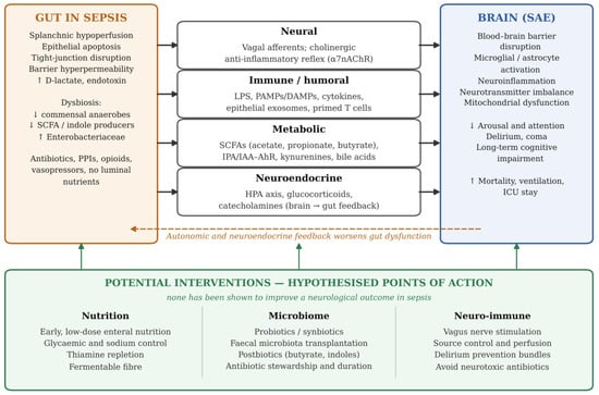
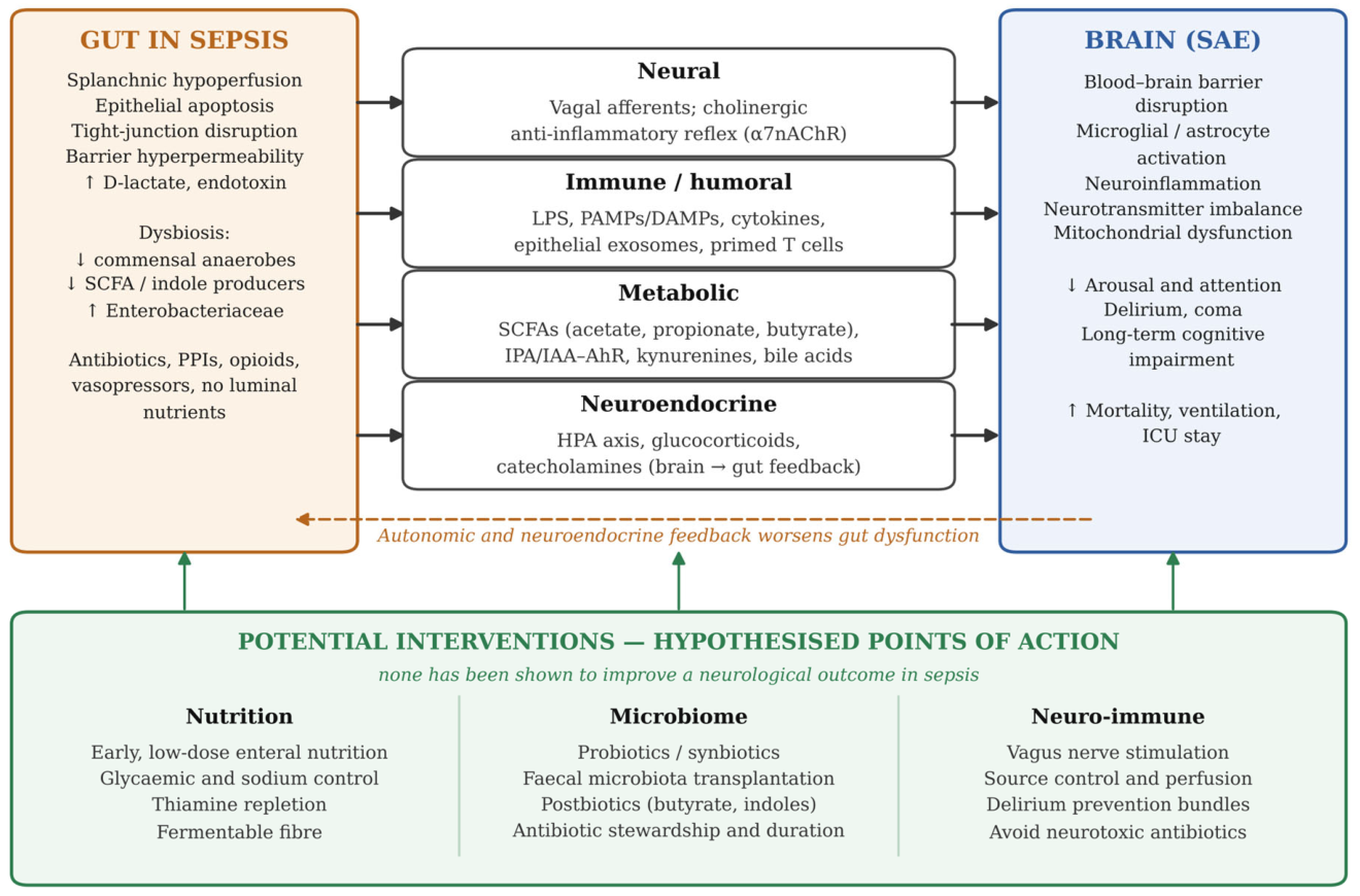

Disclaimer/Publisher’s Note: The statements, opinions and data contained in all publications are solely those of the individual author(s) and contributor(s) and not of MDPI and/or the editor(s). MDPI and/or the editor(s) disclaim responsibility for any injury to people or property resulting from any ideas, methods, instructions or products referred to in the content. |
© 2026 by the authors. Licensee MDPI, Basel, Switzerland. This article is an open access article distributed under the terms and conditions of the Creative Commons Attribution (CC BY) license.