


Abstract
Postmenopausal estrogen deficiency induces complex physiological alterations, notably osteoporosis and metabolic dysfunction. This study investigated the effect of High Active Calcium (HAC) using in vitro and ovariectomized (OVX) rat models. In vitro, HAC significantly inhibited RANKL-induced osteoclastogenesis in RAW264.7 cells. In vivo, HAC administration improved bone mineral density (BMD) and trabecular microarchitecture while normalizing serum calcium and reducing bone resorption markers. Qualitative H&E assessment showed differences in bone morphology among groups. HAC treatment attenuated the OVX-induced elevation in serum ALP activity and reduced the Glu-OC/Gla-OC ratio, indicating changes in bone turnover-related markers. HAC partially restored uterine estrogen receptor-alpha (ERα) expression and markedly downregulated hepatic inflammatory cytokines (TNF-α, IL-6, IL-1β). Overall, HAC treatment was associated with improvements in bone remodeling-related markers and reduced hepatic inflammatory cytokine expression in OVX rats. These findings support further preclinical evaluation of HAC as a calcium-based nutritional supplement for estrogen deficiency-associated bone loss.
1. Introduction
Postmenopausal women are susceptible to osteoporosis, which is characterized by a reduction in bone density and an increased risk of fractures. This is attributable to postmenopausal estrogen deficiency, a primary cause of osteoporosis [1]. Estrogen plays a pivotal role in preserving bone mineral density (BMD) by maintaining the balance between bone formation and resorption. It has been reported that one in three women will experience a fragility fracture due to osteoporosis [2]. Such osteoporosis fractures frequently require surgical intervention, hospitalization, and prolonged rehabilitation, thereby imposing a substantial socioeconomic burden on affected individuals and healthcare systems [3].
Following menopause, estrogen deficiency disrupts the balance between receptor activator of nuclear factor-κB ligand (RANKL) and osteoprotegerin (OPG), promoting osteoclast differentiation and survival, while impairing osteoblast differentiation and activity [4,5]. This imbalance leads to excessive bone resorption, progressive loss of bone mineral density, deterioration of trabecular microarchitecture, and cortical bone thinning [6,7,8]. Consequently, postmenopausal osteoporosis markedly increases fracture risk at mechanically vulnerable sites such as the lumbar spine and hip [9]. These fractures are frequently associated with chronic pain, functional impairment, and reduced mobility, ultimately leading to decreased quality of life and increased mortality among affected patients [10].
Calcium supplementation is widely used as a standard strategy for the management of osteoporosis in postmenopausal women. Previous studies have demonstrated its beneficial effects in mitigating bone loss and reducing fracture risk, particularly in individuals with inadequate dietary calcium intake [11,12]. Bone strength is derived from a composite structure in which hydroxyapatite mineralization reinforces the type I collagen matrix, thereby contributing to bone density and structural integrity [13,14]. However, the clinical efficacy of calcium supplementation varies across populations and is influenced by formulation-dependent factors such as dosage and bioavailability [15,16]. Calcium absorption and tolerability differ substantially according to the chemical form of the supplement. Calcium carbonate, despite its high elemental calcium content [17], is highly dependent on gastric acid for absorption and is frequently associated with gastrointestinal side effects, including abdominal distension and constipation, which may reduce tolerability and adherence [18,19,20]. In contrast, calcium citrate exhibits greater aqueous solubility, lower dependence on gastric acidity, and more consistent bioavailability and is generally better tolerated [21]. Nevertheless, its lower elemental calcium content compared with calcium carbonate often necessitates a larger tablet size or higher dosing frequency, potentially increasing cost and reducing convenience [22,23]. Therefore, the development and evaluation of alternative calcium formulations remain important for improving calcium supplementation strategies.
In ovariectomized (OVX) rats, estrogen deficiency induces an imbalance in bone remodeling characterized by enhanced osteoclast-mediated bone resorption and progressive trabecular bone loss, thereby recapitulating key features of postmenopausal osteoporosis [24,25]. Accordingly, the OVX rat model has been widely employed as a well-established and reliable preclinical model for investigating postmenopausal bone loss and evaluating therapeutic interventions. In this study, we evaluated the effects of High Active Calcium (HAC) on bone loss and bone remodeling in OVX rats. In addition, serum lipid profiles, hepatic inflammatory markers, and uterine ERα expression were assessed to characterize broader changes associated with estrogen deficiency and HAC treatment.
2. Materials and Methods
2.1. Sample Preparation
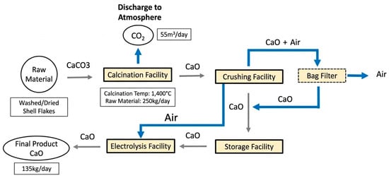
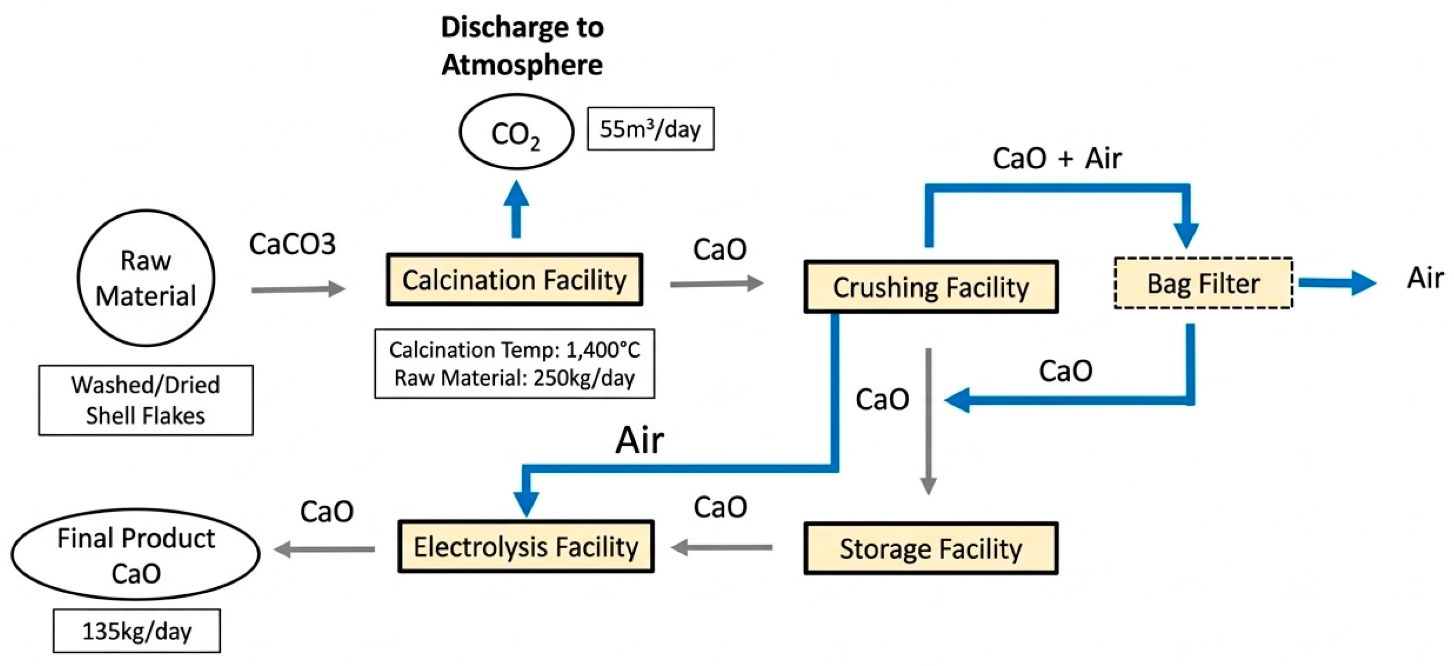
High Active Calcium (HAC) is the manufacturer-designated product name for a cockle-shell-derived, CaO-based powder supplied by Eco-Biotech Co., Ltd. (Hwaseong, Republic of Korea). According to the manufacturer, the shells were obtained from Tegillarca granosa collected in Yeoja Bay, Korea. They were high-pressure washed for 45–60 min using 0.5 mm abrasive particles, manually sorted, and dried. The dried shells were calcined at approximately 1400 °C to produce CaO, which was subsequently crushed and processed by high-voltage electrolysis (≥30 kV) to produce HAC (Figure 1). The manufacturer reports a CaO purity of ≥98%. The term “High Active Calcium” is used solely as the product name; superior calcium absorption or bioavailability was not established in the present study.
Figure 1.
Manufacturer-provided process flow diagram for the production of HAC, a cockle-shell-derived CaO-based product.
2.2. Cell Culture and Treatment
Mouse macrophage RAW 264.7 cells were purchased from the Korean Cell Line Bank (Seoul, Republic of Korea) and cultured in Dulbecco’s Modified Eagle Medium (DMEM; Welgene, Daegu, Republic of Korea) supplemented with 10% fetal bovine serum (FBS; Welgene, Republic of Korea), and 100 U/mL penicillin, 100 μg/mL streptomycin (Welgene, Korea) at 37 °C, 5% CO2 incubation. Cells were seeded in 96-well plates at 1 × 10⁴ cells/well. After 24 h, osteoclast differentiation was initiated by replacing the medium with DMEM containing RANKL (50 ng/mL) and HAC at the indicated concentrations, and cultures were maintained for 7 days with medium changes every 2–3 days. HAC concentrations used in the in vitro experiments are expressed as elemental calcium concentration (µM).
2.3. Cell Viability Assay
Cell viability was determined using a 3-(4,5-dimethylthiazol-2-yl)-2,5-diphenyltetrazolium bromide (MTT) assay kit (Sigma-Aldrich, St. Louis, MO, USA) in RAW264.7 cultures under undifferentiated and differentiated conditions. HAC was added for the final 24 h of the differentiation period, while undifferentiated RAW264.7 cells were treated with HAC for 24 h. MTT was added to each well, and the plate was incubated for 2 h at 37 °C in 5% CO2. The medium was removed, formazan crystals were dissolved in 100 µL DMSO, and absorbance at 570 nm was measured (BioTek Instruments, Winooski, VT, USA).
2.4. Alkaline Phosphatase Activity
Alkaline phosphatase (ALP) activity was determined using a TRACP & ALP Assay Kit (MK301, TaKaRa Bio Inc., Kusatsu, Shiga, Japan) following the manufacturer’s protocol. Cells were washed once with Dulbecco’s phosphate-buffered saline (DPBS), then 50 µL of extraction solution and 50 µL of substrate solution were added per well and incubated for 60 min at 37 °C. Reactions were stopped by adding 50 µL of stop solution, and absorbance was measured at 405 nm using a microplate reader (BioTek Instruments, Winooski, VT, USA).
2.5. Animal Experiments
Female Sprague–Dawley rats (n = 40, 6 weeks old) were obtained from Damool (Daejeon, Republic of Korea). Animals were housed under a 12 h light/dark cycle at 22–24 °C and 50–60% relative humidity. After a one-week acclimatization period, the rats were randomly allocated to five experimental groups (n = 8 per group), with body weight used to ensure comparable baseline distributions. No formal allocation concealment procedure was used. Ovariectomy (OVX) was performed according to a practical guide [26] under isoflurane anesthesia via a ventral abdominal incision. Animals were allowed to recover for one week before the experimental procedures.
- Group 1: Sham control group (Sham): rats were sham-operated; one week later, they were administered water once daily by oral gavage for 8 weeks.
- Group 2: OVX control group (OVX): rats were OVX-operated; 1 week later, they were administered water by oral gavage once daily for 8 weeks.
- Group 3: HAC low-dose group (HAC-L): rats were OVX-operated; one week later, they were administered HAC (20 mg/kg) by oral gavage for 8 weeks.
- Group 4: HAC medium-dose group (HAC-M): rats were OVX-operated; 1 week later, they were administered HAC (40 mg/kg) by oral gavage for 8 weeks.
- Group 5: HAC high-dose group (HAC-H): rats were OVX-operated; one week later, they were administered HAC (80 mg/kg) by oral gavage for 8 weeks.
All procedures were approved by the Institutional Animal Care and Use Committee (IACUC) of Jeonbuk National University (approval no. 2023-043). All animal care and experimental procedures were conducted in accordance with institutional guidelines and internationally accepted standards for the ethical treatment of laboratory animals. The administered does refer to the mass of HAC product. Assuming 98 wt% CaO based on the manufacturer-reported purity, the doses of 20, 40, and 80 mg/kg correspond to theoretical elemental calcium doses of approximately 14, 28, and 56 mg/kg, respectively. These estimates were not confirmed by direct elemental analysis.
2.6. Serum Biochemical Analysis
The blood was collected and centrifuged at 2000 rpm for 15 min at 4 °C. The serum samples were then collected and stored at −80 °C until used. ALT, AST, and calcium levels were measured using the VetTest 8008 (IDEXX Laboratories Inc., Westbrook, Cumberland County, ME, USA) with the VetTest serum biochemistry single chip. Levels of Gla-OC, Glu-OC, ALP activity (Takara, Tokyo, Japan), CTX1, NTX1 (MyBioSource, Inc., San Diego, CA, USA), LDL, HDL, TC, and TG (BIOMAX, Guri, Republic of Korea) in rat serum were measured using ELISA kits according to the manufacturer’s protocols.
2.7. Micro-Computed Tomography Analysis
The rats’ left femurs were dissected, fixed in 10% formalin, and stored at 4 °C until analysis. To evaluate the effects of the treatment on bone structure, the femurs were scanned using a micro-computed tomography (μ-CT) system (SkyScan 1076, Bruker Corporation, Billerica, MA, USA) at the Center for University-wide Research Facilities (CURF), Jeonbuk National University (Jeonju, Republic of Korea). For µ-CT analysis, femoral samples from three animals per group were randomly selected for μ-CT analysis (n = 3/group). High-resolution μ-CT imaging was used to analyze the trabecular bone microarchitecture, focusing on bone volume and density. Bone mineral density (BMD) was quantified using calibration phantoms with predefined Hounsfield unit (HU) values, corresponding to low (1067.1 HU; 0.25 g/cm3) and high (3249.3 HU; 0.75 g/cm3) density standards. Image reconstruction and data analysis were performed using the CTAn software (version 1.20.3.0, Bruker Corporation, Billerica, MA, USA).
2.8. Histopathological Analysis
2.8.1. Decalcification
The rats’ right femurs were dissected, fixed in 10% neutral buffer formalin (NBF) for 3 days, and stored at 4 °C until analysis. Following the procedure described by Liu et al. [27], the femurs were rinsed under running tap water for 24 h to remove residual fixative. Decalcification was carried out using 10% neutral buffer EDTA (pH 7.4) at 4 °C with continuous shaking to ensure uniform decalcification. The decalcifying solution was refreshed every 48 h. Decalcification was complete when needle penetration was no longer resisted.
2.8.2. Tissue Processing and Sectioning
After decalcification, the femurs were rinsed in running tap water for 24 h, processed for paraffin embedding, and then longitudinal sections of 4 μm were cut using a microtome and mounted on poly-L-lysine-coated slides for histological analysis.
2.8.3. Hematoxylin and Eosin (H&E) Staining
Paraffin-embedded femur sections (4 μm thickness) were deparaffinized in xylene and rehydrated through a graded ethanol series (100–70%). After rehydration, the sections were stained with hematoxylin for 5 min and then rinsed in running tap water. Differentiation was performed by dipping the sections in 1% acid ethanol (1% hydrochloric acid in 70% ethanol) five times, followed by a thorough rinse in distilled water to remove excess stain. The sections were then counterstained with eosin for 3 min, dehydrated through graded ethanol, cleared in xylene, and mounted with a synthetic resin mounting medium. Stained sections were observed under a light microscope to evaluate the trabecular bone morphology and structural changes. Osteoblasts and osteoclasts were identified morphologically based on their characteristic cellular appearance and location along the bone surface.
2.9. Western Blot Analysis
Proteins were extracted from liver and uterus tissues using RIPA buffer supplemented with a protease and phosphatase inhibitor cocktail (P3300, GenDEPOT, Barker, TX, USA). The protein concentrations were determined using a bicinchoninic acid (BCA) protein assay kit (Thermo Fisher Scientific, Waltham, MA, USA). Equal amounts of protein samples were separated on SDS-PAGE gels and transferred onto polyvinylidene difluoride (PVDF) membranes. The membranes were blocked with 5% bovine serum albumin (BSA) or 5% non-fat milk in Tris-buffered saline with Tween 20 (TBS-T) for 1 h at room temperature to prevent nonspecific binding. The membranes were then incubated overnight at 4 °C with anti-Estrogen receptor alpha (ab32063, Abcam, Cambridge, UK, 1:1000 in 5% BSA). After incubation, the membranes were washed in TBS-T and then incubated with secondary antibodies such as goat anti-rabbit IgG or goat anti-mouse IgG at a 1:2000 dilution for 2 h at room temperature. Protein bands were visualized using an enhanced chemiluminescence (ECL) solution (BIOFACT, Daejeon, Republic of Korea) and detected using a Chemi-imaging system (Alpha Innotech, San Leandro, CA, USA). Band intensities were quantified using ImageJ software version 1.8.0, and all results were normalized to β-actin.
2.10. Quantitative Reverse Transcription Polymerase Chain Reaction (RT-qPCR)
Total RNA was extracted from RAW 264.7 cells using TRIzol reagent (Invitrogen, Carlsbad, CA, USA), and complementary DNA (cDNA) was synthesized using a PrimeScript™ RT reagent Kit with gDNA Eraser (TaKaRa, Kyoto, Japan). Quantitative PCR was performed using BioFACT™ Real-Time PCR Series (With SYBR® Green I) (BioFACT, Daejeon, Republic of Korea), and gene-specific primers are presented in Table 1. The relative expression levels were determined by the 2−∆∆Ct method with GAPDH as the internal control.
Table 1.
Primer sequences of RT-qPCR.
2.11. Statistical Analysis
All data were analyzed using GraphPad Prism 9 software (GraphPad Software, San Diego, CA, USA). No formal blinding procedures were used during outcome assessment or statistical analysis. Statistical comparisons between experimental groups were performed using a one-way analysis of variance (ANOVA) followed by Tukey’s post hoc test to determine significant differences among groups. No animals were excluded, and data points were omitted only in cases of technical failure. Data are expressed as the mean ± SEM, and statistical significance was considered at p < 0.05.
3. Results
3.1. HAC Suppresses RANKL-Induced Osteoclast Differentiation in RAW264.7 Cells
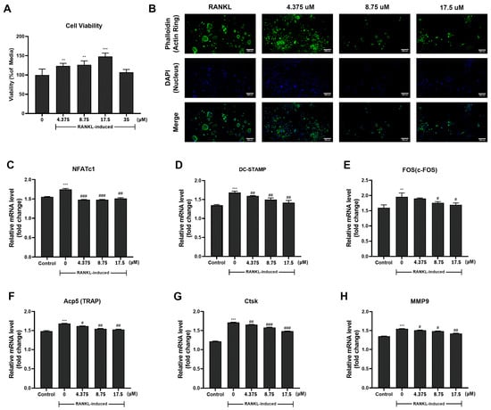
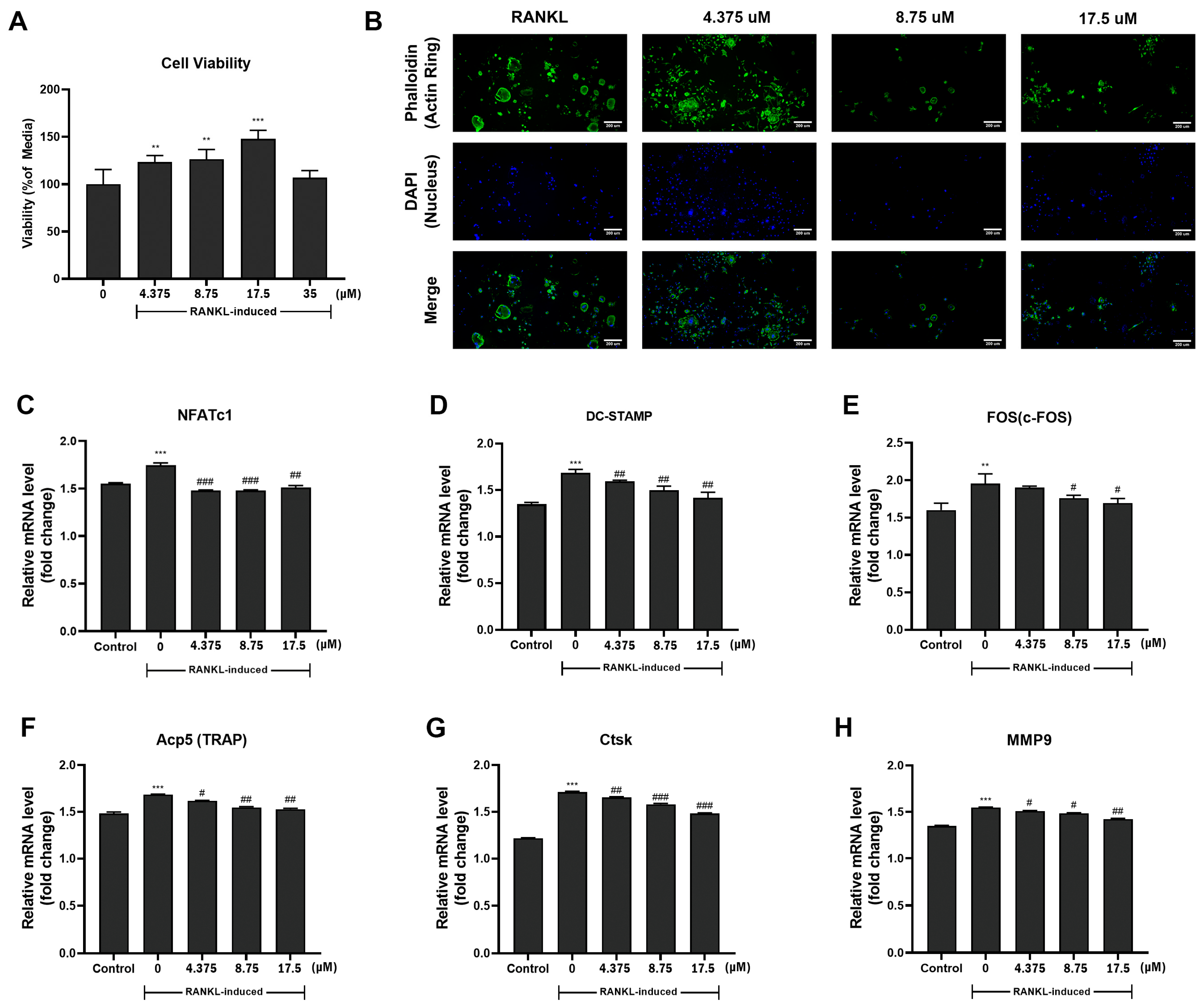
The MTT signal was highest at 17.5 µM and was not reduced at 35 µM (Figure 2A). Therefore, 17.5 µM was not considered the maximum non-cytotoxic concentration. The MTT assay was used to assess whether the tested concentrations reduced the signal, while the effects on osteoclastogenesis were evaluated separately using F-actin ring formation and osteoclast-related gene expression. Phalloidin staining revealed that RANKL treatment induced the formation of multinucleated osteoclasts with well-defined peripheral actin rings (Figure 2B). HAC treatment suppressed actin ring formation in a dose-dependent manner. At 17.5 μM, the formation of large multinucleated cells was markedly reduced, and actin ring structures were largely disrupted, with most cells remaining mononuclear.
Figure 2.
Effects of HAC on osteoclastogenesis and osteoclast-related gene expression in RANKL-induced RAW264.7 cells. (A) Cell viability following HAC treatment. (B) Representative fluorescence images of F-actin ring formation (phalloidin, green) and nuclei (DAPI, blue), with merged images. Scale bar = 200 μm. (C–H) Relative mRNA expression levels of osteoclastogenesis-related markers, including NFATc1, DC-STAMP, c-FOS, Acp5 (TRAP), Ctsk, and MMP9. Data are expressed as mean ± SEM. All in vitro experiments were performed in three independent experiments (n = 3). HAC concentrations are expressed as elemental calcium concentrations. Statistical analysis was performed using one-way ANOVA followed by appropriate Tukey post hoc tests. ** p < 0.01 compared to the Control, *** p < 0.001 compared to the Control. # p < 0.05 compared to the 0, ## p < 0.01 compared to the 0, ### p < 0.001 compared to the 0.
To further evaluate the molecular effects of HAC on osteoclast differentiation, the mRNA expression levels of osteoclast-related genes were analyzed by qRT-PCR (Figure 2C–H). RANKL stimulation significantly increased the expression of NFATc1, c-FOS, DC-STAMP, Acp5 (TRAP), Ctsk, and MMP9 compared with the control group. HAC treatment significantly attenuated the RANKL-induced upregulation of these genes. These findings indicate that HAC suppresses RANKL-induced changes in osteoclast differentiation in RAW264.7 cells.
3.2. Effects of HAC Treatment on Weight Gain and Food Intake in OVX Rats
The initial body weight of all experimental groups was 183.58 ± 0.48 g, with no significant differences among groups (Table 2). Then, 8 weeks after OVX, body weight differed significantly between Sham (267.8 ± 6.8 g) and OVX (338.0 ± 13.0 g). HAC treatment at all three doses did not prevent substantial body weight gain. Over the 8 weeks, body-weight gain was greater in OVX (154.2 ± 6.7 g) than in Sham (84.7 ± 4.6 g). Although not statistically significant, the HAC-H group showed a lower gain (144.2 ± 9.5 g) than the OVX group. A similar pattern was observed for food intake.
Table 2.
Effects of HAC treatment on body composition and food efficiency in ovariectomized (OVX) rats over 8 weeks.
3.3. Effects of HAC on Liver Injury and Serum Lipid Profiles in OVX Rats
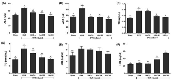
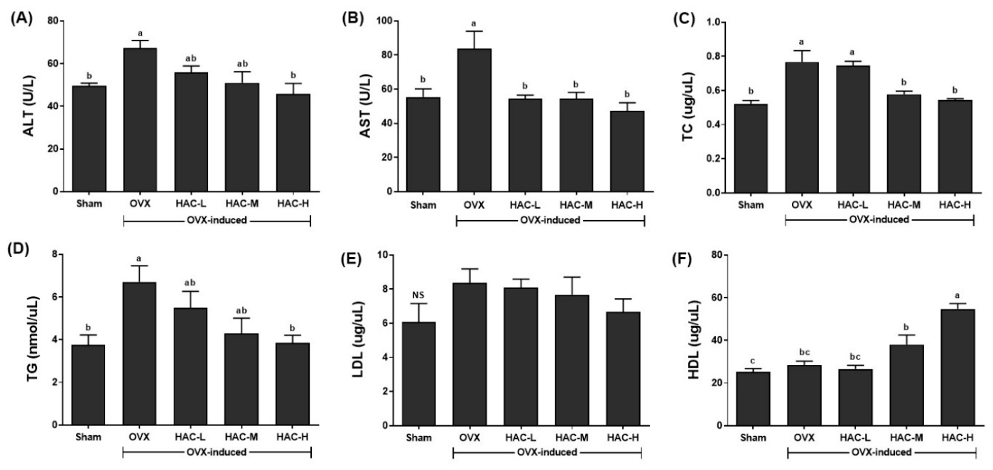
To evaluate the effects of OVX and HAC on liver injury and the serum lipid profile, serum ALT, AST, TC, TG, LDL, and HDL were measured in serum from HAC-treated rats (Figure 3). Serum ALT and AST levels were significantly increased in the OVX group compared with the Sham group (Figure 3A,B). These elevations were dose-dependently attenuated by HAC treatment, with a reduction observed in the HAC-H group. In addition, serum TC and TG levels were elevated in the OVX group compared with the Sham group (Figure 3C,D). HAC treatment reduced TC levels in the HAC-M and HAC-H groups, whereas TG levels significantly decreased only in the HAC-H group. No significant differences in serum LDL levels were observed among the groups (Figure 3E). In contrast, HDL levels were higher in the HAC-H group than in the OVX group (Figure 3F).
Figure 3.
Effects of HAC on serum lipid profile and liver injury markers in OVX rats. (A) ALT; (B) AST; (C) TC; (D) TG; (E) LDL; (F) HDL. All values are expressed as the mean ± SEM (n = 8). a–c Different letters in a column mean significant differences among groups, TUKEY’s multiple range test (p < 0.05). NS denotes no significant difference among the groups.
3.4. HAC Modulates Osteoclastogenesis-Related Markers and Bone Resorption in OVX Rats
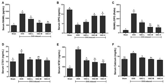
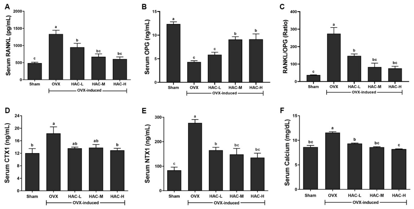
Serum RANKL levels were significantly increased, whereas OPG levels were decreased in the OVX group compared with the Sham group (Figure 4A,B). As a result, the RANKL/OPG ratio was markedly elevated in the OVX group (Figure 4C), reflecting a shift toward increased osteoclastogenesis and bone resorption. HAC treatment dose-dependently reversed these changes.
Figure 4.
Effects of HAC on bone turnover markers and the RANKL/OPG axis in OVX rats. (A) Serum RANKL levels. (B) Serum OPG levels. (C) RANKL/OPG ratio. (D) Serum C-terminal telopeptide of type I collagen (CTX1) levels. (E) Serum N-terminal telopeptide of type I collagen (NTX1) levels. (F) Serum calcium levels. Data are expressed as mean ± SEM (n = 8). Statistical analysis was performed using one-way ANOVA followed by appropriate post hoc tests. a–c Different letters indicate statistically significant differences among groups (p < 0.05).
Serum CTX1 and NTX1 levels were significantly elevated in the OVX group compared with the Sham group (Figure 4D,E), indicating increased osteoclast-mediated bone resorption. Compared with the OVX group, serum CTX1 levels were significant lower only in the HAC-H groups, whereas NTX1 levels were significantly lower in all HAC-treated groups. Although NTX1 showed a numerical downward trend with increasing HAC dose, no significant differences were observed among the HAC-treated groups. Serum calcium levels were also elevated in the OVX group (Figure 4F), reflecting increased bone turnover, and were reduced following HAC treatment.
3.5. HAC Improves Trabecular Microarchitecture in OVX Rats
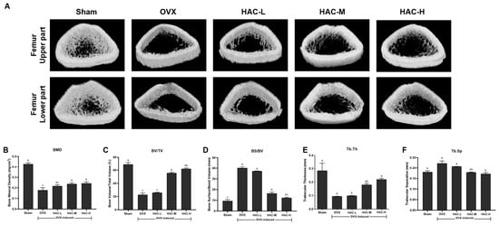
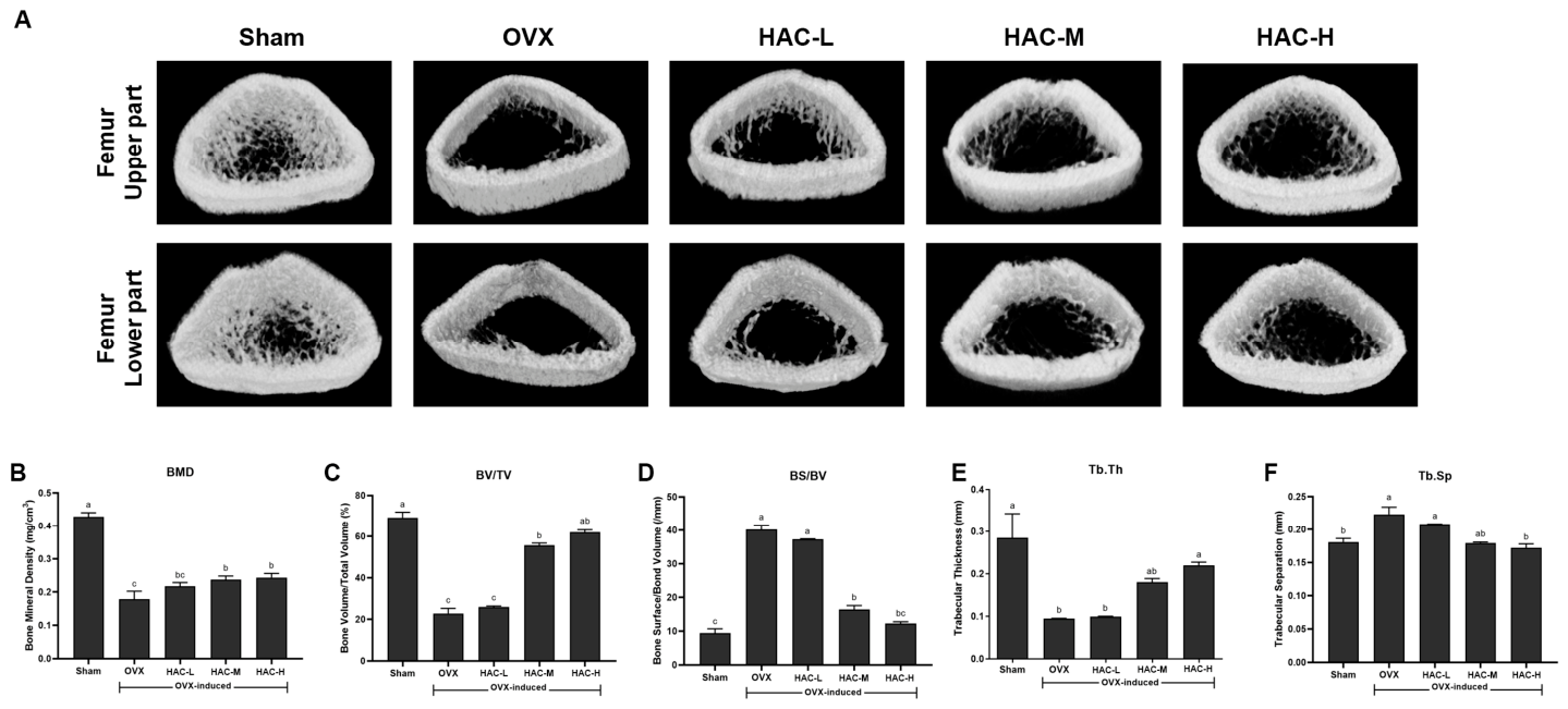
Representative micro-CT images demonstrated severe trabecular bone loss and increased porosity in the OVX group compared with the Sham group (Figure 5A). Quantitative analysis showed that OVX significantly decreased BMD, BV/TV, and Tb.Th and increased BS/BV and Tb.Sp compared with the Sham group (Figure 5B–F). Compared with the OVX group, HAC-M and HAC-H significantly increased BMD and BV/TV and reduced BS/BV and Tb.Sp. Tb.Th was significantly increased only in the HAC-H group. HAC-L did not differ significantly from the OVX group in these micro-CT parameters. Although the group means showed generally favorable trends with increasing HAC dose, significant dose-dependent differences among HAC-treated groups were not observed.
Figure 5.
Effects of HAC on trabecular bone microarchitecture in OVX rats as assessed via micro-CT. (A) Representative micro-CT images of femoral trabecular bone (upper and lower regions). (B) Bone mineral density (BMD). (C) Bone volume/total volume (BV/TV). (D) Bone surface/bone volume (BS/BV). (E) Trabecular thickness (Tb.Th). (F) Trabecular separation (Tb.Sp). Data are presented as mean ± SEM (n = 3). Statistical significance was determined using one-way ANOVA followed by post hoc multiple-comparison tests. a–c Different letters indicate significant differences among groups (p < 0.05).
3.6. HAC Modulates Bone Formation-Related Markers and Bone Remodeling in OVX Rats
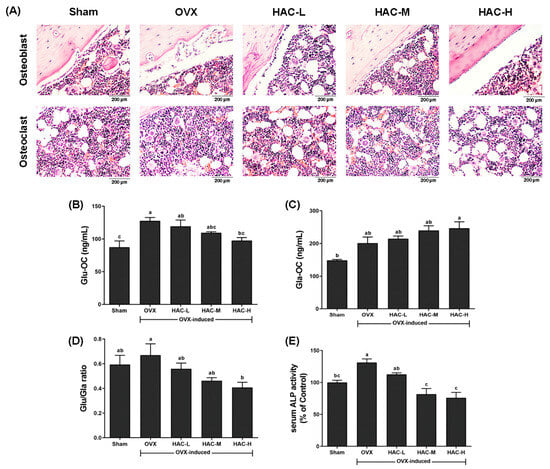
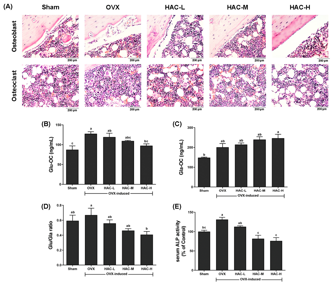
To evaluate bone remodeling, H&E-stained sections of rat femora were analyzed (Figure 6A). Qualitative examination of H&E-stained femoral sections showed differences in bone morphology between the Sham and OVX groups. HAC-treated sections showed visually apparent preservation of bone morphology, particularly in the HAC-H group. Because osteoblasts and osteoclasts were not assessed using cell-specific staining or quantitative histomorphometry, these observations are presented as qualitative histological findings and do not establish changes in bone-cell numbers. Consistent with the histological findings, serum osteocalcin (OC) and ALP levels exhibited similar trends (Figure 6B–E). Compared with the OVX group, Serum Glu-OC levels were significantly lower only in the HAC-H group (Figure 6B). Serum Gla-OC levels were numerically higher in the HAC-H group than in the OVX group, although this difference was not statistically significant (Figure 6C) The Glu-OC/Gla-OC ratio was significantly lower in the HAC-H group than in the OVX group (Figure 6D). Furthermore, the OVX-induced elevation in serum ALP activity was dose-dependently attenuated by HAC treatment, with significant reductions observed in both the HAC-M and HAC-H groups (Figure 6E).
Figure 6.
Effects of HAC on bone histology and bone formation-related markers in OVX rats. (A) Representative H&E-stained femoral sections showing qualitative bone morphology. Scale bar = 200 μm. (B) Serum undercarboxylated osteocalcin (Glu-OC) levels. (C) Serum carboxylated osteocalcin (Gla-OC) levels. (D) Ratio of Glu-OC to Gla-OC. (E) Serum alkaline phosphatase (ALP) activity. Data are presented as mean ± SEM (n = 8). Statistical significance was determined using one-way ANOVA followed by post hoc multiple-comparison tests. a–c Different letters indicate significant differences among groups (p < 0.05).
3.7. Effects of HAC on Uterine ERα Expression and Hepatic Inflammatory Markers in OVX Rats
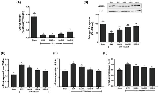
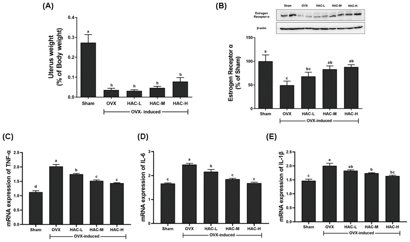
Uterine weight was significantly lower in the OVX group than in the Sham group, confirming the effect of ovariectomy (Figure 7A). HAC treatment did not significantly restore uterine weight compared with the OVX group. Uterine ERα protein expression was lower in the OVX group than in the Sham group, whereas it was significantly higher in the HAC-M and HAC-H groups than in the OVX group (Figure 7B). Hepatic TNF-α and IL-6 mRNA levels were lower in all HAC-treated groups than in the OVX group, while IL-1β mRNA levels were significantly lower only in the HAC-M and HAC-H groups (Figure 7C–E).
Figure 7.
Effects of HAC on uterine atrophy and inflammatory cytokine expression in OVX rats. (A) Uterus weight expressed as a percentage of body weight. (B) Protein expression of estrogen receptor alpha (ER-α) in uterine tissue, with representative Western blot images and corresponding quantification. (C–E) Relative mRNA expression levels in liver tissues of TNF-α (C), IL-6 (D), and IL-1β (E). Data are expressed as mean ± SEM (n = 8). Statistical analysis was performed using one-way ANOVA followed by Tukey’s post hoc tests. a–d Different letters indicate statistically significant differences among groups (p < 0.05).
4. Discussion
Postmenopausal bone loss is characterized by high bone turnover and deterioration of trabecular microarchitecture, which together reduce bone strength and increase the risk of fragility fractures [26,27]. Although calcium supplementation is widely used as a preventive strategy, increasing evidence suggests that its efficacy may vary by formulation and bioavailability [28,29,30]. However, many previous studies have focused primarily on changes in areal BMD or single biochemical markers, with limited evidence of concurrent improvements in bone microstructure and cellular functional parameters of osteoblasts and osteoclasts. Therefore, in the present study, we comprehensively evaluated the structural and functional effects of high active calcium (HAC) in an ovariectomized rat model, integrating μ-CT-based trabecular analysis, serum bone turnover markers, and in vitro osteoclastogenic assays.
Successful establishment of the OVX model was confirmed by a reduction in uterine weight and a significant elevation in bone resorption markers, including CTX-1 and NTX-1 [26,28]. Since CTX is widely recognized as a reliable marker of bone resorption due to its low diurnal variability and analytical precision [29], the observed increases in these markers in our study clearly validate the successful induction of estrogen deficiency-associated bone loss. Importantly, eight weeks of HAC administration significantly improved BMD and restored trabecular microarchitecture, as demonstrated by μ-CT analysis. The concomitant reduction in resorption markers suggests that the structural recovery was associated with suppression of excessive bone turnover. Because postmenopausal osteoporosis is characterized by an imbalance favoring bone resorption over formation, attenuation of bone resorption by HAC indicates a partial restoration of bone remodeling balance in OVX-induced osteoporosis.
Osteocalcin (OC), the most abundant non-collagenous protein in bone, serves as a crucial biomarker for osteoblast activity and matrix mineralization due to its high calcium-binding affinity [30]. At the cellular level, osteoblasts drive bone formation by regulating ALP activity, promoting OC expression, and facilitating matrix mineralization [31]. In the present study, HAC treatment modulated serum ALP activity and osteocalcin-related markers, consistent with changes in bone remodeling. Serum Glu-OC levels and the Glu-OC/Gla-OC ratio were significantly lower in the HAC-H group than in the OVX group. Although serum Gla-OC levels were numerically higher in the HAC-H group, this difference was not statistically significant. These findings suggest changes in osteocalcin-related markers but do not directly establish enhanced bone formation or matrix mineralization.
Interestingly, our study demonstrated that HAC administration decreased serum calcium levels compared to the OVX control group. While this may seem counterintuitive for a calcium supplement, it aligns perfectly with the physiological mechanisms underlying postmenopausal bone loss [32]. Estrogen deficiency typically accelerates osteoclast-mediated bone resorption, leading to an abnormal efflux of calcium from the bone matrix into the bloodstream, which often results in elevated serum calcium levels [33]. In the present study, HAC treatment reduced serum calcium levels compared with the OVX group. This change occurred together with reductions in bone resorption markers, suggesting that the decrease in serum calcium may be associated with attenuation of excessive bone resorption. The accompanying changes in serum ALP activity and the Glu-OC/Gla-OC ratio were consistent with changes in bone turnover-related markers following HAC treatment. Therefore, the decrease in serum calcium observed following HAC treatment may be associated with changes in bone remodeling, although the underlying mechanisms were not directly assessed.
Beyond its direct effects on bone remodeling, estrogen is a critical hormone that regulates body composition and lipid metabolism in females, and its levels decline significantly after menopause [4,34]. Consequently, OVX rats commonly exhibit metabolic alterations, including changes in body composition and lipid profiles, that closely mimic the postmenopausal state [35]. Estrogen exerts its effects primarily through three distinct receptors: ERα, ERβ, and GPER1. Among these, ERα is specifically known to regulate cellular metabolism via the AMPK signaling pathway [36,37]. Beyond its metabolic roles, ERα signaling is closely associated with the modulation of inflammatory responses. Indeed, previous studies have reported that ERα knockout significantly alters the expression of hepatic inflammatory cytokine genes [38]. In the present study, HAC administration in OVX rats upregulated uterine ERα expression while concurrently downregulating hepatic inflammatory genes. These findings suggest a possible association between increased uterine ERα expression and the regulation of hepatic inflammatory gene expression following HAC treatment.
Estrogen plays a crucial role in metabolic processes, and its decline significantly impairs the regulatory functions of adipose tissue. Numerous studies have reported that the reduction in estrogen levels following menopause is closely associated with elevated levels of total cholesterol (TC), triglycerides (TG), and low-density lipoprotein (LDL) [39]. Furthermore, estrogen deficiency exacerbates inflammation and oxidative stress, which, in turn, impair high-density lipoprotein (HDL) metabolism [40]. Collectively, these changes indicate that postmenopausal estrogen loss induces dyslipidemia. In our study, HAC administration effectively ameliorated OVX-induced dyslipidemia. Additionally, HAC treatment attenuated the OVX-induced increases in serum AST and ALT levels, consistent with improved liver injury markers.
The present study demonstrated the beneficial effects of HAC on OVX-induced bone loss and related metabolic alterations. However, the absence of a conventional calcium comparator limits our ability to determine whether the observed effects reflect properties unique to HAC beyond those associated with calcium supplementation itself. In addition, the lack of an established positive-control treatment, such as estradiol or an anti-osteoporotic agent, limits direct comparison with known therapeutic responses. Detailed analytical characterization of the administered product was unavailable, including measured elemental calcium content, particle size, quantitative solubility, impurities and heavy metals, stability, and batch-to-batch consistency. Dietary calcium intake was not quantified, preventing comparison of the supplemental doses with background dietary calcium intake.
5. Conclusions
HAC treatment improved bone microarchitecture and altered serum bone turnover markers in OVX rats. These changes were accompanied by increased uterine ERα expression and improvements in selected lipid and hepatic inflammatory markers. Further preclinical evaluation is required to establish the comparative efficacy, bioavailability, safety, and underlying mechanisms of HAC.
Author Contributions
J.C. Data curation, formal analysis, investigation, methodology, validation, and writing—original draft. H.C. Validation, formal analysis, and writing—original draft. J.K. (Jajun Koo) Conceptualization, resources, funding acquisition, and writing—review and editing. J.K. (Jungkee Kwon) led conceptualization, project administration, supervision, and writing—review and editing. All authors have read and agreed to the published version of the manuscript.
Funding
This work was supported by the Technology Development Program (RS-2024-00487194) funded by the Ministry of SMEs and Startups (MSS, Republic of Korea).
Institutional Review Board Statement
All animal experiments were approved by the Institutional Animal Care and Use Committee of Jeonbuk National University (Approval No. 2023-043, on 24 April 2023) and were conducted in accordance with the guidelines for the care and use of laboratory animals established by the National Institutes of Health (NIH, USA).
Informed Consent Statement
Not applicable.
Data Availability Statement
The data presented in this study are available on request from the corresponding author. The data are not publicly available due to ethical restrictions.
Conflicts of Interest
Jajun Koo is an executive of Eco-Biotech Co., Ltd., which manufactures the High Active Calcium (HAC) formulation evaluated in this study. This relationship represents a potential competing interest. The remaining authors declare no conflicts of interest.
References
- Walker, M.D.; Shane, E. Postmenopausal Osteoporosis. N. Engl. J. Med. 2023, 389, 1979–1991. [Google Scholar] [CrossRef] [Scilit] [PubMed]
- Söreskog, E.; Ström, O.; Spångéus, A.; Åkesson, K.E.; Borgström, F.; Banefelt, J.; Toth, E.; Libanati, C.; Charokopou, M. Risk of major osteoporotic fracture after first, second and third fracture in Swedish women aged 50 years and older. Bone 2020, 134, 115286. [Google Scholar] [CrossRef] [Scilit] [PubMed]
- Lorentzon, M.; Johansson, H.; Harvey, N.C.; Liu, E.; Vandenput, L.; McCloskey, E.V.; Kanis, J.A. Osteoporosis and fractures in women: The burden of disease. Climacteric 2022, 25, 4–10. [Google Scholar] [CrossRef] [Scilit] [PubMed]
- Khosla, S.; Oursler, M.J.; Monroe, D.G. Estrogen and the skeleton. Trends Endocrinol. Metab. 2012, 23, 576–581. [Google Scholar] [CrossRef] [Scilit] [PubMed]
- Chi, G.; Qiu, L.; Ma, J.; Wu, W.; Zhang, Y. The association of osteoprotegerin and RANKL with osteoporosis: A systematic review with meta-analysis. J. Orthop. Surg. Res. 2023, 18, 839. [Google Scholar] [CrossRef] [Scilit] [PubMed]
- Liang, Y.; Yuan, X.; Dai, X.; Zhang, G.; Li, C.; Yang, H.; Zhang, T.; Qin, J. The effects of simvastatin on the bone microstructure and mechanics of ovariectomized mice: A micro-CT and micro-finite element analysis study. BMC Musculoskelet. Disord. 2024, 25, 748. [Google Scholar] [CrossRef] [Scilit] [PubMed]
- Geraldi, M.V.; Gregori, G.; Johansson, L.; Hjertonsson, U.; Brättemark, E.; Lorentzon, M. Associations between body composition and bone loss in early postmenopausal women. J. Bone Min. Res. 2026, 41, 251–258. [Google Scholar] [CrossRef] [Scilit] [PubMed]
- Jones, B.C.; Lee, H.; Cheng, C.C.; Al Mukaddam, M.; Song, H.K.; Snyder, P.J.; Kamona, N.; Rajapakse, C.S.; Wehrli, F.W. MRI Quantification of Cortical Bone Porosity, Mineralization, and Morphologic Structure in Postmenopausal Osteoporosis. Radiology 2023, 307, e221810. [Google Scholar] [CrossRef] [Scilit] [PubMed]
- Schini, M.; Lui, L.Y.; Vilaca, T.; Ewing, S.K.; Thompson, A.; Bauer, D.C.; Bouxsein, M.L.; Black, D.M.; Eastell, R. The relationship between baseline bone mineral density and fracture incidence in the placebo groups of randomized controlled trials using individual patient data from the FNIH-ASBMR-SABRE project. J. Bone Min. Res. 2025, 40, 307–314. [Google Scholar] [CrossRef] [Scilit] [PubMed]
- Greig, A.M.; Briggs, A.M.; Bennell, K.L.; Hodges, P.W. Trunk muscle activity is modified in osteoporotic vertebral fracture and thoracic kyphosis with potential consequences for vertebral health. PLoS ONE 2014, 9, e109515. [Google Scholar] [CrossRef] [Scilit] [PubMed]
- Spangler, M.; Phillips, B.B.; Ross, M.B.; Moores, K.G. Calcium supplementation in postmenopausal women to reduce the risk of osteoporotic fractures. Am. J. Health Syst. Pharm. 2011, 68, 309–318. [Google Scholar] [CrossRef] [Scilit] [PubMed]
- Heidari, B.; Hajian-Tilaki, K.; Babaei, M. Effectiveness and safety of routine calcium supplementation in postmenopausal women. A narrative review. Diabetes Metab. Syndr. 2020, 14, 435–442. [Google Scholar] [CrossRef] [Scilit] [PubMed]
- Eastell, R.; Rosen, C.J.; Black, D.M.; Cheung, A.M.; Murad, M.H.; Shoback, D. Pharmacological Management of Osteoporosis in Postmenopausal Women: An Endocrine Society* Clinical Practice Guideline. J. Clin. Endocrinol. Metab. 2019, 104, 1595–1622. [Google Scholar] [CrossRef] [Scilit] [PubMed]
- Stock, S.R. The Mineral-Collagen Interface in Bone. Calcif. Tissue Int. 2015, 97, 262–280. [Google Scholar] [CrossRef] [Scilit] [PubMed]
- Cao, J.J.; Gregoire, B.R. Calcium Deficiency Decreases Bone Mass without Affecting Adiposity in Ovariectomized Rats Fed a High-Fat Diet. Nutrients 2024, 16, 478. [Google Scholar] [CrossRef] [Scilit] [PubMed]
- Tang, B.M.; Eslick, G.D.; Nowson, C.; Smith, C.; Bensoussan, A. Use of calcium or calcium in combination with vitamin D supplementation to prevent fractures and bone loss in people aged 50 years and older: A meta-analysis. Lancet 2007, 370, 657–666. [Google Scholar] [CrossRef] [Scilit] [PubMed]
- Jain, G.K.; Hasan, N.; Chandra, N.; Mishra, A. Absorption, Scintigraphy, Efficacy and Tolerability of Short-Term Calcium Carbonate Supplementation in Healthy Subjects. Cureus 2025, 17, e87642. [Google Scholar] [CrossRef] [Scilit] [PubMed]
- Recker, R.R. Calcium absorption and achlorhydria. N. Engl. J. Med. 1985, 313, 70–73. [Google Scholar] [CrossRef] [Scilit] [PubMed]
- Prince, R.L.; Devine, A.; Dhaliwal, S.S.; Dick, I.M. Effects of calcium supplementation on clinical fracture and bone structure: Results of a 5-year, double-blind, placebo-controlled trial in elderly women. Arch. Intern. Med. 2006, 166, 869–875. [Google Scholar] [CrossRef] [Scilit] [PubMed]
- Reid, I.R.; Mason, B.; Horne, A.; Ames, R.; Reid, H.E.; Bava, U.; Bolland, M.J.; Gamble, G.D. Randomized controlled trial of calcium in healthy older women. Am. J. Med. 2006, 119, 777–785. [Google Scholar] [CrossRef] [Scilit] [PubMed]
- Naciu, A.M.; Tabacco, G.; Bilezikian, J.P.; Santonati, A.; Bosco, D.; Incognito, G.G.; Gaspa, G.; Manfrini, S.; Falchetti, A.; Trimboli, P.; et al. Calcium Citrate Versus Calcium Carbonate in the Management of Chronic Hypoparathyroidism: A Randomized, Double-Blind, Crossover Clinical Trial. J. Bone Min. Res. 2022, 37, 1251–1259. [Google Scholar] [CrossRef] [Scilit] [PubMed]
- Bauer, D.C. Calcium supplements and fracture prevention. N. Engl. J. Med. 2014, 370, 387–388. [Google Scholar] [CrossRef] [Scilit] [PubMed]
- Wiria, M.; Tran, H.M.; Nguyen, P.H.B.; Valencia, O.; Dutta, S.; Pouteau, E. Relative bioavailability and pharmacokinetic comparison of calcium glucoheptonate with calcium carbonate. Pharmacol. Res. Perspect. 2020, 8, e00589. [Google Scholar] [CrossRef] [Scilit] [PubMed]
- Høegh-Andersen, P.; Tankó, L.B.; Andersen, T.L.; Lundberg, C.V.; Mo, J.A.; Heegaard, A.M.; Delaissé, J.M.; Christgau, S. Ovariectomized rats as a model of postmenopausal osteoarthritis: Validation and application. Arthritis Res. Ther. 2004, 6, R169–R180. [Google Scholar] [CrossRef] [Scilit] [PubMed]
- Kang, Y.; Li, N.; Yu, Y.; Wang, D.; Zhao, T.; Sun, L.; Liu, C.; Tang, L. Weight-Bearing Ladder Climbing Exercise Improves Bone Loss and Bone Microstructural Damage While Promoting Bone Injury Healing in OVX Rats. Biology 2025, 15, 55. [Google Scholar] [CrossRef] [Scilit] [PubMed]
- Yousefzadeh, N.; Kashfi, K.; Jeddi, S.; Ghasemi, A. Ovariectomized rat model of osteoporosis: A practical guide. EXCLI J. 2020, 19, 89–107. [Google Scholar] [CrossRef] [Scilit] [PubMed]
- Liu, H.; Zhu, R.; Liu, C.; Ma, R.; Wang, L.; Chen, B.; Li, L.; Niu, J.; Zhao, D.; Mo, F.; et al. Evaluation of Decalcification Techniques for Rat Femurs Using HE and Immunohistochemical Staining. BioMed Res. Int. 2017, 2017, 9050754. [Google Scholar] [CrossRef] [Scilit] [PubMed]
- Kim, M.; Park, M. The Brown Algae Ishige sinicola Extract Ameliorates Ovariectomy-Induced Bone Loss in Rats and Suppresses Osteoclastogenesis through Downregulation of NFATc1/c-Fos. Nutrients 2022, 14, 1683. [Google Scholar] [CrossRef] [Scilit] [PubMed]
- Cheng, C.H.; Chen, L.R.; Chen, K.H. Osteoporosis Due to Hormone Imbalance: An Overview of the Effects of Estrogen Deficiency and Glucocorticoid Overuse on Bone Turnover. Int. J. Mol. Sci. 2022, 23, 1376. [Google Scholar] [CrossRef] [Scilit] [PubMed]
- Guisado-Cuadrado, I.; Romero-Parra, N.; Recacha-Ponce, P.; Peinado, A.B. Understanding Bone Metabolism Biomarker Variability Across the Menstrual Cycle: A Systematic Review. Calcif. Tissue Int. 2026, 117, 23. [Google Scholar] [CrossRef] [Scilit] [PubMed]
- Li, H.; Cao, W.; Tao, L.; Zhu, Y. The role of fat-soluble vitamins on bone metabolism and osteoporosis: A literature review. Ann. Med. 2025, 57, 2533429. [Google Scholar] [CrossRef] [Scilit] [PubMed]
- Yang, L.; Zhou, H.; Wang, Q.; Wang, Z.; Bai, L.; Wei, J.; Song, L.; Zhang, F.; Tian, X.; Liang, T. Prevention of osteoporosis in ovariectomized mice with calcium yak caseinate. J. Dairy Sci. 2025, 108, 4544–4557. [Google Scholar] [CrossRef] [Scilit] [PubMed]
- López-Pérez, T.V.; Jiménez-Ortega, R.F.; Cruz-Rangel, A.; Aparicio-Bautista, D.I.; Fernández-López, J.C.; Becerra-Cervera, A.; Reyes-Grajeda, J.P.; Salmerón, J.; Hidalgo-Bravo, A.; Rivera-Paredez, B.; et al. Serum Calcium Concentration Is Associated with Bone Mineral Density and Synonymous Variants in the RYR1 Gene in a Mexican-Mestizo Population. Med. Sci. 2025, 13, 324. [Google Scholar] [CrossRef] [Scilit] [PubMed]
- Zheng, W.; Rogoschin, J.; Niehoff, A.; Oden, K.; Kulling, S.E.; Xie, M.; Diel, P. Combinatory effects of phytoestrogens and exercise on body fat mass and lipid metabolism in ovariectomized female rats. J. Steroid Biochem. Mol. Biol. 2018, 178, 73–81. [Google Scholar] [CrossRef] [Scilit] [PubMed]
- Mandal, C.C. High Cholesterol Deteriorates Bone Health: New Insights into Molecular Mechanisms. Front. Endocrinol. 2015, 6, 165. [Google Scholar] [CrossRef] [Scilit] [PubMed]
- Pedram, A.; Razandi, M.; O’Mahony, F.; Harvey, H.; Harvey, B.J.; Levin, E.R. Estrogen reduces lipid content in the liver exclusively from membrane receptor signaling. Sci. Signal. 2013, 6, ra36. [Google Scholar] [CrossRef] [Scilit] [PubMed]
- Dong, J.; Dennis, K.M.J.H.; Venkatakrishnan, R.; Hodson, L.; Tomlinson, J.W. The Impact of Estrogen Deficiency on Liver Metabolism: Implications for Hormone Replacement Therapy. Endocr. Rev. 2025, 46, 790–809. [Google Scholar] [CrossRef] [Scilit] [PubMed]
- Bryzgalova, G.; Gao, H.; Ahren, B.; Zierath, J.R.; Galuska, D.; Steiler, T.L.; Dahlman-Wright, K.; Nilsson, S.; Gustafsson, J.A.; Efendic, S.; et al. Evidence that oestrogen receptor-alpha plays an important role in the regulation of glucose homeostasis in mice: Insulin sensitivity in the liver. Diabetologia 2006, 49, 588–597. [Google Scholar] [CrossRef] [Scilit] [PubMed]
- He, Q.; Fang, Y.; Wang, L.; Liang, M.; Chen, X.; Hu, R.; Zhong, J. Determining the status of small dense low-density lipoprotein cholesterol level in women undergoing menopausal transition. Front. Endocrinol. 2025, 15, 1500712. [Google Scholar] [CrossRef] [Scilit] [PubMed]
- Patel, P.; Patil, S.; Kaur, N. Estrogen and Metabolism: Navigating Hormonal Transitions from Perimenopause to Postmenopause. J. Mid-Life Health 2025, 16, 247–256. [Google Scholar] [CrossRef] [Scilit] [PubMed]

Disclaimer/Publisher’s Note: The statements, opinions and data contained in all publications are solely those of the individual author(s) and contributor(s) and not of MDPI and/or the editor(s). MDPI and/or the editor(s) disclaim responsibility for any injury to people or property resulting from any ideas, methods, instructions or products referred to in the content. |
© 2026 by the authors. Licensee MDPI, Basel, Switzerland. This article is an open access article distributed under the terms and conditions of the Creative Commons Attribution (CC BY) license.