


Abstract
X-ray powder diffraction (XRPD) occupies a central place in the structural characterization toolbox of materials engineering, providing phase identification, lattice-parameter determination, and microstructural information within a single analytical framework. Over the past two decades, modern position-sensitive and area detectors have substantially reduced acquisition times and enabled second- to minute-scale measurements in selected laboratory and synchrotron configurations. During the same period, Rietveld refinement software has become increasingly accessible for quantitative phase analysis of multiphase materials, while Machine-Learning methods have opened new possibilities for automated pattern classification, adaptive measurement, and high-throughput experimentation. This review provides a critical and integrative account of these developments through four interconnected themes: the quantitative capabilities and limitations of laboratory and synchrotron XRPD; industrial applications in cement, pharmaceuticals, energy storage, catalysis, and mining; multimodal characterization strategies combining XRPD with Raman spectroscopy, X-ray fluorescence, electron microscopy, and pair distribution function analysis; and in situ, operando, and data-driven methodologies. Particular attention is given to amorphous-phase quantification, peak overlap in complex systems, uncertainty assessment, and the generalization limitations of current Machine-Learning approaches. Emerging opportunities include laboratory total scattering, diffraction computed tomography, adaptive data acquisition, and the integration of XRPD into autonomous materials discovery workflows.
1. Introduction
Materials engineering rests on the principle that structure governs performance. Whether the material in question is a Portland cement clinker, a lithium-ion cathode, a pharmaceutical polymorph, or a heterogeneous catalyst, the capacity to identify and quantify crystalline phases, follow structural changes under service conditions, and connect those changes to macroscopic behavior is foundational to the discipline. X-ray powder diffraction has served this purpose for over a century, yet characterizing it as a mature or static technique would be a serious misreading of the current literature.
XRPD is routinely used in cement production, pharmaceutical solid-form development, battery research, mineral processing, and catalyst characterization [1,2,3,4,5]. Across these fields, its principal value lies in the direct identification of crystalline phases and, when appropriate structural models and calibration procedures are available, their quantitative determination.
Three compounding developments define the current analytical landscape: faster position-sensitive detectors, increasingly mature scripted Rietveld workflows, and Machine-Learning (ML) methods for pattern classification and autonomous experimentation [6,7,8,9,10]. Together, these developments improve throughput and information extraction, but important trade-offs remain among amorphous-phase quantification, temporal resolution, spatial coverage, model complexity, and transferability across instruments. Multimodal and data-driven workflows provide promising routes for addressing these limitations.
The discussion that follows is structured around the following sections. Section 2 establishes the physical and analytical foundations: diffraction physics, the Rietveld method, the software ecosystem, and the principal sources of systematic error in industrial quantitative phase analysis (QPA). Section 3 surveys the major industrial application domains. Section 4 examines multimodal characterization frameworks. Section 5 addresses in situ and operando methodologies. Section 6 treats Machine-Learning and data-driven approaches. Section 7 identifies open research challenges and future directions, and Section 8 provides conclusions.
Review methodology. This work follows a critical narrative approach instead of the formal methodology associated with systematic reviews and meta-analyses. Relevant studies were gathered through focused searches conducted across major bibliographic databases using combinations of the terms “powder X-ray diffraction”, “Rietveld quantitative phase analysis”, “operando XRD”, “pair distribution function”, “industrial XRD”, and “Machine-Learning diffraction”. Priority was given to methodological studies, interlaboratory comparisons, authoritative reviews, and representative primary applications published through 2024. Quantitative values are reported only as study-dependent or indicative ranges and should not be interpreted as universal performance specifications.
2. Fundamentals of XRPD and Quantitative Phase Analysis
2.1. Diffraction from Crystalline Powders
When a monochromatic X-ray beam of wavelength λ illuminates a polycrystalline powder, Bragg’s law governs the angles 2θ at which constructive interference produces diffraction maxima: nλ = 2dsin θ, where d is the interplanar spacing of the reflecting lattice planes and n is an integer. A powder diffraction pattern represents the three-dimensional reciprocal lattice collapsed into a one-dimensional intensity profile, with each reflection encoded in three separable attributes: position (related to d-spacing and therefore to unit cell parameters), integrated intensity (related to structure factor magnitude and multiplicity), and peak shape (related to crystallite size, microstrain, and instrumental contributions).
Crystallite size broadening is often estimated phenomenologically using the Scherrer equation D = Kλ/βcosθ, where β is the specimen-related integral breadth and K is a shape-dependent constant [11]. While K is commonly approximated as 0.9 for spherical crystallites, its value can vary significantly (typically between 0.62 and 2.08) depending on the Miller indices (hkl), crystallite morphology, and the specific definition of breadth used. Microstrain broadening contributes an angular dependence that can be explored using Williamson–Hall [12], Warren–Averbach [13], or whole-pattern methods [14,15]. These approaches do not necessarily yield equivalent size or strain parameters: their results depend on instrumental broadening, crystallite shape and size distribution assumptions, peak profile models, anisotropy, and defect structure. The approaches developed by Popa [16] and Stephens [17] provide complementary descriptions of anisotropic diffraction-line broadening. Popa addressed the reflection dependence of size- and strain-related broadening for the Laue groups, whereas Stephens introduced a phenomenological model for anisotropic microstrain broadening. These approaches should not automatically be interpreted as unique physical descriptions of the underlying microstructure.
Preferred orientation represents one of the most practically consequential sources of systematic error in powder diffraction. In flat-plate diffractometer geometries with platy or acicular crystals, intensity ratios can be distorted by an order of magnitude if texture is unaddressed. Corrections based on the March–Dollase function [18] or generalized spherical harmonics [19,20] are routinely incorporated into Rietveld refinement. A compelling demonstration of the analytical power of the multiaxial March–Dollase approach is provided by Luis-Raya et al. [21], who applied it to Glancing Incidence X-ray Diffraction data from polycrystalline surfaces of long-chain 1,ω-alkanediols, successfully distinguishing platelet-like from needle-like preferred orientation patterns and correlating textural differences with crystalline morphology and molecular packing.
2.2. The Rietveld Method: Principles and Accuracy
The Rietveld method minimizes the weighted sum of squared residuals Sy over all measured data points, where yio and yic are observed and calculated intensities [22]. Under an ideal Poisson counting model, Wi = 1/σi2 is commonly approximated using ; alternative treatments may be required when detector corrections, preprocessing, background subtraction, overdispersion, or zero count observations invalidate this approximation. Within a multiphase system, the weight fraction Wp of a given phase can be expressed in terms of its corresponding scale factor Sp through , subject to the assumptions of the model [23]. Here, Z is the number of formula units per unit cell, M is the molar mass of the formula unit, and V is the unit-cell volume. The subscript i denotes summation over all crystalline phases included in the refinement. Peak profiles are commonly represented using pseudo-Voigt functions, including Thompson–Cox–Hastings pseudo-Voigt formulation [24].
Refinement quality is assessed using profile residuals, the goodness-of-fit statistic, inspection of the observed/calculated difference curve, parameter correlations, and the chemical and crystallographic plausibility of the model [25,26]. No universal χ2 threshold establishes that a refinement is valid: its magnitude depends on counting statistics, data preprocessing, model completeness, instrumental resolution, and correlations between observations. Numerical R factors therefore cannot replace critical inspection of the fit or tests of stability against reasonable changes in refinement strategy. The background level also influences profile residuals, particularly Rp, since a high background contribution may result in lower residual values without necessarily indicating better agreement between the observed and calculated Bragg intensities.
For favorable mixtures of well crystallized phases with adequate structural models, interlaboratory and benchmark studies show that errors near 1 wt% may be achievable for major phases [27]. Complex industrial materials commonly exhibit larger, phase-dependent errors because of peak overlap, preferred orientation, micro-absorption, particle statistics, imperfect structural models, and unmodeled amorphous content [27,28]. As in single-crystal crystallography and other least-squares refinement procedures, the estimated standard deviations obtained from Rietveld refinement describe the precision of fitted parameters under the assumptions of the adopted model, conventionally at the one standard deviation (1σ) level. These estimates should not be interpreted as measures of accuracy, since systematic effects and model inadequacies may introduce parameter bias that is not captured by the least-squares covariance matrix [27]. Where metrological traceability is required, replicate preparation and measurement, suitable reference materials, bias assessment, and an explicitly defined uncertainty model should be used to estimate combined and, where appropriate, expanded uncertainty following the Guide to the Expression of Uncertainty in Measurement (GUM) principles [29]. The standard iterative workflow is summarized in Figure 1.
Figure 1.
Conceptual workflow of Rietveld refinement for quantitative phase analysis. Structural, microstructural, instrumental, and background parameters are refined iteratively; convergence statistics must be evaluated together with the difference curve and physical plausibility. In the calculated diffraction profile, S denotes the scale factor, K indexes Bragg reflections, Lk represents the geometrical and intensity correction factors, FK is the structure factor, ϕ is the peak-profile function, 2θK is the Bragg position, PK is the preferred-orientation correction, A is the absorption correction, and Yb is the background contribution. In the least-squares expression, yobs and ycalc denote observed and calculated intensities, respectively, wi is the statistical weight of data point i, and Sy is the weighted sum of squared residuals.
2.3. Amorphous Content: The Structural Challenge
Amorphous content presents a fundamental challenge for conventional Bragg-based QPA because amorphous phases contribute broad diffuse scattering rather than discrete Bragg reflections. Several complementary strategies are available, but their applicability and performance are strongly material-dependent (Table 1). Pair distribution function analysis provides information addressing local-scale structural organization in amorphous and nanocrystalline systems and has expanded to molecular materials including pharmaceuticals, polymers, and coordination compounds [30]. It is not, however, intrinsically quantitative without appropriate experimental corrections, phase models, and scale factor calibration.
Table 1.
Comparison of approaches used to estimate amorphous content in XRPD. Reported performance is strongly dependent on material, concentration range, calibration, model, and uncertainty definition; values from different methods are not directly comparable.
Unlike the amorphous-content quantification methods summarized in Table 1, total-scattering and pair distribution function (PDF) approaches primarily provide information on local atomic structure in disordered materials rather than a direct determination of amorphous-phase fractions [30,35]. The decision workflow for selecting an appropriate amorphous-phase quantification strategy is summarized in Figure 2.
Figure 2.
Decision workflow for selecting an amorphous-phase quantification strategy in XRPD. The appropriate method depends on the availability of a representative amorphous model or reference, the feasibility of adding an internal standard, and demonstrated instrumental stability; uncertainty is material- and protocol-dependent.
2.4. Laboratory Versus Synchrotron Sources
Laboratory diffractometers driven by sealed tubes, microfocus sources, or rotating anodes remain the workhorses of industrial XRPD. Cu Kα radiation (λ = 1.5406 Å) is widely used. For Fe-rich materials, Cu Kα radiation can generate substantial Fe fluorescence, increasing the background and reducing the signal-to-noise ratio. Co Kα radiation or energy-discriminating detectors can therefore be used to mitigate fluorescence, whereas higher energy Mo or Ag radiation may be selected when reduced absorption and greater penetration are required [26,36,37]. Modern position-sensitive detectors have reduced acquisition times substantially, although second-scale measurements are feasible only for selected configurations and analytical objectives.
Dedicated synchrotron beamlines provide substantially higher flux and brilliance than conventional laboratory sources, together with tunable photon energy, specialized sample environments, and detector configurations optimized for time or angular resolution. These capabilities enable time-resolved experiments and high-resolution measurements suitable for detecting subtle structural changes [38]. Performance depends on beamline optics, photon energy, beam size, detector, sample scattering power, and the required signal-to-noise ratio; flux, flux density, and brilliance should not be treated as interchangeable quantities.
The practical advantage of synchrotron radiation does not arise exclusively from higher photon flux. High brilliance, low beam divergence, broad energy tunability, and compatibility with fast area detectors allow experimental configurations that cannot generally be reproduced with conventional laboratory diffractometers. Depending on the beamline and experimental objective, synchrotron XRPD can be optimized either for very high angular resolution or for very high temporal resolution. These two objectives are not equivalent: high-resolution powder diffraction typically sacrifices acquisition speed, whereas time-resolved experiments commonly employ area detectors and wider angular acceptance to acquire complete or near-complete diffraction patterns on sub-second to second timescales. Daniels and Drakopoulos demonstrated the feasibility of high-energy diffraction using a large-area flat-panel detector, illustrating how detector architecture can substantially increase acquisition speed in synchrotron experiments [6]. Modern synchrotron configurations further enable high-energy transmission geometries, small illuminated volumes, capillary sample environments, anomalous or resonant diffraction, and high-Q total scattering measurements. Consequently, the choice between laboratory and synchrotron XRPD should be dictated not simply by nominal resolution, but by the required compromise among penetration depth, spatial resolution, angular resolution, temporal resolution, sample environment, and statistical quality of the diffraction pattern.
A useful methodological precedent is provided by Cheng et al., who reviewed synchrotron radiation XRPD applications in hydrogen storage materials by distinguishing conventional structural characterization, ex situ synchrotron measurements, and time-resolved in situ experiments. Although hydrogen storage materials represent only one materials class, this example illustrates the broader evolution of XRPD from static phase characterization toward the dynamic investigation of structural transformations and structure–processing–property relationships in materials engineering [39].
2.5. Software Ecosystem
The software ecosystem for Rietveld refinement includes packages with different strengths, interfaces, licensing models, and levels of automation (Table 2). TOPAS supports macro scripting and batch refinement [7]; GSAS-II integrates Rietveld analysis with texture, small-angle scattering, and PDF-related workflows [40]; MAUD emphasizes texture and microstructure analysis [41]; and FullProf remains widely used for neutron diffraction and magnetic structure analysis [42]. JANA2020 is designed for complex, modulated, and composite structures [43], while Profex provides a graphical interface for the BGMN refinement engine [44]. Software choice should be based on the analytical problem, validation requirements, available models, licensing conditions, and user expertise rather than an assumed universal ranking.
Table 2.
Comparison of widely used software tools for Rietveld refinement and related diffraction analyses. Licensing and platform availability should be confirmed from official distribution sites because conditions may change between versions.
2.6. Synthesis: The Analytical Gap
The physical and methodological foundations reviewed above define several analytical limitations that directly affect the industrial applications discussed in Section 3. First, amorphous-phase determination is particularly sensitive to the analytical method, material characteristics, and experimental conditions. The uncertainty depends on the nature and concentration of the amorphous component, sample preparation, counting statistics, calibration strategy, and the quantification approach employed [27,30,31,32,33,34,35]. For this reason, no universal uncertainty interval should be assigned to amorphous-phase quantification without material-specific validation. Second, decreasing coherent-domain size progressively broadens Bragg reflections and increases reflection overlap, ultimately reducing the amount of independent information available for phase identification, microstructural analysis, and QPA [14,15]. Rather than defining a universal coherent-domain size threshold, the practical limit should be regarded as system- and instrument-dependent. Third, the estimated standard deviations produced by least-squares refinement do not represent the complete analytical uncertainty. Sampling, sample preparation, instrumental effects, phase-model errors, preferred orientation, micro-absorption, and analyst-dependent refinement decisions must also be considered [26,27]. These limitations motivate the multimodal and total-scattering strategies discussed in Section 4 and the automated analysis approaches reviewed in Section 6.
3. Industrial Applications of XRPD
Table 3 summarizes the principal XRPD applications, analytical challenges, complementary techniques, and practical performance considerations across the six industrial sectors discussed in this section. These applications span different levels of technological maturity. Routine industrial quality-control and process-support applications are therefore distinguished from laboratory-scale, synchrotron-based, in situ, and operando research demonstrations; inclusion in Table 3 does not imply equivalent levels of industrial deployment.
Table 3.
Summary of major XRPD applications, analytical challenges, complementary techniques, and practical performance considerations across the industrial sectors discussed in Section 3.
3.1. Cement and Construction Materials
The cement industry represents one of the most mature and widespread industrial applications of XRPD (Table 3, row 1). Portland cement clinker consists principally of alite (C3S), belite (C2S), aluminate (C3A), and ferrite (C4AF), along with minor phases including free lime, periclase, and alkali sulfates [53]. (In cement chemistry notation, C, S, A, and F represent CaO, SiO2, Al2O3, and Fe2O3, respectively. Thus, C3S, C2S, C3A, and C4AF correspond to the nominal compositions 3CaO·SiO2, 2CaO·SiO2, 3CaO·Al2O3, and 4CaO·Al2O3·Fe2O3, respectively.) XRPD-based Rietveld QPA provides a rapid and reliable means of determining clinker phase composition and is widely used for process and quality control in modern cement production [1].
Time-resolved XRPD has substantially improved the understanding of cement hydration because crystalline reactants and products can be quantified continuously rather than inferred from a sequence of independently prepared specimens. Early quantitative in situ studies demonstrated correlations between heat flow evolution and the consumption or formation of crystalline phases during the first stages of Portland cement hydration. Hesse et al. combined quantitative in situ XRD with calorimetry to relate phase evolution directly to heat flow features [54], while Jansen et al. used time-resolved QXRD and the G-factor method to follow alite hydration and compare phase evolution with calorimetric heat release [32]. Together, these studies, along with related in situ XRD investigations of C-S-H formation in hydrating alite pastes [55], illustrate that the value of time-resolved XRPD lies not simply in faster data acquisition, but in its ability to distinguish successive reaction regimes and establish kinetic relationships between the dissolution of clinker phases and the formation of hydration products. More recent approaches have extended quantitative diffraction to supplementary cementitious materials, including the application of Partial Or No Known Crystal Structure (PONKCS) models to components lacking a complete crystal-structure description [56]. Nevertheless, the inherent constraints associated with PONKCS-type models should also be considered when the amorphous “halo” significantly overlaps with very broad crystalline peaks (e.g., in nanocrystalline slags or highly disordered calcined clays). These models assume a constant diffuse scattering profile, which may fail if the degree of disorder or the coherent domain size evolves dynamically during the reaction.
The quantification of supplementary cementitious materials (SCMs) is challenged by substantial amorphous fractions. Fly ash, granulated blast furnace slag, and silica fume may require internal or external standard approaches, calibrated whole-pattern methods, or complementary mass-balance procedures, depending on the analytical question. Snellings et al. [34] demonstrated the usefulness of XRPD for quantifying amorphous SCM fractions in anhydrous and hydrated blended cements. The development of low-carbon binders, such as limestone calcined clay cement (LC3), has further highlighted these challenges. Quantifying reactive metakaolin and tracking the formation of secondary Al2O3-Fe2O3-mono (AFm) phases, including hemicarboaluminate and monocarboaluminate, requires careful treatment of both amorphous and crystalline contributions to the diffraction pattern [57].
In cement manufacturing, laboratory XRPD–Rietveld QPA is established for routine clinker and cement characterization, whereas synchrotron in situ measurements are primarily research tools used to resolve hydration mechanisms and reaction kinetics rather than routine plant quality control.
3.2. Pharmaceutical Solid Forms
XRPD is a primary tool for pharmaceutical solid-form characterization (Table 3, row 2). Polymorphs, solvates, hydrates, co-crystals, and amorphous forms may differ in solubility, stability, manufacturability, and bioavailability, making diffraction an important component of pharmaceutical development and solid-form characterization [2,45]. Detection and quantification limits are method- and matrix-specific and depend on peak overlap, crystallinity, specimen preparation, counting statistics, and calibration. DSC, solid-state NMR, Raman spectroscopy, dynamic vapor sorption, and variable-humidity XRPD can provide complementary information on solid-state transformations, molecular environment, and hydration or solvation behavior.
Laboratory XRPD is routinely used for pharmaceutical solid-form identification and quality control, whereas synchrotron XRPD is primarily employed when higher resolution, sensitivity, or temporal resolution is required for specialized structural investigations.
3.3. Energy Storage and Conversion
XRPD is central to battery materials research and development (Table 3, row 3). Operando XRPD provides direct information on phase transformations, phase coexistence, and lattice evolution during electrochemical cycling [3,46]. The AMPIX-type electrochemical cell is a widely cited example of a versatile design for combined in situ X-ray scattering, spectroscopy, and electrochemical measurements [46], although the measured diffraction pattern may also contain contributions from cell components such as windows, current collectors, separators, electrolyte, and the counter electrode. Solid electrolytes are commonly examined by XRPD to assess phase purity and structural changes, as electrochemical performance can depend strongly on phase purity, polymorphism, secondary phases, and processing-induced structural changes [47]. Operando synchrotron characterization has been used to follow structural evolution during LLZO synthesis and, when combined with mesoscale modeling, to investigate the coupling between phase formation, grain growth, and densification [58]. In Ni-rich layered oxides, phase coexistence and anisotropic lattice evolution during electrochemical cycling have been revealed by operando diffraction studies [59].
These examples should be distinguished from routine industrial characterization. Laboratory XRPD is widely applicable to phase identification and structural characterization of battery materials, whereas many operando and synchrotron implementations remain specialized research tools requiring dedicated cells, beamline access, and application-specific experimental configurations. Their principal industrial relevance currently lies in materials development, failure analysis, and process optimization rather than routine production line control.
3.4. Catalysis
Heterogeneous catalysts are often complex multiphase solids whose activity depends on bulk structure, defects, dispersion, interfaces, and surface composition. In situ and operando methods can reveal transformations that may be obscured by cooling, depressurization, or air exposure [5]. Fischer–Tropsch systems illustrate the structural complexity of catalytic materials: Claeys et al. [48] investigated cobalt carbide formation, whereas de Smit and Weckhuysen [49] reviewed the structural evolution and deactivation of iron-based Fischer–Tropsch catalysts. Studies of Cu/ZnO/Al2O3 catalysts have shown that catalytic performance is strongly influenced by Cu–ZnO interactions, defects, dispersion, and surface composition, illustrating the value of combining diffraction with complementary spectroscopic, microscopic, theoretical, and catalytic measurements [60,61]. Combined in situ XAS and parametric Rietveld refinement has also been used to investigate structural changes in zeolite catalysts [62,63].
Accordingly, conventional XRPD can support phase identification and quantitative characterization of catalyst materials, whereas operando synchrotron XRPD and combined XRPD–XAS experiments generally represent specialized research methodologies used to resolve structural transformations under reaction conditions. The latter should therefore not be interpreted as routine industrial process-control implementations.
3.5. Metallurgy and Additive Manufacturing
Metallic materials represent a particularly important application domain for diffraction because phase constitution, lattice strain, texture, precipitation, and residual stress are directly linked to thermomechanical history. Conventional laboratory XRPD is routinely employed for identifying equilibrium and metastable phases after heat treatment, deformation, welding, or additive manufacturing. However, ex situ measurements provide only the structural state retained after cooling and therefore cannot necessarily reconstruct the transformation pathway that produced it. This limitation becomes particularly important in processes involving heating and cooling rates sufficiently high to suppress equilibrium transformations.
Synchrotron diffraction provides a major advantage in this context because high-energy X-rays can penetrate metallic specimens and permit rapid acquisition in transmission geometry. Consequently, phase fractions and lattice parameters can be monitored continuously during thermal cycling, mechanical deformation, and reactive processing. In Ti- and Fe-based alloys, such experiments can distinguish transient phases that may disappear during cooling and can provide transformation temperatures and kinetic information directly rather than inferring them from post-process microstructures. High-energy synchrotron diffraction has also been applied to additively manufactured alloys to compare surface- and bulk-deformation-induced phase transformations [50]. Additive manufacturing is particularly demanding because laser- or electron-beam powder-bed fusion involves local melting, rapid solidification, steep thermal gradients, repeated reheating, residual-stress development, and strong spatial heterogeneity. These conditions can promote metastable phases and crystallographic textures that differ from those of conventionally processed materials. Laboratory XRPD remains valuable for post-build-phase analysis and residual-stress evaluation, whereas synchrotron diffraction can provide access to phase-transformation kinetics under in situ conditions.
Beyond additive manufacturing, in situ synchrotron diffraction has also been used to follow reaction pathways in Ti-Al3Ti-based composites and to investigate the effect of transition metals on the stabilization of the ordered L12 structure [51].
Diffraction should nevertheless be distinguished from synchrotron X-ray imaging in additive manufacturing. High-speed synchrotron X-ray imaging can directly visualize dynamic phenomena such as pore motion and elimination within the melt pool during laser powder-bed fusion [64], whereas diffraction provides information on crystalline phase evolution and lattice response. The two approaches therefore address complementary aspects of materials processing.
For metallurgy and additive manufacturing, ex situ laboratory XRPD provides an accessible route for phase analysis and related structural characterization, whereas high-energy synchrotron and in situ diffraction experiments are primarily used to investigate transient phase evolution, residual-stress development, and processing mechanisms. These advanced measurements currently serve predominantly as research and process-development tools rather than routine production-line inspection methods.
3.6. Mining, Geology, and Mineral Processing
XRPD-based quantitative mineralogical analysis is widely used in ore characterization, process control, and environmental mineralogy (Table 3, row 6). Variable mineral compositions, structural and stacking disorder, preferred orientation, and poorly crystalline or X-ray amorphous components can complicate the quantitative analysis of natural materials. Automated mineralogy systems such as Quantitative Evaluation of Minerals by Scanning Electron Microscopy (QEMSCAN) and Mineral Liberation Analyzer (MLA) provide spatially resolved information complementary to bulk XRPD. Reynolds Cup exercises have demonstrated that sample preparation, selection of appropriate mineral standards and structural models, treatment of clay minerals, and analyst experience can substantially influence quantitative mineralogical results [4].
In mining and mineral processing, laboratory XRPD and Rietveld-based QPA can directly support mineralogical characterization and process-related decisions, although performance depends strongly on representative sampling, particle statistics, preferred orientation, structural disorder, and the availability of appropriate phase models. More specialized diffraction or multimodal measurements should therefore be distinguished from routine mineralogical analysis used for operational decision support.
3.7. Synthesis: Converging Analytical Requirements
The six industrial sectors reviewed in this section differ in the materials and phases of interest, the timescales over which structural changes occur, and the environmental conditions under which measurements must be performed. They converge, however, on a common set of analytical requirements: validated amorphous-content quantification with an uncertainty compatible with the intended industrial decision; multimodal constraints that resolve ambiguities beyond the reach of diffraction alone; integration of XRPD into automated, high-throughput workflows; and connection of structural data to functional performance metrics accessible only through complementary techniques. Section 4 examines the multimodal strategies that address these requirements.
4. Multimodal Characterization Frameworks
4.1. Rationale and Design Principles
No single analytical technique provides a complete description of a complex material. XRPD identifies and quantifies crystalline phases, determines lattice parameters, and can probe coherent domain size, strain, texture, and residual stress under appropriate measurement and modeling conditions. Conventional Bragg-based XRPD is not directly quantitative for amorphous components without standards, calibration, or explicit treatment of diffuse scattering, and it provides no direct elemental composition or surface chemistry measurement.
Multimodal workflows should therefore distinguish post hoc comparison, cross-constrained interpretation, and true joint refinement. Post hoc comparison involves datasets that are analyzed independently and subsequently compared; cross-constrained interpretation uses information from one technique to guide or restrict the interpretation of another; and true joint refinement incorporates multiple data types simultaneously into a unified optimization. Most of the examples discussed below fall within the first two categories, whereas true joint refinement remains less common.
4.2. XRPD and Raman Spectroscopy
This distinction is particularly relevant for C-S-H and other poorly crystalline hydration products, for which local bonding information and long-range crystallographic information is not interchangeable. Assignments of silicate Qn-type environments from vibrational spectra should be treated cautiously because polymerization state, hydration, and composition can influence spectral features; structural interpretation should therefore be supported by suitable reference compounds or complementary methods such as solid-state NMR [65,66]. In battery electrodes, Raman and XRPD can provide complementary information on local bonding and long-range structural evolution, but Raman mode changes should not automatically be interpreted as preceding diffraction visible changes without time-resolved evidence from the same system [3].
4.3. XRPD and X-Ray Fluorescence
XRF provides bulk elemental composition that can be used as an external compositional constraint or mass-balance check for XRPD-derived phase fractions. This is particularly useful for solid solutions and mineral assemblages with uncertain compositions. Bulk chemical information can also be used to constrain structural models during Rietveld refinement, for example, by restricting site occupancies or enforcing chemically plausible compositions. Such constraints can reduce parameter correlation and improve the physical plausibility of the refined model. However, the complementary use of XRF-derived compositional information should be distinguished from a true simultaneous refinement of XRF and XRPD datasets, which requires a specific mathematical implementation.
4.4. XRPD and Electron Microscopy
SEM-EDS and automated mineralogy provide spatially resolved elemental and phase information at particle and grain scales. Comparison of TEM particle-size distributions with diffraction-derived coherent domain sizes can help distinguish particle size from coherent scattering domain size; differences may arise from polycrystalline particles, anisotropic domains, defects, strain, agglomeration, or limited TEM sampling. Four-dimensional STEM provides spatially resolved diffraction information [67], whereas quantitative electron PDF analysis requires dedicated ePDF methodology [68,69].
Electron pair distribution function analysis extends total scattering concepts to electron diffraction and is particularly attractive for nanoscale volumes that are difficult to access using conventional bulk X-ray measurements. However, quantitative ePDF analysis requires careful treatment of electron diffraction data and associated experimental effects [68,69]. Consequently, spatially resolved diffraction data obtained by 4D-STEM should not automatically be interpreted as quantitative ePDF measurements.
4.5. Total Scattering and Pair Distribution Function Analysis
Pair distribution function analysis is one of the most informative approaches for probing short-range order in amorphous and nanocrystalline materials. Programs such as PDFgui support real-space structural modeling [70]. Total scattering data collected to sufficiently high-Q and corrected for experimental effects are Fourier transformed to obtain G(r) [35]. Terban and Billinge [30] review applications to molecular materials using data from synchrotron and laboratory X-ray sources. DiffPy-CMI supports complex modeling using multiple information sources [71], providing a framework in which multiple datasets and structural constraints can be incorporated within model-based analysis; however, it does not by itself guarantee true joint refinement or unique and quantitative separation of crystalline and amorphous components. Szymanski et al. [10] demonstrated machine-learned phase identification using combined XRD and PDF representations. This represents a combined data approach rather than joint crystallographic refinement; the distinction between experimentally measured and virtual PDFs should be stated when interpreting data requirements. Their complementary information domains are summarized in Figure 3.
Figure 3.
Conceptual map of complementary characterization methods centered on XRPD. The techniques provide different, non-interchangeable information: phase and long-range structure (XRPD), molecular bonding (Raman), elemental composition (XRF/EDS), microstructure (SEM/TEM), thermal behavior (DSC/TGA), local coordination (XAS), and short-range order (total scattering/PDF).
4.6. Synthesis: Information Complementarity
The multimodal frameworks reviewed in this section share a common analytical architecture: XRPD provides the crystalline phase fraction framework, which is then constrained, validated, and extended by spectroscopic, elemental, and imaging techniques that each address specific information gaps. These approaches span different levels of integration, from post hoc comparison and cross-constrained interpretation to true joint refinement. Most of the examples discussed in this section fall within the first two categories. The frontier toward which research is advancing is genuine quantitative data fusion in which constraints from all techniques are incorporated simultaneously into a unified structural model. Frameworks such as DiffPy-CMI [71] provide capabilities for incorporating multiple datasets and structural constraints within model-based analysis, although the degree of joint inference depends on the specific implementation. Therefore, the use of multiple characterization techniques should not automatically be interpreted as true joint refinement. Section 6 examines how Machine-Learning is opening new pathways toward automated multimodal integration.
5. In Situ and Operando XRPD
5.1. Principles and the Operando Philosophy
The distinction between in situ and operando measurements reflects a conceptual distinction in experimental approach. In situ measurements characterize a sample under controlled non-ambient conditions without necessarily reproducing its actual operating state. Operando measurements go further by combining characterization under relevant operating conditions with simultaneous measurement of the material or device function. The operando approach emerged from the catalysis community in the early 2000s and has since been adopted across a broad range of materials science applications [5].
5.2. High-Temperature and Reactive Atmosphere Studies
High-temperature XRPD supports phase diagram determination, reaction pathway analysis, sintering studies, and thermal expansion measurements. Commercial non-ambient stages cover broad temperature ranges, but maximum temperature and compatible atmosphere depend strongly on furnace design, heating element, sample holder, window material, gas handling, and instrument geometry. Cu/ZnO/Al2O3 catalysts illustrate the value of combining bulk diffraction with surface-sensitive, imaging, theoretical, and catalytic measurements [60,61]. These studies illustrate the complexity of structural evolution in Cu/ZnO-based catalysts and should not be interpreted as evidence for a universal operando CuO → Cu2O → Cu0 sequence.
5.3. Electrochemical Operando Studies
Battery research is a major application of electrochemical operando XRPD. The AMPIX cell [46] enables combined X-ray and electrochemical measurements and is an influential, but not universal, architecture. Depending on energy, geometry, composition, and cell design, diffraction patterns can contain contributions from the active material, windows, current collectors, separator, electrolyte, and counter electrode. Modern synchrotron experiments can achieve temporal resolutions of seconds in selected configurations [59]. The temporal resolution required in an operando experiment should be selected according to the characteristic timescale of the electrochemical process. Shorter exposure times generally reduce counting statistics unless compensated by increased incident flux, detector efficiency, or scattering volume, whereas long acquisition intervals may obscure short-lived structural changes.
Cell design introduces additional compromises because X-ray transparent windows, current collectors, separators, and electrolyte may contribute absorption and parasitic scattering. For quantitative analysis, these contributions should therefore be minimized experimentally or appropriately considered during data analysis. The representative nature of the electrochemical state is equally important: operando diffraction should be synchronized with relevant electrochemical variables, such as current, potential, and capacity, so that structural transitions can be correlated with functional states rather than acquisition time alone. Studies of in situ and operando X-ray methods at electrochemical interfaces provide a broader framework for defining and interpreting such measurements [72]. A generic transmission-geometry cell concept is shown in Figure 4.
Figure 4.
Conceptual cross-section of a transmission-geometry operando electrochemical cell for XRPD. Actual layer sequence, window materials, beam path, cell pressure, and electrochemical configuration must be adapted and validated for the experiment.
5.4. Mechanical Loading and Residual Stress
Strain diffraction measures changes in lattice spacing associated with applied or residual stress and is widely used in structural engineering. Individual stress tensor components can be derived from diffraction measurements acquired at sufficient sample orientations, provided that an appropriate stress-free lattice spacing and diffraction elastic constants are available [73,74]. Texture, elastic anisotropy, stress gradients, penetration depth, and type-II and type-III stresses must be considered when interpreting the result.
5.5. Emerging In Situ Capabilities: Diffraction-CT and Autonomous Pipelines
Synchrotron diffraction contrast tomography and X-ray diffraction computed tomography (XRD-CT) provide three-dimensional structural information, but they should not be treated as synonymous techniques. Grain-resolved diffraction contrast methods can provide information on crystallographic orientation and grain morphology [75], whereas XRD-CT provides spatially resolved diffraction information that can be used to map phase distributions and structural changes across heterogeneous samples. Spatial resolution, acquisition strategy, and the type of information reconstructed depend on the specific modality and experimental configuration. XRD-CT can be particularly informative for heterogeneous functional materials because bulk-averaged diffraction may obscure localized reaction behavior. Operando XRD-CT of battery electrodes has demonstrated this advantage by resolving three-dimensional heterogeneity in electrochemical reaction rates [76].
6. Machine-Learning and Data-Driven Approaches
6.1. Phase Identification by Deep Learning
The application of Machine-Learning to XRPD phase identification has expanded rapidly since the mid-2010s, driven by the availability of large crystallographic databases, the maturation of deep learning frameworks, and the recognition that manual phase identification cannot scale to high-throughput workflows. Table 4 provides a critical comparative assessment of the major ML architectures that have been applied to XRPD.
Convolutional neural network (CNN)-based approaches have reported high classification performance on synthetic or curated datasets [77,78]. Performance on experimental data is generally less stable because of preferred orientation, background, fluorescence, peak broadening, compositional variation, and instruments’ specific distortions [79,80]. Direct numerical comparison across publications is difficult because datasets, labels, phase multiplicity, train/test splitting, and evaluation metrics are not standardized. The study of Aguiar et al. [81] concerns electron imaging and electron diffraction and is therefore relevant to diffraction ML broadly, but it is not a benchmark of laboratory XRPD generalization. The principal implication of these studies is that synthetic-to-experimental transfer should be treated as an explicit domain adaptation problem rather than as a routine classification task. Data augmentation that accounts for experimentally relevant variations in peak positions, peak widths, intensities, background, and noise can improve model robustness, provided that the simulated perturbations adequately represent the experimental domain [79,80]. Cross-instrument validation remains particularly important because differences in instrumental resolution, wavelength, detector response, specimen preparation, and measurement geometry can introduce additional domain shifts.
Maffettone et al. [8] introduced a crystallography companion agent that produces probabilistic classifications intended to reduce reliance on uncalibrated deterministic predictions. Szymanski et al. [9] coupled Machine-Learning to a physical diffractometer so that measurements could be steered toward informative angular regions; in their Li-La-Zr-O and Li-Ti-P-O test spaces, adaptive sampling achieved an F1 score above 0.88 in approximately 10–15 min, compared with 25–30 min for conventional sampling, and enabled detection of a short-lived intermediate during an in situ solid-state reaction.
Table 4.
Machine-Learning approaches applied to XRPD. Performance values are included only where they are directly traceable to the cited study; results from different datasets and metrics are not directly comparable. CNN = convolutional neural network; PLS = partial least squares; PDF = pair distribution function.
6.2. Machine-Learning-Assisted Diffraction Analysis and Structural Interpretation
Machine-Learning has begun to support selected stages of powder diffraction analysis, including phase classification, rapid estimation of profile parameters, candidate ranking, and uncertainty-aware interpretation. Dong et al. [82] developed the phase quantification network (PQ-Net), a deep regression CNN for real-time full-profile analysis that predicts quantities including scale factors, lattice parameters, and crystallite-size-related parameters from multiphase diffraction data. Conventional indexing, structure solution, and crystallographic validation remain necessary, particularly for previously unreported structures and multiphase samples. Databases such as the Materials Project [86] and AFLOW [87] expand the pool of candidate structures but do not remove the need for experimental validation.
6.3. Quantitative Analysis and Autonomous Discovery
Autonomous materials discovery pipelines combine robotic synthesis, characterization, and algorithmic experiment selection [84,85]. In A-Lab, XRPD served as a central phase-identification and verification component within the autonomous workflow [85]. The XRD + PDF study of Szymanski et al. [10] provides a proof of concept for combined representations in phase identification, but does not establish a universal automated multimodal refinement workflow. A validation-centered implementation concept is shown in Figure 5.
Figure 5.
Proposed ML-assisted XRPD workflow. Machine-Learning is used for candidate generation and triage, followed by crystallographic refinement, inspection of the fit and parameter plausibility, uncertainty reporting, and expert review whenever confidence or validation criteria are not met.
Autonomous materials laboratories also expose an important distinction between phase recognition and materials verification. A classifier may rapidly assign likely phases, but establishing that a synthesized product corresponds to the intended target requires crystallographic consistency, appropriate assessment of phase purity, reproducibility, and, where necessary, quantitative refinement. Consequently, XRPD in autonomous laboratories should be understood as part of a hierarchical decision process in which Machine-Learning prioritizes candidate interpretations, whereas diffraction analysis and complementary characterization establish whether those interpretations are physically defensible.
6.4. Critical Assessment of Current Limitations
The ML–XRPD literature consistently highlights the challenge of transferring models from synthetic or curated datasets to experimental measurements. Interpretation is complicated by nonstandard datasets and metrics, incomplete reporting of out-of-distribution behavior, and limited cross-instrument validation. Deployment in QPA requires validation for the target instrument, phase assemblage, concentration range, and sample preparation protocol. Shared experimental datasets with transparent raw data, metadata, and crystallographically validated labels would substantially improve reproducibility and comparison among methods.
To mitigate the domain shift between synthetic and experimental data, future developments should increasingly integrate physically informed representations and crystallographic constraints into Machine-Learning workflows. Incorporating information such as crystallographic symmetry, physically meaningful diffraction perturbations, and structural relationships may reduce reliance on purely empirical correlations and improve model robustness. However, such approaches should complement rather than replace validation against experimental data acquired across different instruments, sample types, and measurement conditions.
A complementary future direction is to combine predictive performance with interpretable and independently corroborated physical reasoning. Predict–Explain–Corroborate strategies developed in advanced manufacturing combine Machine-Learning prediction, explainable AI analysis, and physics-based simulation to assess whether learned relationships remain consistent with underlying process physics [88]. An analogous validation philosophy could be explored for XRPD, in which model explanations are tested against crystallographic constraints, instrumental response, and physically simulated diffraction behavior rather than evaluated solely through predictive accuracy.
When sufficiently large experimental datasets are impractical to obtain, virtual-sample generation may provide an additional strategy for data-efficient learning. Studies in other engineering domains have demonstrated improved predictive performance by augmenting experimentally constrained small datasets with virtual samples [89]. For XRPD, however, such augmentation should reproduce physically realistic variations in peak position, intensity, width, background, noise, preferred orientation, and instrumental response; otherwise, synthetic-data augmentation may itself reinforce the synthetic-to-experimental domain shift discussed above.
7. Research Needs and Future Perspectives
7.1. Amorphous Quantification: The Primary Open Challenge
Quantification of amorphous content remains a major challenge in routine Bragg-based QPA. Reported performance is material- and protocol-dependent, and no single uncertainty range applies across cementitious, pharmaceutical, geological, and battery materials. Progress requires improved background and diffuse scattering models, representative reference materials, replicate preparation, validated internal or external standards, and complementary constraints. Laboratory total scattering instruments broaden access to PDF analysis [30], but routine industrial adoption will require validated protocols, uncertainty budgets, and reference mixtures.
A particularly important future direction is the distinction between quantifying an “amorphous fraction” operationally and determining the structure of the amorphous component. Internal and external standard QPA can provide an estimate of the fraction not accounted for by the explicitly modeled crystalline phases, whereas total-scattering/PDF approaches probe short- and intermediate-range structural correlations within disordered components. These are different analytical questions and should not be treated as interchangeable. A material may therefore require both calibrated QPA and total scattering analysis when the engineering objective involves not only the amount of disordered material but also its local structural state.
7.2. Complex and Disordered Structures
Multiphase systems containing solid solutions, low-symmetry structures, stacking disorder, or incomplete structural models remain challenging for Rietveld QPA. Stacking fault models [90], partial structure approaches [31], and superspace refinement [43] address different aspects of this problem and should not be conflated.
7.3. Standardization of Performance Characterization
A universally recognized performance standard for XRPD-based QPA has yet to be established across the principal regulatory and industrial jurisdictions. The IUCr CPD round-robin studies [27,28] have provided important interlaboratory benchmarks for evaluating the performance of quantitative phase analysis, but they represent discrete comparative exercises rather than ongoing certification standards. The development of standardized performance-characterization protocols defining methodologies for accuracy assessment, uncertainty reporting, and interlaboratory comparison would facilitate objective comparisons among QPA methodologies and support their broader acceptance in pharmaceutical and process-control applications.
7.4. Data Infrastructure and Community Resources
The sustainable path for ML in XRPD is integration into rather than replacement of established crystallographic analysis workflows. Achieving genuine improvements in ML generalization to experimental data requires the construction of large, diverse, publicly available experimental XRPD datasets with high-quality Rietveld validated labels. Open data initiatives through crystallographic databases (CSD, ICSD, COD) provide the structural foundation; what is additionally needed is an archive of experimental raw diffractograms with associated Rietveld solutions, calibrated for specific instruments and phase assemblages, analogous to what the Worldwide Protein Data Bank provides for structural biology.
7.5. Integrated Autonomous Pipelines and Next-Generation Operando
The demonstrations by Szymanski et al. [9,85] and MacLeod et al. [84] provide proof of concept for increasingly autonomous materials characterization and discovery workflows. An important research frontier is extending these approaches to materials spaces that are poorly represented in existing training datasets. Simultaneously, advances in operando characterization, synchrotron diffraction tomography under functional conditions [75,76], electron-based total-scattering and pair-distribution-function methodologies using dedicated ePDF protocols [68,69], and real-time multimodal data integration are expanding the structural information accessible during materials processing across increasingly broad spatial and temporal scales.
Recent publications further illustrate the continued development of X-ray powder diffraction methodologies. The comprehensive methodological review by Kaduk et al. established a broad reference framework covering phase identification, quantitative analysis, structure determination, microstructural characterization, and pair distribution function analysis [91]. More recently, Mahmood et al. revisited the principles, techniques, and applications of X-ray diffraction in materials science [92], while Fei et al. introduced Dara, an automated framework combining multiple phase-identification hypotheses with Rietveld refinement [93]. Together, these contributions highlight the continued relevance of established diffraction methodologies and the growing importance of computational approaches for improving crystallographic analysis.
8. Conclusions
The analysis presented throughout this review leads to a conclusion that can be stated straightforwardly: XRPD retains a central position in materials engineering because few analytical techniques combine crystalline phase identification, quantitative multiphase analysis, structural refinement, microstructural sensitivity, and compatibility with non-ambient measurements within a single experimental framework.
The Rietveld method remains a quantitative backbone of XRPD analysis, supported by a diverse software ecosystem (Table 2) and established methodological texts [94,95]. Position-sensitive detectors have broadened access to time-resolved measurements, while synchrotron and laboratory total scattering methods extend structural analysis beyond the Bragg regime to local order [30,35]. The principal challenges are material- and application-dependent, but recurring limitations include amorphous-phase quantification, model dependence in complex mixtures, microstructural interpretation, and transferability of automated analysis across instruments.
Across the industrial applications surveyed in Section 3, XRPD is most reliable when sampling, preparation, calibration, structural models, and uncertainty assessment are matched to the analytical objective. Multimodal approaches can reduce ambiguity, provided that post hoc correlation is distinguished from true joint refinement.
The comparison across cement, pharmaceutical, energy storage, catalytic, metallurgical, and mineral processing applications shows that the primary limitation of modern XRPD is increasingly not data acquisition itself, but the validity of the analytical model used to convert diffraction intensity into engineering information. Improved sources and detectors increase measurement speed and sensitivity, but they do not eliminate errors arising from sampling, preferred orientation, incomplete structural models, amorphous components, micro-absorption, or inappropriate interpretation of microstructural parameters. The next stage of XRPD development must therefore combine improved instrumentation with validation-centered analysis, multimodal constraints, explicit uncertainty treatment, and physically informed automation.
Operando measurements connect structure with function only when structural and performance data are collected simultaneously under relevant conditions. Machine-Learning approaches, including probabilistic classification [8], adaptive measurement [9], combined XRD–PDF representations [10], and autonomous synthesis platforms [84,85], are promising, but require transparent datasets, domain-specific validation, uncertainty-aware outputs, and expert crystallographic oversight.
Author Contributions
Conceptualization, G.L.-R. and M.R.-C.; methodology, G.L.-R. and M.P.F.-L.; writing—original draft preparation, M.P.F.-L. and M.H.-C.; writing—review and editing, G.L.-R., M.R.-C., M.H.-C., and M.P.F.-L.; supervision, M.R.-C. All authors have read and agreed to the published version of the manuscript.
Funding
This research received no external funding.
Institutional Review Board Statement
Not applicable.
Informed Consent Statement
Not applicable.
Data Availability Statement
This review does not report original experimental data. All quantitative findings cited are derived from published peer-reviewed literature, regulatory documents, and publicly available reports. No new datasets were generated in connection with this review.
Conflicts of Interest
The authors declare no conflicts of interest.
Abbreviations
The following abbreviations are used in this manuscript:
| 4D-STEM | Four-dimensional scanning transmission electron microscopy |
| AFm | Al2O3–Fe2O3–mono |
| API | Active pharmaceutical ingredient |
| BET | Brunauer–Emmett–Teller surface area measurement |
| CNN | Convolutional neural network |
| COD | Crystallography Open Database |
| CSD | Cambridge Structural Database |
| DFT | Density functional theory |
| DSC | Differential scanning calorimetry |
| DVS | Dynamic vapor sorption |
| EIS | Electrochemical impedance spectroscopy |
| GIXD | Grazing-incidence X-ray diffraction |
| GUM | Guide to the Expression of Uncertainty in Measurement |
| ICSD | Inorganic Crystal Structure Database |
| LAGP | Li1.5Al0.5Ge1.5(PO4)3 |
| LATP | Li1.3Al0.3Ti1.7(PO4)3 |
| LFP | LiFePO4 |
| LLZO | Li7La3Zr2O12 |
| LMO | LiMn2O4 |
| MAE | Mean absolute error |
| ML | Machine-Learning |
| MLA | Mineral Liberation Analyzer |
| NCA | LiNi0.8Co0.15Al0.05O2 |
| NMC | LiNixMnᵧCo1−x−ᵧO2 |
| Pair distribution function | |
| PLS | Partial least squares |
| PQ-Net | Phase Quantification Network |
| PONKCS | Partial Or No Known Crystal Structure |
| QEMSCAN | Quantitative Evaluation of Minerals by Scanning Electron Microscopy |
| QPA | Quantitative phase analysis |
| RMSE | Root mean square error |
| SAXS | Small-angle X-ray scattering |
| SCM | Supplementary cementitious material |
| ssNMR | Solid-state nuclear magnetic resonance |
| TEM | Transmission electron microscopy |
| TGA | Thermogravimetric analysis |
| XAS | X-ray absorption spectroscopy |
| XRF | X-ray fluorescence |
| XRPD | X-ray powder diffraction |
References
- Scrivener, K.; Snellings, R.; Lothenbach, B. (Eds.) A Practical Guide to Microstructural Analysis of Cementitious Materials; CRC Press: Boca Raton, FL, USA, 2016. [Google Scholar]
- Thakral, N.K.; Zanon, R.L.; Kelly, R.C.; Thakral, S. Applications of Powder X-Ray Diffraction in Small Molecule Pharmaceuticals: Achievements and Aspirations. J. Pharm. Sci. 2018, 107, 2969–2982. [Google Scholar] [CrossRef] [Scilit] [PubMed]
- Grey, C.P.; Tarascon, J.M. Sustainability and in Situ Monitoring in Battery Development. Nat. Mater. 2017, 16, 45–56. [Google Scholar] [CrossRef] [Scilit] [PubMed]
- Omotoso, O.; McCarty, D.K.; Hillier, S.; Kleeberg, R. Some Successful Approaches to Quantitative Mineral Analysis as Revealed by the 3rd Reynolds Cup Contest. Clays Clay Min. 2006, 54, 748–760. [Google Scholar] [CrossRef] [Scilit]
- Topsoe, H. Developments in operando studies and in situ characterization of heterogeneous catalysts. J. Catal. 2003, 216, 155–164. [Google Scholar] [CrossRef] [Scilit]
- Daniels, J.E.; Drakopoulos, M. High-Energy X-Ray Diffraction Using the Pixium 4700 Flat-Panel Detector. J. Synchrotron Radiat. 2009, 16, 463–468. [Google Scholar] [CrossRef] [Scilit] [PubMed]
- Coelho, A.A. TOPAS and TOPAS-Academic: An Optimization Program Integrating Computer Algebra and Crystallographic Objects Written in C++. J. Appl. Crystallogr. 2018, 51, 210–218. [Google Scholar] [CrossRef] [Scilit]
- Maffettone, P.M.; Banko, L.; Cui, P.; Lysogorskiy, Y.; Little, M.A.; Olds, D.; Ludwig, A.; Cooper, A.I. Crystallography Companion Agent for High-Throughput Materials Discovery. Nat. Comput. Sci. 2021, 1, 290–297. [Google Scholar] [CrossRef] [Scilit] [PubMed]
- Szymanski, N.J.; Bartel, C.J.; Zeng, Y.; Diallo, M.; Kim, H.; Ceder, G. Adaptively Driven X-Ray Diffraction Guided by Machine Learning for Autonomous Phase Identification. NPJ Comput. Mater. 2023, 9, 31. [Google Scholar] [CrossRef] [Scilit]
- Szymanski, N.J.; Fu, S.; Persson, E.; Ceder, G. Integrated Analysis of X-Ray Diffraction Patterns and Pair Distribution Functions for Machine-Learned Phase Identification. NPJ Comput. Mater. 2024, 10, 45. [Google Scholar] [CrossRef] [Scilit]
- Langford, J.I.; Wilson, A.J.C. Scherrer after Sixty Years: A Survey and Some New Results in the Determination of Crystallite Size. J. Appl. Crystallogr. 1978, 11, 102–113. [Google Scholar] [CrossRef] [Scilit]
- Williamson, G.K.; Hall, W.H. X-Ray Line Broadening from Filed Aluminium and Wolfram. Acta Metall. 1953, 1, 22–31. [Google Scholar] [CrossRef] [Scilit]
- Warren, B.E.; Averbach, B.L. The Effect of Cold Work Distortion on X-Ray Patterns. J. Appl. Phys. 1950, 21, 595–599. [Google Scholar] [CrossRef] [Scilit]
- Balzar, D.; Audebrand, N.; Daymond, M.R.; Fitch, A.; Hewat, A.; Langford, J.I.; Le Bail, A.; Louër, D.; Masson, O.; McCowan, C.N.; et al. Size-Strain Line-Broadening Analysis of the Ceria Round-Robin Sample. J. Appl. Crystallogr. 2004, 37, 911–924. [Google Scholar] [CrossRef] [Scilit]
- Scardi, P.; Leoni, M. Whole Powder Pattern Modelling. Acta Crystallogr. A 2002, 58, 190–200. [Google Scholar] [CrossRef] [Scilit] [PubMed]
- Popa, N.C. The (hkl) Dependence of Diffraction-Line Broadening Caused by Strain and Size for All Laue Groups in Rietveld Refinement. J. Appl. Crystallogr. 1998, 31, 176–180. [Google Scholar] [CrossRef] [Scilit]
- Stephens, P.W. Phenomenological Model of Anisotropic Peak Broadening in Powder Diffraction. J. Appl. Crystallogr. 1999, 32, 281–289. [Google Scholar] [CrossRef] [Scilit]
- Dollase, W.A. Correction of Intensities for Preferred Orientation in Powder Diffractometry: Application of the March Model. J. Appl. Crystallogr. 1986, 19, 267–272. [Google Scholar] [CrossRef] [Scilit]
- Järvinen, M. Application of Symmetrized Harmonics Expansion to Correction of the Preferred Orientation Effect. J. Appl. Crystallogr. 1993, 26, 525–531. [Google Scholar] [CrossRef] [Scilit]
- Von Dreele, R.B. Quantitative Texture Analysis by Rietveld Refinement. J. Appl. Crystallogr. 1997, 30, 517–525. [Google Scholar] [CrossRef] [Scilit]
- Luis-Raya, G.; Ramírez-Cardona, M.; Luna-Bárcenas, G.; Hernández-Landaverde, M.A.; Jiménez-Nieto, A.; García-Rivas, J.L.; España-Sánchez, B.L.; Sanchez, I.C. From Intermolecular Interactions to Texture in Polycrystalline Surfaces of 1,ω-Alkanediols (ω = 10–13). Molecules 2017, 22, 956. [Google Scholar] [CrossRef] [Scilit] [PubMed]
- Rietveld, H.M. A Profile Refinement Method for Nuclear and Magnetic Structures. J. Appl. Crystallogr. 1969, 2, 65–71. [Google Scholar] [CrossRef] [Scilit]
- Hill, R.J.; Howard, C.J. Quantitative phase analysis from neutron powder diffraction data using the Rietveld method. J. Appl. Crystallogr. 1987, 20, 467–474. [Google Scholar] [CrossRef] [Scilit]
- Thompson, P.; Cox, D.E.; Hastings, J.B. Rietveld refinement of Debye–Scherrer synchrotron X-ray data from Al2O3. J. Appl. Crystallogr. 1987, 20, 79–83. [Google Scholar] [CrossRef] [Scilit]
- Toby, B.H. R Factors in Rietveld Analysis: How Good Is Good Enough? Powder Diffr. 2006, 21, 67–70. [Google Scholar] [CrossRef] [Scilit]
- McCusker, L.B.; Von Dreele, R.B.; Cox, D.E.; Louër, D.; Scardi, P. Rietveld refinement guidelines. J. Appl. Crystallogr. 1999, 32, 36–50. [Google Scholar] [CrossRef] [Scilit]
- Madsen, I.C.; Scarlett, N.V.Y.; Cranswick, L.M.D.; Lwin, T. Outcomes of the International Union of Crystallography Commission on Powder Diffraction Round Robin on Quantitative Phase Analysis: Samples 1a to 1h. J. Appl. Crystallogr. 2001, 34, 409–426. [Google Scholar] [CrossRef] [Scilit]
- Scarlett, N.V.Y.; Madsen, I.C.; Cranswick, L.M.D.; Lwin, T.; Groleau, E.; Stephenson, G.; Aylmore, M.; Agron-Olshina, N. Outcomes of the International Union of Crystallography Commission on Powder Diffraction Round Robin on Quantitative Phase Analysis: Samples 2, 3, 4, Synthetic Bauxite, Natural Granodiorite and Pharmaceuticals. J. Appl. Crystallogr. 2002, 35, 383–400. [Google Scholar] [CrossRef] [Scilit]
- JCGM 100:2008; Evaluation of Measurement Data—Guide to the Expression of Uncertainty in Measurement (GUM). Bureau International des Poids et Mesures: Sèvres, France, 2008.
- Terban, M.W.; Billinge, S.J.L. Structural Analysis of Molecular Materials Using the Pair Distribution Function. Chem. Rev. 2022, 122, 1208–1272. [Google Scholar] [CrossRef] [Scilit] [PubMed]
- Scarlett, N.V.Y.; Madsen, I.C. Quantification of Phases with Partial or No Known Crystal Structures. Powder Diffr. 2006, 21, 278–284. [Google Scholar] [CrossRef] [Scilit]
- Jansen, D.; Bergold, S.T.; Götz-Neunhoeffer, F.; Neubauer, J. The Hydration of Alite: A Time-Resolved Quantitative X-Ray Diffraction Approach Using the G-Factor Method Compared with Heat Release. J. Appl. Crystallogr. 2011, 44, 895–901. [Google Scholar] [CrossRef] [Scilit]
- Whitfield, P.S.; Mitchell, L.D. Quantitative Rietveld Analysis of the Amorphous Content in Cements and Clinkers. J. Mater. Sci. 2003, 38, 4415–4421. [Google Scholar] [CrossRef] [Scilit]
- Snellings, R.; Salze, A.; Scrivener, K.L. Use of X-Ray Diffraction to Quantify Amorphous Supplementary Cementitious Materials in Anhydrous and Hydrated Blended Cements. Cem. Concr. Res. 2014, 64, 89–98. [Google Scholar] [CrossRef] [Scilit]
- Egami, T.; Billinge, S.J.L. Underneath the Bragg Peaks: Structural Analysis of Complex Materials, 2nd ed.; Pergamon Press: Oxford, UK, 2012; Volume 16. [Google Scholar]
- Mos, Y.M.; Vermeulen, A.C.; Buisman, C.J.N.; Weijma, J. X-Ray Diffraction of Iron Containing Samples: The Importance of a Suitable Configuration. Geomicrobiol. J. 2018, 35, 511–517. [Google Scholar] [CrossRef] [Scilit]
- León-Reina, L.; Cuesta, A.; García-Maté, M.; Álvarez-Pinazo, G.; Santacruz, I.; Vallcorba, O.; Torre, A.G.; Aranda, M.A.G. Accuracy in Rietveld Quantitative Phase Analysis: A Comparative Study of Strictly Monochromatic Mo and Cu Radiations. J. Appl. Crystallogr. 2016, 49, 722–735. [Google Scholar] [CrossRef] [Scilit] [PubMed]
- Fitch, A.N. The High Resolution Powder Diffraction Beam Line at ESRF. J. Res. Natl. Inst. Stand. Technol. 2004, 109, 133–142. [Google Scholar] [CrossRef] [Scilit] [PubMed]
- Cheng, H.; Lu, C.; Liu, J.; Yan, Y.; Han, X.; Jin, H.; Wang, Y.; Liu, Y.; Wu, C. Synchrotron Radiation X-Ray Powder Diffraction Techniques Applied in Hydrogen Storage Materials—A Review. Prog. Nat. Sci. Mater. Int. 2017, 27, 66–73. [Google Scholar] [CrossRef] [Scilit]
- Toby, B.H.; Von Dreele, R.B. GSAS-II: The Genesis of a Modern Open-Source All Purpose Crystallography Software Package. J. Appl. Crystallogr. 2013, 46, 544–549. [Google Scholar] [CrossRef] [Scilit]
- Lutterotti, L.; Matthies, S.; Wenk, H.R. MAUD: A Friendly Java Program for Material Analysis Using Diffraction. IUCr Newsl. CPD 1999, 21, 14–15. [Google Scholar]
- Rodríguez-Carvajal, J. Recent Advances in Magnetic Structure Determination by Neutron Powder Diffraction. Phys. B 1993, 192, 55–69. [Google Scholar] [CrossRef] [Scilit]
- Petříček, V.; Palatinus, L.; Plášil, J.; Dušek, M. Jana2020—A New Version of the Crystallographic Computing System Jana. Z. Krist. Cryst. Mater. 2023, 238, 271–282. [Google Scholar] [CrossRef] [Scilit]
- Doebelin, N.; Kleeberg, R. Profex: A Graphical User Interface for the Rietveld Refinement Program BGMN. J. Appl. Crystallogr. 2015, 48, 1573–1580. [Google Scholar] [CrossRef] [Scilit] [PubMed]
- Chieng, N.; Rades, T.; Aaltonen, J. An Overview of Recent Studies on the Analysis of Pharmaceutical Polymorphs. J. Pharm. Biomed. Anal. 2011, 55, 618–644. [Google Scholar] [CrossRef] [Scilit] [PubMed]
- Borkiewicz, O.J.; Shyam, B.; Wiaderek, K.M.; Kurtz, C.; Chupas, P.J.; Chapman, K.W. The AMPIX Electrochemical Cell: A Versatile Apparatus for in Situ X-Ray Scattering and Spectroscopic Measurements. J. Appl. Crystallogr. 2012, 45, 1261–1269. [Google Scholar] [CrossRef] [Scilit]
- Janek, J.; Zeier, W.G. A Solid Future for Battery Development. Nat. Energy 2016, 1, 16141. [Google Scholar] [CrossRef] [Scilit]
- Claeys, M.; Dry, M.E.; van Steen, E.; du Plessis, E.; van Berge, P.J.; Saib, A.M.; Moodley, D.J. In Situ Magnetometer Study on the Formation and Stability of Cobalt Carbide in Fischer–Tropsch Synthesis. J. Catal. 2014, 318, 193–202. [Google Scholar] [CrossRef] [Scilit]
- de Smit, E.; Weckhuysen, B.M. The Renaissance of Iron-Based Fischer–Tropsch Synthesis: On the Multifaceted Catalyst Deactivation Behaviour. Chem. Soc. Rev. 2008, 37, 2758–2781. [Google Scholar] [CrossRef] [Scilit] [PubMed]
- Antunes, L.H.M.; Hoyos, J.J.; Andrade, T.C.; Sarvezuk, P.W.C.; Wu, L.; Ávila, J.A.; Oliveira, J.P.; Schell, N.; Jardini, A.L.; Žilková, J.; et al. Deformation-Induced Martensitic Transformation in Co–28Cr–6Mo Alloy Produced by Laser Powder Bed Fusion: Comparison Surface vs. Bulk. Addit. Manuf. 2021, 46, 102100. [Google Scholar] [CrossRef] [Scilit]
- Lazurenko, D.V.; Lozanov, V.V.; Stark, A.; Pyczak, F.; Ruktuev, A.A.; Emurlaev, K.I.; Song, L.; Bataev, I.A.; Ivanov, I.V.; Ogneva, T.S.; et al. In Situ Synchrotron X-Ray Diffraction Study of Reaction Routes in Ti-Al3Ti-Based Composites: The Effect of Transition Metals on L12 Structure Stabilization. J. Alloys Compd. 2021, 875, 160004. [Google Scholar] [CrossRef] [Scilit]
- Raudsepp, M.; Pani, E. Application of Rietveld Analysis to Environmental Mineralogy. In Environmental Aspects of Mine; Mineralogical Association of Canada Short Course; Mineralogical Association of Canada: Sudbury, ON, Canada, 2003; Volume 31, pp. 165–180. [Google Scholar]
- Taylor, H.F.W. Cement Chemistry, 2nd ed.; Thomas Telford: London, UK, 1997. [Google Scholar]
- Hesse, C.; Götz-Neunhoeffer, F.; Neubauer, J. A New Approach in Quantitative In-Situ XRD of Cement Pastes: Correlation of Heat Flow Curves with Early Hydration Reactions. Cem. Concr. Res. 2011, 41, 123–128. [Google Scholar] [CrossRef] [Scilit]
- Bergold, S.T.; Götz-Neunhoeffer, F.; Neubauer, J. Quantitative Analysis of C-S-H in Hydrating Alite Pastes by in-Situ XRD. Cem. Concr. Res. 2013, 53, 119–126. [Google Scholar] [CrossRef] [Scilit]
- Naber, C.; Stegmeyer, S.; Jansen, D.; Götz-Neunhoeffer, F.; Neubauer, J. The PONKCS Method Applied for Time Resolved XRD Quantification of Supplementary Cementitious Material Reactivity in Hydrating Mixtures with Ordinary Portland Cement. Constr. Build. Mater. 2019, 214, 449–457. [Google Scholar] [CrossRef] [Scilit]
- Avet, F.; Li, X.; Scrivener, K. Determination of the Amount of Reacted Metakaolin in Calcined Clay Blends. Cem. Concr. Res. 2018, 106, 40–48. [Google Scholar] [CrossRef] [Scilit]
- Barai, P.; Fister, T.T.; Liang, Y.; Libera, J.; Wolfman, M.; Wang, X.; Garcia, J.; Iddir, H.; Srinivasan, V. Investigating the Calcination and Sintering of Li7La3Zr2O12 (LLZO). Solid Electrolytes Using Operando Synchrotron X-ray Characterization and Mesoscale Modeling. Chem. Mater. 2021, 33, 4337–4352. [Google Scholar] [CrossRef] [Scilit]
- Xu, C.; Reeves, P.J.; Jacquet, Q.; Grey, C.P. Phase Behavior during Electrochemical Cycling of Ni-Rich Cathode Materials for Li-Ion Batteries. Adv. Energy Mater. 2021, 11, 2003404. [Google Scholar] [CrossRef] [Scilit]
- Behrens, M.; Studt, F.; Kasatkin, I.; Kühl, S.; Hävecker, M.; Abild-Pedersen, F.; Zander, S.; Girgsdies, F.; Kurr, P.; Kniep, B.L.; et al. The Active Site of Methanol Synthesis over Cu/ZnO/Al2O3 Industrial Catalysts. Science 2012, 336, 893–897. [Google Scholar] [CrossRef] [Scilit] [PubMed]
- Grandjean, D.; Pelipenko, V.; Batyrev, E.D.; Heuvel, J.C.; Khassin, A.A.; Yurieva, T.M.; Weckhuysen, B.M. Dynamic Cu/ZnO Interaction in Room Temperature Methanol Synthesis Catalysts Unraveled by in Situ XAFS. J. Phys. Chem. C 2011, 115, 20175–20191. [Google Scholar] [CrossRef] [Scilit]
- Agostini, G.; Lamberti, C.; Palin, L.; Milanesio, M.; Danilina, N.; Xu, B.; Bokhoven, J.A. Parametric Rietveld Refinement To Understand Dealumination of Y Zeolite Catalyst. J. Am. Chem. Soc. 2010, 132, 667–678. [Google Scholar] [CrossRef] [Scilit] [PubMed]
- Bordiga, S.; Groppo, E.; Agostini, G.; Bokhoven, J.A.; Lamberti, C. Reactivity of Surface Species in Heterogeneous Catalysts Probed by in Situ X-Ray Absorption Techniques. Chem. Rev. 2013, 113, 1736–1850. [Google Scholar] [CrossRef] [Scilit] [PubMed]
- Hojjatzadeh, S.M.H.; Parab, N.D.; Yan, W.; Guo, Q.; Xiong, L.; Zhao, C.; Qu, M.; Escano, L.I.; Xiao, X.; Fezzaa, K.; et al. Pore Elimination Mechanisms during 3D Printing of Metals. Nat. Commun. 2019, 10, 3088. [Google Scholar] [CrossRef] [Scilit] [PubMed]
- Richardson, I.G. The calcium silicate hydrates. Cem. Concr. Res. 2008, 38, 137–158. [Google Scholar] [CrossRef] [Scilit]
- Kirkpatrick, R.J.; Yarger, J.L.; McMillan, P.F.; Yu, P.; Cong, X. Raman spectroscopy of C-S-H, tobermorite, and jennite. Adv. Cem. Based Mater. 1997, 5, 93–99. [Google Scholar] [CrossRef] [Scilit]
- Ophus, C. Four-Dimensional Scanning Transmission Electron Microscopy (4D-STEM): From Scanning Nanodiffraction to Ptychography and Beyond. Microsc. Microanal. 2019, 25, 563–582. [Google Scholar] [CrossRef] [Scilit] [PubMed]
- Gorelik, T.E.; Neder, R.; Terban, M.W.; Lee, Z.; Mu, X.; Jung, C.; Jacob, T.; Kaiser, U. Towards Quantitative Treatment of Electron Pair Distribution Function. Acta Crystallogr. Sect. B 2019, 75, 532–549. [Google Scholar] [CrossRef] [Scilit] [PubMed]
- Souza Junior, J.B.; Schleder, G.R.; Bettini, J.; Nogueira, I.C.; Fazzio, A.; Leite, E.R. Pair Distribution Function Obtained from Electron Diffraction: An Advanced Real-Space Structural Characterization Tool. Matter 2021, 4, 441–460. [Google Scholar] [CrossRef] [Scilit]
- Farrow, C.L.; Juhas, P.; Liu, J.W.; Bryndin, D.; Božin, E.S.; Bloch, J.; Billinge, S.J.L. PDFfit2 and PDFgui: Computer Programs for Studying Nanostructure in Crystals. J. Phys. Condens. Matter 2007, 19, 335219. [Google Scholar] [CrossRef] [Scilit] [PubMed]
- Juhas, P.; Farrow, C.L.; Yang, X.; Knox, K.R.; Billinge, S.J.L. Complex Modeling: A Strategy and Software Program for Combining Multiple Information Sources to Solve Ill-Posed Structure and Nanostructure Inverse Problems. Acta Crystallogr. A 2015, 71, 562–568. [Google Scholar] [CrossRef] [Scilit] [PubMed]
- Magnussen, O.M.; Drnec, J.; Qiu, C.; Martens, I.; Huang, J.J.; Chattot, R.; Singer, A. In Situ and Operando X-ray Scattering Methods in Electrochemistry and Electrocatalysis. Chem. Rev. 2024, 124, 629–721. [Google Scholar] [CrossRef] [Scilit] [PubMed]
- Noyan, I.C.; Cohen, J.B. Residual Stress: Measurement by Diffraction and Interpretation; Springer: New York, NY, USA, 1987. [Google Scholar]
- Withers, P.J.; Bhadeshia, H.K.D.H. Residual stress. Part 1—Measurement techniques. Mater. Sci. Technol. 2001, 17, 355–365. [Google Scholar] [CrossRef] [Scilit]
- King, A.; Johnson, G.; Engelberg, D.; Ludwig, W.; Marrow, J. Observations of Intergranular Stress Corrosion Cracking in a Grain-Mapped Polycrystal. Science 2008, 321, 382–385. [Google Scholar] [CrossRef] [Scilit] [PubMed]
- Liu, H.; Kazemiabnavi, S.; Grenier, A.; Vaughan, G.; Michiel, M.; Polzin, B.J.; Thornton, K.; Chapman, K.W.; Chupas, P.J. Quantifying Reaction and Rate Heterogeneity in Battery Electrodes in 3D through Operando X-Ray Diffraction Computed Tomography. ACS Appl. Mater. Interfaces 2019, 11, 18386–18394. [Google Scholar] [CrossRef] [Scilit] [PubMed]
- Park, W.B.; Chung, J.; Jung, J.; Sohn, K.-S.; Singh, S.P.; Pyo, M.; Shin, N.; Sohn, K.S. Classification of Crystal Structure Using a Convolutional Neural Network. IUCrJ 2017, 4, 486–494. [Google Scholar] [CrossRef] [Scilit] [PubMed]
- Lee, J.-W.; Park, W.B.; Lee, J.H.; Singh, S.P.; Sohn, K.-S. A Deep-Learning Technique for Phase Identification in Multiphase Inorganic Compounds Using Synthetic XRD Powder Patterns. Nat. Commun. 2020, 11, 86. [Google Scholar] [CrossRef] [Scilit] [PubMed]
- Lee, B.D.; Lee, J.-W.; Ahn, J.; Kim, S.; Park, W.B.; Sohn, K.-S. A Deep Learning Approach to Powder X-Ray Diffraction Pattern Analysis: Addressing Generalizability and Perturbation Issues Simultaneously. Adv. Intell. Syst. 2023, 5, 2300140. [Google Scholar] [CrossRef] [Scilit]
- Oviedo, F.; Ren, Z.; Sun, S.; Settens, C.; Liu, Z.; Titan, N.; Hartono, P.; Ramasamy, S.; DeCost, B.L.; Tian, S.I.P.; et al. Fast and Interpretable Classification of Small X-Ray Diffraction Datasets Using Data Augmentation and Deep Neural Networks. NPJ Comput. Mater. 2019, 5, 60. [Google Scholar] [CrossRef] [Scilit]
- Aguiar, J.A.; Gong, M.L.; Unocic, R.R.; Tasdizen, T.; Miller, B.D. Decoding Crystallography from High-Resolution Electron Imaging and Diffraction Datasets with Deep Learning. Sci. Adv. 2019, 5, 1949. [Google Scholar] [CrossRef] [Scilit] [PubMed]
- Dong, H.; Butler, K.T.; Matras, D.; Price, S.W.T.; Ordarchenko, Y.; Thompson, A.; Middelkoop, V.; Jacques, S.D.M.; Beale, A.M.; Vamvakerons, A. A Deep Convolutional Neural Network for Real-Time Full Profile Analysis of Big Powder Diffraction Data. NPJ Comput. Mater. 2021, 7, 74. [Google Scholar] [CrossRef] [Scilit]
- Guccione, P.; Lopresti, M.; Milanesio, M.; Caliandro, R. Multivariate Analysis Applications in X-Ray Diffraction. Crystals 2021, 11, 12. [Google Scholar] [CrossRef] [Scilit]
- MacLeod, B.P.; Parlane, F.G.L.; Morrissey, T.D.; Häse, F.; Roch, L.M. Self-Driving Laboratory for Accelerated Discovery of Thin-Film Materials. Sci. Adv. 2020, 6, 8867. [Google Scholar] [CrossRef] [Scilit] [PubMed]
- Szymanski, N.J.; Rendy, B.; Fei, Y.; Kumar, R.E.; He, T.; Milsted, A. An Autonomous Laboratory for the Accelerated Synthesis of Novel Materials. Nature 2023, 624, 86–91. [Google Scholar] [CrossRef] [Scilit] [PubMed]
- Jain, A.; Ong, S.P.; Hautier, G.; Chen, W.; Richards, W.D.; Dacek, S. Commentary: The Materials Project: A Materials Genome Approach to Accelerating Materials Innovation. APL Mater. 2013, 1, 011002. [Google Scholar] [CrossRef] [Scilit]
- Curtarolo, S.; Setyawan, W.; Hart, G.L.W.; Jahnatek, M.; Chepulskii, R.V.; Taylor, R.H.; Wang, S.; Xue, J.; Yang, K.; Levy, O.; et al. AFLOW: An Automatic Framework for High-Throughput Materials Discovery. Comput. Mater. Sci. 2012, 58, 218–226. [Google Scholar] [CrossRef] [Scilit]
- Wu, M.; Yao, Z.; Ye, L.; Ge, J.; Zha, J.; Zhu, Y. A synergistic framework combining explainable AI and multiphysical simulation for uncovering process–structure relationships in mask electrolyte jet machining. Adv. Eng. Inform. 2026, 76C, 105089. [Google Scholar] [CrossRef] [Scilit]
- Ge, J.; Yao, Z.; Wu, M.; Almeida, J.H.S., Jr.; Jin, Y.; Sun, D. Tackling data scarcity in machine learning-based CFRP drilling performance prediction through a broad learning system with virtual sample generation (BLS-VSG). Compos. Part B Eng. 2025, 305, 112701. [Google Scholar] [CrossRef] [Scilit]
- Treacy, M.M.J.; Newsam, J.M.; Deem, M.W. A General Recursion Method for Calculating Diffracted Intensities from Crystals Containing Planar Faults. Proc. R. Soc. 1991, 433, 499–520. [Google Scholar] [CrossRef] [Scilit]
- Kaduk, J.A.; Billinge, S.J.L.; Dinnebier, R.E.; Henderson, N.; Madsen, I.; Černý, R.; Leoni, M.; Lutterotti, L.; Thakral, S.; Chateigner, D. Powder diffraction. Nat. Rev. Methods Primers 2021, 1, 77. [Google Scholar] [CrossRef] [Scilit]
- Mahmood, H.F.; Ahmad, S.A.; Abu-Bakr, M. Comprehensive Overview of X-Ray Diffraction: Principles, Techniques, and Applications in Material Science. Technobius Phys. 2025, 3, 0035. [Google Scholar] [CrossRef] [Scilit]
- Fei, Y.; McDermott, M.J.; Rom, C.L.; Wang, S.; Ceder, G. Dara: Automated Multiple-Hypothesis Phase Identification and Refinement from Powder X-ray Diffraction. Chem. Mater. 2026, 38, 1364–1376. [Google Scholar] [CrossRef] [Scilit] [PubMed]
- Young, R.A. (Ed.) The Rietveld Method; Oxford University Press: Oxford, UK, 1993. [Google Scholar]
- Dinnebier, R.E.; Billinge, S.J.L. (Eds.) Powder Diffraction: Theory and Practice; RSC Publishing: Cambridge, UK, 2008. [Google Scholar]
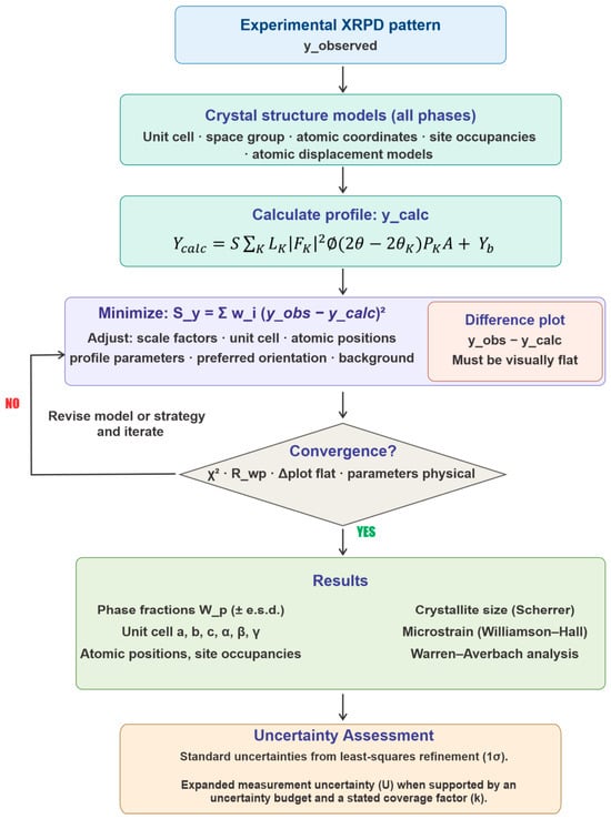
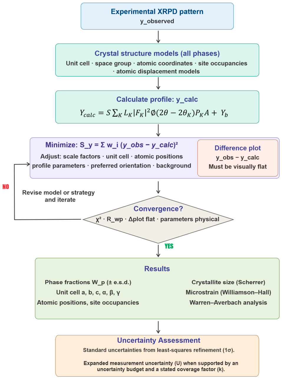
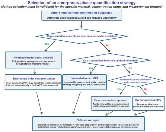
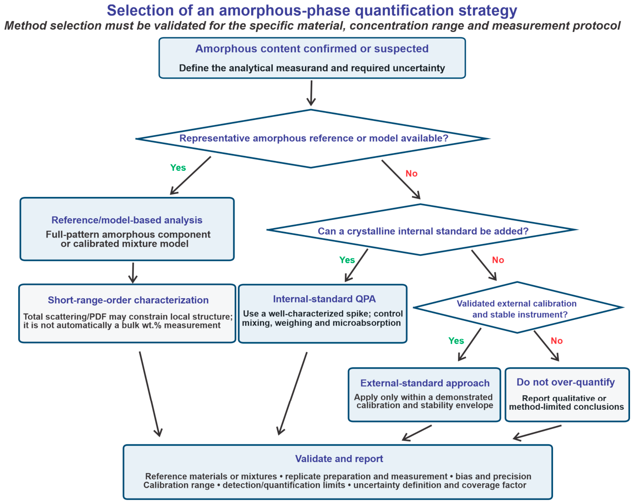
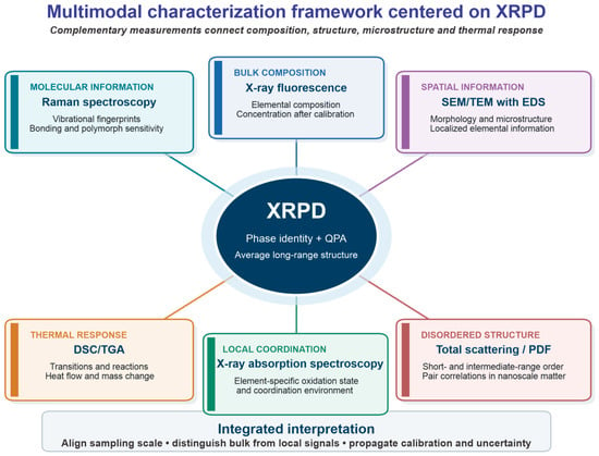
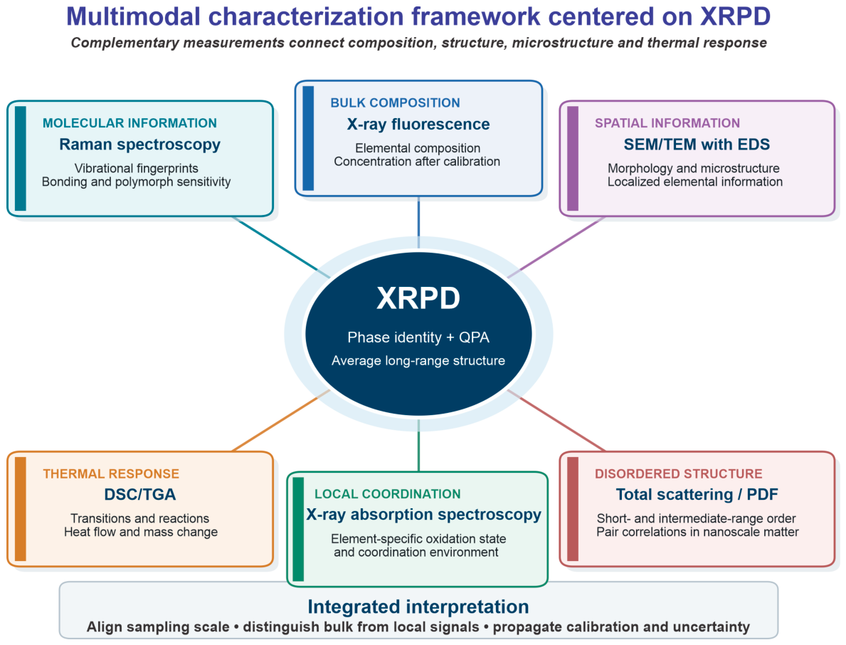
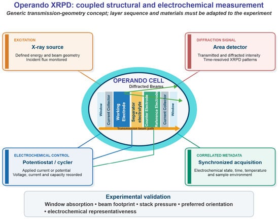
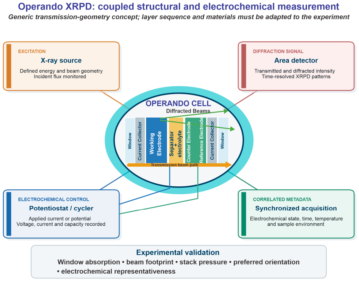
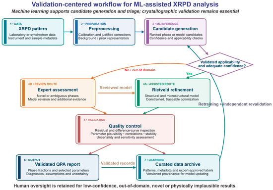
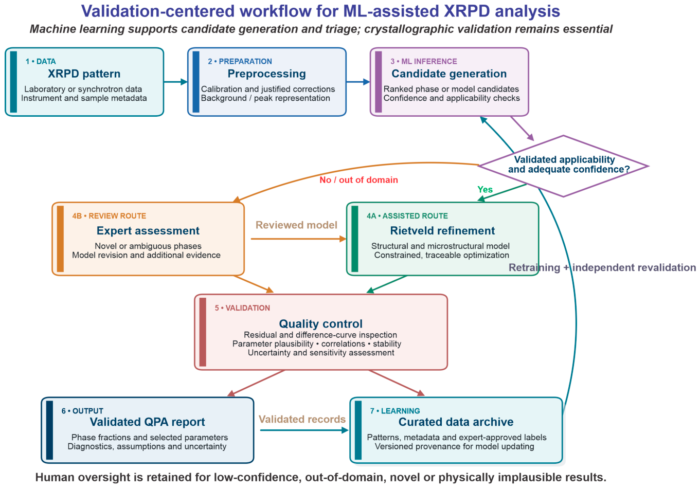

Disclaimer/Publisher’s Note: The statements, opinions and data contained in all publications are solely those of the individual author(s) and contributor(s) and not of MDPI and/or the editor(s). MDPI and/or the editor(s) disclaim responsibility for any injury to people or property resulting from any ideas, methods, instructions or products referred to in the content. |
© 2026 by the authors. Licensee MDPI, Basel, Switzerland. This article is an open access article distributed under the terms and conditions of the Creative Commons Attribution (CC BY) license.