


Abstract
This study examined whether direct pre-ozonation changes inhibition-zone diameters and selected chemical properties of endodontic irrigants. Sodium hypochlorite (NaOCl; 5.25%), chlorhexidine digluconate (CHX; 2%), ethylenediaminetetraacetic acid (EDTA; 17%), and saline underwent a fixed procedure (gas-phase ozone, 80 µg/mL; 30 L/h; 5 min; 200 mL). Disk diffusion against Candida albicans, Staphylococcus aureus, and Enterococcus faecalis was evaluated in eight independent paired runs. pH was measured, and available chlorine in NaOCl was determined in three independent paired batches. Mean inhibition-zone diameters were smaller after ozonation in all nine comparisons, and eight comparisons were statistically significant after Holm correction; the EDTA comparison against C. albicans was not statistically significant. CHX showed the largest pH change (9.05 to 6.34; −2.71 units). Available chlorine decreased from 5.10 ± 0.06% to 2.84 ± 0.04%, a paired reduction of 2.26 percentage points (95% CI, 2.02–2.50). Under this protocol, pre-ozonation did not increase inhibition-zone diameters. These findings cannot distinguish changes in intrinsic antimicrobial activity from altered agar diffusion and do not establish a clinical effect. Dissolved ozone was not measured, and chemical transformation products were not characterized.
1. Introduction
One of the primary goals of root canal treatment is the removal of inorganic and organic debris, pulp tissue, microorganisms, and their metabolic by-products from the root canal system [1]. The root canal system has a highly complex anatomy [2], and mechanical instrumentation alone cannot clean all regions [3]. Microorganisms persisting in inaccessible areas can contribute to treatment failure [1]. Chemical irrigation therefore plays a critical role in cleaning regions that are difficult to reach with mechanical preparation alone [4].
Research on irrigation has examined solution concentration and temperature as well as activation techniques intended to improve irrigant penetration and cleaning [5]. Ozone applications have attracted attention because of ozone’s oxidative and antimicrobial properties and have been investigated in oral surgery, pediatric dentistry, and endodontics [6].
In endodontics, ozone has been applied as a gas or dissolved in water [7]. Direct gaseous application requires specialized delivery and scavenging. Pre-ozonating a conventional irrigant could, in principle, provide a single prepared liquid for delivery through standard irrigation procedures. This operational rationale differs from separate or sequential ozone treatment. Its biological advantage, however, cannot be assumed because ozone may react with the active components of the irrigant before application.
Previous endodontic studies have evaluated gaseous ozone, ozonated water, or combinations with conventional irrigants [8,9,10,11]. Evidence on direct pre-ozonation of NaOCl, CHX, and EDTA remains limited. A controlled comparison of each irrigant before and after the same pre-ozonation procedure can therefore determine whether the process changes an initial laboratory antimicrobial endpoint and selected physicochemical measures.
This study evaluated inhibition-zone diameters produced by directly pre-ozonated and non-ozonated NaOCl, CHX, and EDTA against Staphylococcus aureus, Enterococcus faecalis, and Candida albicans. Changes in pH were recorded for all solutions, and available chlorine was measured for NaOCl. The agar assay was used as a preliminary screening model. The null hypothesis was that direct pre-ozonation would not change the inhibition-zone diameter within any irrigant–microorganism combination.
2. Materials and Methods
This study was designed as an in vitro laboratory investigation using standard microbial strains and irrigation solutions. No human participants, animals, or patient-derived samples were involved. The manuscript was prepared with reference to the Preferred Reporting Items for Laboratory studies in Endodontology (PRILE) 2021 guidelines [12].
2.1. Determination of Sample Size
The sample-size rationale was guided by a published agar-diffusion study [13], which reported a G*Power calculation (version 3.1.9.7) using α = 0.05, 80% power, and Cohen’s f = 0.680 and allocated eight specimens per group. That study compared three independent material groups and did not use the present matched design. Eight independent paired runs were completed for each irrigant microorganism combination. The external rationale does not establish 80% power for the present nine paired comparisons after multiplicity correction; the analyses are interpreted as exploratory, with uncertainty reported using confidence intervals.
Each run was a separate experimental preparation. For each irrigant, a newly opened bottle was divided within that run to provide matched non-ozonated and ozonated aliquots; the next run used a different newly opened bottle. The run-level pair was the experimental unit. The analysis dataset contained one recorded value per condition within each run, giving eight paired observations per irrigant–microorganism combination. For each irrigant, the eight newly opened bottles bore eight distinct manufacturer-labeled lot numbers; thus, the runs did not all use bottles from a single recorded manufacturing lot.
2.2. Experimental Design and Ozonation Procedure
Four irrigation solutions were evaluated: 5.25% sodium hypochlorite (NaOCl) (Saver, Prime Dental, Izmir, Turkey), 17% ethylenediaminetetraacetic acid (EDTA) (Werax, Spot Kimya, Izmir, Turkey), 2% chlorhexidine digluconate (CHX) (Microvem, Altun Kimya, Istanbul, Turkey), and sterile saline (Polifleks, Polifarma, Istanbul, Turkey). Each active irrigant was evaluated in matched non-ozonated and ozonated aliquots across eight paired experimental runs for each irrigant–microorganism combination. Sterile saline was included in both forms as the negative control. The non-ozonated aliquot of each active irrigant served as the matched reference for the effect of the pre-ozonation procedure.
Ozonation was performed using an Ozonette generator (SEDECAL, Madrid, Spain) set to a gas-phase ozone concentration of 80 µg/mL, a gas flow rate of 30 L/h, and a contact time of 5 min. The liquid volume (200 mL), container geometry, and room temperature (23 ± 1 °C) were kept constant. No validated standard for direct pre-ozonation of endodontic irrigants was available; the fixed 5 min exposure and device settings were selected to provide a reproducible screening condition within the generator’s operating range. The same protocol was applied to every irrigant.
The concentration of ozone dissolved in each irrigant after treatment was not measured. Gas-phase exposure therefore cannot be interpreted as an equal dissolved-ozone dose across chemically different solutions. The experiment evaluated the overall effect of the specified pre-ozonation process rather than a dose–response relationship for dissolved ozone.
2.3. pH Measurements
The pH of each solution was measured using a calibrated digital pH meter (Orion 3-Star, Thermo Scientific, MA, USA) at 23 ± 1 °C in accordance with ISO 10523:2008 [14]. Three independent paired measurement runs were performed for each solution. Within each run, the matched non-ozonated and ozonated aliquots were prepared from the same newly opened bottle; each subsequent run used a different newly opened bottle. The pH results are reported as mean ± standard deviation for the three paired runs in Table 1. The exact elapsed time from the end of ozonation to the pH reading was not recorded.
Table 1.
pH before and after direct pre-ozonation.
2.4. Iodometric Titration of Available Chlorine
Available chlorine was measured in 5.25% NaOCl before and after ozonation. Each of three paired replicates was prepared from a newly opened bottle with a different batch code, and each bottle was ozonated separately under the same conditions. After appropriate dilution, the samples were acidified in the presence of potassium iodide. Liberated iodine was titrated with standardized sodium thiosulfate, using starch as the endpoint indicator. Available chlorine was calculated from sodium thiosulfate consumption according to iodometric stoichiometry and expressed as a percentage. The paired difference and its 95% CI were calculated from these three paired batch measurements.
2.5. Microbial Strains and Disk Diffusion Assay
The microorganisms tested were Staphylococcus aureus (ATCC 25923, Manassas, VA, USA), Enterococcus faecalis (ATCC 29212, Manassas, VA, USA), and Candida albicans (ATCC 14053, Manassas, VA, USA). Test solutions were aseptically applied to sterile 6 mm paper disks (20 µL per disk). Culture media included 5% sheep blood agar, eosin methylene blue agar, and Sabouraud 2% dextrose agar. Inoculum turbidity was adjusted to a 0.5 McFarland standard using sterile Mueller–Hinton broth. Bacterial disk-diffusion assays were performed on Mueller–Hinton agar. Impregnated disks were placed on the inoculated assay plates immediately after solution preparation in the same experimental session. Plates were incubated at 35 ± 2 °C for 18–24 h. Inhibition-zone diameters were measured in millimeters. For positive inhibition zones, the measured diameter included the 6 mm paper disk. The absence of an inhibition zone was coded as 0, not as the physical disk diameter.
For each microorganism irrigant ozonation condition within a run, one full inhibition zone diameter was measured directly with a precision caliper along a line passing through the disk center. The 6 mm paper disk was included in the diameter. This direct reading was the single run-level value used for analysis; no technical readings were averaged or otherwise combined.
Different operators performed ozonation, pH measurement, and microbiological procedures. Before disk impregnation and placement, the solutions were assigned numeric codes. The operators who impregnated the disks and measured the zones had access only to those codes and were blinded to solution identity (Supplementary Figure S1).
Disk diffusion was selected as a preliminary screen under fixed assay conditions. Its endpoint depends on both growth inhibition and movement of the test material through agar. Accordingly, a change in zone diameter cannot by itself distinguish a change in intrinsic antimicrobial potency from a change in diffusion behavior.
2.6. Statistical Analysis
Continuous data are summarized as mean and standard deviation (SD). Normality of paired within-run differences was assessed using the Shapiro–Wilk test (α = 0.05). Each non-ozonated–ozonated pair was compared using a two-sided paired-samples t-test when the normality test did not reject normality; otherwise, an exact two-sided Wilcoxon signed-rank test was used. For the latter, zero differences were omitted, tied absolute differences received average ranks, and all sign assignments were enumerated. Test choice for each comparison is identified in Table 2. Unadjusted p-values were corrected using Holm’s step-down procedure across the nine active-irrigant–microorganism comparisons, with statistical significance defined as adjusted p < 0.05. Saline was excluded from hypothesis testing. The effect estimate is the mean within-run difference (non-ozonated minus ozonated). Nominal 95% t-based confidence intervals (CIs) for mean differences were calculated using seven degrees of freedom for the disk-diffusion data and two for the three paired chlorine measurements. These CIs are not multiplicity-adjusted and, for Wilcoxon comparisons, are descriptive mean-based intervals rather than intervals corresponding to the rank-based test. Their coverage is uncertain for small, non-normal samples. Between-irrigant hypothesis tests were omitted because the research question concerned within-irrigant changes. Original analyses used IBM SPSS Statistics version 27 (IBM Corp., Armonk, NY, USA); the paired calculations and Holm adjustment were verified during revision using Python 3.13.5 and SciPy 1.17.0.
Table 2.
Paired changes in inhibition zone diameter (mm) after direct pre-ozonation.
3. Results
All eight planned experimental runs were completed, and no measurements were excluded. Neither non-ozonated nor ozonated saline produced an inhibition zone in any replicate for any microorganism; saline was therefore not included in inferential comparisons.
pH decreased after ozonation in all four solutions (Table 1). The mean changes were −0.20 pH units for NaOCl, −0.09 for EDTA, −2.71 for CHX, and −0.99 for saline. These are differences in pH units, not percentage changes.
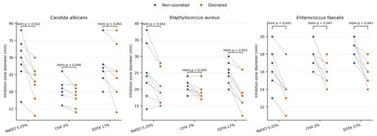
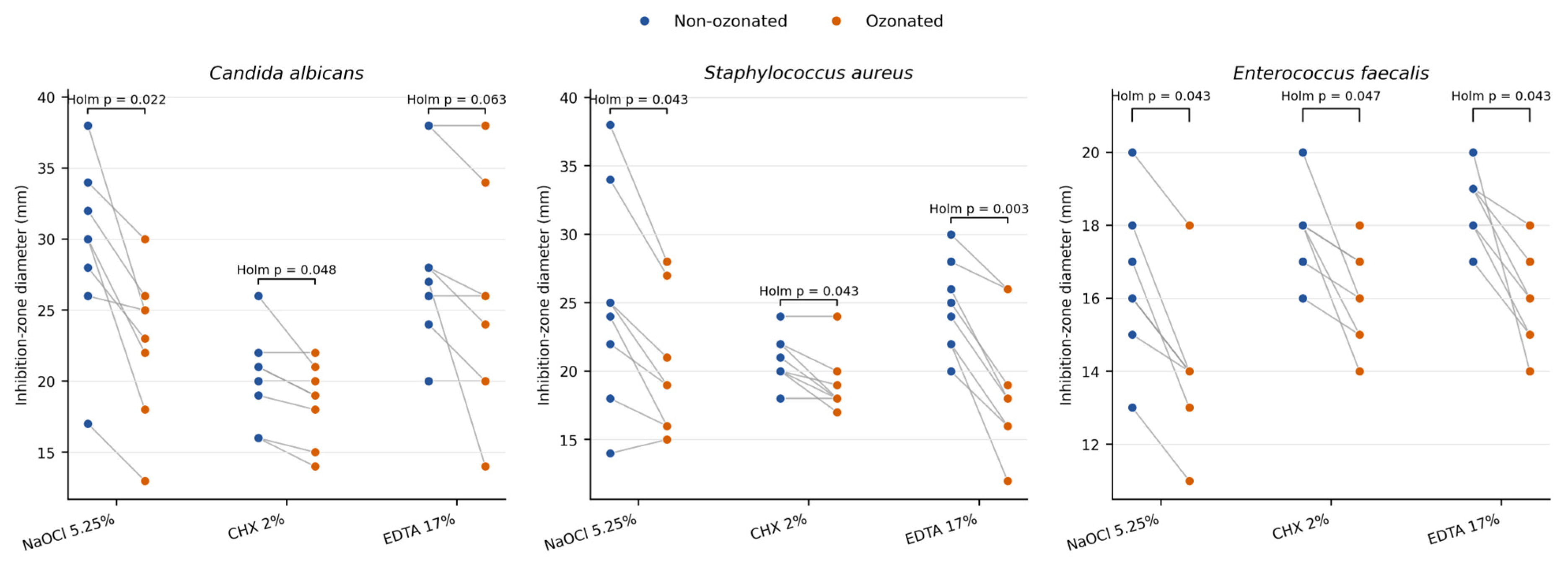
The paired disk-diffusion results are shown in Figure 1 and Table 2. Mean inhibition zone diameters were smaller after ozonation in all nine irrigant–microorganism comparisons. Eight comparisons were statistically significant after Holm correction: NaOCl and CHX against C. albicans, and all three irrigants against S. aureus and E. faecalis. The EDTA comparison against C. albicans did not reach statistical significance (mean paired difference, 3.38 mm; 95% CI, −0.23 to 6.98; Holm-adjusted p = 0.063). Across the eight runs, the direction and magnitude of change varied by irrigant–microorganism combination. These results describe changes in the disk-diffusion endpoint.
Figure 1.
Paired inhibition zone diameters in eight independent experimental runs. Each gray line joins non-ozonated and ozonated aliquots prepared from the same bottle within one run; coincident observations may overlap. p-values are Holm-adjusted across the nine paired comparisons. Saline produced no inhibition zones and is not plotted.
Available Chlorine in NaOCl
Available chlorine values were 5.16%, 5.04%, and 5.10% before ozonation and 2.80%, 2.87%, and 2.85% after ozonation. The mean decreased from 5.10 ± 0.06% to 2.84 ± 0.04%, corresponding to an approximate relative reduction of 44.3%. The mean paired reduction was 2.26 percentage points (95% CI, 2.02 to 2.50), calculated from the three paired batch measurements. Because only three independent batches were analyzed, the estimate should be interpreted as preliminary.
4. Discussion
Under the tested conditions, direct pre-ozonation did not increase the inhibition zone diameter of any active irrigant against any tested microorganism. Mean inhibition zone diameters were smaller after ozonation in all nine irrigant–microorganism comparisons, and eight comparisons were statistically significant after Holm correction. The EDTA–C. albicans comparison did not reach statistical significance; this does not establish equivalence or absence of an effect. These results are limited to the disk-diffusion endpoint and the specified ozonation procedure.
NaOCl remains a widely used endodontic irrigant because of its broad antimicrobial action and tissue-dissolving capacity [4]. In this study, ozonation was followed by smaller inhibition zones for NaOCl against all three organisms. The available chlorine measurements provide a related chemical observation: mean available chlorine decreased from 5.10% to 2.84%. Loss of available chlorine could contribute to the change in zone diameter, but the experiment did not characterize chlorine species or establish a causal pathway. The titration result is also based on only three paired batches and requires confirmation with a larger analytical series.
Previous investigations have compared ozonated water with NaOCl or examined ozone as a separate adjunct [8,10,11]. Those designs do not reproduce the direct pre-ozonation procedure examined here. Consequently, the present results neither confirm nor refute the efficacy of ozone delivered by other methods; they describe changes following the specific gas-phase setting, contact time, liquid volume, and apparatus used in this experiment.
CHX showed smaller zones after ozonation against all three organisms and the largest observed pH change, from 9.05 to 6.34. The pH result demonstrates a physicochemical change, but it does not identify degradation of CHX or explain the microbiological endpoint. No chromatographic, spectroscopic, or mass-spectrometric characterization was performed. Any structural-change mechanism is therefore a hypothesis for future study rather than a finding of this experiment. Kurylo et al. examined crystallographic changes after ozonation of 0.05% CHX [15]. Their different concentration and analytical endpoint provide context but do not identify transformation products or confirm a mechanism in the present 2% CHX experiment.
EDTA is used primarily as a chelating agent in endodontics [16]. Ozonated EDTA showed smaller zones against S. aureus and E. faecalis, whereas the C. albicans comparison was not statistically significant. No direct analysis of EDTA transformation products or chelating performance was performed. Its zone changes therefore cannot establish degradation or loss of chelating function. Chemical characterization would be required before assigning a mechanism.
The disk-diffusion assay is useful for comparative laboratory screening, but zone diameter reflects both microbial growth inhibition and agar diffusion. Ozonation may change pH or other physicochemical properties that influence diffusion. The smaller zones observed here therefore cannot be interpreted as direct proof that intrinsic antimicrobial potency decreased. Quantitative suspension or direct-contact testing with validated neutralization and viable-count measurement would help separate killing from diffusion effects. Dentin-block and mature-biofilm models would then be needed to examine behavior in a root-canal-like substrate. The design did not independently vary ozone and irrigant concentrations or generate minimum inhibitory concentration data; it therefore cannot establish synergy or antagonism or provide a fractional inhibitory concentration index.
The dissolved ozone concentration was not measured. Because NaOCl, CHX, EDTA, and saline have different chemical compositions, equal gas-phase exposure may produce different dissolved-ozone concentrations and decay profiles. The exact time from ozonation to pH measurement was also not recorded. A matched ozone-free gas-bubbling control was not included, so ozone-specific effects cannot be separated from other effects of the gas-treatment procedure. The sample size was small, and the reported sample-size rationale does not establish power for the multiplicity-adjusted paired analysis. Shapiro–Wilk screening has limited sensitivity at n = 8, and the nominal t-based intervals, especially for non-normal differences, should be interpreted cautiously.
The 200 mL liquid volume, fixed container geometry, and controlled room conditions were chosen for reproducibility. They do not reproduce the small volumes, delivery dynamics, dentin contact, organic load, or biofilm structure encountered during root canal irrigation. This study should therefore be viewed as an initial laboratory screen and does not support clinical use or avoidance of directly pre-ozonated irrigants.
Strengths of this study include the matched within-run design, the use of a new bottle for every run, blinded disk coding and measurement, reporting of paired effect estimates with confidence intervals, and analytical measurement of available chlorine in separate NaOCl batches.
Priorities for Further Study
The next steps are to quantify dissolved ozone and its decay in each irrigant, characterize chemical changes with appropriate analytical methods, verify the microbiological findings with a quantitative direct-contact or suspension assay using validated neutralization, and test selected conditions in dentin and mature-biofilm models.
5. Conclusions
Under the tested gas-phase setting (80 µg/mL, 30 L/h, 5 min; 200 mL), direct pre-ozonation did not increase inhibition zone diameters for NaOCl, CHX, or EDTA. Mean inhibition zone diameters were smaller after ozonation in all nine paired comparisons, and eight comparisons were statistically significant after Holm correction. CHX showed the largest pH change, and available chlorine in NaOCl decreased in three paired batches. These findings apply to the agar-diffusion endpoint and do not establish reduced intrinsic antimicrobial activity or a specific chemical mechanism. Dissolved-ozone measurement, chemical characterization, and quantitative direct-contact testing are required before the biological effect of direct pre-ozonation can be defined.
Supplementary Materials
The following supporting information can be downloaded at: https://www.mdpi.com/article/10.3390/app16199770/s1, Supplementary Figure S1: Composite of representative agar plates showing solution-impregnated disks used in the antimicrobial assay. Numbers identify the test materials: 1 = NaOCl, 2 = EDTA, 3 = CHX, and 4 = saline; “O” denotes the ozonated form. Images document disk coding and placement; no measurements were derived from the photographs. Supplementary Dataset S1: Run-level disk-diffusion measurements in XLSX format, including all eight runs, three microorganisms, three active irrigants, both ozonation conditions, and saline controls (192 records).
Author Contributions
Conceptualization, E.D. and A.H.; methodology, E.D.; software, A.H.; validation, E.D., A.H. and A.K.; formal analysis, E.D., A.H. and F.A.; investigation, E.D., A.H. and A.K.; resources, E.D., A.H., A.K. and F.A.; data curation, A.H.; writing—original draft preparation, E.D. and A.H.; writing—review and editing, E.D. and A.H.; visualization, E.D., A.K. and F.A.; supervision, E.D., A.K. and F.A.; project administration, A.H. All authors have read and agreed to the published version of the manuscript.
Funding
This research received no external funding.
Institutional Review Board Statement
This study was approved by the Bolu Abant Izzet Baysal University Non-Interventional Clinical Research Ethics Committee (decision no. 2024/247, 24 September 2024).
Informed Consent Statement
Not applicable.
Data Availability Statement
Run-level disk-diffusion data are provided in Supplementary Dataset S1. Individual paired available-chlorine measurements are reported in Section Available Chlorine in NaOCl, and pH summary measurements are presented in Table 1. Further information may be obtained from the corresponding author upon reasonable request.
Acknowledgments
The authors thank Hatice Ergin for assistance with the chemistry analyses.
Conflicts of Interest
The authors declare no conflicts of interest.
Abbreviations
| Abbreviation | Definition |
| ATCC | American Type Culture Collection |
| CHX | chlorhexidine digluconate |
| CI | confidence interval |
| EDTA | ethylenediaminetetraacetic acid |
| NaOCl | sodium hypochlorite |
| SD | standard deviation |
References
- Wong, J.; Manoil, D.; Näsman, P.; Belibasakis, G.N.; Neelakantan, P. Microbiological aspects of root canal infections and disinfection strategies: An update review on the current knowledge and challenges. Front. Oral Health 2021, 2, 672887. [Google Scholar] [CrossRef] [Scilit]
- Versiani, M.A.; Martins, J.N.R.; Ordinola-Zapata, R. Anatomical complexities affecting root canal preparation: A narrative review. Aust. Dent. J. 2023, 68, S5–S23. [Google Scholar] [CrossRef] [Scilit]
- Boutsioukis, C.; Arias-Moliz, M.T. Present status and future directions—Irrigants and irrigation methods. Int. Endod. J. 2022, 55, 588–612. [Google Scholar] [CrossRef] [Scilit]
- Haapasalo, M.; Shen, Y.; Wang, Z.; Gao, Y. Irrigation in endodontics. Br. Dent. J. 2014, 216, 299–303. [Google Scholar] [CrossRef] [Scilit]
- Raducka, M.; Piszko, A.; Piszko, P.J.; Jawor, N.; Dobrzyński, M.; Grzebieluch, W.; Mikulewicz, M.; Skośkiewicz-Malinowska, K. Narrative review on methods of activating irrigation liquids for root canal treatment. Appl. Sci. 2023, 13, 7733. [Google Scholar] [CrossRef] [Scilit]
- Liaqat, S.; Tariq, S.; Hayat, I.; Mobeen, B.; Fayyaz, S.; Jabeen, H.; Khalid, S.; Muhammad, N.; Khan, M.A. Therapeutic effects and uses of ozone in dentistry: A systematic review. Ozone Sci. Eng. 2023, 45, 387–397. [Google Scholar] [CrossRef] [Scilit]
- Rezaeianjam, M.; Khabazian, A.; Khabazian, T.; Ghorbani, F.; Abbasi, T.; Asghari, S.; Heidari, F.; Shiri, A.; Naderi, M. Efficacy of ozone therapy in dentistry with approach of healing, pain management, and therapeutic outcomes: A systematic review of clinical trials. BMC Oral Health 2025, 25, 433. [Google Scholar] [CrossRef] [Scilit]
- Merçon, I.R.; De Campos, F.U.F.; Fontana, C.E.; Pelegrine, R.A.; De Martin, A.S.; Da Silveira Bueno, C.E. Analysis of antimicrobial efficacy of sodium hypochlorite and ozonated water against biofilm in oval canals. Braz. Dent. J. 2023, 34, 33–41. [Google Scholar] [CrossRef] [Scilit]
- Noites, R.; Pina-Vaz, C.; Rocha, R.; Carvalho, M.F.; Gonçalves, A.; Pina-Vaz, I. Synergistic antimicrobial action of chlorhexidine and ozone in endodontic treatment. BioMed Res. Int. 2014, 2014, 592423. [Google Scholar] [CrossRef] [Scilit]
- Pinheiro, S.L.; da Silva, C.C.; da Silva, L.A.; Cicotti, M.P.; Bueno, C.E.D.S.; Fontana, C.E.; Pagrion, L.R.; Dalmora, N.P.; Daque, T.T.; de Campos, F.U. Antimicrobial efficacy of 2.5% sodium hypochlorite, 2% chlorhexidine, and ozonated water as irrigants in mesiobuccal root canals with severe curvature of mandibular molars. Eur. J. Dent. 2018, 12, 94–99. [Google Scholar] [CrossRef] [Scilit]
- Silva, E.J.N.L.; Prado, M.C.; Soares, D.N.; Hecksher, F.; Martins, J.N.R.; Fidalgo, T.K.S. The effect of ozone therapy in root canal disinfection: A systematic review. Int. Endod. J. 2020, 53, 317–332. [Google Scholar] [CrossRef] [Scilit]
- Nagendrababu, V.; Murray, P.E.; Ordinola-Zapata, R.; Peters, O.A.; Rôças, I.N.; Siqueira, J.F., Jr.; Priya, E.; Jayaraman, J.; Pulikkotil, S.J.; Camilleri, J.; et al. PRILE 2021 guidelines for reporting laboratory studies in Endodontology: A consensus-based development. Int. Endod. J. 2021, 54, 1482–1490. [Google Scholar] [CrossRef] [Scilit]
- Afify, Y.M.; Allam, G.G.; Abd El-Geleel, O. Evaluating the antimicrobial effect, compressive strength and fluoride release of glass ionomer cement modified with Triphala. Sci. Rep. 2025, 15, 39372. [Google Scholar] [CrossRef] [Scilit]
- ISO 10523:2008; Water Quality—Determination of pH. International Organization for Standardization: Geneva, Switzerland, 2008.
- Kurylo, V.O.; Korol, D.M.; Rozhko, M.M.; Kindii, D.D.; Toncheva, K.D.; Kozak, R.V. Effects of ozone treatment on 0.05% chlorhexidine bigluconate solution: Assessment of crystallography results in an in vitro experiment. Med. Ecol. Probl. 2024, 28, 19–28. [Google Scholar] [CrossRef] [Scilit]
- Mohammadi, Z.; Shalavi, S.; Jafarzadeh, H. Ethylenediaminetetraacetic acid in endodontics. Eur. J. Dent. 2013, 7, S135–S142. [Google Scholar] [CrossRef] [Scilit]
Disclaimer/Publisher’s Note: The statements, opinions and data contained in all publications are solely those of the individual author(s) and contributor(s) and not of MDPI and/or the editor(s). MDPI and/or the editor(s) disclaim responsibility for any injury to people or property resulting from any ideas, methods, instructions or products referred to in the content. |
© 2026 by the authors. Licensee MDPI, Basel, Switzerland. This article is an open access article distributed under the terms and conditions of the Creative Commons Attribution (CC BY) license.