


Abstract
Background: Hip resurfacing arthroplasty (HRA) was developed as a bone-preserving alternative to conventional total hip arthroplasty (THA), particularly for young and physically active patients. However, concerns regarding metal-on-metal bearings have substantially restricted its use. This study evaluated the long-term clinical, functional, radiographic, and implant survivorship outcomes of HRA. Methods: This retrospective cohort study included all 53 consecutive primary HRA procedures performed at a single institution between January 2001 and December 2010. No patients were excluded or lost to follow-up. The mean age at surgery was 43.2 years (range, 18–56 years), and the mean follow-up was 20 years (range, 15–25 years). Clinical outcomes were assessed using the Harris Hip Score (HHS) and Western Ontario and McMaster Universities Osteoarthritis Index (WOMAC). Paired preoperative and final scores were analyzed only in the 47 patients who retained their HRA implant. Radiographic evaluation included implant fixation, radiolucent lines, osteolysis, component alignment, and heterotopic ossification. Implant survivorship was estimated using the Kaplan–Meier method, with revision of any component for any reason as the endpoint. Results: Among the 47 patients with retained HRA implants, the mean HHS increased from 31.3 ± 17.9 preoperatively to 95.3 ± 7.4 at final follow-up, while the mean total WOMAC score decreased from 65.9 ± 10.4 to 11.5 ± 3.5 (both p < 0.001). Six hips (11.3%) underwent revision to conventional THA: three for metallosis, two for femoral neck fracture, and one for persistent pain associated with acetabular radiolucency. One additional patient developed postoperative thrombophlebitis that did not require implant revision. Radiographic assessment of unrevised hips demonstrated stable component fixation without progressive radiolucency, clinically significant osteolysis, implant migration, or aseptic loosening. Kaplan–Meier implant survivorship was 88.7% (95% confidence interval, 76.5–94.8%) at approximately 20 years. Conclusions: In this selected cohort of predominantly young and active patients, HRA provided substantial long-term clinical improvement, with an HHS of 95.3 ± 7.4, a WOMAC score of 11.5 ± 3.5, and implant survivorship of 88.7% at approximately 20 years. Nevertheless, the 11.3% revision rate and complications associated with metal-on-metal bearings support restricting HRA to carefully selected patients and maintaining long-term clinical, radiographic, and metal-ion surveillance.
1. Introduction
Total hip arthroplasty (THA) is widely regarded as one of the most successful procedures in modern orthopedic surgery, providing reliable pain relief, restoration of joint function, and substantial improvements in quality of life for patients with advanced hip disease [1,2]. Continuous advances in implant design, bearing surfaces, biomaterials, and surgical techniques have significantly enhanced implant longevity and clinical outcomes over recent decades. However, despite these achievements, the long-term management of younger and more physically active patients remains challenging. Their greater functional demands and longer life expectancy expose prosthetic implants to increased mechanical stress, resulting in a higher cumulative risk of wear, revision surgery, and implant failure during their lifetime [3,4].
Hip resurfacing arthroplasty (HRA) was developed as a bone-preserving alternative to conventional THA, specifically targeting young and active patients with end-stage hip disease. Unlike standard THA, HRA preserves the femoral head and neck, thereby maintaining proximal femoral bone stock and allowing a more physiological transfer of mechanical loads across the hip joint. This bone-conserving strategy may simplify future revision procedures while reducing alterations in proximal femoral biomechanics [5,6]. Furthermore, the use of large-diameter femoral heads has been associated with improved joint stability, a lower risk of postoperative dislocation, and restoration of a more physiological range of motion compared with conventional THA [7,8].
These biomechanical characteristics have made HRA particularly attractive for patients wishing to maintain a high level of physical activity after surgery. Several studies have reported that patients treated with HRA achieve higher postoperative activity levels, a more physiological gait pattern, and an earlier return to sports than those undergoing conventional THA [9,10]. In addition, preservation of the native femoral anatomy facilitates conversion to conventional THA should revision become necessary, representing one of the principal theoretical advantages of the resurfacing procedure [11].
Despite these potential benefits, the role of HRA remains controversial. Procedure-specific complications, particularly femoral neck fractures, continue to represent an important cause of early failure and are closely related to patient selection, implant positioning, and surgical technique [12]. Moreover, increasing evidence regarding complications associated with metal-on-metal (MoM) bearing surfaces—including elevated serum cobalt and chromium ion concentrations, adverse local tissue reactions, metallosis, and pseudotumor formation—has substantially reduced the worldwide use of HRA over the past decade [13,14,15]. These concerns have reinforced the importance of meticulous surgical technique, accurate component positioning, and careful patient selection to optimize long-term outcomes.
Appropriate patient selection is now recognized as one of the most important determinants of successful hip resurfacing. Current indications are generally restricted to younger patients with good bone quality, preserved femoral head morphology, and high functional demands, whereas patients with osteoporosis, extensive femoral head deformity, or compromised bone stock are considered poor candidates for the procedure [16]. Consequently, the highly selective indications for HRA may partially explain the variability in clinical outcomes reported across different studies.
Although the popularity of HRA has declined because of concerns surrounding MoM bearings, several high-volume centers continue to report excellent mid-term clinical and functional outcomes together with satisfactory implant survivorship when the procedure is performed in carefully selected patients by experienced surgeons [17,18]. Nevertheless, evidence regarding very long-term outcomes remains limited, as relatively few studies have reported clinical and radiographic results beyond 15–20 years of follow-up.
Therefore, further long-term investigations are needed to better define the durability of hip resurfacing arthroplasty, evaluate implant survivorship, characterize late complications, and determine whether the functional benefits observed in the early postoperative period are maintained over time.
The aim of this retrospective cohort study was to evaluate the twenty-year clinical, functional, radiographic, and survivorship outcomes of hip resurfacing arthroplasty in a consecutive series of patients treated at a single institution.
2. Materials and Methods
2.1. Study Design and Patient Population
This retrospective, single-center cohort study included the complete inception cohort of consecutive patients who underwent primary hip resurfacing arthroplasty (HRA) at a tertiary referral center between January 2001 and December 2010. Institutional medical records, operative reports, outpatient clinical assessments, and radiographic examinations were retrospectively reviewed to evaluate long-term clinical, functional, radiographic, and implant survivorship outcomes.
The surgical inclusion period was predefined because, at the time of the final study assessment, it provided a minimum potential follow-up of 15 years. A total of 53 patients (53 hips) underwent primary HRA during this period. All 53 procedures were included in the survivorship analysis. No patients were excluded because of incomplete follow-up, and no patients were lost to follow-up.
Revision of any resurfacing component for any reason was considered the endpoint of implant survivorship. Final clinical outcome analysis was restricted to patients with a retained HRA implant and complete paired preoperative and final follow-up data. Patients converted to total hip arthroplasty (THA) were excluded from the final HHS and WOMAC analyses because subsequent scores would represent outcomes of the revision THA rather than outcomes attributable to the original HRA.
The demographic and baseline clinical characteristics of the cohort are summarized in Table 1, and the indications for surgery are reported in Table 2. The cohort comprised 40 men (75.5%) and 13 women (24.5%), with a mean age of 43.2 years (range, 18–56 years) at the time of surgery. The principal indication was primary osteoarthritis (n = 40), followed by osteonecrosis of the femoral head (n = 7), slipped capital femoral epiphysis (n = 2), Perthes disease (n = 2), post-traumatic osteoarthritis (n = 1), and rheumatoid arthritis (n = 1).
Table 1.
Demographic and clinical characteristics of the study cohort.
Table 2.
Indications for hip resurfacing arthroplasty.
Patients with primary osteoarthritis presented with advanced symptomatic hip degeneration, persistent pain, functional limitations, and inadequate improvement following conservative treatment. Eligibility for HRA was determined according to patient age, functional demands, bone quality, proximal femoral morphology, and the extent of femoral head involvement. Candidates were generally young and physically active patients with preserved femoral bone stock and satisfactory femoral head and neck anatomy. The final decision was confirmed intraoperatively; when inadequate bone quality, extensive femoral head involvement, or severe structural deformity was identified, conventional THA was preferred.
All patients received detailed information regarding the expected benefits and potential risks of HRA, including femoral neck fracture, implant failure, and complications associated with metal-on-metal bearing surfaces, and provided written informed consent before surgery.
2.2. Surgical Technique
All procedures were performed by experienced orthopedic surgeons using a standardized posterolateral Gibson–Moore approach, with the patient positioned in the lateral decubitus position. The short external rotators were detached close to their femoral insertion, and the posterior capsule was opened to expose and dislocate the hip. Acetabular preparation and femoral head resurfacing were then performed according to the implant manufacturer’s surgical protocol.
Hip resurfacing was performed using the Birmingham Hip Resurfacing system (Smith & Nephew, Warwick, UK) in 44 hips and the Durom Hip Resurfacing system (Zimmer, Warsaw, IN, USA) in 9 hips. Implant selection reflected surgeon preference and implant availability during the study period.
The acetabular component was implanted with a target inclination of approximately 45° and a target anteversion of approximately 20°, with the aim of limiting edge loading while optimizing implant stability and bearing function. Femoral preparation was performed carefully to preserve femoral head vascularity and avoid notching of the femoral neck.
The femoral component was intentionally positioned in slight valgus relative to the native femoral neck axis to reduce the risk of postoperative femoral neck fracture. The femoral component was fixed using low-viscosity polymethylmethacrylate bone cement according to the manufacturer’s recommendations. Before wound closure, implant stability, hip range of motion, restoration of leg length, and joint stability were systematically assessed.
At closure, the posterior capsule and short external rotators were repaired using non-absorbable sutures to restore posterior soft-tissue continuity and improve postoperative stability.
2.3. Postoperative Management and Rehabilitation
The postoperative rehabilitation regimen described below was derived from the contemporaneous institutional rehabilitation pathway and corroborated, when available, by the individual medical records. Given the retrospective nature of the study, minor variations in timing and progression may have occurred according to each patient’s clinical condition and the treating surgeon’s assessment.
Postoperative mobilization began on the first postoperative day under the supervision of a physiotherapist. Patients were initially allowed protected partial weight-bearing using two crutches for six weeks. Progression to full weight-bearing was permitted between six and eight weeks, provided that pain was controlled, gait was stable, and radiographs showed no evidence of femoral neck fracture, component migration, or other mechanical complications.
During the first six postoperative weeks, patients were instructed to avoid hip flexion beyond 90°, adduction beyond the midline, and forced internal rotation. Early rehabilitation included ankle-pump exercises, isometric quadriceps and gluteal contractions, and progressive active-assisted hip range-of-motion exercises within the permitted range.
Muscle strengthening was progressively advanced from isometric exercises to active and resisted exercises targeting the hip abductors, extensors, and flexors. Walking aids were discontinued when patients demonstrated a pain-free, non-antalgic gait and adequate hip abductor control.
Low-impact activities, including stationary cycling and swimming, were generally resumed after three months. Return to running, contact sports, and other high-impact activities was not considered before six months and required the absence of pain, satisfactory hip range of motion, recovery of muscle strength, stable radiographic findings, and the ability to perform sport-specific movements safely.
2.4. Clinical Evaluation
Clinical assessment was performed preoperatively and at the latest available follow-up using the Harris Hip Score (HHS) and the Western Ontario and McMaster Universities Osteoarthritis Index (WOMAC).
The full HHS ranges from 0 to 100 points, with higher scores indicating better hip function. Its pain, function, deformity, and range-of-motion components were directly assessed at both the preoperative and final follow-up evaluations. Clinical improvement was evaluated by comparing paired preoperative and final HHS values in patients with a retained HRA implant and complete data at both time points.
The validated Italian-language WOMAC questionnaire consisted of 24 items divided into pain (5 items; range, 0–20), stiffness (2 items; range, 0–8), and physical function (17 items; range, 0–68) subscales. Items were scored using the five-point Likert version, providing a raw total score ranging from 0 to 96, with lower scores indicating less pain and stiffness and better physical function. Raw scores were used without conversion to a 0–100 normalized scale. WOMAC pain, stiffness, physical function, and total scores were evaluated separately.
Patients converted to THA were excluded from the final clinical outcome analysis. Their clinical course, reason for revision, and timing of revision were evaluated separately in the complications and revision analyses. No scores obtained after revision THA were attributed to the original resurfacing procedure.
Whenever possible, final follow-up evaluations were performed by orthopedic surgeons who had not participated in the index surgical procedure, thereby limiting observer-related bias.
2.5. Metal-Ion Surveillance and Assessment of Adverse Reactions
Because the procedures were performed over an extended historical period, serum cobalt and chromium measurements were not included in a uniform routine surveillance protocol for the entire cohort. Metal-ion testing was performed when patients developed otherwise unexplained hip pain, swelling, reduced function, radiographic abnormalities, or other clinical findings suggestive of metal-related complications.
When available, serum cobalt and chromium concentrations were recorded together with the units of measurement, timing of testing, clinical indication, and subsequent trend. Because laboratory surveillance protocols and intervention thresholds evolved during the study period, no single predefined metal-ion threshold was used as an isolated indication for revision. Clinical decisions were based on the combined evaluation of symptoms, serial metal-ion concentrations, radiographic findings, cross-sectional imaging, and implant status.
Patients with suspected adverse reactions to metal debris were assessed for local manifestations, including pain, swelling, reduced hip function, and periarticular masses. Additional cross-sectional imaging using metal artifact reduction sequence magnetic resonance imaging (MARS MRI) or ultrasonography was performed when clinically indicated and available. Medical records were also reviewed for potential systemic manifestations of cobalt or chromium toxicity, including neurological, cardiac, thyroid, renal, auditory, and visual abnormalities. Because metal-ion testing was not systematically performed in all patients, no population-level analysis of serum metal-ion concentrations was undertaken.
2.6. Radiographic Evaluation
Radiographic assessment included standardized anteroposterior pelvic radiographs and axial lateral views obtained preoperatively, immediately postoperatively, and during scheduled follow-up.
Acetabular component fixation was evaluated according to the DeLee and Charnley classification. Radiolucent lines were recorded according to their location within the three acetabular zones and, when measurable, according to their maximum width in millimeters. Radiolucent lines were considered progressive when their width or extent increased on serial radiographs compared with the immediate postoperative examination.
Acetabular osteolysis was defined as a focal area of progressive periprosthetic bone loss that was absent on the immediate postoperative radiograph. Radiographic loosening was defined by progressive component migration, a change in component position, or progressive circumferential radiolucency. Acetabular inclination was measured on anteroposterior pelvic radiographs.
Femoral component alignment was assessed relative to the longitudinal axis of the femoral shaft and classified as neutral, varus, or valgus. Serial radiographs were also evaluated for femoral neck narrowing, component migration, subsidence, periprosthetic osteolysis, and signs of aseptic loosening.
Heterotopic ossification was classified according to the Brooker classification. All follow-up radiographs were compared with the immediate postoperative examination to identify progressive radiographic changes.
2.7. Follow-Up and Survivorship Assessment
Patients were followed according to the institutional arthroplasty surveillance protocol at approximately six weeks, three months, six months, and twelve months postoperatively, and annually thereafter. Patients were subsequently invited to undergo a comprehensive long-term clinical and radiographic assessment.
At each follow-up visit, clinical examination, functional outcome scores, standard radiographs, postoperative complications, and revision procedures were recorded. The mean follow-up was approximately 20 years, ranging from 15 to 25 years according to the date of the index procedure.
The primary endpoint of the survivorship analysis was revision of any HRA component for any reason. For revised hips, follow-up was calculated from the date of the index HRA to the date of revision. Unrevised hips were followed until their latest documented clinical or radiographic assessment.
2.8. Statistical Analysis
Continuous variables are reported as mean ± standard deviation (SD), whereas categorical variables are reported as absolute frequencies and percentages. Normality of continuous variables and of the within-patient differences between preoperative and final HHS and WOMAC values was assessed using the Shapiro–Wilk test.
Because normality of the paired differences could not be assumed, paired preoperative and final HHS and WOMAC values were compared using the Wilcoxon signed-rank test. The exact number of patients contributing to each paired outcome analysis was reported. All tests were two-tailed, and a p-value < 0.05 was considered statistically significant.
Because of the limited and markedly unbalanced subgroup sizes, particularly for the Durom implant group and female patients, the study was not adequately powered for reliable inferential comparisons according to implant type or sex. No formal subgroup hypothesis testing was therefore performed to avoid unstable or potentially misleading estimates. Implant type and sex were reported descriptively.
Implant survivorship was estimated using the Kaplan–Meier method, with revision of any resurfacing component for any reason defined as the endpoint. Time to revision was calculated from the date of the index HRA to the date of revision. Unrevised hips were censored at the date of their latest available clinical or radiographic assessment. All 53 primary HRA procedures were included in the survivorship analysis. Survivorship estimates were reported with their corresponding 95% confidence intervals (95% CIs), and the numbers of hips at risk were reported at predefined follow-up intervals.
3. Results
3.1. Patient Demographics and Follow-Up
A total of 53 consecutive patients (53 hips) underwent primary HRA between January 2001 and December 2010. All 53 hips were included in the implant survivorship analysis. No patients were excluded, and no patients were lost to follow-up.
The cohort comprised 40 men (75.5%) and 13 women (24.5%), with a mean age of 43.2 years (range, 18–56 years) at the time of surgery. The mean length of hospital stay was 5 days (range, 4–6 days). The mean follow-up was 20 years (range, 15–25 years), according to the date of the index procedure.
At the latest assessment, 47 hips retained the original resurfacing implant and were included in the final clinical and radiographic outcome analyses. Six hips had undergone conversion to conventional THA and were analyzed separately as revision events.
3.2. Clinical Outcomes
The clinical outcome analysis included the 47 patients with a retained HRA implant and complete paired preoperative and final follow-up data. Patients converted to THA were excluded from this analysis, and no post-revision THA scores were attributed to the original resurfacing procedure.
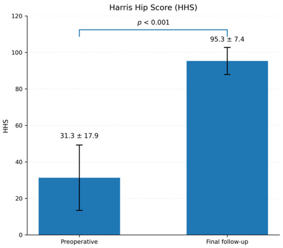
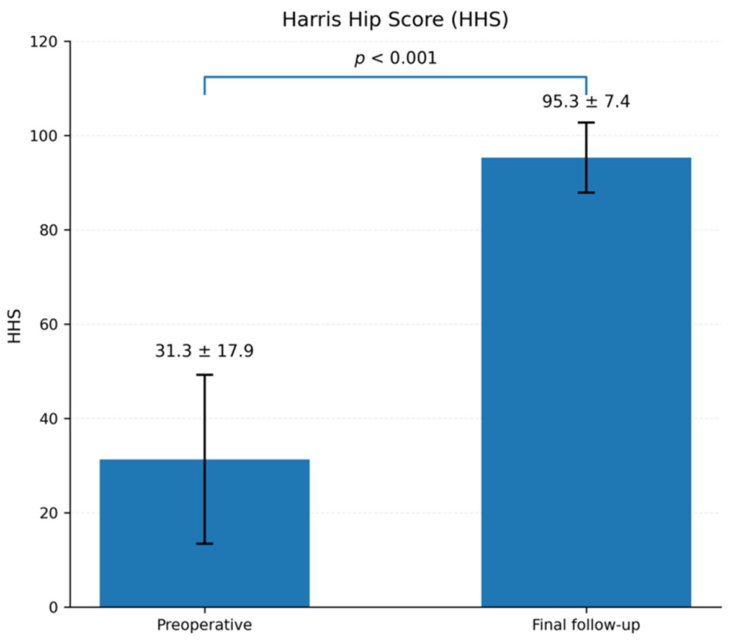
The mean Harris Hip Score (HHS) increased from 31.3 ± 17.9 preoperatively to 95.3 ± 7.4 at final follow-up. This improvement was statistically significant according to the Wilcoxon signed-rank test (p < 0.001) (Table 3; Figure 1).
Table 3.
Paired comparison of Harris Hip Score before surgery and at final follow-up in patients with a retained HRA implant (n = 47).
Figure 1.
Preoperative and final follow-up Harris Hip Score (HHS) values in 47 patients with a retained HRA implant and complete paired data. Bars represent mean values, and error bars represent standard deviations. Statistical significance was assessed using the Wilcoxon signed-rank test (p < 0.001).
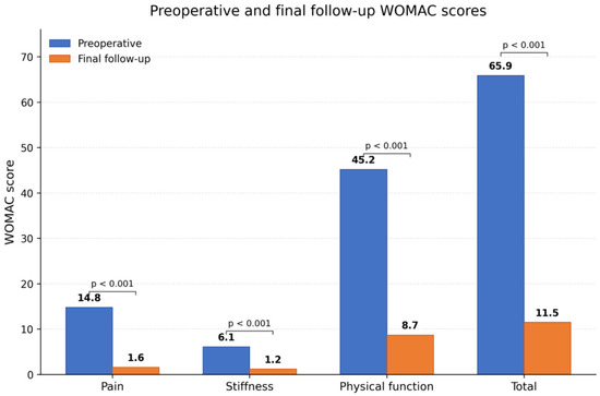
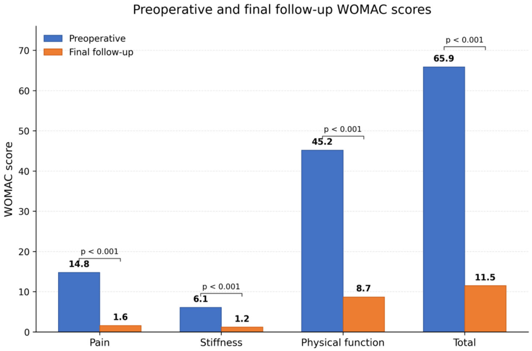
The mean total Western Ontario and McMaster Universities Osteoarthritis Index (WOMAC) score decreased from 65.9 ± 10.4 preoperatively to 11.5 ± 3.5 at final follow-up (p < 0.001). Analysis of the WOMAC subscales showed a reduction in mean pain score from 14.8 to 1.6, stiffness score from 6.1 to 1.2, and physical function score from 45.2 to 8.7 (all p < 0.001) (Table 4; Figure 2). Lower postoperative WOMAC values indicated substantial long-term improvements in pain, stiffness, and physical function.
Table 4.
Paired comparison of WOMAC total score before surgery and at final follow-up in patients with a retained HRA implant (n = 47).
Figure 2.
Separate comparison of preoperative and final WOMAC pain, stiffness, physical function, and total scores in 47 patients with a retained HRA implant and complete paired data. Lower values indicate better outcomes. All paired comparisons were statistically significant (p < 0.001, Wilcoxon signed-rank test).
3.3. Complications and Revisions
Seven complications were documented during follow-up, corresponding to an overall complication rate of 13.2%. Six complications resulted in revision of the resurfacing implant, corresponding to an implant revision rate of 11.3%.
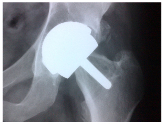
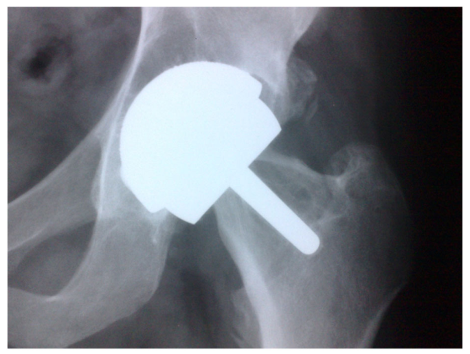
The revision procedures included three conversions to THA for metallosis, two conversions for femoral neck fracture, and one conversion for persistent hip pain associated with acetabular radiolucency (Figure 3).
Figure 3.
Femoral neck fracture identified seven years after HRA.
The remaining complication was one episode of postoperative thrombophlebitis, which did not require implant revision.
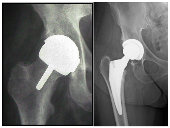
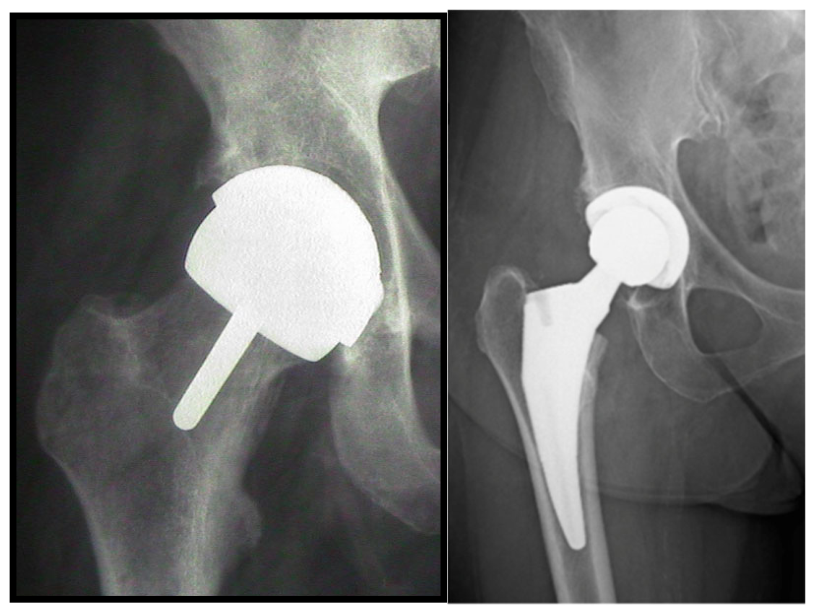
All six failed resurfacing implants were successfully converted to conventional THA, with no major postoperative complications documented following revision (Figure 4).
Figure 4.
Postoperative radiograph obtained after conversion of the failed hip resurfacing arthroplasty to conventional total hip arthroplasty (THA).
Serum cobalt and chromium measurements were not routinely obtained throughout the entire study period, and complete numerical metal-ion data were not available for population-level analysis. Metal-ion testing was performed only when clinically indicated. No clinical manifestations of systemic cobalt or chromium toxicity were documented in the available medical records. The radiographic images associated with the metallosis cases were therefore interpreted as representative imaging of revised HRA rather than as direct evidence of elevated serum metal-ion concentrations.
3.4. Radiographic Outcomes
Radiographic evaluation of the 47 unrevised hips demonstrated stable fixation of both the femoral and acetabular components at final follow-up. No progressive radiolucent lines, clinically significant osteolysis, component migration, subsidence, or aseptic loosening were observed among the retained implants.
One case of symptomatic acetabular radiolucency was identified during follow-up and was treated by conversion to THA; this case was therefore classified as a revision event and was not included among the final radiographic outcomes of the retained implants.
The mean acetabular component inclination was 44.1°, and the mean valgus alignment of the femoral component was 7.2°. Heterotopic ossification was observed in three of the 47 unrevised hips (6.4%). The quantitative radiographic findings are summarized in Table 5.
Table 5.
Radiographic outcomes of the 47 unrevised hips at final follow-up.
3.5. Implant Survivorship
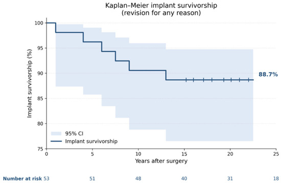
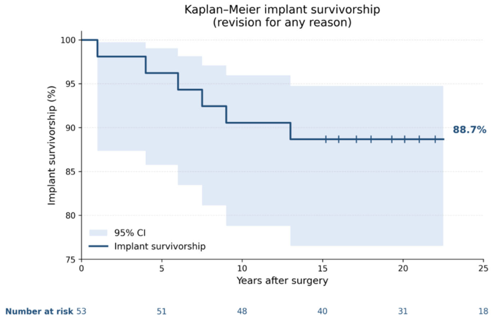
Six revisions of the resurfacing implant were recorded during follow-up. Kaplan–Meier analysis, with revision of any resurfacing component for any reason defined as the endpoint, demonstrated an estimated implant survivorship of 88.7% at approximately 20 years. The corresponding 95% confidence interval was 76.5–94.8%.
All 53 hips were included in the survivorship analysis. Unrevised hips were censored at their latest available clinical or radiographic assessment. The numbers of hips at risk at the predefined follow-up intervals are displayed beneath the Kaplan–Meier curve (Figure 5).
Figure 5.
Kaplan–Meier curve showing implant survivorship after hip resurfacing arthroplasty, with revision of any resurfacing component for any reason defined as the endpoint. Estimated implant survivorship was 88.7% (95% CI, 76.5–94.8%) at approximately 20 years. The numbers of hips at risk are shown below the curve.
4. Discussion
The present retrospective cohort study evaluated the clinical, functional, radiographic, and survivorship outcomes of HRA after a mean follow-up of 20 years. Among the 47 patients who retained their original resurfacing implant, the mean HHS improved from 31.3 ± 17.9 to 95.3 ± 7.4, while the mean WOMAC score decreased from 65.9 ± 10.4 to 11.5 ± 3.5. Six of the 53 hips underwent revision to conventional THA, corresponding to a revision rate of 11.3% and an estimated implant survivorship of 88.7%. Radiographic evaluation of the unrevised hips demonstrated stable component fixation without progressive radiolucency, clinically significant osteolysis, migration, or aseptic loosening. These findings suggest that HRA can provide durable long-term outcomes in carefully selected patients, although the risks associated with metal-on-metal articulation remain clinically relevant.
The substantial improvements in HHS and WOMAC indicate that favorable function and symptom relief may be maintained over an extended period in patients with a retained HRA implant. However, these results must be interpreted in light of the outcome-analysis strategy. Patients converted to THA were excluded from the final clinical analysis because scores obtained after revision could not be attributed to the original resurfacing procedure. Consequently, the final HHS and WOMAC values describe the outcomes of surviving HRA implants and should not be interpreted as representing the entire inception cohort. Previous studies have reported that HRA may provide functional outcomes comparable to conventional THA and, in selected young and physically active patients, may facilitate higher activity levels and more physiological gait patterns [19,20].
The contemporary role of HRA must nevertheless be considered in the context of improvements in conventional THA. Modern ceramic bearings and highly cross-linked polyethylene have substantially improved wear performance and implant durability, including in younger patients. Contemporary THA therefore provides reliable pain relief and long-term function without the metal-ion exposure inherent to metal-on-metal HRA. The potential advantages of resurfacing—including preservation of the femoral neck and proximal femoral bone stock, large-head stability, and restoration of hip biomechanics—must outweigh these bearing-related risks. Accordingly, HRA should not be regarded as a universal alternative to THA but as a selective option for young and highly active patients, particularly men with good bone quality, preserved femoral head and neck anatomy, and high functional demands. Bipolar hemiarthroplasty is not an appropriate comparator for this population because it addresses different indications and patient characteristics.
The extended follow-up represents an important feature of the present study. Although several investigations have reported favorable short- and mid-term outcomes, relatively few studies have evaluated HRA beyond 15–20 years [21,22]. The present series therefore provides additional information regarding implant durability and late complications. The estimated survivorship of 88.7% at approximately 20 years is broadly consistent with long-term findings reported by experienced, high-volume centers, although differences in patient selection, implant design, surgical technique, and surveillance protocols limit direct comparison between studies [21,23].
The complications observed in this cohort reflect the principal limitations of HRA. Two patients developed femoral neck fractures requiring revision to THA. Femoral neck fracture following resurfacing has been associated with inadequate bone quality, femoral neck notching, disruption of femoral head vascularity, and varus positioning of the femoral component [23,24,25]. In the present series, the femoral component was intentionally positioned in slight valgus, and postoperative loading progressed cautiously. Nevertheless, the occurrence of two fractures confirms that this complication cannot be eliminated completely and reinforces the importance of appropriate patient selection, meticulous femoral preparation, and structured postoperative rehabilitation.
Three patients underwent revision for metallosis, emphasizing the biological limitations of metal-on-metal articulation. Elevated cobalt and chromium concentrations, adverse local tissue reactions, metallosis, and pseudotumor formation have contributed substantially to the decline in the use of HRA [24,25,26]. In the present cohort, metal-ion measurements were obtained when clinically indicated rather than through a uniform routine surveillance protocol. Complete numerical metal-ion data were therefore unavailable for population-level analysis, and the radiographs should not be interpreted as direct evidence of elevated serum metal-ion concentrations. No clinical manifestations of systemic cobalt or chromium toxicity were documented in the available records. When adverse reactions to metal debris were suspected, clinical findings, serial metal-ion measurements, radiographs, and, when available, MARS MRI or ultrasonography were considered together. This experience supports the need for long-term surveillance while also representing an important limitation of the study.
All six failed resurfacing implants were converted to conventional THA. Preservation of the femoral canal may facilitate conversion compared with revision of a failed femoral stem and remains a theoretical advantage of resurfacing, particularly in young patients who may require further surgery during their lifetime [27,28]. However, the revised patients were not included in the final HHS and WOMAC analyses, and no post-revision clinical outcomes were attributed to the original HRA. Therefore, the present study cannot establish the functional superiority or relative ease of conversion to THA, and conclusions regarding revision outcomes should remain cautious.
Radiographic assessment of the 47 unrevised hips demonstrated stable fixation at final follow-up. No progressive radiolucent lines, clinically significant osteolysis, component migration, subsidence, or aseptic loosening were observed. One additional patient developed symptomatic acetabular radiolucency and underwent revision to THA; this case was therefore analyzed as a revision event rather than as part of the final radiographic assessment of the retained implants. These findings are consistent with previous long-term reports showing stable fixation among surviving HRA components [29]. Nevertheless, restricting the final radiographic analysis to unrevised implants may produce a favorable survivor profile and should be considered when interpreting these results.
Patient selection is likely to have contributed substantially to the outcomes. The cohort had a mean age of 43.2 years and was predominantly male, with men accounting for 75.5% of the study population. Candidates were selected according to age, functional demands, bone quality, proximal femoral morphology, and preservation of the femoral head and neck. The results are therefore most applicable to young, active patients with favorable anatomy and cannot be generalized to older patients, individuals with impaired bone quality, or populations with a greater proportion of women [30]. The small and unbalanced subgroups, including only nine Durom implants and 13 women, also precluded reliable inferential comparisons according to implant type or sex.
All procedures were performed through a posterolateral Gibson–Moore approach, with repair of the posterior capsule and short external rotators. This repair may have contributed to postoperative stability, whereas the approach itself and the associated soft-tissue dissection may have influenced early recovery. Because no comparison with other surgical approaches was performed, the findings should be generalized cautiously to HRA undertaken through anterolateral, lateral, or anterior approaches.
Alternative bearing technologies may modify the future risk–benefit profile of resurfacing. Ceramic-on-ceramic resurfacing systems and ceramic resurfacing hybrid concepts have been proposed to preserve the biomechanical advantages of HRA while avoiding cobalt and chromium exposure. However, these technologies require robust long-term evidence before they can be considered established alternatives to contemporary THA [31]. At present, metal-on-metal HRA should remain limited to carefully selected patients treated in experienced centers with appropriate long-term surveillance.
Return to sporting activity represents a potentially important advantage of HRA in young patients. Previous studies have reported favorable return-to-sport rates and restoration of physiological gait following resurfacing [19,32]. However, postoperative activity and return to sport were not systematically quantified in the present cohort. Consequently, no study-specific return-to-sport rate can be reported, and the functional advantages observed through HHS and WOMAC should not be interpreted as direct evidence of return to high-impact activity.
This study has several limitations. Its retrospective, single-center design introduces the possibility of selection and information bias. The relatively small sample size, male predominance, young mean age, and highly selected indications limit generalizability. The absence of a contemporary THA control group prevents direct comparison between the procedures. Clinical outcome analysis was restricted to the 47 patients with retained HRA implants, potentially producing a favorable survivor profile. Metal-ion measurements and cross-sectional imaging were not routinely performed in the entire cohort, and return to activity was not systematically recorded. Furthermore, all procedures were performed using a posterolateral approach, and the subgroup sizes were insufficient for reliable comparisons according to sex or implant type.
Despite these limitations, the study includes the complete consecutive cohort of 53 primary HRA procedures performed during the predefined study period, with no patients lost to follow-up. Additional strengths include the approximately 20-year mean follow-up, separate analysis of revised and unrevised hips, standardized radiographic evaluation, and Kaplan–Meier survivorship analysis. Overall, the findings support HRA as a durable but highly selective procedure rather than a general alternative to contemporary THA. Its potential functional and bone-preserving advantages must be balanced against the risks of femoral neck fracture, metal-related adverse reactions, and the need for long-term surveillance.
5. Conclusions
At a mean follow-up of 20 years, HRA was associated with substantial and sustained clinical improvement among the 47 patients who retained their original resurfacing implant. The mean HHS was 95.3 ± 7.4, and the mean WOMAC score was 11.5 ± 3.5 at final follow-up. Six of the 53 hips underwent revision to conventional THA, corresponding to a revision rate of 11.3% and an estimated implant survivorship of 88.7% (95% CI, 76.5–94.8%). The unrevised implants demonstrated stable radiographic fixation without progressive radiolucency, clinically significant osteolysis, migration, or aseptic loosening.
HRA should not be considered a general alternative to contemporary THA. Its potential advantages, including preservation of proximal femoral bone stock, large-head stability, and restoration of hip biomechanics, must be balanced against the risks of femoral neck fracture, metallosis, adverse local tissue reactions, and systemic exposure to metal ions. Its current role should therefore be restricted to carefully selected young and highly active patients, particularly men with good bone quality, preserved femoral head and neck anatomy, and high functional demands. Careful surgical technique and long-term clinical and radiographic surveillance, with metal-ion testing and cross-sectional imaging when clinically indicated, remain essential.
Author Contributions
Conceptualization, G.R., P.P. and D.L.; methodology, G.R., P.P. and D.L.; software, G.R. and S.L.; validation, G.R. and S.L.; investigation, F.L., P.F., G.S. and L.M.; resources, G.R., V.D.L. and F.L.; data curation, P.F., F.D.M., P.P., G.S. and L.M.; visualization, P.F., F.D.M. and P.P.; supervision, G.R.; writing—original draft preparation, G.R., P.P. and D.L.; writing—review and editing, G.R. All authors have read and agreed to the published version of the manuscript.
Funding
This research received no external funding.
Institutional Review Board Statement
All study procedures were conducted in accordance with the ethical principles outlined in the 1964 Helsinki Declaration and its subsequent amendments. Informed consent for the scientific use of anonymized clinical data was obtained from all patients, in accordance with institutional regulations and the guidelines of the Italian Data Protection Authority (“Garante per la protezione dei dati personali”). In compliance with Italian Legislative Decree No. 196/2003, as amended by Legislative Decree No. 101/2018, and Regulation (EU) 2016/679 (GDPR), the use of fully anonymized clinical data for research purposes does not require prior Institutional Review Board approval. Considering that this was a retrospective observational study based exclusively on clinical and radiographic data collected during routine follow-up, that no additional interventions or examinations were performed for research purposes, and that all data were analyzed in fully anonymized form, after consultation with our Internal Review Committee, formal approval from the Regional Ethics Committee was deemed unnecessary.
Informed Consent Statement
Informed consent was obtained from all subjects involved in the study.
Data Availability Statement
The data presented in this study are available on request from the corresponding author. The data are not publicly available due to privacy and ethical restrictions related to patient-level clinical and radiographic information.
Conflicts of Interest
Sandro Luziatelli and Luca Mancini are employed by Villa Letizia, a private healthcare institution. Villa Letizia had no role in the design of the study; in the collection, analysis, or interpretation of the data; in the writing of the manuscript; or in the decision to publish the results. The remaining authors declare no conflicts of interest.
Abbreviations
The following abbreviations are used in this manuscript:
| HRA | Hip Resurfacing Arthroplasty |
| THA | Total Hip Arthroplasty |
| HHS | Harris Hip Score |
| WOMAC | Western Ontario and McMaster Universities Osteoarthritis Index |
| AP | Anteroposterior |
| MoM | Metal on Metal |
| SD | Standard Deviation |
| CI | Confidence Interval |
| IRB | Institutional Review Board |
References
- Learmonth, I.D.; Young, C.; Rorabeck, C. The operation of the century: Total hip replacement. Lancet 2007, 370, 1508–1519. [Google Scholar] [CrossRef] [Scilit] [PubMed]
- Evans, J.T.; Evans, J.P.; Walker, R.W.; Blom, A.W.; Whitehouse, M.R.; Sayers, A. How long does a hip replacement last? A systematic review and meta-analysis of case series and national registry reports. Lancet 2019, 393, 647–654. [Google Scholar] [CrossRef] [Scilit] [PubMed]
- Bayliss, L.E.; Culliford, D.; Monk, A.P.; Glyn-Jones, S.; Prieto-Alhambra, D.; Judge, A.; Cooper, C.; Carr, A.J.; Arden, N.K.; Beard, D.J.; et al. The effect of patient age at intervention on risk of implant revision after total replacement of the hip or knee. The Lancet 2017, 389, 1424–1430, Correction in The Lancet 2017, 389, 1398. [Google Scholar] [CrossRef] [Scilit] [PubMed]
- Mäkelä, K.T.; Eskelinen, A.; Pulkkinen, P.; Paavolainen, P.; Remes, V. Results of 3,668 primary total hip replacements for primary osteoarthritis from the Finnish Arthroplasty Register. Acta Orthop. 2011, 82, 521–529. [Google Scholar] [PubMed]
- McMinn, D.; Treacy, R.; Lin, K.; Pynsent, P. Metal-on-metal surface replacement of the hip: Experience of the McMinn prosthesis. Clin. Orthop. Relat. Res. 1996, 329, S89–S98. [Google Scholar]
- Daniel, J.; Pynsent, P.B.; McMinn, D.J.W. Metal-on-metal resurfacing of the hip in patients under the age of 55 years with osteoarthritis. J. Bone Jt. Surg. Br. 2004, 86, 177–184. [Google Scholar] [CrossRef] [Scilit] [PubMed]
- Amstutz, H.C.; Le Duff, M.J. Hip resurfacing: A review of results and indications. Clin. Orthop. Relat. Res. 2011, 469, 364–371. [Google Scholar]
- Girard, J.; Lavigne, M.; Vendittoli, P.A.; Roy, A.G. Biomechanics of hip resurfacing. Orthop. Clin. N. Am. 2005, 36, 159–165. [Google Scholar]
- Mont, M.A.; Marker, D.R.; Smith, J.M.; Ulrich, S.D.; McGrath, M.S. Resurfacing is comparable to total hip arthroplasty at short-term follow-up. Clin. Orthop. Relat. Res. 2009, 467, 66–71. [Google Scholar] [CrossRef] [Scilit] [PubMed]
- Lavigne, M.; Ganapathi, M.; Mottard, S.; Girard, J.; Vendittoli, P.A. Gait analysis after resurfacing hip arthroplasty versus total hip arthroplasty. J. Arthroplast. 2008, 23, 117–123. [Google Scholar] [CrossRef] [Scilit] [PubMed]
- Ball, S.T.; Le Duff, M.J.; Amstutz, H.C. Early results of conversion of failed hip resurfacing arthroplasty to total hip arthroplasty. J. Bone Jt. Surg. Am. 2007, 89, 735–741. [Google Scholar] [CrossRef] [Scilit] [PubMed]
- Shimmin, A.J.; Back, D. Femoral neck fractures following Birmingham hip resurfacing: A national review of 50 cases. J. Bone Jt. Surg. Br. 2005, 87, 463–464. [Google Scholar]
- Pandit, H.; Glyn-Jones, S.; McLardy-Smith, P.; Gundle, R.; Whitwell, D.; Gibbons, C.L.M.; Ostlere, S.; Athanasou, N.; Gill, H.S.; Murray, D.W. Pseudotumours associated with metal-on-metal hip resurfacings. J. Bone Jt. Surg. Br. 2008, 90, 847–851. [Google Scholar] [CrossRef] [Scilit] [PubMed]
- Hart, A.J.; Sabah, S.A.; Bandi, A.S.; Maggiore, P.; Tarassoli, P.; Sampson, B.; Skinner, J.A. Sensitivity and specificity of blood cobalt and chromium metal ions for predicting failure of metal-on-metal hip replacement. J. Bone Jt. Surg. Br. 2011, 93, 1308–1313. [Google Scholar] [CrossRef] [Scilit] [PubMed]
- Langton, D.J.; Jameson, S.S.; Joyce, T.J.; Hallab, N.J.; Natu, S.; Nargol, A.V.F. Early failure of metal-on-metal bearings in hip resurfacing and large-diameter total hip replacement. J. Bone Jt. Surg. Br. 2010, 92, 38–46. [Google Scholar] [CrossRef] [Scilit] [PubMed]
- Lachiewicz, P.F.; Watters, T.S. The use of hip resurfacing arthroplasty in young patients. J. Am. Acad. Orthop. Surg. 2012, 20, 481–489. [Google Scholar] [PubMed]
- Treacy, R.B.C.; McBryde, C.W.; Pynsent, P.B. Birmingham hip resurfacing arthroplasty: A minimum follow-up of 5 years. J. Bone Jt. Surg. Br. 2005, 87, 167–170. [Google Scholar]
- Amstutz, H.C.; Le Duff, M.J.; Campbell, P.A.; Gruen, T.A.; Wisk, L.E. Clinical and radiographic results of metal-on-metal hip resurfacing with a minimum ten-year follow-up. J. Bone Jt. Surg. Am. 2010, 92, 2663–2671. [Google Scholar] [CrossRef] [Scilit] [PubMed]
- Treacy, R.B.C.; McBryde, C.W.; Pynsent, P.B. Birmingham hip resurfacing arthroplasty: A minimum follow-up of ten years. J. Bone Jt. Surg. Br. 2011, 93, 27–33. [Google Scholar] [CrossRef] [Scilit] [PubMed]
- Rovere, G.; De Mauro, D.; D’Orio, M.; Fulchignoni, C.; Matrangolo, M.R.; Perisano, C.; Ziranu, A.; Pataia, E. Use of muscular flaps for the treatment of hip prosthetic joint infection: A systematic review. BMC Musculoskelet. Disord. 2021, 22, 1059. [Google Scholar] [CrossRef] [Scilit] [PubMed]
- Daniel, J.; Pradhan, C.; Ziaee, H.; Pynsent, P.B.; McMinn, D.J.W. Results of Birmingham hip resurfacing at 12 to 15 years: A single-surgeon series. Bone Jt. J. 2014, 96-B, 1298–1306. [Google Scholar] [CrossRef] [Scilit] [PubMed]
- Matharu, G.S.; Pandit, H.G.; Murray, D.W.; Judge, A. Adverse reactions to metal debris after hip resurfacing. J. Bone Jt. Surg. Am. 2016, 98, 617–626. [Google Scholar] [CrossRef] [Scilit] [PubMed]
- Meschini, C.; Cauteruccio, M.; Oliva, M.S.; Sircana, G.; Vitiello, R.; Rovere, G.; Muratori, F.; Maccauro, G.; Ziranu, A. Hip and knee replacement in patients with ochronosis: Clinical experience and literature review. Orthop. Rev. 2020, 12, 8687. [Google Scholar] [CrossRef] [Scilit] [PubMed]
- Kwon, Y.M.; Ostlere, S.J.; McLardy-Smith, P.; Athanasou, N.A.; Gill, H.S.; Murray, D.W. “Asymptomatic” pseudotumors after metal-on-metal hip resurfacing arthroplasty: Prevalence and metal ion study. J. Arthroplast. 2011, 26, 511–518. [Google Scholar]
- Langton, D.J.; Joyce, T.J.; Jameson, S.S.; Lord, J.; Van Orsouw, M.; Holland, J.P.; Nargol, A.V.F.; De Smet, K.A. Adverse reaction to metal debris following hip resurfacing: The influence of component type, orientation and volumetric wear. J. Bone Jt. Surg. Br. 2011, 93-B, 164–171. [Google Scholar] [CrossRef] [Scilit] [PubMed]
- Rovere, G.; Bosco, F.; Miceli, A.; Ratano, S.; Freddo, G.; D’Itri, L.; Ferruzza, M.; Maccauro, G.; Farsetti, P.; Camarda, L. Adoption of blockchain as a step forward in orthopedic practice. Eur. J. Transl. Myol. 2024, 34, 12197. [Google Scholar] [CrossRef] [Scilit] [PubMed]
- Giustra, F.; Cacciola, G.; Pirato, F.; Bosco, F.; De Martino, I.; Sabatini, L.; Rovere, G.; Camarda, L.; Massè, A. Indications, complications, and clinical outcomes of fixation and acute total hip arthroplasty for the treatment of acetabular fractures: A systematic review. Eur. J. Orthop. Surg. Traumatol. 2024, 34, 47–57. [Google Scholar] [CrossRef] [Scilit] [PubMed]
- McGrath, M.S.; Marker, D.R.; Seyler, T.M.; Ulrich, S.D.; Mont, M.A. Surface replacement is comparable to primary THA in revision scenarios. J. Arthroplast. 2009, 24, 68–74. [Google Scholar]
- Coulter, G.; Young, D.A.; Dalziel, R.E.; Shimmin, A.J. Birmingham hip resurfacing at a mean of ten years: Results from an independent centre. J. Bone Jt. Surg. Br. 2012, 94-B, 315–321. [Google Scholar] [CrossRef] [Scilit] [PubMed]
- Nunley, R.M.; Della Valle, C.J.; Barrack, R.L. Is patient selection important for hip resurfacing? Clin. Orthop. Relat. Res. 2009, 467, 56–65. [Google Scholar] [CrossRef] [Scilit] [PubMed]
- Lin, D.; Xu, J.; Weinrauch, P.; Young, D.A.; De Smet, K.; Manktelow, A.; Beaulé, P.E.; Walter, W.L. Two-Year Results of Ceramic-on-Ceramic Hip Resurfacing in an International Multicenter Cohort. J. Arthroplast. 2024, 39, 2800–2806. [Google Scholar] [CrossRef] [Scilit] [PubMed]
- Naal, F.D.; Maffiuletti, N.A.; Munzinger, U.; Hersche, O. Sports activity after hip resurfacing arthroplasty. Am. J. Sports Med. 2007, 35, 705–711. [Google Scholar] [CrossRef] [Scilit] [PubMed]

Disclaimer/Publisher’s Note: The statements, opinions and data contained in all publications are solely those of the individual author(s) and contributor(s) and not of MDPI and/or the editor(s). MDPI and/or the editor(s) disclaim responsibility for any injury to people or property resulting from any ideas, methods, instructions or products referred to in the content. |
© 2026 by the authors. Licensee MDPI, Basel, Switzerland. This article is an open access article distributed under the terms and conditions of the Creative Commons Attribution (CC BY) license.