


Abstract
Glabridin is a hydrophobic isoflavonoid isolated from licorice that exhibits potent tyrosinase inhibitory activity and is therefore regarded as a promising skin-whitening agent. However, its pharmaceutical application is limited by its poor aqueous solubility and low skin permeability. To improve the dissolution and membrane permeation of glabridin, glabridin-loaded poly(vinyl alcohol) (PVA) nanofibers were prepared by electrospinning. Four PVA grades differing in their degrees of polymerization and hydrolysis were evaluated as nanofiber matrices. Bead-free nanofibers were successfully fabricated regardless of the PVA grade employed. Differential scanning calorimetry and powder X-ray diffraction analyses indicated that glabridin was predominantly present in an amorphous state within the nanofiber matrix, while ATR-FTIR spectroscopy suggested possible intermolecular interactions between glabridin and PVA. In vitro membrane permeation studies using Strat-M® membranes demonstrated that the nanofiber formulations enhanced the apparent membrane permeation of glabridin relative to the control formulation. In addition, the release and membrane permeation behavior of glabridin depended on the PVA grade, reflecting differences in the dissolution characteristics of the polymer matrix. Collectively, these findings suggest that electrospun PVA nanofibers provide a promising formulation platform for improving glabridin delivery while enabling control of its release and membrane permeation through appropriate selection of the PVA matrix.
1. Introduction
Glabridin is an isoflavonoid isolated from the roots and rhizomes of licorice species, including Glycyrrhiza glabra L [1,2]. Glabridin has attracted considerable attention because of its diverse biological activities, including antioxidant, anti-inflammatory, antimicrobial, anti-obesity, and skin-whitening effects [1,2]. Consequently, it has been extensively investigated for pharmaceutical and cosmetic applications [1,3,4,5,6]. Among these biological activities, the skin-whitening effect of glabridin has been studied most extensively and is primarily attributed to its inhibition of tyrosinase, a key enzyme involved in melanin biosynthesis [1,5,6,7,8,9,10]. Previous studies have demonstrated that glabridin exhibits potent tyrosinase inhibitory activity comparable to that of several commercially available whitening agents [11].
For cosmetic and dermatological applications, topical delivery of glabridin is advantageous because skin hyperpigmentation disorders, such as freckles and age spots, originate locally within the skin. However, its practical application in topical formulations remains challenging because of its poor aqueous solubility and limited topical delivery [1,5,12]. Human skin possesses multiple barrier structures, including the stratum corneum and tight junctions within the epidermis, which restrict the transport of exogenous compounds [13,14]. In addition, glabridin exhibits relatively high lipophilicity, with a reported logP value exceeding 3 [4,15], which may limit its distribution into the hydrophilic layers beneath the stratum corneum [16,17,18,19]. Therefore, formulation strategies that improve the dissolution and delivery behavior of glabridin are required.
Several approaches have been explored to improve glabridin delivery, including formulations containing emulsifiers, organic solvents, and penetration enhancers, as well as microneedle-based systems [1,5,6,20]. Although these strategies can enhance the solubilization and delivery of glabridin, excessive use of organic additives may cause skin irritation and hypersensitivity. Microneedle-based systems, while highly effective, require physical disruption of the skin barrier and may also present challenges related to invasiveness and manufacturing costs. Consequently, there remains a need for a safe, noninvasive, and skin-friendly formulation that improves glabridin delivery.
Because of their high specific surface area and ability to incorporate poorly water-soluble compounds into polymer matrices, electrospun nanofibers have emerged as promising drug delivery platforms [21,22]. Nanofibers produced by electrospinning often function as solid dispersion systems, in which incorporated compounds are molecularly dispersed or amorphized within the polymer matrix [23]. Consequently, the dissolution behavior of poorly water-soluble compounds can be enhanced. Electrospun nanofibers can also be fabricated as thin, flexible sheet-type dosage forms. When composed of water-soluble polymers, these sheets rapidly dissolve or disintegrate upon contact with moisture [24,25,26], making them attractive for topical delivery applications [26,27]. In addition, recent studies have demonstrated that electrospun nanofiber formulations improve both the dissolution and membrane permeation of poorly water-soluble compounds [21,22,28,29].
Among the available matrix polymers, poly(vinyl alcohol) (PVA) was selected for the present study. PVA is a water-soluble, biocompatible, low-toxicity polymer that has been widely used in pharmaceutical and food applications [30,31]. Aqueous PVA solutions can be directly used for electrospinning, thereby reducing the reliance on the organic solvents commonly required for processing many water-insoluble biodegradable polymers, such as biodegradable polyesters. Although ethanol was added in the present study to incorporate the poorly water-soluble glabridin-containing material, the main polymer solution was aqueous. The physicochemical properties, including water solubility and dissolution behavior, vary according to the degree of polymerization and hydrolysis, and numerous grades are commercially available [30,31,32,33,34,35]. This grade-dependent tunability makes it possible to modify formulation performance without changing the fundamental chemical structure of the polymer. In addition, the water-responsive dissolution behavior of PVA nanofiber sheets may enable the development of topical formulations that dissolve following application with a small amount of water, without requiring removal as an intact sheet. Thus, selecting an appropriate PVA grade may provide a practical means to modulate the release characteristics of incorporated compounds. These properties formed the basis for selecting PVA as the nanofiber matrix in the present study.
Based on this rationale, glabridin-loaded PVA nanofiber sheets were prepared by electrospinning, and the effects of glabridin loading and PVA grade on physicochemical properties, drug release behavior, and membrane permeation were investigated. To evaluate their potential as topical delivery formulations, Strat-M® membranes were employed as a reproducible model membrane system [36,37,38].
2. Materials and Methods
2.1. Materials
Polyol Licorice Extract HG (containing 4% glabridin in butylene glycol) was purchased from Maruzen Pharmaceuticals Co., Ltd. (Hiroshima, Japan). A glabridin reference standard was purchased from Tokyo Chemical Industry Co., Ltd. (Tokyo, Japan). Four grades of poly(vinyl alcohol) (PVA), GOHSENOL™ EG-05P, EG-40P, KH-20, and NH-18, were used as matrix polymers for nanofiber fabrication and were kindly supplied by Mitsubishi Chemical Corporation (Tokyo, Japan). The characteristics of the PVA grades used in this study are summarized in Table 1. Ethanol (99.5%) was purchased from Fujifilm Wako Pure Chemical Corporation (Osaka, Japan). Acetonitrile (HPLC grade, 99.8%) was purchased from Kishida Chemical Co., Ltd. (Osaka, Japan).
Table 1.
Characteristics of the PVA grades used in this study.
2.2. Fabrication of PVA Nanofibers by Electrospinning
Aqueous PVA solutions were prepared by dissolving PVA in distilled water under magnetic stirring overnight at temperatures above 85 °C. PVA concentrations were adjusted to 25% (w/w) for EG-05P and 8% (w/w) for EG-40P, KH-20, and NH-18. The compositions of the electrospinning solutions are summarized in Table 2. Briefly, Polyol Licorice Extract HG (glabridin BG solution) was first mixed with ethanol, and the resulting mixture was then added to the PVA aqueous solutions after they had cooled to room temperature. For Exp. 2–5, 7, and 9, the amount of ethanol was adjusted to achieve a distilled water-to-ethanol mass ratio of 9:1, based on the amount of distilled water contained in the PVA solution. For Exp. 11, which contained fully hydrolyzed NH-18, the amount of ethanol was reduced to prevent precipitation of the PVA. The prepared solutions were loaded into 5 mL syringes (Terumo Corporation, Tokyo, Japan) connected via Teflon tubing to 22G nonbeveled needles (15 mm in length; MECC Co., Ltd., Fukuoka, Japan). Under the conditions summarized in Table 2, electrospinning was carried out using a NANON-04 electrospinning apparatus (MECC Co., Ltd., Fukuoka, Japan). Because changes in the PVA grade and solution composition affected the electrospinning behavior, the applied voltage and feed rate were adjusted slightly when necessary. The preparation conditions were selected to obtain continuous, bead-free nanofibers that could be collected as self-supporting sheets. To maintain low humidity throughout fabrication, a desiccant dehumidifier (F-YZVX, Panasonic Corporation, Tokyo, Japan) was connected to the electrospinning system, while ambient temperature and relative humidity were continuously monitored and recorded. Glabridin-free blank nanofibers were also prepared following the same procedure as reference controls for evaluating the morphological and physicochemical changes associated with the incorporation of the glabridin-containing solution. To confirm glabridin incorporation, the fabricated nanofibers were dissolved in a water/ethanol mixture and analyzed by HPLC. The HPLC conditions are described in Section 2.6.
Table 2.
Composition of electrospinning solutions, fabrication conditions, and average fiber diameters of prepared nanofibers.
2.3. Morphological Evaluation of Nanofibers
The morphology of the prepared nanofiber sheets was examined using a scanning electron microscope (JSM-6510LV, JEOL Ltd., Tokyo, Japan). After mounting the samples on specimen stubs with double-sided carbon tape, they were sputter-coated with gold using a Smart Coater (DII-29010SCTR, JEOL Ltd., Tokyo, Japan). SEM images were acquired at an accelerating voltage of 10 kV. Fiber diameter was determined by measuring 100 randomly selected fibers using ImageJTM software (version 1.54d, National Institutes of Health, Bethesda, MD, USA), and the results are presented as mean ± SD.
2.4. Thermal and Crystalline Analysis
To evaluate the thermal properties of glabridin in the nanofibers, differential scanning calorimetry was performed using a DSC8000 instrument (PerkinElmer, Waltham, MA, USA). Samples (4–7 mg) were sealed in aluminum pans and heated from 50 °C to 250 °C at a rate of 10 °C/min under a nitrogen flow of 20 mL/min. The physical state of glabridin in the nanofibers was further characterized by powder X-ray diffraction (PXRD). Using a D8 ADVANCE diffractometer (Bruker, Billerica, MA, USA) equipped with Cu Kα radiation (λ = 1.5406 Å) and operated at 40 kV and 40 mA, diffraction patterns were recorded over a 2θ range of 5–40° with a step size of approximately 0.018°.
2.5. ATR-FTIR Spectroscopy
To investigate possible intermolecular interactions between glabridin and PVA in the nanofibers, attenuated total reflectance Fourier transform infrared spectroscopy was performed. ATR-FTIR spectra were collected using a Frontier FT-IR spectrometer (PerkinElmer, Inc., Waltham, MA, USA) equipped with a diamond/ZnSe crystal over a wavenumber range of 4000–650 cm−1 at a spectral resolution of 4 cm−1 by averaging four scans for each spectrum.
2.6. In Vitro Release and Permeation Studies
In vitro release and membrane permeation studies were performed using vertical Franz diffusion cells equipped with Strat-M® membranes (Merck Millipore, Burlington, MA, USA). The effective diffusion area and receptor volume were 1.77 cm2 and 10 mL, respectively. Based on a previous report [39], a mixture of distilled water and acetonitrile (60:40, v/v) was selected as the receptor medium. Liu et al. [39] used a receptor medium containing 38% (v/v) acetonitrile to maintain sink conditions for glabridin. Because the receptor medium used in the present study contained a nearly identical proportion of acetonitrile (40%, v/v), it was considered to provide sink conditions for glabridin. Throughout the experiment, the receptor medium was maintained at 32 ± 1 °C and continuously stirred at 600 rpm. Approximately 10 mg of the nanofiber sample was placed on the membrane surface, after which 200 μL of distilled water was added. As a control, Polyol Licorice Extract HG diluted 100-fold with distilled water was applied at an equivalent glabridin dose. At 0.5, 1, 1.5, and 3 h, 300 μL of the receptor medium was withdrawn and immediately replaced with an equal volume of fresh medium. For Exp. 3, additional samples were collected at 6, 9, and 24 h. The collected samples were filtered through a 0.45 μm PTFE membrane filter and analyzed by HPLC (JASCO Corporation, Tokyo, Japan). The cumulative amount of permeated glabridin was corrected for the amount removed during sampling. HPLC analysis was performed using a reverse-phase column (Inertsil ODS-3, 4.6 × 250 mm, 5 μm; GL Sciences Inc., Tokyo, Japan) with a mobile phase consisting of acetonitrile and 2.0% (v/v) aqueous acetic acid solution (60:40, v/v) at a flow rate of 1.3 mL/min. The column temperature was maintained at 40 °C, and glabridin was detected at 282 nm. The chromatographic conditions were adapted from an HPLC method for glabridin described in a report by the Ministry of Health, Labour and Welfare of Japan [40]. The calibration curve showed good linearity over the concentration range of 0.03–60 μg/mL (Figure S1). For the present analysis, 0.03 μg/mL, corresponding to the lowest concentration of the calibration curve, was used as the limit of quantification (LOQ). Values below the LOQ were treated as zero.
3. Results
3.1. Preparation and Morphology of Glabridin-Loaded PVA Nanofibers
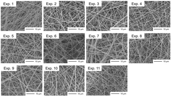
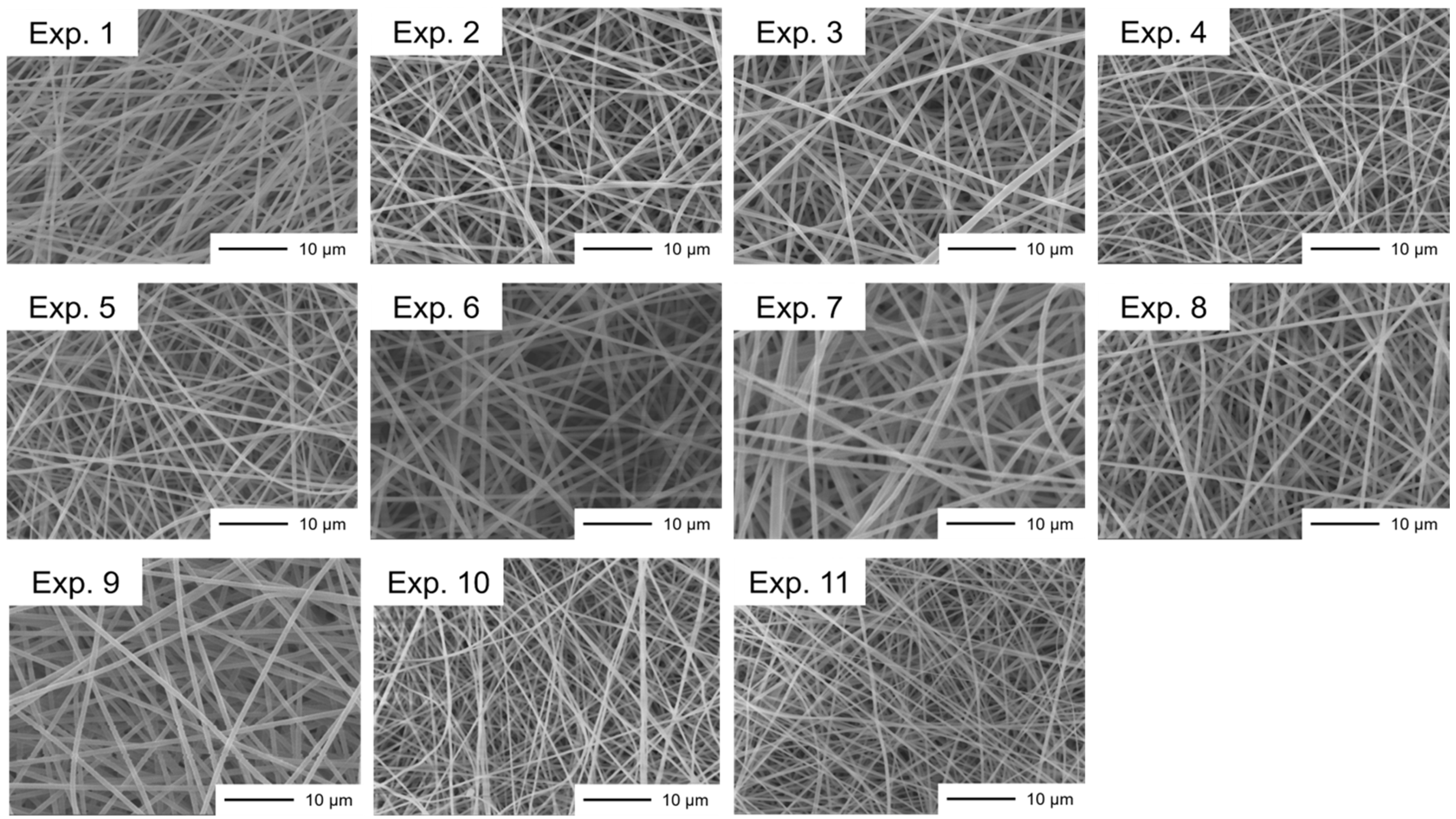
The formulations investigated in this study are summarized in Table 2. EG-05P and EG-40P were selected to evaluate the influence of the PVA degree of polymerization on nanofiber properties. In addition, KH-20 and NH-18/EG-40P formulations were included to examine the effects of the degree of hydrolysis. Although the absolute amount of glabridin BG solution differed among the formulations, Exp. 3, Exp. 7, Exp. 9, and Exp. 11 were designed to have the same theoretical glabridin-to-PVA weight ratio. The different absolute amounts resulted from the different PVA concentrations required to obtain electrospinnable solutions for each PVA grade. For the EG-05P-based formulations (Exp. 2–5), the glabridin loading level was varied to investigate its influence on nanofiber characteristics and membrane permeation behavior. Representative SEM images of the fabricated nanofibers are shown in Figure 1. Regardless of the PVA grade or glabridin loading, all formulations exhibited bead-free fibrous structures, indicating the successful fabrication of nanofibers by electrospinning. The average fiber diameters are summarized in Table 2. For the EG-05P- and NH-18/EG-40P-based formulations, glabridin loading had little effect on fiber diameter. In contrast, glabridin loading increased the average fiber diameter in the EG-40P- and KH-20-based formulations. The glabridin content of the nanofibers was determined by HPLC analysis, and the measured values were generally consistent with the theoretical values for most formulations.
Figure 1.
SEM images of PVA nanofibers prepared using different PVA grades and glabridin loading levels. All images were acquired at a magnification of 2500×. Scale bar = 10 μm.
3.2. Thermal and Crystalline Properties of Glabridin-Loaded PVA Nanofibers
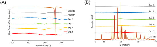
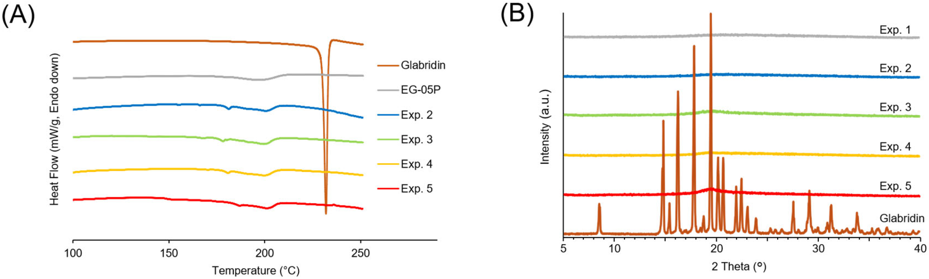
Figure 2A shows the DSC thermograms of the EG-05P-based nanofibers. Raw glabridin exhibited a sharp endothermic peak at 230.0 °C. Following electrospinning, however, no corresponding peak was detected in any nanofiber formulation. Instead, all nanofiber samples exhibited a broad endothermic peak around 200 °C. The DSC thermograms of the other formulations are presented in Figure S2A and showed similar profiles. Figure 2B shows the PXRD patterns of the EG-05P-based nanofibers. Although raw glabridin exhibited several characteristic diffraction peaks, all nanofiber formulations displayed broad diffraction patterns without distinct crystalline peaks. Similar PXRD patterns were also obtained for the other formulations (Figure S2B).
Figure 2.
Thermal and crystalline properties of glabridin-loaded nanofibers. (A) DSC thermograms of raw glabridin, EG-05P powder, and glabridin-loaded EG-05P nanofibers (Exp. 2–5). (B) PXRD patterns of raw glabridin, blank EG-05P nanofibers (Exp. 1), and glabridin-loaded EG-05P nanofibers (Exp. 2–5).
3.3. ATR-FTIR Spectra of Glabridin-Loaded Nanofibers
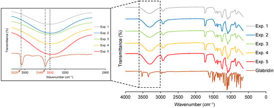
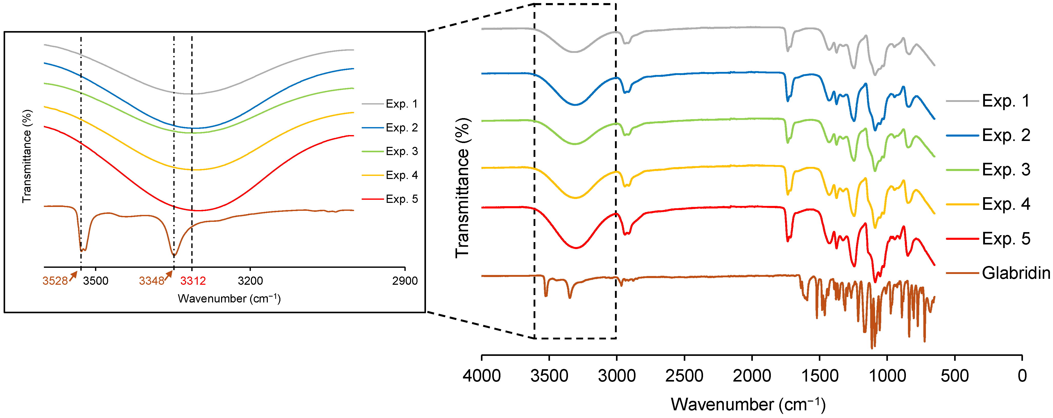
Figure 3 shows the ATR-FTIR spectra of the EG-05P-based nanofibers. The spectra are vertically offset for clarity. Raw glabridin exhibited characteristic absorption peaks at 3528 and 3348 cm−1. In contrast, the spectrum of the blank EG-05P nanofibers (Exp. 1) exhibited a broad absorption band at 3312 cm−1. Following glabridin loading, this band shifted to 3310, 3307, 3310, and 3301 cm−1 for Exp. 2–5, respectively. ATR-FTIR spectra of the other formulations are presented in Figure S3. Similar shifts in the broad absorption band within the 3000–3500 cm−1 region were observed following glabridin loading. In addition, differences in the spectral profiles within the 950–1750 cm−1 region were observed among the various PVA-based nanofiber formulations.
Figure 3.
ATR-FTIR spectra of raw glabridin and EG-05P-based nanofibers. Spectra are vertically offset for clarity. The analyzed samples included raw glabridin, blank EG-05P nanofibers (Exp. 1), and glabridin-loaded EG-05P nanofibers (Exp. 2–5). The inset shows an enlarged view of the 3000–3600 cm−1 region. The characteristic O–H stretching peaks of raw glabridin at 3528 and 3348 cm−1 and the broad O–H stretching band of the blank EG-05P nanofibers at approximately 3312 cm−1 are indicated.
3.4. In Vitro Release and Membrane Permeation Studies
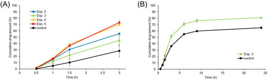
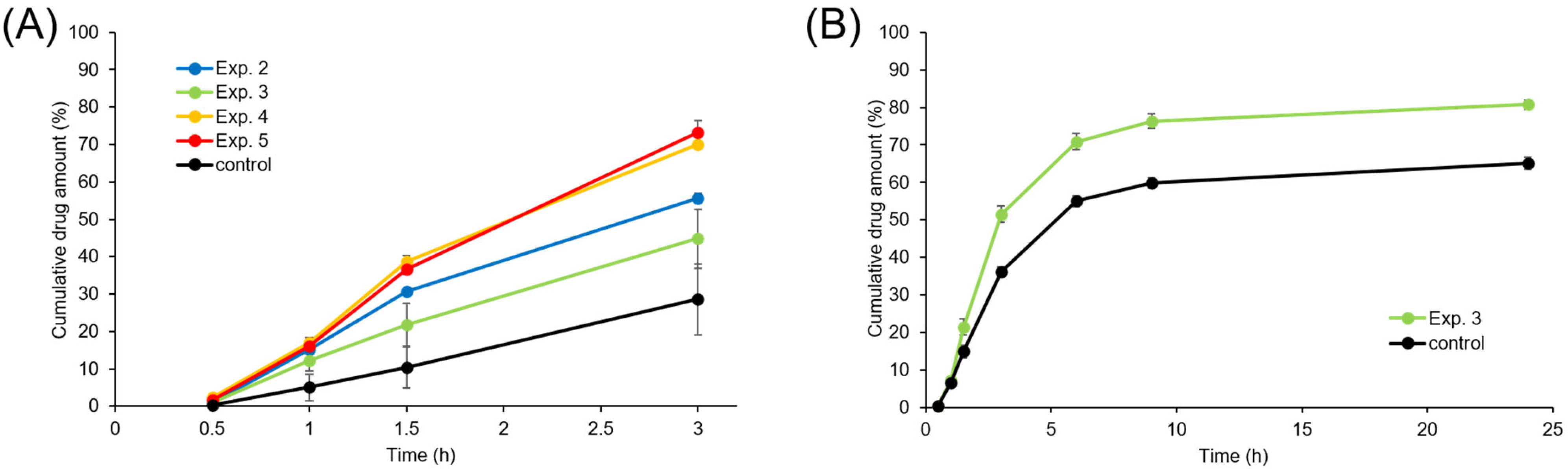
The in vitro release and membrane permeation profiles of the EG-05P-based nanofibers are shown in Figure 4. Figure 4A compares formulations containing different amounts of glabridin (Exp. 2–5) with the control formulation over a 3 h period. Relative to the control formulation, all nanofiber formulations exhibited greater cumulative permeation, which generally increased with increasing glabridin loading.
Figure 4.
Cumulative membrane permeation profiles of glabridin from the EG-05P nanofiber formulations and control through the Strat-M® membrane: (A) 3-h comparison of Exp. 2–5 and (B) independent 24-h evaluation of Exp. 3. Panel B was obtained from an experiment conducted independently of that shown in panel (A), using a separately prepared batch of Exp. 3 and an independently measured control. Data are presented as mean ± SD. (A) Nanofiber formulations, n = 3; control, n = 12. (B) n = 3.
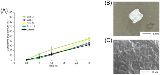
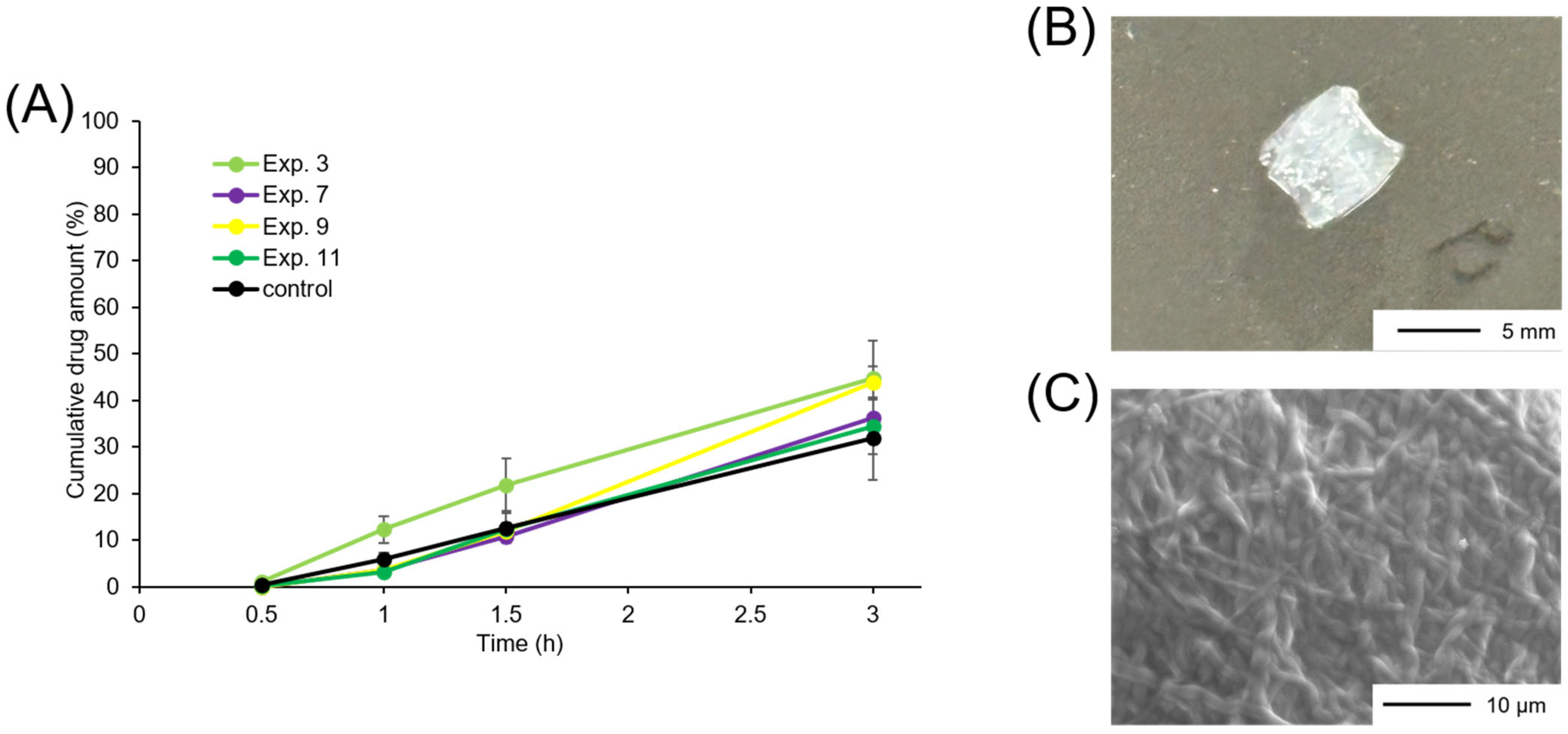
Figure 4B presents the results of an extended 24 h study of Exp. 3 and the control formulation. The cumulative permeation increased up to 6 h and subsequently approached a plateau. After 24 h, the cumulative permeation of Exp. 3 reached approximately 80% and remained higher than that of the control formulation. At the end of the 24-h study, the Exp. 3 nanofiber sheet was no longer visually discernible as an intact sheet on the membrane surface. The effects of PVA grade on glabridin permeation are shown in Figure 5A. During the early stage of the study, EG-05P (Exp. 3) exhibited greater cumulative permeation than the control formulation. In contrast, EG-40P (Exp. 7), KH-20 (Exp. 9), and NH-18/EG-40P (Exp. 11) showed lower cumulative permeation than the control formulation up to 1.5 h, although their cumulative permeation exceeded that of the control formulation after 3 h. Among these formulations, cumulative permeation at 3 h was highest for KH-20, followed by EG-40P and NH-18/EG-40P.
Figure 5.
Effect of PVA grade on the in vitro release and membrane permeation behavior of glabridin-loaded nanofibers. (A) Membrane permeation profiles of Exp. 3 (EG-05P), Exp. 7 (EG-40P), Exp. 9 (KH-20), and Exp. 11 (NH-18/EG-40P). Data are presented as mean ± SD (nanofiber formulations, n = 3; control, n = 12). (B) Photograph of Exp. 11 after the permeation study. (C) SEM image of Exp. 11 recovered after the permeation study.
Representative images of NH-18/EG-40P nanofibers (Exp. 11) after the permeation study are shown in Figure 5B, and SEM images of the recovered sample are shown in Figure 5C. Unlike the EG-05P nanofibers, which completely dissolved during the study, the Exp. 11 nanofiber sheet retained its overall morphology after 24 h. Furthermore, the recovered fibers appeared thicker than those observed before the permeation study.
4. Discussion
Glabridin possesses tyrosinase inhibitory activity and is expected to exert skin-whitening effects by suppressing melanin production in the skin [1,5,6,7,8,9]. However, its pharmaceutical application has been limited by its poor aqueous solubility and low transdermal absorbability [1,5,12]. To address these limitations, glabridin-loaded PVA nanofibers were fabricated by electrospinning, and the effects of nanofiber formation and PVA grade on membrane permeation behavior were investigated.
The glabridin-loaded PVA nanofibers were prepared by blend electrospinning. Upon application of a high voltage, the droplet at the needle tip deforms into a Taylor cone when the electrostatic force overcomes the surface tension, followed by the ejection of a charged liquid jet. The jet is elongated and thinned under the electric field, while the solvents evaporate during its travel toward the collector, resulting in the solidification of PVA into continuous fibers [41,42,43]. During this process, glabridin becomes dispersed within the PVA matrix. Consequently, the fibers deposited on the collector subsequently accumulate and overlap to form a nonwoven, self-supporting nanofiber sheet.
Bead formation during electrospinning is influenced by solution properties, particularly polymer concentration, viscosity, and polymer-chain entanglement, as well as process parameters such as the applied voltage and feed rate [44]. In the present study, the PVA concentration was selected according to the PVA grade to ensure continuous fiber formation. It should be noted that the ethanol content and some electrospinning parameters, including the applied voltage and feed rate, differed among the formulations. These conditions were adjusted when necessary to obtain continuous, bead-free nanofibers that could be collected as self-supporting sheets. Nevertheless, these differences may also have contributed to the observed nanofiber properties; therefore, the effects cannot be attributed exclusively to the PVA grade. Further studies that systematically control and independently evaluate the formulation and process parameters will be necessary to more precisely determine the effects of the PVA grade.
Ethanol was selected as an organic solvent because it is classified as a Class 3 solvent with low toxic potential according to the ICH guideline and is highly volatile. Ethanol has also been used in previous electrospinning studies involving poorly water-soluble drugs [45,46]. After electrospinning, the nanofibers appeared dry and could be collected as self-supporting sheets. However, the residual ethanol content was not quantitatively determined in the present study; therefore, complete removal of ethanol could not be confirmed. Future studies should quantitatively evaluate residual ethanol and determine whether additional post-electrospinning drying is required.
Overall, electrospinning successfully produced bead-free nanofibers regardless of the PVA grade employed. In addition, the measured glabridin contents were generally close to the theoretical values, indicating efficient incorporation of glabridin into the nanofiber matrices. Together, these findings demonstrate that glabridin-loaded nanofibers can be prepared using a range of PVA grades with different physicochemical properties.
A key advantage of electrospun nanofibers is their ability to maintain incorporated drugs in an amorphous state [23]. Consistent with this property, the characteristic melting and crystalline diffraction peaks of glabridin disappeared after electrospinning, indicating a loss of crystallinity within the nanofiber matrix. Accordingly, glabridin was considered to be predominantly present in an amorphous state in the fabricated nanofibers. The broad endothermic event observed around 200 °C was considered to be associated with the thermal behavior of PVA [31,47,48]. Minor endothermic events were also observed below 200 °C. Because the DSC profiles in this temperature range contained broad and overlapping thermal events, their origin could not be unambiguously determined by comparison with the PVA raw materials alone. These events may reflect the thermal behavior of PVA, changes associated with electrospinning, and/or components introduced by the commercial glabridin BG solution. Furthermore, grade-matched blank nanofibers were not analyzed by DSC. Additional thermal analyses using the corresponding blank nanofibers and glabridin BG solution would therefore be required to clarify the origin of these events.
Further evidence of interactions between glabridin and PVA was provided by the ATR-FTIR spectra. Following glabridin loading, the broad absorption band around 3300 cm−1 in the PVA nanofibers shifted slightly toward lower wavenumbers relative to the characteristic O–H stretching bands of raw glabridin. Such shifts are commonly associated with intermolecular interactions involving hydroxyl groups and may therefore reflect hydrogen-bond formation between glabridin and PVA. The differences in spectral profiles observed among the various PVA grades are likely attributable to differences in the degree of hydrolysis and, consequently, the ratio of hydroxyl and residual acetyl groups within the polymer structure.
Regarding the permeability evaluation, although in vitro release and membrane permeation were evaluated using Strat-M® membranes, the receptor medium contained acetonitrile to maintain sink conditions. Therefore, the present study should be regarded as a comparative membrane permeation model rather than a direct simulation of physiological skin conditions.
In this system, the control formulation exhibited substantially lower permeation. During preparation of the control samples, glabridin precipitation was visually observed after dilution of the glabridin BG solution with water. Consequently, only a fraction of the administered glabridin was likely present in a dissolved state and available for membrane permeation. In contrast, all nanofiber formulations exhibited greater membrane permeation compared with the control formulation. The most plausible explanation for this improvement is the amorphization of glabridin during electrospinning. Because amorphous drugs generally exhibit higher solubility and faster dissolution than their crystalline counterparts [49], glabridin incorporated into the nanofibers likely dissolved more readily in the small volume of water applied to the donor chamber. As a result, a greater amount of dissolved glabridin became available for membrane permeation. Therefore, this difference in the amount of dissolved glabridin available at the membrane surface may account for the higher permeation observed with the nanofiber formulations. Furthermore, the increase in permeation with increasing glabridin loading is consistent with Fick’s law, whereby a larger concentration gradient between the donor and receptor compartments promotes drug transport across the membrane. Hespeler et al. reported that amorphous, nanodispersed glabridin incorporated into mesoporous silica exhibited a more than four-fold increase in saturation solubility and a markedly increased dissolution rate compared with crystalline glabridin [6]. In the present study, the increased dissolved concentration resulting from the amorphous state of glabridin may have contributed to its enhanced permeation across the Strat-M® membrane. However, the observed enhancement cannot be attributed exclusively to amorphization because other formulation characteristics, including the hydrophilicity and dissolution or gel-forming behavior of the PVA matrix, may also affect glabridin release and membrane permeation. Therefore, whether glabridin amorphized by another method or incorporated into a polymer matrix other than PVA would exhibit a similar improvement in membrane permeation remains to be determined.
As another potential factor, fiber diameter influences the specific surface area of nanofibers and may consequently affect polymer dissolution and drug release. In the present study, all formulations consisted of continuous, bead-free fibers in the submicron range. However, because fiber diameter was not independently controlled, its contribution to glabridin release and membrane permeation could not be determined. Control of the mean fiber diameter and its variation will be important for ensuring consistent product quality in future development.
The results also demonstrated that the membrane permeation behavior of glabridin could be modulated by changing the PVA grade used as the nanofiber matrix. The membrane permeation behavior of glabridin varied among the PVA grades. EG-05P exhibited relatively rapid permeation during the early stage, whereas EG-40P, KH-20, and NH-18/EG-40P showed delayed permeation. These differences may be related to the hydration, swelling, and dissolution behavior of the PVA matrix. EG-05P has a lower degree of polymerization than EG-40P and is therefore expected to hydrate and dissolve more rapidly because of reduced chain entanglement and intermolecular interactions. In contrast, the higher degrees of polymerization of EG-40P and KH-20 may retard hydration and dissolution of the nanofiber matrix.
The aqueous behavior of PVA is also affected by its degree of hydrolysis, which increases in the order of KH-20, EG-40P, and NH-18. A low degree of hydrolysis increases the proportion of hydrophobic residual acetate groups, whereas fully hydrolyzed PVA forms extensive intra- and intermolecular hydrogen bonds and has higher crystallinity. Thus, the relationship between the degree of hydrolysis and water solubility is not necessarily monotonic, and both low-hydrolysis and fully hydrolyzed grades may exhibit slower hydration or dissolution than an appropriate partially hydrolyzed grade. Our previous studies using different PVA grades demonstrated that these characteristics affected the dissolution and release performance of electrospun PVA nanofibers [22,33,50]. These relationships have also been summarized in our recent review of the pharmaceutical applications of PVA nanofibers [51]. Furthermore, Jackson et al. demonstrated that blending PVA grades with different degrees of hydrolysis enabled control of the dissolution of PVA films and electrospun nanofiber mats, resulting in sustained release of incorporated silver salts [52]. Thus, differences in the hydration, swelling, and dissolution behavior of the PVA matrix likely contributed to the observed differences in glabridin release and subsequent permeation through the Strat-M® membrane. However, the individual contributions of release from the nanofibers and subsequent transport through the Strat-M® membrane could not be distinguished in the present experimental system.
Although fully hydrolyzed PVA is attractive for controlling the dissolution behavior of nanofibers, preparing nanofibers using fully hydrolyzed PVA alone can be challenging because of its high solution viscosity and limited spinnability. In our previous study, stable electrospinning of fully hydrolyzed PVA alone was difficult because of frequent nozzle clogging, and blending with partially hydrolyzed PVA was required to obtain uniform nanofibers [50]. Therefore, the NH-18/EG-40P formulation employed in the present study was selected as a practical approach to utilize the advantages of fully hydrolyzed PVA while maintaining sufficient electrospinnability.
Visual observations after the permeation study supported this interpretation. The EG-05P-based nanofiber sheet was no longer visually discernible as an intact sheet after the 24-h study. Because EG-05P is a partially hydrolyzed, water-soluble PVA grade, prolonged contact with water likely resulted in the loss of structural integrity and dissolution or disintegration of the nanofiber sheet. However, complete dissolution was not quantitatively confirmed. In contrast, the EG-40P- and KH-20-based nanofibers remained as gel-like residues, while the NH-18/EG-40P nanofiber sheet retained its overall morphology after 24 h. SEM observations revealed that the recovered fibers were thicker than those before the study. These findings suggest that dissolution of the polymer matrix was suppressed while water uptake occurred, resulting in swelling of the nanofibers. Consequently, the overall delivery behavior of glabridin, including its release from the nanofiber matrix and subsequent membrane permeation, may be regulated by selecting the PVA grade to control the dissolution characteristics of the polymer matrix.
However, it should be noted that the initial comparison of the formulations was limited to 3 h because this experiment was designed as an exploratory screening of their early glabridin membrane permeation profiles. Exp. 3 was subsequently selected as a representative EG-05P formulation and evaluated for up to 24 h. Although the Exp. 11 sheet containing fully hydrolyzed PVA retained its overall morphology after 24 h, quantitative membrane permeation profiles beyond 3 h were not obtained for Exp. 11 or the other formulations used to compare the effects of PVA grade. Therefore, the present results do not allow a comprehensive comparison of longer-term release and permeation behavior among the different PVA grades. Further studies evaluating all relevant formulations over a common 24-h period are warranted.
Taken together, these results demonstrate that electrospun PVA nanofibers can improve the membrane permeation of glabridin while providing a platform for controlling drug release and membrane permeation behavior through PVA grade selection.
To build upon these findings, several limitations of the present study should be addressed in future work. First, future studies using excised biological skin are required to verify whether the trends observed in the Strat-M® model are maintained under physiologically relevant conditions. Second, because the water contact angle was not measured in the present study, the effects of PVA grade and glabridin loading on nanofiber wettability should be evaluated using time-dependent measurements appropriate for water-soluble PVA nanofibers [21]. Third, exploratory kinetic analysis did not allow a definitive interpretation of the glabridin release mechanism or a quantitative explanation of the differences among PVA grades. Because the measured profiles reflect both drug release from the nanofiber sheets and subsequent permeation through the Strat-M® membrane, further studies specifically designed to evaluate release kinetics are required. Fourth, although all formulations could be collected and handled as self-supporting sheets, quantitative mechanical testing was not performed; thus, their tensile properties and the effects of PVA grade and glabridin loading should be evaluated. Fifth, the storage stability of the glabridin-loaded nanofibers was not evaluated, necessitating future examinations of their morphology, glabridin content, physical state, and release/membrane permeation behavior after storage under defined conditions.
Finally, the pharmacological performance of glabridin-loaded nanofibers should be evaluated in biologically relevant models. We believe that these future investigations will enhance the understanding and clinical potential of glabridin-loaded nanofibers.
5. Conclusions
In this study, glabridin-loaded PVA nanofibers were successfully fabricated by electrospinning using various PVA grades. DSC and PXRD analyses indicated that glabridin was predominantly present in an amorphous state within the nanofiber matrix, while ATR-FTIR spectroscopy suggested possible intermolecular interactions between glabridin and PVA. Membrane permeation studies using Strat-M® membranes demonstrated that the nanofiber formulations enhanced the apparent membrane permeation of glabridin relative to the control formulation. In addition, selecting different PVA grades modulated the release and membrane permeation behavior of glabridin, reflecting differences in the dissolution characteristics of the polymer matrix. Collectively, these findings demonstrate that electrospun PVA nanofibers provide a promising formulation platform for improving the delivery of poorly water-soluble compounds while enabling control of their release and membrane permeation behavior through selection of the polymer matrix.
Supplementary Materials
The following supporting information can be downloaded at https://www.mdpi.com/article/10.3390/app16189258/s1. Figure S1: Calibration curve for the HPLC determination of glabridin over the concentration range of 0.03–60 μg/mL. Figure S2: Thermal and crystalline characterization of glabridin-loaded nanofibers. (A) DSC thermograms and (B) PXRD patterns of raw glabridin and glabridin-loaded nanofibers (Exp. 7, Exp. 9, Exp. 11). Figure S3: ATR-FTIR spectra of glabridin-loaded nanofibers with different PVA matrices containing the same glabridin content. Spectra are vertically offset for clarity.
Author Contributions
Conceptualization, J.H., A.T., K.H. and K.T. (Kohei Tahara); methodology, K.T. (Kota Takahashi), Y.K., M.Y., A.Y., E.Y., T.I., J.H. and K.T. (Kohei Tahara); validation, K.T. (Kota Takahashi), Y.K., M.Y., Y.T., A.Y., E.Y. and T.I.; formal analysis, K.T. (Kota Takahashi), Y.K., M.Y., J.H. and K.T. (Kohei Tahara); investigation, K.T. (Kota Takahashi), Y.K., M.Y., Y.T. and J.H.; resources, K.T. (Kohei Tahara); writing—original draft preparation, K.T. (Kota Takahashi); writing—review and editing, K.T. (Kota Takahashi) and K.T. (Kohei Tahara); visualization, J.H., A.T. and K.T. (Kohei Tahara); supervision, A.T., K.H. and K.T. (Kohei Tahara); project administration, K.H. and K.T. (Kohei Tahara).; funding acquisition, K.H. and K.T. (Kohei Tahara). All authors have read and agreed to the published version of the manuscript.
Funding
This work was supported in part by a collaborative research program between Gifu Pharmaceutical University and ZeroGravity Inc. The Laboratory of Nanofiber Technology at Gifu Pharmaceutical University is supported by an endowed chair funded by Mitsubishi Chemical Corporation.
Institutional Review Board Statement
Not applicable.
Informed Consent Statement
Not applicable.
Data Availability Statement
The data presented in this study are available from the corresponding author upon reasonable request.
Conflicts of Interest
Jumpei Horikoshi is an employee of PlasMEDi Inc., Atsushi Takeoka is an employee of ZeroGravity Inc., and Kouji Hara is an employee of Mitsubishi Chemical Corporation. The remaining authors declare that the research was conducted in the absence of any commercial or financial relationships that could be construed as a potential conflict of interest.
References
- Pan, X.; Li, Y.; Pang, W.; Xue, Y.; Wang, Z.; Jiang, C.; Shen, C.; Liu, Q.; Liu, L. Preparation, Characterisation and Comparison of Glabridin-Loaded Hydrogel-Forming Microneedles by Chemical and Physical Cross-Linking. Int. J. Pharm. 2022, 617, 121612. [Google Scholar] [CrossRef] [Scilit] [PubMed]
- Simmler, C.; Pauli, G.F.; Chen, S.-N. Phytochemistry and Biological Properties of Glabridin. Fitoterapia 2013, 90, 160–184. [Google Scholar] [CrossRef] [Scilit] [PubMed]
- Hsu, Y.; Wu, L.; Hou, M.; Tsai, E.; Lee, J.; Liang, H.; Jong, Y.; Hung, C.; Kuo, P. Glabridin, an Isoflavan from Licorice Root, Inhibits Migration, Invasion and Angiogenesis of MDA-MB-231 Human Breast Adenocarcinoma Cells by Inhibiting Focal Adhesion Kinase/Rho Signaling Pathway. Mol. Nutr. Food Res. 2011, 55, 318–327. [Google Scholar] [CrossRef] [Scilit] [PubMed]
- Bombelli, A.; Calligari, P.; Bocchinfuso, G.; Vincken, J.-P.; Abee, T.; Den Besten, H.M.W.; Stella, L.; Araya-Cloutier, C. Mechanistic Insights into the Membrane Permeabilization Activity of Antimicrobial Prenylated Isoflavonoids: A Comparative Study of Glabridin, Wighteone, and Lupiwighteone. J. Agric. Food Chem. 2025, 73, 6668–6677. [Google Scholar] [CrossRef] [Scilit] [PubMed]
- Cheng, L.; Xu, J.; Wang, A.; Hou, S.; Li, A. Glabridin-Encapsulated Liposomes Targeting Melanocytes through the Melanocortin 1 Receptor. Colloids Surf. B Biointerfaces 2025, 255, 114979. [Google Scholar] [CrossRef] [Scilit] [PubMed]
- Hespeler, D.; Kaltenbach, J.; Pyo, S.M. Glabridin smartPearls–Silica Selection, Production, Amorphous Stability and Enhanced Solubility. Int. J. Pharm. 2019, 561, 228–235. [Google Scholar] [CrossRef] [Scilit] [PubMed]
- Wang, Y.; Wu, K.; Li, S.; Li, X.; He, Y. In Vivo Confocal Raman Spectroscopy Investigation of Glabridin Liposomes Dermal Penetration Process in Human Skin. Vib. Spectrosc. 2023, 129, 103610. [Google Scholar] [CrossRef] [Scilit]
- Yokota, T.; Nishio, H.; Kubota, Y.; Mizoguchi, M. The Inhibitory Effect of Glabridin from Licorice Extracts on Melanogenesis and Inflammation. Pigment Cell Res. 1998, 11, 355–361. [Google Scholar] [CrossRef] [Scilit] [PubMed]
- Brenner, M.; Hearing, V.J. The Protective Role of Melanin Against UV Damage in Human Skin. Photochem. Photobiol. 2008, 84, 539–549. [Google Scholar] [CrossRef] [Scilit] [PubMed]
- Riley, P.A. Melanin. Int. J. Biochem. Cell Biol. 1997, 29, 1235–1239. [Google Scholar] [CrossRef] [Scilit] [PubMed]
- Hollinger, J.C.; Angra, K.; Halder, R.M. Are Natural Ingredients Effective in the Management of Hyperpigmentation? A Systematic Review. J. Clin. Aesthet. Dermatol. 2018, 11, 28–37. [Google Scholar] [PubMed]
- Chen, Y.; Liu, A. Skin-Whitening Effect of Hydroxypropyl-β-Cyclodextrin/Glabridin Inclusion Complex Loaded on a Dual Thermo/pH-Sensitive Hydrogel. Polymer 2025, 316, 127788. [Google Scholar] [CrossRef] [Scilit]
- Brito, S.; Baek, M.; Bin, B.-H. Skin Structure, Physiology, and Pathology in Topical and Transdermal Drug Delivery. Pharmaceutics 2024, 16, 1403. [Google Scholar] [CrossRef] [Scilit] [PubMed]
- Lefèvre-Utile, A.; Braun, C.; Haftek, M.; Aubin, F. Five Functional Aspects of the Epidermal Barrier. Int. J. Mol. Sci. 2021, 22, 11676. [Google Scholar] [CrossRef] [Scilit] [PubMed]
- Yu, X.-Y.; Lin, S.-G.; Zhou, Z.-W.; Chen, X.; Liang, J.; Yu, X.-Q.; Chowbay, B.; Wen, J.-Y.; Duan, W.; Chan, E.; et al. Role of P-Glycoprotein in Limiting the Brain Penetration of Glabridin, An Active Isoflavan from the Root of Glycyrrhiza glabra. Pharm. Res. 2007, 24, 1668–1690. [Google Scholar] [CrossRef] [Scilit] [PubMed]
- Finnin, B.C.; Morgan, T.M. Transdermal Penetration Enhancers: Applications, Limitations, and Potential. J. Pharm. Sci. 1999, 88, 955–958. [Google Scholar] [CrossRef] [Scilit] [PubMed]
- Zheng, X.-S.; Duan, C.-Z.; Xiao, Z.-D.; Yao, B.-A. Transdermal Delivery of Praziquantel: Effects of Solvents on Permeation Across Rabbit Skin. Biol. Pharm. Bull. 2008, 31, 1045–1048. [Google Scholar] [CrossRef] [Scilit] [PubMed][Green Version]
- Gao, S.; Cheng, X.; Zhang, M.; Dai, Q.; Liu, C.; Lu, Y. Design Principles and Applications of Ionic Liquids for Transdermal Drug Delivery. Adv. Sci. 2024, 11, 2405983. [Google Scholar] [CrossRef] [Scilit] [PubMed]
- Verdier-Sévrain, S.; Bonté, F. Skin Hydration: A Review on Its Molecular Mechanisms. J. Cosmet. Dermatol. 2007, 6, 75–82. [Google Scholar] [CrossRef] [Scilit] [PubMed]
- Xin, Y.; Yun, S.; Yuhe, L.; Yinxue, M.; Shurui, N.; Yue, Z.; Kunming, Q.; Weidong, L. Development of Licorice Flavonoids Loaded Microemulsion for Transdermal Delivery Using CCD-Optimal Experimental Approach: Formulation Development and Characterization. Front. Nanotechnol. 2021, 3, 748791. [Google Scholar] [CrossRef] [Scilit]
- Miki, A.; Hara, K.; Shibata, T.; Morioka, T.; Kobayashi, A.; Yoshimura, N.; Yamazoe, E.; Ito, T.; Tahara, K. Development of a Solid Dispersion System for Polyvinyl Alcohol Nanofibers Embedded with Silicon Dioxide Particles via Emulsion Electrospinning for Improved Solubility of Poorly Water-Soluble Drugs. J. Drug Deliv. Sci. Technol. 2024, 99, 105915. [Google Scholar] [CrossRef] [Scilit]
- Shibata, T.; Yoshimura, N.; Kobayashi, A.; Ito, T.; Hara, K.; Tahara, K. Emulsion-Electrospun Polyvinyl Alcohol Nanofibers as a Solid Dispersion System to Improve Solubility and Control the Release of Probucol, a Poorly Water-Soluble Drug. J. Drug Deliv. Sci. Technol. 2022, 67, 102953. [Google Scholar] [CrossRef] [Scilit]
- Paaver, U.; Heinämäki, J.; Laidmäe, I.; Lust, A.; Kozlova, J.; Sillaste, E.; Kirsimäe, K.; Veski, P.; Kogermann, K. Electrospun Nanofibers as a Potential Controlled-Release Solid Dispersion System for Poorly Water-Soluble Drugs. Int. J. Pharm. 2015, 479, 252–260. [Google Scholar] [CrossRef] [Scilit] [PubMed]
- Park, J.-C.; Ito, T.; Kim, K.-O.; Kim, K.-W.; Kim, B.-S.; Khil, M.-S.; Kim, H.-Y.; Kim, I.-S. Electrospun Poly(Vinyl Alcohol) Nanofibers: Effects of Degree of Hydrolysis and Enhanced Water Stability. Polym. J. 2010, 42, 273–276. [Google Scholar] [CrossRef] [Scilit]
- Liu, G.; Li, J.; Wang, L.; Zhang, L.; Yang, T.; Guo, Y.; Zhang, G.; Ni, S.; Qiao, L.; Nie, H. Fast-Dissolving Nanofibrous Facial Masks for Delivering Skincare Ingredients. Colloids Surf. B Biointerfaces 2025, 253, 114770. [Google Scholar] [CrossRef] [Scilit] [PubMed]
- Lin, S.-H.; Ou, S.-L.; Hsu, H.-M.; Wu, J.-Y. Preparation and Characteristics of Polyethylene Oxide/Curdlan Nanofiber Films by Electrospinning for Biomedical Applications. Materials 2023, 16, 3863. [Google Scholar] [CrossRef] [Scilit] [PubMed]
- Yilmaz, F.; Celep, G.; Tetik, G. Nanofibers in Cosmetics. In Nanofiber Research—Reaching New Heights; Rahman, M.M., Asiri, A.M., Eds.; InTech: London, UK, 2016; ISBN 978-953-51-2528-0. [Google Scholar]
- So, P.B.; Wang, Y.-C.; Huang, P.-H.; Wu, T.-H.; Yen, F.-L. Physicochemical Characterization, Skin Penetration, Anti-Melanogenesis and Safety Assessment of Flavokawain C Nanofibers. Int. J. Mol. Sci. 2025, 26, 2966. [Google Scholar] [CrossRef] [Scilit] [PubMed]
- Yu, D.-G.; Gao, L.-D.; White, K.; Branford-White, C.; Lu, W.-Y.; Zhu, L.-M. Multicomponent Amorphous Nanofibers Electrospun from Hot Aqueous Solutions of a Poorly Soluble Drug. Pharm. Res. 2010, 27, 2466–2477. [Google Scholar] [CrossRef] [Scilit] [PubMed]
- Nagarkar, R.; Patel, J. Polyvinyl Alcohol: A Comprehensive Study. Acta Sci. Pharm. Sci. 2019, 3, 34–44. [Google Scholar]
- Aslam, M.; Kalyar, M.A.; Raza, Z.A. Polyvinyl Alcohol: A Review of Research Status and Use of Polyvinyl Alcohol Based Nanocomposites. Polym. Eng. Sci. 2018, 58, 2119–2132. [Google Scholar] [CrossRef] [Scilit]
- DeMerlis, C.C.; Schoneker, D.R. Review of the Oral Toxicity of Polyvinyl Alcohol (PVA). Food Chem. Toxicol. 2003, 41, 319–326. [Google Scholar] [CrossRef] [Scilit] [PubMed]
- Kanamori, M.; Hara, K.; Yamazoe, E.; Ito, T.; Tahara, K. Development of Polyvinyl Alcohol (PVA) Nanofibers Containing Cationic Lipid/siRNA Complexes via Electrospinning: The Impact of PVA Characterization. Nanomaterials 2024, 14, 1083. [Google Scholar] [CrossRef] [Scilit] [PubMed]
- Brough, C.; Miller, D.A.; Keen, J.M.; Kucera, S.A.; Lubda, D.; Williams, R.O. Use of Polyvinyl Alcohol as a Solubility-Enhancing Polymer for Poorly Water Soluble Drug Delivery (Part 1). AAPS PharmSciTech 2016, 17, 167–179. [Google Scholar] [CrossRef] [Scilit] [PubMed]
- Murakami, H.; Kawashima, Y.; Niwa, T.; Hino, T.; Takeuchi, H.; Kobayashi, M. Influence of the Degrees of Hydrolyzation and Polymerization of Poly(Vinylalcohol) on the Preparation and Properties of Poly(Dl-Lactide-Co-Glycolide) Nanoparticle. Int. J. Pharm. 1997, 149, 43–49. [Google Scholar] [CrossRef] [Scilit]
- Haq, A.; Goodyear, B.; Ameen, D.; Joshi, V.; Michniak-Kohn, B. Strat-M® Synthetic Membrane: Permeability Comparison to Human Cadaver Skin. Int. J. Pharm. 2018, 547, 432–437. [Google Scholar] [CrossRef] [Scilit] [PubMed]
- Moroni, S.; Curzi, G.; Aluigi, A.; Tiboni, M.; Vergassola, M.; Saviano, M.; Marchitto, L.; Zucchi, S.; Ragni, L.; Casettari, L. Influence of Permeability Enhancers on in Vitro Peptides Delivery through STRAT-M® Membranes. J. Drug Deliv. Sci. Technol. 2023, 89, 104987. [Google Scholar] [CrossRef] [Scilit]
- Nair, R.S.; Billa, N.; Morris, A.P. Optimizing In Vitro Skin Permeation Studies to Obtain Meaningful Data in Topical and Transdermal Drug Delivery. AAPS PharmSciTech 2025, 26, 147. [Google Scholar] [CrossRef] [Scilit] [PubMed]
- Liu, C.; Hu, J.; Sui, H.; Zhao, Q.; Zhang, X.; Wang, W. Enhanced Skin Permeation of Glabridin Using Eutectic Mixture-Based Nanoemulsion. Drug Deliv. Transl. Res. 2017, 7, 325–332. [Google Scholar] [CrossRef] [Scilit] [PubMed]
- HPLC Conditions for Glabridin: Excerpted in Part from Japan’s Specifications and Standards for Food Additives. 000642501.Pdf. Available online: https://warp.ndl.go.jp/web/20260709131922/https://www.mhlw.go.jp/content/11130500/000642501.pdf (accessed on 3 September 2026).
- Ghosh, S.; Yadav, A.; Gurave, P.M.; Srivastava, R.K. Unique Fiber Morphologies from Emulsion Electrospinning—A Case Study of Poly(ε-Caprolactone) and Its Applications. Colloids Interfaces 2023, 7, 19. [Google Scholar] [CrossRef] [Scilit]
- Nikmaram, N.; Roohinejad, S.; Hashemi, S.; Koubaa, M.; Barba, F.J.; Abbaspourrad, A.; Greiner, R. Emulsion-Based Systems for Fabrication of Electrospun Nanofibers: Food, Pharmaceutical and Biomedical Applications. RSC Adv. 2017, 7, 28951–28964. [Google Scholar] [CrossRef] [Scilit]
- McClellan, P.; Landis, W.J. Recent Applications of Coaxial and Emulsion Electrospinning Methods in the Field of Tissue Engineering. BioResearch Open Access 2016, 5, 212–227. [Google Scholar] [CrossRef] [Scilit] [PubMed]
- Jacobs, V.; Anandjiwala, R.D.; Maaza, M. The Influence of Electrospinning Parameters on the Structural Morphology and Diameter of Electrospun Nanofibers. J. Appl. Polym. Sci. 2010, 115, 3130–3136. [Google Scholar] [CrossRef] [Scilit]
- Domokos, A.; Balogh, A.; Dénes, D.; Nyerges, G.; Ződi, L.; Farkas, B.; Marosi, G.; Nagy, Z.K. Continuous Manufacturing of Orally Dissolving Webs Containing a Poorly Soluble Drug via Electrospinning. Eur. J. Pharm. Sci. 2019, 130, 91–99. [Google Scholar] [CrossRef] [Scilit] [PubMed]
- Arakawa, S.; Hara, K.; Koide, Y.; Yamada, M.; Yamazoe, E.; Ito, T.; Tahara, K. Enhanced Solubility and Controlled Release of Probucol Using Electrospun Nanofibers Prepared with Different Pharmaceutical Polymers. Pharm. Dev. Technol. 2025, 30, 1183–1197. [Google Scholar] [CrossRef] [Scilit] [PubMed]
- Couți, N.; Porfire, A.; Iovanov, R.; Crișan, A.G.; Iurian, S.; Casian, T.; Tomuță, I. Polyvinyl Alcohol, a Versatile Excipient for Pharmaceutical 3D Printing. Polymers 2024, 16, 517. [Google Scholar] [CrossRef] [Scilit] [PubMed]
- Ding, W.; Wei, S.; Zhu, J.; Chen, X.; Rutman, D.; Guo, Z. Manipulated Electrospun PVA Nanofibers with Inexpensive Salts. Macro Mater. Eng. 2010, 295, 958–965. [Google Scholar] [CrossRef] [Scilit]
- Alonzo, D.E.; Zhang, G.G.Z.; Zhou, D.; Gao, Y.; Taylor, L.S. Understanding the Behavior of Amorphous Pharmaceutical Systems during Dissolution. Pharm. Res. 2010, 27, 608–618. [Google Scholar] [CrossRef] [Scilit] [PubMed]
- Ogawa, R.; Hara, K.; Kobayashi, A.; Yoshimura, N.; Taniguchi, Y.; Yamazoe, E.; Ito, T.; Tahara, K. Controlled Release of Lysozyme Using Polyvinyl Alcohol-Based Polymeric Nanofibers Generated by Electrospinning. Chem. Pharm. Bull. 2024, 72, 324–329. [Google Scholar] [CrossRef] [Scilit] [PubMed]
- Yavari, A.; Ito, T.; Yamazoe, E.; Hara, K.; Tahara, K. PVA Nanofibers: Electrospinning Techniques and Pharmaceutical Applications. Ther. Deliv. 2025, 16, 1071–1093. [Google Scholar] [CrossRef] [Scilit] [PubMed]
- Jackson, J.; Plackett, D.; Hsu, E.; Lange, D.; Evans, R.; Burt, H. The Development of Solvent Cast Films or Electrospun Nanofiber Membranes Made from Blended Poly Vinyl Alcohol Materials with Different Degrees of Hydrolyzation for Optimal Hydrogel Dissolution and Sustained Release of Anti-Infective Silver Salts. Nanomaterials 2021, 11, 84. [Google Scholar] [CrossRef] [Scilit] [PubMed]

Disclaimer/Publisher’s Note: The statements, opinions and data contained in all publications are solely those of the individual author(s) and contributor(s) and not of MDPI and/or the editor(s). MDPI and/or the editor(s) disclaim responsibility for any injury to people or property resulting from any ideas, methods, instructions or products referred to in the content. |
© 2026 by the authors. Licensee MDPI, Basel, Switzerland. This article is an open access article distributed under the terms and conditions of the Creative Commons Attribution (CC BY) license.