


Abstract
Exercise generally elicits changes in bone and inflammatory biomarkers, but it is unclear if these changes are related. The purpose of this secondary analysis was to investigate associations between bone and inflammatory biomarkers before and after a single bout of high-intensity/impact exercise and post-exercise nutrition. In a repeated measures crossover design, 13 healthy, young adult females (age = 20.3 ± 2.3 y) performed high-intensity resistance and impact exercise followed by nutrient consumption (skim milk and an isoenergetic carbohydrate beverage). Venous blood was obtained pre-exercise, 15 min, 75 min, 24 h, and 48 h post-exercise. Serum bone biomarkers (cross-linked C-telopeptide of type I collagen [CTX], receptor activator of NF-κB ligand [RANKL], sclerostin [SOST], osteoprotegerin [OPG], total osteocalcin [OC]) and cytokines (interleukin [IL]-1β, IL-6, IL-10, and tumor necrosis factor-alpha [TNF-α]) were measured. Separate linear mixed regression models were performed for each bone biomarker. There were significant associations between bone and inflammatory biomarkers, including CTX with TNF-α (positive) and IL-10 (negative), OPG with TNF-α (negative) and IL-10 (positive), RANKL with IL-6 (positive) and SOST with TNF-α (negative) and IL-10 (positive). Thus, relationships exist between inflammatory and bone biomarkers in the context of an exercise and nutrition stimulus, which may have implications for optimizing exercise recovery strategies.
1. Introduction
The emerging field of osteoimmunology explores the interrelationship between the immune system and the skeleton [1], and includes shared cellular lineages [2], transcription factors and receptor targets [3], as well as reciprocal communication (“crosstalk”) that regulates the function of both systems [4]. While intrinsic factors such as genetics, age, and sex influence immune and skeletal health [5], these systems are also modulated by lifestyle factors, particularly nutrition and exercise [6].
Acute exercise primarily stimulates bone remodeling through mechanical loading [7] but also through alterations in calcium kinetics [8], and crosstalk with muscle, adipose tissue, and immune cells [9]. Exercise also provokes a complex, transient immune and inflammatory response that varies according to exercise modality, intensity, and duration [10]. High-intensity exercise is generally associated with increases in circulating immune cells and cytokines, characterized by an initial rise in pro-inflammatory cytokines (e.g., TNF-α, IL-1β), followed by the release of anti-inflammatory cytokines (e.g., IL-4, IL-10) that help resolve inflammation and initiate recovery [10]. This temporal pattern resembles the acute bone biomarker response to exercise, whereby a transient increase in bone resorption markers (e.g., CTX) [11] is often followed by a later upregulation of bone formation markers (~60 min post-exercise) [12]. Given the close functional integration of the immune and skeletal systems, inflammation (mediated by circulating cytokines) may represent an important mechanism linking exercise to bone remodeling.
Human bone can be a challenging tissue to study both in vivo and in vitro due to our inability to obtain bone biopsies, as well as the prolonged time course (months to years) over which bone mass changes occur [13]. The latter factor renders static indicators of bone health, such as BMD measured by dual energy X-ray absorptiometry or bone quality (i.e., microarchitecture) by computed tomography, ill-suited to assess acute responses to stimuli [14]. Instead, analyzing bone turnover biomarkers can be used to evaluate both the dynamic cellular processes that govern/modulate the bone remodeling cycle (e.g., OPG, RANKL, SOST, OC), as well as biological fragments released during these processes (e.g., CTX) [15]. Measurement of circulating biomarkers is therefore commonly used in both clinical/diagnostic settings [15], as well as for research involving acute/short-term experiments and interventions that influence bone (such as nutrition or exercise) [14,16,17]. Considering the close relationship between the immune and skeletal systems, it is plausible that one of the mechanisms mediating bone remodeling following exercise is inflammation, communicated via circulating cytokines.
Despite evidence that both inflammatory and bone biomarkers change with exercise, little is known about how these biomarkers interact acutely. Improving our understanding of how these systems relate has implications for optimizing and enhancing both short-term post-exercise recovery strategies as well as long-term adaptations to training. Therefore, the purpose of this study was to investigate associations between inflammatory and bone biomarkers following a single bout of high-intensity/impact exercise and post-exercise nutrition. Despite limited studies to date assessing biomarker-level associations between the inflammatory and skeletal systems within acute exercise contexts, we hypothesized that relationships linking pro-inflammation with bone resorption and anti-inflammation with bone formation would exist between circulating inflammatory and bone biomarkers within this paradigm.
2. Materials and Methods
The bone biomarker and inflammatory cytokine response data from our acute exercise study (clinical trial registration: NCT03615989) have been previously and separately published [16,18]. This secondary study was performed in line with the principles of the Declaration of Helsinki. Approval was granted by the Ethics Committees of Brock University (REB: 17-402, 3 August 2018) and York University (REB: 2019-045, 31 January 2019), and informed consent was obtained from all individual participants prior to their enrolment into the study. This paper investigates the relationship between these biomarkers as a novel secondary analysis.
Thirteen healthy, normal weight, young female adults (age = 20.3 ± 2.3 y, BMI = 21.0 ± 1.1 kg/m2) were recruited after initial screening ensured they met the following inclusion criteria: (1) free of any metabolic or musculoskeletal medical conditions, (2) body mass index (BMI) 18.5–24.9 kg/m2, (3) recreationally active (<2 structured exercise sessions per week) and not currently participating in a resistance training program, and (4) no known allergy to dairy protein or lactose intolerance.
In a randomized within-subject crossover design, participants completed 2 trials, each consisting of either: (1) exercise bout + post-exercise carbohydrate consumption (CHO) or (2) exercise bout + post-exercise milk consumption (MILK). Prior to their first trial, all participants had baseline measurements taken and completed an exercise familiarization session. These measures included height, weight, and body fat percentage measured using bioelectrical impedance analysis (BIA; InBody 520 BIA system; Biospace Co., Inc., Los Angeles, CA, USA). They also completed a Godin Shephard leisure time physical activity questionnaire, a general medical/health questionnaire and a physical activity readiness questionnaire (2018 Par-Q +). Participants were instructed to keep their diet consistent between trial sessions and were asked to record all their food and drink intake over a 2-day period for each trial (beginning on the day of the exercise bout). Dietary intakes were analyzed using the ESHA Food Processor Program v10.8 (Food Processor SQL, ESHA Research, Salem, OR, USA). Dietary intakes throughout the two trials have been previously reported [16,18]. On trial days, participants were instructed to arrive at the lab following an overnight fast (including 24 h and 48 h blood samples) and to refrain from alcohol and exercise for 48 h before and after the trial session. A minimum of 4 weeks separated the trials in those not on hormonal contraceptives (n = 8), and all testing was completed during the early follicular phase of the menstrual cycle to help ensure consistency between trials. Participants on monophasic hormonal contraceptives (n = 5) completed trials with a minimum 2-week washout between, during the hormone delivery phase. To account for circadian rhythm influences, testing was carried out in the morning between 7:00 a.m. and 9:00 a.m. for all trials and was completed at the same time for both trials for each participant.
2.1. Exercise Protocol
Participants completed a familiarization session to learn how to correctly perform the trial exercises and to determine their estimated 1 repetition maximum (1 RM) on three resistance exercise machines (Chest Press, Seated Row, Leg Press) in accordance with the American College of Sports Medicine exercise testing recommendations. This was then used to determine appropriate loading for the trial exercise bouts, totaling 3–4 sets/exercise, 8–12 reps/set at ∼75% 1 RM. Exercise bouts were performed from approximately 09:00 a.m. to 10:00 a.m. and participants completed the same exercise bout in both trials, consisting of combined plyometric (broad jumps, pogo jumps, box drops, explosive lunges; 198 impacts/session), and resistance exercises (chest press, leg press, seated row). Additional details regarding the exercise protocol have been previously published [16,18].
2.2. Nutrition Protocol
Two isoenergetic and isovolumetric nutritional supplements were given in a randomized trial order, with the two beverages being consumed 5–10 min following exercise cessation, and 1 h following completion of the first trial drink. For the CHO condition, each drink consisted of 52.7 g of maltodextrin (each drink: 200 kcals, 0 g protein, 50 g carbohydrate, 0 mg calcium) mixed with approximately 550 mL of water plus a calorie-free, fruit-flavored sweetener. For the MILK condition, each drink consisted of 550 mL of 0% skim milk (Natrel fine filtered skim milk, Agropur, Saint-Hubert, QC, Canada; each drink: 200 kcals, 20 g protein, 29 g carbohydrate, 660 mg calcium).
2.3. Blood Sample Collection Protocol
Venous blood samples were collected from the median cubital vein pre-exercise as well as 15 min, 75 min, 24 h, and 48 h post-exercise. After each draw, blood samples were centrifuged (serum samples were rested for 25 min, plasma samples were spun immediately) at 1300 g and 4 °C for 15 min, with serum and plasma then aliquoted and stored at –80 °C until analysis.
2.4. Biomarker Analysis
Serum concentrations of CTX (cat# E-EL-H0960; Elabscience, Houstoun, TX, USA) and SOST (cat# DSST00; R&D, Minneapolis, MN, USA) were analyzed in duplicate using enzyme-linked immunosorbent assays (ELISA), RANKL (cat# HRNKLMAG-51K-01; Millipore Corporation, Etobicoke, ON, Canada) using a single-plex magnetic bead assay, and total OC and OPG using a multiplex human bone magnetic bead assay (cat# HBNMAG-51K; Millipore Corporation, Etobicoke, ON, Canada). The average intra-assay coefficients of variation were 8.5 and 6.3%, respectively, and the average inter-assay coefficients of variation were 2.9 and 3.9%, respectively. Serum RANKL was analyzed in duplicate using a single-plex RANKL magnetic bead assay (cat# HRNKLMAG-51K-01; Millipore Corporation, Etobicoke, ON, Canada). The average intra-assay coefficient of variation was 4.8% and the average inter-assay coefficient of variation was 4.6%. Plasma concentrations of the inflammatory markers IL-6, TNF-α, IL-1β, and IL-10 were analyzed in duplicate using microbead multiplex assay kits (Human high sensitivity T cell panel HSTCMAG-28SK, Millipore Corp, Burlington, MA, USA). Average coefficients of variation for each cytokine were 8.7% (IL-6), 5.6% (TNF-α), 7.5% (IL-1β), and 8.0% (IL-10).
2.5. Statistical Analysis
Detailed descriptions for how data were selected and processed have been previously published [16,18]. Dietary intake data and exercise volume load (sets × reps × weight) were compared between trial conditions using a paired samples t-test, with p-values considered significant at p ≤ 0.05.
To examine the relationships between the inflammatory and bone biomarkers, we analyzed the data using linear mixed models, creating a separate regression model for each bone biomarker. Analyses were conducted using the software environment R-studio version 4.3.1 (2023) and utilized the following packages: lme4, lmerTest, performance, ggeffects, and MuMin.
Dependent variables for the models consisted of the bone biomarkers: serum CTX, OPG, RANKL, SOST, and OC. The fixed effects covariates used were time, supplement condition (milk or carbohydrate, with carbohydrate being the baseline), plasma IL-6, IL-10, IL-1β and TNF-α. Participant ID was used as a random effect covariate. Both standardized and unstandardized fixed effect beta coefficients and 95% confidence intervals (CI) were reported. Missing data were handled using listwise deletion, and variance inflation factors were all <2. Marginal (fixed effects only) and conditional (fixed and random effects) R2 values were calculated and reported as the percentage of variance explained by each model. Benjamini and Hochberg’s False Discovery Rate correction was applied (25 tests: 5 models, 5 hypothesis tests) and corrected p-values are reported for each model, with p ≤ 0.05 considered as a significant difference and p ≤ 0.10 considered as a trending difference [19].
3. Results
Absolute serum concentrations of bone and inflammatory biomarkers, originally published in [16,18], are presented in Supplementary Table S1.
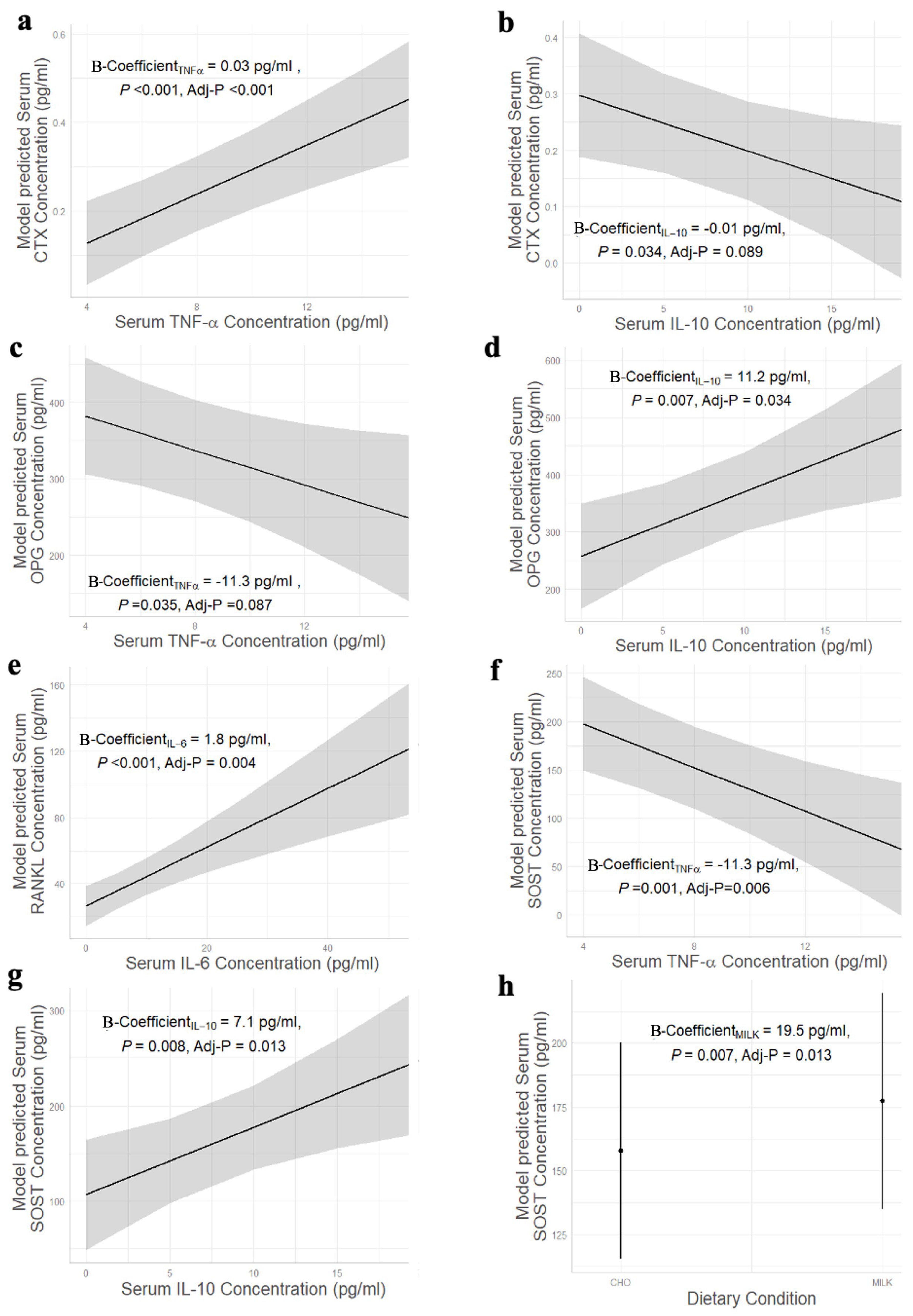
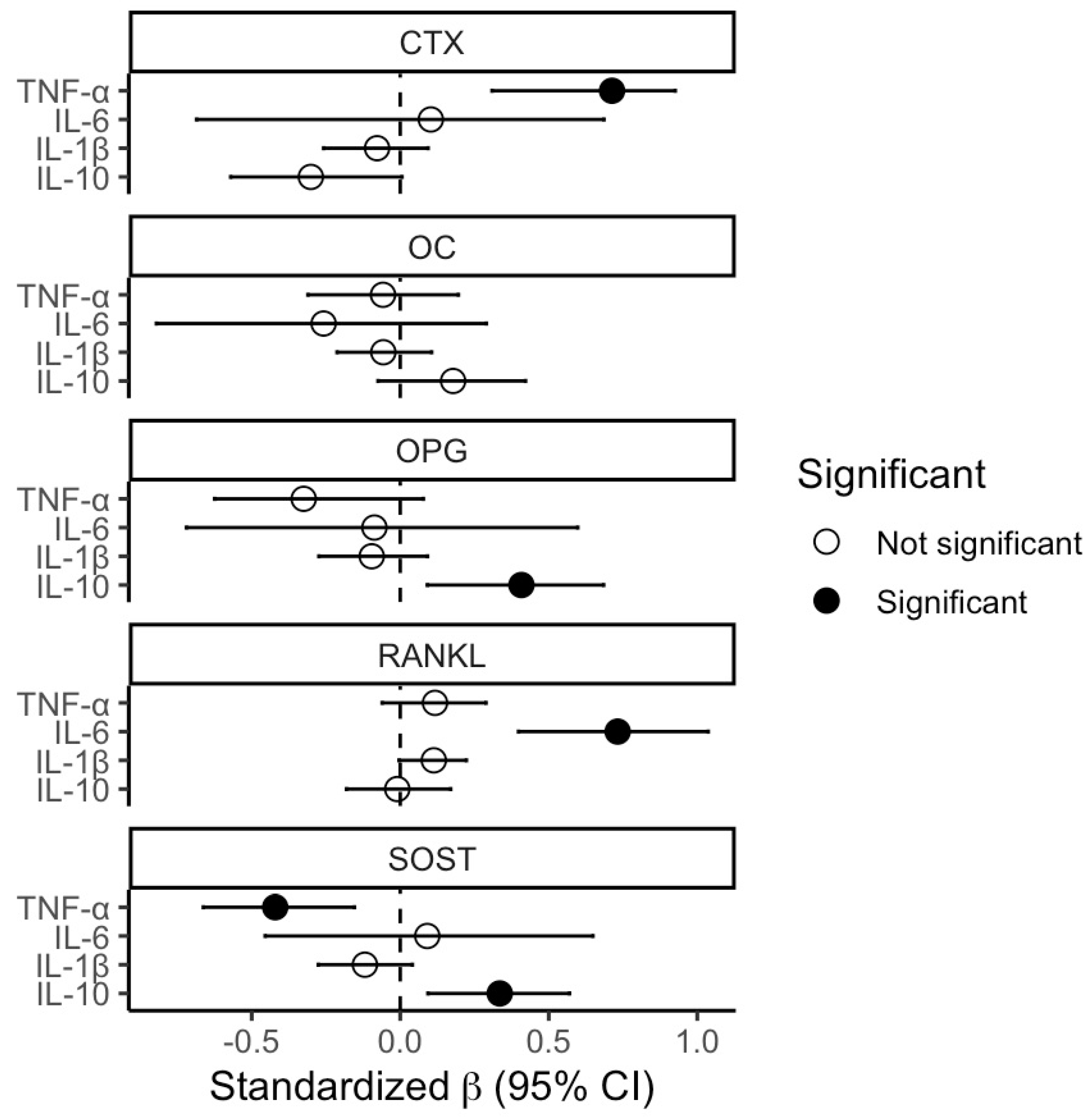
A summary of all associations is presented in Table 1, graphical representations of significant relationships are shown in Figure 1, and a forest plot of the mixed model results is shown in Figure 2.
Table 1.
Summary table presenting the statistical coefficients (Unstandardized [B] and standardized [β] beta coefficients and 95% CIs) for all tested associations.
Figure 1.
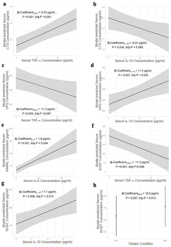
Graphical representation of the significant associations between (a) CTX and TNF-α, (b) CTX and IL-10, (c) OPG and TNF-α, (d) OPG and IL-10, (e) RANKL and IL-6, (f) SOST and TNF-α, (g) SOST and IL-10, and (h) SOST and dietary condition. Shading = 95% CI. CTX: C-telopeptide of type I collagen; TNF-α: tumor necrosis factor-alpha; IL-10: interleukin-10; OPG: osteoprotegerin; RANKL: receptor activator of NF-κB ligand; IL-6: interleukin-6; SOST: sclerostin.
Figure 2.
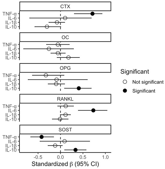
Forest plot showing the standardized beta coefficients from each model, with significant associations indicated by filled circles (p < 0.05).
For CTX, our model explains 86.7% of the variation, with 17.0% coming from fixed effects only. CTX was significantly positively associated with TNF-α (Figure 1a; p < 0.001), but had a trending negative association with IL-10 (Figure 1b; p = 0.089).
For OPG, our model explains 83.1% of the variation, with 10.8% coming from fixed effects only. OPG had a trending negative association with TNF-α (Figure 1c; p = 0.087) but was significantly positively associated with IL-10 (Figure 1d; p = 0.034).
For RANKL, our model explains 88.1% of the variation, with 60.1% coming from fixed effects only. RANKL was significantly positively associated with IL-6 (Figure 1e; p = 0.004).
For SOST, our model explains 83.8% of the variation, with 18.7% coming from fixed effects only. SOST was significantly negatively associated with TNF-α (Figure 1f; p = 0.006), but was significantly positively associated with IL-10 (Figure 1g; p = 0.013). In addition, SOST concentrations were higher by 19.5 pg/mL (Figure 1h; p = 0.013) in MILK compared to CHO (post hoc analysis determined that this was due to higher baseline levels).
For OC, our model explains 81.0% of the variation, with 3.6% coming from fixed effects only. There were no significant associations between OC and any inflammatory markers.
4. Discussion
We observed significant relationships between bone and inflammatory biomarkers in the context of a single bout of high-intensity exercise combined with nutrient consumption. Specifically, we found that CTX was significantly positively associated with TNF-α, that OPG was positively associated with IL-10 and negatively associated with TNF-α, that RANKL was positively associated with IL-6, and that SOST was positively associated with IL-10 and negatively associated with TNF-α.
Assessing circulating bone turnover biomarkers provides insight into acute bone responses, given that months or years may be required to see meaningful alterations in BMD [17]. Increased pro-inflammatory TNF-α being associated with increases in bone breakdown (CTX) and a trend toward increased anti-inflammatory IL-10 being associated with decreases in bone breakdown (CTX) provides supporting evidence for the interconnectedness of the immune and skeletal systems in response to exercise. This also suggests that pro-inflammation processes post-exercise may be involved in bone breakdown and that increased anti-inflammatory signaling may be related to bone formation. Exercise acts as a systemic stressor that can induce damage-associated molecular patterns (DAMPs). When toll-like receptors (TLRs [20]) are stimulated by DAMPs, pro-inflammatory pathways such as the NOD-like receptor protein 3 (NLRP3) inflammasome and nuclear factor kappa B (NF-κB) are activated, leading to recruitment of cytokines and leukocytes (that further produce cytokines) to the site of that damage [20]. In vitro research has shown that these TLRs are also expressed on bone cells [21], and their stimulation may increase the activity of osteoclasts, leading to elevated (and potentially inflammatory-mediated) bone resorption [21,22]. Indeed, individuals with chronically elevated inflammation (rheumatoid arthritis, inflammatory bowel disease) often display reduced BMD and increased fracture risk [23,24]. In addition, when drugs that block/blunt cytokines (e.g., TNF inhibitors: Enbrel, Remicade and Humira) are given to patients with increased inflammation, there appear to be long-term improvements in BMD [25]. By reducing inflammatory cytokine levels, RANK-RANKL-mediated osteoclastogenesis seems to be attenuated, shifting bone towards a less catabolic state [26]. Two recent studies have also found significant correlations between inflammatory and bone biomarkers in different human contexts. Damani et al. (2023) performed a secondary analysis on data from the PRUNE Study in post-menopausal females and found a significant negative correlation between C-reactive protein (CRP; an acute phase pro-inflammatory marker) and P1NP (a bone formation marker), but no relationship between TNF-α and CTX (like we observed) [27]. Osalou and colleagues (2025) assessed changes in cytokines, adipokines and bone markers in females with obesity after 12 weeks of high-intensity interval training and found significant positive correlations between CRP and CTX and a trending correlation between TNF-α and CTX [28]. They also observed negative correlations between TNF-α and OC, and CRP and bone alkaline phosphatase (a bone formation marker) [28]. Differences in population and study design likely account for the different relationships observed within these studies, but these findings along with ours provide evidence of mechanistic links between the skeletal and immune systems in generally healthy people both habitually and after undergoing an acute inflammatory stimulus (i.e., intense exercise). It must be noted that while we did include nutritional condition as a fixed effect covariate (non-significant for outcomes assessed in this analysis), it is still possible that anti-inflammatory components within milk (e.g., whey protein, calcium, vitamin D) may have differentially modulated cytokine-to-bone signaling compared with the isoenergetic CHO control. Regardless, these findings have implications for athletes seeking to optimize acute recovery and adaptations to training. For example, if an athlete is in a highly pro-inflammatory state (post-competition, overreaching/overtraining syndrome, etc.), then bone remodeling could also be affected, and steps may need to be taken by coaches/support staff to prevent long-term deleterious bone outcomes. Consumption of dairy foods can help supply adequate bone-supporting nutrients to benefit recovery and musculoskeletal health [16,18].
The OPG/RANKL signaling mechanism is essential for maintenance of bone homeostasis [29], and our results showed that as the anti-inflammatory cytokine IL-10 increased, so did OPG, but as the pro-inflammatory cytokine TNF-α increased, OPG decreased. This suggests that cytokine signaling may affect bone resorption by altering OPG release, but this potential relationship warrants further mechanistic investigation. If IL-10 (anti-inflammatory) was found to increase OPG production/release, then this would block RANKL binding, reduce osteoclast activity and shift bone turnover to a more anabolic state. More research is needed to better understand the local and systemic responses of bone in relation to OPG, but to our knowledge, our study is the first to demonstrate a relationship between OPG and inflammatory cytokines within an exercise context in humans. For RANKL, we observed a significant positive association between its response and the IL-6 response. This cytokine is complex and multifunctional, with recent research showing its importance for metabolism/energy allocation and that exercise-derived IL-6 (considered a ‘myokine’) may have positive effects on health, including providing anti-inflammatory function [30,31,32]. In bone, IL-6 has been shown to have both positive and negative effects on osteoblast and osteoclast differentiation [33]. Our findings indicate that it may be involved in bone remodeling, perhaps through modulation of RANKL; however, the lack of association with CTX suggests that acute exercise-mediated changes in IL-6 (anti-inflammatory) do not necessarily relate to increased bone breakdown [31].
Mature osteoblast formation is primarily mediated through the canonical Wingless (Wnt) signaling pathway, which is negatively regulated by the proteins SOST and Dickkopf (DKK) 1/2 [13,34]. We observed a significant negative relationship between pro-inflammatory TNF-α and SOST. In contrast, an increase in the anti-inflammatory IL-10 cytokine was associated with an increase in circulating SOST. These findings are unexpected considering TNF-α has been shown to upregulate the expression of SOST through the NF-κB signaling pathway in mice, contributing to apoptosis of osteoblasts [35]. In previous exercise studies performed by our group, we also observed a significant positive correlation between post-exercise SOST and TNF-α [36], which is the opposite of what was found during this analysis. It is currently unclear why our results are divergent from previous observations, but it is likely related to differences in study design, such as human vs. animal data, differences in exercise modality used (high-impact jumping and resistance exercise vs. running and cycling) or the post-exercise provision of energy that was involved with this study vs. no post-exercise provisions in previous studies [35,36]. One (or multiple) of these factors may have suppressed the expected SOST response or altered TNF-α release/signaling in some way.
We observed several significant relationships between bone and inflammatory markers in this analysis; however, there are some limitations to consider. First, we assessed associations, so we cannot determine if causality exists, nor the direction of effect. Second, our sample size for this study was small, which may have reduced power, increased model instability and impeded our ability to more definitively determine whether an observation was truly non-significant or inconclusive. We also evaluated a cohort of young female adults, some of whom were using monophasic hormonal contraceptives. Although we used a crossover design with the same monthly sampling time to ensure a consistent hormonal milieu between visits, the findings are not necessarily generalizable to other populations, such as male athletes, post-menopausal females or older adults, generally. Future research should build upon these novel findings and continue to explore how these interconnected systems modulate one another, perhaps by evaluating more timepoints, with a larger sample size, and/or through investigation into how different exercise modalities may influence these system interconnections. To increase the generalizability of these findings, it would also be valuable to study these relationships in a male or older, post-menopausal cohorts to determine if the identified crosstalk is estrogen-dependent or whether it is a universal response to high-intensity and impact exercise.
5. Conclusions
In conclusion, findings from this secondary analysis suggest that inflammatory and bone biomarkers are physiologically related in the context of an exercise and nutrition stimulus. Our results generally report that pro-inflammatory and anti-inflammatory cytokines were positively and negatively associated with catabolic bone biomarkers, respectively. We also observed the opposite for anabolic bone biomarkers, with pro-inflammatory and anti-inflammatory cytokines negatively and positively associated, respectively. Improving our understanding of the underlying mechanisms and relationships between these systems could help optimize both acute exercise recovery (i.e., resolving inflammation and supporting bone turnover/formation) and, eventually, therapeutic techniques for those with chronic diseases to ensure exercise most effectively supports bone remodeling.
Supplementary Materials
The following supporting information can be downloaded at: https://www.mdpi.com/article/10.3390/app16146987/s1, Supplementary Table S1. Serum concentrations of bone biomarkers and cytokines at pre-exercise, 15 min, 75 min, 24 h, and 48 h post-exercise in the carbohydrate (CHO) and milk (MILK) trials. Originally published in Prowting et al. and Fraschetti et al. [16,18].
Author Contributions
Conceptualization, J.L.P. and A.R.J.; methodology, J.L.P., E.C.F., P.K. and A.R.J.; software, A.E.; formal analysis, A.E. and J.L.P.; writing—original draft preparation, J.L.P.; writing—review and editing, J.L.P., A.E., E.C.F., P.K. and A.R.J.; visualization, A.E.; supervision, A.R.J.; project administration, A.R.J.; funding acquisition, A.R.J. All authors have read and agreed to the published version of the manuscript.
Funding
This research was supported by an unrestricted research grant, a Junior Faculty Fund/Minor Research Grant (JFF/MRG) from York University and a Brock University Advancement Fund (BUAF) grant. J.L.P. was supported by a Canadian Institutes of Health Research Doctoral Fellowship (#181430). E.C.F. was supported by a Natural Sciences and Engineering Research Council of Canada Postgraduate Scholarship-Doctoral (PGS D–580026-2023).
Institutional Review Board Statement
This study was performed in line with the principles of the Declaration of Helsinki. Approval was granted by the Ethics Committees of Brock University (REB: 17-402, 3 August 2018) and York University (REB: 2019-045, 31 January 2019).
Informed Consent Statement
Informed consent was obtained from all subjects involved in the study.
Data Availability Statement
The data presented in this study are available on request from the corresponding author. The data are not publicly available due to due to ethical restrictions protecting participant confidentiality and privacy.
Acknowledgments
The authors would like to thank the participants for their commitment to the study. We also wish to thank H. Snider and J. Brown for their help with data collection.
Conflicts of Interest
All authors declare no conflicts of interest with the present work. For projects outside the present work, A.R.J. and P.K. report grant funding from Dairy Farmers of Canada, NSERC and non-financial support from Lactalis. A.R.J. reports speaker and grant review fees from Dairy Farmers of Canada.
Abbreviations
The following abbreviations are used in this manuscript:
| BMD | Bone mineral density |
| BMI | Body mass index |
| CRP | C-reactive protein |
| CTX | Cross-linked C-telopeptide of type I collagen |
| DAMP | Damage-associated molecular patterns |
| IL- | Interleukin- |
| NF-κB | Nuclear factor kappa B |
| NLRP3 | NOD-like receptor protein 3 |
| OPG | Osteoprotegerin |
| OC | Total osteocalcin |
| P1NP | Procollagen type I N-propeptide |
| RANKL | Receptor activator of NF-κB ligand |
| SOST | Sclerostin |
| TLRs | Toll-like receptors |
| TNF-α | Tumor necrosis factor-alpha |
References
- Caetano-Lopes, J.; Canhao, H.; Fonseca, J.E. Osteoimmunology--the hidden immune regulation of bone. Autoimmun. Rev. 2009, 8, 250–255. [Google Scholar] [CrossRef] [Scilit] [PubMed]
- Walsh, M.C.; Choi, Y. Biology of the RANKL–RANK–OPG system in immunity, bone, and beyond. Front. Immunol. 2014, 5, 511. [Google Scholar] [CrossRef] [Scilit] [PubMed]
- Tsukasaki, M.; Takayanagi, H. Osteoimmunology: Evolving concepts in bone–immune interactions in health and disease. Nat. Rev. Immunol. 2019, 19, 626–642. [Google Scholar] [CrossRef] [Scilit] [PubMed]
- Nakashima, T.; Takayanagi, H. Osteoimmunology: Crosstalk between the immune and bone systems. J. Clin. Immunol. 2009, 29, 555–567. [Google Scholar] [CrossRef] [Scilit] [PubMed]
- Snelling, A.M.; Crespo, C.J.; Schaeffer, M.; Smith, S.; Walbourn, L. Modifiable and nonmodifiable factors associated with osteoporosis in postmenopausal women: Results from the Third National Health and Nutrition Examination Survey, 1988–1994. J. Women’s Health Gend.-Based Med. 2001, 10, 57–65. [Google Scholar] [CrossRef] [Scilit] [PubMed]
- Wilson-Barnes, S.L.; Lanham-New, S.A.; Lambert, H. Modifiable risk factors for bone health & fragility fractures. Best Pract. Res. Clin. Rheumatol. 2022, 36, 101758. [Google Scholar] [CrossRef] [Scilit] [PubMed]
- Kenkre, J.; Bassett, J. The bone remodelling cycle. Ann. Clin. Biochem. 2018, 55, 308–327. [Google Scholar] [CrossRef] [Scilit] [PubMed]
- Kohrt, W.M.; Wherry, S.J.; Wolfe, P.; Sherk, V.D.; Wellington, T.; Swanson, C.M.; Weaver, C.M.; Boxer, R.S. Maintenance of serum ionized calcium during exercise attenuates parathyroid hormone and bone resorption responses. J. Bone Miner. Res. 2018, 33, 1326–1334. [Google Scholar] [CrossRef] [Scilit] [PubMed]
- Kirk, B.; Feehan, J.; Lombardi, G.; Duque, G. Muscle, bone, and fat crosstalk: The biological role of myokines, osteokines, and adipokines. Curr. Osteoporos. Rep. 2020, 18, 388–400. [Google Scholar] [CrossRef] [Scilit] [PubMed]
- Cerqueira, É.; Marinho, D.A.; Neiva, H.P.; Lourenço, O. Inflammatory effects of high and moderate intensity exercise—A systematic review. Front. Physiol. 2020, 10, 489354. [Google Scholar] [CrossRef] [Scilit] [PubMed]
- Rantalainen, T.; Heinonen, A.; Linnamo, V.; Komi, P.V.; Takala, T.E.; Kainulainen, H. Short-term bone biochemical response to a single bout of high-impact exercise. J. Sports Sci. Med. 2009, 8, 553. [Google Scholar] [PubMed]
- Dolan, E.; Varley, I.; Ackerman, K.E.; Pereira, R.M.R.; Elliott-Sale, K.J.; Sale, C. The bone metabolic response to exercise and nutrition. Exerc. Sport Sci. Rev. 2020, 48, 49–58. [Google Scholar] [CrossRef] [Scilit] [PubMed]
- Eriksen, E.F. Cellular mechanisms of bone remodeling. Rev. Endocr. Metab. Disord. 2010, 11, 219–227. [Google Scholar] [CrossRef] [Scilit] [PubMed]
- Dolan, E.; Dumas, A.; Keane, K.M.; Bestetti, G.; Freitas, L.H.M.; Gualano, B.; Kohrt, W.M.; Kelley, G.A.; Pereira, R.M.R.; Sale, C. The bone biomarker response to an acute bout of exercise: A systematic review with meta-analysis. Sports Med. 2022, 52, 2889–2908. [Google Scholar] [CrossRef] [Scilit] [PubMed]
- Kuo, T.-R.; Chen, C.-H. Bone biomarker for the clinical assessment of osteoporosis: Recent developments and future perspectives. Biomark. Res. 2017, 5, 18. [Google Scholar] [CrossRef] [Scilit] [PubMed]
- Prowting, J.L.; Skelly, L.E.; Kurgan, N.; Fraschetti, E.C.; Klentrou, P.; Josse, A.R. Acute effects of Milk vs. carbohydrate on bone turnover biomarkers following loading exercise in young adult females. Front. Nutr. 2022, 9, 840973. [Google Scholar] [CrossRef] [Scilit] [PubMed]
- Dolan, E.; Dumas, A.; Esteves, G.P.; Takarabe, L.L.; Perfeito, L.A.M.; Keane, K.M.; Gualano, B.; Kelley, G.A.; Burke, L.; Sale, C. The Influence of Nutrition Intervention on the P1NP and CTX-1 Response to an Acute Exercise Bout: A Systematic Review with Meta-Analysis. Sports Med. 2024, 54, 2889–2906. [Google Scholar] [CrossRef] [Scilit] [PubMed]
- Fraschetti, E.C.; Skelly, L.E.; Prowting, J.L.; Abdul-Sater, A.A.; Josse, A.R. The acute effects of milk consumption on systemic inflammation after combined resistance and plyometric exercise in young adult females. Nutrients 2022, 14, 4532. [Google Scholar] [CrossRef] [Scilit] [PubMed]
- Benjamini, Y.; Hochberg, Y. Controlling the False Discovery Rate: A Practical and Powerful Approach to Multiple Testing. J. R. Stat. Soc. Ser. B (Methodol.) 2018, 57, 289–300. [Google Scholar] [CrossRef] [Scilit]
- Alonso-Pérez, A.; Franco-Trepat, E.; Guillán-Fresco, M.; Jorge-Mora, A.; López, V.; Pino, J.; Gualillo, O.; Gómez, R. Role of toll-like receptor 4 on osteoblast metabolism and function. Front. Physiol. 2018, 9, 504. [Google Scholar] [CrossRef] [Scilit] [PubMed]
- Bar-Shavit, Z. Taking a toll on the bones: Regulation of bone metabolism by innate immune regulators. Autoimmunity 2008, 41, 195–203. [Google Scholar] [CrossRef] [Scilit] [PubMed]
- Epsley, S.; Tadros, S.; Farid, A.; Kargilis, D.; Mehta, S.; Rajapakse, C.S. The Effect of Inflammation on Bone. Front. Physiol. 2021, 11, 1695. [Google Scholar] [CrossRef] [Scilit]
- Gkastaris, K.; Goulis, D.G.; Potoupnis, M.; Anastasilakis, A.D.; Kapetanos, G. Obesity, osteoporosis and bone metabolism. J. Musculoskelet. Neuronal Interact. 2020, 20, 372–381. [Google Scholar] [PubMed]
- Briot, K.; Geusens, P.; Em Bultink, I.; Lems, W.F.; Roux, C. Inflammatory diseases and bone fragility. Osteoporos. Int. 2017, 28, 3301–3314. [Google Scholar] [CrossRef] [Scilit] [PubMed]
- Zerbini, C.A.F.; Clark, P.; Mendez-Sanchez, L.; Pereira, R.M.R.; Messina, O.D.; Uña, C.R.; Adachi, J.D.; Lems, W.F.; Cooper, C.; Lane, N.E.; et al. Biologic therapies and bone loss in rheumatoid arthritis. Osteoporos. Int. 2017, 28, 429–446. [Google Scholar] [CrossRef] [Scilit] [PubMed]
- Redlich, K.; Smolen, J.S. Inflammatory bone loss: Pathogenesis and therapeutic intervention. Nat. Rev. Drug Discov. 2012, 11, 234–250. [Google Scholar] [CrossRef] [Scilit] [PubMed]
- Damani, J.J.; De Souza, M.J.; Strock, N.C.A.; Koltun, K.J.; Williams, N.I.; Weaver, C.; Rogers, C.J. Associations Between Inflammatory Mediators and Bone Outcomes in Postmenopausal Women: A Cross-Sectional Analysis of Baseline Data from the Prune Study. J. Inflamm. Res. 2023, 16, 639–663. [Google Scholar] [CrossRef] [Scilit] [PubMed]
- Osalou, M.A.; Atakan, M.M.; Güzel, Y.; Koşar, Ş.N. Twelve-week interval training alters cytokines, adipokines and bone markers in women with obesity. Int. J. Sports Med. 2025, 46, 402–411. [Google Scholar] [CrossRef] [Scilit] [PubMed]
- Tobeiha, M.; Moghadasian, M.H.; Amin, N.; Jafarnejad, S. RANKL/RANK/OPG pathway: A mechanism involved in exercise-induced bone remodeling. BioMed Res. Int. 2020, 2020, 6910312. [Google Scholar] [CrossRef] [Scilit] [PubMed]
- Ellingsgaard, H.; Hojman, P.; Pedersen, B.K. Exercise and health—Emerging roles of IL-6. Curr. Opin. Physiol. 2019, 10, 49–54. [Google Scholar] [CrossRef] [Scilit]
- Nara, H.; Watanabe, R. Anti-Inflammatory Effect of Muscle-Derived Interleukin-6 and Its Involvement in Lipid Metabolism. Int. J. Mol. Sci. 2021, 22, 9889. [Google Scholar] [CrossRef] [Scilit] [PubMed]
- Kistner, T.M.; Pedersen, B.K.; Lieberman, D.E. Interleukin 6 as an energy allocator in muscle tissue. Nat. Metab. 2022, 4, 170–179. [Google Scholar] [CrossRef] [Scilit] [PubMed]
- Blanchard, F.; Duplomb, L.; Baud’huin, M.; Brounais, B. The dual role of IL-6-type cytokines on bone remodeling and bone tumors. Cytokine Growth Factor Rev. 2009, 20, 19–28. [Google Scholar] [CrossRef] [Scilit] [PubMed]
- Clevers, H.; Nusse, R. Wnt/β-catenin signaling and disease. Cell 2012, 149, 1192–1205. [Google Scholar] [CrossRef] [Scilit] [PubMed]
- Zhao, B. TNF and bone remodeling. Curr. Osteoporos. Rep. 2017, 15, 126–134. [Google Scholar] [CrossRef] [Scilit] [PubMed]
- Kouvelioti, R.; Kurgan, N.; Falk, B.; Ward, W.E.; Josse, A.R.; Klentrou, P. Cytokine and Sclerostin Response to High-Intensity Interval Running versus Cycling. Med. Sci. Sports Exerc. 2019, 51, 2458–2464. [Google Scholar] [CrossRef] [Scilit] [PubMed]

Disclaimer/Publisher’s Note: The statements, opinions and data contained in all publications are solely those of the individual author(s) and contributor(s) and not of MDPI and/or the editor(s). MDPI and/or the editor(s) disclaim responsibility for any injury to people or property resulting from any ideas, methods, instructions or products referred to in the content. |
© 2026 by the authors. Licensee MDPI, Basel, Switzerland. This article is an open access article distributed under the terms and conditions of the Creative Commons Attribution (CC BY) license.