


Abstract
Background: Panoramic radiography is one of the most widely used imaging examinations in dental practice, providing a broad view of the jaws and adjacent head and neck structures. Although primarily prescribed for odontogenic assessment, its field of view may reveal non-odontogenic findings with potential clinical significance. Methods: A structured narrative review was conducted according to SANRA criteria. A literature search was performed in PubMed/MEDLINE, Embase, Scopus, and Web of Science for English-language publications from January 2010 to May 2026. Backward and forward citation tracking of relevant articles, key reviews, and reference textbooks was also performed. Eligible studies and authoritative sources addressing non-odontogenic findings detectable on panoramic radiographs were qualitatively synthesized. Results: The review focuses on carotid artery calcifications, maxillary sinus abnormalities, mandibular radiomorphometric signs related to low skeletal bone mineral density, elongation or calcification of the stylohyoid complex, sialoliths, tonsilloliths, calcified lymph nodes, phleboliths, and laryngeal cartilage calcifications. These findings range from benign anatomical variants to radiographic indicators that may require medical or specialist evaluation. Conclusions: Panoramic radiography should be regarded as a tool for recognition and clinical suspicion rather than definitive diagnosis of extraoral or systemic disease. Dentists play a central role in systematically assessing the entire image, documenting relevant abnormalities, correlating them with patient history and risk factors, and initiating appropriate referral when indicated.
1. Introduction
Orthopantomography (OPG) is one of the most widely used imaging examinations in dental and orthodontic practice, as it provides, through a single exposure, an overall representation of the dental arches, the maxillary and mandibular bony structures, the temporomandibular joints, and several adjacent anatomical regions [1,2]. This comprehensive view supports its extensive use in the initial assessment of patients, treatment planning, and monitoring of various odontostomatological conditions [1,2]. However, because of its broad field of acquisition, OPG should not be interpreted as an examination limited exclusively to the teeth and jaws, but rather as a complex image that also includes anatomical areas where findings unrelated to dental disease may be detected [3,4,5]. From this perspective, systematic analysis of the entire OPG has clinical value that extends beyond the dentomaxillofacial field alone [3,4,5]. Indeed, OPG may reveal non-dental findings of variable nature and significance, including carotid artery calcifications, maxillary sinus abnormalities, radiographic signs suggestive of reduced bone mineral density, elongation or calcification of the styloid process, sialolithiasis, tonsilloliths, lymph node calcifications, phleboliths, and calcifications of the laryngeal cartilages [6,7,8,9,10,11]. Some of these findings correspond to anatomical variants, benign conditions, or calcified sequelae without immediate clinical relevance, whereas others may represent indirect indicators of extraoral or systemic diseases requiring further investigation [3,5,6,12]. It is therefore essential to emphasize that OPG cannot, in most cases, be considered a definitive diagnostic examination for non-dental diseases. The intrinsic limitations of this imaging modality, related to the two-dimensional nature of the image, anatomical superimposition, geometric distortion, and variability associated with patient positioning, require cautious interpretation of the observed findings [1,2,4]. In this context, the role of OPG is primarily that of a first-line imaging examination, useful for the incidental recognition of abnormalities, for guiding clinical suspicion, and for initiating, when appropriate, further diagnostic assessment through dedicated imaging examinations or specialist consultation [2,3,4,5,12]. In this regard, dentists may play a particularly relevant role not only in the diagnosis of oral diseases but also in the preliminary identification of radiographic findings that fall outside the strictly dental field. The ability to recognize these alterations, distinguish them from odontogenic diseases, and assess their potential clinical significance positions the dentist as a sentinel figure, potentially involved in the early detection of extraoral conditions of medical interest [3,5,6,7,12,13]. In light of these considerations, the present study aims to provide a narrative analysis of the main non-dental findings that can be identified on OPG, with particular attention to their radiological features, differential diagnosis, and potential clinical relevance. The objective is not to assign OPG an autonomous diagnostic value for extraoral diseases, but rather to highlight how a systematic, informed, and methodologically appropriate interpretation of the examination may contribute to the recognition of findings deserving clinical attention, while also providing practical guidance for their proper radiological assessment and for the possible referral of patients for targeted diagnostic investigation [1,2,3,4,5,12].
2. Materials and Methods
2.1. Study Design
This study was designed as a structured narrative review to identify, describe, and clinically contextualize non-odontogenic findings that may be detected on OPG in routine dental practice. A structured narrative review was considered more appropriate than a systematic review because the aim was not to answer a narrowly defined clinical question or perform a quantitative synthesis, but to provide a clinically oriented overview of multiple non-odontogenic findings detectable on OPG. Given the heterogeneity of the available literature in terms of study design, populations, imaging methods, and clinical outcomes, a qualitative synthesis guided by SANRA (Scale for the Assessment of Narrative Review Articles) criteria was deemed the most suitable approach [14]. The primary unit of analysis was the non-odontogenic radiographic finding identifiable on OPG. The findings were synthesized qualitatively and organized into predefined diagnostic categories relevant to dental practice.
2.2. Information Sources and Search Strategy
A literature search was conducted in PubMed/MEDLINE, Embase, Scopus, and Web of Science covering the period from January 2010 to May 2026. To maximize retrieval of relevant publications, backward and forward citation tracking was also performed on all included articles, key review papers, and major reference textbooks in oral radiology and head and neck imaging. Before the formal search, a preliminary scoping phase was undertaken to map the terminology used to describe non-odontogenic or extra-dental findings detectable on OPG and to identify the main categories most frequently addressed in the literature. These categories included, among others, carotid artery calcifications, maxillary sinus abnormalities, tonsilloliths, elongated styloid process and/or calcified stylohyoid complex, calcified cervical lymph nodes, osteoporosis-related mandibular cortical changes, and other soft-tissue calcifications of the head and neck, as well as the distinction between dental age estimation and skeletal/auxological age assessment. Based on this exploratory phase, and informed by standard textbooks and previous reviews, an a priori list of diagnostic domains was defined and used to guide the search strategy. Search strings were then developed for each domain by combining terms related to OPG, incidental or non-odontogenic findings, head and neck calcifications, maxillary sinus alterations, mandibular radiomorphometric indices, and related disease-specific terminology. The complete search strategies for each database are reported in the Supplementary Material (Supplementary S1).
2.3. Eligibility Criteria
Studies were considered eligible if they reported extra-dental findings detectable on OPG and provided information relevant to their radiographic appearance, differential diagnosis, clinical interpretation, prevalence, or implications for referral and patient management. Both studies specifically focused on incidental findings on OPG and studies addressing individual extra-dental entities identifiable on OPG were considered for inclusion. Publications primarily addressing odontogenic disease, dentoalveolar pathology, or jaw lesions strictly related to the teeth and their supporting structures were excluded unless they contributed directly to the differential diagnosis of an extra-dental radiographic finding. Animal studies and articles not published in English were also excluded. Because the purpose of the review was clinical and interpretive rather than quantitative, no restrictions were placed on study design. Accordingly, observational studies, diagnostic accuracy studies, case series, case reports, review articles, and authoritative reference sources were all considered when relevant to the topic. All radiographic images used in this review were derived from anonymized archival material; all patient-identifying information was removed, and consent was obtained when applicable according to institutional policies and regulations.
3. Non-Odontogenic Findings on Panoramic Radiography
3.1. Carotid Calcifications
Vascular calcifications located in the carotid region or along the course of the cervicofacial arteries are incidental but clinically significant findings that may be detected on OPG (Table 1) [15]. Several studies have reported prevalence rates of vascular calcifications on OPG ranging from 2% to 11% in adult populations, with higher rates observed in specific subgroups, including men, individuals over 40 years of age, smokers, patients with hypertension, diabetes, or hypercholesterolemia, and those who have undergone kidney transplantation or head and neck radiotherapy [16,17]. Carotid calcifications are the most frequent incidental finding in radiographs of patients over 60 years of age [18]. Several studies have suggested a relationship between carotid-region calcifications detected on panoramic radiographs and periodontal risk or poor oral health. Although this association does not establish causality and OPG cannot be used to diagnose vascular disease, it highlights the potential relevance of integrating radiographic findings with periodontal status and systemic cardiovascular risk factors during dental assessment [7,19,20,21].
Table 1.
Carotid calcifications on orthopantomography: a summary of the literature from the last 15 years.
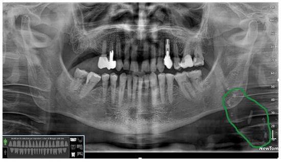
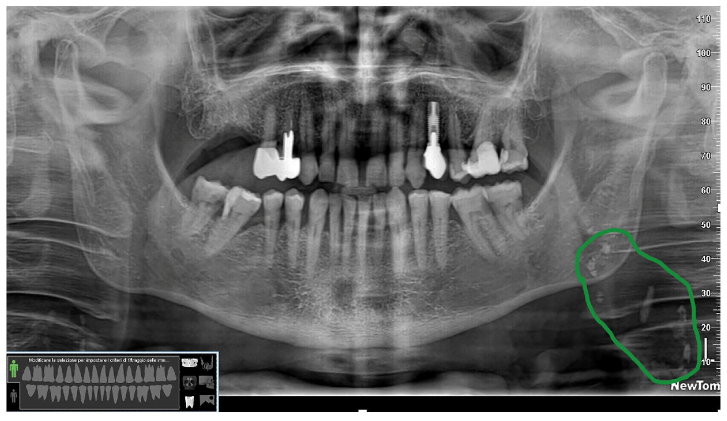
The OPG used in daily clinical dental practice has early diagnostic potential in the detection of radiopaque findings in the carotid region. Signs compatible with carotid artery calcifications visible on OPG appear as heterogeneous radiopaque nodular masses, localized in 78.22% of cases in the region of the carotid bifurcation, that is, in the area adjacent to the cervical vertebrae, at or below the C3–C4 intervertebral space, at a 45-degree angle to the mandibular angle (Figure 1).
Figure 1.
Routine orthopantomography of a patient showing radiopaque formations at the left mandibular angle and C3–C4 vertebral level, compatible with carotid calcifications (green circle). The image is derived from anonymized archival radiographic material; all patient-identifying information was removed.
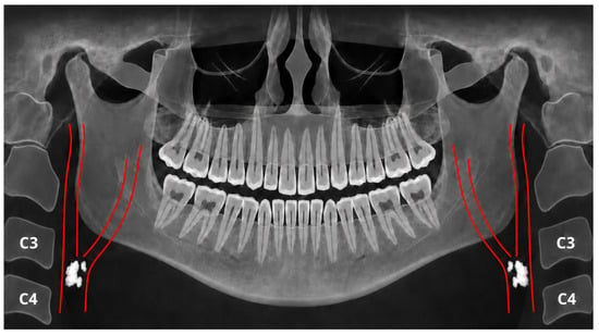
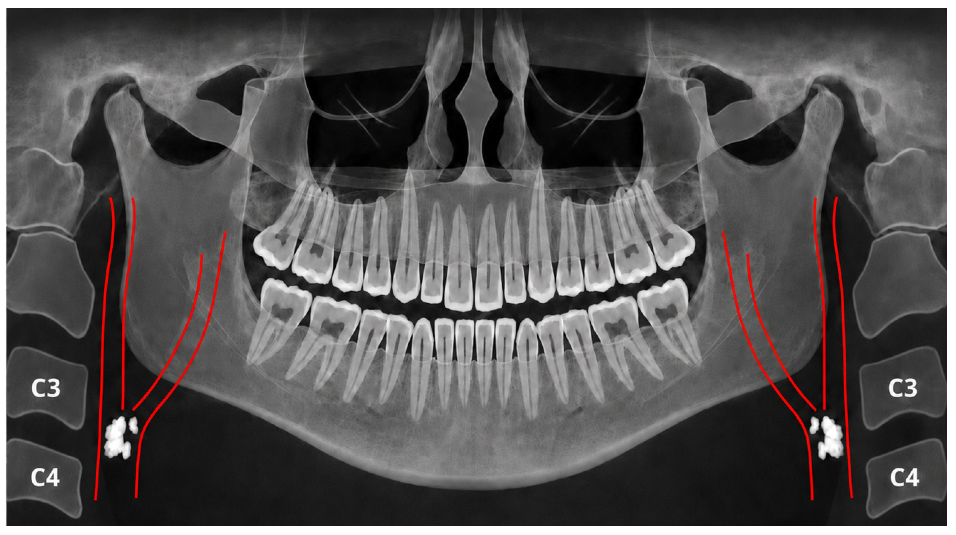
Calcifications may also be identified at the level of the carotid bifurcation, in the superior region of the external carotid artery along the course of the superficial temporal artery, or as multiple tortuous “tram-track” calcifications adjacent to the posterior border of the mandibular ramus, consistent with Mönckeberg arteriosclerosis involving the maxillary and facial arteries (Figure 2) [49]. Such superiorly located calcifications, as observed in these cases, have been associated with an increased risk of intracranial ischemic disease and are frequently reported in patients with chronic kidney disease [50,51,52].
Figure 2.
Schematic representation of the course of the common carotid artery and its external and internal branches in relation to key landmarks on panoramic radiography.
3.2. Maxillary Sinus
OPG provides a broad overview of the maxillofacial region and allows simultaneous assessment of the maxillary bones and maxillary sinuses [53]. In dental practice, this imaging modality may contribute to the preliminary recognition of maxillary sinus abnormalities, particularly because several sinonasal conditions are asymptomatic and are detected incidentally during routine radiographic evaluation. Radiographic alterations involving the maxillary sinus may include mucosal thickening, polyps, mucous retention cysts, antral cysts, and neoplastic lesions (Table 2) [53,54].
Table 2.
Main maxillary sinus pathologies observable on OPG.
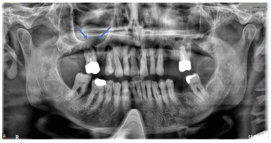
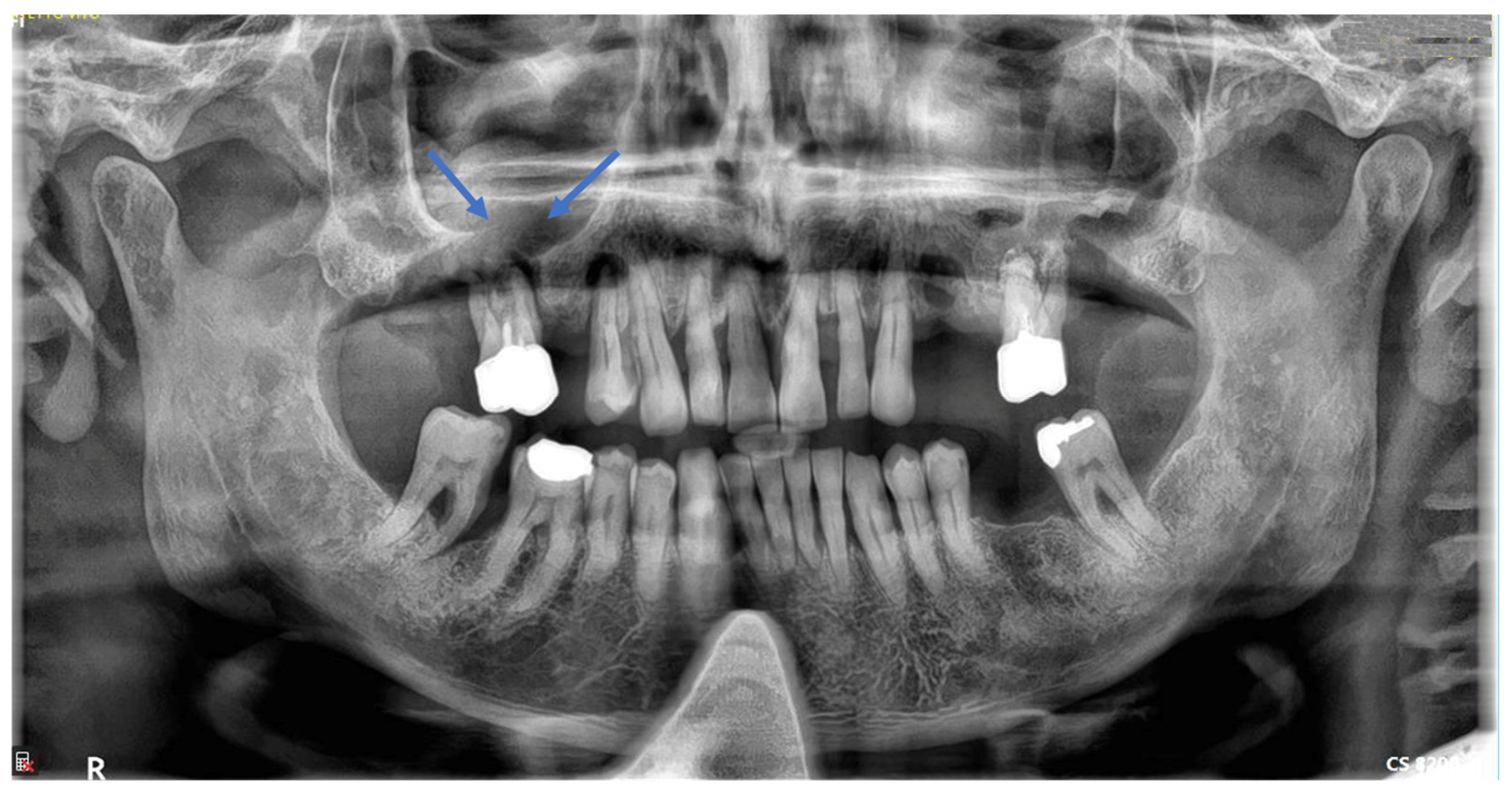
These abnormalities may present with variable radiographic appearances, ranging from localized dome-shaped opacities to more extensive changes affecting the sinus cavity. Calcified radiopaque bodies with heterogeneous morphology may also be detected and are referred to as antroliths when located within the maxillary sinus and rhinoliths when located within the nasal cavity [6]. The prevalence of pathological changes affecting the maxillary sinuses ranges from 45% to 70%. Among these findings, mucosal thickening represents the most frequently reported inflammatory alteration, irrespective of its etiological origin. This finding may be odontogenic in 10–40% of cases or non-odontogenic in 5–12% of cases [62]. Odontogenic involvement is particularly relevant in dental imaging, as inflammatory processes arising from periapical infection, periodontal disease, endodontic pathology, or post-surgical complications may extend to the sinus floor and induce localized mucosal reactions (Figure 3) [3,63].
Figure 3.
A clear oroantral communication is present in the region of the upper right first molar (blue arrows), associated with a recurrent odontogenic abscess. The image is derived from anonymized archival radiographic material; all patient-identifying information was removed.
3.3. Osteoporosis
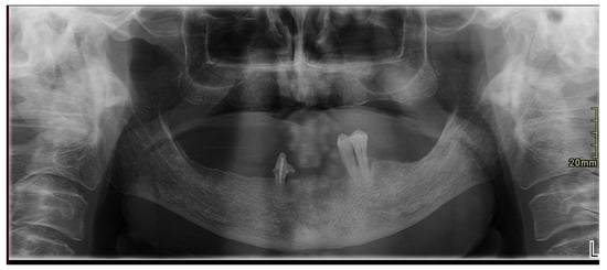
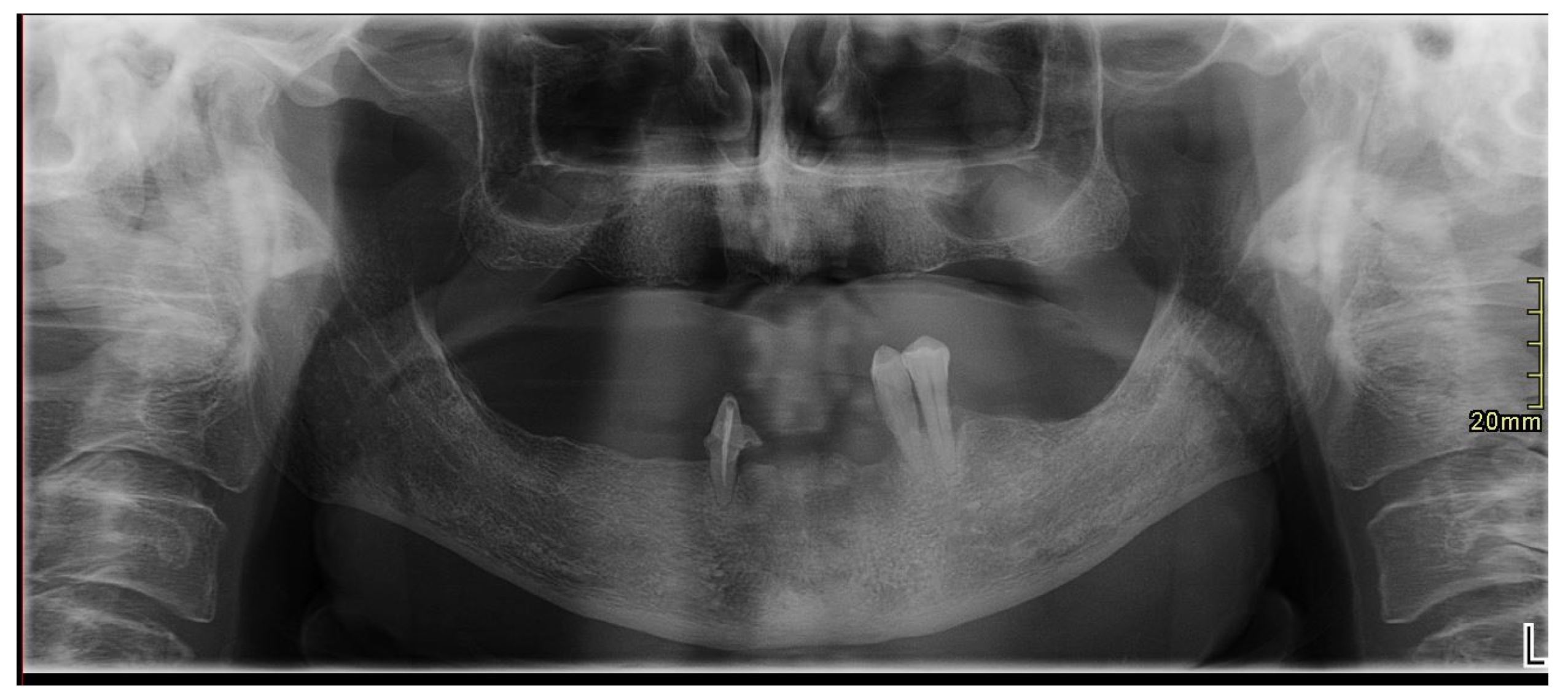
Osteoporosis is a skeletal disorder characterized by progressive loss of bone mass, which predominantly affects older individuals and tends to occur earlier in women [64]. Early signs of osteopenia and osteoporosis may be detectable on OPG [65]. Over the past four decades, numerous studies have reported incidental radiographic findings on dental OPG that may be suggestive of the early stages of osteoporosis [64]. OPG may provide adjunctive radiographic information that can raise suspicion of reduced bone mineral density in selected patients; however, it should not be considered a standalone screening tool for osteoporosis, and any suspected finding requires confirmation by dedicated densitometric assessment. This can be achieved through the evaluation of mandibular radiomorphometric indices, including the number of missing teeth, alveolar bone resorption, lamina dura width, mandibular cortical thickness, and the morphology of the inferior mandibular cortex, commonly assessed using the mandibular cortical index (MCI) (Figure 4) [66].
Figure 4.
OPG in which some mandibular radiomorphometric indices can be assessed: number of missing teeth, alveolar bone resorption, mandibular cortical width, and mandibular cortical morphology. The image is derived from anonymized archival radiographic material; all patient-identifying information was removed.
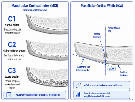
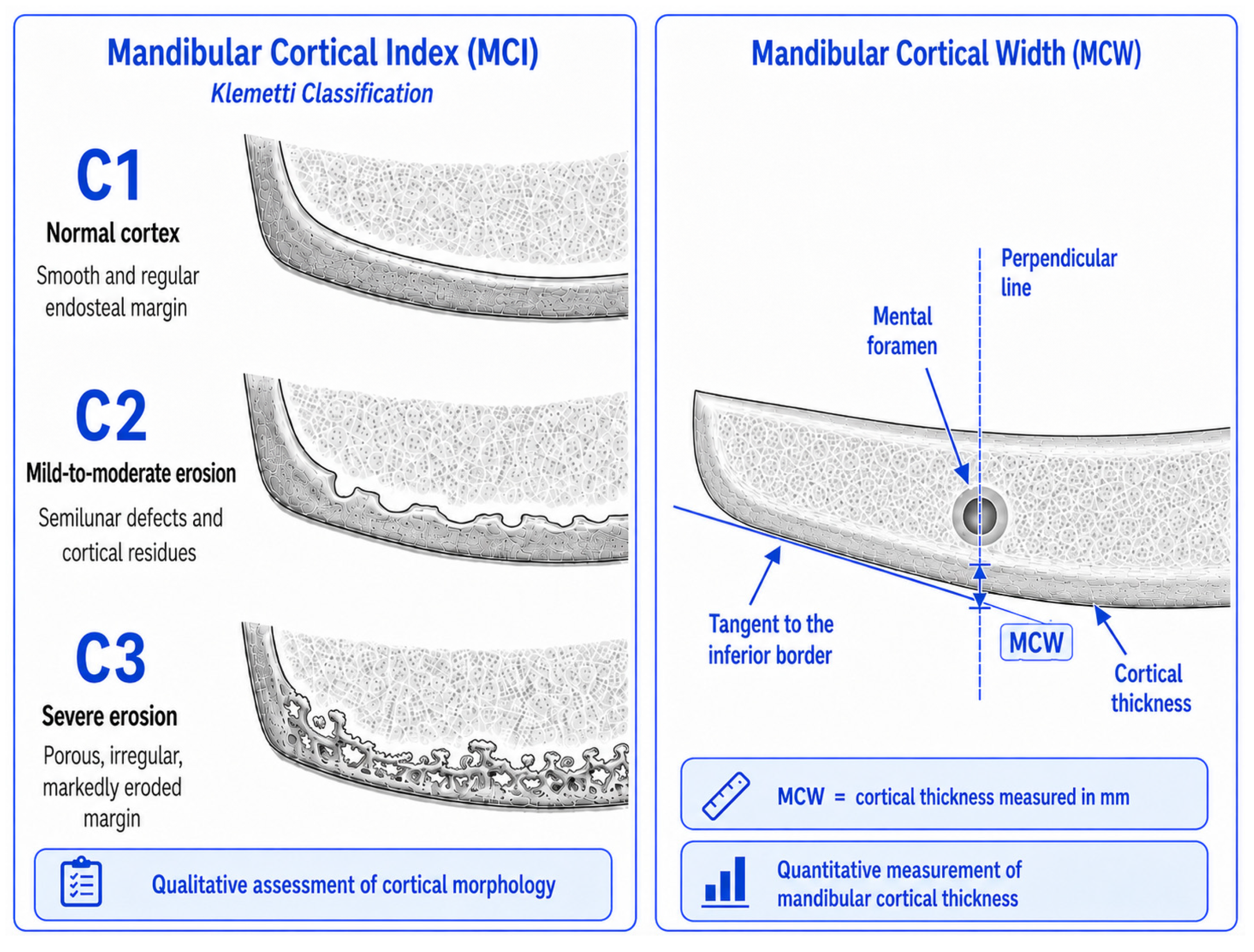
Trabecular bone is more sensitive to changes in bone metabolism than cortical bone [67]. For this reason, a more objective radiographic assessment may be obtained by evaluating mandibular indices such as the MCI, which describes the appearance of the endosteal margin of the mandibular cortex, and mandibular cortical width (MCW), measured at the region of the mental foramen (Table 3, Figure 5) [68].
Table 3.
Mandibular radiomorphometric indices assessable on orthopantomography: measurement methods and clinical significance.
Figure 5.
Mandibular Cortical Index and Mandibular Cortical Width on OPG: qualitative and quantitative assessment of the mandibular cortex.
The main advantage of these indices is that they may provide dentists with simple and cost-effective tools for the early detection of radiographic signs suggestive of osteopenia or osteoporosis. However, their diagnostic accuracy and clinical applicability should be interpreted with caution, as the demographic characteristics of the available studies are highly heterogeneous, particularly with regard to sex, age, and menopausal status [69].
3.4. Styloid Process
The styloid process is a cylindrical bony projection that arises from the temporal bone, anterior to the stylomastoid foramen. Its length varies considerably among individuals, between the two sides of the same individual, and across different populations. It also tends to increase with age, is commonly bilateral, and is usually clearly visible on OPG [70]. Langlais proposed a radiographic classification of elongated and mineralized stylohyoid ligament complexes based on three morphological types: Type I, elongated; Type II, pseudoarticulated; and Type III, segmented. These were further characterized according to the pattern of calcification as calcified outline, partially calcified, nodular, or completely calcified [71]. According to a 2022 systematic review, elongation of the styloid process may result from a chronic age-related calcification process, although it does not appear to be significantly associated with sex [72]. The normal length of the styloid process generally ranges from 20 to 25 mm, and it is considered clinically relevant when it is markedly elongated, typically exceeding 30 mm [73].
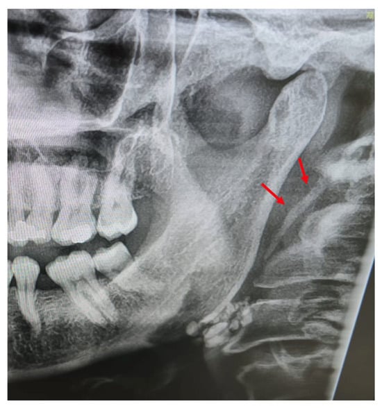
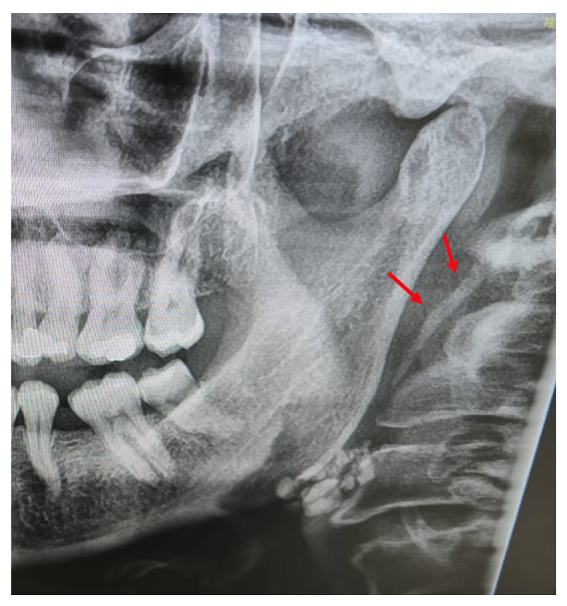
An elongated styloid process may remain entirely asymptomatic and therefore be detected incidentally on radiographic or tomographic examinations performed for unrelated reasons (Figure 6). In some cases, however, this anatomical variation may give rise to a range of symptoms, presumably due to compression or irritation of adjacent neurovascular and soft-tissue structures, particularly the cranial nerves, carotid vessels, and surrounding soft tissues. The most frequently reported symptoms include pain during head movement, cervicofacial pain, otalgia, headache, dysphagia, odynophagia, tinnitus, and trismus. This clinical spectrum is commonly associated with Eagle syndrome, a condition in which elongation or calcification of the stylohyoid complex becomes clinically significant [74].
Figure 6.
Marked elongation of the styloid process (red arrows). The image is derived from anonymized archival radiographic material; all patient-identifying information was removed.
3.5. Sialolithiasis
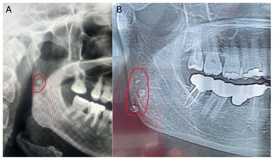
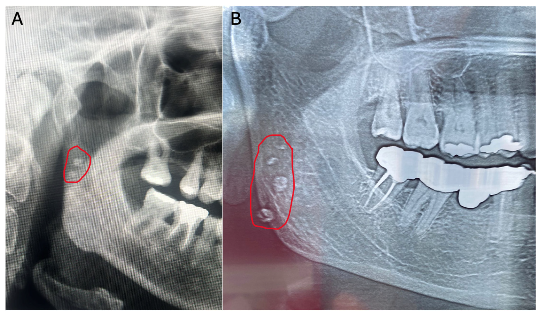
Sialolithiasis is one of the most common obstructive disorders of the major salivary glands, affecting approximately 0.01% to 1% of the population [75]. It results from the deposition of calcium salts around accumulations of organic debris, including mucous substances, ductal epithelial cells, bacteria, or even foreign bodies, leading to the formation of so-called sialoliths [76]. The submandibular glands are the salivary glands most frequently affected by sialolithiasis, accounting for approximately 80% of cases. This predominance is related to the morphology of the salivary duct and to the alkaline environment, which is characterized by a high phosphate concentration [77]. According to a study that analyzed 500 OPG, sialoliths are often incidental findings, with a reported prevalence of 3%. Submandibular sialoliths were the most frequently observed and were more commonly detected in female patients, whereas parotid sialoliths were more frequent in male patients [78]. On OPG, sialoliths may be identified as unilateral, diffusely calcified radiopacities with well-defined borders, usually located below the mandibular margin and near the region of the mandibular ramus (Figure 7) [18]. However, the radiographic diagnosis of sialoliths can be challenging, as conditions such as osteosclerosis, enostosis, osteoma, phleboliths, and calcified lymph nodes may show similar radiographic features (Table 4) [6].
Figure 7.
Sialoliths of the parotid gland (A) and submandibular gland (B) (red circle). The image is derived from anonymized archival radiographic material; all patient-identifying information was removed.
Table 4.
Main differential diagnoses of sialoliths and overlapping soft-tissue calcifications on panoramic radiographs.
In the past, conventional occlusal radiographs were the initial diagnostic modality of choice. They were relatively effective in detecting large radiopaque ductal stones, but they were unable to identify smaller intraparenchymal stones, although approximately 80% of sialoliths are radiopaque [86]. OPG taken during dental examinations can detect sialoliths with good overall accuracy, but with limited precision. This limitation is mainly due to the distortion that frequently occurs in OPG and to the superimposition of anatomical structures in the mandibular body. Therefore, computed tomography is often required for an accurate diagnosis and for determining the exact location of the stone, particularly when treatment planning is needed [76]. The ability of dentists to identify sialoliths on OPG is essential in order to avoid confusion with other soft-tissue calcifications, which are very common in the head and neck region [78].
3.6. Tonsilloliths
Tonsilloliths are calcified concretions that develop within the crypts of the palatine tonsils, usually as a result of the progressive deposition of calcium salts on retained organic material, including cellular debris, food remnants, bacteria, and mucous secretions. In most cases, they represent benign and asymptomatic findings and are often identified incidentally on radiographs obtained for dental purposes. On OPG, tonsilloliths may appear as single or multiple radiopaque masses of variable size, typically projected over the mandibular ramus region, most commonly near the area superimposed on the soft palate and the oropharyngeal space [87]. From an epidemiological perspective, tonsilloliths show a generally low prevalence among patients examined with OPG, with a higher frequency reported in males [3]. Their radiographic detection depends on several factors, including the degree of mineralization, size, number of calcified deposits, and their spatial relationship with adjacent anatomical structures. Small or poorly calcified tonsilloliths may not be visible on OPG, whereas more heavily mineralized concretions tend to appear as clearly recognizable radiopaque masses in the mandibular ramus region. A relevant radiographic feature is the possible occurrence of ghost images or contralateral artefacts. Because of the anatomical position of the palatine tonsils relative to the focal trough of OPG, tonsilloliths may produce a double projection, consisting of a real radiopacity on the side of origin and a ghost image on the contralateral side. This phenomenon may complicate radiographic interpretation and requires careful assessment of the symmetry, margin definition, and location of the observed radiopacities [75]. According to the classification proposed by Oda et al., tonsilloliths detected on OPG can be divided into three regions in relation to the mandibular ramus and the soft palate: region 1, located superior to the soft palate; region 2, corresponding to the level of the soft palate; and region 3, located inferior to the soft palate [88,89]. This classification provides a systematic description of the topographic distribution of these findings and may facilitate their radiographic recognition, particularly when tonsilloliths appear as small multiple radiopacities or are superimposed on adjacent anatomical structures [89]. The low detection rate of tonsilloliths on OPG may be related not only to their morphological characteristics and degree of calcification but also to the inherent limitations of two-dimensional imaging, in which anatomical superimposition and ghost images may interfere with interpretation. Since these calcifications are frequently projected over the mandibular ramus region, they should be distinguished from other radiopaque findings in the same area, including bone islands, lymph node calcifications, phleboliths, sialoliths, vascular calcifications, and other mineralized soft-tissue lesions [89]. Accurate identification therefore requires combined evaluation of the location, morphology, number, distribution, and possible bilaterality of the radiopacities.
3.7. Lymph Nodes
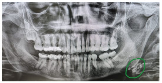
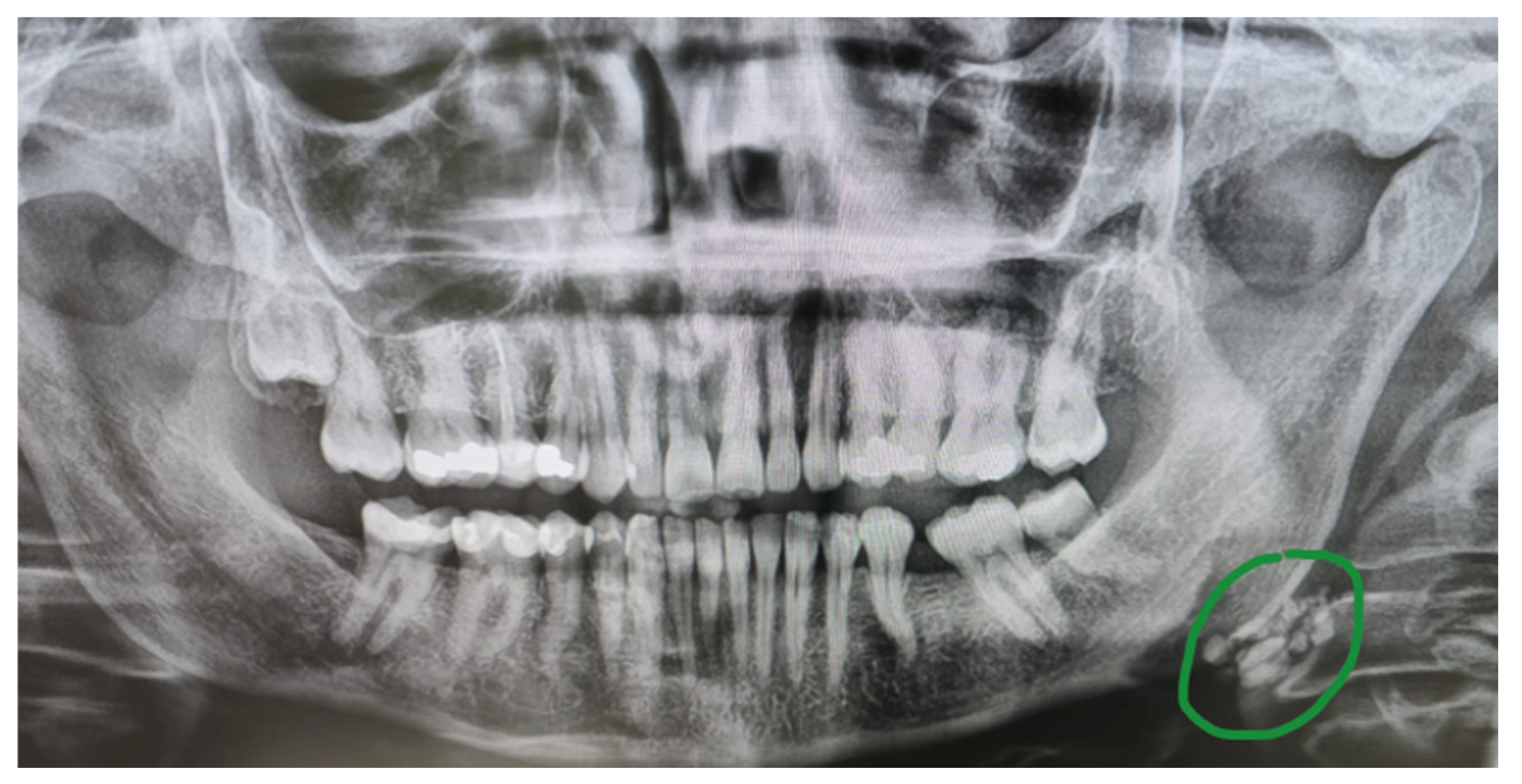
Calcified lymph nodes may be detected on routine OPG as soft-tissue radiopaque masses of variable shape and size. Their radiographic appearance may be laminated or heterogeneous, with both radiopaque and radiolucent areas [6,82,90]. They typically present as single or multiple irregular and lobulated radiopacities of variable size, often with a characteristic cauliflower-like appearance, and are usually located along the submental, submandibular, or cervical lymph node chains (Figure 8) [18]. Lymph node calcifications may occur as a result of previous chronic inflammatory, infectious, granulomatous, or neoplastic processes. They have been described in association with tuberculosis and other granulomatous diseases, including sarcoidosis, histoplasmosis, coccidioidomycosis, and blastomycosis, as well as in lymphoma, thyroid and breast metastases, and systemic inflammatory conditions such as rheumatoid arthritis, scleroderma, and amyloidosis [91,92]. From a radiographic perspective, these calcifications should be distinguished from other soft-tissue radiopacities in the cervicomandibular region, including sialoliths, phleboliths, tonsilloliths, and vascular calcifications [83,84].
Figure 8.
Panoramic radiograph showing multiple irregular and lobulated radiopacities in the left laterocervical region (green circle), consistent with calcified lymph nodes. The image is derived from anonymized archival radiographic material; all patient-identifying information was removed.
3.8. Phleboliths
Phleboliths are intravascular calcifications resulting from the mineralization of thrombi that develop as a consequence of slow or stagnant blood flow, particularly within venous vascular malformations or other low-flow vascular lesions [93]. On OPG, they are relatively uncommon findings, and their prevalence tends to increase with age [18,94,95]. Radiographically, phleboliths appear as round or oval radiopacities, either solitary or more commonly multiple, with variable size and an occasionally scattered distribution within the soft tissues. A characteristic feature is their laminated or concentric morphology, often described as a “target-like” appearance, with either a radiopaque or radiolucent center and variably defined margins [6]. From a differential diagnostic perspective, phleboliths may resemble sialoliths, particularly when projected over the submandibular region [96]. However, unlike sialoliths, which are typically aligned along the course of the salivary ducts, phleboliths more often show a multiple and irregular distribution that is not necessarily related to ductal anatomy [96,97]. The presence of multiple concentric radiopacities, especially when associated with soft-tissue swelling or a clinical history suggestive of a vascular malformation or hemangioma, is more consistent with phleboliths [93,96,97,98,99].
3.9. Laryngeal Cartilages
Calcifications of the triticeal and thyroid cartilages can frequently be visualized on OPG, and their identification is useful for the differential diagnosis of carotid calcifications [100]. In fact, triticeal cartilage calcifications are radiographically located at the level of the third and fourth cervical vertebrae (C3–C4), between the greater horn of the hyoid bone and the superior horn of the thyroid cartilage. Compared with carotid calcifications, however, they appear more oval, with a smooth, well-defined cortical border, and are centrally positioned within the lateral thyrohyoid ligament [101]. Calcifications of the thyroid cartilage are generally visible on OPG for only 2–3 mm, below the hyoid bone, medial to C4, at the lower margin of the radiograph [6], and may range from linear shadows to dense laminar calcifications [65].
4. Implications for Dental Practice
The findings summarized in this review reinforce the view that OPG should not be interpreted solely as an examination of the teeth and jaws. Owing to its wide field of view, an OPG may include anatomical regions in which vascular, sinonasal, skeletal, salivary, lymphatic, and other soft-tissue calcifications can be incidentally detected. For dental practitioners, the clinical relevance of these findings does not lie in establishing a definitive medical diagnosis but rather in recognizing radiographic patterns that may require documentation, clinical correlation, and, when appropriate, referral for specialist assessment. A first practical implication is the need for a systematic evaluation of the entire OPG [3,4,5,102]. In everyday dental practice, attention is often focused primarily on dentoalveolar structures, periodontal status, impacted teeth, periapical disease, implant planning, or temporomandibular joint morphology. However, the evidence reviewed here indicates that clinically meaningful extra-dental findings may be located outside the usual dental areas of interest, particularly in the laterocervical region, the maxillary sinuses, the mandibular angle, and the soft tissues of the floor of the mouth and neck [3,4,5,6,12,102]. Therefore, interpretation of OPG should include a structured assessment of both odontogenic and non-odontogenic regions, with particular attention to the number, morphology, distribution, laterality, and anatomical projection of radiopaque or radiolucent abnormalities [4,5,6,12].
Among the findings discussed, carotid artery calcifications are of particular importance because of their possible association with systemic vascular disease and cardiovascular risk factors [15]. When radiopacities compatible with carotid calcifications are identified near the C3–C4 level, inferior and posterior to the mandibular angle, the dentist should not infer the presence of carotid stenosis, nor should OPG be regarded as a substitute for dedicated vascular imaging. Nevertheless, such findings should be reported, particularly in older patients or in individuals with known cardiovascular risk factors, including hypertension, diabetes, smoking, dyslipidaemia, chronic kidney disease, previous radiotherapy, or a history of vascular events [16,17,18,19,50]. In these cases, appropriate communication with the patient and referral to the general practitioner, cardiologist, vascular specialist, or another suitable physician may enable targeted assessment with Doppler ultrasonography or other dedicated investigations when clinically indicated [3,50].
Findings involving the maxillary sinus represent another area in which dentists may play a clinically relevant role. OPG may reveal mucosal thickening, dome-shaped opacities, discontinuity of the sinus floor, antroliths, foreign bodies, or more extensive sinus opacification [53,54,56,59]. In dental practice, the main implication is the need to distinguish findings that may be related to dental disease from those more suggestive of rhinogenic, inflammatory, or neoplastic conditions [57,60,61,103]. When sinus alterations are adjacent to periapical lesions, periodontal defects, extraction sites, endodontic overfilling, implants, or oroantral communications, an odontogenic origin should be considered [55,58,62,63]. In such cases, Cone Beam Computed Tomography (CBCT) may be appropriate to evaluate the relationship between the maxillary posterior teeth, the sinus floor, and the extent of bony involvement [53,55,59]. Conversely, persistent unilateral opacification, irregular margins, bone destruction, soft-tissue extension, unexplained tooth displacement, paraesthesia, or symptoms not accounted for by dental disease should prompt referral to an otolaryngologist or oral and maxillofacial specialist, with further evaluation by Computed Tomography (CT) and/or Magnetic Resonance Imaging (MRI) [57,58,61].
Assessment of mandibular radiomorphometric indices also has practical implications. Evaluation of the mandibular cortical index and mandibular cortical width may help dentists identify patients, particularly peri- and postmenopausal women or older individuals, who may be at increased risk of low skeletal bone mineral density [64,65]. These indices should be regarded as opportunistic screening tools rather than diagnostic criteria for osteoporosis. Their clinical value is greatest when they are interpreted in conjunction with the patient’s age, sex, menopausal status, fracture history, medical history, and other clinical risk factors [66,67,68]. A markedly eroded mandibular cortex or reduced mandibular cortical width may justify referral for formal densitometric assessment, usually by Dual-energy X-ray Absorptiometry (DXA), but should not be used in isolation to diagnose osteoporosis or to initiate medical treatment [66,67,68,69].
Elongation or calcification of the styloid process and stylohyoid complex illustrates the importance of correlating radiographic findings with clinical symptoms [70,71,72]. An elongated styloid process is often an incidental and asymptomatic finding. Therefore, its detection on OPG does not automatically require treatment [72,73]. However, when elongation or calcification is associated with cervicofacial pain, dysphagia, odynophagia, otalgia, headache, tinnitus, trismus, or pain during head movement, Eagle syndrome may be considered in the differential diagnosis [74]. In such cases, the OPG may provide the first diagnostic clue, whereas CT or CBCT may be required to assess the morphology, length, angulation, and relationship of the stylohyoid complex to adjacent neurovascular structures before specialist management [73,74]. With regard to salivary gland calcifications, the practical role of OPG is mainly preliminary recognition. Sialoliths, particularly in the submandibular region, may appear as radiopaque masses along the expected course of the salivary ducts or glands [75,76,77,78,79,80,81]. Their detection should be integrated with clinical findings such as swelling, meal-related pain, recurrent sialadenitis, or reduced salivary flow [79,86]. When symptoms are present, or when localization is uncertain, additional imaging such as ultrasonography, occlusal radiography, CBCT, CT, or MR sialography may be required, depending on the suspected site and the differential diagnosis [76,79,82,83,84]. Distinguishing sialoliths from phleboliths is particularly important, as phleboliths may indicate an underlying venous malformation and should not be managed as simple salivary stones without further evaluation [96,97,98,99]. Tonsilloliths, calcified lymph nodes, phleboliths, and laryngeal cartilage calcifications further emphasize the importance of differential diagnosis. Tonsilloliths are generally benign and asymptomatic; however, their projection over the mandibular ramus and their possible ghost images may mimic other radiopacities [87,88,89]. Calcified lymph nodes, particularly when multiple, irregular, lobulated, or “cauliflower-like” in appearance, may represent sequelae of previous inflammatory or granulomatous disease, but may also require clinical correlation when unexplained or associated with systemic signs [90,91,92]. Phleboliths, especially when multiple and concentrically laminated, should raise suspicion for a vascular malformation when supported by clinical findings [93,94,95,96]. Triticeous and thyroid cartilage calcifications are relatively common anatomical or age-related findings and are particularly relevant because they may be mistaken for carotid calcifications. Recognition of their typical location, shape, and smooth, well-corticated margins can reduce false-positive interpretations and unnecessary referrals [100,101]. A central message for dental practice is therefore the need to balance vigilance with diagnostic caution [1,104]. OPG is a two-dimensional projection technique affected by magnification, distortion, anatomical superimposition, ghost images, and variability related to patient positioning; consequently, its sensitivity and specificity may vary, and both false-positive and false-negative interpretations may occur [1,104]. For this reason, non-odontogenic findings detected on OPG should generally be described as “compatible with”, “suggestive of”, or “requiring differential diagnosis”, rather than as definitive diagnoses [3,5,102]. The report should include the anatomical location, laterality, size when measurable, morphology, number of lesions, relationship to relevant landmarks, and the recommended clinical action [105,106]. This approach improves communication with both the patient and the receiving medical specialist [105]. The dentist’s responsibility is not to replace the physician, radiologist, otolaryngologist, vascular specialist, endocrinologist, or oral and maxillofacial surgeon, but to act as a sentinel healthcare professional. This role involves recognizing abnormalities, distinguishing likely benign variants from potentially relevant findings, avoiding overinterpretation, and initiating appropriate referral pathways. This is particularly important because many patients undergo OPG more frequently than other head and neck imaging examinations, and some systemic or extraoral conditions may first be suspected in the dental setting [3,5,13,102,106]. These implications also support the need for improved education and standardized reporting in dental radiology [105,107]. Undergraduate and postgraduate training should emphasize not only odontogenic pathology but also the recognition of common extra-dental calcifications and maxillary sinus abnormalities [106,107]. In clinical practice, checklists or structured reporting templates may help reduce missed findings and improve interpretive consistency [105]. However, OPG should not be prescribed solely for the purpose of screening for systemic diseases [2]. Its contribution should instead be understood as opportunistic: when an OPG is already justified for dental reasons, the entire image should be interpreted responsibly, and any relevant extra-dental findings should be managed according to their clinical significance.
Finally, future research should aim to clarify the diagnostic accuracy, interobserver reliability, referral thresholds, and patient outcomes associated with the detection of non-odontogenic findings on OPG. Prospective studies comparing OPG findings with Doppler ultrasonography, CT, CBCT, MRI, DXA, or clinical follow-up would help define which radiographic patterns have the highest predictive value and which findings warrant specialist referral. Until stronger evidence is available, the most appropriate approach remains cautious, systematic, and clinically integrated interpretation of OPG.
5. Conclusions
OPG offers dentists an opportunity to observe not only odontogenic disease but also findings that may reflect conditions beyond the dentoalveolar region. Its clinical value therefore depends on the dentist’s ability to interpret the image as a whole, rather than as a strictly dental examination. The dentist should be regarded as a first-line observer within a broader diagnostic pathway. Recognition of non-odontogenic findings, appropriate documentation, and timely communication with patients and medical specialists may contribute to earlier identification of clinically relevant conditions. At the same time, this role must be exercised with diagnostic caution. OPG should guide suspicion and support referral when needed, but it should not be used as a stand-alone diagnostic tool for extraoral or systemic disease. Strengthening radiological training and promoting systematic whole-image assessment may enhance the contribution of dental practice to interdisciplinary patient care.
Supplementary Materials
The following supporting information can be downloaded at: https://www.mdpi.com/article/10.3390/app16136344/s1, Supplementary S1. Search strategies.
Author Contributions
Conceptualization, D.D.F., M.P. and N.M.; methodology, D.D.F. and M.P.; validation, D.D.F., M.P. and N.M.; formal analysis, N.M. and D.D.F.; investigation, D.D.F., N.M., M.R. and F.S.; resources, D.D.F. and N.M.; data curation, D.D.F., M.K. and M.P.; writing—original draft preparation, D.D.F., N.M. and M.P.; writing—review and editing, D.D.F. and M.P.; visualization, M.R., F.S., M.P. and M.K.; supervision, D.D.F. and M.P. All authors have read and agreed to the published version of the manuscript.
Funding
This research received no external funding.
Institutional Review Board Statement
Not applicable.
Informed Consent Statement
Not applicable.
Data Availability Statement
No new datasets were generated or analyzed during the current study. The data supporting the findings of this review are derived from previously published articles, which are cited in the reference list. The archival radiographic images included in this article were anonymized and are not publicly available due to privacy and ethical restrictions.
Acknowledgments
The authors gratefully acknowledge D. Boscia for providing the radiographic material used in this study.
Conflicts of Interest
The authors declare no conflicts of interest.
References
- Izzetti, R.; Nisi, M.; Aringhieri, G.; Crocetti, L.; Graziani, F.; Nardi, C. Basic Knowledge and New Advances in Panoramic Radiography Imaging Techniques: A Narrative Review on What Dentists and Radiologists Should Know. Appl. Sci. 2021, 11, 7858. [Google Scholar] [CrossRef] [Scilit]
- Benavides, E.; Krecioch, J.R.; Connolly, R.T.; Allareddy, T.; Buchanan, A.; Spelic, D.; O’Brien, K.K.; Keels, M.A.; Mascarenhas, A.K.; Duong, M.-L.; et al. Optimizing radiation safety in dentistry. J. Am. Dent. Assoc. 2024, 155, 280–293.e4. [Google Scholar] [CrossRef] [Scilit] [PubMed]
- Ghassemzadeh, S.; Sbricoli, L.; Frigo, A.C.; Bacci, C. Incidental findings detected with panoramic radiography: Prevalence calculated on a sample of 2017 cases treated at a major Italian trauma and cancer centre v. Oral Radiol. 2021, 37, 507–517. [Google Scholar] [CrossRef] [Scilit] [PubMed]
- Cederhag, J.; Lundegren, N.; Alstergren, P.; Shi, X.-Q.; Hellén-Halme, K. Evaluation of Panoramic Radiographs in Relation to the Mandibular Third Molar and to Incidental Findings in an Adult Population. Eur. J. Dent. 2021, 15, 266–272. [Google Scholar] [CrossRef] [Scilit] [PubMed]
- Hlongwa, P.; Moshaoa, M.A.L.; Musemwa, C.; Khammissa, R.A.G. Incidental Pathologic Findings from Orthodontic Pretreatment Panoramic Radiographs. Int. J. Environ. Res. Public Health 2023, 20, 3479. [Google Scholar] [CrossRef] [Scilit] [PubMed]
- Acikgoz, A.; Akkemik, O. Prevalence and Radiographic Features of Head and Neck Soft Tissue Calcifications on Digital Panoramic Radiographs: A Retrospective Study. Cureus 2023, 15, e46025. [Google Scholar] [CrossRef] [Scilit] [PubMed]
- Brar, A.; DeColibus, K.; Rasner, D.S.; Haynes, A.R.; Pancratz, F.; Oladiran, O.; Gbadamosi, S.O.; Owosho, A.A. Carotid Artery Calcification Detected on Panoramic Radiography Is Significantly Related to Cerebrovascular Accident, Coronary Artery Disease, and Poor Oral Health: A Retrospective Cross-Sectional Study. Dent. J. 2024, 12, 99. [Google Scholar] [CrossRef] [Scilit] [PubMed]
- Ahmed, J.; Gupta, A.; Shenoy, N.; Sujir, N.; Muralidharan, A. Prevalence of Incidental Maxillary Sinus Anomalies on CBCT Scans: A Radiographic Study. Diagnostics 2023, 13, 2918. [Google Scholar] [CrossRef] [Scilit] [PubMed]
- Heuchert, J.; Kozieł, S.; Spinek, A.E. Radiomorphometric indices of the mandible as indicators of decreased bone mineral density and osteoporosis—Meta-analysis and systematic review. Osteoporos. Int. 2024, 35, 401–412. [Google Scholar] [CrossRef] [Scilit] [PubMed]
- Munhoz, L.; Morita, L.; Nagai, A.Y.; Moreira, J.; Arita, E.S. Mandibular cortical index in the screening of postmenopausal at low mineral density risk: A systematic review. Dentomaxillofac. Radiol. 2021, 50, 20200514. [Google Scholar] [CrossRef] [Scilit] [PubMed]
- Assiri Ahmed, H.; Estrugo-Devesa, A.; Roselló Llabrés, X.; Egido-Moreno, S.; López-López, J. The prevalence of elongated styloid process in the population of Barcelona: A cross-sectional study & review of literature. BMC Oral Health 2023, 23, 674. [Google Scholar] [CrossRef] [Scilit] [PubMed]
- Syed, A.Z. Soft Tissue Calcifications in the Head and Neck Region. Dent. Clin. N. Am. 2024, 68, 375–391. [Google Scholar] [CrossRef] [Scilit] [PubMed]
- De Falco, D.; Barone, B.; Iaquinta, F.; Pedone, D.; Roselli, L.; Petruzzi, M. Profiling of Patients Attending the Initial Dental Consultation at a Dental Clinic in Southern Italy: A Single-Centre Retrospective Cross-Sectional Study. Appl. Sci. 2026, 16, 1186. [Google Scholar] [CrossRef] [Scilit]
- Baethge, C.; Goldbeck-Wood, S.; Mertens, S. SANRA—A scale for the quality assessment of narrative review articles. Res. Integr. Peer Rev. 2019, 4, 5. [Google Scholar] [CrossRef] [Scilit] [PubMed]
- Prados-Privado, M.; García Villalón, J.; Blázquez Torres, A.; Martínez-Martínez, C.H.; Prados-Frutos, J.C.; Ivorra, C. Are Panoramic Images a Good Tool to Detect Calcified Carotid Atheroma? A Systematic Review. Biology 2022, 11, 1684. [Google Scholar] [CrossRef] [Scilit] [PubMed]
- Ertas, E.T.; Sisman, Y. Detection of incidental carotid artery calcifications during dental examinations: Panoramic radiography as an important aid in dentistry. Oral Surg. Oral Med. Oral Pathol. Oral Radiol. Endodontol. 2011, 112, e11–e17. [Google Scholar] [CrossRef] [Scilit] [PubMed]
- Bayer, S.; Helfgen, E.-H.; Bös, C.; Kraus, D.; Enkling, N.; Mues, S. Prevalence of findings compatible with carotid artery calcifications on dental panoramic radiographs. Clin. Oral Investig. 2011, 15, 563–569. [Google Scholar] [CrossRef] [Scilit] [PubMed]
- Maia, P.R.L.; Tomaz, A.F.G.; Maia, E.F.T.; Lima, K.C.; Oliveira, P.T. de Prevalence of soft tissue calcifications in panoramic radiographs of the maxillofacial region of older adults. Gerodontology 2022, 39, 266–272. [Google Scholar] [CrossRef] [Scilit] [PubMed]
- Chang, T.I.; Wu, X.; Boström, K.I.; Tran, H.-A.; Couto-Souza, P.H.; Friedlander, A.H. Panoramic imaged carotid atheromas are associated with increased neutrophil count: Both validated, independent predictors of near-term myocardial infarction. Dentomaxillofac. Radiol. 2021, 50, 20210045. [Google Scholar] [CrossRef] [Scilit] [PubMed]
- De Falco, D.; Zacà, S.; Ringold, M.; Sodero, F.; Angiletta, D.; Petruzzi, M. Periodontitis and peripheral artery disease: A mini-review. Front. Oral Health 2026, 7, 1728706. [Google Scholar] [CrossRef] [Scilit] [PubMed]
- Tiller, R.; Bengel, W.; Rinke, S.; Ziebolz, D. Association between carotid area calcifications and periodontal risk: A cross sectional study of panoramic radiographic findings. BMC Cardiovasc. Disord. 2011, 11, 67. [Google Scholar] [CrossRef] [Scilit] [PubMed]
- Dolatabadi, M.A.; Motamedi, M.H.K.; Lassemi, E.; Janbaz, Y. Asymptomatic carotid artery calcifications discovered on panoramic radiography. Gen. Dent. 2010, 58, 97–99. [Google Scholar] [PubMed]
- Christou, P.; Leemann, B.; Schimmel, M.; Kiliaridis, S.; Müller, F. Carotid artery calcification in ischemic stroke patients detected in standard dental panoramic radiographs—A preliminary study. Adv. Med. Sci. 2010, 55, 26–31. [Google Scholar] [CrossRef] [Scilit] [PubMed]
- De Nazaré Alves De Oliveira, C.; Pimenta Amaral, T.M.; Abdo, E.N.; Mesquita, R.A. Bilateral tonsilloliths and calcified carotid atheromas: Case report and literature review. J. Cranio-Maxillofac. Surg. 2013, 41, 179–182. [Google Scholar] [CrossRef] [Scilit] [PubMed]
- Alman, A.C.; Johnson, L.R.; Calverley, D.C.; Grunwald, G.K.; Lezotte, D.C.; Hokanson, J.E. Validation of a method for quantifying carotid artery calcification from panoramic radiographs. Oral Surg. Oral Med. Oral Pathol. Oral Radiol. 2013, 116, 518–524. [Google Scholar] [CrossRef] [Scilit] [PubMed]
- Friedlander, A.H.; Aghazadehsanai, N.; Chang, T.I.; Harada, N.; Garrett, N.R. Prevalence of calcified carotid artery atheromas on panoramic images of individuals with primary hyperparathyroidism. Dentomaxillofac. Radiol. 2013, 42, 20130118. [Google Scholar] [CrossRef] [Scilit] [PubMed]
- Lee, J.Y.; Antoniazzi, M.C.C.; Perozini, C.; Ruivo, G.F.; Pallos, D. Prevalence of carotid artery calcification in patients with chronic renal disease identified by panoramic radiography. Oral Surg. Oral Med. Oral Pathol. Oral Radiol. 2014, 118, 612–618. [Google Scholar] [CrossRef] [Scilit] [PubMed]
- Moshfeghi, M.; Beigom Taheri, J.; Bahemmat, N.; Ebrahim Evazzadeh, M.; Hadian, H. Relationship Between Carotid Artery Calcification and Dental Panoramic Images, Hypertension and Myocardial Infarction. Iran. J. Radiol. 2014, 11, e8714. [Google Scholar] [CrossRef] [Scilit] [PubMed]
- Garoff, M.; Johansson, E.; Ahlqvist, J.; Jäghagen, E.L.; Arnerlöv, C.; Wester, P. Detection of calcifications in panoramic radiographs in patients with carotid stenoses ≥ 50%. Oral Surg. Oral Med. Oral Pathol. Oral Radiol. 2014, 117, 385–391. [Google Scholar] [CrossRef] [Scilit] [PubMed]
- Lee, J.-S.; Kim, O.-S.; Chung, H.-J.; Kim, Y.-J.; Kweon, S.-S.; Lee, Y.-H.; Shin, M.-H.; Yoon, S.-J. The correlation of carotid artery calcification on panoramic radiographs and determination of carotid artery atherosclerosis with ultrasonography. Oral Surg. Oral Med. Oral Pathol. Oral Radiol. 2014, 118, 739–745. [Google Scholar] [CrossRef] [Scilit] [PubMed]
- Atalay, Y.; Asutay, F.; Agacayak, K.S.; Koparal, M.; Adali, F.; Gulsun, B. Evaluation of calcified carotid atheroma on panoramic radiographs and Doppler ultrasonography in an older population. Clin. Interv. Aging 2015, 10, 1121–1129. [Google Scholar] [CrossRef] [Scilit] [PubMed]
- Patil, S. Prevalence of carotid artery calcification in postmenopausal women and its correlation with atherogenic risk factors. J. Nat. Sci. Biol. Med. 2015, 6, 1. [Google Scholar] [CrossRef] [Scilit] [PubMed]
- Gonçalves, J.R.d.S.N.; Yamada, J.L.Y.; Berrocal, C.; Westphalen, F.H.; Franco, A.; Fernandes, Â. Prevalence of Pathologic Findings in Panoramic Radiographs: Calcified Carotid Artery Atheroma. Acta Stomatol. Croat. 2016, 50, 230–234. [Google Scholar] [CrossRef] [Scilit] [PubMed]
- Markman, R.; Conceicao-Vasconcelos, K.; Brandao, T.; Prado-Ribeiro, A.; Santos-Silva, A.; Ajudarte Lopes, M. Calcified carotid artery atheromas on panoramic radiographs of head and neck cancer patients before and after radiotherapy. Med. Oral 2017, 22, e153. [Google Scholar] [CrossRef] [Scilit] [PubMed]
- Gustafsson, N.; Ahlqvist, J.B.; Näslund, U.; Wester, P.; Buhlin, K.; Gustafsson, A.; Levring Jäghagen, E. Calcified carotid artery atheromas in panoramic radiographs are associated with a first myocardial infarction: A case-control study. Oral Surg. Oral Med. Oral Pathol. Oral Radiol. 2018, 125, 199–204.e1. [Google Scholar] [CrossRef] [Scilit] [PubMed]
- Santos, J.; Soares, G.; Alves, A.; Kurita, L.; Silva, P.; Costa, F. Prevalence of carotid artery calcifications among 2500 digital panoramic radiographs of an adult Brazilian population. Med. Oral 2018, 23, e256. [Google Scholar] [CrossRef] [Scilit] [PubMed]
- Maia, P.R.L.; Medeiros, A.M.C.; Pereira, H.S.G.; Lima, K.C.; Oliveira, P.T. Presence and associated factors of carotid artery calcification detected by digital panoramic radiography in patients with chronic kidney disease undergoing hemodialysis. Oral Surg. Oral Med. Oral Pathol. Oral Radiol. 2018, 126, 198–204. [Google Scholar] [CrossRef] [Scilit] [PubMed]
- Friedlander, A.H.; Lee, U.K.; Polanco, J.C.; Tran, H.-A.; Chang, T.I.; Redman, R.S. Positive Association Between Neutrophil-Lymphocyte Ratio and Presence of Panoramically Imaged Carotid Atheromas Among Men. J. Oral Maxillofac. Surg. 2019, 77, 321–327. [Google Scholar] [CrossRef] [Scilit] [PubMed]
- Akkemik, O.; Kazaz, H.; Tamsel, S.; Dündar, N.; Sahinalp, S.; Ellidokuz, H. A 5 years follow-up for ischemic cardiac outcomes in patients with carotid artery calcification on panoramic radiographs confirmed by doppler ultrasonography in Turkish population. Dentomaxillofac. Radiol. 2020, 49, 20190440. [Google Scholar] [CrossRef] [Scilit] [PubMed]
- Çetin, M.B.; Sezgin, Y.; Yilmaz, M.N.N.; Seçgin, C.K. Assessment of Carotid Artery Calcifications on Digital Panoramic Radiographs and Their Relationship With Periodontal Condition and Cardiovascular Risk Factors. Int. Dent. J. 2021, 71, 160–166. [Google Scholar] [CrossRef] [Scilit] [PubMed]
- Rocha, B.A.; Carneiro, L.D.O.B.; Vespasiano, A.; Horta, M.C.R. Detection of calcified carotid atheroma on panoramic dental radiography and its confirmation by Doppler ultrasound. Einstein 2021, 19, eAI5707. [Google Scholar] [CrossRef] [Scilit] [PubMed]
- Kwon, Y.-E.; An, C.-H.; Choi, K.-S.; An, S.-Y. Comparison of carotid artery calcification between stroke and nonstroke patients using CT angiographic and panoramic images. Dentomaxillofac. Radiol. 2022, 51, 20210500. [Google Scholar] [CrossRef] [Scilit] [PubMed]
- Janiszewska-Olszowska, J.; Jakubowska, A.; Gieruszczak, E.; Jakubowski, K.; Wawrzyniak, P.; Grocholewicz, K. Carotid Artery Calcifications on Panoramic Radiographs. Int. J. Environ. Res. Public Health 2022, 19, 14056. [Google Scholar] [CrossRef] [Scilit] [PubMed]
- Zaghden, O.; Jaziri, R.; Kammoun, R.; Chaabani, I.; Alaya, T.B. Benefit of Panoramic Radiography in the Detection of Carotid Calcifications: Clinical Case Reports and Review of the Literature. Case Rep. Dent. 2023, 2023, 3989502. [Google Scholar] [CrossRef] [Scilit] [PubMed]
- Mehdizadeh, M.; Shahbazi, S.; Taheri, H.; Eslami, A. Evaluation of Using Panoramic Radiography and Ultrasonography for Diagnosing Carotid Artery Calcifications. Adv. Biomed. Res. 2023, 12, 226. [Google Scholar] [CrossRef] [Scilit] [PubMed]
- Bladh, M.; Gustafsson, N.; Engström, G.; Kennbäck, C.; Klinge, B.; Nilsson, P.M.; Jönsson, D.; Levring Jäghagen, E. Defined shapes of carotid artery calcifications on panoramic radiographs correlate with specific signs of cardiovascular disease on ultrasound examination. Oral Surg. Oral Med. Oral Pathol. Oral Radiol. 2024, 137, 408–420. [Google Scholar] [CrossRef] [Scilit] [PubMed]
- Garoff, M.; Ahlqvist, J.; Levring Jäghagen, E.; Wester, P.; Johansson, E. Carotid calcifications in panoramic radiographs can predict vascular risk. Dentomaxillofac. Radiol. 2025, 54, 28–34. [Google Scholar] [CrossRef] [Scilit] [PubMed]
- Karlsson, A.; Gustafsson, N.; Wester, P.; Zamure-Damberga, L.; Levring Jäghagen, E. Carotid artery calcifications on panoramic radiographs are associated with vascular disease severity on carotid ultrasound. Dentomaxillofac. Radiol. 2026, 55, 28–36. [Google Scholar] [CrossRef] [Scilit] [PubMed]
- Micheletti, R.G.; Fishbein, G.A.; Currier, J.S.; Fishbein, M.C. Mönckeberg Sclerosis Revisited: A Clarification of the Histologic Definition of Mönckeberg Sclerosis. Arch. Pathol. Lab. Med. 2008, 132, 43–47. [Google Scholar] [CrossRef] [Scilit] [PubMed]
- S, A.; Thambi, J.; Christy, W. A retrospective diagnostic study of prevalence of orofacial calcifications using panoramic radiograph: To insinuate the unseen. J. Clin. Exp. Dent. 2023, 15, e289–e297. [Google Scholar] [CrossRef] [Scilit] [PubMed]
- Goodman, W.G.; Goldin, J.; Kuizon, B.D.; Yoon, C.; Gales, B.; Sider, D.; Wang, Y.; Chung, J.; Emerick, A.; Greaser, L.; et al. Coronary-Artery Calcification in Young Adults with End-Stage Renal Disease Who Are Undergoing Dialysis. N. Engl. J. Med. 2000, 342, 1478–1483. [Google Scholar] [CrossRef] [Scilit] [PubMed]
- Anwar, Z.; Zan, E.; Carone, M.; Ozturk, A.; Sozio, S.M.; Yousem, D.M. Superficial temporal artery calcification in patients with end-stage renal disease: Association with vascular risk factors and ischemic cerebrovascular disease. Indian J. Radiol. Imaging 2011, 21, 215–220. [Google Scholar] [CrossRef] [Scilit] [PubMed]
- Ege, B.; Kurt, M.Y.; Koparal, M.; Geyik, A.; Kaplanoglu, K. Comparison of Diagnostic Accuracy of Panoramic Radiography to CBCT in Maxillary Sinus Diseases. Indian J. Otolaryngol. Head Neck Surg. 2023, 75, 881–892. [Google Scholar] [CrossRef] [Scilit] [PubMed]
- Vallo, J.; Suominen-Taipale, L.; Huumonen, S.; Soikkonen, K.; Norblad, A. Prevalence of mucosal abnormalities of the maxillary sinus and their relationship to dental disease in panoramic radiography: Results from the Health 2000 Health Examination Survey. Oral Surg. Oral Med. Oral Pathol. Oral Radiol. Endodontol. 2010, 109, e80–e87. [Google Scholar] [CrossRef] [Scilit] [PubMed]
- Mahasneh, S.A.; Al-Hadidi, A.; Hassona, Y.; Sawair, F.A.; Al-Nazer, S.; Bakain, Y.; Cunliffe, J. Maxillary Sinusitis of Odontogenic Origin: Prevalence among 3D Imaging—A Retrospective Study. Appl. Sci. 2022, 12, 3057. [Google Scholar] [CrossRef] [Scilit]
- Ha, E.-G.; Jeon, K.J.; Choi, H.; Lee, C.; Choi, Y.J.; Han, S.-S. Automatic diagnosis of retention pseudocyst in the maxillary sinus on panoramic radiographs using a convolutional neural network algorithm. Sci. Rep. 2023, 13, 2734. [Google Scholar] [CrossRef] [Scilit] [PubMed]
- Lin, J.; Wang, C.; Wang, X.; Chen, F.; Zhang, W.; Sun, H.; Yan, F.; Pan, Y.; Zhu, D.; Yang, Q.; et al. Expert consensus on odontogenic maxillary sinusitis multi-disciplinary treatment. Int. J. Oral Sci. 2024, 16, 11. [Google Scholar] [CrossRef] [Scilit] [PubMed]
- Craig, J.R.; Tataryn, R.W.; Aghaloo, T.L.; Pokorny, A.T.; Gray, S.T.; Mattos, J.L.; Poetker, D.M. Management of odontogenic sinusitis: Multidisciplinary consensus statement. Int. Forum Allergy Rhinol. 2020, 10, 901–912. [Google Scholar] [CrossRef] [Scilit] [PubMed]
- Günaçar, D.N.; Köse, T.E.; Ceren, F. Radiodiagnostic properties of maxillary antroliths: A retrospective cone beam computed tomography study. BMC Oral Health 2025, 25, 259. [Google Scholar] [CrossRef] [Scilit] [PubMed]
- Jang, H.-B.; Lee, D.H.; Lim, S.C. Factors affecting the occurrence of maxillary sinus fungus ball. Front. Surg. 2024, 11, 1491155. [Google Scholar] [CrossRef] [Scilit] [PubMed]
- Kumar, J.; Daga, R.; Pradhan, G.; Meher, R. Sinonasal Inflammation or Neoplasm: Raise the Red Flagsǃ-A Pictorial Review. Indian J. Radiol. Imaging 2023, 33, 522–531. [Google Scholar] [CrossRef] [Scilit] [PubMed]
- Fischborn, A.R.; Andreis, J.D.; Wambier, L.M.; Pedroso, C.M.; Claudino, M.; Franco, G.C.N. Performance of panoramic radiography compared with computed tomography in the evaluation of pathological changes in the maxillary sinuses: A systematic review and meta-analysis. Dentomaxillofac. Radiol. 2023, 52, 20230067. [Google Scholar] [CrossRef] [Scilit] [PubMed]
- Terlemez, A.; Tassoker, M.; Kizilcakaya, M.; Gulec, M. Comparison of cone-beam computed tomography and panoramic radiography in the evaluation of maxillary sinus pathology related to maxillary posterior teeth: Do apical lesions increase the risk of maxillary sinus pathology? Imaging Sci. Dent. 2019, 49, 115. [Google Scholar] [CrossRef] [Scilit] [PubMed]
- Selvakumar, R.; Chandran, A.; Patil, A.; Harini, T.; Dandekeri, S.; Verma, D.; Babu, J.S.; Swarnalatha, C.; Nayyar, A.S. Osteoporosis risk group: Screening for osteoporosis in dental clinics using panoramic radiographs. J. Educ. Health Promot. 2022, 11, 271. [Google Scholar] [CrossRef] [Scilit] [PubMed]
- Mupparapu, M.; Akintoye, S.O. Application of Panoramic Radiography in the Detection of Osteopenia and Osteoporosis—Current State of the Art. Curr. Osteoporos. Rep. 2023, 21, 354–359. [Google Scholar] [CrossRef] [Scilit] [PubMed]
- Vlasiadis, K.Z.; Damilakis, J.; Velegrakis, G.A.; Skouteris, C.A.; Fragouli, I.; Goumenou, A.; Matalliotakis, J.; Koumantakis, E.E. Relationship between BMD, dental panoramic radiographic findings and biochemical markers of bone turnover in diagnosis of osteoporosis. Maturitas 2008, 59, 226–233. [Google Scholar] [CrossRef] [Scilit] [PubMed]
- Horner, K.; Devlin, H. The relationships between two indices of mandibular bone quality and bone mineral density measured by dual energy X-ray absorptiometry. Dentomaxillofac. Radiol. 1998, 27, 17–21. [Google Scholar] [CrossRef] [Scilit] [PubMed]
- Hwang, J.J.; Lee, J.-H.; Han, S.-S.; Kim, Y.H.; Jeong, H.-G.; Choi, Y.J.; Park, W. Strut analysis for osteoporosis detection model using dental panoramic radiography. Dentomaxillofac. Radiol. 2017, 46, 20170006. [Google Scholar] [CrossRef] [Scilit] [PubMed]
- Calciolari, E.; Donos, N.; Park, J.C.; Petrie, A.; Mardas, N. Panoramic Measures for Oral Bone Mass in Detecting Osteoporosis: A Systematic Review and Meta-Analysis. J. Dent. Res. 2015, 94, 17S–27S. [Google Scholar] [CrossRef] [Scilit] [PubMed]
- More, C.B.; Asrani, M.K. Evaluation of the styloid process on digital panoramic radiographs. Indian J. Radiol. Imaging 2010, 20, 261–265. [Google Scholar] [CrossRef] [Scilit] [PubMed]
- Langlais, R.P.; Miles, D.A.; Van Dis, M.L. Elongated and mineralized stylohyoid ligament complex: A proposed classification and report of a case of Eagle’s syndrome. Oral Surg. Oral Med. Oral Pathol. 1986, 61, 527–532. [Google Scholar] [CrossRef] [Scilit] [PubMed]
- Bruno, G.; De Stefani, A.; Barone, M.; Costa, G.; Saccomanno, S.; Gracco, A. The validity of panoramic radiograph as a diagnostic method for elongated styloid process: A systematic review. CRANIO 2022, 40, 33–40. [Google Scholar] [CrossRef] [Scilit] [PubMed]
- Dudde, F.; Bergmann, W.; Schuck, O.; Schunk, J.; Barbarewicz, F. Accuracy in the Diagnostics of Styloid Process—Panoramic Radiograph vs. Computed Tomography: A Comparative Study. In Vivo 2024, 38, 1390–1396. [Google Scholar] [CrossRef] [Scilit] [PubMed]
- Nogueira-Reis, F.; De Oliveira Reis, L.; Fontenele, R.C.; Freitas, D.Q.; Tabchoury, C.P.M. Prevalence and features of elongated styloid process on imaging studies: A systematic review and meta-analysis. Clin. Oral Investig. 2022, 26, 1199–1215. [Google Scholar] [CrossRef] [Scilit] [PubMed]
- Garay, I.; Netto, H.D.; Olate, S. Soft tissue calcified in mandibular angle area observed by means of panoramic radiography. Int. J. Clin. Exp. Med. 2014, 7, 51–56. [Google Scholar] [PubMed]
- Kim, J.H.; Aoki, E.M.; Cortes, A.R.G.; Abdala-Júnior, R.; Asaumi, J.; Arita, E.S. Comparison of the diagnostic performance of panoramic and occlusal radiographs in detecting submandibular sialoliths. Imaging Sci. Dent. 2016, 46, 87. [Google Scholar] [CrossRef] [Scilit] [PubMed]
- Franco, A.; De Carvalho Mattos, M.J.; Ferrari, F.; Dos Reis Neto, J.M.; Carta Gambus, L.C.; Couto Souza, P.H.; Berti-Couto, S.D.A. Massive Submandibular Sialolith: Complete Radiographic Registration and Biochemical Analysis through X-Ray Diffraction. Case Rep. Surg. 2014, 2014, 659270. [Google Scholar] [CrossRef] [Scilit] [PubMed]
- Aoun, G.; El-Outa, A.; Nasseh, I. Sialoliths: A Radiographic Retrospective Study. Curr. Res. Dent. 2020, 11, 1–5. [Google Scholar] [CrossRef] [Scilit]
- Beumer, L.; Van Der Meij, E.; Kamstra, J.; De Visscher, J. Interobserver and intraobserver variability in the radiological assessment of sialolithiasis using cone beam computed tomography. Med. Oral 2022, 27, e94–e98. [Google Scholar] [CrossRef] [Scilit] [PubMed]
- Sánchez Barrueco, A.; Díaz Tapia, G.; Guerra Gutiérrez, F.; Salvador Álvarez, E.; Villacampa Aubá, J.M. The key role of magnetic resonance sialography in the differential diagnosis between sialolith and phlebolith of the floor of the mouth. BMC Oral Health 2025, 25, 1615. [Google Scholar] [CrossRef] [Scilit] [PubMed]
- Özçelik, N.; Vehbi, H.; Alaskarov, E. Comparison of the Inadequacies of Ultrasonography and Computed Tomography in the Diagnosis of Sialolithiasis. Ear Nose Throat J. 2024, 103, 37S–44S. [Google Scholar] [CrossRef] [Scilit] [PubMed]
- Celik Ozsoy, S.; Zirek, T.; Bahrilli, S.; Yuksel, I.B.; Altindag, A. Examination of the Frequency of Soft Tissue Ossification and Calcifications in Panoramic Radiographs: A Retrospective Study. Diagnostics 2025, 15, 2013. [Google Scholar] [CrossRef] [Scilit] [PubMed]
- Özkan, G.; Köse, E.; Yeşiltepe, S. Ultrasonographic Evaluation of Soft Tissue Calcifications in the Head and Neck Region Detected on Panoramic Radiographs. J. Ultrasound Med. 2023, 42, 1057–1064. [Google Scholar] [CrossRef] [Scilit] [PubMed]
- Yeşilova, E.; Bayrakdar, İ.Ş. Radiological evaluation of maxillofacial soft tissue calcifications with cone beam computed tomography and panoramic radiography. Int. J. Clin. Pract. 2021, 75, e14086. [Google Scholar] [CrossRef] [Scilit] [PubMed]
- Gurbuz, E.; Gungor, M.; Hatipoglu, H. Radiographic Detection of the Relationship between Tonsilloliths and Dental Plaque-Related Pathologies in a Series of Digital Panoramic Radiographs. Med. Princ. Pract. 2022, 31, 149–155. [Google Scholar] [CrossRef] [Scilit] [PubMed]
- Mohsin, S.F.; Riyaz, M.A.; Alqazlan, A.A. Detection and Management of Giant Submandibular Gland Sialolith. Pak. J. Med. Sci. 2022, 38, 2030. [Google Scholar] [CrossRef] [Scilit] [PubMed]
- Crameri, M.; Bassetti, R.; Werder, P.; Kuttenberger, J. Tonsillolithen auf dem Orthopantomogramm: Zwei Fallberichte über Kalzifikationen in den Tonsillen. Swiss Dent. J. SSO 2016, 126, 29–32. [Google Scholar] [CrossRef] [Scilit] [PubMed]
- Takahashi, A.; Sugawara, C.; Kudoh, T.; Ohe, G.; Takamaru, N.; Tamatani, T.; Nagai, H.; Miyamoto, Y. Prevalence and imaging characteristics of palatine tonsilloliths evaluated on 2244 pairs of panoramic radiographs and CT images. Clin. Oral Investig. 2017, 21, 85–91. [Google Scholar] [CrossRef] [Scilit] [PubMed]
- Oda, M.; Kito, S.; Tanaka, T.; Nishida, I.; Awano, S.; Fujita, Y.; Saeki, K.; Matsumoto-Takeda, S.; Wakasugi-Sato, N.; Habu, M.; et al. Prevalence and imaging characteristics of detectable tonsilloliths on 482 pairs of consecutive CT and panoramic radiographs. BMC Oral Health 2013, 13, 54. [Google Scholar] [CrossRef] [Scilit] [PubMed]
- Aydin, Ü. Tuberculous lymph node calcification detected on routine panoramic radiography: A case report. Dentomaxillofac. Radiol. 2003, 32, 252–254. [Google Scholar] [CrossRef] [Scilit] [PubMed]
- Hirschfeld, J.J. Calcification in lymph nodes. Oral Surg. Oral Med. Oral Pathol. 1986, 61, 412. [Google Scholar] [CrossRef] [Scilit] [PubMed]
- Romano, N.; Silvestri, G.; Castaldi, A. The “ABC” of Neck Calcifications: A Practical Guide. SN Compr. Clin. Med. 2021, 3, 2455–2464. [Google Scholar] [CrossRef] [Scilit]
- Brahmbhatt, A.N.; Skalski, K.A.; Bhatt, A.A. Vascular lesions of the head and neck: An update on classification and imaging review. Insights Imaging 2020, 11, 19. [Google Scholar] [CrossRef] [Scilit] [PubMed]
- Jha, R.; Gohil, J.; Verma, O.; Jani, Y.; Thakkar, S.; Gupta, M. Prevalence of Soft Tissue Calcifications on Digital Panoramic Radiographs: A Cross-Sectional Study. J. Pharm. Bioallied Sci. 2026, 18, S140–S142. [Google Scholar] [CrossRef] [Scilit] [PubMed]
- Uzun, C.; Celenk, P.; van der Stelt, P.F. Venous malformation with multiple phleboliths: A case report. Quintessence Int. 2017, 48, 51–56. [Google Scholar] [CrossRef] [Scilit] [PubMed]
- Sato, S.; Takahashi, M.; Takahashi, T. A Case of Multiple Phleboliths on the Medial Side of the Right Mandible. Case Rep. Dent. 2020, 2020, 6694402. [Google Scholar] [CrossRef] [Scilit] [PubMed]
- Ibáñez-Mancera, N.; Partida-Rodríguez, E.; Partida-Rodríguez, O. Phleboliths of venous malformation studied with scan electron microscopy. Case report and literature review. J. Clin. Exp. Dent. 2024, 16, e921–e925. [Google Scholar] [CrossRef] [Scilit] [PubMed]
- Gallant, S.C.; Chewning, R.H.; Orbach, D.B.; Trenor, C.C.; Cunningham, M.J. Contemporary Management of Vascular Anomalies of the Head and Neck—Part 1: Vascular Malformations: A Review. JAMA Otolaryngol. Head Neck Surg. 2021, 147, 197. [Google Scholar] [CrossRef] [Scilit] [PubMed]
- Bertino, F.; Trofimova, A.V.; Gilyard, S.N.; Hawkins, C.M. Vascular anomalies of the head and neck: Diagnosis and treatment. Pediatr. Radiol. 2021, 51, 1162–1184. [Google Scholar] [CrossRef] [Scilit] [PubMed]
- Çağırankaya, L.B.; Akkaya, N.; Akçiçek, G.; Boyacıoğlu Doğru, H. Is the diagnosis of calcified laryngeal cartilages on panoramic radiographs possible? Imaging Sci. Dent. 2018, 48, 121. [Google Scholar] [CrossRef] [Scilit] [PubMed]
- Ahmad, M.; Madden, R.; Perez, L. Triticeous cartilage: Prevalence on panoramic radiographs and diagnostic criteria. Oral Surg. Oral Med. Oral Pathol. Oral Radiol. Endodontol. 2005, 99, 225–230. [Google Scholar] [CrossRef] [Scilit] [PubMed]
- MacDonald, D.; Yu, W. Incidental findings in a consecutive series of digital panoramic radiographs. Imaging Sci. Dent. 2020, 50, 53. [Google Scholar] [CrossRef] [Scilit] [PubMed]
- Craig, J.R.; Poetker, D.M.; Aksoy, U.; Allevi, F.; Biglioli, F.; Cha, B.Y.; Chiapasco, M.; Lechien, J.R.; Safadi, A.; Simuntis, R.; et al. Diagnosing odontogenic sinusitis: An international multidisciplinary consensus statement. Int. Forum Allergy Rhinol. 2021, 11, 1235–1248. [Google Scholar] [CrossRef] [Scilit] [PubMed]
- Różyło-Kalinowska, I. Panoramic radiography in dentistry. Clin. Dent. Rev. 2021, 5, 26. [Google Scholar] [CrossRef] [Scilit]
- Schnitzer, M.L.; Biechele, G.; Schober, E.; Herr, F.L.; Dascalescu, C.; Heimer, M.; Ebner, R.; Fusch, V.; Marschner, S.; Corradini, S.; et al. Structured reporting of dental panoramic images in a hospital-based radiology setting: A comparative study. Front. Dent. Med. 2026, 7, 1769864. [Google Scholar] [CrossRef] [Scilit] [PubMed]
- Duran, M.H.; Coşgun Baybars, S. Evaluation of Soft Tissue Calcifications in the Head and Neck Region on Panoramic Radiography of Edentulous Patients. Necmettin Erbakan Üniv. Diş Hekim. Derg. 2024, 2, 208–215. [Google Scholar] [CrossRef] [Scilit]
- Math, S.Y.; Sultan, O.S.; Anuar, M.F.B.Z.; Pacheco-Pereira, C. Interactive E-Learning Module: Enhancing Panoramic Radiograph Interpretation Skills of Dental Students. J. Dent. Educ. 2026, 90, 136–146. [Google Scholar] [CrossRef] [Scilit] [PubMed]

Disclaimer/Publisher’s Note: The statements, opinions and data contained in all publications are solely those of the individual author(s) and contributor(s) and not of MDPI and/or the editor(s). MDPI and/or the editor(s) disclaim responsibility for any injury to people or property resulting from any ideas, methods, instructions or products referred to in the content. |
© 2026 by the authors. Licensee MDPI, Basel, Switzerland. This article is an open access article distributed under the terms and conditions of the Creative Commons Attribution (CC BY) license.