


Abstract
Dietary factors, including the consumption of isoflavones-rich foods of plant origin, may contribute to the reduced incidence of prostate cancer. Isoflavones, natural phytoestrogens often found in legumes, can modulate estrogen and androgen receptor signaling. This study aimed to evaluate the biological potential of isoflavone-rich extracts obtained from twelve species from the Fabaceae family, targeting prostate cancer cell viability, proliferation, inflammatory markers, prostate-specific antigen secretion, and 5α-reductase activity. The tested extracts showed moderate cytotoxic activity against prostate cancer cell lines, apart from highly susceptible PC3 cells, and only weak toxicity to normal prostate epithelial cells. Significant antiproliferative activity was observed, especially for Cytisus scoparius, Ononis arvensis, and Genista tinctoria, while most extracts reduced prostate-specific antigen (PSA) secretion in normal prostate cells. Furthermore, the extracts showed anti-inflammatory properties by reducing the pro-inflammatory cytokine interleukin 6 (IL-6) and improving cytokine balance indices. Multivariate analyses revealed correlations between total isoflavone content and antiproliferative activity.
1. Introduction
Prostate cancer is one of the main causes of death among men who have been diagnosed with the disease. Globally, the number of new cases exceeds 1.5 million annually, making prostate cancer the fourth most commonly detected cancer overall [1]. A characteristic epidemiological feature of prostate cancer is its marked geographical variability: Asian populations display significantly lower incidence and mortality rates compared with Western countries [2]. After migrating to Western countries, Asian men exhibit a higher incidence of prostate cancer compared to those who remain in Asia. However, their risk generally remains lower than that observed in non-Asian men, suggesting that environmental factors such as lifestyle and diet, rather than genetics alone, contribute to prostate cancer risk [3,4]. One proposed explanation for these differences is the higher consumption of soy products and other legume plants rich in isoflavones—such as genistein, daidzein, and glycitein—in the diets of Asian populations compared with Western diets [5,6].
Isoflavones are natural phytoestrogens which can bind to the estrogen receptors, with a particular preference for ERβ, the predominant estrogen receptor subtype in prostate tissue [7]. Notably, some isoflavones, and their metabolites, also demonstrate binding affinities toward the androgen receptor (AR) that are comparable to testosterone, suggesting they may compete with endogenous androgens for AR binding and thereby modulate androgen-dependent pathways in prostate cancer cells [8]. Isoflavones are characteristic for Fabaceae (legume) plants, with soy (Glycine max (L.) Merr.), and red clover (Trifolium pratense L.) as the best-known representatives, with exceptionally high isoflavones content [9,10]. Interestingly, apart from some edible legumes with notable isoflavones content (e.g., soybeans, chickpea sprouts), which can provide health benefits as an element of the daily diet, there are also other Fabaceae species not classified as food plants whose biological potential warrants further investigation. Thus, the search for new, isoflavones-rich plant sources is fully justified.
The influence of isoflavones on the development and progression of prostate cancer has been the subject of extensive epidemiological, clinical, and experimental research. Population-based studies consistently indicate that high isoflavone intake is associated with a reduced risk of developing prostate cancer and with slower disease progression; these findings are supported across various cohorts of healthy men as well as individuals at elevated risk of prostate malignancy [11]. Preclinical studies have demonstrated that isoflavones inhibit the proliferation of both androgen-dependent (LNCaP) and androgen-independent (PC3, DU145) prostate cancer cell lines. This can be probably related to the modulation of key regulators of the cell cycle—such as increased expression of p21 and p27, reduction of cyclins D1 and B1, and inhibition of cyclin-dependent kinases 2 and 4—leading to cell-cycle arrest at various stages, suppression of proliferation, and induction of apoptosis [12].
A crucial aspect of isoflavones activity is their effect on the androgen balance, which is central to the development of prostate cancer. Studies have shown that isoflavones reduce AR expression, decrease its transcriptional activity, and diminish PSA secretion at both the protein and mRNA levels. They also downregulate the expression of genes involved in invasion and metastasis, such as matrix metalloproteinase-2 (MMP-2), urokinase-type plasminogen activator (uPA) and also its receptor, thereby limiting the migratory and invasive properties of prostate cancer cells [13]. Importantly, many of these effects occur not only at pharmacological concentrations but also at physiological levels that can be achieved through dietary intake, particularly in the case of genistein and equol [14].
Prostate cancer is considered an ideal target for chemoprevention due to its long latency period, high incidence, and the presence of well-characterized preneoplastic lesions such as prostatic intraepithelial neoplasia [15]. Chemoprevention is defined as the use of natural or synthetic agents to reverse, inhibit, or delay carcinogenesis by interfering with specific molecular events in the cancer-initiating and cancer-promoting pathways. Notably, isoflavones exert chemopreventive effects in the early stages of carcinogenesis through activation of the DNA damage response, a crucial cellular mechanism safeguarding genomic integrity. Such an effect, observed for genistein, has been suggested as one of the earliest barriers that delay progression from premalignant lesions to invasive cancer [16]. Although benign prostatic hyperplasia (BPH) is not considered a definitive precancerous lesion, BPH and prostate cancer often coexist, and epidemiological data suggest a positive association between BPH and prostate cancer risk [17]. Inhibiting or delaying prostate cell hyperproliferation and the processes that promote tissue growth is also important from a chemopreventive perspective, as limiting proliferation and growth signaling may reduce the risk of these lesions and slow down unfavorable changes in the prostate microenvironment [18,19]. This mechanistic insight provides a strong rationale for considering dietary isoflavones as functional nutraceuticals in prostate cancer chemoprevention.
The reported total isoflavone content in soybean seeds ranges from 525 to 986 mg/kg, depending on the cultivar. Interestingly, in our study, several Fabaceae species exhibited substantially higher total isoflavone content, including Trifolium medium (26.70 mg/g d.m.), Genista tinctoria (19.65 mg/g d.m.), and Trifolium pratense (12.56 mg/g d.m.), highlighting their potential as alternative natural sources of these biologically active compounds. In our previous study, we indicated 12 legume species (see Section 2.1), from the “non-food” category, as new and promising candidates for further chemopreventive studies based on their high isoflavone content and diverse isoflavone profiles. The selected species represented diverse isoflavone profiles and varying total isoflavone contents, including both highly abundant and moderate sources of these compounds. Additionally, we proved that the extracts from these species decreased not only viability but also proliferation of human breast cancer cell lines, with a high safety profile against normal cells and interesting antioxidant potential [20]. These findings not only confirmed the biological potential of isoflavone-rich Fabaceae extracts but also highlighted the need to further investigate their chemopreventive properties in other hormone-related tumors. Taking these into account, the present study focused on the evaluation of the biological potential of the previously obtained and quantitatively characterized, isoflavone-rich extracts from twelve Fabaceae species against prostate cancer and normal cells, with particular emphasis on cell viability and proliferation. Furthermore, we wanted to evaluate the ability of the extracts to influence some features associated with prostate carcinogenesis, namely release of inflammatory markers, PSA level or 5α-reductase activity, to better highlight their potential preventive role in prostate diseases.
2. Materials and Methods
2.1. Plant Material and Extraction
Above-ground parts of 12 Fabaceae species, namely Cytisus scoparius (L.) Link, Genista tinctoria (L.), Medicago x varia Martyn, Melilotus albus Medik., Ononis arvensis L., Trifolium arvense L., T. fragiferum L., T. incarnatum L., T. medium. L., T. pratense L., T. resupinatum L., Vicia grandiflora Scop., that were preselected in our previous study [18] were used. Plant material was collected in 2024 in Lesser Poland province (Table S1). Voucher specimens of all collected plant species were deposited at the Department of Pharmacognosy, Jagiellonian University Medical College. The dried and ground plant material was heat-reflux extracted for 60 min, with 50% methanol (a plant-to-solvent of ratio 1:125). Then the extracts were subjected to qualitative and quantitative isoflavone HPLC analysis. The data on plant collection, voucher specimens, complete extraction and quantitative analytical procedures were described in detail in our previous publication [20]. Additionally, the antioxidant capacity (DPPH and FRAP) of the extracts was previously determined [20]. The total isoflavone content of the analyzed extracts and the results of antioxidant potential, which were also included in the chemometric analysis in the present study, are summarized in Table S1.
The obtained extracts were evaporated to dryness and dissolved (50 mg/mL) in dimethyl sulfoxide to obtain stock solutions for further cellular assays.
2.2. Cell Lines and Conditions
Different human prostate cancer cell lines were used in the study: androgen-negative DU145 and PC3, and androgen-responsive LNCaP cells. Safety assessment was performed using normal human prostate epithelial cells PNT2. All cell lines were cultured in an incubator at 37 °C, 5% CO2, and proper humidity), using appropriate media: DMEM/F-12 for PNT2 and PC3, MEM with low glucose concentration for DU145, and RPMI 1640 without phenol-red for LNCaP, supplemented with fetal bovine serum and antibiotic mixture. All cell lines and media were obtained from Merck (Darmstadt, Germany).
2.3. Cell Viability and Proliferation
The DMSO solutions of extracts were subsequently diluted in the culture media (25 to 500 μg/mL), to avoid the toxic effect of DMSO alone (its final concentration in culture wells did not exceed 0.1%). Cell viability after 48 h of incubation was determined using the MTT assay, which assesses the metabolic activity of viable cells based on the reduction of tetrazolium salt to formazan crystals by mitochondrial oxidoreductases, as previously described [18]. Doxorubicin was used as a reference cytostatic. Absorbance for the viability assay was measured at 490 nm.
Cell proliferation-related changes after 48 h and 72 h were evaluated using the crystal violet assay, which measures the total biomass of adherent cells according to the previously reported methodology [18]. For this assay, extract working concentrations ranged from 50 to 300 μg/mL. Briefly, the cells were washed with PBS and fixed with 3.7% formaldehyde. Then, crystal violet solution was added for 10 min, followed by washing with PBS. Crystal violet was extracted from cells using 1.33% citric acid and 1.09% sodium citrate in water/methanol (1:1) solution. Absorbance was recorded at 570 nm using BioTek Synergy microplate reader (BioTek Instruments Inc., Winooski, VT, USA). The results are presented as the mean ± SD from three independent experiments, as a percentage of untreated cells.
2.4. Prostate-Specific Antigen and 5α-Reductase Determination
Briefly, PNT2 cells were seeded onto 96-multi-well plates and incubated with the tested extracts (50, 100, and 300 µg/mL) or dutasteride (reference drug) for 72 h, as previously described [17]. Cell culture supernatants were collected and used for analysis of PSA and the 5α-reductase level was analyzed in cell culture supernatants, using ELISA kits (Bioassay Technology Laboratory, Shanghai, China), according to the manufacturer’s instructions. Absorbance was measured using a microplate reader Synergy H1 (Agilent Technologies, Inc., Santa Clara, Ca, USA). The results were presented as a % of control (untreated cells), from three separate experiments.
2.5. Anti-Inflammatory Activity
PNT2 cells were stimulated with interferon-γ (10 ng/mL) and of TNF-α (20 ng/mL) to induce inflammation, after which the examined extracts or dexamethasone (reference drug) were added accordingly and incubated for 48 h. Cytokine (TNF-α, IL-6, IL-10) release was determined in cell supernatants by ELISA kits (Bioassay Technology Laboratory, Shanghai, China), according to the manufacturer’s protocol. The absorbance was measured using a microplate reader Synergy H1 (Agilent Technologies, Inc., Santa Clara, CA, USA). The results, taken from three separate experiments, were determined as a % of the control (cells stimulated with cytokine mixtures).
2.6. Statistical Analysis
Statistical analysis was implemented using Statistica v.13.3 (TIBCO Software Inc., Palo Alto, CA, USA). Shapiro–Wilk and Levene’s tests were used to evaluate the normality of data distribution, and homogeneity of variances, respectively, followed by Student’s t-test, to compare the differences between two groups, or one-way ANOVA and Tukey’s post hoc test among more than two groups. Statistical significance was considered with a p-value ≤ 0.05.
Principal component analysis and hierarchical cluster analysis were conducted in Statistica v.13.3 (TIBCO Software Inc., Palo Alto, CA, USA). Bartlett’s test of sphericity and the KMO index were applied to confirm the suitability of the data for PCA. In the model, the examined plant species were the cases, and the activities (proliferation (after 48 h incubation), Il-6, antioxidant), Il-10/Il-6, and the isoflavone sum were the parameters. PCA model was developed based on the correlation matrix. Hierarchical cluster analysis (CA) was performed on standardized data using Ward’s method with Euclidean distance, with clustering using Mojena’s rule. Graphs were prepared using Statistica v.13.3.
3. Results and Discussion
3.1. Effect on Cell Viability
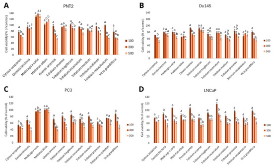
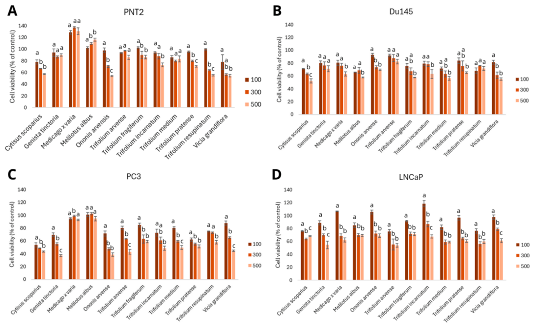
In our previous study, we demonstrated a chemopreventive effect of the above-mentioned extracts from twelve Fabaceae species, with the defined isoflavones content, directed to breast cancer in vitro. As a continuation, in the present study, the cytotoxic potential of the same extracts was evaluated in a human prostate cancer in vitro model, comprising not only androgen-sensitive (LNCaP) but also androgen-independent (DU145, PC3) cell lines, to mimic the heterogenous nature of the tumor. The effect was determined across a broad concentration range (25–500 µg/mL). However, since no effects were observed at concentrations below 100 µg/mL, the results shown in Figure 1 focus on the three highest concentrations tested. Overall, the extracts exhibited a moderate impact on cell viability, as referred to the criteria of the National Cancer Institute and the Geran protocol for plant extracts [21]. Notably, PC3 cells were the most sensitive to the extracts. This observation is particularly significant given that PC3 represents a highly aggressive, metastatic, and therapy-resistant prostate cancer phenotype, which is typically considered difficult to eradicate using standard treatment strategies. Despite this inherent resilience, exposure to Cytisus scoparius, Genista tinctoria and Ononis arvensis extracts resulted in a pronounced reduction in PC3 cell viability (Figure 1C), with IC50 167.1, 323.0, and 231.2 µg/mL, respectively (IC50 for doxorubicin >50 µg/mL). In contrast, the DU145 and LNCaP cells displayed a milder cytotoxic response. Among DU145 cells, the greatest decrease in viability was observed after treatment with Cytisus scoparius and Trifolium medium extracts, which, at the highest tested concentration, reduced cell viability to 52.46 ± 3.28 and 56.29 ± 3.13%, respectively (IC50 for doxorubicin 3.2 µg/mL). Although these effects were moderate, they nonetheless indicate measurable sensitivity of DU145 cells to some of the tested extracts (Figure 1B). In hormone-dependent LNCaP cells, the strongest responses were recorded following treatment with the highest tested concentration of Trifolium arvense and Genista tinctoria (Figure 1D), reducing cell viability to 53.76 ± 2.96 and 54.55 ± 5.69%, respectively (IC50 for doxorubicin 1.8 µg/mL). Doxorubicin was used exclusively as a reference control for the assay.
Figure 1.
Cytotoxic effects of Fabaceae plant extracts (100, 300, and 500 μg/mL, 48 h) on normal PNT2 (A), and cancer DU145 (B), PC3 (C), and LNCaP (D) prostate cells. The results are presented as the mean ± SD from three separate experiments. Significant differences (p ≤ 0.05) between extract doses of each species are marked with different lowercase letters. Statistical comparisons were performed between different concentrations of the same extract within each cell line (one-way ANOVA and Tukey’s post hoc test). Groups sharing at least one lowercase letter are not significantly different.
Comparison of our findings with the available literature data supports these observations. The ethanol extracts from Genista monspessulana seeds showed IC50 values ranging from 26.3 to 211.0 μg/mL against PC-3 cells, depending on the seed collection site, placing them within a similar activity range to the extracts analyzed in the present study. Notably, the study applied a broad concentration range (0.8–500 μg/mL), with meaningful cytotoxic effects occurring mainly at doses above 50 μg/mL, which is similar to our observations [22]. A comparable pattern was reported by Asadi-Samani et al., who demonstrated that extracts from the aerial parts of Medicago sativa displayed markedly different cytotoxic profiles depending on the prostate cancer cell line tested. For PC3 cells, concentrations of 1–10 μg/mL were ineffective, and the IC50 exceeded 300 μg/mL, classifying the extract as weakly active. In contrast, for androgen-independent DU145 cells, the same extract was moderately active, with IC50 77 μg/mL [23]. Despite these findings, it should be emphasized that the studies on the cytotoxic effects of isoflavone-rich Fabaceae extracts on prostate cancer cells still remain limited.
To assess the selectivity and safety of the tested extracts, normal prostate cells PNT2 were also examined. Most of the examined extracts revealed no cytotoxic effect, with the exception of Ononis arvensis, Vicia grandiflora, and Trifolium resupinatum, which reduced cell viability to 53.78 ± 1.04, 54.45 ± 1.90, and 55.40 ± 1.36%, respectively, at the highest tested dose (Figure 1A). However, these effects can be classified as only weak, according to Geran protocol [21], and compared to the reference doxorubicin (IC50 1.4 µg/mL). In our previous study, we also proved that the examined extracts did not reveal hepatotoxic nor goitrogenic effects in vitro, as well as were not toxic to normal breast cells [20]. These observations support the overall high safety profile of the examined extracts, important for the future studies.
3.2. Effect on Cell Proliferation
Since certain phytochemicals—particularly isoflavones—may exert hormone-dependent proliferative effects, we aimed to evaluate the antiproliferative potential of the examined extracts using the same prostate cancer in vitro model as in the viability study. The antiproliferative effect was determined at subcytotoxic doses (50–300 µg/mL), and at two time points (48 and 72 h).
The antiproliferative activity revealed substantial differences among the three prostate cancer cell lines. After 48 h of treatment, the proliferation of PC3 cells exposed to Genista tinctoria and Cytisus scoparius extracts decreased to 34.67 ± 3.10 and 35.84 ± 3.40% of control, respectively (Figure S1, supplementary). In DU145 cells, the strongest responses were observed after treatment with Vicia grandiflora (61.92 ± 2.36%) and Ononis arvensis (62.12 ± 4.68%), indicating a moderate suppression of proliferation at this time point. In contrast, androgen-dependent LNCaP cells showed high sensitivity to the extracts from Trifolium pratense (37.03 ± 0.32%) and Trifolium medium (37.81 ± 0.28%), with proliferation levels comparable to those observed in PC3 cells.
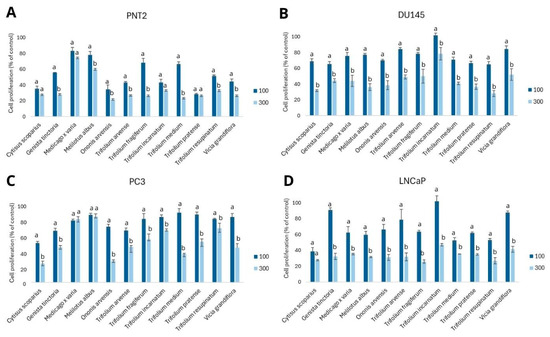
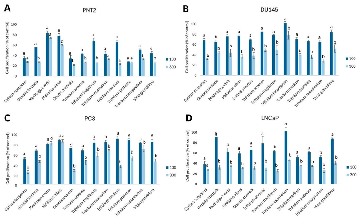
Extending the incubation time to 72 h markedly enhanced the antiproliferative effects across nearly all extracts and cell lines. In PC3 cells, Cytisus scoparius and Ononis arvensis extracts at the highest tested concentration decreased the proliferation to 24.05 ± 2.92 and 27.17 ± 1.47%, respectively (Figure 2C). DU145 cells, which were more resistant at 48 h, exhibited pronounced sensitivity at 72 h, especially to Trifolium resupinatum (28.01 ± 3.65%) and Cytisus scoparius (31.85 ± 1.17%) (Figure 2B) at the dose of 300 µg/mL. The strongest inhibitory effect was observed in LNCaP cells, in which Trifolium fragiferum and T. resupinatum decreased cell proliferation to 25.78 ± 2.16 and 26.86 ± 4.00%, respectively, demonstrating a potent antiproliferative response in the androgen-dependent model (Figure 2D). Interestingly, some extracts showed notable antiproliferative effects even at lower concentrations, as observed for Cytisus scoparius, which inhibited the proliferation of androgen-dependent LNCaP cells to 38.87 ± 4.65% at the dose of 100 µg/mL.
Figure 2.
Antiproliferative effect of Fabaceae plant extracts on normal PNT2 (A), and cancer DU145 (B), PC3 (C), and LNCaP (D) prostate cells, treated with the extracts (100 and 300 µg/mL) for 72 h. Results are presented as mean ± SD from three separate experiments. Significant differences (p ≤ 0.05) between extract doses for each species are indicated by different lowercase letters. Statistical comparisons were performed between different concentrations of the same extract within each cell line (one-way ANOVA and Tukey’s post hoc test).
Some of the examined extracts also exerted an inhibitory effect on the proliferation of non-cancerous prostate epithelial PNT2 cells. The strongest inhibitory effects were observed for Trifolium incarnatum and T. medium extracts at the highest tested concentration (300 µg/mL) at both time points. After 48 h of treatment, inhibition of cell proliferation reached 26.25 ± 2.58 and 31.34 ± 4.24%, respectively (Figure S1, Supplementary Materials), while after 72 h the values were 21.57 ± 4.13 and 23.28 ± 1.86% (Figure 2A). The observed effect is important in terms of the increased cell proliferation observed during benign prostate hyperplasia, preventing the incidence of such a problem. However, it should be noted that the observed effects on PNT2 cells may also suggest partially non-selective cytostatic activity of some extracts under the applied experimental conditions. Nevertheless, the inhibitory effects observed in normal cells were generally less pronounced than those detected in prostate cancer cell lines.
The literature lacks data describing the impact of Fabaceae plants extracts towards prostate cancer or normal cells’ proliferation, making our study the first to comprehensively address this issue. The obtained results clearly indicate that the tested Fabaceae extracts exhibit significant antiproliferative potential against prostate cancer cells, with the strongest activity observed for Cytisus scoparius, Ononis arvensis, Trifolium resupinatum, T. fragiferum, and Genista tinctoria. In some cases the correlation between the isoflavones content and the observed activity was noted, which will be discussed below.
3.3. Impact on Prostate-Specific Antigen Level and 5α-Reductase Activity
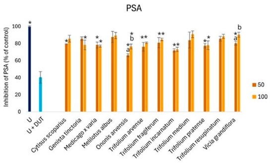
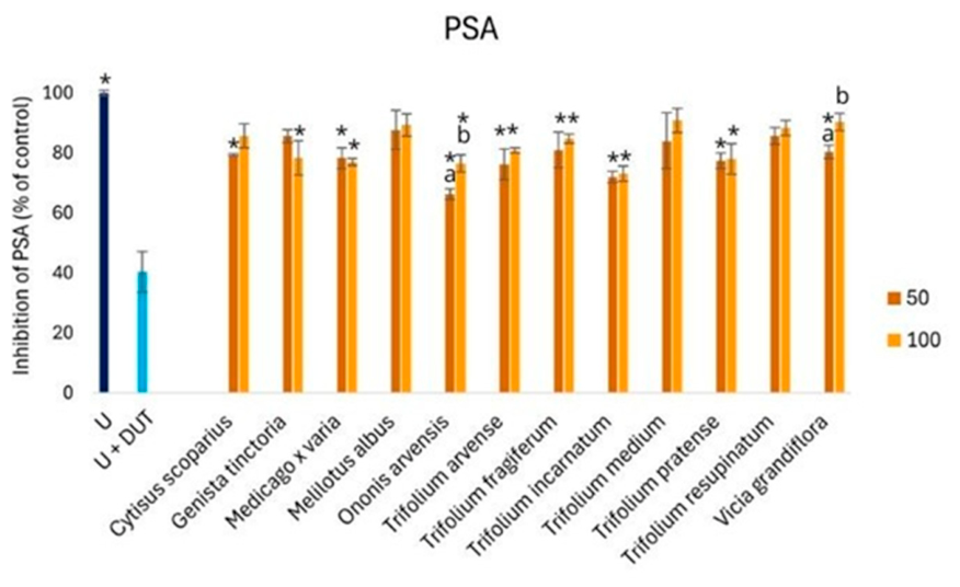
Prostate-specific antigen (PSA) is a serine protease secreted by prostate epithelial cells, and its expression is tightly regulated by androgen receptor signaling. Clinically, PSA is a widely used biomarker of prostate activity, and elevated levels are associated with benign prostatic hyperplasia (BPH), prostate cancer, and inflammation [24]. Thus, to better address the study, we additionally examined the effect of isoflavone-rich plant extracts on PSA levels in non-cancerous prostate epithelial cells. All tested extracts reduced PSA levels compared with controls, with inhibition rates ranging from approximately 66% to 90% (Figure 3). No clear dose–response relationship was observed. The strongest inhibitory effect was observed for Ononis arvensis (50 µg/mL) and Trifolium incarnatum at both tested concentrations, where PSA levels decreased to 66.23 ± 1.83, 71.95 ± 1.99, and 73.06 ± 2.59%, respectively. None of the tested extracts exhibited stronger activity than dutasteride, and the extracts were clearly less potent overall. Moreover, no consistent dose dependency was noted for the extracts. Therefore, the observed effects should be interpreted cautiously, particularly considering the lack of significant 5α-reductase inhibitory activity demonstrated by the tested extracts. Reducing PSA levels may involve mechanisms independent of direct modulation of androgen-related pathways and requires further mechanistic investigation. Nevertheless, some of the tested extracts demonstrated measurable biological activity in prostate cell models, which may warrant further investigation in future in vivo studies related to prostate hyperplasia and prostate cancer.
Figure 3.
The activity of the tested extracts on PSA release in PNT2 cells. Cells were incubated without (untreated, U) or with various concentrations of the extracts (50 and 100 μg/mL), or with the reference drug dutasteride (U + DUT). The results are presented as mean % U ± SD of control from three independent experiments. Significant differences between extract doses of the same species are marked with different letters, while those between the tested concentrations and control (U) are marked with * (p ≤ 0.05). All tested extract samples differ significantly (p ≤ 0.05) from U + DUT.
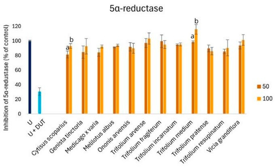
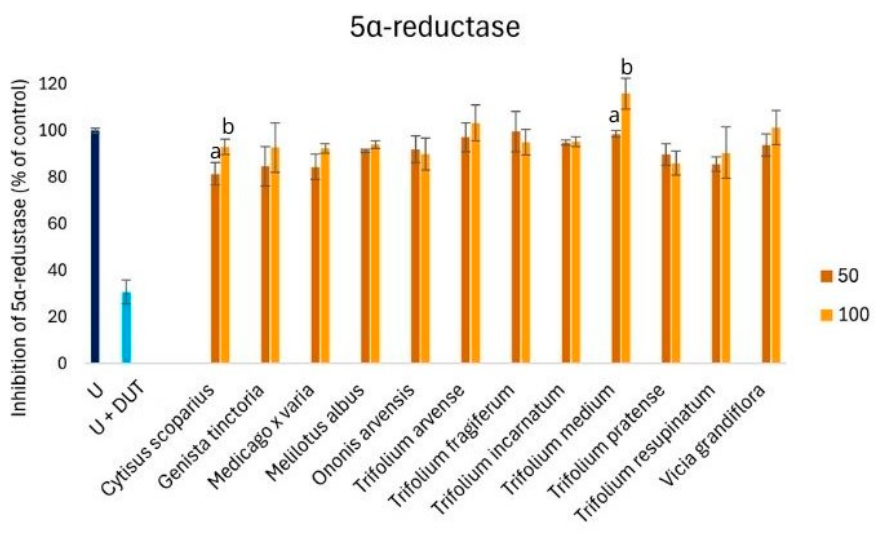
5α-reductase is a key enzyme involved in catalyzing the conversion of testosterone to dihydrotestosterone (DHT), which exhibits significantly higher affinity for the androgen receptor. Increased 5α-reductase activity and elevated DHT levels are directly involved in the pathogenesis of BPH and prostate cancer progression; therefore, achieving this was an additional goal of our study. The obtained results indicate that none of the extracts at either dose demonstrated 5α-reductase inhibitory activity, as the percent of inhibition did not fall below 90% of control (Figure 4). As with PSA inhibition, no clear dose dependency was noted. Interestingly, despite the lack of significant inhibition of 5α-reductase, all tested extracts reduced PSA secretion in prostate epithelial cells. This suggests that the observed decrease in PSA levels may result from mechanisms independent of direct 5α-reductase inhibition. PSA expression may also be regulated by modulation of androgen receptor signaling, inflammatory pathways, oxidative stress, or other cell signaling mechanisms [6]. This study is the first in the literature to describe the effect of extracts from the tested plants on PSA and 5α-reductase. In our previous study, we demonstrated similar impact of extracts from chickpea sprouts (Cicer arietinum L.), a species also belonging to the Fabaceae family, on 5α-reductase activity, and PSA secretion in the same cellular model [25], which supports the biological potential of Fabaceae species.
Figure 4.
Effect of the tested Fabaceae extracts on 5α-reductase activity in PNT2 cells. Cells were incubated without treatment (untreated control, U), with the reference drug dutasteride (U + DUT), or with plant extracts (50 and 100 μg/mL). The results are presented as mean ± SD from three separate experiments. Significant differences between concentrations of the same extract were determined using one-way ANOVA followed by Tukey’s post hoc test and are indicated by different lowercase letters (p ≤ 0.05). All tested extract samples differed significantly (p ≤ 0.05) from the dutasteride-treated group.
3.4. Anti-Inflammatory Activity
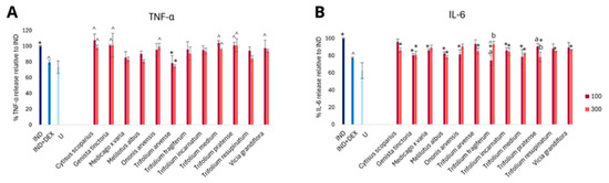
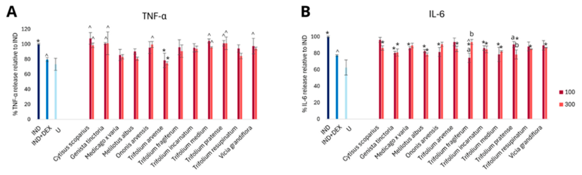
Chronic inflammation is an important contributing factor in the pathogenesis and progression of prostate cancer, but also benign prostatic hyperplasia, influencing cell proliferation, tissue remodeling, and local androgen signaling. Interleukin-6 (IL-6) and tumor necrosis factor alpha (TNF-α) are key pro-inflammatory cytokines involved in the regulation of immune responses and the development of chronic inflammation, while interleukin-10 (IL-10) is a major anti-inflammatory cytokine responsible for limiting excessive inflammatory reactions. Therefore, to complete the study, we evaluated the impact of the tested extracts on IL-6, TNF-α, and IL-10 release from cytokine-stimulated PNT2 cells. Moreover, the proportions of IL-6 and TNF-α to IL-10, used as the indicators of the relationships between pro- and anti-inflammatory signaling [26,27], were calculated to better describe the observed effect. Analysis of IL-6 levels demonstrated that the majority of the tested plant extracts significantly reduced the secretion of this pro-inflammatory cytokine compared to the stimulated control (Figure 5B). This inhibitory effect was observed at both 100 µg/mL and 300 µg/mL, with a tendency toward stronger suppression at the higher concentration, indicating partial dose dependence. However, no consistent dose–response relationship was observed across all extracts, with the exception of Trifolium fragiferum and T. pratense.
Figure 5.
Activity of the tested extracts on TNF-α (A) and IL-6 (B) release in PNT2 cells stimulated with cytokine mix (see the Materials and Methods for details). Cells were incubated with various concentrations of the extracts (100 and 300 μg/mL), with the reference drug dexamethasone (IND + DEX), or treated with cytokine mix to induce inflammation (IND). Control, untreated cells (U) were also included. The results represent the mean ± SD of control from three independent experiments. Significant differences between doses of extracts of the same species are marked with pairs of letters, between the tested concentrations and IND are marked with *, while between the tested concentrations and IND + DEX are marked with ^.
Analysis of TNF-α levels showed that only the extract of Trifolium arvense significantly reduced the secretion of this pro-inflammatory cytokine compared to the stimulated control (IND) (Figure 5A). No dose–response relationship was noted for the tested extracts. Interestingly, more than half of the extracts did not differ significantly from the effect observed after short-term dexamethasone treatment.
The IL-6/IL-10 and TNF-α/IL-10 ratios were calculated to reveal the relationship between pro- and anti-inflammatory responses induced by the tested plant extracts. The lowest ratios (approximately 0.60), compared to the cytokine-induced inflammation control (0.78), were observed for Trifolium incarnatum and T. medium, suggesting a particularly strong ability of these extracts to suppress IL-6 relative to release of IL-10. Notably, Trifolium incarnatum also exhibited the lowest TNF-α/IL-10 ratio among the tested extracts, indicating a shift toward an anti-inflammatory response characterized by reduced TNF-α relative to IL-10. In contrast, the highest IL-6/IL-10 (above 1.0) and TNF-α/IL-10 (above 1.5) ratios were observed in comparison to the corresponding values in the inflammation control group; (1.1) values were noted for Trifolium fragiferum, which may reflect a less favorable modulation of the cytokine balance by this species. For the remaining tested species, the IL-6/IL-10 and TNF-α/IL-10 ratios did not differ significantly from the reference group with cytokine-induced inflammation in human prostate cells.
3.5. Principal Component Analysis (PCA)
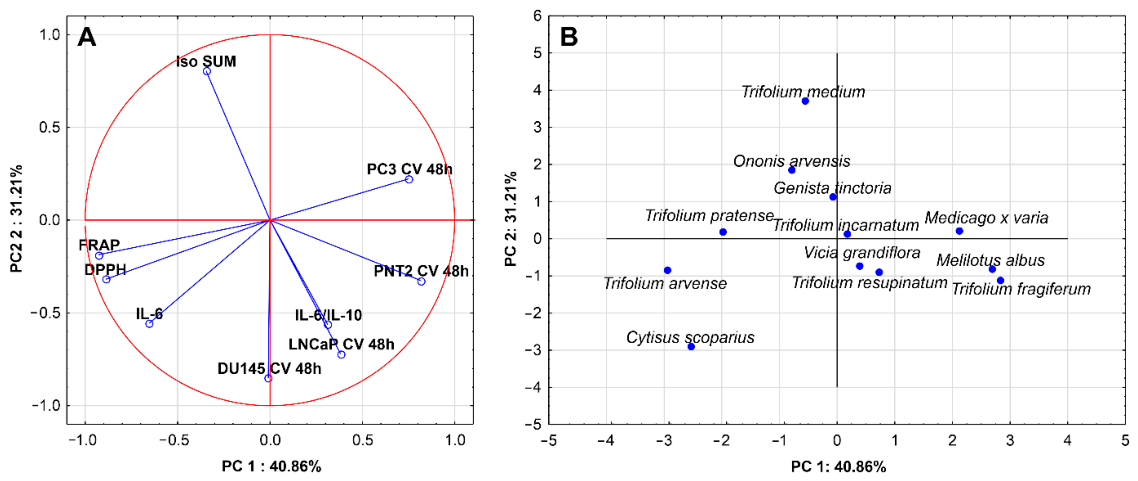
To reveal underlying patterns and correlations among variables, the PCA was implemented. As the extracts analyzed in the present study had been previously characterized in terms of their isoflavone content and antioxidant activity [18], these data, reported in our previous publication, were also incorporated into the chemometric analysis. The constructed PCA model had two significant components, with eigenvalues above 1 (3.68 and 2.80, respectively), resulting in the explanation of 72% of the variance in the dataset. Strong negative correlation for the first principal component with variables such as antioxidant activity and (positively) to the proliferation of PNT2 cell line was noted, whereas the second principal component was positively correlated with isoflavones sum and negatively with the impact on the LNCaP and DU145 cell proliferation. The basic features of the PCA model are presented in Table S2 of the Supplementary Materials.
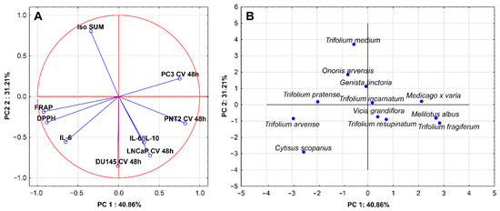
The most important observation from PCA is the statistically significant (p < 0.05) strong negative correlation between isoflavone content and the proliferation of androgen-dependent LNCaP cell line (r = −0.67) and the moderate correlation with the IL-6/IL-10 ratio (r = −0.55) (Figure 6A). No correlation between antioxidant activity and the proliferation of LNCaP cells was noted, suggesting that probably isoflavones, but not other polyphenols, reveal the impact on LNCaP cells proliferation. However, it should be emphasized that the observed correlations do not imply a direct causal relationship between total isoflavone content and the measured biological effects. The obtained associations should be interpreted cautiously, as other bioactive constituents present in the extracts may also contribute to the observed activities. No correlation was observed between antioxidant activity and IL-6/Il-10 ratio. The figure also shows a weak negative correlation between isoflavone content and proliferation of the DU145 line (r = 0.22; p > 0.05), but it is statistically insignificant. However, there is a statistically significant strong notable negative correlation between antioxidant activity and proliferation of the PC3 line (FRAP r = −0.67; DPPH r = −0.68, p < 0.05), while there is no correlation between this cell line and isoflavone content. Furthermore, a similar negative correlation between antioxidant activity and proliferation of the PNT2 line (FRAP r = −0.60; DPPH r = −0.50, p < 0.05) was also noted.
Figure 6.
PCA correlation circle presenting the vectors of the variables within the two principal components (PC1 and PC2) (A); PCA score scatterplot for examined plant species in the space defined by PC1 and PC2 (B). Abbreviations: iso SUM—total isoflavones content of the extracts; CV 48 h—cell proliferation assessed by crystal violet method after 48 h; DU145, LNCaP, PNT2, PC3—cell lines; FRAP, DPPH—antioxidant assays; IL-6—Interleukin-6; IL-10—Interleukin-10.
The PCA allowed further investigation of the potential similarity in the activity profile between the examined plant species (Figure 6B). Based on the model, it can be seen that Trifolium medium alone stood out from the others, while Cytisus scoparius and T. arvense clustered together, but also stood out from the others. Furthermore, Ononis arvensis and Genista tinctoria, as well as Melilotus albus and T. fragiferum, formed two distinct clusters, respectively.
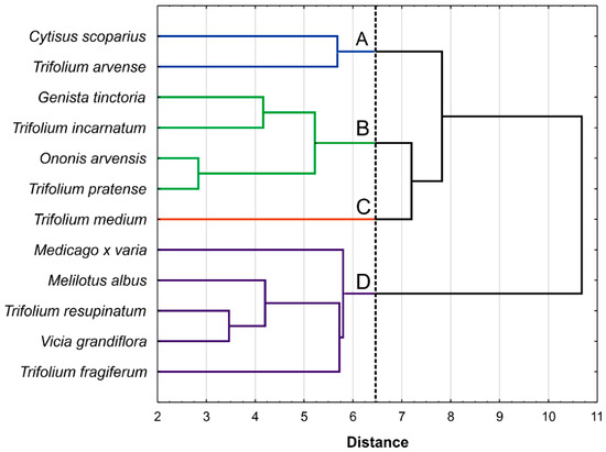
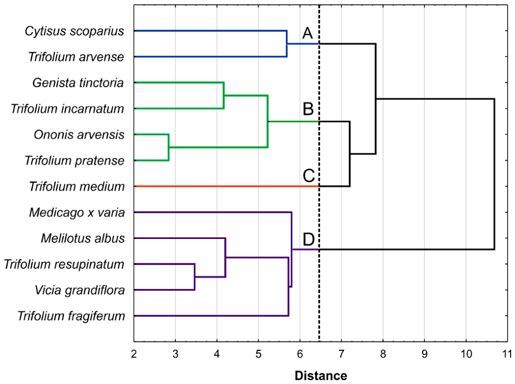
Additionally, to examine and more precisely classify which species exert a similar effect on all parameters examined in our study, a hierarchical cluster analysis (CA) was included as the next step of the analysis. As can be seen, the analysis confirmed the preliminary data from the PCA regarding species grouping. Four clusters can be distinguished within the studied species (Figure 7). Cluster A, with Trifolium arvense and Cytisus scoparius revealing high antioxidant activity and strong inhibition of PC3 cell proliferation, but only weak inhibition of DU145 cell proliferation and IL-6 release. Cluster B gathered four species (Genista tinctoria, Trifolium incarnatum, Ononis arvensis, T. pratense) with strong inhibition of LNCaP, DU145 and PNT2 cells proliferation, but also the best scores in the inhibition of PSA. Single-element cluster C with Trifolium medium is characterized by the highest isoflavones sum, the strongest inhibition of LNCaP cell proliferation, and the strongest anti-inflammatory effect, observed as the inhibition of IL6 and the lowest IL-6/Il-10 ratio value, while almost no impact on PSA and 5α-reductase was noted. In multi-element cluster D (Trifolium resupinatum, Vicia grandifolia, Medicago x varia, Melilotus albus, T. fragiferum), species with the lowest isoflavones sum, the weakest cytotoxic impact on PNT2 and PC3, the weakest inhibitory impact on the proliferation of all cell lines examined, and the weakest antioxidant potential were gathered.
Figure 7.
Dendrogram of similarity for examined plant species. Clusters A–C identified using Mojena’s rule (dashed line).
4. Conclusions and Limitations
We are aware of some of the limitations of our study. First, all experiments were performed exclusively in vitro using established human prostate cancer cell lines and normal prostate epithelial cells. Although such models are important for initial screening, they do not fully replicate the biological complexity of in vivo systems, including tumor–immune system interactions, vascularization, hormonal regulation, bioavailability and metabolic transformations of isoflavones. Second, the biological effects observed in this work were evaluated at the level of cell viability, proliferation and inflammation; more detailed mechanistic analyses were not performed on a molecular level. Therefore, the precise molecular pathways responsible for the cytotoxic and antiproliferative effects of Fabaceae extracts, including their potential influence on androgen receptor signaling, oxidative stress, apoptosis induction, and cell cycle regulation, have yet to be elucidated.
It should also be emphasized that the study was conducted using crude plant extracts, not isolated compounds. Although PCA showed an association between total isoflavone content and observed biological activity, the direct effect of individual isoflavones could not be clearly determined. Therefore, the observed effects should not be attributed solely to isoflavones, as other phytochemicals present in the extracts, including phenolic acids and flavonoids, may also contribute to the observed biological activity. Further studies including fractionation, compound isolation and mechanistic analyses are needed to identify the components responsible for the observed effects and to elucidate their molecular mechanisms of action.
Moreover, relatively high concentrations of the tested extracts were required to obtain significant biological effects in some assays. This may limit the direct physiological and translational significance of the obtained results, especially considering the bioavailability, metabolism, and tissue distribution of phytochemicals in vivo. However, the tested samples were crude plant extracts consisting of complex mixtures of compounds, which generally require higher concentrations than isolated pure compounds in cellular models. Therefore, this study should be considered primarily as a preliminary in vitro assessment of the biological potential of selected Fabaceae species.
Our study indicates that some of the examined species, namely Trifolium medium, T. pratense, Ononis arvensis or Genista tinctoria, with high isoflavones content, reveal interesting biological potential with regard to prostate cancer. Although none of the examined species are classified as food (in terms of their inclusion in the daily diet), some of them may represent promising candidates for further studies focused on prostate-related biological activity. Despite the above-mentioned limitations of our study, the indicated species may be interesting candidates for further examinations.
Supplementary Materials
The following supporting information can be downloaded at: https://www.mdpi.com/article/10.3390/app16136289/s1, Figure S1: Antiproliferative activity of Fabaceae extracts after 48 h on PNT2 (A), DU145 (B), PC3 (C), and LNCaP (D) cells; Figure S2: Variability of the total mean content (mg/g d.m.) of all isoflavones (with specified content of individual compounds, where possible), determined by HPLC, within the tested species of the Fabaceae family.; Table S1: Basic parameters of the PCA model. Table S2: Antioxidant capacity and total isoflavone content of the analyzed Fabaceae extracts.
Author Contributions
Conceptualization, W.P., P.P. and A.G.; methodology, W.P., K.G. and A.G.; formal analysis, W.P., K.G., P.P. and A.G.; investigation, W.P. and E.P.; writing—original draft preparation, W.P., K.G., P.P. and A.G.; writing—review and editing, K.G., P.P., I.P. and A.G.; visualization, W.P. and K.G.; supervision, A.G. All authors have read and agreed to the published version of the manuscript.
Funding
This research was funded by The Priority Research Area qLIFE under the Strategic Programme Excellence Initiative at Jagiellonian University.
Data Availability Statement
Data is available on request.
Conflicts of Interest
The authors declare no conflicts of interest.
References
- Hah, H.; Diabasana, Z.; Soultani, S.; Idoux-Gillet, Y.; Massfelder, T. Prostate cancer: A journey through its history and recent developments. Cancers 2025, 17, 194. [Google Scholar] [CrossRef] [PubMed]
- Wang, L.; Lu, B.; He, M.; Wang, Y.; Wang, Z.; Du, L. Prostate Cancer Incidence and Mortality: Global Status and Temporal Trends in 89 Countries from 2000 to 2019. Front. Public Health 2022, 10, 811044. [Google Scholar] [CrossRef] [PubMed]
- Chung, B.H.; Horie, S.; Chiong, E. The Incidence, Mortality, and Risk Factors of Prostate Cancer in Asian Men. Prostate Int. 2019, 7, 1–8. [Google Scholar] [CrossRef] [PubMed]
- Kimura, T. East meets West: Ethnic differences in prostate cancer epidemiology between East Asians and Caucasians. Chin. J. Cancer 2012, 31, 421–429. [Google Scholar] [CrossRef] [PubMed]
- Vergne, S.; Sauvant, P.; Lamothe, V.; Chantre, P.; Asselineau, J.; Perez, P.; Durand, M.; Moore, N.; Bennetau-Pelissero, C. Influence of ethnic origin (Asian v. Caucasian) and background diet on the bioavailability of dietary isoflavones. Br. J. Nutr. 2009, 102, 1642–1653. [Google Scholar] [CrossRef] [PubMed]
- Pejčić, T.; Zeković, M.; Bumbaširević, U.; Kalaba, M.; Vovk, I.; Bensa, M.; Popović, L.; Tešić, Ž. The Role of Isoflavones in the Prevention of Breast Cancer and Prostate Cancer. Antioxidants 2023, 12, 368. [Google Scholar] [CrossRef] [PubMed]
- Křížová, L.; Dadáková, K.; Kašparovská, J.; Kašparovský, T. Isoflavones. Molecules 2019, 24, 1076. [Google Scholar] [CrossRef] [PubMed]
- Gök, A. In silico analysis of soy isoflavones and their metabolites in prostate cancer. J. Braz. Chem. Soc. 2025, 36, 11. [Google Scholar] [CrossRef]
- Soyata, A.; Hasanah, A.N.; Rusdiana, T. Isoflavones in soybean as a daily nutrient: The mechanisms of action and how they alter the pharmacokinetics of drugs. Turk. J. Pharm. Sci. 2021, 18, 799–810. [Google Scholar] [CrossRef] [PubMed]
- Bu, Y.; Zhang, X.; Xiong, Z.; Li, K.; Zhang, S.; Lin, M.; Zhao, G.; Zheng, N.; Wang, J.; Zhao, S. Effect of red clover isoflavones on ruminal microbial composition and fermentation in dairy cows. Appl. Microbiol. Biotechnol. 2025, 109, 107. [Google Scholar] [CrossRef] [PubMed]
- Zhang, H.Y.; Cui, J.; Zhang, Y.; Wang, Z.L.; Chong, T.; Wang, Z.M. Isoflavones and prostate cancer: A review of some critical issues. Chin. Med. J. 2016, 129, 341–347. [Google Scholar] [CrossRef] [PubMed]
- Van der Eecken, H.; Joniau, S.; Berghen, C.; Rans, K.; De Meerleer, G. The use of soy isoflavones in the treatment of prostate cancer: A focus on the cellular effects. Nutrients 2023, 15, 4856. [Google Scholar] [CrossRef] [PubMed]
- Sivoňová, M.K.; Kaplán, P.; Tatarková, Z.; Lichardusová, L.; Dušenka, R.; Jurečeková, J. Androgen Receptor and Soy Isoflavones in Prostate Cancer. Mol. Clin. Oncol. 2019, 10, 191–204. [Google Scholar] [CrossRef] [PubMed]
- Messina, M. Soy and Health Update: Evaluation of the Clinical and Epidemiologic Literature. Nutrients 2016, 8, 754. [Google Scholar] [CrossRef] [PubMed]
- Rawla, P. Epidemiology of Prostate Cancer. World J. Oncol. 2019, 10, 63–89. [Google Scholar] [CrossRef] [PubMed]
- Zhang, K.; Wang, J.; Xu, B. Critical Review on Molecular Mechanisms for Genistein’s Beneficial Effects on Health Through Oxidative Stress Reduction. Antioxidants 2025, 14, 904. [Google Scholar] [CrossRef] [PubMed]
- Dai, X.; Fang, X.; Ma, Y.; Xianyu, J. Benign prostatic hyperplasia and the risk of prostate cancer and bladder cancer: A meta-analysis of observational studies. Medicine 2016, 95, e3493. [Google Scholar] [CrossRef] [PubMed]
- Markiewicz, M.; Galanty, A.; Prochownik, E.; Kołodziejczyk, A.; Paśko, P. Innovative production of bioactive white clover sprouts under microgravity: Towards functional foods supporting prostate health. Appl. Sci. 2025, 15, 11668. [Google Scholar] [CrossRef]
- Prieto, J.M.; Hanafi, M.M.M. Advances in Molecular Regulation of Prostate Cancer Cells by Top Natural Products of Malaysia. Curr. Issues Mol. Biol. 2023, 45, 1536–1567. [Google Scholar] [CrossRef] [PubMed]
- Paździora, W.; Grabowska, K.; Zagrodzki, P.; Paśko, P.; Prochownik, E.; Podolak, I.; Galanty, A. Quantitative analysis of isoflavones from Fabaceae species and their chemopreventive potential on breast cancer cells. Molecules 2025, 30, 2379. [Google Scholar] [CrossRef] [PubMed]
- Grever, M.R.; Schepartz, S.A.; Chabner, B.A. The National Cancer Institute: Cancer drug discovery and development program. Semin. Oncol. 1992, 19, 622–638. [Google Scholar] [PubMed]
- Díaz, L.; Cely-Veloza, W.; Coy-Barrera, E. Identification of anti-proliferative compounds from Genista monspessulana seeds through covariate-based integration of chemical fingerprints and bioactivity datasets. Molecules 2022, 27, 3996. [Google Scholar] [CrossRef] [PubMed]
- Asadi-Samani, M.; Rafieian-Kopaei, M.; Lorigooini, Z.; Shirzad, H. A screening of growth inhibitory activity of Iranian medicinal plants on prostate cancer cell lines. Biomedicine 2018, 8, 8. [Google Scholar] [CrossRef] [PubMed]
- Abdulla, A.; Leslie, S.W. Biomarker assays for elevated prostate-specific antigen risk analysis. In StatPearls; StatPearls Publishing: Treasure Island, FL, USA, 2025. [Google Scholar]
- Galanty, A.; Prochownik, E.; Grudzińska, M.; Paśko, P. Chickpea sprouts as a potential dietary support in different prostate disorders—A preliminary in vitro study. Molecules 2024, 29, 1044. [Google Scholar] [CrossRef] [PubMed]
- de Brito, R.C.; Lucena-Silva, N.; Torres, L.C.; Luna, C.F.; Correia, J.B.; da Silva, G.A. The balance between the serum levels of IL-6 and IL-10 cytokines discriminates mild and severe acute pneumonia. BMC Pulm. Med. 2016, 16, 170. [Google Scholar] [CrossRef] [PubMed]
- Zhou, L.; Tang, C.; Li, X.; Feng, F. IL-6/IL-10 mRNA expression ratio in tumor tissues predicts prognosis in gastric cancer patients without distant metastasis. Sci. Rep. 2022, 12, 19427. [Google Scholar] [CrossRef] [PubMed]
Disclaimer/Publisher’s Note: The statements, opinions and data contained in all publications are solely those of the individual author(s) and contributor(s) and not of MDPI and/or the editor(s). MDPI and/or the editor(s) disclaim responsibility for any injury to people or property resulting from any ideas, methods, instructions or products referred to in the content. |
© 2026 by the authors. Licensee MDPI, Basel, Switzerland. This article is an open access article distributed under the terms and conditions of the Creative Commons Attribution (CC BY) license.