


Highlights
What are the main findings?
- Full-arch digital impression accuracy differed significantly among intraoral scanner generations (TRIOS 3, TRIOS 5, TRIOS 6).
- Model type influenced accuracy; dentate models generally showed lower deviation values than fully prepared models, with scanner-dependent variations.
What are the implications of the main findings?
- Digital impression accuracy is influenced not only by scanner generation but also by surface morphology and reference richness.
- Careful scanner selection and optimized scanning protocols are essential to ensure reliable outcomes in full-arch restorative workflows.
Abstract
The accuracy of full-arch digital impressions remains a topic of debate despite advancements in intraoral scanning technologies. This in vitro study aimed to evaluate the effect of different intraoral scanner generations (TRIOS 3, TRIOS 5, and TRIOS 6) on full-arch digital impression accuracy by comparing dentate and fully prepared models. A dentate and a fully prepared maxillary model were digitized using a high-accuracy desktop scanner to create reference datasets. The models were fabricated with a 3D printer, and a total of 60 digital impressions were obtained, with 10 repeated scans for each scanner–model combination. Accuracy was assessed by comparing STL datasets with the reference models using root mean square (RMS) deviation values. The results showed that TRIOS 3 and TRIOS 5 demonstrated similar accuracy performance across both model types, whereas TRIOS 6 exhibited higher deviation values, particularly in the fully prepared model. Furthermore, model type had a significant effect on accuracy, and a statistically significant interaction was observed between scanner type and model type. These findings indicate that digital impression accuracy is influenced not only by scanner generation but also by surface morphology and reference characteristics.
1. Introduction
Dentistry applications are increasingly moving toward digitalization. Digital impressions have emerged as a more advantageous alternative to conventional impression techniques due to their benefits, including speed, patient comfort, ease of data storage, and direct integration with CAD/CAM production systems [1]. Clinical and in vitro studies have demonstrated that the accuracy of digital full-arch scans is, in most cases, within clinically acceptable limits, and that digital and conventional impressions generally exhibit comparable accuracy values [1,2]. However, full-arch scanning remains a topic of debate in terms of accuracy due to factors such as the accumulation of stitching/registration errors over large surface areas, insufficient surface texture, clinical conditions, and variations in scanning protocols [3].
Digital dental workflows are integrated systems consisting of data acquisition, computer-aided design (CAD), and computer-aided manufacturing (CAM) stages [4,5]. In these workflows, the data acquisition stage is performed using intraoral and extraoral scanners. Intraoral scanners (IOS) enable the direct digital recording of intraoral tissues, thereby reducing the need for conventional impression materials, whereas extraoral scanners (EOS) represent an indirect digital workflow based on the scanning of conventional impressions or stone models [6,7]. The obtained three-dimensional digital data are integrated with CAD/CAM systems, allowing restorations to be fabricated in a faster, more accurate, and more predictable manner [8,9]. In addition, digital workflows have been reported to improve patient comfort, facilitate clinical and laboratory procedures, and enhance data storage and interdisciplinary communication [5]. Particularly in complex treatments such as implantology and full-mouth rehabilitation, digital workflows have been emphasized to optimize surgical and prosthetic planning while increasing treatment accuracy and time efficiency [4,10].
Several factors should be considered when selecting an intraoral scanner for digital dental workflows. The most important parameters include trueness and precision, scanning speed, ease of use, ergonomics, software capabilities, and compatibility with CAD/CAM systems [11]. In addition, the accuracy of intraoral scanners may be influenced by operator experience, scanning strategy, calibration, oral anatomy, ambient conditions, and the morphology of the scanned surface [12]. Recent literature has also emphasized that scanner selection should not rely solely on hardware specifications, but should additionally consider the intended clinical application, treatment complexity, patient comfort, and workflow efficiency [5].
IOS systems provide advantages such as improved patient comfort, rapid data acquisition, simplified data storage, and direct integration with CAD/CAM systems; however, factors including saliva, patient movement, limited working area, and stitching errors occurring during complete-arch scans may affect their accuracy [7]. EOS systems, on the other hand, can achieve high precision under controlled laboratory conditions, although they may be influenced by dimensional changes associated with impression materials and stone models [13]. Nevertheless, both scanning systems have been reported to provide clinically acceptable levels of accuracy and are widely used in digital dental workflows [6,7]. In particular, desktop (extraoral) scanners are considered the gold standard for obtaining reference data in many studies due to their high accuracy and precision under controlled laboratory conditions.
The main characteristics, advantages, and limitations of intraoral and extraoral scanner systems used in digital dentistry are summarized in Table 1.
Table 1.
Classification and characteristics of intraoral and extraoral scanner systems used in digital dental workflows.
The accuracy of digital impressions is a two-dimensional concept consisting of trueness and precision, as defined by ISO standards in metrology [14]. In dentistry, ANSI/ADA standards also provide a framework for evaluating optical scanners in terms of accuracy and repeatability [15]. Therefore, comparing different intraoral scanners using standardized methodologies in clinically relevant scenarios is crucial for both device selection and the reliability of digital workflows.
Recent evidence suggests that full-arch scanning accuracy varies among devices and may change depending on the clinical scenario. Studies in the literature report that the trueness and precision performance of intraoral scanners in complete-arch scans differ depending on arch and reference conditions such as dentate, edentulous, or implant-supported models [16]. Similarly, more recent in vitro and clinical studies indicate that measurable differences in complete-arch accuracy exist among contemporary intraoral scanner systems, and that scanning technologies and underlying algorithms may influence these outcomes [17].
One of the key factors affecting full-arch accuracy is the morphology of the scanned surface and the richness of reference features. In dentate arches, cusp–fossa anatomy and natural surface texture facilitate image matching, whereas after full preparation, surfaces become more homogeneous, sharp transitions decrease, and local loss of tissue/contrast may challenge the scanner’s alignment algorithms, contributing to cumulative errors [18].
It has been reported that preparation geometry and scanning protocols play a decisive role in the trueness and precision performance of intraoral scanners. In particular, accuracy tends to decrease on prepared surfaces, and this effect varies depending on the scanner type and scanning strategy [19,20,21].
This issue is especially critical for the reliability of digital impressions in restorative rehabilitations requiring full-mouth preparation.
Although numerous studies have evaluated the accuracy of intraoral scanners, studies comparing different generations of the same scanner system under varying full-arch surface morphologies remain limited. In particular, evidence regarding the performance of TRIOS 3, TRIOS 5, and TRIOS 6 on dentate and fully prepared full-arch models using repeated measurements and a standardized reference approach is still insufficient. Therefore, the novelty of the present study lies in the comparative evaluation of these three scanner generations under different surface morphologies and in investigating the interaction between scanner generation and surface morphology on full-arch digital impression accuracy.
Therefore, the aim of this in vitro study was to compare the accuracy of digital impressions obtained from TRIOS 3, TRIOS 5, and TRIOS 6 intraoral scanners on dentate and fully prepared full-arch models. The null hypothesis was that scanner generation and model type would have no significant effect on the accuracy of full-arch digital impressions.
2. Materials and Methods
This in vitro study was designed to evaluate the effect of different generations of intraoral scanners on the accuracy of full-arch digital impressions. In the study, three different intraoral scanner generations (TRIOS 3, TRIOS 5, and TRIOS 6; 3Shape, Copenhagen, Denmark) were compared using two types of full-arch models: an unprepared (dentate) model and a fully prepared model. The abbreviations for the scanner types and model conditions used in this study are summarized in Table 2.
Table 2.
Abbreviations related to scanner types and model conditions.
A priori power analysis was performed using G*Power software (v3.1, Heinrich Heine University, Düsseldorf, Germany) to determine the minimum required sample size. Based on a medium-to-large effect size assumption (Cohen’s f = 0.35), a significance level of α = 0.05, and a statistical power of 95%, the required sample size was calculated prior to data collection. Accordingly, 10 scans per group were considered sufficient and consistent with previous in vitro studies evaluating intraoral scanner accuracy [22,23].
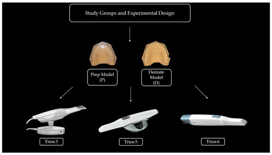
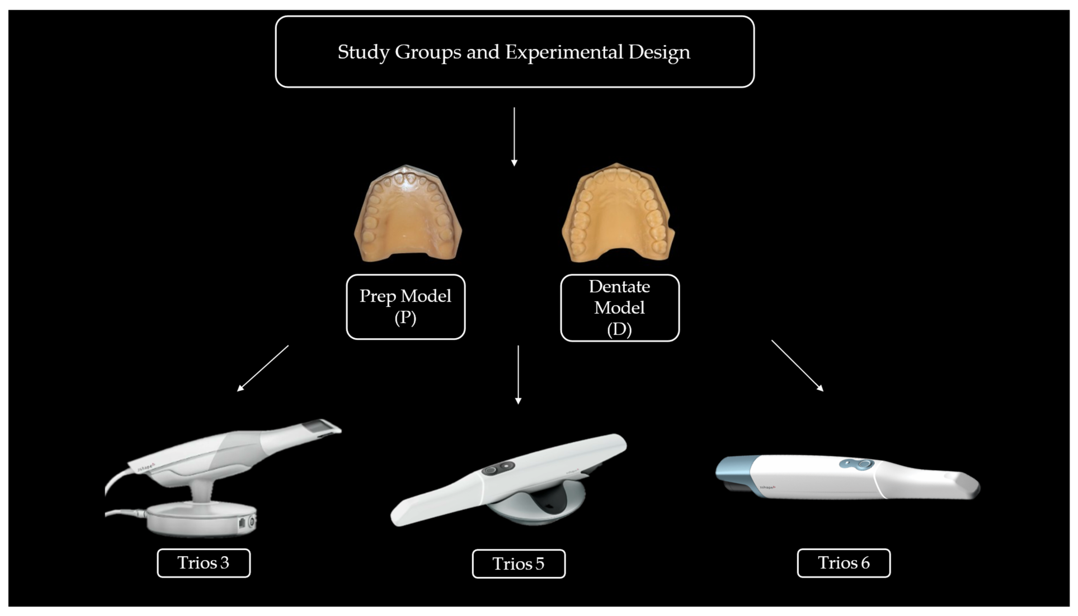
The study design consisted of six subgroups based on two model types (D and P) and three intraoral scanner generations (T3, T5, and T6). For each scanner–model combination, 10 repeated scans were obtained under standardized conditions, resulting in a total of 60 scans. Repeated measurements were included to reduce measurement variability and to assess the intra-scanner repeatability (precision) of the scanners. Each scan was performed independently and treated as an independent dataset during statistical analysis. The distribution of the experimental groups is presented in Figure 1.
Figure 1.
Schematic illustration of the experimental design showing the grouping of intraoral scanners (TRIOS 3, TRIOS 5, and TRIOS 6) and the two model conditions (dentate and fully prepared) used in the study.
- 1.
- Preparation of Reference Models
In this study, a maxillary phantom model (Frasaco maxillary phantom model, Frasaco GmbH, Tettnang, Germany) was used. All teeth from the right second molar to the left second molar (teeth #17 to #27) were prepared in accordance with the principles accepted for full-coverage crown restorations. All preparation procedures were performed by a single operator using standardized diamond burs.
After completion of the preparations, the model was scanned using a high-accuracy desktop scanner (Shining 3D AutoScan-DS-MIX Dental 3D Scanner, Shining 3D, Hangzhou, China), which has a manufacturer-reported accuracy of approximately 5 µm under standardized laboratory conditions. Prior to reference data acquisition, the scanner was calibrated according to the manufacturer’s instructions using the original calibration object supplied by the manufacturer. The obtained data were saved in standard tessellation language (STL) format and used as the fully prepared reference STL model. In addition, a second maxillary Frasaco model without any tooth preparation was scanned using the same desktop scanner to obtain the dentate reference STL model.
- 2.
- Fabrication of 3D-Printed Reference Models
Both reference STL files were manufactured using a 3D printer (Asiga Max UV, Asiga, Australia) with a photopolymer resin (Asiga DentaMODEL Resin, Asiga, Australia). The printing process was performed under UV light in the wavelength range of 385–405 nm, with a layer thickness of 50 µm.
Following the printing process, the models were subjected to ultrasonic cleaning in isopropyl alcohol and subsequently post-cured using a UV curing unit (Asiga Flash, Asiga, Australia) operating at 385 nm to complete polymerization [24]. All polymerization and post-curing procedures were performed strictly according to the manufacturers’ instructions.
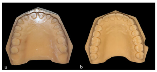
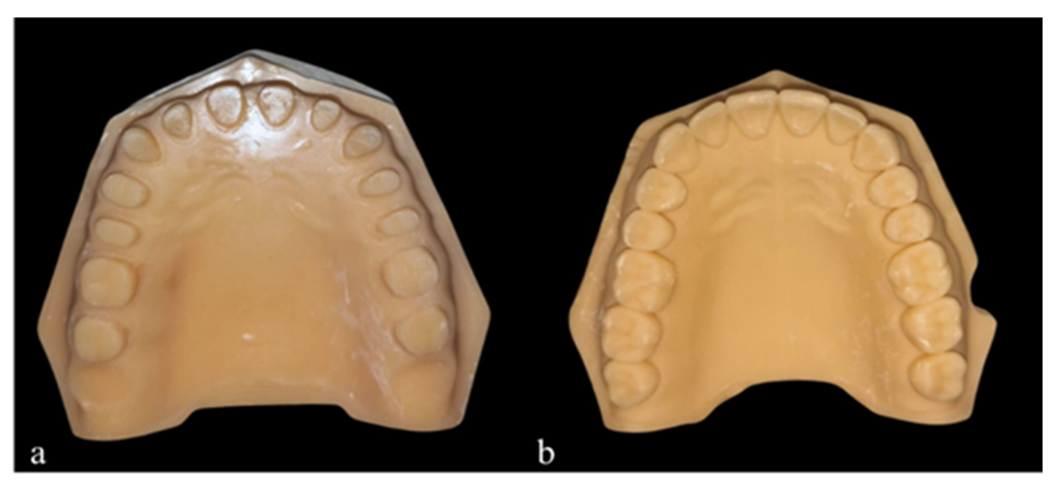
This manufacturing and post-processing protocol was standardized for all specimens to ensure dimensional accuracy and reproducibility of the models. In addition, to minimize potential dimensional alterations during the scanning procedures, all scans were performed immediately after model fabrication, and the models not actively being scanned were stored in a closed container under standardized environmental conditions. Representative occlusal views of the dentate and fully prepared three-dimensional (3D) printed reference models are shown in Figure 2.
Figure 2.
Occlusal views of the maxillary three-dimensional (3D) printed reference models used in the study: (a) dentate model and (b) fully prepared model.
- 3.
- Acquisition of Digital Impressions and Scanning Procedure
All intraoral scans were performed by a single operator with at least five years of experience in intraoral scanning. Prior to the study, five preliminary scans were conducted on the reference models to ensure the operator’s adaptation to the scanning protocol. Before each scanning session, the TRIOS 3 intraoral scanner was calibrated according to the manufacturer’s recommendations. For TRIOS 5 and TRIOS 6, routine user calibration was not required; instead, the built-in system self-check mechanisms were utilized.
A waiting period of approximately 5 min was applied between consecutive scans to prevent potential effects on device performance. During scanning, the models were kept in a fixed position, the distance between the scanner tip and the surface was maintained in accordance with the manufacturer’s instructions, and all surfaces were cleaned prior to scanning. All scans were performed in a controlled laboratory environment with constant ambient lighting, providing homogeneous white illumination at approximately 500–1000 lux.
The reference models were scanned using the TRIOS 3, TRIOS 5, and TRIOS 6 intraoral scanners (3Shape, Copenhagen, Denmark) in accordance with the manufacturer’s protocols. For each scanner–model combination, 10 repeated scans were performed, and all datasets were saved in standard tessellation language (STL) format.
The obtained STL files were processed following a standardized digital workflow and archived in separate folders according to scanner type and model group. The reference STL data obtained from the desktop scanner were used as the reference dataset in all accuracy analyses, and file integrity was verified prior to analysis.
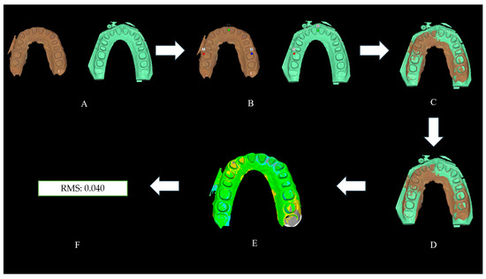
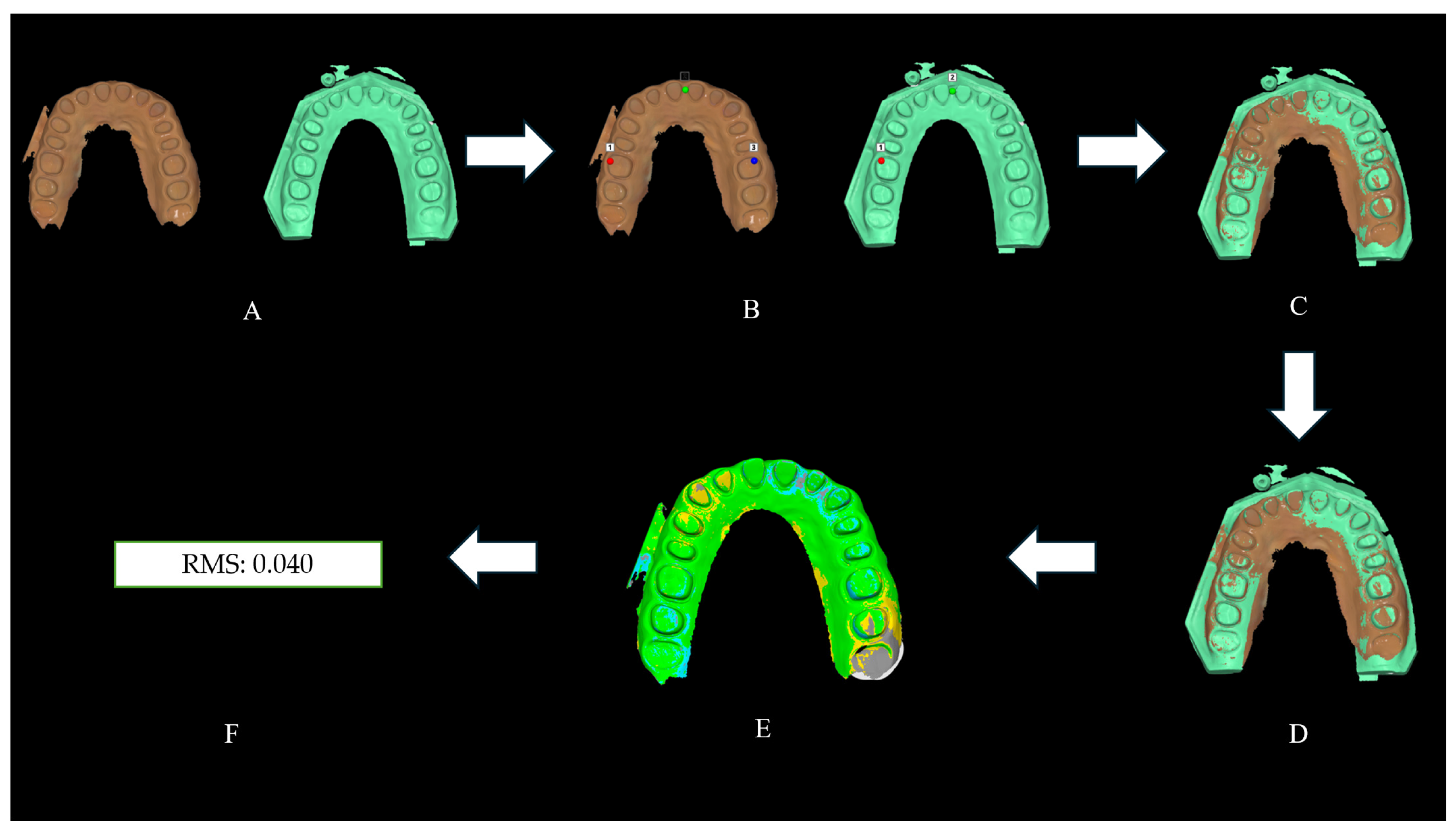
All STL datasets were imported into Medit Design software (version 2.1.4.97) and aligned with the corresponding reference STL datasets. An initial manual alignment was first performed to establish approximate positioning, followed by automatic best-fit alignment using the software’s surface-based registration algorithm. During deviation analysis, low-fidelity data were excluded and outlier removal was performed using a sigma multiplier of 1.0. RMS deviation calculations were performed using the “Find Nearest Position” calculation method with a percentile range of interest set at 95%. Color map analyses were subsequently generated to visualize three-dimensional surface deviations between the reference and test datasets.
As shown in Figure 3, all STL datasets were imported into Medit Design software, where initial manual alignment was first performed, followed by automatic alignment and three-dimensional comparison with the corresponding reference STL datasets. Subsequently, surface deviations were visualized using color map analysis, and accuracy was quantitatively evaluated based on RMS values.
Figure 3.
Workflow of the deviation analysis: (A) model import, (B) reference point selection, (C) initial alignment, (D) best-fit alignment, (E) color map generation, and (F) RMS value calculation.
3. Results
In this study, the trueness of digital impressions obtained from dentate (D) and prepared (P) full-arch models using three different generations of intraoral scanners (T3, T5, and T6) was evaluated based on root mean square (RMS) values. A total of 60 scans were analyzed, with 10 repetitions for each scanner–model combination. The mean, standard deviation, median, minimum, and maximum RMS values according to scanner and model type are presented in Table 3.
Table 3.
Descriptive statistics and confidence intervals of RMS deviations for each scanner and model type.
Overall, T3 and T5 demonstrated similar performance in both model types, whereas T6 exhibited higher RMS values, particularly in the P group. In D models, T3 and T5 showed similar mean RMS values (34.7 µm for both), while T6 presented a higher mean RMS value (42.0 µm). In P models, T3 (21.2 µm) and T5 (21.4 µm) again showed comparable values, whereas T6 demonstrated a markedly higher RMS value (58.0 µm).
Levene’s test results indicated that the assumption of homogeneity of variances was violated (p < 0.05), and normality violation was detected in one subgroup. Therefore, robust two-way ANOVA was performed to evaluate the effects of scanner type and model type on RMS values. All statistical analyses were conducted using Jamovi software (Version 2.7.6). In addition to p values, effect sizes (partial eta squared, ηp2) were calculated to determine the magnitude of the observed effects. The scanner type demonstrated a large effect size (ηp2 = 0.556), whereas the interaction between scanner type and model type also showed a large effect size (ηp2 = 0.359). In contrast, the main effect of model type showed a relatively small effect size (ηp2 = 0.036). Furthermore, 95% confidence intervals were considered during statistical interpretation.
The robust analysis revealed that model type had a significant effect on RMS values (D vs. P, p = 0.023). Overall, P models exhibited different accuracy behavior compared with D models. In addition, an interaction between scanner type and model type was identified, indicating that the significant differences among scanners mainly emerged in the P models.
Post hoc Mann–Whitney U comparisons demonstrated no statistically significant differences among T3, T5, and T6 in D models (all comparisons, p > 0.05). In contrast, in P models, the RMS values of T6 were significantly higher than those of T3 and T5 (p < 0.001 for both T6–T3 and T6–T5 comparisons). No significant difference was observed between T3 and T5 (p > 0.05). The results of the robust two-way ANOVA and post hoc Mann–Whitney U comparisons are presented in Table 4.
Table 4.
Robust two-way ANOVA and post hoc comparison results for RMS values.
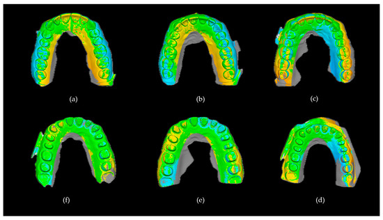
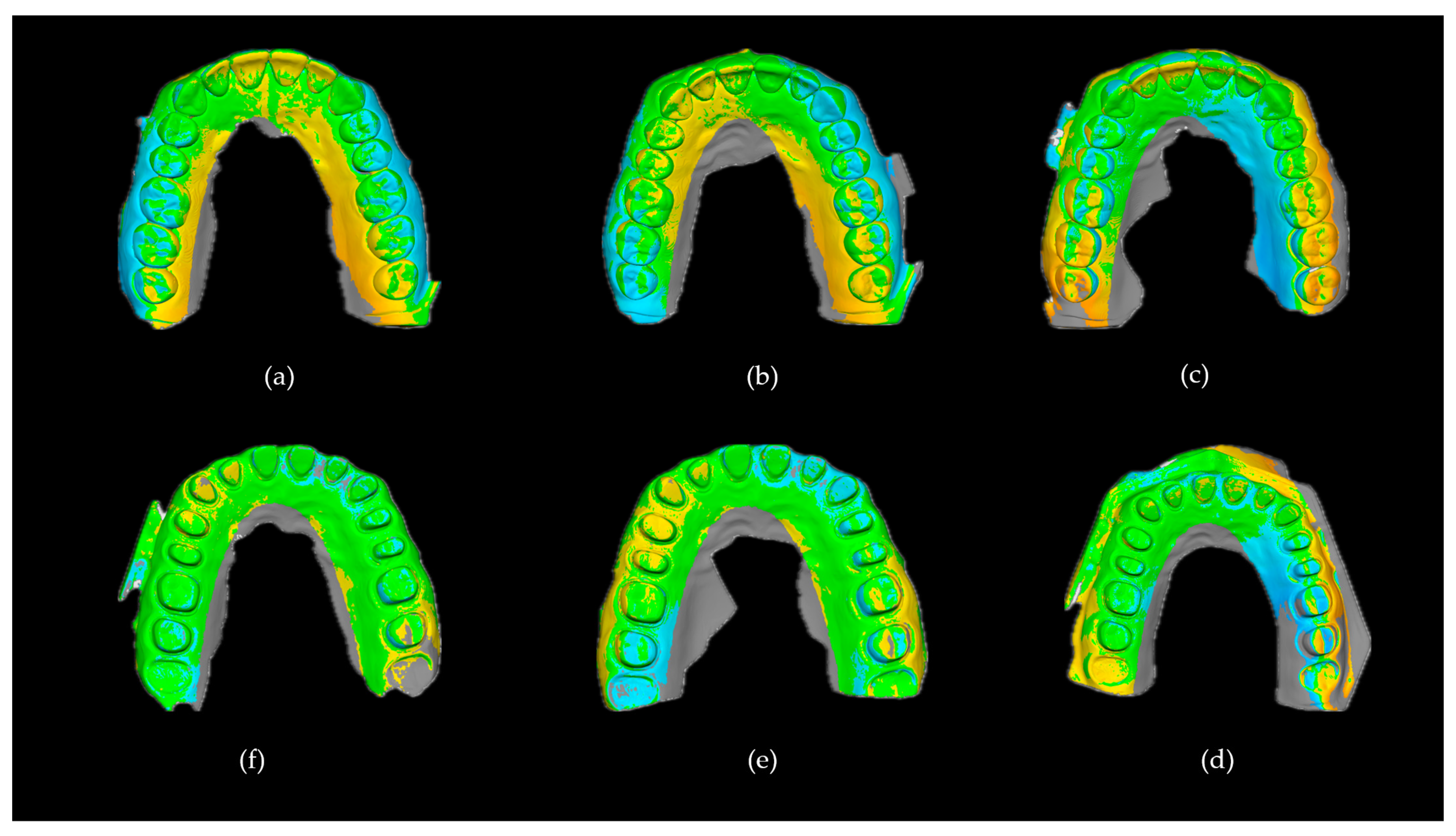
These findings indicate that different scanner generations exhibited comparable accuracy performance in D models; however, in P models, T6 demonstrated higher RMS values compared with T3 and T5 under the conditions of this study. This finding may be associated with the homogeneous surface morphology and reduced reference point distribution of the prepared models. Representative three-dimensional color map analyses of each scanner–model combination are presented in Figure 4.
Figure 4.
Representative color map deviation analyses of full-arch digital impressions obtained from different intraoral scanners and model types. (a) T3-D, (b) T5-D, (c) T6-D, (d) T6-P, (e) T5-P, and (f) T3-P. Green areas indicate minimal deviation, whereas blue and yellow regions represent negative and positive deviations, respectively.
Precision Analysis
The precision analysis results obtained from repeated scans performed under identical experimental conditions are presented in Table 5. Precision was evaluated based on the variability of repeated RMS measurements within each scanner–model combination. Lower standard deviation and coefficient of variation values indicated higher repeatability and more consistent scanner performance.
Table 5.
Precision analysis of repeated scans obtained from different intraoral scanner generations and model types.
Precision analysis revealed differences in repeatability among the scanner groups. Among all groups, T5-D demonstrated the lowest coefficient of variation value (6.08%) and the lowest standard deviation, indicating the highest repeatability and the most consistent scanning performance. Similarly, T3-D also exhibited low variability with a CV value below 10%, suggesting stable repeated measurements in dentate models.
In contrast, higher variability values were observed in the prepared model groups. T5-P and T3-P demonstrated increased coefficient of variation values (33.35% and 26.76%, respectively), indicating reduced repeatability on homogeneous prepared surfaces. The highest variability was observed in the T6-D group (CV = 36.42%), while T6-P exhibited both high mean RMS values and high variability.
4. Discussion
In this study, the effect of different generations of intraoral scanners on full-arch digital impression accuracy was compared using D and P models. The findings demonstrated that scanner generation alone was not the sole determinant of accuracy; however, significant differences emerged when scanner generation was evaluated together with model type. Accordingly, the null hypothesis of the study, which stated that “scanner generation and model type (D vs. P) would not have a significant effect on full-arch digital impression accuracy,” was partially rejected based on the obtained results.
Full-arch scans of dentate models are frequently used in clinical practice, particularly for obtaining opposing arch records in cases with limited tooth loss, fabricating diagnostic models, and manufacturing occlusal splints. In contrast, fully prepared arches are commonly encountered in extensive prosthetic rehabilitation cases, such as patients requiring an increase in vertical dimension, individuals with genetic disorders such as Amelogenesis Imperfecta, or cases in which all teeth require restoration. Therefore, evaluating these two clinically relevant scenarios together is important for more realistically demonstrating the performance of intraoral scanners under different surface morphologies. The present study was conducted to contribute to the existing literature regarding digital impression accuracy in these clinical situations and to support the existing body of knowledge.
In the present study, RMS (Root Mean Square) deviation values were used to evaluate digital impression accuracy. RMS is a parameter that reflects global accuracy by combining all point-to-point deviations between the reference and test models into a single value. Particularly in full-arch scans, it enables the assessment of cumulative errors, thereby providing a more comprehensive and reliable analysis. Furthermore, RMS is a standard method widely used in three-dimensional surface comparisons. Therefore, it was preferred in this study to evaluate the overall accuracy of digital impressions [25].
No statistically significant difference in accuracy was observed between T3 and T5 in this study. This finding is largely consistent with the current literature. In an in vitro study conducted by Büyükhatipoğlu et al., the digital accuracy of different intraoral scanners was compared in posterior implant and opposing molar regions. Data obtained from T3, T5, and Medit i700 scanners using a Frasaco maxillary model were compared with reference STL datasets. The authors reported that T5 demonstrated higher trueness than T3 in the implant region; however, this superiority was not maintained in the molar region, and no significant difference in precision was found among the scanners [26]. These findings are in agreement with the results of the present study. This similarity may be explained by the fact that both scanners are based on similar imaging principles and advanced stitching algorithms. In addition, it has been reported that digital impression accuracy is influenced not only by scanner generation, but also by several factors such as surface morphology, distribution of reference points, and scanning protocol. Therefore, small differences between scanners may often result in comparable accuracy outcomes [27].
In a study conducted by Ciocan et al., the digital impression accuracy of different intraoral scanner systems was compared, with particular emphasis on the performance of newer-generation scanners. The authors reported that T5 could provide higher trueness with lower deviation values under certain scanning conditions; however, these differences generally did not exceed clinically meaningful thresholds. This finding suggests that small accuracy differences measured among intraoral scanners may not directly translate into clinical outcomes and that most contemporary systems perform within clinically acceptable accuracy ranges [28].
Although statistically significant differences were observed among some scanner groups, the reported RMS values remained within the clinically acceptable range for full-arch digital impressions reported in the literature. In the systematic review by Pesce et al., contemporary intraoral scanner systems were reported to generally provide clinically acceptable accuracy ranges in full-arch scans. Accordingly, deviations within the range of approximately 50–100 µm have been considered clinically acceptable for many restorative applications [1]. Therefore, even the deviations observed in T6 should not be interpreted as direct evidence of clinical inadequacy. Rather, the findings may reflect relative differences in scanner performance under standardized in vitro conditions.
It has also been reported in the literature that T3 is capable of maintaining its accuracy even during long-term use and demonstrates high trueness and precision values in many clinical scenarios [29]. The similar imaging principles and data processing algorithms used in T5 systems may also help explain these findings. Furthermore, current evidence suggests that newer-generation intraoral scanners do not provide a marked superiority in terms of accuracy, and that technological developments are primarily focused on ergonomics, scanning speed, and user experience [26,27].
In the in vitro study conducted by Büyükhatipoğlu et al., T5 demonstrated statistically significant differences compared with T3, particularly during the scanning of scan body regions. However, in the present study, a scanning protocol involving localized reference structures such as scan bodies was not used; instead, full-arch scans were evaluated. This methodological difference may be considered one of the main reasons for the discrepancies between the findings of the two studies [26].
In the present study, the absence of significant differences between T6 and both T3 and T5 in the D model may be explained by the rich reference points provided by the complex morphology of the dentate arch, which likely stabilized the data alignment (stitching) process [30,31]. Similarly, Fratila et al. reported that one of the most important factors affecting intraoral scanner accuracy is the morphology of the scanned surface, and that complex anatomical structures may improve the performance of alignment algorithms and reduce inter-scanner differences. Although scanner generation is considered a factor influencing impression accuracy, its effect has been suggested to be relatively limited compared with the determining role of surface morphology [30].
In contrast, T6 demonstrated significantly higher RMS values in the P model compared with T3 and T5 under the conditions of this study. A possible explanation for this finding may be related to the interaction between surface morphology and scanner data acquisition/alignment algorithms. Since P surfaces exhibit a more homogeneous structure with fewer anatomical details compared with D models, the number of stable reference points available for intraoral scanners may become limited, thereby increasing error accumulation during the stitching process based on sequential image alignment. Particularly in full-arch scans, these small alignment errors may accumulate and negatively affect global accuracy [11,32]. Revilla-León et al. reported that increasing the vertical dimension increases the number of visible reference points and the amount of acquired data in the occlusal region, thereby improving the alignment process and enhancing impression accuracy [33]. This finding supports the importance of surface morphology and reference geometry in digital impression accuracy. In addition, the rapid data acquisition and wide-area scanning approach of T6 may also have contributed to insufficient local reference formation on homogeneous surfaces, resulting in increased alignment errors. Therefore, the higher deviations observed in the P model should not be interpreted as direct evidence of technical inadequacy, but rather as a possible interaction between surface morphology and algorithmic processes.
The precision findings generally supported the trueness results. T3 and T5 demonstrated more stable and consistent repeated measurements, particularly in dentate models. In contrast, increased variability was observed in the prepared model groups. Although T6 showed higher variation values, this should not be interpreted as directly indicating inferior scanner performance. The reduced anatomical reference geometry of homogeneous prepared surfaces may have increased stitching-related deviations and affected scan repeatability. Therefore, the findings suggest that scanner repeatability may be influenced not only by scanner generation but also by surface morphology.
From a clinical perspective, the findings of the present study suggest that scanner selection may become increasingly important in full-arch rehabilitation workflows involving extensive tooth preparation. While all evaluated scanners demonstrated comparable performance in dentate models, greater variability was observed in prepared models characterized by reduced anatomical landmarks and more homogeneous surfaces. Therefore, clinicians performing full-mouth rehabilitations, vertical dimension increase procedures, or comprehensive fixed prosthodontic treatments should consider not only scanner generation but also the complexity of the scanning surface and the availability of reference structures. Under such conditions, careful scanning strategies and adequate surface reference distribution may contribute to improving the reliability of digital impressions and reducing cumulative alignment errors.
In addition, recent developments in intraoral scanning technology have increasingly focused on software integration, artificial intelligence-assisted diagnostics, and patient communication tools rather than solely on hardware improvements. AI-supported systems have been reported to enhance diagnostic workflows and patient engagement; however, the clinical impact of increased scan resolution and AI-based analyses on impression accuracy remains to be fully elucidated [34].
In an in vitro study conducted by Mangano et al., the accuracy of extraoral photogrammetry (EPG), intraoral photogrammetry (IPG), and different intraoral scanners (TRIOS 6®, iTero Lumina™, i900®, and CS 3800®) was compared on a fully edentulous model for full-arch implant impressions. According to the findings, the highest accuracy was observed with EPG, followed by IPG and iTero Lumina, whereas i900 and T6 demonstrated higher deviation values. The authors reported that T6 showed lower accuracy compared with other new-generation intraoral scanners and significantly higher deviations than photogrammetry-based systems, particularly in full-arch implant impressions. This reduced performance was attributed to stitching errors occurring during long-span scans and the limited availability of reference points. These findings are consistent with the results of the present study and support the concept that the accuracy of full-arch digital impressions depends not only on scanner technology, but also on scanning distance and the distribution of reference points [35].
Another possible explanation for the higher RMS values observed in D models is that these models possess a larger volume and wider surface area compared with P models. This may increase the number of points evaluated during the alignment process and enlarge the dataset size, thereby causing small deviations to accumulate cumulatively. However, this trend was reversed for T6, where higher RMS values were observed in the P model. This finding may suggest that the alignment performance of T6 could be negatively affected on homogeneous prepared surfaces.
The main limitation of this study is that it was conducted under standardized in vitro conditions, which cannot fully replicate the complex clinical environment encountered during intraoral scanning procedures. Important clinical variables, including saliva, soft tissue mobility, restricted intraoral access, patient movement, humidity, and operator-related variability, were not simulated. These factors may influence image acquisition, data stitching, and scanner performance during full-arch digital impression procedures. Therefore, the findings of the present study should be interpreted with caution and should not be directly extrapolated to clinical situations. Furthermore, all scans were performed by a single experienced operator using a standardized scanning protocol. Although this approach minimized methodological variability, it did not allow assessment of inter-operator differences that may occur in daily clinical practice. In addition, only maxillary models were evaluated, which may further limit the generalizability of the results.
The use of 3D-printed models may represent a potential source of dimensional deviation due to factors such as polymerization shrinkage, printing resolution, layer thickness, and post-curing procedures. In particular, additive manufacturing processes may introduce minor dimensional alterations that could affect the geometric accuracy of experimental models. However, all models were fabricated using the same validated printing system, standardized printing parameters, identical layer thickness, and manufacturer-recommended post-processing and post-curing protocols to minimize such variability. Furthermore, because the same standardized reference models were consistently used for all scanner comparisons, the potential influence of additive manufacturing-related distortions was considered limited and equally distributed among the experimental groups.
Although a high-accuracy desktop scanner was used as the reference system, a minor degree of measurement uncertainty originating from the reference scanner itself may have propagated into the RMS deviation calculations. However, because the same calibrated reference dataset was consistently used for all comparisons under standardized conditions, this effect was considered minimal. Finally, the relatively limited sample size and the evaluation of only three scanner generations represent additional limitations of the study.
5. Conclusions
Within the limitations of this in vitro study, T3 and T5 demonstrated similar full-arch digital impression accuracy in both D and P models. In contrast, T6 showed higher RMS values in P models under the conditions of this study; however, this finding should be interpreted cautiously, as prepared surface morphology and reduced reference features may have influenced the results. The findings suggest that digital impression accuracy is influenced not only by scanner generation but also by surface morphology and reference point distribution.
Author Contributions
Conceptualization, A.Ç. and Y.K.Ş.; methodology, Y.K.Ş. and E.B.B.; software, A.Ç. and E.B.B.; validation, A.Ç. and Y.K.Ş.; formal analysis, A.Ç.; investigation, Y.K.Ş.; resources, A.Ç.; data curation, A.Ç.; writing—original draft preparation, A.Ç.; writing—review and editing, Y.K.Ş.; visualization, A.Ç., Y.K.Ş. and E.B.B.; supervision, A.Ç.; project administration, A.Ç. and Y.K.Ş.; funding acquisition, Y.K.Ş., A.Ç. and E.B.B. All authors have read and agreed to the published version of the manuscript.
Funding
This research received no external funding.
Data Availability Statement
The raw data supporting the conclusions of this article will be made available by the authors on request.
Conflicts of Interest
The authors declare no conflicts of interest.
Abbreviations
| CAD/CAM | Computer-Aided Design/Computer-Aided Manufacturing |
| RMS | Root Mean Square |
| STL | Standard Tessellation Language |
| T3 | TRIOS 3 intraoral scanner |
| T5 | TRIOS 5 intraoral scanner |
| T6 | TRIOS 6 intraoral scanner |
| D | Dentate model |
| P | Prepared model |
| EPG | Extraoral photogrammetry |
| IPG | Intraoral photogrammetry |
| UV | Ultraviolet |
References
- Pesce, P.; Nicolini, P.; Caponio, V.C.A.; Zecca, P.A.; Canullo, L.; Isola, G.; Baldi, D.; De Angelis, N.; Menini, M. Accuracy of Full-Arch Intraoral Scans Versus Conventional Impression: A Systematic Review with a Meta-Analysis and a Proposal to Standardise the Analysis of the Accuracy. J. Clin. Med. 2024, 14, 71. [Google Scholar] [CrossRef] [Scilit]
- Ahmed, S.; Hawsah, A.; Rustom, R.; Alamri, A.; Althomairy, S.; Alenezi, M.; Shaker, S.; Alrawsaa, F.; Althumairy, A.; Alteraigi, A. Digital Impressions Versus Conventional Impressions in Prosthodontics: A Systematic Review. Cureus 2024, 16, e51537. [Google Scholar] [CrossRef] [Scilit] [PubMed]
- Garcia-Gomez, S.; Maneiro-Lojo, J.; Pérez-Barquero, J.A.; Kois, J.C.; Fernandez-Estevan, L.; Revilla-León, M. Influence of Interdental Space and Operator Experience on the Accuracy, Scanning Time, and Number of Photograms of Veneer Preparations Digitized by Using an Intraoral Scanner. J. Prosthet. Dent. 2025, 134, 2391–2398. [Google Scholar] [CrossRef] [Scilit]
- Stanley, M.; Paz, A.G.; Miguel, I.; Coachman, C. Fully Digital Workflow, Integrating Dental Scan, Smile Design and CAD-CAM: Case Report. BMC Oral Health 2018, 18, 134. [Google Scholar] [CrossRef] [Scilit]
- Lee, Y.; Ku, H.-M.; Jun, M.-K. Clinical Application of Intraoral Scanners in Dentistry: A Narrative Review. Oral 2024, 4, 639–652. [Google Scholar] [CrossRef] [Scilit]
- Lee, S.J.; Kim, S.-W.; Lee, J.J.; Cheong, C.W. Comparison of Intraoral and Extraoral Digital Scanners: Evaluation of Surface Topography and Precision. Dent. J. 2020, 8, 52. [Google Scholar] [CrossRef] [Scilit]
- Shah, N.; Thakur, M.; Gill, S.; Shetty, O.; Alqahtani, N.M.; Al-Qarni, M.A.; Alqahtani, S.M.; Elagib, M.F.A.; Chaturvedi, S. Validation of Digital Impressions’ Accuracy Obtained Using Intraoral and Extraoral Scanners: A Systematic Review. J. Clin. Med. 2023, 12, 5833. [Google Scholar] [CrossRef] [Scilit]
- Brunetto, J.L.; Adriazola Ique, M.M.; Melo Neto, C.L.D.M.; Sachi, V.P.; Dos Santos, D.M.; Goiato, M.C. Intraoral and Extraoral Scanning Technologies in the Digital Workflow Era: An Integrative Review with Artificial Intelligence. Odovtos Int. J. Dent. Sci. 2026, 27, 34–42. [Google Scholar] [CrossRef] [Scilit]
- Baradee, A.A.; Spies, B.C. Are Conventional Impressions Obsolete? A Narrative Review on the Applicability of Intraoral Scanners. Aust. Dent. J. 2025, 70, S93–S104. [Google Scholar] [CrossRef] [Scilit]
- Donker, V.J.J.; Raghoebar, G.M.; Vissink, A.; Meijer, H.J.A. Digital Workflow for Immediate Implant Placement and Chairside Provisionalization in the Esthetic Zone. Case Rep. Dent. 2022, 2022, 5114332. [Google Scholar] [CrossRef] [Scilit] [PubMed]
- Mangano, F.; Gandolfi, A.; Luongo, G.; Logozzo, S. Intraoral Scanners in Dentistry: A Review of the Current Literature. BMC Oral Health 2017, 17, 149. [Google Scholar] [CrossRef] [Scilit]
- Alkadi, L. A Comprehensive Review of Factors That Influence the Accuracy of Intraoral Scanners. Diagnostics 2023, 13, 3291. [Google Scholar] [CrossRef] [Scilit]
- Ellakany, P.; Tantawi, M.E.; Mahrous, A.A.; Al-Harbi, F. Evaluation of the Accuracy of Digital Impressions Obtained from Intraoral and Extraoral Dental Scanners with Different CAD/CAM Scanning Technologies: An In Vitro Study. J. Prosthodont. 2022, 31, 314–319. [Google Scholar] [CrossRef] [Scilit]
- ISO 5725-1:2023; Accuracy (Trueness and Precision) of Measurement Methods and Results—Part 1: General Principles and Definitions. International Organization for Standardization: Geneva, Switzerland, 2023.
- Cui, N.; Wang, J.; Hou, X.; Sun, S.; Huang, Q.; Lim, H.-K.; Cai, H.; Jia, Q.; Lee, E.-S.; Jiang, H.B. Bias Evaluation of the Accuracy of Two Extraoral Scanners and an Intraoral Scanner Based on ADA Standards. Scanning 2021, 2021, 5535403. [Google Scholar] [CrossRef] [Scilit]
- Vitai, V.; Németh, A.; Sólyom, E.; Czumbel, L.M.; Szabó, B.; Fazekas, R.; Gerber, G.; Hegyi, P.; Hermann, P.; Borbély, J. Evaluation of the Accuracy of Intraoral Scanners for Complete-Arch Scanning: A Systematic Review and Network Meta-Analysis. J. Dent. 2023, 137, 104636. [Google Scholar] [CrossRef] [Scilit]
- Buhl, J.; Ender, A.; Mehl, A. Accuracy of Current Intraoral Scanning Systems for Full-Arch Impressions: An In Vitro Study. J. Dent. 2025, 161, 105984. [Google Scholar] [CrossRef] [Scilit] [PubMed]
- Revilla-León, M.; Ntovas, P.; Li, J.; Barmak, A.B.; Kois, J.C.; Gómez-Polo, M. Tooth Preparation-Related Factors That Impact the Accuracy of Intraoral Scanners for Fabricating Tooth-Supported Restorations: A Systematic Review. J. Prosthet. Dent. 2025, 134, 1555.e1–1555.e20. [Google Scholar] [CrossRef] [Scilit] [PubMed]
- Abduo, J.; Laskey, D. Effect of Preparation Type on the Accuracy of Different Intraoral Scanners: An In Vitro Study at Different Levels of Accuracy Evaluation. J. Esthet. Restor. Dent. 2022, 34, 1221–1229. [Google Scholar] [CrossRef] [Scilit]
- Mai, H.Y.; Mai, H.-N.; Lee, C.-H.; Lee, K.-B.; Kim, S.; Lee, J.-M.; Lee, K.-W.; Lee, D.-H. Impact of Scanning Strategy on the Accuracy of Complete-Arch Intraoral Scans: A Preliminary Study on Segmental Scans and Merge Methods. J. Adv. Prosthodont. 2022, 14, 88–95. [Google Scholar] [CrossRef] [Scilit]
- Mandelli, F.; Gherlone, E.F.; Keeling, A.; Gastaldi, G.; Ferrari, M. Full-Arch Intraoral Scanning: Comparison of Two Different Strategies and Their Accuracy Outcomes. J. Osseointegration 2018, 10, 65–74. [Google Scholar] [CrossRef] [Scilit]
- Mangano, F.G.; Hauschild, U.; Veronesi, G.; Imburgia, M.; Mangano, C.; Admakin, O. Trueness and Precision of 5 Intraoral Scanners in the Impressions of Single and Multiple Implants: A Comparative In Vitro Study. BMC Oral Health 2019, 19, 101. [Google Scholar] [CrossRef] [Scilit]
- Giuliodori, G.; Rappelli, G.; Aquilanti, L. Intraoral Scans of Full Dental Arches: An In Vitro Measurement Study of the Accuracy of Different Intraoral Scanners. Int. J. Environ. Res. Public Health 2023, 20, 4776. [Google Scholar] [CrossRef] [Scilit] [PubMed]
- Titihazan, F.; Veja, I.; Zaharia, C.; Hajaj, T.; Sinescu, C.; Constantin, G.D.; Rominu, M. Comparative Evaluation of Color Stability and Fracture Resistance of CAD/CAM and Chairside Provisional Restorations: An In Vitro Study. J. Funct. Biomater. 2025, 16, 426. [Google Scholar] [CrossRef] [Scilit]
- Gao, H.; Liu, X.; Zhou, T.; Tan, J. Trueness of 4 Sectional-Cast Digital Methods for Transferring the Interocclusal Relationship in Complete Mouth Rehabilitation. J. Prosthet. Dent. 2025, 133, 575.e1–575.e10. [Google Scholar] [CrossRef] [Scilit] [PubMed]
- Kecik Buyukhatipoglu, I.; Ozdemir, M.; Sari, F. Comparative Evaluation of Digital Accuracy Among Three Intraoral Scanners for Posterior Implant and Tooth Scans. Appl. Sci. 2026, 16, 495. [Google Scholar] [CrossRef] [Scilit]
- Mourouzis, P. Critical Methodological Factors Influencing the Accuracy of Intraoral Scanners in Digital Dentistry Research. Comput. Biol. Med. 2025, 187, 109780. [Google Scholar] [CrossRef] [Scilit]
- Ciocan, L.T.; Vasilescu, V.G.; Răuță, S.-A.; Pantea, M.; Pițuru, S.-M.; Imre, M. Comparative Analysis of Four Different Intraoral Scanners: An In Vitro Study. Diagnostics 2024, 14, 1453. [Google Scholar] [CrossRef] [Scilit] [PubMed]
- Kanmaz, M.G.; Agani Sabah, G.; Balcı, M.; Erden, M.B. Comparison of Intraoral Scanner Accuracy Before and After Calibration: An In Vitro Study. BMC Oral Health 2025, 25, 1186. [Google Scholar] [CrossRef] [Scilit]
- Fratila, A.M.; Saceleanu, A.; Arcas, V.C.; Fratila, N.; Earar, K. Enhancing Intraoral Scanning Accuracy: From the Influencing Factors to a Procedural Guideline. J. Clin. Med. 2025, 14, 3562. [Google Scholar] [CrossRef] [Scilit]
- Kontis, P.; Güth, J.-F.; Keul, C. Accuracy of Intraoral Scans of the Edentulous Maxilla: An In Vitro Study. Clin. Oral Investig. 2025, 29, 342. [Google Scholar] [CrossRef] [Scilit]
- Ender, A.; Mehl, A. Accuracy of Complete-Arch Dental Impressions: A New Method of Measuring Trueness and Precision. J. Prosthet. Dent. 2013, 109, 121–128. [Google Scholar] [CrossRef] [Scilit]
- Revilla-León, M.; Gómez-Polo, M.; Zeitler, J.M.; Barmak, A.B.; Kois, J.C.; Pérez-Barquero, J.A. Does the Available Interocclusal Space Influence the Accuracy of the Maxillomandibular Relationship Captured with an Intraoral Scanner? J. Prosthet. Dent. 2024, 132, 435–440. [Google Scholar] [CrossRef] [Scilit]
- Alghauli, M.A.; Aljohani, W.; Almutairi, S.; Aljohani, R.; Alqutaibi, A.Y. Advancements in Digital Data Acquisition and CAD Technology in Dentistry: Innovation, Clinical Impact, and Promising Integration of Artificial Intelligence. Clin. eHealth 2025, 8, 32–52. [Google Scholar] [CrossRef] [Scilit]
- Mangano, F.G.; Lerner, H.; Valle, G.; Biaggini, F.; Mangano, C.; Dybov, A. Accuracy of Extraoral Photogrammetry, Intraoral Photogrammetry, and Direct Intraoral Scanning in Full-Arch Digital Implant Impressions: A Comparative In Vitro Study. J. Dent. 2026, 165, 106269. [Google Scholar] [CrossRef] [Scilit]

Disclaimer/Publisher’s Note: The statements, opinions and data contained in all publications are solely those of the individual author(s) and contributor(s) and not of MDPI and/or the editor(s). MDPI and/or the editor(s) disclaim responsibility for any injury to people or property resulting from any ideas, methods, instructions or products referred to in the content. |
© 2026 by the authors. Licensee MDPI, Basel, Switzerland. This article is an open access article distributed under the terms and conditions of the Creative Commons Attribution (CC BY) license.