


Abstract
Pleomorphism in cells encompasses a variety of morphological features, such as heterogeneity in the size and shape of cells and nuclei, irregularity of the nuclear membrane, hyperchromasia, and peculiarities of cell organelles. Pleomorphism in mammalian cells is associated with genetically unstable neoplasms and is frequently accompanied by high rates of proliferation and low differentiation status. Among soft tissue sarcomas, a group of tumors characterized by the pleomorphic phenotype can be distinguished. Pleomorphic soft tissue sarcomas are a rare, understudied group of soft tissue sarcomas with aggressive behavior, low survival rates, and high resistance to standard chemotherapy treatment. Knowledge of the mechanisms behind the pleomorphic features, common genetic and epigenetic alterations, and their association with aggressive tumorigenesis is essential for advancing patient care for this group of sarcomas. In this narrative review, we consolidate existing evidence concerning clinical features of pleomorphic sarcomas, their possible histogenesis, the common drivers and transcriptional networks, and the potentially available approaches for treatment.
1. Introduction
Pleomorphism in cytology refers to the morphological heterogeneity between individual cells in a given population. The description of this phenomenon includes variations in cell size and shape, peculiarities of nuclei including shape, structure, membrane irregularities, hyperchromasia and location, and ratio and spatial distribution of cellular organelles [1,2]. In some strains of bacteria, yeast, and mycoplasma, this phenomenon is a part of the adaptation to environmental cues [3,4,5,6]. However, in mammalian cells, pleomorphism, especially nuclear pleomorphism, is commonly associated with genomic instability and neoplastic growths [7,8,9]. The pleomorphic component is observed in a wide range of neoplasms, from epithelial to mesenchymal and from benign to fully malignant tumors. Pleomorphic cancers demonstrate aggressive characteristics and poor prognosis, in comparison with their non-pleomorphic counterparts. For example, pleomorphic rhabdomyosarcoma (pRMS) has a worse prognosis and survival than other rhabdomyosarcoma (RMS) subtypes [10]. Many pleomorphic neoplasms are rare and understudied, which results in a lack of therapy options for patients with this diagnosis.
Pleomorphic soft tissue sarcomas (STS) are rare malignant neoplasms originating from mesenchymal cells and united by high morphological heterogeneity [1]. This list includes several subtypes of STS that are defined as pleomorphic entities according to the latest classification from the World Health Organization (WHO): undifferentiated pleomorphic sarcoma (UPS), pRMS, pleomorphic liposarcoma (pLPS), and myxoid pleomorphic liposarcoma (mpLPS) [11]. Additionally, several other subtypes of STS are frequently included in the discussion of pleomorphism [1] due to the pleomorphic component they display. Myxofibrosarcoma (MFS), previously grouped together with UPS under the umbrella term of malignant fibrous histiocytoma (MFH), is a myofibroblastic tumor [11] with a significant pleomorphic component [12]. Dedifferentiated liposarcoma (DDLPS) displays pleomorphism as a part of its dedifferentiated component [13]. Poorly differentiated leiomyosarcoma (LMS) and anaplastic malignant peripheral nerve sheath tumor (MPNST) display marked pleomorphism and an almost UPS-like appearance [14,15]. For the purposes of this review, we use the umbrella term pleomorphic STS to refer to UPS, MFS, pRMS, pLPS, DDLPS, LMS, and MPNST.
The common clinical features of pleomorphic STS include high ability to mimic other tumors, aggressive rapid growth, plasticity and atypical growth patterns, high rates of local recurrences and distant metastasis, and chemoresistance [16,17,18,19,20]. Selective treatment of pleomorphic STS relies primarily on accurate histological and molecular verification of these neoplasms. The diversity of tissues giving rise to these tumors results in a highly heterogeneous cellular structure, making definitive histogenesis and diagnostics of pleomorphic STS subtypes one of the most complicated areas in cancer pathology.
The molecular characteristics of pleomorphic STS leave many unresolved questions at present. Data on the genomic drivers and transcriptional programs are available for the most common pleomorphic subtypes. However, even for these subtypes, an examination of larger cohorts of patients may lead to the discovery of new molecular alterations. Most importantly, the molecular mechanism behind the emergence, evolution, and functioning of the pleomorphic tumor component in STS has not been elucidated at present. New data are required to fill in these gaps, while careful examination of the available studies can help identify common links and processes between the pleomorphic STS subtypes.
This review brings together the currently available knowledge on pleomorphic STS as a group. We outline the documented clinical features of each pleomorphic STS subtype and analyze the common genetic drivers, transcriptional and epigenetic traits, cells of origin, and available therapeutic options. Finally, we summarize open challenges and directions for future studies for this area of sarcoma research.
2. Review Methodology
This narrative review was informed by searching the databases Pubmed (MEDLINE), Google Scholar, Cochrane, and ClinicalTrials.gov, and it covered the time period from 1994 to 2026. To find an optimal amount of information about all pleomorphic STS, even the rare subtypes, we searched for the following terms: “undifferentiated pleomorphic sarcoma”, “myxofibrosarcoma”, “pleomorphic liposarcoma”, “dedifferentiated liposarcoma”, “pleomorphic rhabdomyosarcoma”, “leiomyosarcoma”, and “malignant peripheral nerve sheath tumor”. These primary search terms were also combined with the following terms: “genetics”, “transcriptomics”, “clinical trials”, “clinical features”, “cell signaling”, and “morphology”. The acquired results were filtered to include articles with full text available in English. Reviews and full-length experimental studies were prioritized. Additionally, articles published after 2000 were prioritized, since the classification of STS is constantly evolving. For example, until the early 2000s, the outdated term MFH was frequently used for UPS and MFS.
3. Clinical Traits of Pleomorphic STS
The pleomorphic STS group encompasses the following histotypes: UPS, MFS, pRMS, pLPS, DDLPS, LMS, and MPNST [11]. Additionally, the mpLPS and pleomorphic dermal sarcoma (PDS) also display pleomorphic traits [21,22], but due to their extreme rarity and insufficient data, they are not analyzed in this review. The main histopathological features of the pleomorphic STS subtypes are summarized in Table 1.
Table 1.
Characteristics of pleomorphic STS.
3.1. Undifferentiated Pleomorphic Sarcoma
UPS is defined by WHO as the malignant tumor of uncertain differentiation [11]. It constitutes up to 15–20% of all STS cases [39,40], manifests mostly in the extremities and trunk, and predominantly affects patients over 40 years of age [23]. Its frequency of local recurrences is 20–30%, and the frequency of metastases is 30–35% [24,25]. The most common metastases locations include lungs, bone, and liver [24,41]. The 5-year overall survival rate is 50–60% [25,42], with the prognostic factors including tumor size, histological grade, and presence of distant metastases during the primary diagnosis [24,25]. Due to its definition, UPS is considered to be a “wastebasket” of different undifferentiated sarcomas, and therefore is also the most heterogeneous STS type [43]. Another unique trait of UPS is that it constitutes the majority (42%) of radio-associated sarcomas, even capable of being induced by therapeutic radiotherapy [44,45]. It is also the most common sarcoma subtype to occur in patients with a predisposition to tumorigenesis, including Li–Fraumeni syndrome [46].
3.2. Myxofibrosarcoma
MFS is a malignant myofibroblastic tumor [11] with the marked presence of myxoid and pleomorphic components [12,43]. It manifests as a slow-growing subcutaneous tumor mass in extremities and adjacent areas, and is one of the most frequent sarcomas in elderly patients, with its occurrence peaking in the sixth to eighth decade of life [26]. The overall 5-year survival rate is 70% [47]. The characteristic trait of MFS is the longitudinal extension of malignant cells along the fascial planes, which is detected by magnetic resonance imaging (MRI) as peritumoral infiltration outside of visible tumor boundaries (“tail-like growth”) [19]. This quality presents a challenge for surgical excision and leads to the high frequency of MFS recurrences (50–60%). The metastatic potential is reliably predicted by the histological grade: low-grade MFS almost never metastasizes, while mid-grade and high-grade MFS with a higher degree of pleomorphism may result in a 16–38% metastasis rate [12]. In addition to classical clinical predictors (tumor size, histological grade, and resection margins status), the percentage of the myxoid component in MFS correlates with survival rate: the presence of more than 5% of the myxoid component is associated with a better outcome [48].
3.3. Pleomorphic Rhabdomyosarcoma
PRMS is the pleomorphic subtype of the malignant skeletal muscle tumor, RMS [11]. It is the most frequent RMS subtype in elderly patients [11]. The tumor occurs predominantly in the lower extremities of elderly male patients in the sixth to seventh decade of life [27], as confirmed by a recent population study on the Japanese collection of soft tissue and bone tumors [28]. The prognosis for this tumor type is one of the worst out of all the pleomorphic STS. The frequency of local recurrences is 53.8%, and more than 80% of the tumors metastasize over the course of five years. The median survival rate for localized tumors is 12.8 months and for metastatic tumors it is only 7.1 months, with the 2-year and 5-year pRMS survival rates suggested to be 66.3% and 54.1%, respectively [28]. Large tumor size, old age, and distant metastases are associated with worse prognosis [28,49]. Studies suggest a strong similarity between pRMS and UPS [50], perhaps due to RMS and UPS having overlapping cells of origin [51]. Like UPS, pRMS sometimes arises as a result of radiation treatment [52,53], and there have been cases of pRMS in pediatric patients associated with the Li–Fraumeni syndrome [54].
3.4. Pleomorphic Liposarcoma
PLPS is the rarest, pleomorphic subtype of the malignant adipogenic tumor, liposarcoma (LPS) [11,55]. It is the most aggressive subtype of LPS [56], with local recurrence and metastasis rates of higher than 30%, and a 5-year overall survival rate of 60% [29]. PLPS mainly targets elderly patients with incidence rates peaking around the sixth decade of life, with slight prevalence in male patients [29]. Its predominant location is in the deep soft tissues of limbs, especially the hip and lumbo-pelvic area, with the notable exception being the retroperitoneal area [1]. Adverse prognostic factors include localization in places other than extremities, large tumor size, and high mitotic index [29]. Several studies have described cases of pLPS having an additional epithelioid differentiation or myxoid component [29,57,58], demonstrating high plasticity of this sarcoma subtype. Modern studies also emphasize this subtype’s ability to mimic other LPS subtypes and the need for its fast and correct diagnosis, considering its high rate of local recurrences and metastases [59].
3.5. Dedifferentiated Liposarcoma
DDLPS is an LPS subtype that develops from the well-differentiated liposarcoma (WDLPS) [60]. It predominantly affects patients over 50, with its key localization being in the retroperitoneum, followed by the extremities, trunk, scrotum, and spermatic cord [30]. DDLPS displays lower metastatic potential (19%) than other pleomorphic sarcomas but is associated with a higher recurrence rate (40–80%), especially in the retroperitoneum [31]. The disease-related mortality rate is 42% [61], with the histological grade of the dedifferentiated component correlating with the survival rate [62]. Interestingly, in rare cases, DDLPS displays the presence of myogenic, rhabdomyoblastic, or osteoblastic differentiation [62,63,64]. Reports vary on how this line of differentiation impacts clinical prognosis. Myogenic and rhabdomyoblastic DDLPS have demonstrated worse survival statistics than regular DDLPS [62], but there are also reports of a prognostically favorable DDLPS subtype with rhabdomyogenic differentiation [65].
3.6. Leiomyosarcoma
Leiomyosarcoma is classified by WHO as a malignant smooth muscle tumor [11], and it is considered as one of the more common STS types [66]. Depending on the location, the reported rate of local recurrence is 10–43% [33], metastasis rate is 40–89%, mortality rate is 50–65%, and median survival rate is 9–17 months. The most frequent localization of LMS metastases is the lungs [34]. Less than 10% of LMS may be poorly differentiated with explicit pleomorphism, but usually some parts with classical LMS histology are still present [35]. The loss of myogenic markers in LMS has been reported to have adverse prognostic value [14]. However, there is also data that poorly differentiated LMS with some myogenic gene expression might have higher metastatic potential than the undifferentiated pleomorphic sarcoma (UPS), but lower potential than LMS [67]. On the molecular level, these pleomorphic LMS are close to or indistinguishable from UPS, so the term “pleomorphic sarcomas with partial myogenic phenotype” did not receive wide distribution in clinical practice [67].
3.7. Malignant Peripheral Nerve Sheath Tumor
MPNST is a rare STS type (3–10%) that originates from the peripheral nerves or displays differentiation of components of the nerve sheath, and it was known formerly as the malignant schwannoma [68,69,70]. The average 5-year survival rate is 30–60% [71,72], with around 40% of patients developing distant metastases within 5 years and around 30% of patients having local recurrence within 10 years [37,38]. The median onset is 30–60 years, which is slightly younger than for other genomically complex sarcomas [36]. The main predisposing factor to MPNST is the presence of neurofibromatosis type I (NF1), with NF1 patients being 1000–2000 more likely to develop MPNST than the non-NF1 population [73]. Around 10% of MPNST can occur as a result of radiation therapy for a different tumor, but these cases are more rare than for UPS [74]. The presence of NF1 association and induction through radiation are considered to be major negative prognostic factors [75,76]. Pleomorphic features are frequently reported for MPNST [77,78,79], although, similarly to LMS, there appears to be a morphological spectrum ranging from cases with distinguishable neurogenic differentiation to pleomorphic cases [15].
3.8. Chemoresistance in Pleomorphic STS
The question of chemoresistance is critically important for understanding the clinical dynamics of pleomorphic STS. Patients with advanced or metastatic pleomorphic STS face a dismal prognosis, and chemotherapy regimens, including doxorubicin as a monotherapy and in combination with other drugs, remain the standard for treatment [80]. This indicates the lack of alternatives for the patients who are unresponsive to anthracyclines. Molecular studies emphasize that pleomorphic sarcomas have a complex genetic background with inactivation of common tumor suppressors, but they are missing the identified initiating oncogenic events, which could serve as targets for therapy [1]. Present studies confirm that pRMS may be less chemosensitive than other pleomorphic STS subtypes: neoadjuvant/adjuvant chemotherapy does not improve overall, metastasis-free, or local recurrence-free survivals [20,81]. In the future, molecular analysis may provide theranostic and important prognostic information.
4. Genomic Drivers
Pleomorphism in STS cytology mostly arises from genetic instability, followed by multiple genetic abnormalities, including gene mutations, insertions and deletions, chromosome translocations, and DNA copy number variations [1]. Complex genetic features of pleomorphic STS are difficult to classify, but several similarities can be highlighted (Figure 1).
Figure 1.
Schematic overview of the common mutations and transcriptomic changes in various pleomorphic STS subtypes. The common mutations in pleomorphic STS subtypes (shown in gray) lead to diverse downstream effects. The Hippo pathway is often negatively dysregulated due to NF2 (Merlin), SAV1, and LATS2 inactivation (shown in purple), which leads to the YAP/TAZ complex stabilization and relocation to the nucleus (shown in orange). Conversely, Hedgehog and RTK-RAS pathways are frequently activated due to upregulation of their components SHH/DHH, HHIP, PTCH1/2, SMO, SUFU, GLI1/2/3 (Hedgehog) and RTKs (AXL), RAS, RAC, TRIO-RICTOR-mTOR-AKT, and PAK (RTK-RAS) (shown in orange). An activator of the Wnt/β-catenin pathway (RSPO2) and its inhibitor (DKK1) are upregulated in some pleomorphic STS (shown in orange). Upregulation of Wnt/β-catenin pathway components (APC, AXIN1, CTNNB1) is also observed in some pleomorphic STS (shown in orange). The signaling network enables a transcriptional program that leads to increased proliferation, dedifferentiation, metabolic reprogramming and immune checkpoint activation.
4.1. TP53/RB1
The key drivers of genetic instability present the abnormalities associated with DNA damage and repair signaling as well as apoptotic pathways. Indeed, the mutations in TP53, encoding the main regulator of genome integrity and apoptosis induction, as well as related signaling, are reported to be found in pleomorphic STS. More specifically, mutations in TP53 are not usually high in LPS, but for pleomorphic LPS and DDLPS, TP53 mutations are detected in 60% of cases [82,83,84]. This is tightly associated with MDM2 amplification, leading to functional inactivation of non-mutated TP53 [85,86,87,88]. The abnormalities in another tumor suppressor gene, RB1, are also reported for PLPS [83,88]. Additionally, DDLPS is characterized by the genetic abnormalities specific for chromosome 12 and affected, in addition to the MDM2 gene, the CDK4, HMGA, SAS, and GL1 genes [86]. Mutated TP53 and oncogenic KRAS in pRMS are not only associated with progression and metastasis but also result in the development of the pleomorphic phenotype, which was supported by the study in vivo in adult mice [89,90,91,92]. PRMS with the complete loss of both TP53 and RB1 are associated with lower immune infiltrate than pRMS with higher levels of either of those proteins [93]. TP53 mutations/deletions as well as loss of chromosome 13q, which leads to RB1 inactivation, occur in approximately 80% of pLMS and 70% of UPS [84,94,95,96,97,98]. Mutations in TP53 and other genomic changes have been described in MPNST [99].
4.2. MSI
Other drivers of genome instability (GIN) and high tumor mutation burden (TMB) are the disruptions in the mismatch repair system. Microsatellite instability (MSI) is frequent in colorectal cancers (CRC). However, several clinical cases of patients with a family history of MSI-driven colorectal cancer or with primary colorectal tumor/Lynch syndrome/Muir-Torre syndrome developing pRMS, pLMS, and UPS have been described [20,100,101,102,103,104,105]. The disruption of MSH2 and MSH6 genes was reported for UPS and pRMS associated with hereditary non-polyposis colorectal cancer [106]. In line with this observation, LMS development could be tightly associated with Li–Fraumeni syndrome, with germline TP53 mutation as the key pathogenic driver, or with primary hereditary retinoblastoma with RB1 loss [107,108,109,110].
4.3. ATRX/Telomeres
Alternative lengthening of telomeres and loss of tumor suppressor-encoding ATRX (α-thalassemia/mental retardation syndrome X-linked) gene expression is reported for UPS, pRMS, pLPS, DDLPS, and pLMS [111,112,113].
4.4. PIK3CA/AKT/mTOR Pathway
Chromosomal deletions encompassing PTEN, the gene encoding the suppressor of the PIK3CA/AKT/mTOR pathway, have been described for 40–50% of LMS and 20–25% of UPS [85]. For pRMS, the genomic alterations in the AKT pathway genes occur in 80% of the cases [81].
4.5. Hippo Pathway
According to CNV analysis of 108 sarcoma samples, 40% of LMS, DDLPS, MFS, UPS, and UPS with giant cells display copy number loss of the NF2, SAV1, and LATS2 genes corresponding to the Hippo pathway [114]. This is a high percentage, considering that mutations in this pathway are uncommon, and only NF2 is recognized as a bona fide tumor suppressor gene [115].
5. Transcriptomic Landscape of Pleomorphic Sarcoma
Genomic alterations in pleomorphic STS lead to a network of downstream transcriptomic changes (Figure 1). TP53 is a crucial regulator of multiple networks, which include the Hippo, Hedgehog, Wnt, Notch, and PI3K-TOR pathways [116,117]. Its inactivation is known to alter the lipid and glucose metabolism, lysosome autophagy, and properties of the immune microenvironment [116,118]. Inactivation of RB1 affects cell cycle genes and lipid metabolism [117,119]. Mutations in the RAS, PTEN, and GLI genes dysregulate the Ras, PI3K-TOR, and Hedgehog signaling pathways, respectively. In this section, we discuss transcriptional alterations in these pathways in pleomorphic STS and their possible role in maintaining the pleomorphic phenotype.
5.1. Hippo/YAP/TAZ Signaling
The Hippo pathway is a control system of cell proliferation and organ size. Negative dysregulation of the Hippo pathway leads to the translocation of YAP/TAZ proteins into the nucleus and the activation of genes related to proliferation, apoptosis, and stem-cell renewal [120].
Negative deregulation of the Hippo pathway and stabilization of YAP/TAZ transcription activation occur in UPS, UPS with giant cells, MFS, DDLPS, and LMS [114]. Pleomorphic sarcomas display activation of FOXM1, one of the downstream effectors of the YAP/TAZ transcriptional complex. Elevated expression of FOXM1 is detected in UPS, LPS, and LMS, while in a non-pleomorphic, translocation-positive synovial sarcoma (SS), its expression is less uniform [114]. FOXM1 leads to activation of proliferation and induction of pluripotency genes (SOX2, KLF4, OCT4, and NANOG) in UPS and LPS [121]. In RAS-driven RMS (eRMS and pRMS), YAP is critical for tumor maintenance, as it supports tumor cell proliferation and inhibition of differentiation [122]. In the autochthonous mouse model of UPS, YAP promotes proliferation and dedifferentiation through suppression of USP1 expression and subsequent activation of NF-κB signaling [123].
Based on the present data, there is some correlation between the pleomorphic phenotype in STS (UPS, LPS, LMS) and the negative dysregulation of the Hippo pathway, when compared to non-pleomorphic sarcomas (SS). The mouse model of UPS also provides a possible mechanism linking YAP/TAZ-mediated transcription to the maintenance of the pleomorphic state in pleomorphic STS. More evidence needs to be acquired for pLPS and poorly differentiated MPNST.
5.2. Hedgehog Signaling
Hedgehog signaling regulates the transcription of cell cycle components, thereby controlling development, tissue repair, stem cell homeostasis, and tumorigenesis [124]. It is implicated in CSC maintenance in multiple leukemias and solid cancers [125].
Components of Hedgehog (GLI1, PTCH1, and HHIP) and Notch signaling (HES1, HEY1, and HEY2) are upregulated in the stem-like tumor population in UPS, and the combined inhibition of these pathways leads to tumor size and transplantation potential reduction [126]. In pRMS, PTCH1/2 and SMO are overexpressed [81]. Anaplastic eRMS, a pediatric equivalent of pRMS, displays elevated levels of GLI1 compared to non-pleomorphic eRMS cases [127]. GLI2-mediated signaling is upregulated in the dedifferentiated component of DDLPS compared to WDLPS, and is considered to be linked to dedifferentiation, ECM remodeling, and an immune-poor tumor microenvironment (TME) [128]. Elevated expression of SMO, SUFU, and GLI1 occurs in uterine LMS and leiomyomas with atypical and pleomorphic features [129]. NKX-6 upregulates the SHH pathway in LMS, and its overexpression promotes the stem-like program (KLF8, MYC, CD49F) in LMS tumor cells [130]. In MPNST, Hedgehog signaling components have variable levels of expression. SHH and GLI1 are activated in NF1, while in the MPNST, DHH and PTCH2 are downregulated [131]. It is proposed that MPNST has two subtypes, and the SHH signaling (the SMO, GLI2, GLI3, CCNE1, and TGFB2 genes) is activated in one of them [132]. This MPNST-G1 subtype has poorer immune TME and worse prognosis and may originate from neural crest cells, which might suggest that it is the poorly differentiated, pleomorphic MPNST variant.
Based on these data, upregulation of Hedgehog signaling has correlative ties to the pleomorphic STS. It is preferentially activated in pleomorphic and poorly differentiated STS subtypes (stem-like tumor cells in UPS, anaplastic eRMS, DDLPS, poorly differentiated MPNST) compared to STS subtypes with higher differentiation (WDLPS, well differentiated MPNST), although more comparative studies are required.
5.3. Wnt Signaling
Wnt networks (Wnt/β-catenin, noncanonical Wnt signaling, and Wnt/Ca2+ pathways) are implicated in stem cell maintenance, proliferation, metabolic processes, and cell migration [133]. The Wnt/β-catenin pathway is crucial for the maintenance of cancer stem cells [134].
Activation of the Wnt/β-catenin pathway through uncomplexed β-catenin is detected in around 50% of STS across subtypes: UPS—60%; LMS—75%; and LPS—25% [135]. For MPNST, it has been proposed that there are two subtypes, with one of them having a better prognosis and activated Wnt/β-catenin signaling (WNT10A, RAC2, AXIN1, and FZD1) [132]. In pRMS, CTNNB1 and APC1 are upregulated [81]. Heterogeneous activation of the Wnt/β-catenin pathway among UPS cases is linked to their immune status. UPS with activated Wnt/β-catenin is immune-cold, while UPS with downregulated Wnt/β-catenin is immune-hot [136].
Expression of the R-spondins (the RSPO gene family), the Wnt/β-catenin pathway activators, is elevated in some pleomorphic STS. In pRMS, RSPO2 expression is upregulated in one of the pleomorphic, non-myogenic tumor clusters [137]. In MPNST, RSPO2 knockdown triggers a loss of viability in tumor cells [138].
DKK1, an inhibitor of Wnt/β-catenin signaling, is upregulated in a subgroup of UPS, which coincides with the immune-hot TME [136,137]. The DKK1+ UPS tumor cluster with stem-like properties was detected in the single-cell study [139]. DKK1 is commonly upregulated in MPNST cell lines [140]. Furthermore, downregulation of the Wnt/β-catenin pathway in a sarcoma cell line leads to a decrease in proliferation but fails to override the differentiation block [135]. The inactivation of the Wnt pathway in hMSC cells leads to the formation of MFH (UPS-like) tumors [134].
Based on present data, the role of the Wnt/β-catenin pathway in the pleomorphic STS remains contradictory. There is an association of both inhibitors (UPS, MPNST) and activators (pRMS, MPNST) of this pathway with pleomorphic, stem-like tumor populations in STS. However, this pathway is involved in genesis of some non-pleomorphic sarcoma types, like SS [141,142], which indicated that the activation of this pathway is not pleomorphic subtype specific.
5.4. Notch Signaling
Notch signaling is a conserved network involving a system of Notch receptors, enzymes, and specialized ligands to perform functions related to cell differentiation, apoptosis, NF-kB activation and metastasis, angiogenesis, and proliferation [143].
In UPS, components of Notch signaling (HES1, HEY1, and HEY2) and the Hedgehog pathway are upregulated in the stem-like tumor population, which affects tumor growth and viability [126]. However, another UPS study identified a stem-like tumor population with elevated expression of DLK1, an inhibitory ligand of Notch signaling [139].
At present, the data on Notch signaling in the pleomorphic and non-pleomorphic STS are not sufficient to evaluate its role for pleomorphic STS as a united group.
5.5. RTK-Ras Signaling: PI3K-AKT-TOR and MAPK/ERK Pathway
Growth factor signaling through receptor tyrosine kinases (RTK) and the activation of the Ras GTPase lead to the activation of several pathways, including PI3K-TOR and MAPK signaling. The PI3K-AKT pathway regulates cell survival, metabolism, angiogenesis, and metastasis in cancer [144,145]. The MAPK/ERK (MAPK) pathway controls development, cell proliferation, differentiation, and death [145,146].
PI3K-AKT and RAS-MAPK pathways are activated in most UPS cases, with hyperactivation of RAS-MAPK correlating with UPS aggressiveness [136,147]. MFS has a tumor-specific mechanism by which integrin 10 initiates TRIO-RICTOR-mTOR-AKT signaling to drive tumorigenesis [43]. In pRMS, RTK/MAPK and AKT/PIK3CA/mTOR pathways are consistently upregulated [81,148], with studies on mice suggesting that the RAS-MAPK-ERK pathway may be important for disease initiation [90,92]. In pLPS and DDLPS, AKT and MAPK pathways are activated through the AXL RTK, with the activated expression of AXL being linked to increased migratory and invasive tumor characteristics [149]. In LMS, the well-differentiated subtype may display preferential loss of PTEN expression and overexpression of RICTOR [150], although there are also reports of all LMS subtypes having this feature [85]. In MPNST, MAPK signaling is frequently activated overall [151].
RTK-RAS signaling plays a substantial role in pleomorphic STS (UPS, MFS, pRMS, pLPS, DDLPS, LMS, MPNST) growth and progression, although the link to pleomorphism is unclear. Additionally, a subset of mLPS displays activation of AKT and inactivation of PTEN [152,153], and aggressive SS shows activation of AKT, mTOR, 4E-BP1, and CyclinD1 [154]. These data should be taken into account when considering the impact of RTK-RAS signaling on pleomorphic and non-pleomorphic STS.
5.6. Glucose and Lipid Metabolic Transcriptional Programs
Glucose and lipid metabolism dysregulation are common hallmarks of cancer progression. An upregulation in the glycolysis rate and mitochondrial biogenesis in pleomorphic STS (UPS, MFS, LMS, pLPS, and DDLPS but not mLPS) is associated with severe downregulation in gluconeogenetic enzyme FBP2 [155]. PLPS, and in particular pleomorphic areas, are marked by the elevated expression of SETD5, a glycolysis regulator of cancer stem-like cells [156]. A subset of UPS displays an upregulation of succinate dehydrogenase subunits (SDHB, SDHC, and SDHD), which leads to impaired SDH complex activity and accumulation of oncometabolite succinate [157]. The TP53/RB1 mouse model of pleomorphic sarcoma displays an upregulation of ALDH1A3 and PGAM2 glycolytic enzymes [158].
Adipogenic genes and sometimes even signatures are associated with many pleomorphic STS. CD248, suggested to function as a pre-adipocyte glycoprotein [159], is upregulated in UPS, LMS, and RMS compared to STS without the pleomorphic component, such as SS [160]. The expression of the adipogenic receptor protein PPARy is shared by pLPS, mLPS, DDLPS, and UPS [161]. The murine model of pRMS is characterized by pleomorphic morphology, intermediate differentiation, and a strong expression of lipid transporter Lamp1 and adipogenic genes (Pparg, Fabp4, Cd36, AdipoQ, and AdipoR1) [162]. Single-cell transcriptomics of pRMS tumor cells reveals an upregulation of lipid transport genes (LAMP1, SORT1, FLOT1, PGRMC2) [137].
Based on the available data, some components of the glucose and lipid metabolic pathways are differentially expressed in the pleomorphic STS (UPS, MFS, LMS, pLPS, DDLPS), compared to non-pleomorphic STS (mLPS). However, there is no evidence to tie these pathways to pleomorphism at present.
5.7. Immune TME Transcriptional Programs
Pleomorphic sarcoma tumor cells frequently lead to a pronounced immune response. More than 50% of pleomorphic sarcomas exhibit elevated PD-L1 and PD-1 expression, increased T-cell infiltration, and abundant tumor-associated macrophages [94,163,164,165].
The presence of two immune groups has been documented for many pleomorphic sarcomas. For UPS and pRMS, the existence of immune-high tumors with pro-inflammatory TME and multiple immune checkpoint markers (UPS—LAG3, TIGIT, TIMD4, IDO1, and PD-L1; pRMS—LAG3, IDO1, IFI30, EOMES, and HAVCR2) and immune-low tumors with poorer immune TME have been described [93,136,137]. Immune-low UPS is additionally enriched in stemness markers, Wnt/β-catenin, FGFR2-PI3K/Akt, and MAPK pathways [136]. A divide into two immune groups has also been documented for DDLPS [166]. Compared to UPS, MFS has decreased overall immunogenicity, with lower MHC antigen presentation, neutrophil activation, and unbalanced mitochondrial activity [167]. LMS is usually not described as having enrichment in immune-pathway genes or checkpoint genes [168,169,170,171]. This corresponds with data on pLMS displaying a lower response to single-agent immunotherapy compared to UPS [170,172,173]. However, a recent proteomic study identified immune-cold and immune-hot (classical and dedifferentiated) LMS groups [174].
Pleomorphic STS, especially UPS and DDLPS, demonstrate high counts of CD68+ and CD163+ macrophages compared to translocation-induced sarcomas [175]. Macrophage infiltration in UPS is uneven, with CD68+ high spots having enrichment in the FOLR2, IL4R, IL13RA1, PDCD1LG2, and IDO1 genes, glycolysis, inflammatory response, and mTORC1 signaling [176]. CD47/SIRPA-positive macrophages are detected in UPS, pRMS, and pLPS [175,176,177]. Cell fusions between UPS tumor clusters and macrophages are observed in UPS and MFS but not in pRMS [137,139,178]. Additionally, pRMS macrophages display the presence of the immunosuppressive molecules SIGLEC1, SIRPA, CSF1R, and HAVCR2 [137].
Based on the available data, pleomorphic STS have predominantly high immune infiltration, with a group of immune-low cases observed as a part of heterogeneity within the same subtype (UPS, pRMS, DDLPS, pLMS). This quality contrasts with non-pleomorphic, translocation-positive STS, like SS and mLPS, that are typically completely immune-cold [179,180]. The link between MSI and strong lymphatic activation and overexpression of immune checkpoint proteins has been previously described for CRC [181], with studies further suggesting that the degree of MSI plays a crucial role in determining immune infiltration [182]. We can hypothesize that a similar mechanism exists for pleomorphic STS, with MSI-high pleomorphic STS forming an immune-hot group with high expression of immune checkpoints, while the MSI-lower group is detected by the immune system to a lesser degree. However, further studies are required to confirm this link and elucidate the concrete mechanisms.
5.8. Alternative Splicing Deregulation
Splicing dysregulation is a common feature present in many STS. Expression of 25, 16, and 6 splicing factors is associated with survival in the DDLPS, LMS, and UPS, respectively. For UPS, these splicing factors were identified as hnRNPA0, KHDRBS2, HNRNPA3, RBFOX2, PTBP2, and ZRANB2 [183]. Single-cell sequencing of UPS and MFS identified a tumor cluster with upregulated splicing factor genes (SNRNP70, RSRP1, and LUC7L) [178]. A possibly similar tumor cluster with increased expression of nuclear RNA retention factor LENG8 was reported in a case of pRMS [137]. The long non-coding RNA NEAT1 is upregulated during UPS metastasis and presumably exerts its action through interaction with RNA splicing factors [184].
At present, data on splicing regulation in pleomorphic STS are scarce, so no conclusions for the group as a whole can be drawn. Additionally, splicing dysregulation does occur in non-pleomorphic STS as well [185,186], so if this process does have an effect on pleomorphism it likely has to do with the involvement of specific mRNA-editing and splicing factors.
6. Epigenetics
Classification of pleomorphic STS is rarely improved by methylation profiling [187]. Several abnormalities leading to epigenetic machinery disruption were reported for distinct pleomorphic STS subtypes, for example, somatic mutations in the components of the polycomb repressive complex 2 (PRC2) or the complete loss of trimethylated histone 3 lysine 27 (H3K27me3) in MPNST [188,189]. Interestingly, 90% of radiation therapy-associated MPNST are characterized by a loss of H3K27me3 expression [190].
7. Cells of Origin
In addition to the mutational landscape, described earlier in this review, cells of origin may play a role in establishing the pleomorphic phenotype of the STS. The discussion of this topic is still mainly composed of questions, rather than answers. One of the reasons for this is the methodologic difficulties, such as the rarity of pleomorphic tumor material, the need for single-cell analysis and in vitro and in vivo proof of the cell-of-origin theory. Additionally, pleomorphic STS exhibit amazing plasticity, potential dedifferentiation and transdifferentiation [18,191]. This further complicates the discussion of their origin, as the least differentiated cell population may not equate to the earliest cell state in this case. Single-cell results must be viewed with caution, distinguishing between cell phenotype, state, and cells-of-origin.
Currently, single-cell datasets and analysis of developmental trajectories are available only for UPS and DDLPS. For UPS, pseudotime with verification by mRNA velocity analysis supports the hypothesis that UPS originates from mesenchymal stem cells. Murine models were also used to study the possibly overlapping origins of UPS and RMS. The Ptch1, p53, and/or Rb1 conditional model showed that UPS is more likely to evolve from Pax-7-expressing satellite stem cells, while maturing myoblasts are more likely to serve as sources for eRMS [192]. However, a conflicting murine model study posited that eRMS can originate from satellite stem cells, while UPS have fibroblastic cells of origin [193]. Single-cell analysis of DDLPS suggested that a tumor population resembling multipotent progenitors of white adipose tissue are at the common origins of both WDLPS and DDLPS [194].
At present, the limited single-cell and murine model evidence acquired on UPS and DDLPS suggests that multipotent stem cells (mesenchymal stem cells, satellite stem cells, adipose stem cells) may be potential candidates for cells of origin for pleomorphic STS. However, this hypothesis needs to be tested on other pleomorphic STS subtypes.
8. Therapeutic Approaches
Analysis of therapeutic strategies shows that direct targeting of the pleomorphic phenotype does not exist at present. For some pleomorphic STS subtypes, targeting of the molecular aspects that lead to dedifferentiation or the increased immunogenicity associated with genomic instability may be possible. The therapeutic landscape for pleomorphic STS encompasses established clinical treatments, investigational approaches supported by clinical trials, and preclinical/emerging strategies.
8.1. Established Clinical Treatments
Surgical resection with negative margins remains the cornerstone of treatment for localized pleomorphic STS, as complete resection is the only potentially curative approach [195]. For retroperitoneal and anatomically complex tumors, achieving R0 resection is often challenging, and complex multivisceral resection may be required.
Chemotherapy, primarily doxorubicin in monotherapy or in combination with ifosfamide, remains a first-line treatment strategy for the majority of pleomorphic STS [80]. According to retrospective studies, the frequency of objective response to this regimen for UPS and pLPS does not exceed 15–25%, while the median survival without progression is 4–6 months [196]. The dedifferentiated component in DDLPS frequently demonstrates primary resistance to anthracyclines [197]. For MFS and UPS that show progression on anthracyclines, the combination of gemcitabine and docetaxel may be effective [198]. However, there is no proof of chemotherapy having selective effects on the pleomorphic component.
Radiotherapy plays an important role in the multidisciplinary management of pleomorphic STS, particularly in the neoadjuvant and adjuvant settings. For localized high-risk STS, neoadjuvant radiotherapy is frequently used to facilitate resection and improve local control. The SU2C-SARC032 trial demonstrated that the addition of pembrolizumab to preoperative radiotherapy significantly improved 2-year disease-free survival in patients with high-grade UPS and DDLPS of the extremity [199]. However, the efficacy of radiotherapy varies across histotypes. MFS demonstrates relatively higher radiosensitivity, while UPS and DDLPS show more variable responses [26]. It is important to note that pleomorphic STS can also arise as radiation-induced sarcomas (particularly UPS, pRMS, MPNST), typically developing 5–15 years after therapeutic irradiation, with a prolonged latency period of up to 25 years reported in some cases [44,45,52,74,200]. Radiation-induced UPS carries a poor prognosis due to its aggressive biological behavior, high local recurrence rates, and limited therapeutic options. Surgical excision with negative margins remains the primary treatment, while chemotherapy and re-irradiation have more limited roles depending on disease extent [201]. For recurrent or unresectable disease, radiotherapy may provide palliative benefit and, in select cases, durable local tumor control.
8.2. Investigational Approaches (Clinical Trials)
8.2.1. MDM2/CDK4 Inhibitors
DDLPS, in almost 100% of cases, displays amplification of the MDM2 gene and co-amplification of CDK4 in the 12q13-15 region [197]. This molecular defect is present in both the well-differentiated and the dedifferentiated (pleomorphic) components. Inhibitors of MDM2 (milademetan, brigimadlin) and CDK4/6 (palbociclib, abemaciclib, ribociclib) are being evaluated as candidates for targeting the pleomorphic component. In early clinical studies, MDM2 inhibitor monotherapy led to disease stabilization in some patients with DDLPS, but objective responses were rare, and acquired resistance developed rapidly [202]. The phase II/III Brightline-1 trial (NCT05218499) is comparing brigimadlin with doxorubicin in patients with advanced DDLPS, representing a pivotal investigation of MDM2 inhibition in this setting. Combinations of MDM2 and CDK4/6 inhibitors (siremadlin + ribociclib) have demonstrated moderate efficacy [203]. MDM2 and CDK4 amplifications are not typical for UPS, MFS, and pRMS [153].
8.2.2. Immunotherapy
The use of immune checkpoint inhibitors has proven effective for some pleomorphic STS (Table 2). As mentioned above, the link between a high degree of MSI and immune-hot TME with increased expression of immune checkpoints has been demonstrated for cancers, like CRC [181,182]. A similar mechanism between the degree of genomic instability, immune phenotype and sensitivity to ICI therapy is discussed for pleomorphic STS [204]. The degree of MSI and genomic instability may determine the split into immune-hot and immune-cold cases and determine the susceptibility to immunotherapy, even within one pleomorphic STS subtype.
According to the SARC028 study, the anti-PD-1 agent pembrolizumab led to an objective response in 23% of patients with metastatic UPS [205,206]. In the neoadjuvant SU2C-SARC032 study, addition of pembrolizumab to radiotherapy in patients with high-grade UPS improved 2-year recurrence-free survival from 52% to 67% [199]. Double blockade of CTLA-4 and PD-1 (nivolumab and ipilimumab) in UPS demonstrated a 16% response rate in the Alliance A091401 study [207]. Response to immunotherapy correlates with the presence of dense CD8+ T cell infiltrates and tertiary lymphoid structures in the pleomorphic component TME [208].
However, responses are highly histotype-dependent. In DDLPS, pembrolizumab monotherapy shows lower efficacy (ORR ~ 10%) [206]. For pRMS and pLPS, no significant response to immunotherapy has been documented. The ENVASARC trial (NCT04480502) evaluates envafolimab (a PD-L1 inhibitor) alone or in combination with ipilimumab in patients with advanced UPS or MFS who have progressed on chemotherapy [209]. This histotype-specific approach reflects the recognition that immunotherapy benefit is not uniform across pleomorphic STS. Although, some studies suggest that doxorubicin treatment might help boost the efficacy of ICI therapy for immune-cold STS [210]. Immune cell therapy (TCR-T, targeted at cancer testis antigens MAGE-A4 and NY-ESO-1) also benefits SS and myxoid LPS, but not pleomorphic histotypes, where expression of these antigens is low or absent [211].
Table 2.
Selected clinical trials of immunotherapy in pleomorphic STS.
8.3. Preclinical and Emerging Strategies
Promising new directions that are still in preclinical or early clinical trial stages include modulation of the myeloid compartment (inhibitors of CSF1R, e.g., vimseltinib) in combination with PD-L1 inhibition [209], as well as inhibitors of nuclear export (selinexor) for DDLPS [197]. Recent fundamental studies showed that deletions and loss of function of RB1 and TP53 in UPS and MFS correlate with increased levels of the oncoprotein Skp2 [213], opening new therapeutic possibilities for these subtypes.
Single-cell studies have revealed the complexity of the tumor microenvironment and identified potential markers of chemoresistance, including the expression of KLF4, ULK1, LUM, and GPNMB in tumor cells associated with doxorubicin resistance [214]. Characterization of chemosensitive and chemoresistant tumor clusters at the single-cell level may guide future personalized therapeutic approaches.
The TP53 mutations present in most pleomorphic sarcoma subtypes may open up new opportunities for their treatment, but so far this approach has not been thoroughly explored. ONYX-015, an oncolytic adenovirus therapy targeting missense p53, was used on advanced sarcoma patients, but partial response was observed only for one patient with MPNST [215]. PC14374 showed a positive effect on the murine p53-Y220C sarcoma model by reducing the mutant p53 levels and increasing the levels of WT p53 [216]. Inhibition of HSP90, a stabilizer of mutant p53 in tumor cells, has inhibited the growth of mutp53-positive mouse sarcoma cells [217]. Perhaps, the possibility of targeting mutant p53 in pleomorphic STS will be explored in the future.
In summary, specific targeting of the pleomorphic tumor component is absent at present. MDM2/CDK4 inhibitors are aimed at the molecular driver of the dedifferentiated component in DDLPS only. Immunotherapy is effective for UPS and possibly MFS, but with marked histotype-dependent heterogeneity. For most pleomorphic STS subtypes (pRMS, pLPS, high-grade MFS without MDM2 amplification), no effective targeted therapy strategies exist. At present, pleomorphic STS are mainly treated through surgery, chemotherapy, radiotherapy, and palliative approaches, with immunotherapy for a limited number of histotypes.
9. Challenges and Future Perspectives
At present, the amount of scientific data on the pleomorphic STS, especially the rarer subtypes, is far from sufficient. There is an overall clinical picture for all pleomorphic STS subtypes, although studies on more patients could provide for more accurate diagnostics and improve the estimations of prognosis and survival for each subtype. The bulk transcriptomic data for most pleomorphic STS subtypes are available, but there is not enough information to characterize the pleomorphic to non-pleomorphic continuum for LMS and MPNST. The epigenetics of pleomorphic STS is largely undocumented and needs further research. However, the most evident gap in knowledge for pleomorphic STS is the single-cell studies. Currently, single-cell transcriptomic and/or spatial datasets are available for UPS [139,178,218], MFS [178], and DDLPS [194,219,220]. For pRMS, the published scRNA-seq dataset is based on a single case [137]. For pLPS, pLMS, and MPNST, there is no data on single-cell populations, even at the transcriptomic level. Considering the heterogeneous nature of pleomorphic STS, single-cell genomic, transcriptomic, proteomic, and spatial omics data are crucial for figuring out the mechanics behind these tumors. Single-cell studies would likely provide an answer to the question of cells of origin for some of the rarer pleomorphic subtypes. This method could also be used to integrate data from all pleomorphic STS and describe the common features and functions of the pleomorphic component in all of them. Most importantly, if supplemented with in vitro experimental approaches for chemoresistance prognosis as well as patient-derived xenografts in vivo, single-cell studies would hopefully provide candidate genes and proteins for targeted therapy for pleomorphic STS subtypes.
10. Conclusions
This review summarizes the available data on clinical genomic, transcriptional, and epigenetic features of pleomorphic STS, their cells of origin, and available therapy options. At present, the most common denominator for pleomorphic STS subtypes appears to be the complex karyotype with TP53 and RB1 mutations and genomic instability. The dysregulation of Hippo, Hedgehog and RTK-RAS signaling and the presence of immune-hot and immune-cold groups within one subtype are also common traits, but need verification on some of the rarer pleomorphic STS. UPS remains the most well-studied pleomorphic STS, while the other subtypes are associated with much less data. These gaps in knowledge, especially at the single-cell level, prevent deeper understanding of the pleomorphic sarcoma cells, their role in the tumor architecture, progression, and therapy resistance. Acquiring data on these topics will hopefully lead to effective and safe strategies for treatment of pleomorphic STS in the future.
Author Contributions
Conceptualization: E.E.K., E.V.D.; visualization: V.E.T., N.A.K., E.E.K.; funding acquisition: K.I.K. and M.G.Y.; writing—original draft preparation: E.E.K., A.S.R., E.A.L.; writing—review and editing: E.V.D., M.G.Y., E.E.K., A.S.R., E.A.L., V.E.T., N.A.K., T.I.F., K.I.K. All authors have read and agreed to the published version of the manuscript.
Funding
This work was funded by the Russian Science Foundation (Grant # 23-65-00003).
Institutional Review Board Statement
Not applicable.
Informed Consent Statement
Not applicable.
Data Availability Statement
No new data were created or analyzed in this study. Data sharing is not applicable to this article.
Conflicts of Interest
The authors declare no conflicts of interest.
References
- Carvalho, S.D.; Pissaloux, D.; Crombe, A.; Coindre, J.M.; Le Loarer, F. Pleomorphic Sarcomas: The State of the Art. Surg. Pathol. Clin. 2019, 12, 63–105. [Google Scholar] [CrossRef] [Scilit]
- Singh, P.; Mittal, A. Pleomorphism in Biological Units of Life: Morphological Heterogeneity in Cells Does Not Translate Uniformly to Subcellular Components. ACS Omega 2024, 9, 23377–23389. [Google Scholar] [CrossRef] [Scilit]
- Caccamo, P.D.; Brun, Y.V. The Molecular Basis of Noncanonical Bacterial Morphology. Trends Microbiol. 2018, 26, 191–208. [Google Scholar] [CrossRef] [Scilit]
- Hallgren, J.; Jonas, K. Diversity and evolution of alphaproteobacterial dimorphism. Curr. Opin. Microbiol. 2025, 88, 102661. [Google Scholar] [CrossRef] [Scilit]
- Goralska, K.; Lis, S.; Brzezianska-Lasota, E. Cell pleomorphism and changes in the enzymatic profile of selected Candida albicans strains in interaction with Escherichia coli—Pilot study. J. Mycol. Med. 2024, 34, 101458. [Google Scholar] [CrossRef] [Scilit]
- Su, F.; Zhang, J.; Zhu, Y.; Lv, H.; Ge, Y. Identification of sacrococcygeal and pelvic abscesses infected with invasive Mycoplasma hominis by MALDI-TOF MS. J. Clin. Lab. Anal. 2022, 36, e24329. [Google Scholar] [CrossRef] [Scilit]
- Bignold, L.P. Pathogenetic mechanisms of nuclear pleomorphism of tumour cells based on the mutator phenotype theory of carcinogenesis. Histol. Histopathol. 2003, 18, 657–664. [Google Scholar] [CrossRef] [Scilit]
- Fischer, A.H. The diagnostic pathology of the nuclear envelope in human cancers. Adv. Exp. Med. Biol. 2014, 773, 49–75. [Google Scholar] [CrossRef] [Scilit]
- Mousavikhamene, Z.; Sykora, D.J.; Mrksich, M.; Bagheri, N. Morphological features of single cells enable accurate automated classification of cancer from non-cancer cell lines. Sci. Rep. 2021, 11, 24375. [Google Scholar] [CrossRef] [Scilit]
- Amer, K.M.; Thomson, J.E.; Congiusta, D.; Dobitsch, A.; Chaudhry, A.; Li, M.; Chaudhry, A.; Bozzo, A.; Siracuse, B.; Aytekin, M.N.; et al. Epidemiology, Incidence, and Survival of Rhabdomyosarcoma Subtypes: SEER and ICES Database Analysis. J. Orthop. Res. 2019, 37, 2226–2230. [Google Scholar] [CrossRef] [Scilit]
- Sbaraglia, M.; Bellan, E.; Dei Tos, A.P. The 2020 WHO Classification of Soft Tissue Tumours: News and perspectives. Pathologica 2021, 113, 70–84. [Google Scholar] [CrossRef] [Scilit]
- Nishio, J.; Nakayama, S. Biology and Management of High-Grade Myxofibrosarcoma: State of the Art and Future Perspectives. Diagnostics 2023, 13, 3022. [Google Scholar] [CrossRef] [Scilit]
- Marino-Enriquez, A.; Hornick, J.L.; Dal Cin, P.; Cibas, E.S.; Qian, X. Dedifferentiated liposarcoma and pleomorphic liposarcoma: A comparative study of cytomorphology and MDM2/CDK4 expression on fine-needle aspiration. Cancer Cytopathol. 2014, 122, 128–137. [Google Scholar] [CrossRef] [Scilit]
- Demicco, E.G.; Boland, G.M.; Brewer Savannah, K.J.; Lusby, K.; Young, E.D.; Ingram, D.; Watson, K.L.; Bailey, M.; Guo, X.; Hornick, J.L.; et al. Progressive loss of myogenic differentiation in leiomyosarcoma has prognostic value. Histopathology 2015, 66, 627–638. [Google Scholar] [CrossRef] [Scilit]
- Jimenez-Heffernan, J.A.; Lopez-Ferrer, P.; Vicandi, B.; Hardisson, D.; Gamallo, C.; Viguer, J.M. Cytologic features of malignant peripheral nerve sheath tumor. Acta Cytol. 1999, 43, 175–183. [Google Scholar] [CrossRef] [Scilit]
- Chandrasiri, N.; Taiwo, O.; Ahmed, M.; Malhotra, A.; El-Sheikh, S. Undifferentiated Pleomorphic Sarcoma Mimicking Breast Implant-Associated Anaplastic Large Cell Lymphoma. J. Breast Cancer 2024, 27, 215–222. [Google Scholar] [CrossRef] [Scilit]
- Steiner, D.; Smolle, M.A.; Brcic, I.; Leithner, A. Rhabdomyosarcoma and pleomorphic sarcoma in the same location: Recurrence or new entity? Wien. Klin. Wochenschr. 2022, 134, 76–79. [Google Scholar] [CrossRef] [Scilit]
- Yamada, Y.; Mizoguchi, K.; Shiba, E.; Mishima, H.; Otsuki, S.; Hoki, M.; Hirata, M.; Sakamoto, A.; Matsuda, S.; Marx, A.; et al. A Case of Dedifferentiated Liposarcoma That Contributes to Accompanying Vessels of Various Size. Diagnostics 2024, 14, 1679. [Google Scholar] [CrossRef] [Scilit]
- Kikuta, K.; Kubota, D.; Yoshida, A.; Morioka, H.; Toyama, Y.; Chuuman, H.; Kawai, A. An analysis of factors related to the tail-like pattern of myxofibrosarcoma seen on MRI. Skelet. Radiol. 2015, 44, 55–62. [Google Scholar] [CrossRef] [Scilit]
- Tlemsani, C.; Leroy, K.; Gimenez-Roqueplo, A.P.; Mansuet-Lupo, A.; Pasmant, E.; Larousserie, F.; Boudou-Rouquette, P.; Vidaud, M.; Cadranel, J.; Blons, H.; et al. Chemoresistant pleomorphic rhabdomyosarcoma: Whole exome sequencing reveals underlying cancer predisposition and therapeutic options. J. Med. Genet. 2020, 57, 104–108. [Google Scholar] [CrossRef] [Scilit]
- Ozcan, I.; Zepeda Mendoza, C.J.; Dermawan, J.K.; Sukov, W.R.; Larsen, B.T.; Folpe, A.L. Myxoid Pleomorphic Liposarcoma: A Clinicopathological and Cytogenomic Study of 24 Cases. Mod. Pathol. 2026, 101077. [Google Scholar] [CrossRef] [Scilit]
- Carletti, M.; Nguyen, D.A.; Malouf, P.; Ingersoll, Z.; Hosler, G.A.; Weis, S.E. Pleomorphic Dermal Sarcoma: A Clinical and Histopathologic Emulator of Atypical Fibroxanthoma, but Different Biologic Behavior. HCA Healthc. J. Med. 2022, 3, 299–304. [Google Scholar] [CrossRef] [Scilit]
- Makris, E.A.; Tran, T.B.; Delitto, D.J.; Lee, B.; Ethun, C.G.; Grignol, V.; Harrison Howard, J.; Bedi, M.; Clark Gamblin, T.; Tseng, J.; et al. Natural history of undifferentiated pleomorphic sarcoma: Experience from the US Sarcoma Collaborative. J. Surg. Oncol. 2024, 129, 1354–1363. [Google Scholar] [CrossRef] [Scilit]
- Gonzalez, M.R.; Clunk, M.J.; Bedi, A.D.S.; Werenski, J.O.; Lang, J.H.; Karczewski, D.; Sodhi, A.; Lozano-Calderon, S.A. Prognostic and predictive factors in undifferentiated pleomorphic sarcoma: A long-term study from a large tertiary care urban center. J. Surg. Oncol. 2023, 128, 322–331. [Google Scholar] [CrossRef] [Scilit]
- Chen, S.; Huang, W.; Luo, P.; Cai, W.; Yang, L.; Sun, Z.; Zheng, B.; Yan, W.; Wang, C. Undifferentiated Pleomorphic Sarcoma: Long-Term Follow-Up from a Large Institution. Cancer Manag. Res. 2019, 11, 10001–10009. [Google Scholar] [CrossRef] [Scilit]
- Mentzel, T.; Calonje, E.; Wadden, C.; Camplejohn, R.S.; Beham, A.; Smith, M.A.; Fletcher, C.D. Myxofibrosarcoma. Clinicopathologic analysis of 75 cases with emphasis on the low-grade variant. Am. J. Surg. Pathol. 1996, 20, 391–405. [Google Scholar] [CrossRef] [Scilit]
- Stock, N.; Chibon, F.; Binh, M.B.; Terrier, P.; Michels, J.J.; Valo, I.; Robin, Y.M.; Guillou, L.; Ranchere-Vince, D.; Decouvelaere, A.V.; et al. Adult-type rhabdomyosarcoma: Analysis of 57 cases with clinicopathologic description, identification of 3 morphologic patterns and prognosis. Am. J. Surg. Pathol. 2009, 33, 1850–1859. [Google Scholar] [CrossRef] [Scilit]
- Kobayashi, H.; Okajima, K.; Zhang, L.; Hirai, T.; Ishibashi, Y.; Tsuda, Y.; Ikegami, M.; Kawai, A.; Tanaka, S. Prognostic factors and treatment outcomes in patients with pleomorphic rhabdomyosarcoma: A population-based cohort study. Jpn. J. Clin. Oncol. 2024, 54, 471–478. [Google Scholar] [CrossRef] [Scilit]
- Hornick, J.L.; Bosenberg, M.W.; Mentzel, T.; McMenamin, M.E.; Oliveira, A.M.; Fletcher, C.D. Pleomorphic liposarcoma: Clinicopathologic analysis of 57 cases. Am. J. Surg. Pathol. 2004, 28, 1257–1267. [Google Scholar] [CrossRef] [Scilit]
- Coindre, J.M.; Hostein, I.; Maire, G.; Derre, J.; Guillou, L.; Leroux, A.; Ghnassia, J.P.; Collin, F.; Pedeutour, F.; Aurias, A. Inflammatory malignant fibrous histiocytomas and dedifferentiated liposarcomas: Histological review, genomic profile, and MDM2 and CDK4 status favour a single entity. J. Pathol. 2004, 203, 822–830. [Google Scholar] [CrossRef] [Scilit]
- Mussi, C.; Collini, P.; Miceli, R.; Barisella, M.; Mariani, L.; Fiore, M.; Casali, P.G.; Gronchi, A. The prognostic impact of dedifferentiation in retroperitoneal liposarcoma: A series of surgically treated patients at a single institution. Cancer 2008, 113, 1657–1665. [Google Scholar] [CrossRef] [Scilit]
- Heda, S.; Gattani, P.; Heda, A.; Zade, R.D.; Gattani, R.G. Uterine Leiomyosarcoma in a Young Woman: A Report of a Rare Case. Cureus 2024, 16, e63116. [Google Scholar] [CrossRef] [Scilit]
- Burns, J.; Jones, R.L.; Huang, P.H. Molecular subtypes of leiomyosarcoma: Moving toward a consensus. Clin. Transl. Discov. 2022, 2, e149. [Google Scholar] [CrossRef] [Scilit]
- Stork, T.; Hegedus, B.; Guder, W.; Hamacher, R.; Hardes, J.; Kaths, M.; Plones, T.; Pottgen, C.; Schildhaus, H.U.; Streitburger, A.; et al. Prognostic Factors for Leiomyosarcoma with Isolated Metastases to the Lungs: Impact of Metastasectomy. Ann. Surg. Oncol. 2022, 29, 4429–4436. [Google Scholar] [CrossRef] [Scilit]
- Nicolas, M.M.; Tamboli, P.; Gomez, J.A.; Czerniak, B.A. Pleomorphic and dedifferentiated leiomyosarcoma: Clinicopathologic and immunohistochemical study of 41 cases. Hum. Pathol. 2010, 41, 663–671. [Google Scholar] [CrossRef] [Scilit]
- Widemann, B.C. Current status of sporadic and neurofibromatosis type 1-associated malignant peripheral nerve sheath tumors. Curr. Oncol. Rep. 2009, 11, 322–328. [Google Scholar] [CrossRef] [Scilit]
- Acem, I.; Martin, E.; van Houdt, W.J.; van de Sande, M.A.J.; Grunhagen, D.J.; Verhoef, C.; Monaco, C. The Association of Metastasis Pattern and Management of Metastatic Disease with Oncological Outcomes in Patients with Malignant Peripheral Nerve Sheath Tumors: A Multicenter Cohort Study. Cancers 2021, 13, 5115. [Google Scholar] [CrossRef] [Scilit]
- Jansma, C.; Acem, I.; Grunhagen, D.J.; Verhoef, C.; Martin, E.; Collaborators, M. Local recurrence in malignant peripheral nerve sheath tumours: Multicentre cohort study. BJS Open 2024, 8, zrae024. [Google Scholar] [CrossRef] [Scilit]
- Fletcher, C.D.; Gustafson, P.; Rydholm, A.; Willen, H.; Akerman, M. Clinicopathologic re-evaluation of 100 malignant fibrous histiocytomas: Prognostic relevance of subclassification. J. Clin. Oncol. 2001, 19, 3045–3050. [Google Scholar] [CrossRef] [Scilit]
- Ma, J.; Groisberg, R.; Shao, C.; Zhong, W. Incidence of Undifferentiated Pleomorphic Sarcoma (UPS) in the United States. Sarcoma 2024, 2024, 6735002. [Google Scholar] [CrossRef] [Scilit]
- Wix, S.; Scambler, W.; Trang, V.; Malik, R.A. TET2-mutant undifferentiated pleomorphic sarcoma metastatic to lung and brain. BMJ Case Rep. 2024, 17, e258139. [Google Scholar] [CrossRef] [Scilit]
- Vodanovich, D.A.; Spelman, T.; May, D.; Slavin, J.; Choong, P.F.M. Predicting the prognosis of undifferentiated pleomorphic soft tissue sarcoma: A 20-year experience of 266 cases. ANZ J. Surg. 2019, 89, 1045–1050. [Google Scholar] [CrossRef] [Scilit]
- Widemann, B.C.; Italiano, A. Biology and Management of Undifferentiated Pleomorphic Sarcoma, Myxofibrosarcoma, and Malignant Peripheral Nerve Sheath Tumors: State of the Art and Perspectives. J. Clin. Oncol. 2018, 36, 160–167. [Google Scholar] [CrossRef] [Scilit]
- Patel, S.R. Radiation-induced sarcoma. Curr. Treat. Options Oncol. 2000, 1, 258–261. [Google Scholar] [CrossRef] [Scilit]
- Pitcher, M.E.; Davidson, T.I.; Fisher, C.; Thomas, J.M. Post irradiation sarcoma of soft tissue and bone. Eur. J. Surg. Oncol. 1994, 20, 53–56. [Google Scholar]
- Ono, T.; Yoshimatsu, Y.; Noguchi, R.; Sin, Y.; Tsuchiya, R.; Akiyama, T.; Sugaya, J.; Fukushima, S.; Kojima, N.; Yoshida, A.; et al. Establishment and characterization of NCC-UPS4-C1: A novel cell line of undifferentiated pleomorphic sarcoma from a patient with Li-Fraumeni syndrome. Hum. Cell 2022, 35, 756–766. [Google Scholar] [CrossRef] [Scilit]
- Penel, N.; Coindre, J.M.; Giraud, A.; Terrier, P.; Ranchere-Vince, D.; Collin, F.; Guellec, S.L.E.; Bazille, C.; Lae, M.; de Pinieux, G.; et al. Presentation and outcome of frequent and rare sarcoma histologic subtypes: A study of 10,262 patients with localized visceral/soft tissue sarcoma managed in reference centers. Cancer 2018, 124, 1179–1187. [Google Scholar] [CrossRef] [Scilit]
- Lee, A.Y.; Agaram, N.P.; Qin, L.X.; Kuk, D.; Curtin, C.; Brennan, M.F.; Singer, S. Optimal Percent Myxoid Component to Predict Outcome in High-Grade Myxofibrosarcoma and Undifferentiated Pleomorphic Sarcoma. Ann. Surg. Oncol. 2016, 23, 818–825. [Google Scholar] [CrossRef] [Scilit]
- Noujaim, J.; Thway, K.; Jones, R.L.; Miah, A.; Khabra, K.; Langer, R.; Kasper, B.; Judson, I.; Benson, C.; Kollar, A. Adult Pleomorphic Rhabdomyosarcoma: A Multicentre Retrospective Study. Anticancer Res. 2015, 35, 6213–6217. [Google Scholar]
- Kawasaki, T.; Watanabe, T.; Hirasaki, M.; Kanno, S.; Kawamura, R.; Kokubo, A.; Furuya, S.; Wako, M.; Ichikawa, J. Commentary: Undifferentiated pleomorphic sarcoma of the adrenal gland: A case report and literature review. Front. Oncol. 2026, 16, 1685761. [Google Scholar] [CrossRef] [Scilit]
- Blum, J.M.; Ano, L.; Li, Z.; Van Mater, D.; Bennett, B.D.; Sachdeva, M.; Lagutina, I.; Zhang, M.; Mito, J.K.; Dodd, L.G.; et al. Distinct and overlapping sarcoma subtypes initiated from muscle stem and progenitor cells. Cell Rep. 2013, 5, 933–940. [Google Scholar] [CrossRef] [Scilit]
- Nicholson, A.; Jolley, A.; Gowda, R.; Oehler, M.K. Radiation-induced pleomorphic sarcoma with rhabdomyoblastic differentiation of the lower limb following treatment of vaginal squamous cell carcinoma—A case report and review of the literature. Gynecol. Oncol. Rep. 2023, 48, 101201. [Google Scholar] [CrossRef] [Scilit]
- Oancea, B.; Mirica, R.E. Adult Pleomorphic Rhabdomyosarcoma: Case Report. Reports 2025, 8, 166. [Google Scholar] [CrossRef] [Scilit]
- Hettmer, S.; Archer, N.M.; Somers, G.R.; Novokmet, A.; Wagers, A.J.; Diller, L.; Rodriguez-Galindo, C.; Teot, L.A.; Malkin, D. Anaplastic rhabdomyosarcoma in TP53 germline mutation carriers. Cancer 2014, 120, 1068–1075. [Google Scholar] [CrossRef] [Scilit]
- Dei Tos, A.P. Liposarcomas: Diagnostic pitfalls and new insights. Histopathology 2014, 64, 38–52. [Google Scholar] [CrossRef] [Scilit]
- Lee, A.T.J.; Thway, K.; Huang, P.H.; Jones, R.L. Clinical and Molecular Spectrum of Liposarcoma. J. Clin. Oncol. 2018, 36, 151–159. [Google Scholar] [CrossRef] [Scilit]
- Creytens, D.; Folpe, A.L.; Koelsche, C.; Mentzel, T.; Ferdinande, L.; van Gorp, J.M.; Van der Linden, M.; Raman, L.; Menten, B.; Fritchie, K.; et al. Myxoid pleomorphic liposarcoma-a clinicopathologic, immunohistochemical, molecular genetic and epigenetic study of 12 cases, suggesting a possible relationship with conventional pleomorphic liposarcoma. Mod. Pathol. 2021, 34, 2043–2049. [Google Scholar] [CrossRef] [Scilit]
- Nautiyal, H.; Egiz, A.; Farajzadeh Asl, S.; Fazail, A.H.; Nautiyal, S. Epithelioid Variant of Pleomorphic Liposarcoma: A Rare Challenging Diagnosis Causing Severe Medial Thigh Pain. Cureus 2021, 13, e19531. [Google Scholar] [CrossRef] [Scilit]
- Ciongariu, A.M.; Tapoi, D.A.; Dumitru, A.V.; Bejenariu, A.; Marin, A.; Costache, M. Pleomorphic Liposarcoma Unraveled: Investigating Histopathological and Immunohistochemical Markers for Tailored Diagnosis and Therapeutic Innovations. Medicina 2024, 60, 950. [Google Scholar] [CrossRef] [Scilit]
- Henricks, W.H.; Chu, Y.C.; Goldblum, J.R.; Weiss, S.W. Dedifferentiated liposarcoma: A clinicopathological analysis of 155 cases with a proposal for an expanded definition of dedifferentiation. Am. J. Surg. Pathol. 1997, 21, 271–281. [Google Scholar] [CrossRef] [Scilit]
- Keung, E.Z.; Hornick, J.L.; Bertagnolli, M.M.; Baldini, E.H.; Raut, C.P. Predictors of outcomes in patients with primary retroperitoneal dedifferentiated liposarcoma undergoing surgery. J. Am. Coll. Surg. 2014, 218, 206–217. [Google Scholar] [CrossRef] [Scilit]
- Gronchi, A.; Collini, P.; Miceli, R.; Valeri, B.; Renne, S.L.; Dagrada, G.; Fiore, M.; Sanfilippo, R.; Barisella, M.; Colombo, C.; et al. Myogenic differentiation and histologic grading are major prognostic determinants in retroperitoneal liposarcoma. Am. J. Surg. Pathol. 2015, 39, 383–393. [Google Scholar] [CrossRef] [Scilit]
- Yamashita, K.; Kohashi, K.; Yamada, Y.; Ishii, T.; Nishida, Y.; Urakawa, H.; Ito, I.; Takahashi, M.; Inoue, T.; Ito, M.; et al. Osteogenic differentiation in dedifferentiated liposarcoma: A study of 36 cases in comparison to the cases without ossification. Histopathology 2018, 72, 729–738. [Google Scholar] [CrossRef] [Scilit]
- Kurzawa, P.; Mullen, J.T.; Chen, Y.L.; Johnstone, S.E.; Deshpande, V.; Chebib, I.; Nielsen, G.P. Prognostic Value of Myogenic Differentiation in Dedifferentiated Liposarcoma. Am. J. Surg. Pathol. 2020, 44, 799–804. [Google Scholar] [CrossRef] [Scilit]
- Zhang, X.; Shen, X.; Chen, P.; Xian, X.; Chunhua, Q.; Yang, Y.; Chen, J.; Chen, J.; Lu, C.; Chen, X.; et al. Multi-omics profiling identifies HMGA2 fusions as defining a distinct and prognostically favorable subtype of dedifferentiated liposarcoma with rhabdomyosarcomatous differentiation. Hum. Pathol. 2026, 176, 106190. [Google Scholar] [CrossRef] [Scilit]
- Pearce, H.; Chang, Y.C.; Metalonis, S.W.; Rose, B.E.; Jonczak, E.E.; Subhawong, T.; D’Amato, G.; Grossman, J.; Castillo, P.; Huang, M.; et al. Tumor Genomics, Metastatic Patterns, and Prognosis in Leiomyosarcoma: A Single-Center Retrospective Cohort Study. Cancers 2025, 17, 3544. [Google Scholar] [CrossRef] [Scilit]
- Perot, G.; Mendiboure, J.; Brouste, V.; Velasco, V.; Terrier, P.; Bonvalot, S.; Guillou, L.; Ranchere-Vince, D.; Aurias, A.; Coindre, J.M.; et al. Smooth muscle differentiation identifies two classes of poorly differentiated pleomorphic sarcomas with distinct outcome. Mod. Pathol. 2014, 27, 840–850. [Google Scholar] [CrossRef] [Scilit]
- He, W.; Hu, Z.M.; Yang, Z.H.; Wang, Q.; Schwenzer, T.C. Malignant Peripheral Nerve Sheath Tumor (MPNST) of the Uterine Cervix—A Systematic Literature Review and New Case Report with Possible New Therapeutic Approaches. Geburtshilfe Frauenheilkd. 2025, 85, 395–404. [Google Scholar] [CrossRef] [Scilit]
- Williams, L.A.; Moertel, C.L.; Richardson, M.; Marcotte, E.L. Incidence of second malignancies in individuals diagnosed with malignant peripheral nerve sheath tumors. J. Neurooncol. 2020, 147, 701–709. [Google Scholar] [CrossRef] [Scilit]
- Farid, M.; Demicco, E.G.; Garcia, R.; Ahn, L.; Merola, P.R.; Cioffi, A.; Maki, R.G. Malignant peripheral nerve sheath tumors. Oncologist 2014, 19, 193–201. [Google Scholar] [CrossRef] [Scilit]
- Chang, Y.W.; Liao, W.C. Survival analysis of malignant peripheral nerve sheath tumor: Experience of a tertiary center in Taiwan. J. Chin. Med. Assoc. 2023, 86, 584–588. [Google Scholar] [CrossRef] [Scilit]
- Sobczuk, P.; Teterycz, P.; Czarnecka, A.M.; Switaj, T.; Kosela-Paterczyk, H.; Kozak, K.; Falkowski, S.; Goryn, T.; Zdzienicki, M.; Morysinski, T.; et al. Malignant peripheral nerve sheath tumors—Outcomes and prognostic factors based on the reference center experience. Surg. Oncol. 2020, 35, 276–284. [Google Scholar] [CrossRef] [Scilit]
- Uusitalo, E.; Rantanen, M.; Kallionpaa, R.A.; Poyhonen, M.; Leppavirta, J.; Yla-Outinen, H.; Riccardi, V.M.; Pukkala, E.; Pitkaniemi, J.; Peltonen, S.; et al. Distinctive Cancer Associations in Patients with Neurofibromatosis Type 1. J. Clin. Oncol. 2016, 34, 1978–1986. [Google Scholar] [CrossRef] [Scilit]
- Yamanaka, R.; Hayano, A. Radiation-Induced Malignant Peripheral Nerve Sheath Tumors: A Systematic Review. World Neurosurg. 2017, 105, 961–970 e968. [Google Scholar] [CrossRef] [Scilit]
- Lim, Z.; Gu, T.Y.; Tai, B.C.; Puhaindran, M.E. Survival outcomes of malignant peripheral nerve sheath tumors (MPNSTs) with and without neurofibromatosis type I (NF1): A meta-analysis. World J. Surg. Oncol. 2024, 22, 14. [Google Scholar] [CrossRef] [Scilit]
- Miao, R.; Wang, H.; Jacobson, A.; Lietz, A.P.; Choy, E.; Raskin, K.A.; Schwab, J.H.; Deshpande, V.; Nielsen, G.P.; DeLaney, T.F.; et al. Radiation-induced and neurofibromatosis-associated malignant peripheral nerve sheath tumors (MPNST) have worse outcomes than sporadic MPNST. Radiother. Oncol. 2019, 137, 61–70. [Google Scholar] [CrossRef] [Scilit]
- Peng, W.; Gong, Q.X.; Fan, Q.H.; Liu, Y.; Song, G.X.; Wei, Y.Z. Malignant peripheral nerve sheath tumor: A clinicopathological analysis. Zhonghua Bing Li Xue Za Zhi 2023, 52, 924–930. [Google Scholar] [CrossRef]
- Cheung, H.M.H.; Liu, A.P.Y.; Yeung, M.C.F. Malignant Peripheral Nerve Sheath Tumor (MPNST) with Smooth Muscle Differentiation of the Uterus-A Case Report with Emphasis on Diagnostic Pitfalls and Value of DNA Methylation Analysis. Genes Chromosom. Cancer 2024, 63, e70006. [Google Scholar] [CrossRef] [Scilit]
- Zawawi, M.S.F.; Wan Ismail, W.F.N.; Mustapar, N.; Mohamad, N.; Nasir, A.; Tuan Sharif, S.E. Sporadic malignant peripheral nerve sheath tumour (MPNST) in a 3-year-old girl: A diagnostic challenge. Malays. J. Pathol. 2022, 44, 115–120. [Google Scholar]
- Judson, I.; Verweij, J.; Gelderblom, H.; Hartmann, J.T.; Schoffski, P.; Blay, J.Y.; Kerst, J.M.; Sufliarsky, J.; Whelan, J.; Hohenberger, P.; et al. Doxorubicin alone versus intensified doxorubicin plus ifosfamide for first-line treatment of advanced or metastatic soft-tissue sarcoma: A randomised controlled phase 3 trial. Lancet Oncol. 2014, 15, 415–423. [Google Scholar] [CrossRef] [Scilit]
- Chelsky, Z.L.; Paulson, V.A.; Chen, E.Y. Molecular analysis of 10 pleomorphic rhabdomyosarcomas reveals potential prognostic markers and druggable targets. Genes Chromosom. Cancer 2022, 61, 138–147. [Google Scholar] [CrossRef] [Scilit]
- Thoenen, E.; Curl, A.; Iwakuma, T. TP53 in bone and soft tissue sarcomas. Pharmacol. Ther. 2019, 202, 149–164. [Google Scholar] [CrossRef] [Scilit]
- Crago, A.M.; Dickson, M.A. Liposarcoma: Multimodality Management and Future Targeted Therapies. Surg. Oncol. Clin. N. Am. 2016, 25, 761–773. [Google Scholar] [CrossRef] [Scilit]
- Takahira, T.; Oda, Y.; Tamiya, S.; Yamamoto, H.; Kobayashi, C.; Izumi, T.; Ito, K.; Iwamoto, Y.; Tsuneyoshi, M. Alterations of the RB1 gene in dedifferentiated liposarcoma. Mod. Pathol. 2005, 18, 1461–1470. [Google Scholar] [CrossRef] [Scilit]
- Cancer Genome Atlas Research Network; Abeshouse, A.; Adebamowo, C.; Adebamowo, S.N.; Akbani, R.; Akeredolu, T.; Ally, A.; Anderson, M.L.; Anur, P.; Appelbaum, E.L.; et al. Comprehensive and Integrated Genomic Characterization of Adult Soft Tissue Sarcomas. Cell 2017, 171, 950–965 e928. [Google Scholar] [CrossRef] [Scilit]
- Lesovaya, E.A.; Fetisov, T.I.; Bokhyan, B.Y.; Maksimova, V.P.; Kulikov, E.P.; Belitsky, G.A.; Kirsanov, K.I.; Yakubovskaya, M.G. Genetic, Epigenetic and Transcriptome Alterations in Liposarcoma for Target Therapy Selection. Cancers 2024, 16, 271. [Google Scholar] [CrossRef] [Scilit]
- Stocker, M.; Le Nail, L.R.; De Belenet, H.; Wunder, J.S.; Andrulis, I.L.; Gokgoz, N.; Levy, N.; Mattei, J.C.; Olschwang, S. Inhibition of P53-mediated cell cycle control as the determinant in dedifferentiated liposarcomas development. Am. J. Cancer Res. 2021, 11, 3271–3284. [Google Scholar]
- Nishio, J.; Nakayama, S.; Aoki, M. Myxoid Pleomorphic Liposarcoma: A Review and Update. Cancer Genom. Proteom. 2026, 23, 1–11. [Google Scholar] [CrossRef] [Scilit]
- Post, S.M.; Lozano, G. You can win by losing: p53 mutations in rhabdomyosarcomas. J. Pathol. 2010, 222, 124–128. [Google Scholar] [CrossRef] [Scilit]
- Doyle, B.; Morton, J.P.; Delaney, D.W.; Ridgway, R.A.; Wilkins, J.A.; Sansom, O.J. p53 mutation and loss have different effects on tumourigenesis in a novel mouse model of pleomorphic rhabdomyosarcoma. J. Pathol. 2010, 222, 129–137. [Google Scholar] [CrossRef] [Scilit]
- Saito, H.; Suzuki, N. Establishment of a novel experimental system using single cell-derived pleomorphic rhabdomyosarcoma cell lines expressing K-RasG12V and deficient in p53. Exp. Anim. 2023, 72, 446–453. [Google Scholar] [CrossRef] [Scilit]
- Tsumura, H.; Yoshida, T.; Saito, H.; Imanaka-Yoshida, K.; Suzuki, N. Cooperation of oncogenic K-ras and p53 deficiency in pleomorphic rhabdomyosarcoma development in adult mice. Oncogene 2006, 25, 7673–7679. [Google Scholar] [CrossRef] [Scilit]
- Beird, H.C.; Wu, C.C.; Nakazawa, M.; Ingram, D.; Daniele, J.R.; Lazcano, R.; Little, L.; Davies, C.; Daw, N.C.; Wani, K.; et al. Complete loss of TP53 and RB1 is associated with complex genome and low immune infiltrate in pleomorphic rhabdomyosarcoma. HGG Adv. 2023, 4, 100224. [Google Scholar] [CrossRef] [Scilit]
- Lesovaya, E.A.; Fetisov, T.I.; Bokhyan, B.Y.; Senchenko, M.A.; Rogozhin, D.V.; Maksimova, V.P.; Demko, A.N.; Belitsky, G.A.; Yakubovskaya, M.G.; Kirsanov, K.I. Genetic Heterogeneity of Undifferentiated Pleomorphic Sarcoma: Is There Potential for Targeted Therapy? Cancers 2025, 17, 3613. [Google Scholar] [CrossRef] [Scilit]
- Perot, G.; Chibon, F.; Montero, A.; Lagarde, P.; de The, H.; Terrier, P.; Guillou, L.; Ranchere, D.; Coindre, J.M.; Aurias, A. Constant p53 pathway inactivation in a large series of soft tissue sarcomas with complex genetics. Am. J. Pathol. 2010, 177, 2080–2090. [Google Scholar] [CrossRef] [Scilit]
- Ito, M.; Barys, L.; O’Reilly, T.; Young, S.; Gorbatcheva, B.; Monahan, J.; Zumstein-Mecker, S.; Choong, P.F.; Dickinson, I.; Crowe, P.; et al. Comprehensive mapping of p53 pathway alterations reveals an apparent role for both SNP309 and MDM2 amplification in sarcomagenesis. Clin. Cancer Res. 2011, 17, 416–426. [Google Scholar] [CrossRef] [Scilit]
- Yang, J.; Du, X.; Chen, K.; Ylipaa, A.; Lazar, A.J.; Trent, J.; Lev, D.; Pollock, R.; Hao, X.; Hunt, K.; et al. Genetic aberrations in soft tissue leiomyosarcoma. Cancer Lett. 2009, 275, 1–8. [Google Scholar] [CrossRef] [Scilit]
- Bhalla, A.D.; Landers, S.M.; Singh, A.K.; Landry, J.P.; Yeagley, M.G.; Myerson, G.S.B.; Delgado-Baez, C.B.; Dunnand, S.; Nguyen, T.; Ma, X.; et al. Experimental models of undifferentiated pleomorphic sarcoma and malignant peripheral nerve sheath tumor. Lab. Investig. 2022, 102, 658–666. [Google Scholar] [CrossRef] [Scilit]
- Brohl, A.S.; Kahen, E.; Yoder, S.J.; Teer, J.K.; Reed, D.R. The genomic landscape of malignant peripheral nerve sheath tumors: Diverse drivers of Ras pathway activation. Sci. Rep. 2017, 7, 14992. [Google Scholar] [CrossRef] [Scilit]
- den Bakker, M.A.; Seynaeve, C.; Kliffen, M.; Dinjens, W.N. Microsatellite instability in a pleomorphic rhabdomyosarcoma in a patient with hereditary non-polyposis colorectal cancer. Histopathology 2003, 43, 297–299. [Google Scholar] [CrossRef] [Scilit]
- Tomida, A.; Chiyonobu, T.; Tokuda, S.; Miyachi, M.; Murashima, K.; Hirata, M.; Nakagawa, M.; Iehara, T.; Kuroda, J.; Takayama, K. Pleomorphic rhabdomyosarcoma in a young adult harboring a novel germline MSH2 variant. Hum. Genome Var. 2022, 9, 8. [Google Scholar] [CrossRef] [Scilit]
- Poumeaud, F.; Valentin, T.; Fares, N.; Segier, B.; Watson, S.; Verret, B.; Tlemsani, C.; Penel, N.; Lejeune, S.; Firmin, N.; et al. Sarcomas developed in patients with Lynch Syndrome are enriched in pleomorphic soft-tissue sarcomas and are sensitive to immunotherapy. Eur. J. Cancer 2025, 216, 115196. [Google Scholar] [CrossRef] [Scilit]
- Denu, R.A.; Quintana-Perez, C.D.; Wangsiricharoen, S.; Ingram, D.R.; Wani, K.M.; Lazar, A.J.; Ratan, R.; Roland, C.L.; You, Y.N. DNA Mismatch Repair Deficiency as a Biomarker in Sarcoma. Surg. Oncol. Insight 2024, 1, 100091. [Google Scholar] [CrossRef] [Scilit]
- Cranmer, L.D.; Chen, C.C.; Morgan, S.; Martino, G.; Ray, J. Pleomorphic rhabdomyosarcoma in a patient with hereditary nonpolyposis colorectal cancer. J. Clin. Oncol. 2013, 31, e108–e110. [Google Scholar] [CrossRef] [Scilit]
- Lee, N.; Luthra, R.; Lopez-Terrada, D.; Wang, W.L.; Lazar, A.J. Retroperitoneal undifferentiated pleomorphic sarcoma having microsatellite instability associated with Muir-Torre syndrome: Case report and review of literature. J. Cutan. Pathol. 2013, 40, 730–733. [Google Scholar] [CrossRef] [Scilit]
- Ericson, K.; Engellau, J.; Persson, A.; Lindblom, A.; Domanski, H.; Akerman, M.; Nilbert, M. Immunohistochemical Loss of the DNA Mismatch Repair Proteins MSH2 and MSH6 in Malignant Fibrous Histiocytomas. Sarcoma 2004, 8, 123–127. [Google Scholar] [CrossRef] [Scilit]
- Severino, N.P.; Waisberg, J.; Fragoso, M.; de Lima, L.; Balsamo, F.; Henriques, A.C.; Bianco, B.; de Sousa Gehrke, F. Rectal leiomyosarcoma as the initial phenotypic manifestation of Li-Fraumeni-like syndrome: A case report and review of the literature. J. Med. Case Rep. 2022, 16, 468. [Google Scholar] [CrossRef] [Scilit]
- Liang, S.X.; Lakshmanan, Y.; Woda, B.A.; Jiang, Z. A high-grade primary leiomyosarcoma of the bladder in a survivor of retinoblastoma. Arch. Pathol. Lab. Med. 2001, 125, 1231–1234. [Google Scholar] [CrossRef] [Scilit]
- Dermawan, J.K.; Abramson, D.H.; Chiang, S.; Hensley, M.L.; Tap, W.D.; Movva, S.; Maki, R.G.; Mandelker, D.; Antonescu, C.R. The Impact of Li-Fraumeni and Germline Retinoblastoma Mutations on Leiomyosarcoma Initiation, Outcomes, and Genetic Testing Recommendations. Clin. Cancer Res. 2024, 30, 4780–4790. [Google Scholar] [CrossRef] [Scilit]
- Thibodeau, M.L.; Reisle, C.; Zhao, E.; Martin, L.A.; Alwelaie, Y.; Mungall, K.L.; Ch’ng, C.; Thomas, R.; Ng, T.; Yip, S.; et al. Genomic profiling of pelvic genital type leiomyosarcoma in a woman with a germline CHEK2:c.1100delC mutation and a concomitant diagnosis of metastatic invasive ductal breast carcinoma. Cold Spring Harb. Mol. Case Stud. 2017, 3, a001628. [Google Scholar] [CrossRef] [Scilit]
- Liau, J.Y.; Lee, J.C.; Tsai, J.H.; Yang, C.Y.; Liu, T.L.; Ke, Z.L.; Hsu, H.H.; Jeng, Y.M. Comprehensive screening of alternative lengthening of telomeres phenotype and loss of ATRX expression in sarcomas. Mod. Pathol. 2015, 28, 1545–1554. [Google Scholar] [CrossRef] [Scilit]
- Demanelis, K.; Leix, J.; Burgess, M.A.; Nacev, B.A. ATRX loss in sarcomas is associated with dysregulated gene and transposable element expression, loss of DNA methylation, and worse survival. ESMO Rare Cancers 2026, 5, 100129. [Google Scholar] [CrossRef] [Scilit]
- Saoud, C.; Dermawan, J.K.; Sharma, A.E.; Tap, W.; Wexler, L.H.; Antonescu, C.R. Genomic profiling of pleomorphic rhabdomyosarcoma reveals a genomic signature distinct from that of embryonal rhabdomyosarcoma. Genes Chromosom. Cancer 2024, 63, 23238. [Google Scholar] [CrossRef] [Scilit]
- Eisinger-Mathason, T.S.; Mucaj, V.; Biju, K.M.; Nakazawa, M.S.; Gohil, M.; Cash, T.P.; Yoon, S.S.; Skuli, N.; Park, K.M.; Gerecht, S.; et al. Deregulation of the Hippo pathway in soft-tissue sarcoma promotes FOXM1 expression and tumorigenesis. Proc. Natl. Acad. Sci. USA 2015, 112, E3402–E3411. [Google Scholar] [CrossRef] [Scilit]
- Harvey, K.F.; Zhang, X.; Thomas, D.M. The Hippo pathway and human cancer. Nat. Rev. Cancer 2013, 13, 246–257. [Google Scholar] [CrossRef] [Scilit]
- Wang, H.; Guo, M.; Wei, H.; Chen, Y. Targeting p53 pathways: Mechanisms, structures, and advances in therapy. Signal Transduct. Target. Ther. 2023, 8, 92. [Google Scholar] [CrossRef] [Scilit]
- Engeland, K. Cell cycle regulation: p53-p21-RB signaling. Cell Death Differ. 2022, 29, 946–960. [Google Scholar] [CrossRef] [Scilit]
- Laubach, K.; Zhang, J.; Chen, X. The p53 Family: A Role in Lipid and Iron Metabolism. Front. Cell Dev. Biol. 2021, 9, 715974. [Google Scholar] [CrossRef] [Scilit]
- Muranaka, H.; Hayashi, A.; Minami, K.; Kitajima, S.; Kohno, S.; Nishimoto, Y.; Nagatani, N.; Suzuki, M.; Kulathunga, L.A.N.; Sasaki, N.; et al. A distinct function of the retinoblastoma protein in the control of lipid composition identified by lipidomic profiling. Oncogenesis 2017, 6, e350. [Google Scholar] [CrossRef] [Scilit]
- Fu, M.; Hu, Y.; Lan, T.; Guan, K.L.; Luo, T.; Luo, M. The Hippo signalling pathway and its implications in human health and diseases. Signal Transduct. Target. Ther. 2022, 7, 376. [Google Scholar] [CrossRef] [Scilit]
- Kelleher, F.C.; O’Sullivan, H. FOXM1 in sarcoma: Role in cell cycle, pluripotency genes and stem cell pathways. Oncotarget 2016, 7, 42792–42804. [Google Scholar] [CrossRef] [Scilit]
- Slemmons, K.K.; Crose, L.E.; Rudzinski, E.; Bentley, R.C.; Linardic, C.M. Role of the YAP Oncoprotein in Priming Ras-Driven Rhabdomyosarcoma. PLoS ONE 2015, 10, e0140781. [Google Scholar] [CrossRef] [Scilit]
- Ye, S.; Lawlor, M.A.; Rivera-Reyes, A.; Egolf, S.; Chor, S.; Pak, K.; Ciotti, G.E.; Lee, A.C.; Marino, G.E.; Shah, J.; et al. YAP1-Mediated Suppression of USP31 Enhances NFkappaB Activity to Promote Sarcomagenesis. Cancer Res. 2018, 78, 2705–2720. [Google Scholar] [CrossRef] [Scilit]
- Zhuang, T. Hedgehog pathway, cell cycle, and primary cilium. Cell Death Discov. 2025, 11, 302. [Google Scholar] [CrossRef] [Scilit]
- Cochrane, C.R.; Szczepny, A.; Watkins, D.N.; Cain, J.E. Hedgehog Signaling in the Maintenance of Cancer Stem Cells. Cancers 2015, 7, 1554–1585. [Google Scholar] [CrossRef] [Scilit]
- Wang, C.Y.; Wei, Q.; Han, I.; Sato, S.; Ghanbari-Azarnier, R.; Whetstone, H.; Poon, R.; Hu, J.; Zheng, F.; Zhang, P.; et al. Hedgehog and Notch signaling regulate self-renewal of undifferentiated pleomorphic sarcomas. Cancer Res. 2012, 72, 1013–1022. [Google Scholar] [CrossRef] [Scilit]
- Ragazzini, P.; Gamberi, G.; Pazzaglia, L.; Serra, M.; Magagnoli, G.; Ponticelli, F.; Ferrari, C.; Ghinelli, C.; Alberghini, M.; Bertoni, F.; et al. Amplification of CDK4, MDM2, SAS and GLI genes in leiomyosarcoma, alveolar and embryonal rhabdomyosarcoma. Histol. Histopathol. 2004, 19, 401–411. [Google Scholar] [CrossRef] [Scilit]
- Beadle, E.P.; Bennett, N.E.; Rhoades, J.A. Bioinformatics Screen Reveals Gli-Mediated Hedgehog Signaling as an Associated Pathway to Poor Immune Infiltration of Dedifferentiated Liposarcoma. Cancers 2023, 15, 3360. [Google Scholar] [CrossRef] [Scilit]
- Garcia, N.; Bozzini, N.; Baiocchi, G.; da Cunha, I.W.; Maciel, G.A.; Soares Junior, J.M.; Soares, F.A.; Baracat, E.C.; Carvalho, K.C. May Sonic Hedgehog proteins be markers for malignancy in uterine smooth muscle tumors? Hum. Pathol. 2016, 50, 43–50. [Google Scholar] [CrossRef] [Scilit]
- Su, P.H.; Huang, R.L.; Lai, H.C.; Chen, L.Y.; Weng, Y.C.; Wang, C.C.; Wu, C.C. NKX6-1 mediates cancer stem-like properties and regulates sonic hedgehog signaling in leiomyosarcoma. J. Biomed. Sci. 2021, 28, 32. [Google Scholar] [CrossRef] [Scilit]
- Levy, P.; Vidaud, D.; Leroy, K.; Laurendeau, I.; Wechsler, J.; Bolasco, G.; Parfait, B.; Wolkenstein, P.; Vidaud, M.; Bieche, I. Molecular profiling of malignant peripheral nerve sheath tumors associated with neurofibromatosis type 1, based on large-scale real-time RT-PCR. Mol. Cancer 2004, 3, 20. [Google Scholar] [CrossRef] [Scilit]
- Suppiah, S.; Mansouri, S.; Mamatjan, Y.; Liu, J.C.; Bhunia, M.M.; Patil, V.; Rath, P.; Mehani, B.; Heir, P.; Bunda, S.; et al. Multiplatform molecular profiling uncovers two subgroups of malignant peripheral nerve sheath tumors with distinct therapeutic vulnerabilities. Nat. Commun. 2023, 14, 2696. [Google Scholar] [CrossRef] [Scilit]
- Xue, C.; Chu, Q.; Shi, Q.; Zeng, Y.; Lu, J.; Li, L. Wnt signaling pathways in biology and disease: Mechanisms and therapeutic advances. Signal Transduct. Target. Ther. 2025, 10, 106. [Google Scholar] [CrossRef] [Scilit]
- Yang, K.; Wang, X.; Zhang, H.; Wang, Z.; Nan, G.; Li, Y.; Zhang, F.; Mohammed, M.K.; Haydon, R.C.; Luu, H.H.; et al. The evolving roles of canonical WNT signaling in stem cells and tumorigenesis: Implications in targeted cancer therapies. Lab. Investig. 2016, 96, 116–136. [Google Scholar] [CrossRef] [Scilit]
- Vijayakumar, S.; Liu, G.; Rus, I.A.; Yao, S.; Chen, Y.; Akiri, G.; Grumolato, L.; Aaronson, S.A. High-frequency canonical Wnt activation in multiple sarcoma subtypes drives proliferation through a TCF/beta-catenin target gene, CDC25A. Cancer Cell 2011, 19, 601–612. [Google Scholar] [CrossRef] [Scilit]
- Toulmonde, M.; Lucchesi, C.; Verbeke, S.; Crombe, A.; Adam, J.; Geneste, D.; Chaire, V.; Laroche-Clary, A.; Perret, R.; Bertucci, F.; et al. High throughput profiling of undifferentiated pleomorphic sarcomas identifies two main subgroups with distinct immune profile, clinical outcome and sensitivity to targeted therapies. EBioMedicine 2020, 62, 103131. [Google Scholar] [CrossRef] [Scilit]
- Kopantseva, E.E.; Ikonnikov, A.V.; Menyailo, M.E.; Fetisov, T.I.; Korobeynikova, A.A.; Kirsanov, K.I.; Tararykova, A.A.; Bokhyan, B.Y.; Kozlov, N.A.; Yakubovskaya, M.G.; et al. Single-Cell Sequencing Reveals Novel Tumor Populations and Their Interplay with the Immune Microenvironment in a Pleomorphic Rhabdomyosarcoma. Int. J. Mol. Sci. 2025, 26, 11420. [Google Scholar] [CrossRef] [Scilit]
- Watson, A.L.; Rahrmann, E.P.; Moriarity, B.S.; Choi, K.; Conboy, C.B.; Greeley, A.D.; Halfond, A.L.; Anderson, L.K.; Wahl, B.R.; Keng, V.W.; et al. Canonical Wnt/beta-catenin signaling drives human schwann cell transformation, progression, and tumor maintenance. Cancer Discov. 2013, 3, 674–689. [Google Scholar] [CrossRef] [Scilit]
- Lu, Y.; Chen, D.; Wang, B.; Chai, W.; Yan, M.; Chen, Y.; Zhan, Y.; Yang, R.; Zhou, E.; Dai, S.; et al. Single-cell landscape of undifferentiated pleomorphic sarcoma. Oncogene 2024, 43, 1353–1368. [Google Scholar] [CrossRef] [Scilit]
- Luscan, A.; Shackleford, G.; Masliah-Planchon, J.; Laurendeau, I.; Ortonne, N.; Varin, J.; Lallemand, F.; Leroy, K.; Dumaine, V.; Hivelin, M.; et al. The activation of the WNT signaling pathway is a Hallmark in neurofibromatosis type 1 tumorigenesis. Clin. Cancer Res. 2014, 20, 358–371. [Google Scholar] [CrossRef] [Scilit]
- Barham, W.; Frump, A.L.; Sherrill, T.P.; Garcia, C.B.; Saito-Diaz, K.; VanSaun, M.N.; Fingleton, B.; Gleaves, L.; Orton, D.; Capecchi, M.R.; et al. Targeting the Wnt pathway in synovial sarcoma models. Cancer Discov. 2013, 3, 1286–1301. [Google Scholar] [CrossRef] [Scilit]
- Martinez-Font, E.; Perez-Capo, M.; Vogler, O.; Martin-Broto, J.; Alemany, R.; Obrador-Hevia, A. WNT/beta-Catenin Pathway in Soft Tissue Sarcomas: New Therapeutic Opportunities? Cancers 2021, 13, 5521. [Google Scholar] [CrossRef] [Scilit]
- Zhou, B.; Lin, W.; Long, Y.; Yang, Y.; Zhang, H.; Wu, K.; Chu, Q. Notch signaling pathway: Architecture, disease, and therapeutics. Signal Transduct. Target. Ther. 2022, 7, 95. [Google Scholar] [CrossRef] [Scilit]
- He, Y.; Sun, M.M.; Zhang, G.G.; Yang, J.; Chen, K.S.; Xu, W.W.; Li, B. Targeting PI3K/Akt signal transduction for cancer therapy. Signal Transduct. Target. Ther. 2021, 6, 425. [Google Scholar] [CrossRef] [Scilit]
- Damerell, V.; Pepper, M.S.; Prince, S. Molecular mechanisms underpinning sarcomas and implications for current and future therapy. Signal Transduct. Target. Ther. 2021, 6, 246. [Google Scholar] [CrossRef] [Scilit]
- Bahar, M.E.; Kim, H.J.; Kim, D.R. Targeting the RAS/RAF/MAPK pathway for cancer therapy: From mechanism to clinical studies. Signal Transduct. Target. Ther. 2023, 8, 455. [Google Scholar] [CrossRef] [Scilit]
- Serrano, C.; Romagosa, C.; Hernandez-Losa, J.; Simonetti, S.; Valverde, C.; Moline, T.; Somoza, R.; Perez, M.; Velez, R.; Verges, R.; et al. RAS/MAPK pathway hyperactivation determines poor prognosis in undifferentiated pleomorphic sarcomas. Cancer 2016, 122, 99–107. [Google Scholar] [CrossRef] [Scilit]
- Stickler, S.; Lang, C.; Rieche, M.; Eggerstorfer, M.T.; Hohenegger, M.; Hochmair, M.; Hamilton, G. Characterization of a pleomorphic rhabdomyosarcoma cell line. Sci. Rep. 2025, 15, 2893. [Google Scholar] [CrossRef] [Scilit]
- May, C.D.; Garnett, J.; Ma, X.; Landers, S.M.; Ingram, D.R.; Demicco, E.G.; Al Sannaa, G.A.; Vu, T.; Han, L.; Zhang, Y.; et al. AXL is a potential therapeutic target in dedifferentiated and pleomorphic liposarcomas. BMC Cancer 2015, 15, 901. [Google Scholar] [CrossRef] [Scilit]
- Gibault, L.; Ferreira, C.; Perot, G.; Audebourg, A.; Chibon, F.; Bonnin, S.; Lagarde, P.; Vacher-Lavenu, M.C.; Terrier, P.; Coindre, J.M.; et al. From PTEN loss of expression to RICTOR role in smooth muscle differentiation: Complex involvement of the mTOR pathway in leiomyosarcomas and pleomorphic sarcomas. Mod. Pathol. 2012, 25, 197–211. [Google Scholar] [CrossRef] [Scilit]
- Nagabushan, S.; Lau, L.M.S.; Barahona, P.; Wong, M.; Sherstyuk, A.; Marshall, G.M.; Tyrrell, V.; Wegner, E.A.; Ekert, P.G.; Cowley, M.J.; et al. Efficacy of MEK inhibition in a recurrent malignant peripheral nerve sheath tumor. npj Precis. Oncol. 2021, 5, 9. [Google Scholar] [CrossRef] [Scilit]
- Negri, T.; Virdis, E.; Brich, S.; Bozzi, F.; Tamborini, E.; Tarantino, E.; Jocolle, G.; Cassinelli, G.; Grosso, F.; Sanfilippo, R.; et al. Functional mapping of receptor tyrosine kinases in myxoid liposarcoma. Clin. Cancer Res. 2010, 16, 3581–3593. [Google Scholar] [CrossRef] [Scilit]
- Barretina, J.; Taylor, B.S.; Banerji, S.; Ramos, A.H.; Lagos-Quintana, M.; Decarolis, P.L.; Shah, K.; Socci, N.D.; Weir, B.A.; Ho, A.; et al. Subtype-specific genomic alterations define new targets for soft-tissue sarcoma therapy. Nat. Genet. 2010, 42, 715–721. [Google Scholar] [CrossRef] [Scilit]
- Li, Y.X.; Ding, S.S.; Wen, W.J.; Han, L.; Wang, H.Q.; Shi, H.Y. Impact of the Activation Status of the Akt/mTOR Signalling Pathway on the Clinical Behaviour of Synovial Sarcoma: Retrospective Analysis of 174 Patients at a Single Institution. Cancer Manag. Res. 2020, 12, 1759–1769. [Google Scholar] [CrossRef] [Scilit]
- Huangyang, P.; Li, F.; Lee, P.; Nissim, I.; Weljie, A.M.; Mancuso, A.; Li, B.; Keith, B.; Yoon, S.S.; Simon, M.C. Fructose-1,6-Bisphosphatase 2 Inhibits Sarcoma Progression by Restraining Mitochondrial Biogenesis. Cell Metab. 2020, 31, 1032. [Google Scholar] [CrossRef] [Scilit]
- Abe, M.; Kubota, N.; Yamazaki, K.; Miura, E.; Hirabayashi, K.; Ishii, M.; Shirakawa, H.; Kikuta, K.; Murayama, Y.; Nakagawa, R.; et al. High Immunohistochemical Expression of SETD5 as a Candidate Pathological Factor for Dedifferentiation and Prognosis in Liposarcoma. Pathol. Int. 2026, 76, e70076. [Google Scholar] [CrossRef] [Scilit]
- Esperanca-Martins, M.; Vasques, H.; Ravasqueira, M.S.; Santos, F.; Fonseca, F.; Queiroz, A.S.; Boavida, J.; Jordao, D.M.; Soares do Brito, J.; Corredeira, P.; et al. Succinate Dehydrogenase B (SDHB) Overexpression with Enzymatic Dysfunction Defines a Distinct Subtype of Undifferentiated Pleomorphic Sarcoma. Cancer Res. Commun. 2025, 5, 1934–1945. [Google Scholar] [CrossRef] [Scilit]
- Kohno, S.; Okahashi, N.; Wan, Y.; Yu, H.; Takegami, Y.; Linn, P.; Nagatani, N.; Kitajima, S.; Kawada, T.; Matsuda, F.; et al. RB1 controls differentiation through positive regulation of phosphoglycerate mutases. Cell Death Dis. 2025, 16, 559. [Google Scholar] [CrossRef] [Scilit]
- Benedet, P.O.; Safikhan, N.S.; Pereira, M.J.; Lum, B.M.; Botezelli, J.D.; Kuo, C.H.; Wu, H.L.; Craddock, B.P.; Miller, W.T.; Eriksson, J.W.; et al. CD248 promotes insulin resistance by binding to the insulin receptor and dampening its insulin-induced autophosphorylation. EBioMedicine 2024, 99, 104906. [Google Scholar] [CrossRef] [Scilit]
- Thway, K.; Robertson, D.; Jones, R.L.; Selfe, J.; Shipley, J.; Fisher, C.; Isacke, C.M. Endosialin expression in soft tissue sarcoma as a potential marker of undifferentiated mesenchymal cells. Br. J. Cancer 2016, 115, 473–479. [Google Scholar] [CrossRef] [Scilit]
- Ghadimi, M.P.; Liu, P.; Peng, T.; Bolshakov, S.; Young, E.D.; Torres, K.E.; Colombo, C.; Hoffman, A.; Broccoli, D.; Hornick, J.L.; et al. Pleomorphic liposarcoma: Clinical observations and molecular variables. Cancer 2011, 117, 5359–5369. [Google Scholar] [CrossRef] [Scilit]
- Machado, E.R.; van de Vlekkert, D.; Sheppard, H.S.; Perry, S.; Downing, S.M.; Laxton, J.; Ashmun, R.; Finkelstein, D.B.; Neale, G.A.; Hu, H.; et al. Haploinsufficiency of the lysosomal sialidase NEU1 results in a model of pleomorphic rhabdomyosarcoma in mice. Commun. Biol. 2022, 5, 992. [Google Scholar] [CrossRef] [Scilit]
- Pollack, S.M.; He, Q.; Yearley, J.H.; Emerson, R.; Vignali, M.; Zhang, Y.; Redman, M.W.; Baker, K.K.; Cooper, S.; Donahue, B.; et al. T-cell infiltration and clonality correlate with programmed cell death protein 1 and programmed death-ligand 1 expression in patients with soft tissue sarcomas. Cancer 2017, 123, 3291–3304. [Google Scholar] [CrossRef] [Scilit]
- Kosemehmetoglu, K.; Ozogul, E.; Babaoglu, B.; Tezel, G.G.; Gedikoglu, G. Programmed Death Ligand 1 (PD-L1) Expression in Malignant Mesenchymal Tumors. Turk. J. Pathol. 2017, 1, 192–197. [Google Scholar] [CrossRef] [Scilit]
- Zhu, M.M.T.; Shenasa, E.; Nielsen, T.O. Sarcomas: Immune biomarker expression and checkpoint inhibitor trials. Cancer Treat. Rev. 2020, 91, 102115. [Google Scholar] [CrossRef] [Scilit]
- Petitprez, F.; de Reynies, A.; Keung, E.Z.; Chen, T.W.; Sun, C.M.; Calderaro, J.; Jeng, Y.M.; Hsiao, L.P.; Lacroix, L.; Bougouin, A.; et al. B cells are associated with survival and immunotherapy response in sarcoma. Nature 2020, 577, 556–560. [Google Scholar] [CrossRef] [Scilit]
- Vanni, S.; Fausti, V.; Fonzi, E.; Liverani, C.; Miserocchi, G.; Spadazzi, C.; Cocchi, C.; Calabrese, C.; Gurrieri, L.; Riva, N.; et al. Unveiling the Genomic Basis of Chemosensitivity in Sarcomas of the Extremities: An Integrated Approach for an Unmet Clinical Need. Int. J. Mol. Sci. 2023, 24, 6926. [Google Scholar] [CrossRef] [Scilit]
- Cope, B.M.; Traweek, R.S.; Lazcano, R.; Keung, E.Z.; Lazar, A.J.; Roland, C.L.; Nassif, E.F. Targeting the Molecular and Immunologic Features of Leiomyosarcoma. Cancers 2023, 15, 2099. [Google Scholar] [CrossRef] [Scilit]
- Onagi, H.; Son, R.; Oguchi, A.; Sano, K.; Sasa, K.; Hasegawa, N.; Akaike, K.; Kubota, D.; Takagi, T.; Hayashi, T.; et al. Potential Involvement of Myostatin in Smooth Muscle Differentiation in Pleomorphic Leiomyosarcoma. Int. J. Mol. Sci. 2025, 26, 7676. [Google Scholar] [CrossRef] [Scilit]
- Ben-Ami, E.; Barysauskas, C.M.; Solomon, S.; Tahlil, K.; Malley, R.; Hohos, M.; Polson, K.; Loucks, M.; Severgnini, M.; Patel, T.; et al. Immunotherapy with single agent nivolumab for advanced leiomyosarcoma of the uterus: Results of a phase 2 study. Cancer 2017, 123, 3285–3290. [Google Scholar] [CrossRef] [Scilit]
- Movva, S.; Seier, K.; Avutu, V.; Banks, L.B.; Chan, J.; Chi, P.; Dickson, M.A.; Gounder, M.M.; Kelly, C.M.; Keohan, M.L.; et al. Histology-Specific Clinical Trial of Lenvatinib and Pembrolizumab in Patients with Sarcoma. Clin. Cancer Res. 2024, 30, 5612–5619. [Google Scholar] [CrossRef] [Scilit]
- George, S.; Miao, D.; Demetri, G.D.; Adeegbe, D.; Rodig, S.J.; Shukla, S.; Lipschitz, M.; Amin-Mansour, A.; Raut, C.P.; Carter, S.L.; et al. Loss of PTEN Is Associated with Resistance to Anti-PD-1 Checkpoint Blockade Therapy in Metastatic Uterine Leiomyosarcoma. Immunity 2017, 46, 197–204. [Google Scholar] [CrossRef] [Scilit]
- Kim, M.; Kim, Y.J.; Suh, K.J.; Kim, S.H.; Kim, J.E.; Jeong, J.; Hong, J.Y.; Lee, J.; Lee, S.J.; Oh, S.Y.; et al. Phase 2 trial of avelumab in combination with gemcitabine in advanced leiomyosarcoma as a second-line treatment (EAGLES, Korean Cancer Study Group UN18-09). Cancer 2025, 131, e35609. [Google Scholar] [CrossRef] [Scilit]
- Burns, J.; Wilding, C.P.; Krasny, L.; Zhu, X.; Chadha, M.; Tam, Y.B.; Ps, H.; Mahalingam, A.H.; Lee, A.T.J.; Arthur, A.; et al. The proteomic landscape of soft tissue sarcomas. Nat. Commun. 2023, 14, 3834. [Google Scholar] [CrossRef] [Scilit]
- Dancsok, A.R.; Gao, D.; Lee, A.F.; Steigen, S.E.; Blay, J.Y.; Thomas, D.M.; Maki, R.G.; Nielsen, T.O.; Demicco, E.G. Tumor-associated macrophages and macrophage-related immune checkpoint expression in sarcomas. Oncoimmunology 2020, 9, 1747340. [Google Scholar] [CrossRef] [Scilit]
- Folkert, I.W.; Melendez, B.; Witt, R.G.; Lazcano, R.; Singh, B.; Cope, B.M.; Onuma, A.; Arunkumar, V.; Fong, L.W.; Morey, R.; et al. 899 Spatial transcriptomics reveals heterogeneity of the intratumoral immune microenvironment within undifferentiated pleomorphic sarcoma and regions of perivascular tumor-associated macrophages. J. Immunother. Cancer 2024, 12. [Google Scholar] [CrossRef] [Scilit]
- Ishihara, S.; Iwasaki, T.; Kohashi, K.; Kawaguchi, K.; Toda, Y.; Fujiwara, T.; Setsu, N.; Endo, M.; Matsumoto, Y.; Nakashima, Y.; et al. Clinical significance of signal regulatory protein alpha and T cell immunoreceptor with immunoglobulin and immunoreceptor tyrosine-based inhibition motif domain expression in undifferentiated pleomorphic sarcoma. J. Cancer Res. Clin. Oncol. 2023, 149, 2425–2436. [Google Scholar] [CrossRef] [Scilit]
- Fetisov, T.I.; Ikonnikov, A.V.; Kopantseva, E.E.; Shtompel, P.A.; Khazanova, S.A.; Trapeznikova, E.S.; Zinovieva, V.Y.; Zuevskaya, S.N.; Tararykova, A.A.; Bokhyan, B.Y.; et al. Single-Cell RNA Sequencing Reveals the Cellular and Molecular Differences Between Myxofibrosarcoma and Undifferentiated Pleomorphic Sarcoma. Med. Sci. 2026, 14, 77. [Google Scholar] [CrossRef] [Scilit]
- Resag, A.; Toffanin, G.; Benesova, I.; Muller, L.; Potkrajcic, V.; Ozaniak, A.; Lischke, R.; Bartunkova, J.; Rosato, A.; Johrens, K.; et al. The Immune Contexture of Liposarcoma and Its Clinical Implications. Cancers 2022, 14, 4578. [Google Scholar] [CrossRef] [Scilit]
- Jerby-Arnon, L.; Neftel, C.; Shore, M.E.; Weisman, H.R.; Mathewson, N.D.; McBride, M.J.; Haas, B.; Izar, B.; Volorio, A.; Boulay, G.; et al. Opposing immune and genetic mechanisms shape oncogenic programs in synovial sarcoma. Nat. Med. 2021, 27, 289–300. [Google Scholar] [CrossRef] [Scilit]
- Greco, L.; Rubbino, F.; Dal Buono, A.; Laghi, L. Microsatellite Instability and Immune Response: From Microenvironment Features to Therapeutic Actionability-Lessons from Colorectal Cancer. Genes 2023, 14, 1169. [Google Scholar] [CrossRef] [Scilit]
- Wang, Q.X.; Qu, C.H.; Gao, Y.H.; Ding, P.R.; Yun, J.P.; Xie, D.; Cai, M.Y. The degree of microsatellite instability predicts response to PD-1 blockade immunotherapy in mismatch repair-deficient/microsatellite instability-high colorectal cancers. Exp. Hematol. Oncol. 2021, 10, 2. [Google Scholar] [CrossRef] [Scilit]
- Yang, X.; Huang, W.T.; He, R.Q.; Ma, J.; Lin, P.; Xie, Z.C.; Ma, F.C.; Chen, G. Determining the prognostic significance of alternative splicing events in soft tissue sarcoma using data from The Cancer Genome Atlas. J. Transl. Med. 2019, 17, 283. [Google Scholar] [CrossRef] [Scilit]
- Huang, J.; Sachdeva, M.; Xu, E.; Robinson, T.J.; Luo, L.; Ma, Y.; Williams, N.T.; Lopez, O.; Cervia, L.D.; Yuan, F.; et al. The Long Noncoding RNA NEAT1 Promotes Sarcoma Metastasis by Regulating RNA Splicing Pathways. Mol. Cancer Res. 2020, 18, 1534–1544. [Google Scholar] [CrossRef] [Scilit]
- Hai, Y.; Kawachi, A.; He, X.; Yoshimi, A. Pathogenic Roles of RNA-Binding Proteins in Sarcomas. Cancers 2022, 14, 3812. [Google Scholar] [CrossRef] [Scilit]
- Sun, Z.; Yin, M.; Ding, Y.; Zhu, Z.; Sun, Y.; Li, K.; Yan, W. Integrative analysis of synovial sarcoma transcriptome reveals different types of transcriptomic changes. Front. Genet. 2022, 13, 925564. [Google Scholar] [CrossRef] [Scilit]
- Lyskjaer, I.; De Noon, S.; Tirabosco, R.; Rocha, A.M.; Lindsay, D.; Amary, F.; Ye, H.; Schrimpf, D.; Stichel, D.; Sill, M.; et al. DNA methylation-based profiling of bone and soft tissue tumours: A validation study of the ‘DKFZ Sarcoma Classifier’. J. Pathol. Clin. Res. 2021, 7, 350–360. [Google Scholar] [CrossRef] [Scilit]
- Laugesen, A.; Hojfeldt, J.W.; Helin, K. Role of the Polycomb Repressive Complex 2 (PRC2) in Transcriptional Regulation and Cancer. Cold Spring Harb. Perspect. Med. 2016, 6, a026575. [Google Scholar] [CrossRef] [Scilit]
- Brentlinger, M.N.; Padilla, O.; Qiao, J. Primary Urethral Malignant Peripheral Neural Sheath Tumor in a 58-Year-Old Female in the Absence of Neurofibromatosis Type 1. Cureus 2022, 14, e32634. [Google Scholar] [CrossRef] [Scilit]
- Khanna, L.; Prasad, S.R.; Yedururi, S.; Parameswaran, A.M.; Marcal, L.P.; Sandrasegaran, K.; Tirumani, S.H.; Menias, C.O.; Katabathina, V.S. Second Malignancies after Radiation Therapy: Update on Pathogenesis and Cross-sectional Imaging Findings. Radiographics 2021, 41, 876–894. [Google Scholar] [CrossRef] [Scilit]
- Dry, S.M. Dedifferentiation in bone and soft tissue sarcomas: How do we define it? What is prognostically relevant? Hum. Pathol. 2024, 147, 139–147. [Google Scholar] [CrossRef] [Scilit]
- Rubin, B.P.; Nishijo, K.; Chen, H.I.; Yi, X.; Schuetze, D.P.; Pal, R.; Prajapati, S.I.; Abraham, J.; Arenkiel, B.R.; Chen, Q.R.; et al. Evidence for an unanticipated relationship between undifferentiated pleomorphic sarcoma and embryonal rhabdomyosarcoma. Cancer Cell 2011, 19, 177–191. [Google Scholar] [CrossRef] [Scilit]
- Morena, D.; Maestro, N.; Bersani, F.; Forni, P.E.; Lingua, M.F.; Foglizzo, V.; Scepanovic, P.; Miretti, S.; Morotti, A.; Shern, J.F.; et al. Hepatocyte Growth Factor-mediated satellite cells niche perturbation promotes development of distinct sarcoma subtypes. Elife 2016, 5, e12116. [Google Scholar] [CrossRef] [Scilit]
- Gruel, N.; Quignot, C.; Lesage, L.; El Zein, S.; Bonvalot, S.; Tzanis, D.; Ait Rais, K.; Quinquis, F.; Manciot, B.; Vibert, J.; et al. Cellular origin and clonal evolution of human dedifferentiated liposarcoma. Nat. Commun. 2024, 15, 7941. [Google Scholar] [CrossRef] [Scilit]
- Gonzalez, M.R.; Mendez-Guerra, C.; Goh, M.H.; Pretell-Mazzini, J. Principles of Surgical Treatment of Soft Tissue Sarcomas. Cancers 2025, 17, 401. [Google Scholar] [CrossRef] [Scilit]
- Italiano, A.; Garbay, D.; Cioffi, A.; Maki, R.G.; Bui, B. Advanced pleomorphic liposarcomas: Clinical outcome and impact of chemotherapy. Ann. Oncol. 2012, 23, 2205–2206. [Google Scholar] [CrossRef] [Scilit]
- Zhou, X.P.; Xing, J.P.; Sun, L.B.; Tian, S.Q.; Luo, R.; Liu, W.H.; Song, X.Y.; Gao, S.H. Molecular characteristics and systemic treatment options of liposarcoma: A systematic review. Biomed. Pharmacother. 2024, 178, 117204. [Google Scholar] [CrossRef] [Scilit]
- Maki, R.G.; Wathen, J.K.; Patel, S.R.; Priebat, D.A.; Okuno, S.H.; Samuels, B.; Fanucchi, M.; Harmon, D.C.; Schuetze, S.M.; Reinke, D.; et al. Randomized phase II study of gemcitabine and docetaxel compared with gemcitabine alone in patients with metastatic soft tissue sarcomas: Results of sarcoma alliance for research through collaboration study 002 [corrected]. J. Clin. Oncol. 2007, 25, 2755–2763. [Google Scholar] [CrossRef] [Scilit]
- Mowery, Y.M.; Ballman, K.V.; Hong, A.M.; Schuetze, S.M.; Wagner, A.J.; Monga, V.; Heise, R.S.; Attia, S.; Choy, E.; Burgess, M.A.; et al. Safety and efficacy of pembrolizumab, radiation therapy, and surgery versus radiation therapy and surgery for stage III soft tissue sarcoma of the extremity (SU2C-SARC032): An open-label, randomised clinical trial. Lancet 2024, 404, 2053–2064. [Google Scholar] [CrossRef] [Scilit]
- Lei, T.; Shen, Z.; Shen, M.; Du, L.; Shi, Y.; Peng, Y.; Zhou, Z.; Da, W.; Chen, X.; Li, Q. Clinicopathological and genetic characterization of radiotherapy-induced undifferentiated pleomorphic sarcoma following breast cancer: A case series of three tumors and comprehensive literature review. Diagn. Pathol. 2024, 19, 110. [Google Scholar] [CrossRef] [Scilit]
- Orosz, Z.; Rohonyi, B.; Luksander, A.; Szanto, J. Pleomorphic liposarcoma of a young woman following radiotherapy for epithelioid sarcoma. Pathol. Oncol. Res. 2000, 6, 287–291. [Google Scholar] [CrossRef] [Scilit]
- LoRusso, P.; Yamamoto, N.; Patel, M.R.; Laurie, S.A.; Bauer, T.M.; Geng, J.; Davenport, T.; Teufel, M.; Li, J.; Lahmar, M.; et al. The MDM2-p53 Antagonist Brigimadlin (BI 907828) in Patients with Advanced or Metastatic Solid Tumors: Results of a Phase Ia, First-in-Human, Dose-Escalation Study. Cancer Discov. 2023, 13, 1802–1813. [Google Scholar] [CrossRef] [Scilit]
- Abdul Razak, A.R.; Bauer, S.; Suarez, C.; Lin, C.C.; Quek, R.; Hutter-Kronke, M.L.; Cubedo, R.; Ferretti, S.; Guerreiro, N.; Jullion, A.; et al. Co-Targeting of MDM2 and CDK4/6 with Siremadlin and Ribociclib for the Treatment of Patients with Well-Differentiated or Dedifferentiated Liposarcoma: Results from a Proof-of-Concept, Phase Ib Study. Clin. Cancer Res. 2022, 28, 1087–1097. [Google Scholar] [CrossRef] [Scilit]
- Recine, F.; Vanni, S.; Bongiovanni, A.; Fausti, V.; Mercatali, L.; Miserocchi, G.; Liverani, C.; Pieri, F.; Casadei, R.; Cavaliere, D.; et al. Clinical and translational implications of immunotherapy in sarcomas. Front. Immunol. 2024, 15, 1378398. [Google Scholar] [CrossRef] [Scilit]
- Tawbi, H.A.; Burgess, M.; Bolejack, V.; Van Tine, B.A.; Schuetze, S.M.; Hu, J.; D’Angelo, S.; Attia, S.; Riedel, R.F.; Priebat, D.A.; et al. Pembrolizumab in advanced soft-tissue sarcoma and bone sarcoma (SARC028): A multicentre, two-cohort, single-arm, open-label, phase 2 trial. Lancet Oncol. 2017, 18, 1493–1501. [Google Scholar] [CrossRef] [Scilit]
- Burgess, M.; Bolejack, V.; Schuetze, S.; Van Tine, B.A.; Attia, S.; Riedel, R.F.; Hu, J.S.; Davis, L.E.; Okuno, S.H.; Priebat, D.A.; et al. Clinical activity of pembrolizumab (P) in undifferentiated pleomorphic sarcoma (UPS) and dedifferentiated/pleomorphic liposarcoma (LPS): Final results of SARC028 expansion cohorts. J. Clin. Oncol. 2019, 37, 11015. [Google Scholar] [CrossRef] [Scilit]
- D’Angelo, S.P.; Mahoney, M.R.; Van Tine, B.A.; Atkins, J.; Milhem, M.M.; Jahagirdar, B.N.; Antonescu, C.R.; Horvath, E.; Tap, W.D.; Schwartz, G.K.; et al. Nivolumab with or without ipilimumab treatment for metastatic sarcoma (Alliance A091401): Two open-label, non-comparative, randomised, phase 2 trials. Lancet Oncol. 2018, 19, 416–426. [Google Scholar] [CrossRef] [Scilit]
- Keung, E.Z.; Burgess, M.; Salazar, R.; Parra, E.R.; Rodrigues-Canales, J.; Bolejack, V.; Van Tine, B.A.; Schuetze, S.M.; Attia, S.; Riedel, R.F.; et al. Correlative Analyses of the SARC028 Trial Reveal an Association Between Sarcoma-Associated Immune Infiltrate and Response to Pembrolizumab. Clin. Cancer Res. 2020, 26, 1258–1266. [Google Scholar] [CrossRef] [Scilit]
- Bilani, N.; Al Akoum, N.; Al-Marayaty, R.; Orlando, S.; Alexiev, B.; de Viveiros, P.H.; Pollack, S.M. Revisiting the Immune Frontier in Soft Tissue Sarcomas. Curr. Oncol. Rep. 2026, 28, 24. [Google Scholar] [CrossRef] [Scilit]
- Wilky, B.A.; Julian, K.A.; Maleddu, A.; Mailhot, A.C.; Cartwright, C.R.; Gao, D.; Moreno Tellez, C.; Kemp, L.E.; Therrien, N.R.; Chaudhry, S.S.; et al. A Single-Arm Phase 2 Trial of Doxorubicin Plus Zalifrelimab (Anti-CTLA-4 Antibody) and Balstilimab (Anti-PD-1 Antibody) in Advanced/Metastatic Soft Tissue Sarcomas. Clin. Cancer Res. 2025, 31, 2945–2956. [Google Scholar] [CrossRef] [Scilit]
- Pollack, S.M.; Jungbluth, A.A.; Hoch, B.L.; Farrar, E.A.; Bleakley, M.; Schneider, D.J.; Loggers, E.T.; Rodler, E.; Eary, J.F.; Conrad, E.U., 3rd; et al. NY-ESO-1 is a ubiquitous immunotherapeutic target antigen for patients with myxoid/round cell liposarcoma. Cancer 2012, 118, 4564–4570. [Google Scholar] [CrossRef] [Scilit]
- Seligson, N.D.; Chen, J.L.; Goodrich, A.C.; Van Tine, B.A.; Campbell, J.D.; Richards, A.L.; Antonescu, C.R.; Liebner, D.A.; Milhem, M.M.; Streicher, H.; et al. A multicenter, randomized, non-comparative, phase II study of nivolumab +/− ipilimumab for patients with metastatic sarcoma (Alliance A091401): Expansion cohorts and correlative analyses. J. Immunother. Cancer 2024, 12, e009472. [Google Scholar] [CrossRef] [Scilit]
- Li, G.Z.; Okada, T.; Kim, Y.M.; Agaram, N.P.; Sanchez-Vega, F.; Shen, Y.; Tsubokawa, N.; Rios, J.; Martin, A.S.; Dickson, M.A.; et al. Rb and p53-Deficient Myxofibrosarcoma and Undifferentiated Pleomorphic Sarcoma Require Skp2 for Survival. Cancer Res. 2020, 80, 2461–2471. [Google Scholar] [CrossRef] [Scilit]
- Fetisov, T.I.; Menyailo, M.E.; Ikonnikov, A.V.; Khozyainova, A.A.; Tararykova, A.A.; Kopantseva, E.E.; Korobeynikova, A.A.; Senchenko, M.A.; Bokova, U.A.; Kirsanov, K.I.; et al. Decoding Chemotherapy Resistance of Undifferentiated Pleomorphic Sarcoma at the Single Cell Resolution: A Case Report. J. Clin. Med. 2024, 13, 7176. [Google Scholar] [CrossRef] [Scilit]
- Opyrchal, M.; Aderca, I.; Galanis, E. Phase I clinical trial of locoregional administration of the oncolytic adenovirus ONYX-015 in combination with mitomycin-C, doxorubicin, and cisplatin chemotherapy in patients with advanced sarcomas. Methods Mol. Biol. 2009, 542, 705–717. [Google Scholar] [CrossRef] [Scilit]
- Puzio-Kuter, A.M.; Xu, L.; McBrayer, M.K.; Dominique, R.; Li, H.H.; Fahr, B.J.; Brown, A.M.; Wiebesiek, A.E.; Russo, B.M.; Mulligan, C.L.; et al. Restoration of the Tumor Suppressor Function of Y220C-Mutant p53 by Rezatapopt, a Small-Molecule Reactivator. Cancer Discov. 2025, 15, 1159–1179. [Google Scholar] [CrossRef] [Scilit]
- Alexandrova, E.M.; Yallowitz, A.R.; Li, D.; Xu, S.; Schulz, R.; Proia, D.A.; Lozano, G.; Dobbelstein, M.; Moll, U.M. Improving survival by exploiting tumour dependence on stabilized mutant p53 for treatment. Nature 2015, 523, 352–356. [Google Scholar] [CrossRef] [Scilit]
- Klein, J.C.; Wang, L.; Strand, D.; Lastufka, C.; Hosler, G.A.; Hon, G.C. Single-cell and spatial transcriptomics identify COL6A3 as a prognostic biomarker in undifferentiated pleomorphic sarcoma. Mol. Cancer 2024, 23, 257. [Google Scholar] [CrossRef] [Scilit]
- Pimenta, E.M.; Garza, A.E.; Camp, S.Y.; Park, J.; Hoffman, S.E.; Valderrabano, L.; Fu, J.; Bi, K.; Carson, M.T.; Karam, J.; et al. Epigenetic dysregulation of metabolic programs mediates liposarcoma cell plasticity. Sci. Transl. Med. 2026, 18, eadw4689. [Google Scholar] [CrossRef] [Scilit]
- Denu, R.A.; Kochat, V.; Zheng, Z.; Satpati, S.; Truong, D.D.; Arslan, E.; Weistuch, C.; Divenko, M.; Wu, M.; Padron, W.; et al. Spatially-resolved single cell atlas of liposarcoma reveals lineage hierarchies, immune niches, and regulatory circuits. bioRxiv 2026. [Google Scholar] [CrossRef] [Scilit]
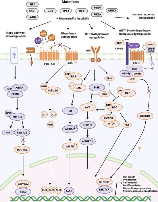
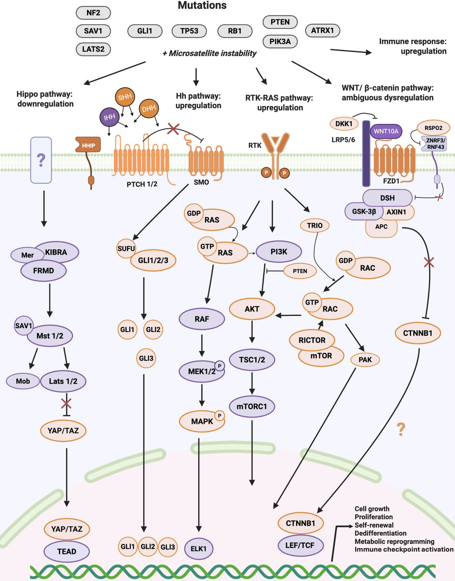

Disclaimer/Publisher’s Note: The statements, opinions and data contained in all publications are solely those of the individual author(s) and contributor(s) and not of MDPI and/or the editor(s). MDPI and/or the editor(s) disclaim responsibility for any injury to people or property resulting from any ideas, methods, instructions or products referred to in the content. |
© 2026 by the authors. Licensee MDPI, Basel, Switzerland. This article is an open access article distributed under the terms and conditions of the Creative Commons Attribution (CC BY) license.