


Abstract
Introduction: Post-hospital rehabilitation is essential for survivors of severe COVID-19, as prolonged immobility and clinical severity often lead to muscle weakness, reduced cardiovascular capacity, and impaired respiratory function. Physical exercise during and after hospitalization may mitigate these effects and support functional recovery. This study aimed to evaluate the effectiveness of a physical exercise-based rehabilitation program in survivors of severe COVID-19. Methodology: A randomized clinical trial was conducted with 30 survivors allocated to two groups: multicomponent exercise (GEm) and multicomponent exercise combined with inspiratory muscle training (GEmTMI). The interventions were performed three times per week for 40–60 min. Quality of life, physical activity level, functional status, and physical capacity were assessed before and after six weeks. Results: Comparisons between GEm and GEmTMI showed significant differences in the 6 min walk test (6MWT) at baseline (p = 0.043) and in the Physical Activity Index (IPAQ) after the intervention (p = 0.002). When the total sample was analyzed, significant improvements were observed across all outcomes after rehabilitation, including quality of life (SF-36), functional capacity (PCFS), physical activity level (IPAQ), respiratory muscle strength, and additional functional tests. Notable improvements included SF-36 Physical Functioning (p = 0.006) and Social Functioning (p = 0.009), PCFS (p = 0.011), IPAQ (p = 0.012), and performance in the 6MWT, STS, STS-1min, TUG, handgrip strength, PEmax, and PImax (all p < 0.001). Discussion: Multicomponent physical rehabilitation, with or without inspiratory muscle training, produced significant gains in physical activity level, functional capacity, dynamic balance, neuromuscular fitness, respiratory muscle strength, and quality of life. These findings underscore the importance of structured post-ICU rehabilitation to support comprehensive physical and psychosocial recovery in survivors of severe COVID-19.
1. Introduction
In the past two decades, several viral diseases have emerged with varying degrees of virulence and transmissibility, posing substantial challenges to public health and healthcare systems worldwide [1], as reported by the World Health Organization (WHO). Among these diseases, coronavirus disease 2019 (COVID-19), caused by SARS-CoV-2, had a particularly devastating impact. As of 7 April 2024, the WHO reported 775,293,630 confirmed cases and 7,044,637 deaths globally. In Brazil, 37.5 million cases and 702.1 thousand deaths were reported [2].
Although the case-fatality rate of SARS-CoV-2 (2.6%) is lower than that of SARS-CoV (11%) and Middle East respiratory syndrome (MERS; 35–50%), COVID-19 has proven far more disruptive because of its high transmission rate, resulting in millions of deaths and overwhelming healthcare systems worldwide [3]. COVID-19 is an infectious disease that can cause severe acute respiratory syndrome and may progress to multi-organ failure [4]. Patients with severe disease frequently develop acute respiratory, neurological, and musculoskeletal manifestations, which can lead to substantial impairments in standing, mobility, and functional independence [5,6].
The literature indicates that many patients discharged after hospitalization for COVID-19 experience persistent symptoms, including fatigue, shortness of breath, cognitive impairment, and sleep dysfunction [7,8,9,10,11]. These symptoms have been reported in patients discharged with or without ICU admission, as well as in “long-haul” patients [10], including those who initially presented with mild to moderate COVID-19 [10]. Moreover, a substantial proportion of individuals develop what has been described as post-viral fatigue syndrome or “long COVID” [5]. Rehabilitation guidelines for post-COVID-19 patients have been released by the WHO and by several countries [12].
Persistent symptoms include fatigue, shortness of breath, joint pain, chest pain, headaches, myalgia, brain fog, and gastrointestinal complaints [7,13,14,15]. These symptoms may be exacerbated by the considerable stress associated with hospitalization, including infrequent and brief interactions with healthcare professionals, absence of family at the bedside, and limited information regarding disease course, treatment options, and prognosis [16]. Accumulating evidence suggests that COVID-19 survivors may develop multiple sequelae beyond pulmonary damage [17], such as weakness, arthralgia, depression, anxiety, and memory impairment [18]. Musculoskeletal dysfunction and muscle weakness can already be observed during hospitalization [19], with the viral infection directly contributing to dysfunction at cellular and tissue levels [20]. In addition, prolonged hospitalization and ICU stays with extended immobilization, inadequate food and protein intake and absorption, inflammation, low vitamin D levels, and acute illness can contribute to anabolic resistance and acute sarcopenia. This metabolic condition—characterized by reduced muscle protein synthesis—is further aggravated by aging, frailty, and obesity [21].
In the current COVID-19 scenario, social isolation requirements contributed to physical inactivity and sedentary behavior, accelerating muscle atrophy and reducing muscle function [22,23,24]. Accordingly, regular physical activity and appropriate exercise training are necessary to preserve muscle mass and prevent atrophy in older age [25]. Sarcopenia—defined as the progressive decline in muscle mass and function associated with aging and physical inactivity—if left untreated, is linked to poor quality of life and increased mortality [26,27]. In this context, inspiratory muscle training (IMT) has shown potential to promote mechanical and clinical improvements in patients affected by COVID-19 and has been considered a promising strategy for pulmonary rehabilitation in this population [28]. However, the available findings remain inconclusive, as they are largely based on very low- to low-quality evidence [29].
These challenges underscore the need for integrated and effective rehabilitation strategies for this population, with strength training representing a promising intervention to enhance physical and functional recovery after COVID-19. Therefore, this study aimed to investigate the impact of a rehabilitation program based on physical exercises in individuals who survived COVID-19 and developed severe disease
2. Materials and Methods
This study was a controlled, randomized clinical trial involving adults (≥18 years) of both sexes who survived COVID-19, were hospitalized, and developed severe disease in Campina Grande (PB). Participants were randomized using a blocked allocation procedure (two blocks) and assigned to one of two rehabilitation groups: multicomponent exercise (GEm) or multicomponent exercise combined with inspiratory muscle training (IMT; GEmTMI). Patients were recruited through public announcements on social media (Instagram Stories and feed). Given that the study was conducted during the period of strict public health restrictions related to the COVID-19 pandemic, which limited in-person attendance, reduced clinic capacity, and constrained recruitment and follow-up logistics, this trial was designed as a pragmatic, two-arm comparison of rehabilitation strategies rather than a no-intervention control trial; therefore, the absence of a control group should be interpreted as a key methodological limitation.
Eligible participants had two negative RT-PCR tests for COVID-19 performed at the hospitalization site, in accordance with discharge criteria for severe patients [30]. Severe disease was defined using the World Health Organization (WHO) clinical progression scale [31]. Participants had been hospitalized and managed in an intensive care unit (ICU), intermediate care unit, or ward and had received invasive mechanical ventilation (via orotracheal tube or tracheostomy) or non-invasive ventilatory support and oxygen therapy (high-flow nasal cannula or reservoir mask). All participants had been discharged from the hospital for at least 30 days before the start of rehabilitation. After receiving a detailed explanation of the study objectives and the exercise-based rehabilitation protocol, all participants provided written informed consent by signing the Informed Consent Form (ICF) approved by the ethics committee.
Participants were excluded if they had evidence of reinfection, rehospitalization, or hospital readmission [31]; were hemodynamically unstable (uncontrolled blood pressure and heart rate); had unstable medical conditions (e.g., cardiovascular or pulmonary disorders such as arrhythmias, decompensated atrial fibrillation, pulmonary thromboembolism, acute heart failure, pulmonary congestion, acute myocardial infarction, or cerebrovascular accident); had osteoarticular or neurological conditions limiting mobility (e.g., paraplegia, quadriplegia, amputation, or severe joint deformities); or had cognitive impairment or dementia that prevented completion of questionnaires and participation in the exercise protocol.
This study complied with Resolution No. 466/12 of the Brazilian National Health Council, which governs research involving human participants, and was submitted to the Research Ethics Committee of the UNIFACISA University Center. The protocol was approved under Opinion No. 4.619.192 and registered in the Brazilian Clinical Trials Registry (ReBEC) under UTN No. U1111-1264-9012 on 29 March 2021. Throughout the study, ethical standards were maintained, including appropriate care and monitoring of participants and protection of data confidentiality and privacy during data handling and after processing for publication. All individuals who voluntarily participated signed the Informed Consent Form (ICF) after reading and understanding its contents, including the study objectives, procedures, risks, and benefits. Participants were informed about all methodological stages (including group allocation) and the full assessment procedures. Each participant was advised of the right to withdraw at any time without any financial or other penalties. Participation was free of charge, and all study-related costs were covered by the researchers.
2.1. Sampling Plan
All participants underwent an initial assessment conducted by a physical education professional and a physiotherapist. At the outset of the study, government-imposed public health restrictions limited the number of individuals allowed in the rehabilitation clinic and therapy gym to reduce the risk of disease transmission. Therefore, recruitment and baseline characterization were conducted using a comprehensive remote assessment, combining medical record review with structured data collection and validated questionnaires. This included a review of medical records and length of hospitalization; collection of sociodemographic data (date of birth, sex, age, educational level, number of household members, and number of rooms in the home); and documentation of disease course (severity, duration of ICU or ward stay, use and duration of mechanical ventilation, surgical procedures, and medications used during treatment). Information on pre-existing comorbidities (cardiovascular, pulmonary, or metabolic diseases) and the use of related medications was also recorded. In addition, three previously validated questionnaires were administered to assess physical activity level (min/week), quality of life (SF-36), and post-COVID-19 functional status.
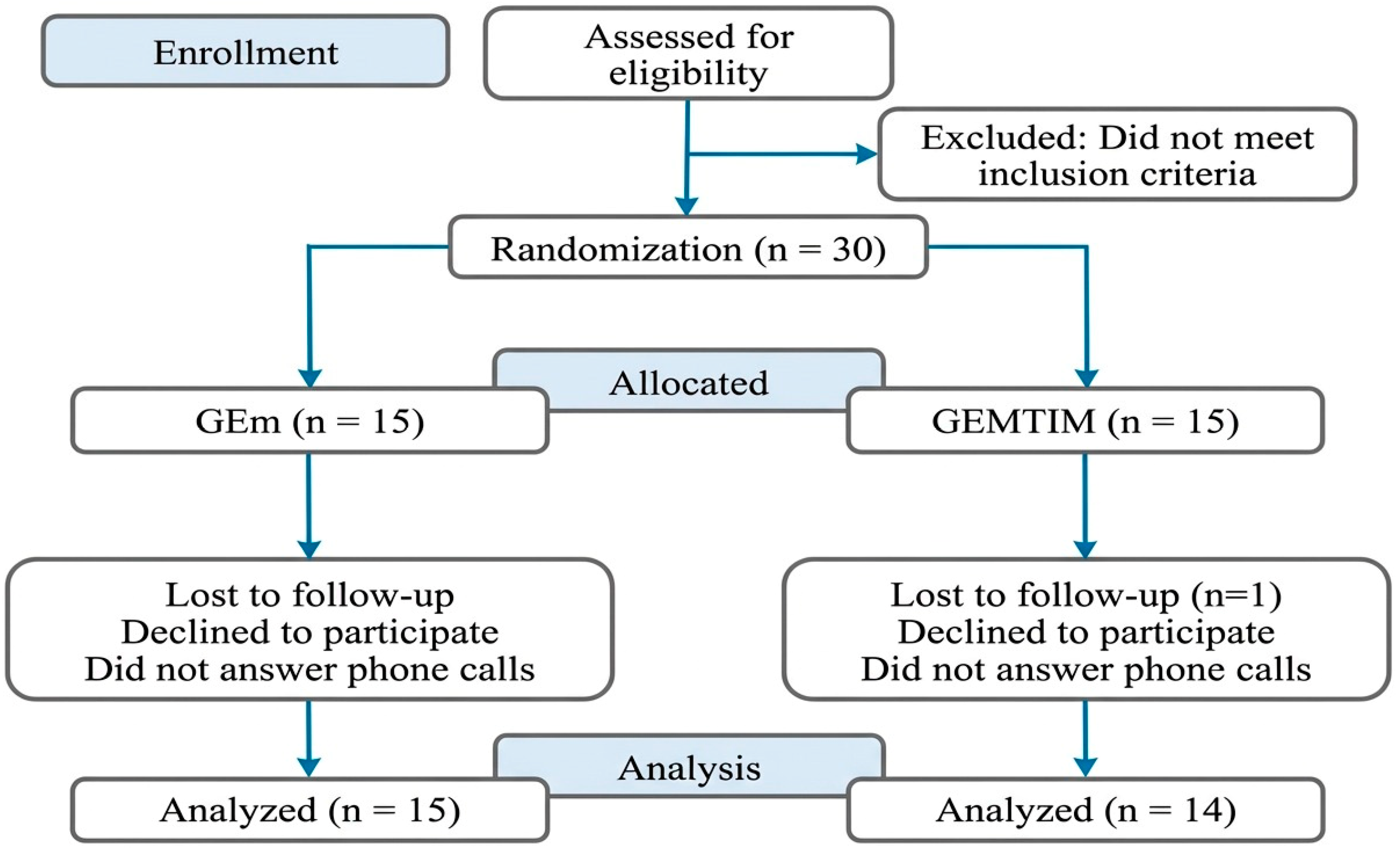
A formal a priori sample size calculation was not feasible because recruitment occurred during a period of strict public health restrictions, which limited clinic capacity and the number of eligible individuals who could be safely assessed in person; additionally, uncertainty regarding the expected effect sizes in this specific population of post-hospitalized patients with severe COVID-19 precluded the reliable specification of parameters for power estimation. Accordingly, the final sample reflects the number of eligible participants who could be recruited and followed within the operational constraints of the service during the study period. Moreover, guidance for external pilot randomized trials indicates that recommended sample sizes per treatment arm may range from approximately 10 to 75 participants, depending on the anticipated standardized effect size (e.g., ~10 per arm for large effects and larger samples for smaller effects), reinforcing that small samples are primarily appropriate for assessing feasibility and informing planning rather than for definitive inference of between-group differences [32]. Therefore, this small sample size should be interpreted as an important limitation, particularly with respect to detecting between-group differences. As shown in Figure 1 below.
Figure 1.
Flowchart of the sample and participant allocation into groups.
2.2. Intervention
Rehabilitation sessions were conducted three times per week, with each session lasting 40–60 min, at the school clinic. All outcome measures were collected at baseline and after six weeks.
Both groups followed the same exercise-based rehabilitation protocol and differed only in the inclusion or absence of inspiratory muscle training (IMT). The exercise-only group performed multicomponent training (aerobic, resistance, and balance exercises) three times per week for 40–50 min. In the IMT group, a PowerBreathe device (HaB International Ltd., Hampshire, UK) was incorporated into the rehabilitation routine under physiotherapist supervision.
Aerobic training lasted 25–30 min and was performed on an ergometer selected by the participant (treadmill or cycle ergometer) to achieve the target heart-rate intensity ranges. Initial intensity was set at or below the heart rate corresponding to the first ventilatory threshold (VT1). Aerobic intensity was progressively increased according to the participant’s exercise tolerance until the second ventilatory threshold was reached. Perceived exertion was monitored using the modified Borg scale, ranging from 0 (no exertion) to 10 (maximal exertion), as proposed by [32].
Muscle-strengthening exercises lasted 10–15 min and included movements performed using body weight or external resistance (free weights, dumbbells, or elastic bands). Training targeted large muscle groups and consisted of 1–3 sets of 8–15 repetition maximum (RM), with 1–2 min rest intervals between sets [33,34]. Load progression was performed weekly with a 5–10% increase. If a participant was unable to complete the prescribed workload, the previous load was maintained, and a new attempt to increase the load was made the following week. Exercise intensity was additionally monitored using an analog perceived exertion scale for resistance training ranging from 0 (no exertion) to 10 (maximal effort), as proposed by [35].
Balance exercises lasted approximately 7–10 min and included motor tasks designed to challenge the center of gravity (e.g., tandem walking and walking on unstable surfaces), activities with reduced sensory input (e.g., sitting-to-standing and standing with eyes closed), and postural muscle challenges (e.g., toe walking and heel walking). These exercises were performed at either the beginning or the end of the session [36,37].
Inspiratory muscle training (IMT) to improve inspiratory muscle strength and endurance was performed by participants three times per week using a linear pressure-threshold loading device (Powerbreathe® Classic line, HaB International Ltd., Hampshire, UK) and a nasal clip. Training began at 40–50% of maximal inspiratory pressure (PImax) and was progressively increased to 60–70% of PImax, up to the highest tolerable intensity. Participants performed 90 breaths per session, divided into three sets, with 1–2 min of rest between sets. PImax was reassessed every two weeks to adjust training intensity to the participant’s current capacity.
The training load was selected to provide the highest tolerable resistance while still allowing inspirations across the full vital capacity, thereby enhancing training specificity by applying a stimulus throughout the entire range of motion of the inspiratory muscles, including the lengths at which these muscles operate during exercise. Perceived respiratory effort (4–6 on the modified Borg 0–10 scale) was also used to guide decisions regarding load progression. IMT was performed either alone or in combination with resistance and balance exercises.
For safety, heart rate was monitored throughout each session using the Firstbeat Bodyguard 2 (Firstbeat Technologies Ltd., Jyväskylä, Finland). Blood pressure was measured by auscultation using an aneroid sphygmomanometer and stethoscope (Diasyst®, São Paulo, Brazil). Peripheral oxygen saturation was monitored using a pulse oximeter (TuffsatTM, São Paulo, Brazil). Capillary blood glucose was measured in participants with diabetes using a glucometer (Accu-Chek Nano, Roche, Germany).
If a participant exhibited peripheral oxygen saturation < 90% or developed symptoms such as palpitations, diaphoresis, chest tightness, or severe dyspnea, the session was immediately interrupted and the participant was placed at rest. The physiotherapist and the medical team then evaluated the participant to determine whether the session could be resumed, and supplemental oxygen was provided as needed.
2.3. Variables
2.3.1. Physical Activity Level
Habitual physical activity level was assessed using the short form of the International Physical Activity Questionnaire (IPAQ) [38,39], which demonstrates moderate validity and reliability, with intraclass correlation coefficients (ICC) ranging from 0.33 to 0.62 [40]. Physical activity was estimated based on the duration and weekly frequency of walking and moderate- and vigorous-intensity activities performed across different domains. Total physical activity was expressed as minutes per week, and individuals accumulating ≥ 150 min/week were classified as physically active [41].
2.3.2. Quality of Life
Quality of life was assessed using the Medical Outcomes Study 36-Item Short Form Health Survey (SF-36), which shows acceptable model fit, with comparative fit indices (CFI) ranging from 0.90 to 0.92 and a root mean square error of approximation (RMSEA) of 0.07. Validity is high, as indicated by significant correlations with other health-related quality-of-life measures, and reliability is also high, with Cronbach’s alpha coefficients often exceeding 0.90 [42]. The instrument has been previously validated in its Brazilian version [43].
The SF-36 comprises 36 items assessing eight domains: physical functioning (10 items), role limitations due to physical health (four items), bodily pain (two items), general health perceptions (five items), vitality (four items), social functioning (two items), role limitations due to emotional problems (three items), and mental health (five items), in addition to a comparative item on current health status relative to one year earlier. Domain scores range from 0 (worst) to 100 (best).
2.3.3. Post-COVID-19 Functional Status Scale (PCFS)
Functional status was assessed using a recently published scale designed to capture relevant aspects of daily life during follow-up after infection [44]. This ordinal scale comprises six levels, ranging from 0 (“no symptoms”) to 5 (“death”), and spans the full spectrum of functional outcomes. It focuses on limitations in daily tasks and activities—both domestic and occupational/educational—as well as lifestyle changes. The scale assesses the domains of “constant care,” “activities of daily living (ADLs),” “instrumental activities of daily living (IADLs),” “participation in usual social roles,” and “symptom checklist.” It demonstrates significant correlations with other health-related quality-of-life measures and good reliability, with Cronbach’s alpha coefficients exceeding 0.76. Model fit indices were also acceptable, with comparative fit indices (CFI) ranging from 0.90 to 0.92 and a root mean square error of approximation (RMSEA) of 0.07 [45].
2.3.4. Anthropometry and Body Composition
Body mass (kg) was measured using a Balmak® Class III digital electronic (BALMAK SA, Athens, Greece) scale with a precision of 0.01 kg. Height was measured using a SANNY® stadiometer (Personal Caprice Portable, São Paulo, Brazil) with a precision of 0.1 cm. Body mass index (BMI) was calculated as body mass divided by height squared (kg/m2). Body composition (fat and total body water percentage) was assessed by bioelectrical impedance analysis (BIA) using the Omron HBF-514C portable scale (OMRON, Kyoto, Japan), which applies a low-intensity, imperceptible electrical current.
2.3.5. Dynamic Balance
Dynamic balance was assessed using the Timed Up and Go (TUG) test [46], which demonstrates high reliability and validity, with an intraclass correlation coefficient (ICC) of 0.82 and significant correlations with other mobility tests [47]. The TUG evaluates balance and functional mobility by measuring the time (in seconds) required for the participant to rise from a chair without using the arms, walk 3 m, turn, walk back, and sit down again. At the start of the test, the participant sat with the back against the backrest; at the end, the participant returned to the seated position with the back against the backrest. The participant was instructed to begin on the verbal cue “Go,” and timing started at the command and stopped when the participant sat back and reclined against the backrest [48].
2.3.6. Functional Capacity
Functional capacity was assessed using the six-minute walk test (6MWT), with functional capacity quantified as the distance walked. The 6MWT has high validity for evaluating functional capacity in chronic diseases and shows significant correlations with other functional capacity measures. Test–retest reliability is high, with intraclass correlation coefficients (ICC) ranging from 0.80 to 0.97 [49]. The test was conducted in accordance with American Thoracic Society guidelines. Required equipment included a stopwatch, measuring tape, pulse oximeter (TuffsatTM, São Paulo, Brazil), heart rate monitor (735xt, Garmin®, Schaffhausen, Switzerland), sphygmomanometer (Diasyst® Sphygmomanometer, São Paulo, Brazil), and stethoscope (Diasyst®, São Paulo, Brazil).
The 6MWT was performed in a 30 m corridor at the clinic. Vital signs—including blood pressure, heart rate, perceived exertion (0–10 analog scale) [32], and oxygen saturation—were measured before, during, and after the test. The outcome was the greatest distance covered in six minutes in a single trial.
Participants were instructed to stop the test if they experienced symptoms such as lower-limb pain, tachycardia, or respiratory discomfort. The pulse oximeter remained in place throughout the test to monitor peripheral oxygen saturation. The test was terminated if oxygen saturation decreased below 92% or if 90% of the age-predicted maximum heart rate was reached [36]. At the end of the test, the highest heart rate value was recorded as maximal heart rate (HRmax) and used to prescribe aerobic exercise intensity.
2.3.7. Neuromuscular Fitness
Lower-limb muscle endurance was assessed using the 60 s sit-to-stand test from the Senior Fitness Test—Physical Fitness Test for Lower Limbs [50]. This test shows good reliability, with intraclass correlation coefficients (ICC) ranging from 0.76 to 0.93, as well as good concurrent validity, demonstrated by significant correlations with other physical fitness tests, and sensitivity to clinical change [51]. The test began with the participant seated on a chair with feet flat on the floor. Before the timed trial, the participant performed three repetitions to familiarize themselves with the task and then completed as many sit-to-stand repetitions as possible in 60 s.
Handgrip strength was assessed using a handgrip dynamometer test, which demonstrates high reliability (ICC: 0.90–0.97) and high validity, with strong correlations with other measures of muscle strength (e.g., isokinetic dynamometry). It is also sensitive to changes in functional status, supporting its use for monitoring therapeutic interventions [52]. Measurements were obtained using a digital dynamometer (EH101 model, Clear; adjustable and calibrated; range: 0–100 kgf). Participants were assessed in the standing position with the elbow fully extended and the arm slightly abducted, following the manufacturer’s instructions. Before testing, participants received standardized instructions and performed several practice contractions until comfortable with the procedure.
2.3.8. Respiratory Muscle Strength
Maximal respiratory pressures were measured using an analog manovacuometer connected to a pressure line, a mouthpiece with a filter, and a connector with a 2 mm leak hole. Participants were seated with feet flat on the floor and an upright posture, without upper-limb support, and wore a nasal clip. They were instructed to perform the maximal inspiratory pressure (PImax) maneuver from residual volume (RV) to total lung capacity (TLC), followed by a maximal, sustained inspiratory effort. Maximal expiratory pressure (PEmax) was measured from TLC with a maximal, sustained expiratory effort.
At least three maneuvers were performed, with 1 min rest intervals between attempts, and reproducibility was defined as a ≤10% variation between maneuvers. The highest value obtained from the acceptable maneuvers was recorded. Predicted values for PImax and PEmax were calculated for each participant using equations validated for the Brazilian population.
2.3.9. Cardiopulmonary Exercise Teste (CPET)
Before the cardiopulmonary exercise test (CPET) to determine peak VO2, participants were informed about the procedures and instructed to exercise to volitional exhaustion. A standard 12-lead ECG and peripheral oxygen saturation were recorded at rest and at the end of each workload stage using a Vyntus® ECG system (Vyaire, Leipzig, Germany). The test could be terminated if any criterion for test termination, according to current guidelines, was met [53,54,55]. Blood pressure was measured by auscultation at rest, at the end of each CPET stage, and during active recovery using a sphygmomanometer (Diasyst®, São Paulo, Brazil) and stethoscope (Diasyst®, São Paulo, Brazil). Oxygen saturation was continuously monitored throughout the test using a pulse oximeter (TuffsatTM, São Paulo, Brazil). The CPET was conducted by a sports physician (member of the multidisciplinary team), with support from a physical education professional and a physiotherapist.
To familiarize participants with treadmill walking, the test began on a flat, motorized treadmill (Movement, R7 127V), during which participants practiced walking without holding the handrails. For participants who could walk safely, a 10 min warm-up was performed at a fixed speed of 3–6 km/h and 0% grade. After warm-up, a face mask was fitted for metabolic measurements using the Vyntus® CPX system (Vyaire, Leipzig, Germany).
The A ramp protocol was used, starting at 4.0 km/h and 1.0% grade, with speed increased by 0.5 km/h each minute until peak VO2 was achieved. The protocol was designed to elicit peak VO2 within 8–12 min, in line with recommendations [56]. The test was terminated upon volitional exhaustion or if any of the following occurred: diastolic blood pressure (DBP) increased to 120 mmHg; systolic blood pressure (SBP) decreased persistently by >10 mmHg; SBP increased disproportionately to 260 mmHg with rising workload; chest discomfort with increasing workload or accompanied by ECG changes suggestive of ischemia; ataxia, dizziness, pallor, cyanosis, or presyncope; dyspnea disproportionate to exercise intensity; ST-segment depression of 0.3 mV (2.5 mm) or ST-segment elevation of 0.2 mV (2.0 mm) in leads without a Q wave; complex ventricular arrhythmias; onset of non-sustained or sustained supraventricular tachycardia; atrial tachycardia; atrial fibrillation; second- or third-degree atrioventricular block; or signs of left ventricular failure [57]. Because of the high cost and logistical constraints of CPET, it was performed only at baseline to determine training intensities prior to the rehabilitation program.
2.4. Statistical Analysis
Results are presented as mean ± standard deviation [95% CI] or as proportions. Data normality was assessed using the Shapiro–Wilk test due to the small sample size. Within-group pre- to post-rehabilitation changes were analyzed using the two-tailed Wilcoxon signed-rank test for nonparametric continuous data and the chi-square test for categorical data, as appropriate, whereas paired t-tests were used for parametric data. Between-group comparisons were performed using the Mann–Whitney U test for nonparametric data and independent-samples t-tests for parametric data. Statistical significance was set at p < 0.05. All analyses were conducted in SPSS version 20 (IBM, Armonk, NY, USA).
3. Results
The 30 eligible patients were allocated to two rehabilitation groups: the physical exercise group (GEm; n = 15) and the physical exercise plus inspiratory muscle training group (GEmTMI; n = 14). One patient was excluded from the study due to absences related to transportation difficulties and was therefore not included in the final evaluation. Participants’ demographic characteristics are presented in Table 1.
Table 1.
Demographic characteristics of the patients Total (GEm + GEmTMI), GEm, and GEmTMI for men (M) and women (F).
No adverse events were reported during the program. SpO2 remained above 90% in all participants throughout the training sessions. Data from 29 participants—assigned to the GEm (n = 15) and GEmTMI (n = 14) groups—were analyzed to characterize baseline demographic and anthropometric features that could influence study outcomes. As shown in Table 1, there was no statistically significant evidence of between-group differences in age, sex, weight, height, BMI, or disease prevalence. Although not reported in Table 1, educational level was also assessed; the chi-square test (χ2 = 4.95) indicated no significant difference in its distribution between groups (p = 0.66).
As shown in Table 2, both groups improved across several domains, particularly social functioning and physical limitations; however, many of these improvements did not reach statistical significance.
Table 2.
Quality of Life (SF 36) of the patients Total (GEm + GEmTMI), GEm, and GEmTMI.
The results presented in Table 3 indicate that the intensive mobility intervention (TMI) yielded significant benefits, including symptom reduction; improvements in instrumental activities of daily living and social role functioning; and increases in physical activity and energy expenditure among participants.
Table 3.
Post-COVID-19 Functional State Scale (PCFS) and physical activity level (IPAQ) of the patients Total (GEm + GEmTMI), GEm, and GEmTMI.
The results presented in Table 3 indicate that the intensive mobility intervention (IMT) yielded significant benefits, including symptom reduction; improvements in instrumental activities of daily living and social role functioning; and increases in physical activity and energy expenditure among participants. Although both groups improved, the magnitude of change was greater in the GEmTMI group. These findings suggest that IMT may be an effective intervention for improving quality of life and functional status in similar populations. The participants’ cardiopulmonary exercise capacity, including heart rate at the first and second ventilatory thresholds (VT1 and VT2), peak heart rate, and peak oxygen uptake (VO2peak; mL·kg−1·min−1), is presented in Table 4.
Table 4.
Cardiopulmonary exercise capacity of the patients Total (GEm + GEmTMI), GEm, and GEmTMI.
The parameters evaluated and presented in Table 5 were TC6 (six-minute walk test), TSL (time to stand and sit), TSL 1min (time to stand and sit in 1 min), TUG (timed up and go test), Right and Left Handgrip, PEMÁX (maximum expiratory pressure), and PImax (maximum inspiratory pressure).
Table 5.
Functional tests of the patients Total (GEm + GEmTMI), GEm, and GEmTMI.
The parameters evaluated and reported in Table 5 were the six-minute walk test (6MWT), time to stand and sit (TSL), one-minute stand-and-sit test (TSL 1 min), the timed up-and-go test (TUG), right and left handgrip strength, maximal expiratory pressure (PEmax), and maximal inspiratory pressure (PImax). Measurements were obtained before and after the interventions and were compared both within groups and between groups at the two time points. Both the GEm and GEmTMI groups showed significant post-intervention improvements across all assessed parameters. No significant between-group differences were observed, suggesting comparable effectiveness of the interventions. Overall, the findings underscore the effectiveness of both training programs in improving functional capacity, muscle strength, and endurance in the participants.
4. Discussion
At the start of rehabilitation (following the acute phase), the GEm and GEmTMI groups were comparable. The mean age (50.67 ± 11.96 years) was consistent with that reported across 11 post-COVID-19 rehabilitation studies (55.81 ± 8.82 years) [58,59,60,61,62,63,64,65,66,67,68]. Other baseline characteristics (Table 1)—including sex, weight, height, and the prevalence of non-communicable diseases (NCDs)—were also well balanced between groups, with obesity being the most prevalent condition (65.52%). Obesity is associated with hypertension, type 2 diabetes, cardiovascular disease, stroke, and cancer [69], which increases clinical vulnerability to SARS-CoV-2 infection and worsens prognosis [70]. Obese patients have a 2.1-fold higher risk of hospitalization, a 2.6-fold higher risk of ICU admission, and a 3.7-fold higher risk of death [71], and overweight/obesity is highly prevalent among hospitalized individuals [70]. The pro-inflammatory and immunomodulatory effects of adipose tissue—particularly visceral fat—may further exacerbate disease progression [72,73]. Accordingly, rehabilitation strategies should incorporate weight-management components to reduce the risks of respiratory failure and systemic inflammation.
During the peak VO2 test, 29 patients achieved maximal effort without complications; however, exercise capacity was markedly reduced (16.14 ± 5.52 mL·kg−1·min−1), below the 20 mL·kg−1·min−1 threshold often cited as necessary for independent activities of daily living [74]. This impairment may reflect COVID-19–related stress and mitochondrial dysfunction, including reduced oxidative phosphorylation and mitochondrial density [75,76,77,78,79,80,81], as well as possible activation of catabolic pathways linked to skeletal muscle atrophy [33,82,83]. After six weeks, meaningful functional gains were observed. Mean 6MWT distance increased from 378.44 ± 103.06 m to 501.33 ± 76.95 m (+122.89 m; +32.47%), consistent with previous reports [84]. This improvement likely reflects reductions in fatigue and dyspnea and, importantly, supports greater autonomy and self-efficacy, thereby benefiting social functioning and quality of life [85]. Performance on the sit-to-stand test (STST) increased by 37.89%, from 19.89 ± 6.23 to 27.30 ± 7.40 repetitions (+7.41), corroborating the improvements observed in the 6MWT. The more pronounced perception of dyspnea relative to lower-limb fatigue is consistent with the predominant pulmonary contribution to functional limitation after COVID-19 [86], supporting the integration of aerobic training, resistance exercise, and respiratory conditioning within rehabilitation protocols.
A meaningful improvement was observed in the Timed Up and Go (TUG) test, with times decreasing from 9.59 ± 3.68 to 7.00 ± 1.88 s. Notably, all six participants who initially exceeded 12 s—a threshold indicating high fall risk according to CDC/STEADI—fell below this cutoff after the intervention [87]. In long COVID and post-critical illness, prolonged TUG and dual-task TUG (DT-TUG) times are common [88,89]. On average, post-COVID patients take longer to complete sit-to-stand (STS), walking, and TUG tests than healthy individuals [90], likely due to central and peripheral neurological changes that impair gait symmetry [91,92], as well as muscle weakness, exercise intolerance, and histological atrophy [93]. These findings support the clinical relevance of rehabilitation for mobility and safety while remaining consistent with the functional tests reported.
Handgrip strength also increased, from 28.81 ± 10.31 to 34.70 ± 11.37 kg in the right hand and from 26.67 ± 9.62 to 32.63 ± 10.61 kg in the left hand—clinically relevant markers of health and functional status [94] that are often reduced after COVID-19 hospitalization [95,96,97]. Aerobic and resistance exercise can reduce dyspnea and restore strength [98], and early rehabilitation may help prevent critical illness neuromyopathy (CINM) and ICU-acquired weakness (ICU-AW) [99,100,101]. Importantly, muscle quality—captured by markers such as myosteatosis and intermuscular adipose tissue (IMAT)—may be more strongly associated with physical performance than muscle mass alone [102,103]. Elevated IMAT, often linked to chronic inflammation and altered lipid metabolism, is a marker of poor muscle quality and may pre-exist in hospitalized patients, thereby impairing functional recovery [104,105]. Accordingly, the improvements observed here may reflect not only gains in strength but also potential rehabilitation-related improvements in neuromuscular performance and muscle quality.
With respect to respiratory function, respiratory muscle strength is critical for sustaining functional gains. Rehabilitation increased maximal inspiratory pressure (PImax) and maximal expiratory pressure (PEmax), which is clinically relevant for reducing dyspnea and supporting mobility, although some studies note limited statistical power due to small sample sizes [106]. Overall, the pattern of improvement underscores the interdependence of respiratory conditioning and physical performance: greater respiratory strength may reduce perceived exertion during daily activities, which is consistent with improvements in psychosocial outcomes, including social functioning on the SF-36.
The combination of inspiratory muscle training (IMT) and aerobic exercise improves respiratory muscle strength, with significant increases in maximal inspiratory pressure (PImax) reported in the literature [107], as well as cardiovascular benefits in hypertensive individuals [108]. Tele-supervised home-based protocols can expand access and have demonstrated effectiveness [61]. Pulmonary rehabilitation reduces dyspnea, improves exercise capacity, and alleviates fatigue; meta-analyses indicate greater improvements in pulmonary function and quality of life compared with no intervention [109]. Physical training may also accelerate functional recovery and improve spirometric parameters such as forced vital capacity (FVC) and forced expiratory volume in 1 s (FEV1) [110], a hypothesis further supported by the potential anti-inflammatory effects of exercise [110]. In this context, evidence from an Individual Rehabilitation Project (IRP) grounded in early re-educational interventions in hospitalized COVID-19 patients also reported improvements in dyspnea and autonomy in activities of daily living and muscle strength, while highlighting that a no-rehabilitation control group was not feasible for clinical and ethical reasons. This evidence reinforces the importance of early intervention in the post-COVID-19 recovery process [111].
In our study, PImax and PEmax increased after rehabilitation in both the GEm and GEmTMI groups. The absence of statistically significant between-group differences may be explained by the non-specific recruitment of respiratory muscles during whole-body exercise, which could attenuate any incremental effect of IMT. In addition, the small sample size limits the precision of between-group comparisons and reduces power to detect modest incremental effects—an aspect that should be interpreted as an important constraint rather than evidence of equivalence. Nevertheless, these findings are consistent with evidence that both resistance and aerobic training can improve PImax and PEmax [112,113,114].
Taken together, improvements in peak VO2, 6MWT distance, TUG performance, and STST repetitions likely contributed to reduced fatigue and dyspnea while enhancing autonomy. These physical gains were accompanied by higher SF-36 social functioning scores and a lower symptom burden (PCFS). Concurrently, restoration of respiratory muscle strength (PImax and PEmax) may have supported functional performance and reduced perceived exertion. Overall, structured interventions integrating aerobic, resistance, and respiratory training appear to contribute to multidimensional recovery, while conclusions about the incremental contribution of IMT should remain cautious given the study design and sample size.
The findings of this study underscore that post-COVID-19 rehabilitation should be viewed as a multidimensional process in which functional, psychosocial, and respiratory gains are interrelated and mutually reinforcing. Improvements in exercise capacity (peak VO2, 6MWT, and STST), functional mobility (TUG), and peripheral muscle strength (handgrip strength) are consistent with structured interventions that can support autonomy and reduce persistent symptoms such as fatigue and dyspnea. These physical gains may also translate into psychosocial benefits, reflected in higher SF-36 social functioning scores and fewer functional limitations on the PCFS, suggesting greater confidence, social participation, and quality of life.
Concurrently, respiratory improvements—evidenced by increases in PImax and PEmax—reinforce the central role of pulmonary function in sustaining physical capacity and reducing perceived exertion, thereby linking physiological recovery to psychosocial reintegration. Together, these results support the clinical relevance of rehabilitation programs that integrate aerobic, resistance, and respiratory training in minimizing sequelae, promoting comprehensive recovery, and potentially mitigating the long-term impact of infection.
Although the study has methodological limitations, including a small sample size and the absence of a control group, data collection occurred during a period of strict restrictions on gatherings, which constrained recruitment and service capacity. Therefore, the results should be interpreted cautiously because improvements may partially reflect natural recovery over time in addition to intervention-related effects. Moreover, while the lack of a no-intervention control arm remains a key limitation for causal inference, this design choice is consistent with the clinical and ethical rationale reported in hospital-based COVID-19 rehabilitation research, where patients are not left without rehabilitation assistance.
Nevertheless, the findings highlight the importance of establishing post-COVID-19 rehabilitation as a priority within longitudinal care pathways. Beyond improving patients’ physical and mental health, such interventions provide a basis for future research to clarify recovery mechanisms, quantify the incremental contribution of IMT, and expand therapeutic options.
5. Conclusions
This study demonstrated that structured physical exercise programs can support recovery in COVID-19 survivors, particularly those who experienced severe disease. Both groups, with or without inspiratory muscle training (IMT), showed significant improvements in physical capacity, strength, and endurance. Although between-group differences were not statistically significant, the findings suggest clinically meaningful benefits, especially in social functioning and reductions in physical limitations. Nevertheless, these results should be interpreted with caution, given the possibility that some improvements may reflect the natural course of recovery.
Author Contributions
Conceptualization: E.F.P., N.J.B.A.F., J.C.L., T.M.E.G., L.N.d.S., R.R.d.S.J., M.I.K. and G.P. Methodology: E.F.P., N.J.B.A.F., T.M.E.G., R.R.d.S.J., M.I.K. and G.P. Ethics Committee e REBEC: N.J.B.A.F., R.R.d.S.J., M.I.K. and G.P. Validation: J.C.L., T.M.E.G., L.N.d.S. and E.F.P. Formal Analysis: E.F.P., J.C.L., L.N.d.S. and R.R.d.S.J. Data Curation: J.C.L., L.N.d.S., T.M.E.G. and N.J.B.A.F. Investigation: E.F.P., N.J.B.A.F., J.C.L., T.M.E.G., L.N.d.S. and R.R.d.S.J. Resources: N.J.B.A.F., M.I.K. and G.P. Visualization: J.C.L., T.M.E.G., L.N.d.S. and R.R.d.S.J. Supervision: M.I.K. and G.P. Project Administration: G.P., M.I.K. and N.J.B.A.F. Funding Acquisition: G.P. Writing—Original Draft: E.F.P., N.J.B.A.F., J.C.L., T.M.E.G., L.N.d.S., R.R.d.S.J., M.I.K. and G.P. Writing—Review and Editing: E.F.P., N.J.B.A.F., J.C.L., T.M.E.G., L.N.d.S., R.R.d.S.J., M.I.K. and G.P. All authors have read and agreed to the published version of the manuscript.
Funding
This study was supported by the Federal University of Rio Grande do Norte (UFRN), Brazil.
Institutional Review Board Statement
The study was conducted in accordance with the Declaration of Helsinki and was approved by the Research Ethics Committee of the Higher Education and Development Center (UNIFACISA), under approval number 4.619.192 and CAAE No. 43001621.2.0000.5175, on 29 March 2021.
Informed Consent Statement
Informed consent was obtained from all subjects involved in the study.
Data Availability Statement
The original contributions presented in this study are included in the article. For further information, please contact the corresponding authors.
Conflicts of Interest
The authors declare no conflicts of interest.
References
- Cascella, M.; Rajnik, M.; Aleem, A.; Dulebohn, S.C.; Di Napoli, R. Features, Evaluation, and Treatment of Coronavirus (COVID-19). In StatPearls [Internet]; StatPearls Publishing: Treasure Island, FL, USA, 2025. Available online: http://www.ncbi.nlm.nih.gov/books/NBK554776/ (accessed on 1 May 2025).
- World Health Organization. WHO Coronavirus (COVID-19) Cases Dashboard; WHO: Geneva, Switzerland, 2023; Available online: https://data.who.int/dashboards/covid19/cases (accessed on 2 August 2024).
- Hasöksüz, M.; Kiliç, S.; Saraç, F. Coronaviruses and SARS-CoV-2. Turk. J. Med. Sci. 2020, 50, 549–556. [Google Scholar] [CrossRef] [Scilit]
- Barazzoni, R.; Bischoff, S.C.; Breda, J.; Wickramasinghe, K.; Krznaric, Z.; Nitzan, D.; Pirlich, M.; Singer, P. ESPEN expert statements and practical guidance for nutritional management of individuals with SARS-CoV-2 infection. Clin. Nutr. 2020, 39, 1631–1638. [Google Scholar]
- Lopez, M.; Bell, K.; Annaswamy, T.; Juengst, S.; Ifejika, N. COVID-19 Guide for the Rehabilitation Clinician: A Review of Nonpulmonary Manifestations and Complications. Am. J. Phys. Med. Rehabil. 2020, 99, 669–673. [Google Scholar]
- Vitacca, M.; Lazzeri, M.; Guffanti, E.; Frigerio, P.; D’abrosca, F.; Gianola, S.; Carone, M.; Paneroni, M.; Ceriana, P.; Pasqua, F.; et al. Italian suggestions for pulmonary rehabilitation in COVID-19 patients recovering from acute respiratory failure: Results of a Delphi process. Monaldi Arch. Chest Dis. 2020, 90. [Google Scholar] [CrossRef] [Scilit]
- Carfì, A.; Bernabei, R.; Landi, F. Gemelli Against COVID-19 Post-Acute Care Study Group. Persistent Symptoms in Patients After Acute COVID-19. JAMA 2020, 324, 603–605. [Google Scholar] [CrossRef] [Scilit] [PubMed]
- Garrigues, E.; Janvier, P.; Kherabi, Y.; Le Bot, A.; Hamon, A.; Gouze, H.; Doucet, L.; Berkani, S.; Oliosi, E.; Mallart, E.; et al. Post-discharge persistent symptoms and health-related quality of life after hospitalization for COVID-19. J. Infect. 2020, 81, e4–e6. [Google Scholar] [CrossRef] [Scilit] [PubMed]
- Li, Z.; Zheng, C.; Duan, C.; Zhang, Y.; Li, Q.; Dou, Z.; Li, J.; Xia, W. Rehabilitation needs of the first cohort of post-acute COVID-19 patients in Hubei, China. Eur. J. Phys. Rehabil. Med. 2020, 56, 339–344. [Google Scholar] [CrossRef] [Scilit]
- Rubin, R. As Their Numbers Grow, COVID-19 “Long Haulers” Stump Experts. JAMA 2020, 324, 1381–1383. [Google Scholar] [CrossRef] [Scilit]
- Simpson, R.; Robinson, L. Rehabilitation After Critical Illness in People With COVID-19 Infection. Am. J. Phys. Med. Rehabil. 2020, 99, 470–474. [Google Scholar] [CrossRef] [Scilit] [PubMed]
- Brugliera, L.; Spina, A.; Castellazzi, P.; Cimino, P.; Arcuri, P.; Deriu, M.G.; Zanetta, C.; Angelone, S.M.; Capitanio, J.F.; Alemanno, F. Rehabilitation of COVID-19 patients. J. Rehabil. Med. 2020, 52, jrm00046. [Google Scholar] [CrossRef] [Scilit] [PubMed]
- Banda, J.; Singh, G.; Alser, O.; Prieto-Alhambra, D. Long-term patient-reported symptoms of COVID-19: An analysis of social media data. medRxiv 2020. [Google Scholar] [CrossRef] [Scilit]
- Davis, H.E.; Assaf, G.S.; McCorkell, L.; Wei, H.; Low, R.J.; Re’em, Y.; Redfield, S.; Austin, J.P.; Akrami, A. Characterizing long COVID in an international cohort: 7 months of symptoms and their impact. eClinicalMedicine 2021, 38, 101019. [Google Scholar] [CrossRef] [Scilit]
- Khashei, M.; Janiczak, S.; St Clair, C.; Liu, W.; Song, J.J.; Hua, W.; Falconer, M.; Eworuke, E. Social media for early characterization of pandemic symptoms: A qualitative analysis of patient-reported COVID-19 experiences. Pharmacoepidemiol. Drug Saf. 2023, 32, 341–351. [Google Scholar] [CrossRef] [Scilit]
- Bo, H.X.; Li, W.; Yang, Y.; Wang, Y.; Zhang, Q.; Cheung, T.; Wu, X.; Xiang, Y.-T. Posttraumatic stress symptoms and attitude toward crisis mental health services among clinically stable patients with COVID-19 in China. Psychol. Med. 2021, 51, 1052–1053. [Google Scholar] [CrossRef] [Scilit]
- Wang, F.; Kream, R.M.; Stefano, G.B. Long-Term Respiratory and Neurological Sequelae of COVID-19. Med. Sci. Monit. 2020, 26, e928996. [Google Scholar] [CrossRef] [Scilit]
- Han, Q.; Zheng, B.; Daines, L.; Sheikh, A. Long-Term Sequelae of COVID-19: A Systematic Review and Meta-Analysis of One-Year Follow-Up Studies on Post-COVID Symptoms. Pathogens 2022, 11, 269. [Google Scholar] [CrossRef] [Scilit]
- Liu, C.; Liu, N.; Zeng, Y.; Xiao, B.; Wang, P.; Zhou, C.; Xia, Y.; Zhao, Z.; Xiao, T.; Li, H. COVID-19 and sarcopenia-related traits: A bidirectional Mendelian randomization study. Front. Endocrinol. 2023, 14, 1162936. [Google Scholar] [CrossRef] [Scilit]
- Disser, N.P.; De Micheli, A.J.; Schonk, M.M.; Konnaris, M.A.; Piacentini, A.N.; Edon, D.L.; Toresdahl, B.G.; Rodeo, S.A.; Casey, E.K.; Mendias, C.L. Musculoskeletal Consequences of COVID-19. J. Bone Jt. Surg. Am. 2020, 102, 1197–1204. [Google Scholar] [CrossRef] [Scilit] [PubMed]
- Welch, C.; Greig, C.; Masud, T.; Wilson, D.; Jackson, T.A. COVID-19 and Acute Sarcopenia. Aging Dis. 2020, 11, 1345–1351. [Google Scholar] [CrossRef] [Scilit] [PubMed]
- Abate, M.; Di Iorio, A.; Di Renzo, D.; Paganelli, R.; Saggini, R.; Abate, G. Frailty in the elderly: The physical dimension. Eur. Medicophys. 2007, 43, 407–415. [Google Scholar]
- Lim, W.S.; Liang, C.K.; Assantachai, P.; Auyeung, T.W.; Kang, L.; Lee, W.J.; Lim, J.-Y.; Sugimoto, K.; Akishita, M.; Chia, S.; et al. COVID-19 and older people in Asia: Asian Working Group for Sarcopenia calls to actions. Geriatr. Gerontol. Int. 2020, 20, 547–558. [Google Scholar] [CrossRef] [Scilit] [PubMed]
- Sheahan, T.P.; Sims, A.C.; Zhou, S.; Graham, R.L.; Pruijssers, A.J.; Agostini, M.L.; Leist, S.R.; Schäfer, A.; Dinnon, K.H., 3rd; Stevens, L.J.; et al. An orally bioavailable broad-spectrum antiviral inhibits SARS-CoV-2 in human airway epithelial cell cultures and multiple coronaviruses in mice. Sci. Transl. Med. 2020, 12, eabb5883. [Google Scholar] [CrossRef] [Scilit] [PubMed]
- Izquierdo, M.; Morley, J.E.; Lucia, A. Exercise in people over 85. BMJ 2020, 368, m402. [Google Scholar] [CrossRef] [Scilit]
- Rosenberg, I.H. Sarcopenia: Origins and clinical relevance. J. Nutr. 1997, 127, 990S–991S. [Google Scholar] [CrossRef] [Scilit]
- Vellas, B.; Fielding, R.A.; Bens, C.; Bernabei, R.; Cawthon, P.M.; Cederholm, T.; Cruz-Jentoft, A.J.; Del Signore, S.; Donahue, S.; Morley, J.; et al. Implications of ICD-10 for Sarcopenia Clinical Practice and Clinical Trials: Report by the International Conference on Frailty and Sarcopenia Research Task Force. J. Frailty Aging 2018, 7, 2–9. [Google Scholar] [CrossRef] [Scilit]
- Chen, Y.; Liu, X.; Tong, Z. Can inspiratory muscle training benefit patients with COVID-19? A systematic review and meta-analysis. J. Med. Virol. 2023, 95, e28956. [Google Scholar] [CrossRef] [Scilit] [PubMed]
- Xavier, D.M.; Abreu, R.A.L.; Corrêa, F.G.; Silva, W.T.; Silva, S.N.; Galvão, E.L.; Junior, M.G.D.N. Effects of respiratory muscular training in post-COVID-19 patients: A systematic review and meta-analysis of randomized controlled trials. BMC Sports Sci. Med. Rehabil. 2024, 16, 181. [Google Scholar] [CrossRef] [Scilit]
- National Center for Immunization and Respiratory Diseases (US), Division of Viral Diseases. Discontinuation of Transmission-Based Precautions and Disposition of Patients with COVID-19 in Healthcare Settings: Interim Guidance; CDC: Atlanta, GA, USA, 2020. Available online: https://stacks.cdc.gov/view/cdc/88538 (accessed on 1 May 2025).
- Marshall, J.C.; Murthy, S.; Diaz, J.; Adhikari, N.K.; Angus, D.C.; Arabi, Y.M.; Baillie, K.; Bauer, M.; Berry, S.; Blackwood, B.; et al. A minimal common outcome measure set for COVID-19 clinical research. Lancet Infect. Dis. 2020, 20, e192–e197. [Google Scholar] [CrossRef] [Scilit]
- Whitehead, A.L.; Julious, S.A.; Cooper, C.L.; Campbell, M.J. Estimating the sample size for a pilot randomised trial to minimise the overall trial sample size for the external pilot and main trial for a continuous outcome variable. Stat. Methods Med. Res. 2016, 25, 1057–1073. [Google Scholar] [CrossRef] [Scilit]
- Utter, A.C.; Robertson, R.J.; Green, J.M.; Suminski, R.R.; McAnulty, S.R.; Nieman, D.C. Validation of the Adult OMNI Scale of perceived exertion for walking/running exercise. Med. Sci. Sports Exerc. 2004, 36, 1776–1780. [Google Scholar] [CrossRef] [Scilit]
- Lau, H.M.C.; Ng, G.Y.F.; Jones, A.Y.M.; Lee, E.W.C.; Siu, E.H.K.; Hui, D.S.C. A randomised controlled trial of the effectiveness of an exercise training program in patients recovering from severe acute respiratory syndrome. Aust. J. Physiother. 2005, 51, 213–219. [Google Scholar] [CrossRef] [Scilit]
- Sheehy, L.M. Considerations for Postacute Rehabilitation for Survivors of COVID-19. JMIR Public Health Surveill. 2020, 6, e19462. [Google Scholar] [CrossRef] [Scilit]
- Robertson, R.J.; Goss, F.L.; Rutkowski, J.; Lenz, B.; Dixon, C.; Timmer, J.; Frazee, K.; Dube, J.; Andreacci, J. Concurrent validation of the OMNI perceived exertion scale for resistance exercise. Med. Sci. Sports Exerc. 2003, 35, 333–341. [Google Scholar] [CrossRef] [Scilit]
- Yang, L.L.; Yang, T. Pulmonary rehabilitation for patients with coronavirus disease 2019 (COVID-19). Chronic Dis. Transl. Med. 2020, 6, 79–86. [Google Scholar] [CrossRef] [Scilit] [PubMed]
- Zhao, H.M.; Xie, Y.X.; Wang, C.; Chinese Association of Rehabilitation Medicine; Respiratory Rehabilitation Committee of Chinese Association of Rehabilitation Medicine; Cardiopulmonary Rehabilitation Group of Chinese Society of Physical Medicine and Rehabilitation. Recommendations for respiratory rehabilitation in adults with coronavirus disease 2019. Chin. Med. J. 2020, 133, 1595–1602. [Google Scholar] [CrossRef] [Scilit]
- Matsudo, S.; Araújo, T.; Matsudo, V.; Andrade, D.; Andrade, E.; Braggion, G. Questionário internacional de atividade física (ipaq): Estudo de validade e reprodutibilidade no brasil. Rev. Bras. Atividade Fís. Saúde 2001, 6, 5–18. [Google Scholar]
- Pardini, R.; Matsudo, S.; Araújo, T.L.; Matsudo, V.; Andrade, E.; Braggion, G.; Andrade, D.; Oliveira, L.; Figueira, A.; Raso, V. Validação do questionário internacional de nível de atividade física (IPAQ-versão 6): Estudo piloto em adultos jovens brasileiros. Rev. Bras. Ciênc. Mov. 2001, 9, 45–52. [Google Scholar]
- Balboa-Castillo, T.; Muñoz, S.; Serón, P.; Andrade-Mayorga, O.; Lavados-Romo, P.; Aguilar-Farias, N. Validity and reliability of the international physical activity questionnaire short form in Chilean adults. PLoS ONE 2023, 18, e0291604. [Google Scholar] [CrossRef] [Scilit]
- Bull, F.C.; Al-Ansari, S.S.; Biddle, S.; Borodulin, K.; Buman, M.P.; Cardon, G.; Carty, C.; Chaput, J.-P.; Chastin, S.; Chou, R. World Health Organization 2020 guidelines on physical activity and sedentary behaviour. Br. J. Sports Med. 2020, 54, 1451–1462. [Google Scholar] [CrossRef] [Scilit] [PubMed]
- Wu, Q.; Chen, Y.; Zhou, Y.; Zhang, X.; Huang, Y.; Liu, R. Reliability, validity, and sensitivity of short-form 36 health survey (SF-36) in patients with sick sinus syndrome. Medicine 2023, 102, e33979. [Google Scholar] [CrossRef] [Scilit]
- Ciconelli, R.; Ferraz, M.; Santos, W.; Meinao, I.; Quaresma, M. Brazilian-Portuguese version of the SF-36. A reliable and valid quality of life outcome measure. Rev. Bras. Reumatol. 1999, 39, 143–150. [Google Scholar]
- Klok, F.A.; Boon, G.J.A.M.; Barco, S.; Endres, M.; Geelhoed, J.J.M.; Knauss, S.; Rezek, S.A.; Spruit, M.A.; Vehreschild, J.; Siegerink, B. The Post-COVID-19 Functional Status scale: A tool to measure functional status over time after COVID-19. Eur. Respir. J. 2020, 56, 2001494. [Google Scholar] [CrossRef] [Scilit]
- Pio, I.D.S.L.; Nunes, D.M.; Barros, J.F.; Silva, F.S. Elaboração e validação de instrumento para rastreio e análise da recuperação de sintomas associados à COVID-19. Epidemiol. Serv. Saúde 2025, 34, e20240220. [Google Scholar]
- Mancini, M.; Horak, F.B. The relevance of clinical balance assessment tools to differentiate balance deficits. Eur. J. Phys. Rehabil. Med. 2010, 46, 239–248. [Google Scholar]
- Reis, M.; Teixeira, M.; Carvão, C.; Martins, A.C. Validity and Reliability of the Self-Administered Timed Up and Go Test in Assessing Fall Risk in Community-Dwelling Older Adults. Geriatrics 2025, 10, 62. [Google Scholar] [CrossRef] [Scilit]
- Podsiadlo, D.; Richardson, S. The timed “Up & Go”: A test of basic functional mobility for frail elderly persons. J. Am. Geriatr. Soc. 1991, 39, 142–148. [Google Scholar] [CrossRef] [Scilit]
- Hamilton, D.M.; Haennel, R.G. Validity and reliability of the 6-minute walk test in a cardiac rehabilitation population. J. Cardiopulm. Rehabil. 2000, 20, 156–164. [Google Scholar] [CrossRef] [Scilit]
- Rikli, R.E.; Jones, C.J. Senior Fitness Test Manual; Human Kinetics: Champaign, IL, USA, 2013. [Google Scholar]
- Hesseberg, K.; Bentzen, H.; Bergland, A. Reliability of the senior fitness test in community-dwelling older people with cognitive impairment. Physiother. Res. Int. 2015, 20, 37–44. [Google Scholar] [CrossRef] [Scilit]
- Gränicher, P.; Maurer, Y.; Spörri, J.; Haller, B.; Swanenburg, J.; de Bie, R.A.; Lenssen, T.A.; Scherr, J. Accuracy and Reliability of Grip Strength Measurements: A Comparative Device Analysis. J. Funct. Morphol. Kinesiol. 2024, 9, 274. [Google Scholar] [CrossRef] [Scilit] [PubMed]
- Herdy, A.H.; Ritt, L.E.F.; Stein, R.; Araújo CGSde Milani, M.; Meneghelo, R.S.; Ferraz, A.S.; Hossri, C.; Almeida, A.E.M.d.; Fernandes-Silva, M.M. Cardiopulmonary Exercise Test: Background, Applicability and Interpretation. Arq. Bras. Cardiol. 2016, 107, 467–481. [Google Scholar] [PubMed]
- Carvalho Tde Milani, M.; Ferraz, A.S.; Silveira ADda Herdy, A.H.; Hossri, C.A.C.; Silva, C.G.S.; Araújo, C.G.S.d.; Rocco, E.A.; Teixeira, J.A.C. Diretriz Brasileira de Reabilitação Cardiovascular–2020. Arq. Bras. Cardiol. 2020, 114, 943–987. [Google Scholar] [CrossRef] [Scilit]
- Grossman, G.B.; Sellera, C.A.C.; Hossri, C.A.C.; Carreira, L.T.F.; Avanza, A.C., Jr.; Albuquerque, P.F.d.; Milani, M.; Mastrocola, L.E.; Ritt, L.E.F.; Freitas, O.G.A.d. Posicionamento do Departamento de Ergometria, Exercício, Cardiologia Nuclear e Reabilitação Cardiovascular (DERC/SBC) sobre a Atuação Médica em suas Áreas Durante a Pandemia por COVID-19. Arq. Bras. Cardiol. 2020, 115, 284–291. [Google Scholar] [CrossRef] [Scilit] [PubMed]
- Agostoni, P.; Dumitrescu, D. How to perform and report a cardiopulmonary exercise test in patients with chronic heart failure. Int. J. Cardiol. 2019, 288, 107–113. [Google Scholar] [CrossRef] [Scilit]
- de Cardiologia, S.B. III Guidelines of Sociedade Brasileira de Cardiologia on the exercise test. Arq. Bras. Cardiol. 2010, 95, 1–26. [Google Scholar]
- Hameed, F.; Palatulan, E.; Jaywant, A.; Said, R.; Lau, C.; Sood, V.; Layton, A.; Gellhorn, A. Outcomes of a COVID-19 recovery program for patients hospitalized with SARS-CoV-2 infection in New York City: A prospective cohort study. PMR 2021, 13, 609–617. [Google Scholar] [CrossRef] [Scilit]
- Martin, I.; Braem, F.; Baudet, L.; Poncin, W.; Fizaine, S.; Aboubakar, F.; Froidure, A.; Pilette, C.; Liistro, G.; De Greef, J.; et al. Follow-up of functional exercise capacity in patients with COVID-19: It is improved by telerehabilitation. Respir. Med. 2021, 183, 106438. [Google Scholar] [CrossRef] [Scilit]
- Tang, Y.; Jiang, J.; Shen, P.; Li, M.; You, H.; Liu, C.; Chen, L.; Wang, Z.; Zhou, C.; Feng, Z. Liuzijue is a promising exercise option for rehabilitating discharged COVID-19 patients. Medicine 2021, 100, e24564. [Google Scholar] [CrossRef] [Scilit] [PubMed]
- Amaral, V.T.D.; Viana, A.A.; Heubel, A.D.; Linares, S.N.; Martinelli, B.; Witzler, P.H.C.; DE Oliveira, G.Y.O.; Zanini, G.D.S.; Silva, A.B.; Mendes, R.G.; et al. Cardiovascular, Respiratory, and Functional Effects of Home-Based Exercise Training after COVID-19 Hospitalization. Med. Sci. Sports Exerc. 2022, 54, 1795–1803. [Google Scholar] [CrossRef] [Scilit]
- Dalbosco-Salas, M.; Torres-Castro, R.; Rojas Leyton, A.; Morales Zapata, F.; Henríquez Salazar, E.; Espinoza Bastías, G.; Díaz, M.E.B.; Allers, K.T.; Fonseca, D.M.; Vilaró, J. Effectiveness of a Primary Care Telerehabilitation Program for Post-COVID-19 Patients: A Feasibility Study. J. Clin. Med. 2021, 10, 4428. [Google Scholar] [CrossRef] [Scilit]
- Gloeckl, R.; Leitl, D.; Jarosch, I.; Schneeberger, T.; Nell, C.; Stenzel, N.; Vogelmeier, C.F.; Kenn, K.; Koczulla, A.R. Benefits of pulmonary rehabilitation in COVID-19: A prospective observational cohort study. ERJ Open Res. 2021, 7, 00108–2021. [Google Scholar] [CrossRef] [Scilit] [PubMed]
- Gobbi, M.; Bezzoli, E.; Ismelli, F.; Trotti, G.; Cortellezzi, S.; Meneguzzo, F.; Arreghini, M.; Seitanidis, I.; Brunani, A.; Aspesi, V.; et al. Skeletal Muscle Mass, Sarcopenia and Rehabilitation Outcomes in Post-Acute COVID-19 Patients. J. Clin. Med. 2021, 10, 5623. [Google Scholar] [CrossRef] [Scilit]
- Ahmed, I.; Bin Inam, A.; Belli, S.; Ahmad, J.; Khalil, W.; Jafar, M.M. Effectiveness of aerobic exercise training program on cardio-respiratory fitness and quality of life in patients recovered from COVID-19. Eur. J. Physiother. 2022, 24, 358–363. [Google Scholar] [CrossRef] [Scilit]
- Li, J.; Xia, W.; Zhan, C.; Liu, S.; Yin, Z.; Wang, J.; Chong, Y.; Zheng, C.; Fang, X.; Cheng, W.; et al. A telerehabilitation programme in post-discharge COVID-19 patients (TERECO): A randomised controlled trial. Thorax 2022, 77, 697–706. [Google Scholar] [CrossRef] [Scilit]
- Nambi, G.; Abdelbasset, W.K.; Alrawaili, S.M.; Elsayed, S.H.; Verma, A.; Vellaiyan, A.; Eid, M.M.; Aldhafian, O.R.; Bin Nwihadh, N.; Saleh, A.K. Comparative effectiveness study of low versus high-intensity aerobic training with resistance training in community-dwelling older men with post-COVID 19 sarcopenia: A randomized controlled trial. Clin. Rehabil. 2022, 36, 59–68. [Google Scholar] [CrossRef] [Scilit]
- Rodríguez-Blanco, C.; Bernal-Utrera, C.; Anarte-Lazo, E.; Saavedra-Hernandez, M.; De-La-Barrera-Aranda, E.; Serrera-Figallo, M.A.; Gonzalez-Martin, M.; Gonzalez-Gerez, J.J. Breathing exercises versus strength exercises through telerehabilitation in coronavirus disease 2019 patients in the acute phase: A randomized controlled trial. Clin. Rehabil. 2021, 36, 486–497. [Google Scholar] [CrossRef] [Scilit] [PubMed]
- Ferreira, A.P.d.S.; Szwarcwald, C.L.; Damacena, G.N. Prevalence of obesity and associated factors in the Brazilian population: A study of data from the 2013 National Health Survey. Rev. Bras. Epidemiol. 2019, 22, e190024. [Google Scholar] [CrossRef] [Scilit] [PubMed]
- Hajifathalian, K.; Kumar, S.; Newberry, C.; Shah, S.; Fortune, B.; Krisko, T.; Ortiz-Pujols, S.; Zhou, X.K.; Dannenberg, A.J.; Kumar, R.; et al. Obesity is Associated with Worse Outcomes in COVID-19: Analysis of Early Data from New York City. Obesity 2020, 28, 1606–1612. [Google Scholar] [CrossRef] [Scilit] [PubMed]
- Silva, C.B.d.; Trindade, L.d.L.; Zuge, S.S.; Ferraz, L.; Kolhs, M.; Heinz, M.K. Associação do índice de massa corporal aos desfechos clínicos dos casos de COVID-19. Cogitare Enferm. 2021, 26, e81396. [Google Scholar] [CrossRef] [Scilit]
- Mraz, M.; Haluzik, M. The role of adipose tissue immune cells in obesity and low-grade inflammation. J. Endocrinol. 2014, 222, R113–R127. [Google Scholar] [CrossRef] [Scilit]
- Chait, A.; den Hartigh, L.J. Adipose Tissue Distribution, Inflammation and Its Metabolic Consequences, Including Diabetes and Cardiovascular Disease. Front. Cardiovasc. Med. 2020, 7, 22. [Google Scholar] [CrossRef] [Scilit]
- Cress, M.E.; Meyer, M. Maximal Voluntary and Functional Performance Needed for Independence in Adults Aged 65 to 97 Years. Phys. Ther. 2003, 83, 37–48. [Google Scholar] [CrossRef] [Scilit] [PubMed]
- Fujita, Y.; Ito, M.; Ohsawa, I. Mitochondrial stress and GDF15 in the pathophysiology of sepsis. Arch. Biochem. Biophys. 2020, 696, 108668. [Google Scholar] [CrossRef] [Scilit]
- McKenna, H.T.; Murray, A.J. Reconsidering critical illness as an uncharacterised acquired mitochondrial disorder. J. Intensiv. Care Soc. 2020, 21, 102–104. [Google Scholar] [CrossRef] [Scilit] [PubMed]
- Romanello, V. The Interplay between Mitochondrial Morphology and Myomitokines in Aging Sarcopenia. Int. J. Mol. Sci. 2020, 22, 91. [Google Scholar] [CrossRef] [Scilit]
- Chung, H.K.; Ryu, D.; Kim, K.S.; Chang, J.Y.; Kim, Y.K.; Yi, H.S.; Kang, S.G.; Choi, M.J.; Lee, S.E.; Jung, S.-B.; et al. Growth differentiation factor 15 is a myomitokine governing systemic energy homeostasis. J. Cell Biol. 2017, 216, 149–165. [Google Scholar] [CrossRef] [Scilit]
- Hyatt, H.; Deminice, R.; Yoshihara, T.; Powers, S.K. Mitochondrial dysfunction induces muscle atrophy during prolonged inactivity: A review of the causes and effects. Arch. Biochem. Biophys. 2019, 662, 49–60. [Google Scholar] [CrossRef] [Scilit] [PubMed]
- Trinity, J.D.; Craig, J.C.; Fermoyle, C.C.; McKenzie, A.I.; Lewis, M.T.; Park, S.H.; Rondina, M.T.; Richardson, R.S. Impact of presymptomatic COVID-19 on vascular and skeletal muscle function: A case study. J. Appl. Physiol. 2021, 130, 1961–1970. [Google Scholar] [CrossRef] [Scilit]
- Zuccarelli, L.; Baldassarre, G.; Magnesa, B.; Degano, C.; Comelli, M.; Gasparini, M.; Manferdelli, G.; Marzorati, M.; Mavelli, I.; Pilotto, A.; et al. Peripheral impairments of oxidative metabolism after a 10-day bed rest are upstream of mitochondrial respiration. J. Physiol. 2021, 599, 4813–4829. [Google Scholar] [CrossRef] [Scilit]
- Friedrich, O.; Reid, M.B.; Van den Berghe, G.; Vanhorebeek, I.; Hermans, G.; Rich, M.M.; Larsson, L. The Sick and the Weak: Neuropathies/Myopathies in the Critically Ill. Physiol. Rev. 2015, 95, 1025–1109. [Google Scholar] [CrossRef] [Scilit]
- Morley, J.E.; Kalantar-Zadeh, K.; Anker, S.D. COVID-19: A major cause of cachexia and sarcopenia? J. Cachexia Sarcopenia Muscle 2020, 11, 863–865. [Google Scholar] [CrossRef] [Scilit]
- Cao, J.; Zheng, X.; Wei, W.; Chu, X.; Chen, X.; Wang, Y.; Liu, Q.; Luo, S.; Weng, J.; Hu, X. Three-month outcomes of recovered COVID-19 patients: Prospective observational study. Ther. Adv. Respir. Dis. 2021, 15, 17534666211009410. [Google Scholar] [CrossRef] [Scilit] [PubMed]
- Daynes, E.; Gerlis, C.; Chaplin, E.; Gardiner, N.; Singh, S.J. Early experiences of rehabilitation for individuals post-COVID to improve fatigue, breathlessness exercise capacity and cognition-A cohort study. Chronic Respir. Dis. 2021, 18, 14799731211015691. [Google Scholar] [CrossRef] [Scilit] [PubMed]
- Spruit, M.A.; Singh, S.J.; Garvey, C.; ZuWallack, R.; Nici, L.; Rochester, C.; Hill, K.; Holland, A.E.; Lareau, S.C.; Man, W.D.-C.; et al. An official American Thoracic Society/European Respiratory Society statement: Key concepts and advances in pulmonary rehabilitation. Am. J. Respir. Crit. Care Med. 2013, 188, e13–e64. [Google Scholar] [CrossRef] [Scilit]
- Department of Health and Human Services; Centers for Disease Control and Prevention, National Center for Injury Prevention and Control. Centers for Disease Control and Prevention National Center for Injury Prevention and Control; US Department of Health and Human Services: Atlanta, GA, USA, 2018.
- Khoja, O.; Silva-Passadouro, B.; Cristescu, E.; McEwan, K.; Doherty, D.; O’Connell, F.; Ponchel, F.; Mulvey, M.; Astill, S.; Tan, A.L.; et al. Clinical Characterization of New-Onset Chronic Musculoskeletal Pain in Long COVID: A Cross-Sectional Study. J. Pain Res. 2024, 17, 2531–2550. [Google Scholar] [CrossRef] [Scilit] [PubMed]
- Morelli, N.; Parry, S.M.; Steele, A.; Lusby, M.; Montgomery-Yates, A.A.; Morris, P.E.; Mayer, K.P. Patients Surviving Critical COVID-19 have Impairments in Dual-task Performance Related to Post-intensive Care Syndrome. J. Intensive Care Med. 2022, 37, 890–898. [Google Scholar] [CrossRef] [Scilit]
- Kowal, M.; Morgiel, E.; Winiarski, S.; Gieysztor, E.; Madej, M.; Sebastian, A.; Madziarski, M.; Wedel, N.; Proc, K.; Madziarska, K.; et al. Effect of COVID-19 on Musculoskeletal Performance in Gait and the Timed-Up and Go Test. J. Clin. Med. 2023, 12, 4184. [Google Scholar] [CrossRef] [Scilit]
- Winiarski, S.; Pietraszewska, J.; Pietraszewski, B. Three-Dimensional Human Gait Pattern: Reference Data for Young, Active Women Walking with Low, Preferred, and High Speeds. BioMed Res. Int. 2019, 2019, 9232430. [Google Scholar] [CrossRef] [Scilit]
- Keklicek, H.; Selçuk, H.; Kurt, İ.; Ulukaya, S.; Öztürk, G. Individuals with a COVID-19 history exhibit asymmetric gait patterns despite full recovery. J. Biomech. 2022, 137, 111098. [Google Scholar] [CrossRef] [Scilit]
- Soares, M.N.; Eggelbusch, M.; Naddaf, E.; Gerrits, K.H.L.; van der Schaaf, M.; van den Borst, B.; Wiersinga, W.J.; van Vugt, M.; Weijs, P.J.; Murray, A.J. Skeletal muscle alterations in patients with acute COVID-19 and post-acute sequelae of COVID-19. J. Cachexia Sarcopenia Muscle 2022, 13, 11–22. [Google Scholar] [CrossRef] [Scilit]
- Cheval, B.; Sieber, S.; Maltagliati, S.; PMillet, G.; Formánek, T.; Chalabaev, A.; Cullati, S.; Boisgontier, M.P. Muscle strength is associated with COVID-19 hospitalization in adults 50 years of age or olde. J. Cachexia Sarcopenia Muscle 2021, 12, 1136–1143. [Google Scholar] [CrossRef] [Scilit]
- Kara, Ö.; Kara, M.; Akın, M.E.; Özçakar, L. Grip strength as a predictor of disease severity in hospitalized COVID-19 patients. Heart Lung 2021, 50, 743–747. [Google Scholar] [CrossRef] [Scilit]
- Paneroni, M.; Simonelli, C.; Saleri, M.; Bertacchini, L.; Venturelli, M.; Troosters, T.; Ambrosino, N.; Vitacca, M. Muscle Strength and Physical Performance in Patients Without Previous Disabilities Recovering From COVID-19 Pneumonia. Am. J. Phys. Med. Rehabil. 2021, 100, 105–109. [Google Scholar] [CrossRef] [Scilit] [PubMed]
- Tuzun, S.; Keles, A.; Okutan, D.; Yildiran, T.; Palamar, D. Assessment of musculoskeletal pain, fatigue and grip strength in hospitalized patients with COVID-19. Eur. J. Phys. Rehabil. Med. 2021, 57, 653–662. [Google Scholar] [CrossRef] [Scilit]
- Tozato, C.; Ferreira, B.F.C.; Dalavina, J.P.; Molinari, C.V.; Alves, V.L.d.S. Cardiopulmonary rehabilitation in post-COVID-19 patients: Case series. Rev. Bras. Ter. Intensiv. 2021, 33, 167–171. [Google Scholar] [CrossRef] [Scilit]
- Bercker, S.; Weber-Carstens, S.; Deja, M.; Grimm, C.; Wolf, S.; Behse, F.; Busch, T.; Falke, K.J.; Kaisers, U. Critical illness polyneuropathy and myopathy in patients with acute respiratory distress syndrome. Crit. Care Med. 2005, 33, 711–715. [Google Scholar] [CrossRef] [Scilit] [PubMed]
- Stevens, R.D.; Dowdy, D.W.; Michaels, R.K.; Mendez-Tellez, P.A.; Pronovost, P.J.; Needham, D.M. Neuromuscular dysfunction acquired in critical illness: A systematic review. Intensive Care Med. 2007, 33, 1876–1891. [Google Scholar] [CrossRef] [Scilit] [PubMed]
- Fuke, R.; Hifumi, T.; Kondo, Y.; Hatakeyama, J.; Takei, T.; Yamakawa, K.; Inoue, S.; Nishida, O. Early rehabilitation to prevent postintensive care syndrome in patients with critical illness: A systematic review and meta-analysis. BMJ Open 2018, 8, e019998. [Google Scholar] [CrossRef] [Scilit]
- Khoja, S.S.; Moore, C.G.; Goodpaster, B.H.; Delitto, A.; Piva, S.R. Skeletal Muscle Fat and Its Association With Physical Function in Rheumatoid Arthritis. Arthritis Care Res. 2018, 70, 333–342. [Google Scholar] [CrossRef] [Scilit]
- West, M.A.; van Dijk, D.P.J.; Gleadowe, F.; Reeves, T.; Primrose, J.N.; Abu Hilal, M.; Edwards, M.R.; Jack, S.; Rensen, S.S.; Grocott, M.P. Myosteatosis is associated with poor physical fitness in patients undergoing hepatopancreatobiliary surgery. J. Cachexia Sarcopenia Muscle 2019, 10, 860–871. [Google Scholar] [CrossRef] [Scilit]
- Puthucheary, Z.A.; Rawal, J.; McPhail, M.; Connolly, B.; Ratnayake, G.; Chan, P.; Hopkinson, N.S.; Padhke, R.; Dew, T.; Sidhu, P.S.; et al. Acute Skeletal Muscle Wasting in Critical Illness. JAMA 2013, 310, 1591. [Google Scholar] [CrossRef] [Scilit]
- Puthucheary, Z.A.; Astin, R.; Mcphail, M.J.W.; Saeed, S.; Pasha, Y.; Bear, D.E.; Constantin, D.; Velloso, C.; Manning, S.; Calvert, L.; et al. Metabolic phenotype of skeletal muscle in early critical illness. Thorax 2018, 73, 926–935. [Google Scholar] [CrossRef] [Scilit] [PubMed]
- Hockele, L.F.; Sachet Affonso, J.V.; Rossi, D.; Eibel, B. Pulmonary and Functional Rehabilitation Improves Functional Capacity, Pulmonary Function and Respiratory Muscle Strength in Post COVID-19 Patients: Pilot Clinical Trial. Int. J. Environ. Res. Public Health 2022, 19, 14899. [Google Scholar] [CrossRef] [Scilit]
- Winkelmann, E.R.; Chiappa, G.R.; Lima, C.O.C.; Viecili, P.R.N.; Stein, R.; Ribeiro, J.P. Addition of inspiratory muscle training to aerobic training improves cardiorespiratory responses to exercise in patients with heart failure and inspiratory muscle weakness. Am. Heart J. 2009, 158, 768.e1–768.e7. [Google Scholar] [CrossRef] [Scilit]
- Ferreira, J.B.; Plentz, R.D.M.; Stein, C.; Casali, K.R.; Arena, R.; Lago, P.D. Inspiratory muscle training reduces blood pressure and sympathetic activity in hypertensive patients: A randomized controlled trial. Int. J. Cardiol. 2013, 166, 61–67. [Google Scholar] [CrossRef] [Scilit]
- Ahmed, I.; Mustafaoglu, R.; Yeldan, I.; Yasaci, Z.; Erhan, B. Effect of Pulmonary Rehabilitation Approaches on Dyspnea, Exercise Capacity, Fatigue, Lung Functions, and Quality of Life in Patients With COVID-19: A Systematic Review and Meta-analysis. Arch. Phys. Med. Rehabil. 2022, 103, 2051–2062. [Google Scholar] [CrossRef] [Scilit]
- Manocchio, N.; Ljoka, C.; Buttarelli, L.; Giordani, L.; Sorbino, A.; Foti, C. Early Motor and Respiratory Re-education in Patients Hospitalized for COVID-19. Adv. Rehabil. 2025, 39, 29–45. [Google Scholar] [CrossRef] [Scilit]
- Ciolac, E.G.; Rodrigues da Silva, J.M.; Vieira, R.P. Physical Exercise as an Immunomodulator of Chronic Diseases in Aging. J. Phys. Act. Health 2020, 17, 662–672. [Google Scholar] [CrossRef] [Scilit]
- Han, J.W.; Kim, Y.M.; Kim, K. Effect of resistance exercises applying maximum expiration on the respiratory muscle strength of elderly women. J. Back Musculoskelet. Rehabil. 2021, 34, 307–312. [Google Scholar] [CrossRef] [Scilit] [PubMed]
- Galdino, G.; Silva, A.M.; Bogão, J.A.; Braz de Oliveira, M.P.; Araújo HAGde, O.; Oliveira, M.S.; Maldonado, A.C.D.; de Oliveira, H.U.; Borges, J.B.C. Association between respiratory muscle strength and reduction of arterial blood pressure levels after aerobic training in hypertensive subjects. J. Phys. Ther. Sci. 2016, 28, 3421–3426. [Google Scholar] [CrossRef] [Scilit] [PubMed]

Disclaimer/Publisher’s Note: The statements, opinions and data contained in all publications are solely those of the individual author(s) and contributor(s) and not of MDPI and/or the editor(s). MDPI and/or the editor(s) disclaim responsibility for any injury to people or property resulting from any ideas, methods, instructions or products referred to in the content. |
© 2026 by the authors. Licensee MDPI, Basel, Switzerland. This article is an open access article distributed under the terms and conditions of the Creative Commons Attribution (CC BY) license.