


Abstract
Instrumental mass fractionation (IMF) occurring during secondary ion mass spectrometry (SIMS) sulfur isotope analysis in geological samples has been evaluated. Given the non-stoichiometric nature and structural variability of pyrrhotite (Fe1-xS), we investigated the role of its chemical composition and crystal structure (matrix effects) on IMF during SIMS sulfur isotope analysis. This study presents a multi-standard linear regression calibration protocol to mitigate the matrix effects and retrieve reliable δ34S values from pyrrhotite. This approach avoids the need for strict compositional and structural matching between pyrrhotite reference materials (RMs) and unknowns. The results demonstrate that the corrected δ34S value of Po-10, treated as an unknown pyrrhotite RM, is 5.97 ± 0.39‰ (2SD, n = 19), in close agreement with its recommended value of 6.0 ± 0.1‰ (2SD), validating the reliability of the protocol. This robust and reproducible protocol is expected to facilitate broader application of in situ sulfur isotope measurements of pyrrhotite to trace the formation processes of Fe sulfides occurring in magmatic–hydrothermal ore deposits and meteorites.
1. Introduction
Pyrrhotite refers to a group of iron monosulfides with the general formula Fe1-xS (0 ≤ x ≤ 0.125) [1]. It is commonly found in ultramafic and mafic rocks, metamorphic rocks, magmatic–hydrothermal deposits, lunar basalts, and Martian meteorites [1,2,3,4,5]. In natural settings, pyrrhotite typically exhibits intergrowth textures and is embedded within complex mineral assemblages [2,6,7]. Advancements in secondary ion mass spectrometry (SIMS) analytical techniques have enabled the application of microscale sulfur isotope measurement of pyrrhotite in several significant scientific contexts, including magma evolution [8,9], sulfur cycling in subduction zones [10,11], ore-forming processes [12,13,14], and global volatile loss on the Moon [15]. However, the accuracy and precision of SIMS isotope data have been called into question, primarily due to a matrix effect [16].
The matrix effect in SIMS isotope analysis can be caused by topography [16], crystal orientation [17], and differences in chemical composition and crystal structure [18,19,20]. Variations in chemical content and crystal structure play a significant role in matrix effects [21], especially for minerals with multiple endmembers. For example, the compositional variations in the dolomite−ankerite solid solution [CaMg(CO3)2−CaFe(CO3)2] can cause δ18O bias up to 11‰ [22]. In addition, the SIMS U-Pb dating of perovskite reported an approximately 8% age offset between cubic and orthorhombic crystal systems [19]. Pyrrhotite exhibits a non-stoichiometric composition. This characteristic typically causes chemical and structural variations [1,23]. Specifically, the Fe content ranges from about 60.4 wt.% in the low-iron endmember (Fe7S8) to 63.5 wt.% in the stoichiometric endmember (FeS). Moreover, pyrrhotite exhibits three crystal systems: hexagonal, monoclinic, and orthorhombic. These pronounced variations raise a fundamental question of whether such compositional and structural variations in pyrrhotite actually induce noticeable sulfur isotope biases during SIMS analysis.
Matrix effects are most effectively corrected by using matrix-matched reference materials (RMs) to normalize the sample measurements [24]. Several pyrrhotite RMs are available for sulfur isotope analysis, including SY-Po [25], JC-Po [24], YP136 [26], Montpellier [13], Po-10 [27], Anderson [28], Enon [29], Alexo [6], and Polo [30]. These RMs exhibit marked compositional variations, with Fe and S concentrations spanning 59.8–61.2 wt.% and 38.9–39.8 wt.%, respectively, alongside variable Ni abundances ranging from below the detection limit (<0.01 wt.%) to approximately 0.8 wt.%. Apart from this chemical heterogeneity, their crystal structures remain poorly constrained. Therefore, assessing and correcting matrix effects remains a critical challenge for obtaining accurate and reliable SIMS sulfur isotope data.
In this study, SIMS sulfur isotope analysis, electron probe microanalysis (EPMA), and single-crystal X-ray diffraction (SCXRD) were conducted on three pyrrhotite RMs. The results indicated that the compositional and structural variations in the three pyrrhotite RMs are principal factors contributing to a significant matrix effect. Based on this finding, a new linear calibration approach was developed to correct these matrix effects in SIMS sulfur isotope analysis. This method effectively mitigates the matrix effect among the pyrrhotite RMs, enabling more accurate data to be obtained for SIMS sulfur isotope analysis.
2. Sample Preparation and Analytical Methods
2.1. Sample Preparation
Two pyrite RMs (Sonora pyrite [31] and in-house Qinghu pyrite) and four pyrrhotite RMs (JC-Po, YP136, Po-10, and SY-Po) were analyzed in this study. Some grains of JC-Po, YP136, and SY-Po pyrrhotite RMs were prepared for crystal structure characterization, and the rest of the grains were embedded in different epoxy mounts (A3250, A4369, and T788) together with pyrite RMs, as detailed in Table S1. Po-10 was treated as an unknown sample, and no crystal structure analysis was performed on it. All the mounts were epoxy disks with a diameter of 25 mm and a thickness of ca. 5 mm. The epoxy mounts were polished until a flat surface was obtained, with surface relief of less than a few micrometers.
2.2. Single-Crystal X-Ray Diffraction (SCXRD)
The crystal structure of three pyrrhotite RMs was determined by single-crystal X-ray diffraction analysis at the State Key Laboratory of Geological Processes and Mineral Resources, China University of Geosciences, Beijing, China. X-ray diffraction data were obtained on a Rigaku Xtalab PRO-007HF diffractometer system. The analytical conditions were set at a voltage of 50 kV and a current of 0.6 mA. Data processing was carried out with CrysAlisPro, and the crystal structures were subsequently solved by direct methods using the SHELXS program [32].
2.3. Electron Probe Microanalysis (EPMA)
Electron probe microanalysis to determine the chemical composition of the pyrrhotite RMs was performed at the State Key Laboratory of Geological Processes and Mineral Resources, China University of Geosciences, Beijing, China, utilizing a JEOL JXA-8100 Microanalyzer. The carbon-coated SIMS mounts were analyzed with an acceleration voltage of 15 kV, a probe current of 20 nA, and a 5 μm beam diameter. The standards used in this study comprised FeS2 for Fe and S, FeAsS for As, PbS for Pb, CuFeS2 for Cu, ZnS for Zn, NiS for Ni, and CoS for Co. The minimum detection limits (in ppm) are Fe (126), S (126), As (478), Pb (616), Cu (111), Zn (189), Ni (96), and Co (88).
2.4. SIMS Sulfur Isotope Measurement
In situ sulfur isotopes (32S, 33S and 34S) of pyrite and pyrrhotite RMs were measured using a Cameca IMS-1280 SIMS at the Analytical Laboratory of Beijing Research Institute of Uranium Geology, following the analytical procedures and instrument parameters described previously [6]. The mounts were coated with a ~30 nm gold film to ensure electrical conductivity. Then, the gold-coated mounts were loaded into the sample chamber associated with a standard block. The Cs+ primary beam with an intensity of ca. 2.5 nA was accelerated at +10 kV and focused in Gaussian mode using a 400 μm primary beam aperture. Rastering the beam over a 20 μm × 20 μm area effectively minimized the sputtering crater depth. The negative secondary ions were extracted at –10 kV potential. We employed a normal-incidence electron flood gun (NEG) to neutralize the possible positive charge accumulation in the analysis area. The transfer-optics magnification was adjusted to produce a field of view of 125 μm, with the field aperture set to 2000 μm. The energy slit width was 25 eV, and its mechanical position was calibrated before starting the session (5 eV gap with respect to the maximum). A multi-collector configuration with three off-axis Faraday cups (L’2, FC2, and H’2) enabled the detection of sulfur isotopes. These detectors were aligned along the focal plane to simultaneously obtain signals for 32S, 33S, and 34S, with entrance and exit slit widths maintained at 60 μm and 500 μm, respectively. The mass resolution power (MRP) was set to 2400 to generate flat-top peaks on all masses. In addition, the mass positions were held constant through NMR (Nuclear Magnetic Resonance) regulation of the magnetic field. The 32S signal intensity typically reached 1 × 10⁹ cps. Each analysis takes approximately 4.5 min, comprising approximately 30 s of pre-sputtering, 60 s of automated centering of secondary ions, and 160 s of integrating sulfur isotope signals (40 cycles × 4 s).
The measured ratios of 34S/32S are normalized using Vienna-Cañon Diablo troilite (V-CDT, 34S/32S = 0.044163) [33] and are calculated as raw δ-value, δ34Sraw, using the equation
For isotopic measurements, the instrumental mass fractionation (IMF) can be corrected through two approaches. Firstly, IMF can be determined using the isotopic ratios of the reference material [34,35]:
IMF = 1000 × (α − 1),
α = (34S/32S) Measured (RM)/(34S/32S) Recommended (RM),
The 34S/32S ratios of unknown samples (Sam) are corrected as follows:
(34S/32S) Corrected (Sam) = (34S/32S) Measured (Sam)/α,
And then converted to the δ34SV-CDT notation via Equation (1).
Alternatively, IMF can also be calculated using the δ34SRaw and δ34SRecommended values of the RMs [36,37,38]:
IMF = δ34S Raw (RM) − δ34S Recommended (RM),
And unknown samples are corrected by:
δ34S Corrected (Sam) = δ34SRaw (Sam) − IMF,
In this study, all analyses were calibrated with the second method (Equations (5) and (6)).
Considering the error propagation during correction, the external reproducibility (2SD, ‰) of all corrected values is defined as the following equation:
where σIMF is the uncertainty of instrumental mass fractionation, and σSIMS denotes the standard deviation (SD) of multiple analyses for the δ34SRaw values throughout the analytical session.
In this study, each mount switch was treated as a separate analytical session (five sessions in total) to evaluate whether the switching procedure itself introduced variability in analytical conditions, even though all instrument parameters remained unchanged. Because the four pyrrhotite RMs (YP136, SY-Po, Po-10, and JC-Po) were distributed across different mounts (A3250, A4369, and T788; Table S1), we employed two pyrite RMs (Sonora and Qinghu) as sentinel standards to track the consistency of the analytical environment throughout all sessions. These pyrite standards were measured repeatedly within each session, so that any systematic variations in signal intensity or mass fractionation would directly reflect condition shifts due to mount switching. This strategy thus tracks instrumental drift and ensures cross-session comparability. Session 1 was conducted to evaluate instrument stability through eight consecutive spot analyses of the Sonora RM on Mount A3250. Subsequently, the Sonora and Qinghu pyrite RMs on Mount A4369 were analyzed in session 2 under identical analytical conditions. The analysis began with four spots from Sonora, followed by seven spots from Qinghu, and concluded with two analyses of Sonora. Then, we conducted sulfur isotope analysis of Qinghu, YP136, and Po-10 RMs on mount T788 in session 3. Three and two continuous analyses of Qinghu RM were performed at the beginning and end of this session, respectively. The analytical sequence was conducted in a repeating cycle of five analyses of YP136, one of Qinghu, five of Po-10, and one of Qinghu. Po-10 pyrrhotite was analyzed as an unknown in this session. Following this, the Sonora, Qinghu, and SY-Po RMs on mount A4369 were analyzed in session 4. Three continuous analyses of Sonora RM were conducted at the beginning and end of this session, respectively. Every five SY-Po analyses were bracketed by one Qinghu RM analysis. Finally, the Qinghu and JC-Po RMs on mount A3250 were analyzed in session 5. Two and one analyses of Qinghu RM were conducted at the beginning and end of this session, respectively. Every five JC-Po analyses were bracketed by one Qinghu RM analysis.
3. Results
3.1. Crystal Structure of Pyrrhotite RMs
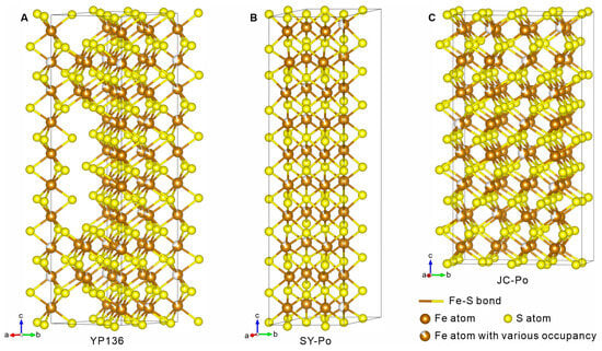
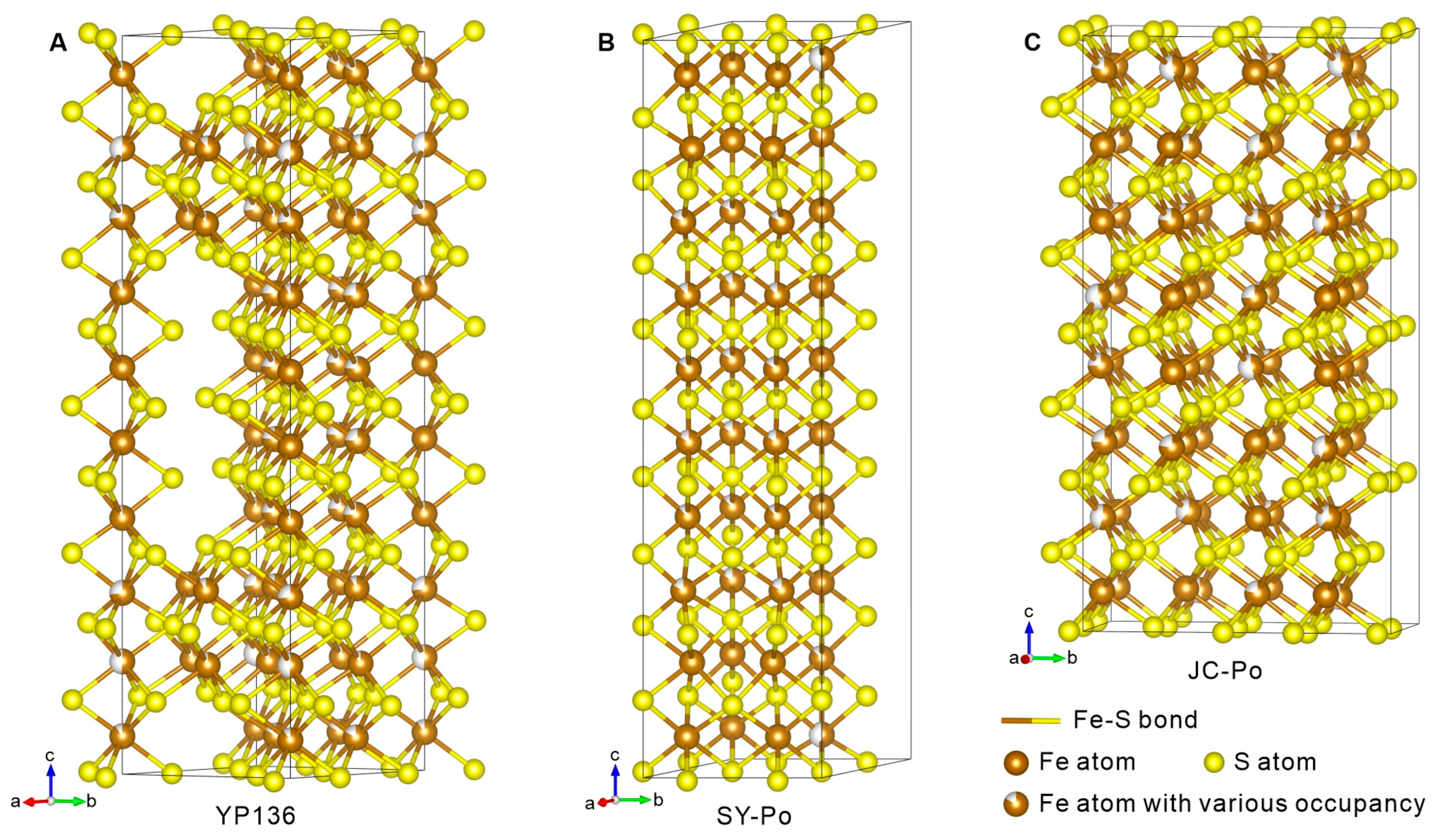
The unit-cell parameters of YP136, SY-Po, and JC-Po pyrrhotite RMs are calculated based on the XRD patterns and summarized in Table 1. YP136 pyrrhotite exhibits a hexagonal crystal system, crystallizing within the P space group (Figure 1A). SY-Po pyrrhotite has a similar crystal structure to YP136, also belonging to the hexagonal crystal system, and P space group (Figure 1B). JC-Po pyrrhotite is characterized by a monoclinic crystal system with a C2 space group (Figure 1C). Further details on data collection and structure refinement can be found in the Supplementary Crystallographic Information File (Supplementary CIF).
Table 1.
The detailed unit-cell parameters of the collected pyrrhotite reference materials (RMs).
Figure 1.
Unit-cell model showing the crystal structure of three pyrrhotite reference materials (RMs). (A) YP136 RM with a hexagonal system. (B) SY-Po RM with a hexagonal system. (C) JC-Po RM with a monoclinic system.
3.2. Chemical Compositions of Pyrrhotite RMs
Results of EPMA for representative grains of the three pyrrhotite RMs are summarized in Table 2, and the detailed compositions for each analysis are listed in Table S2. The concentration of Fe ranges from 58.76 to 60.45 wt.% of YP136, 59.48 to 60.95 wt.% of SY-Po, and 59.80 to 61.38 wt.% of JC-Po. The sulfur abundances are 38.61–39.88 wt.% of YP136, 38.16–40.38 wt.% of SY-Po, and 38.37–39.59 wt.% of JC-Po. These three pyrrhotite RMs contain some elements with low concentrations (<0.63 wt.% for As, Cu, Zn, Ni, and Co in YP136, <0.32 wt.% As, Pb, Cu, Zn, Ni, and Co in SY-Po, and <0.93 wt.% As, Pb, Cu, Zn, Ni, and Co in JC-Po).
Table 2.
The chemical composition (in average, wt.%) of three pyrrhotite RMs.
3.3. Sulfur Isotopes of Pyrite and Pyrrhotite RMs
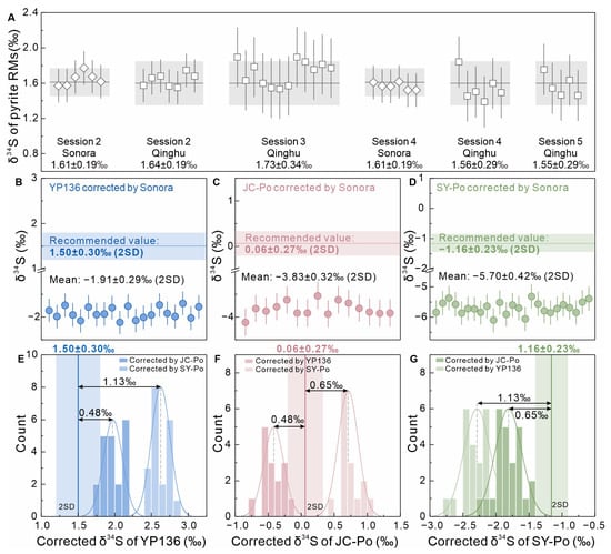
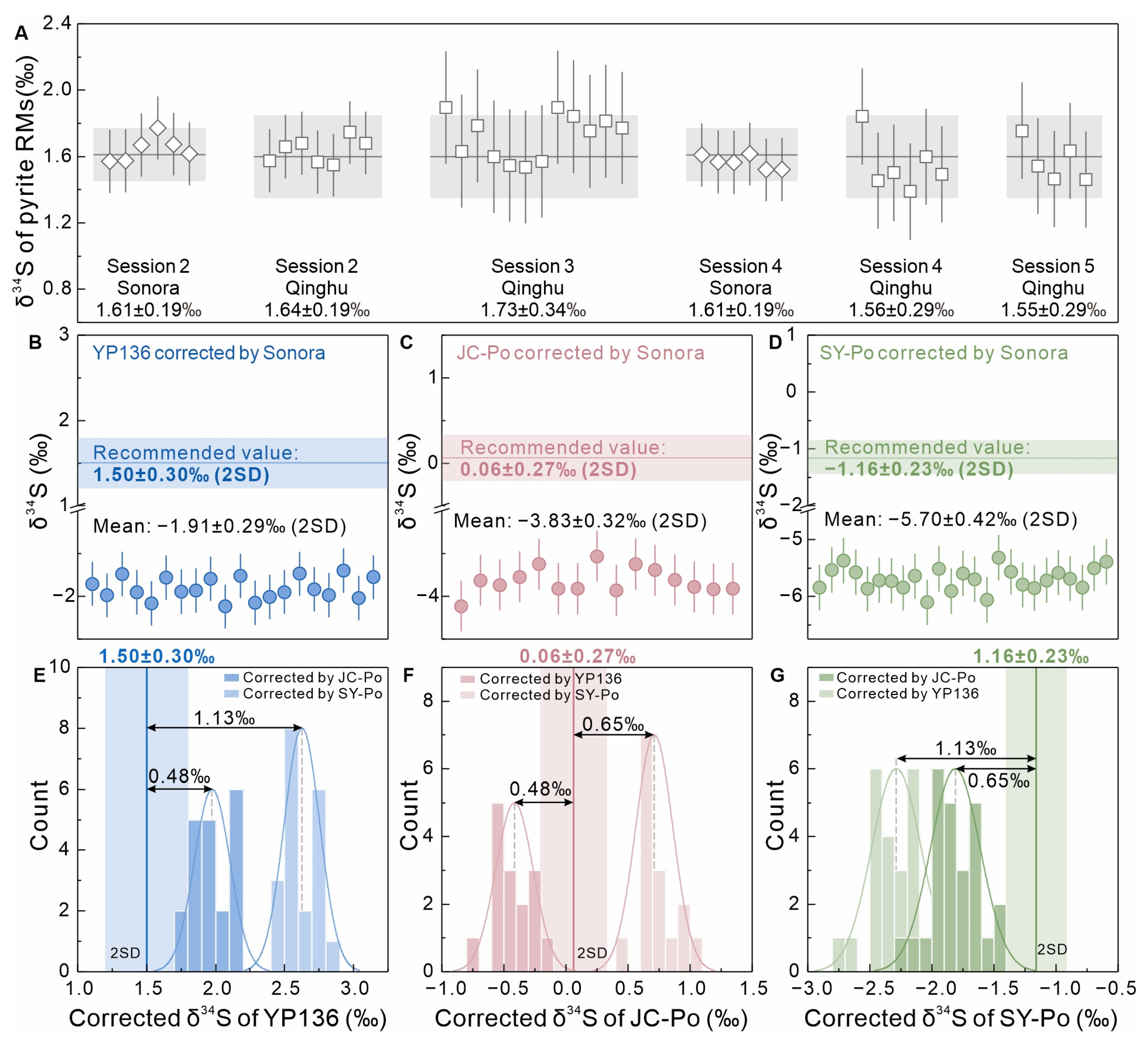
In this study, the Sonora RM tested in session 1 was employed to determine the IMF value (3.10 ± 0.13‰, 2SD), which was used to calibrate the bracketed pyrite RMs from different mounts in sessions 2–5 (Figure 2A, Table S3). The corrected δ34S values of Sonora and Qinghu, tested in session 2, were 1.61 ± 0.19‰ and 1.64 ± 0.19‰, respectively. In session 3, Qinghu RM yielded the corrected δ34S value of 1.76 ± 0.34‰. The corrected δ34S values of pyrite RMs tested in session 4 were 1.61 ± 0.19‰ for Sonora and 1.56 ± 0.29‰ for Qinghu. In session 5, the Qinghu RM had corrected δ34S values of 1.56 ± 0.29‰.
Figure 2.
(A) Reproducibility of δ34S measurements in two pyrite RMs (Sonora pyrite and Qinghu pyrite) through the entire experimental procedure. The dark grey solid line and light grey shaded band denote the recommended value and uncertainty (2SD) for the pyrite RMs, respectively. The recommended δ34S value of Sonora pyrite RM was obtained from [31]. The Qinghu pyrite is an in-house standard. (B–D) The calibrated results of YP136, JC-Po, and SY-Po pyrrhotite RMs are presented. The IMF value was determined by the Sonora pyrite RM analyzed in session 1. After correction, the results show a remarkable offset relative to the recommended values. The recommended δ34S values of pyrrhotite RMs were obtained from [24] for JC-Po, [25] for SY-Po, and [26] for YP136. (E–G) The calibrated δ34S values of YP136, JC-Po, and SY-Po, corrected using other pyrrhotite RMs, show a significant bias ranging from 0.48‰ to 1.13‰.
We employed the IMF value determined by Sonora RM tested in session 1 to normalize the measured δ34S values of three pyrrhotite RMs. The pyrite-normalized δ34S values yielded −1.91 ± 0.29‰ for YP136, −5.70 ± 0.42‰ for SY-Po, and −3.83 ± 0.32‰ for JC-Po (Figure 2B–D, Table 3 and Table S3). The results showed significant bias from the recommended values (bias values of +3.41‰ for YP136, +3.89‰ for JC-Po, and +4.54‰ for SY-Po).
Table 3.
The average of measured δ34S values, instrumental mass fractionation (IMF), and calibrated δ34S values of three pyrrhotite RMs (2SD, ‰).
We used each pyrrhotite RM as a standard to evaluate the other two pyrrhotite RMs, which were treated as unknown samples (Figure 2E,F, Table 3 and Table S3). When JC-Po and SY-Po were used as standards, YP136 yielded corrected δ34S values of 1.98 ± 0.29‰ and 2.63 ± 0.47‰, respectively. Conversely, when JC-Po and YP136 were used to correct SY-Po, the resulting δ34S values were −1.81 ± 0.42‰ and −2.29 ± 0.47‰, respectively. Similarly, JC-Po, when calibrated against YP136 and SY-Po, gave corrected δ34S values of −0.42 ± 0.39‰ and 0.71 ± 0.49‰, respectively.
4. Discussion
4.1. Test for Chemical Composition and Crystal Structure Effects in Pyrrhotite
The disparities in IMF values can be induced by the instrumental conditions, which are typically easy to calibrate [39]. However, the IMF variability during SIMS isotope analysis also depends upon matrix effects, which may arise from sample-dependent properties such as chemical composition, crystal structure, and crystallographic orientation [39,40,41,42,43].
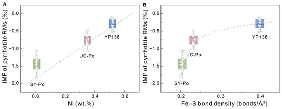
Under stable instrumental conditions, the three pyrrhotite RMs investigated in this study exhibited distinct IMF values of −1.43‰ for SY-Po, −0.78‰ for JC-Po, and −0.30‰ for YP136 (Table 3). Our study finds that these three RMs show distinctly different Ni concentrations (0.01 wt.% in SY-Po, 0.35 wt.% in JC-Po, and 0.52 wt.% in YP136). Although based on a limited dataset of three RMs, their IMF values exhibit a pronounced positive correlation with Ni content (Figure 3A), implying a potential correlation where differences in Ni concentration may influence the observed matrix-dependent IMF variability. This finding is consistent with prior SIMS studies on Ni-bearing sulfides, which attributed similar IMF variations to chemical composition variations during SIMS analysis [6,44]. Furthermore, the slight variation in the Fe/S atomic ratio among the three pyrrhotite RMs (0.87 for YP136 and SY-Po, and 0.89 for JC-Po; Table 2 and Table S2) could also be a contributing factor to the potential matrix effects [45].
Figure 3.
(A) The relationship between the Ni contents and corresponding IMF value of pyrrhotites. (B) The relationship between the Fe-S bond density and corresponding IMF value of pyrrhotites.
In addition, we find that crystal structure may represent another potential factor contributing to matrix effects during pyrrhotite SIMS sulfur isotope measurements. The SCXRD results reveal that YP136 and SY-Po crystallize in the hexagonal system, whereas JC-Po belongs to the monoclinic system. Although YP136 and SY-Po share hexagonal symmetry, they exhibit substantial differences in their unit-cell volumes and the number of Fe-S bonds per unit cell (Table 1). These differences result in marked variations in Fe-S bond densities, i.e., 0.20 bonds/Å3 for SY-Po, 0.23 bonds/Å3 for JC-Po, and 0.40 bonds/Å3 for YP136. The IMF value displays a nonlinear trend with Fe-S bond density (Figure 3B), which we interpret as the result of combined effects from crystal system transitions and variations in Fe-S bond density.
Therefore, these results suggest that both chemical composition and crystal structure play a combined role in matrix effects during pyrrhotite SIMS sulfur isotope analysis.
4.2. Multi-Standard Linear Regression Correction for Matrix Effect in Pyrrhotite
Variations in the IMF value of the three pyrrhotite RMs as a function of chemical composition and crystal structure are large even under unchanged instrumental conditions. This range is considerably wider than the natural δ34S variations in most fine-scale geologic processes [6]. Therefore, establishing an accurate method of calibrating measured isotope ratios for matrix effects is of significance for the application of pyrrhotite SIMS sulfur isotope analysis.
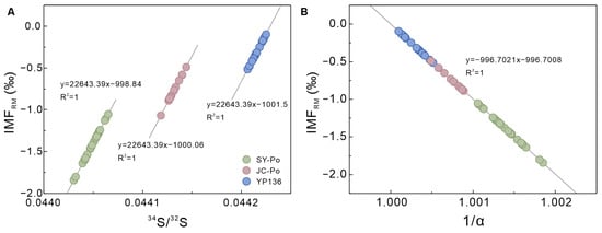
In this study, we implemented a multi-point linear normalization protocol based on the three characterized pyrrhotite RMs from a mathematical point of view. The relationship between the IMFRM and the measured isotope ratios can be described by a linear function of IMFRM = m × (34S/32S) Measured (RM) + n. The linear regressions for the three pyrrhotite RMs yielded a uniform slope but distinct intercepts (Figure 4A). The intercept values represent the recommended isotope value of the respective RMs [46]. To operationalize this function into a direct calibration scheme, the α factor (calculated by Equation (3)) is used to eliminate any drift induced by matrix effects, which normalizes the measured 34S/32S ratio of each spot to the corresponding recommended 34S/32S ratio of each RM. Mathematically, the linear function between IMF and 1/α indeed existed as IMFRM = k × (1/α) + b (Figure 4B), which provides an approach to correct the measured 34S/32S ratio. Combining Equations (1), (3) and (5), the corrected 34S/32S ratio is described as:
where k represents the slope of the linear function, and b denotes the y-intercept. The corrected 34S/32S ratio can be converted to the δ34SV-CDT notation via Equation (1).
Figure 4.
(A) Linear relationship of IMFRM vs. measured 34S/32S ratio. (B) Linear relationship of IMFRM vs. 1/α.
This method requires concurrent measurements of at least three pyrrhotite RMs under unchanged conditions. Rather than relying on a single, potentially mismatched reference material, this multi-standard calibration brackets the instrumental fractionation across the compositional and structural envelope defined by the three RMs. This method provides an operational and robust calibration strategy for SIMS sulfur isotope analyses of natural pyrrhotites when individual matrix-matching is technically difficult to achieve. However, the practical efficacy of this mathematically derived protocol in mitigating the matrix-induced biases must be verified using an independent sample with a known isotopic composition.
4.3. Applicability of the Multi-Standard Linear Regression Correction
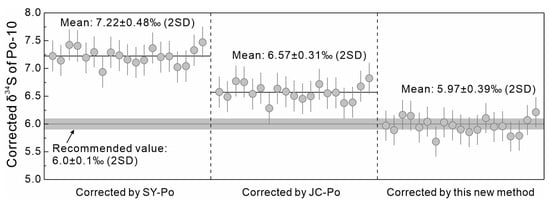
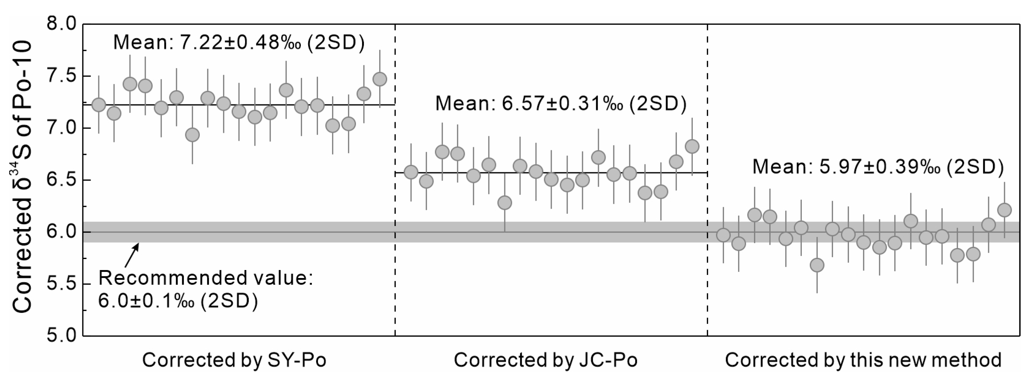
To test the proposed calibration method, the Po-10 pyrrhotite RM (δ34S = 6.0‰ ± 0.1‰ (2SD) [27]) was analyzed as an unknown sample during session 3. When the Po-10 pyrrhotite was calibrated against SY-Po and JC-Po individually, it yielded substantial offsets of 1.22‰ and 0.57‰, respectively (Figure 5). The pronounced δ34S bias directly reflects the severe matrix mismatch (likely arising from disparities in Ni content, Fe/S ratio, and crystal structure). We corrected measured 34S/32S ratios of Po-10 pyrrhotite using the multi-standard linear regression established by three pyrrhotite RMs (YP136, SY-Po, and JC-Po), yielding a calibrated δ34S value of 5.97‰ ± 0.39‰ (2SD, n = 19; Table 4, Figure 5), which is in good agreement with the recommended values.
Figure 5.
The corrected δ34S values of Po-10 pyrrhotite using SY-Po RM, JC-Po RM, and the new multi-standard linear regression method. The recommended δ34S value of Po-10 pyrrhotite RM was obtained from [27].
Table 4.
The calibrated δ34S value by the linear method of four pyrrhotite RMs and the bias relative to the recommended value.
To further validate the robustness of this method, we performed a cross-validation. Specifically, SY-Po, JC-Po, and YP136 were alternatively treated as unknowns and calibrated using the remaining three pyrrhotite RMs, respectively. All corrected values agree well with their recommended values, with the maximum bias being only 0.23‰ (Table 4 and Table S3). This marked improvement in the accuracy of the corrected δ34S value demonstrates that the protocol successfully mitigates the matrix effects, validating its practical reliability.
Mitigating matrix-induced biases is of pronounced importance, as numerous geological processes recorded by pyrrhotite result in only subtle sulfur isotope variations of only 1–2‰. For instance, in situ microanalyses of pyrrhotite have demonstrated that crustal assimilation in magmatic Ni-Cu deposits leads to δ34S shifts of ∼1.5‰ [2], volatile degassing in lunar basalts generates δ34S variations of 1.5–2.0‰ [15], and fluid evolution in hydrothermal systems produces core-to-rim variations of merely ∼1.4‰ [6,47]. Calibrating measured sulfur isotope data using individual standards would allow matrix effects (δ34S bias up to 1.22‰ in this study) to mask these fine-scale geochemical signals. Therefore, the multi-standard calibration protocol provides a reliable analytical tool for deciphering such subtle, fine-scale geological processes.
5. Conclusions
This study demonstrates that matrix effects during SIMS pyrrhotite S isotope analysis are jointly driven by chemical composition and crystal structure. Compositionally, IMF exhibits a pronounced positive correlation with Ni concentration (0.01–0.52 wt.%) and is influenced by minor variations in Fe/S atomic ratios. Crystallographically, the transitions between hexagonal and monoclinic systems, along with variations in Fe-S bond densities (0.20–0.40 bonds/Å3), induce significant matrix effects, with the IMF varying from −1.43‰ to −0.30‰ among three pyrrhotite RMs. To mitigate these matrix effects, we proposed a multi-standard linear regression correction from a mathematical perspective based on the linear function between IMF and (34S/32S) Recommended (RM)/(34S/32S) Measured (RM). This protocol circumvents the stringent requirement for strict matrix matching in both composition and structure between reference materials and unknowns. By employing the independent Po-10 pyrrhotite as an unknown, we obtained the corrected δ34S value (5.97 ± 0.39‰, 2SD; n = 19), which is in good agreement with the recommended value (6.0 ± 0.1‰, 2SD). We also calibrated Po-10 separately against SY-Po and JC-Po, which resulted in significant deviations of up to 1.22‰ and 0.57‰, respectively, from the recommended value. This comparison highlights the accuracy and reliability of the calibration method. Given that many subtle geological processes typically generate slight sulfur isotope shifts of only 1–2‰ in pyrrhotite, we strongly recommend employing this multi-standard calibration approach rather than individual-standard correction (yielding a δ34S bias up to 1.20‰ in this study) to acquire more accurate and reliable sulfur isotope compositions.
While our multi-standard linear regression model yields satisfactory corrections for these four pyrrhotite RMs (JC-Po, YP136, Po-10, and SY-Po), future work should aim to expand this framework by incorporating a wider variety of pyrrhotite RMs from multiple sources and broader compositional ranges. Expanding the RM suite will help to mitigate the influence of potential internal heterogeneities within individual reference materials and will more comprehensively address the complex matrix effects.
Supplementary Materials
The following supporting information can be downloaded at https://www.mdpi.com/article/10.3390/geosciences16110464/s1, Table S1: Detailed information on the collected reference materials (RMs) and the prepared SIMS mounts; Table S2: Major elements of YP136, SY-Po, and JC-Po pyrrhotite RMs measured by EPMA in wt.%; Table S3: Sulfur isotopic compositions of pyrite (Sonora and Qinghu) and pyrrhotite (YP136, SY-Po, JC-Po, and Po-10) measured by SIMS.
Author Contributions
L.C. and X.-H.L. conceived the presented idea and supervised the project. F.-D.Z. and X.-Y.L. carried out the EPMA and SIMS experiments. N.-Y.S. carried out the SCXRD experiments. Q.-L.L. contributed to the interpretation of the results. F.-D.Z. and L.C. wrote the manuscript. All authors provided critical feedback and helped shape the analysis and manuscript. All authors have read and agreed to the published version of the manuscript.
Funding
This study is supported by National Natural Science Foundation of China (42472128), National Key Research and Development Program of China (2023YFF0804200), Natural Science Foundation of Inner Mongolia Autonomous Region of China (2026QA001), the Key Research and Development Program of Gansu Province (24YFGA015), and the “Grassland Talents” Program of Inner Mongolia Autonomous Region.
Data Availability Statement
All data supporting this study are contained within the article and its Supplementary Materials.
Acknowledgments
We thank Hong-Xia Ma, Xiao-Xiao Ling, Ze-Xian Cui, Sheng He, Guo-Qiang Tang, Yu Liu, and Jiao Li for their assistance with sample preparation and measurement.
Conflicts of Interest
Author XiaoYan Liu and Lei Chen was employed by the company Mining Research Institute of Baotou Steel (Group) Corp. The remaining authors declare that the research was conducted in the absence of any commercial or financial relationships that could be construed as a potential conflict of interest.
References
- Wang, H.; Salveson, I. A review on the mineral chemistry of the non-stoichiometric iron sulphide, Fe1− x S (0 ≤ x ≤ 0.125): Polymorphs, phase relations and transitions, electronic and magnetic structures. Phase Transit. 2005, 78, 547–567. [Google Scholar] [CrossRef] [Scilit]
- Caruso, S.; Fiorentini, M.L.; Moroni, M.; Martin, L.A.J. Evidence of magmatic degassing in Archean komatiites: Insights from the Wannaway nickel-sulfide deposit, Western Australia. Earth Planet. Sci. Lett. 2017, 479, 252–262. [Google Scholar] [CrossRef] [Scilit]
- Day, J.M.D.; Paquet, M. Temporally limited late accretion after core formation in the Moon. Meteorit. Planet. Sci. 2021, 56, 683–699. [Google Scholar] [CrossRef] [Scilit]
- Tomkins, A.G.; Alkemade, S.L.; Nutku, S.E.; Stephen, N.R.; Finch, M.A.; Jeon, H. A small S-MIF signal in Martian regolith pyrite: Implications for the atmosphere. Geochim. Cosmochim. Acta 2020, 290, 59–75. [Google Scholar] [CrossRef] [Scilit]
- Stergiou, C.L.; Sakellaris, G.-A.; Melfos, V.; Voudouris, P.; Papadopoulou, L.; Kantiranis, N.; Skoupras, E. Mineralogy, Geochemistry and Fluid Inclusion Study of the Stibnite Vein-Type Mineralization at Rizana, Northern Greece. Geosciences 2023, 13, 61. [Google Scholar] [CrossRef] [Scilit]
- LaFlamme, C.; Martin, L.; Jeon, H.; Reddy, S.M.; Selvaraja, V.; Caruso, S.; Bui, T.H.; Roberts, M.P.; Voute, F.; Hagemann, S.; et al. In situ multiple sulfur isotope analysis by SIMS of pyrite, chalcopyrite, pyrrhotite, and pentlandite to refine magmatic ore genetic models. Chem. Geol. 2016, 444, 1–15. [Google Scholar] [CrossRef] [Scilit]
- Liu, X.Y.; Hao, J.L.; Li, R.Y.; He, Y.Y.; Tian, H.C.; Hu, S.; Li, J.; Gu, L.X.; Yang, W.; Lin, Y.T. Sulfur Isotopic Fractionation of the Youngest Chang’e-5 Basalts: Constraints on the Magma Degassing and Geochemical Features of the Mantle Source. Geophys. Res. Lett. 2022, 49, e2022GL099922. [Google Scholar] [CrossRef] [Scilit]
- Gomide, C.S.; Brod, J.A.; Junqueira-Brod, T.C.; Buhn, B.M.; Santos, R.V.; Barbosa, E.S.R.; Cordeiro, P.F.O.; Palmieri, M.; Grasso, C.B.; Torres, M.G. Sulfur isotopes from Brazilian alkaline carbonatite complexes. Chem. Geol. 2013, 341, 38–49. [Google Scholar] [CrossRef] [Scilit]
- Wang, Z.C.; Li, Y.H.; Zhang, W.; He, Q.; Pan, F.B.; Hu, Z.C.; Zong, K.Q.; Feng, Y.T.; Becker, H.; Day, J.M.D.; et al. Sulfide compositions of young Chang’e-5 basalts and implications for sulfur isotopes in lunar basalt sources. Geochim. Cosmochim. Acta 2024, 368, 168–184. [Google Scholar] [CrossRef] [Scilit]
- Milidragovic, D.; Nott, J.A.; Spence, D.W.; Schumann, D.; Scoates, J.S.; Nixon, G.T.; Stern, R.A. Sulfate recycling at subduction zones indicated by sulfur isotope systematics of Mesozoic ultramafic island arc cumulates in the North American Cordillera. Earth Planet. Sci. Lett. 2023, 620, 118337. [Google Scholar] [CrossRef] [Scilit]
- Walker, A.T.; Evans, K.A.; Kirkland, C.L.; Polito, P.A. Multiple modes of sulphur cycling within a mineralised orogen: A case study from the Fraser Zone, Western Australia. Lithos 2022, 408–409, 106536. [Google Scholar] [CrossRef] [Scilit]
- LaFlamme, C.; Sugiono, D.; Thébaud, N.; Caruso, S.; Fiorentini, M.; Selvaraja, V.; Jeon, H.; Voute, F.; Martin, L. Multiple sulfur isotopes monitor fluid evolution of an Archean orogenic gold deposit. Geochim. Cosmochim. Acta 2018, 222, 436–446. [Google Scholar] [CrossRef] [Scilit]
- LaFlamme, C.; Barré, G.; Rottier, B.; Cartigny, P.; Martin, L.; Beaudoin, G. In situ sulfur isotopes in texturally characterized pyrite, pyrrhotite and chalcopyrite by LA-ICPMS/MS for application in sulfide bearing systems. Chem. Geol. 2025, 691, 122946. [Google Scholar] [CrossRef] [Scilit]
- Coldebella, B.; LaFlamme, C.; Malta, I.S.; Guilmette, C.; Barré, G.; Beaudoin, G.; Martin, L.; Savard, D. The chemical and isotopic characterization of the pyrite to pyrrhotite desulfidation reaction across the metamorphic gradient of a metasedimentary basin. Geochim. Cosmochim. Acta 2025, 392, 119–136. [Google Scholar] [CrossRef] [Scilit]
- Li, Y.H.; Wang, Z.C.; Zhang, W.; Zong, K.Q.; She, Z.B.; He, Q.; Zheng, J.Q.; Li, T.Y.; Pan, F.B.; Chen, X.; et al. Sulfur isotopes from the lunar farside reveal global volatile loss following the giant impact. Nat. Commun. 2025, 16, 5780. [Google Scholar] [CrossRef] [Scilit] [PubMed]
- Kita, N.T.; Ushikubo, T.; Fu, B.; Valley, J.W. High precision SIMS oxygen isotope analysis and the effect of sample topography. Chem. Geol. 2009, 264, 43–57. [Google Scholar] [CrossRef] [Scilit]
- Huberty, J.M.; Kita, N.T.; Kozdon, R.; Heck, P.R.; Fournelle, J.H.; Spicuzza, M.J.; Xu, H.; Valley, J.W. Crystal orientation effects in δ18O for magnetite and hematite by SIMS. Chem. Geol. 2010, 276, 269–283. [Google Scholar] [CrossRef] [Scilit]
- Eiler, J.M.; Graham, C.; Valley, J.W. SIMS analysis of oxygen isotopes: Matrix effects in complex minerals and glasses. Chem. Geol. 1997, 138, 221–244. [Google Scholar] [CrossRef] [Scilit]
- Sun, J.; Wu, F.Y.; Sklyarov, E.; Sarkar, C.; Liu, C.Z.; Pearson, G.D.; Cheng, Z.; Mitchell, R.N. Matrix effects during in situ U-Pb dating of perovskite with variable crystal structure: Evidence from the Tazheran Massif, Russia. Chem. Geol. 2022, 589, 120685. [Google Scholar] [CrossRef] [Scilit]
- Fu, B.; Li, W.C.; Xia, Q.X. First matrix bias calibration for ion microprobe oxygen isotope analysis of Ti-bearing garnet. J. Anal. At. Spectrom. 2025, 40, 3576–3587. [Google Scholar] [CrossRef] [Scilit]
- Li, R.C.; Yu, J.W.; Yan, J.Y.; Wang, X.L.; Liao, J.L.; Guan, Y.; Tian, L.L.; Wang, C.M. Matrix effect during sulfur isotope determination of apatite using SIMS and the potential of using bioapatite to reconstruct the sulfur isotopic composition of seawater. J. Anal. At. Spectrom. 2025, 40, 2833–2840. [Google Scholar] [CrossRef] [Scilit]
- Śliwiński, M.G.; Kitajima, K.; Kozdon, R.; Spicuzza, M.J.; Fournelle, J.H.; Denny, A.; Valley, J.W. Secondary Ion Mass Spectrometry Bias on Isotope Ratios in Dolomite–Ankerite, Part I: δ18O Matrix Effects. Geostand. Geoanal. Res. 2016, 40, 157–172. [Google Scholar] [CrossRef] [Scilit]
- Harries, D.; Pollok, K.; Langenhorst, F. Translation interface modulation in NC-pyrrhotites: Direct imaging by TEM and a model toward understanding partially disordered structural states. Am. Mineral. 2011, 96, 716–731. [Google Scholar] [CrossRef] [Scilit]
- Chen, L.; Liu, Y.; Li, Y.; Li, Q.L.; Li, X.H. New potential pyrrhotite and pentlandite reference materials for sulfur and iron isotope microanalysis. J. Anal. At. Spectrom. 2021, 36, 1431–1440. [Google Scholar] [CrossRef] [Scilit]
- Liu, X.Y.; Chen, L.; Zhao, F.D.; Huang, F.; Li, Q.L.; Yu, H.M.; Li, X.H. A new potential synthetic pyrrhotite reference material for Fe–S isotope microanalysis. J. Anal. At. Spectrom. 2026, 41, 112–120. [Google Scholar] [CrossRef] [Scilit]
- Li, R.C.; Xia, X.P.; Yang, S.H.; Chen, H.Y.; Yang, Q. Off-Mount Calibration and One New Potential Pyrrhotite Reference Material for Sulfur Isotope Measurement by Secondary Ion Mass Spectrometry. Geostand. Geoanal. Res. 2019, 43, 177–187. [Google Scholar] [CrossRef] [Scilit]
- Gilbert, S.E.; Danyushevsky, L.V.; Rodemann, T.; Shimizu, N.; Gurenko, A.; Meffre, S.; Thomas, H.; Large, R.R.; Death, D. Optimisation of laser parameters for the analysis of sulphur isotopes in sulphide minerals by laser ablation ICP-MS. J. Anal. At. Spectrom. 2014, 29, 1042–1051. [Google Scholar] [CrossRef] [Scilit]
- Crowe, D.E.; Vaughan, R.G. Characterization and use of isotopically homogeneous standards for in situ laser microprobe analysis of 34S/32S ratios. Am. Mineral. 1996, 81, 187–193. [Google Scholar] [CrossRef] [Scilit]
- Kitayama, Y.; Thomassot, E.; O’Neil, J.; Wing, B.A. Sulfur- and oxygen-isotope constraints on the sedimentary history of apparent conglomerates from the Nuvvuagittuq Greenstone Belt (Nunavik, Québec). Earth Planet. Sci. Lett. 2012, 355–356, 271–282. [Google Scholar] [CrossRef] [Scilit]
- Molnár, F.; Mänttäri, I.; O’Brien, H.; Lahaye, Y.; Pakkanen, L.; Johanson, B.; Käpyaho, A.; Sorjonen-Ward, P.; Whitehouse, M.; Sakellaris, G. Boron, sulphur and copper isotope systematics in the orogenic gold deposits of the Archaean Hattu schist belt, eastern Finland. Ore Geol. Rev. 2016, 77, 133–162. [Google Scholar] [CrossRef] [Scilit]
- Farquhar, J.; Cliff, J.; Zerkle, A.L.; Kamyshny, A.; Poulton, S.W.; Claire, M.; Adams, D.; Harms, B. Pathways for Neoarchean pyrite formation constrained by mass-independent sulfur isotopes. Proc. Natl. Acad. Sci. USA 2013, 110, 17638–17643. [Google Scholar] [CrossRef] [Scilit] [PubMed]
- Sheldrick, G. A short history of SHELX. Acta Crystallogr., Sect. A: Found. Crystallogr. 2008, 64, 112–122. [Google Scholar] [CrossRef] [Scilit] [PubMed]
- Ding, T.; Valkiers, S.; Kipphardt, H.; De Bièvre, P.; Taylor, P.D.P.; Gonfiantini, R.; Krouse, R. Calibrated sulfur isotope abundance ratios of three IAEA sulfur isotope reference materials and V-CDT with a reassessment of the atomic weight of sulfur. Geochim. Cosmochim. Acta 2001, 65, 2433–2437. [Google Scholar] [CrossRef] [Scilit]
- Paterson, B.A.; Riciputi, L.R.; McSween, H.Y. A comparison of sulfur isotope ratio measurement using two ion microprobe techniques and application to analysis of troilite in ordinary chondrites. Geochim. Cosmochim. Acta 1997, 61, 601–609. [Google Scholar] [CrossRef] [Scilit]
- Hervig, R.L. Anomalous fractionation of sulfur isotopes during sputtering. Rapid Commun. Mass Spectrom. 2002, 16, 1774–1778. [Google Scholar] [CrossRef] [Scilit] [PubMed]
- Wagner, T.; Boyce, A.J.; Fallick, A.E. Laser combustion analysis of δ34S of sulfosalt minerals: Determination of the fractionation systematics and some crystal-chemical considerations. Geochim. Cosmochim. Acta 2002, 66, 2855–2863. [Google Scholar] [CrossRef] [Scilit]
- Crowe, D.E.; Valley, J.W.; Baker, K.L. Micro-analysis of sulfur-isotope ratios and zonation by laser microprobe. Geochim. Cosmochim. Acta 1990, 54, 2075–2092. [Google Scholar] [CrossRef] [Scilit]
- Kelley, S.P.; Fallick, A.E. High precision spatially resolved analysis of δ34S in sulphides using a laser extraction technique. Geochim. Cosmochim. Acta 1990, 54, 883–888. [Google Scholar] [CrossRef] [Scilit]
- Riciputi, L.R.; Paterson, B.A.; Ripperdan, R.L. Measurement of light stable isotope ratios by SIMS: Matrix effects for oxygen, carbon, and sulfur isotopes in minerals. Int. J. Mass Spectrom. 1998, 178, 81–112. [Google Scholar] [CrossRef] [Scilit]
- Gnaser, H.; Hutcheon, I.D. Velocity-dependent isotope fractionation in secondary-ion emission. Phys. Rev. B 1987, 35, 877–879. [Google Scholar] [CrossRef] [Scilit] [PubMed]
- Schroeer, J.M.; Rhodin, T.N.; Bradley, R.C. A quantum-mechanical model for the ionization and excitation of atoms during sputtering. Surf. Sci. 1973, 34, 571–580. [Google Scholar] [CrossRef] [Scilit]
- Gries, W.H.; Rüdenauer, F.G. A quantitative model for the interpretation of secondary ion mass spectra of dilute alloys. Int. J. Mass Spectrom. Ion. Phys. 1975, 18, 111–127. [Google Scholar] [CrossRef] [Scilit]
- Williams, P. The sputtering process and sputtered ion emission. Surf. Sci. 1979, 90, 588–634. [Google Scholar] [CrossRef] [Scilit]
- Liu, L.; Fu, B.; Ding, X.; Gu, J. The 34S/32S homogeneity of Chemical Vapor Transport (CVT) Reaction-synthesized pyrites. Am. Mineral. 2024, 109, 1024–1035. [Google Scholar] [CrossRef] [Scilit]
- Liu, L.; Mavrogenes, J.; Holden, P.; Ireland, T. Quadruple sulfur isotopic fractionation during pyrite desulfidation to pyrrhotite. Geochim. Cosmochim. Acta 2020, 273, 354–366. [Google Scholar] [CrossRef] [Scilit]
- Paul, D.; Skrzypek, G.; Fórizs, I. Normalization of measured stable isotopic compositions to isotope reference scales–a review. Rapid Commun. Mass Spectrom. 2007, 21, 3006–3014. [Google Scholar] [CrossRef] [Scilit] [PubMed]
- Yue, S.W.; Li, D.F.; Bagas, L.; Fang, J.; Lin, Z.W. Geology and Isotope Systematics of the Jianchaling Au Deposit, Shaanxi Province, China: Implications for Mineral Genesis. Geosciences 2018, 8, 120. [Google Scholar] [CrossRef] [Scilit]

Disclaimer/Publisher’s Note: The statements, opinions and data contained in all publications are solely those of the individual author(s) and contributor(s) and not of MDPI and/or the editor(s). MDPI and/or the editor(s) disclaim responsibility for any injury to people or property resulting from any ideas, methods, instructions or products referred to in the content. |
© 2026 by the authors. Licensee MDPI, Basel, Switzerland. This article is an open access article distributed under the terms and conditions of the Creative Commons Attribution (CC BY) license.