


Simple Summary
Some animals can procreate without a mate, producing offspring that are genetic copies of their mother. This unusual form of reproduction is called parthenogenesis. It is rare among vertebrates, but occurs in lizards of the genus Darevskia native to the Caucasus Mountains. In this group, several parthenogenetic species arose when pairs of different sexual species interbred in the distant past. One example is Darevskia armeniaca, whose parents are Darevskia mixta and Darevskia valentini. Although parthenogenesis in Darevskia draws constant attention, the genetic features that make it possible remain poorly understood. Moreover, reliable resources for genomic research on parthenogenesis in Darevskia remain unavailable. In this study, we read long stretches of DNA from D. mixta and D. valentini and, using a reference genome of a related species, assembled each genome into a full set of chromosomes. The comparison revealed substantial structural similarity between the two genome assemblies, along with several rearrangements. This first genomic resource can help in the study of how parthenogenesis arose in Darevskia lizards and identify the genes and chromosome regions involved in this reproductive switch. It also provides a foundation for tracing sex chromosome evolution and establishing relationships among these lizard groups.
Abstract
Darevskia lizards attract the attention of many biologists due to the emergence of parthenogenetic species within the genus. In this study, we present chromosome-level genome assemblies of Darevskia mixta (Méhely, 1909) and Darevskia valentini (Boettger, 1892), two species whose distant ancestors hybridized and formed a parthenogenetic taxon. Genomic DNA was sequenced using the PacBio platform. Primary genome assemblies were generated from ~60 Gb (D. mixta) and ~35 Gb (D. valentini) of the processed PacBio data using HiFiasm and reference-scaffolded. The final genome sequences of D. mixta and D. valentini each span ~1.4 Gb, comprising 19 (18 autosomes and the Z chromosome) and 20 (18 autosomes and the Z and W chromosomes) scaffolds, with N50 scaffold values of 91.1 and 97.5 Mb, respectively. Both genome sequences contain ~42% repeats and encode ~41 thousand predicted genes. The assemblies share major structural similarity, but feature a number of inversions and translocations relative to each other. The obtained assemblies represent the first quality platform for unraveling the genetic features associated with the emergence and maintenance of parthenogenesis in Darevskia lizards.
1. Introduction
Darevskia lizards are small reptiles that have garnered scientific interest due to their unusual reproductive biology [1,2]. The genus is remarkable for comprising both bisexual and parthenogenetic species, which currently coexist in nature [3]. The unisexual lineages arose from several independent interspecific hybridizations between bisexual parental lineages within the genus, so it is possible to describe the parthenospecies as F1 interspecific hybrids [4]. Remarkably, all parthenogenetic Darevskia species owe their origin to only four sexual taxa [5,6]. Despite years of research, it is still unknown which molecular mechanisms underlie this exceptional ability to hybridize with the emergence of parthenogenetic offsprings.
Unlike the molecular pathways, the cellular mechanism of Darevskia parthenogenetic taxa formation has been established recently. In oocytes of a mature viable F1 hybrid, premeiotic endoreplication doubles the genetic material [2,7]. Thus, homologous chromosomes can properly pair and undergo recombination. Finally, a diploid egg cell is formed. The knowledge gap in the understanding of this process concerns two main questions. What is the molecular trigger of endomeiotic replication? Why is the process restricted only to certain parental species? These questions can be addressed using omics data. Genome sequences are particularly useful tools for identifying candidate regions that may be associated with the transition to parthenogenesis.
The first genome assembly of a lizard was obtained for the green anole [8]. Then, many other reptilian genomes followed [9,10,11,12,13,14,15,16,17,18,19]. These early assemblies were still rather fragmented. However, the advancement of sequencing technologies led to improvement in the quality and quantity of the obtained data. Recently, telomere-to-telomere (T2T) or near-T2T reptile genome sequences were reported [20,21,22]. The high accuracy and completeness of the obtained assemblies provide a rich source for extensive and reliable studies relying on genomic data [23,24,25].
High-quality genome assemblies of parthenogenetic species, coupled with transcriptomic data or used in comparative genomic analysis, are brilliant sources of knowledge on the unisexual mode of reproduction, particularly in studies that include parent–hybrid (father/mother/progeny) comparisons [26,27,28]. Using full genome sequences, one can trace the evolution of different genomic regions. For instance, Lv et al. studied the synteny and specific repeat distribution of a flowerpot snake genome to divide chromosomes into three subgenomes and suggest their origin [29]. Another example is a study by Warren et al. [30]. The authors demonstrated a hybrid origin of the Amazon molly (Poecilia formosa) genome, comparing it with genome assemblies of its parental species. The hybrid nature of the species’ genome was also proven with the calculated heterozygosity rate. In addition, linkage analysis confirmed the “frozen” state of the P. formosa genome and the absence of recombination. Thus, high-quality genome sequences serve as a platform for subsequent extensive studies of parthenogenesis and its related aspects. In light of this fact, creating accurate genome assemblies is essential to enable further relevant research.
For Darevskia lizards, a comprehensive genomic resource is still absent. This gap significantly complicates further research on the aspects of parthenogenesis. In our previous study, we assembled the genome of Darevskia valentini (Boettger, 1892) [31], one of the parental species contributing to parthenogenetic taxa formation. Despite the extensive groundwork, the assembly remained fragmented, as it was based solely on 10x linked-reads. However, genome assemblies of both parthenogenetic lineages and the related parental species could provide valuable information on the reasons behind parthenogenesis emergence.
This study aimed to generate and compare high-quality genome assemblies of D. valentini and Darevskia mixta (Méhely, 1909). This pair of sexual taxa underwent possibly multiple hybridization events and produced Darevskia armeniaca, a parthenogenetic Darevskia species [4,6,32]. Thus, the genomes of D. mixta and D. valentini are a suitable model to identify molecular features that could have enabled parthenogenesis in their hybrid offspring. We present reference-scaffolded chromosome-level genome assemblies for both parental species and conduct a primary comparative analysis between them. Our work creates the first framework for further targeted investigation into genomic regions potentially associated with parthenogenesis in Darevskia.
2. Materials and Methods
2.1. Sample Collection and Surgical Procedures
To obtain DNA, blood samples (stored in EDTA buffer at −20 °C) of three male D. mixta individuals and one female D. valentini individual were used. D. valentini sample was collected in Armenia outside of protected areas [31]. D. mixta samples were collected from the Borjomi population in Georgia outside of the protected areas during expeditions in 1998–2000 and kindly provided to the Laboratory of Genome Organization (IGB RAS) by Ilya Darevsky (Zoological Institute of Russian Academy of Sciences). D. mixta and D. valentini are not listed under CITES (https://cites.org/, accessed on 1 May 2026).
2.2. DNA Extraction and Sequencing
Total genomic DNA was extracted from blood samples using the standard phenol–chloroform method [33] and dissolved in TE buffer (pH = 8.0). DNA purity was assessed via the measurement of light absorbance (A230/A260/A280 ratios) on a Genesys UV-Vis spectrophotometer (Thermo Fisher Scientific, Waltham, MA, USA). Only DNA samples with A260/A280 ratio ≥ 1.8 and A260/A230 ratio ≥ 2.0 were taken for sequencing and were analyzed for integrity with 0.5% agarose gel electrophoresis. For one D. mixta and one D. valentini sample, long-read library preparation and PacBio high-fidelity (HiFi) sequencing on PacBio Sequel II/IIE (Pacific Biosciences, Menlo Park, CA, USA) were performed by Novogene (Beijing, China). Three DNA samples of D. mixta, including the specimen sequenced on PacBio, were used to produce short-read libraries (150 PE, Illumina TruSeq DNA PCR Free Sample Prep protocol) and sequenced by Novogene (Beijing, China) on Illumina NovaSeq 6000 (Illumina, San Diego, CA, USA).
2.3. Data Processing and Genome Assembly
PacBio reads in fastq format were cleaned from the adapter sequences with Porechop 0.2.4 (https://github.com/rrwick/porechop, accessed on 1 May 2026) and filtered by length (a minimum of 10 kb) and quality (no lower than Q15) with Seqkit 2.8.2 seq [34]. Raw Illumina reads were processed with Trimmmomatic-0.38 [35] to remove adapter sequences and low-quality bases (TRAILING:28, SLIDINGWINDOW:4:20, MINLEN:50).
Primary genome assemblies were generated with HiFiasm 0.25.0-r726 [36] (for D. mixta and D. valentini) and Verkko v2.2.1 (only for D. mixta) [37] from doubled volumes of PacBio reads. Contamination control was performed with Kraken 2.1.6 [38]. To extract taxids (taxonomic IDs) matching other species than bacteria, grep (“-v”, “-P” options) and awk commands were used. Contigs corresponding to Darevskia were extracted from the Kraken output according to the filtered taxon IDs. For D. mixta, the cleaned assembly was polished with trimmed Illumina reads using Pilon 1.24 [39] to correct possible sequence errors. QUAST v5.0.2 [40], Seqkit 2.8.2, and BUSCO 5.8.3 (sauropsida_odb10) [41] were used for assembly quality assessment.
To identify the positions of the assembled contigs in a reference assembly, the Lacerta agilis L. genome sequence (NCBI Genome, GCA_009819535.1) was used. This species is one of the closest lacertid taxa to Darevskia. Whole-genome alignment was performed with LAST 1595 (https://gitlab.com/mcfrith/last, accessed on 1 May 2026) (with options: “-v”, “--sort2 = 3”, “--strands2 = 1”, “--maxseqs = 100” or “--maxseqs = 1000”). Scaffolding was performed with RagTag v2.1.0 [42] using the L. agilis genome assembly and manually curated according to the output alignment information of both LAST and RagTag. The final contig start coordinates upon assembly scaffolding, as well as their sizes, positions, and orientations, were taken from the “.agp” file generated by RagTag. In the final scaffold headers, the prefix “NC_” was replaced with “scaffold_”. Telomere sequences were searched with Tidk 0.2.41 (repeat window of 1000) (https://github.com/tolkit/telomeric-identifier, accessed on 1 May 2026).
The quality of the final assembly of D. mixta was additionally judged by the consensus quality (QV) score and the percentage of the aligned Illumina data. QV estimation was conducted with Merqury v1.3 [43]. A k-mer database (k = 31) was built using Meryl and the trimmed Illumina reads. The output was passed to merqury.sh along with the final assembly. Samtools 1.10 [44] flagstat was used to calculate the total number of mapped Illumina reads. The total number of bases covered at least once was calculated from bedtools v2.30.0–8-g2fd5181a output piped to an awk command (bedtools genomecov -ibam … -bga 2>/dev/null | awk ‘$4>0 {c+=$3-$2} END {print c+0}’).
Databases of repeat sequences were built with RepeatModeler 2.0.7 (‘-LTRStruct’ option was enabled) [45] and repeats were masked with RepeatMasker 4.1.7 (https://www.repeatmasker.org/, accessed on 1 May 2026) [46]. Gene annotations of the masked assemblies were created with Braker 3.0.8 (‘--softmasking’, ‘--gff3’, ‘--species=lacertidae’) [47], and statistics of the obtained annotation files were calculated with AGAT 1.4.2 [48] and the “grep -c” command. To support the predicted gene models of the D. valentini annotation, raw read counts were obtained with PPLine [49] using the RNA-Seq data from Ochkalova et al. [31] (NCBI SRA: SRX12402759, SRX12751488, and SRX15406814). Counts per million (CPM) values were calculated as a raw count divided by the total library size, multiplied by one million.
For single-nucleotide polymorphism (SNP) detection, PPLine [49] was run on the final D. mixta genome assembly and Illumina reads of three specimens of this species. The number of SNPs was calculated from the output alignments. Due to the low genome coverage with Illumina reads, a low alternate allele fraction threshold was used in freebayes for SNP detection to retain true heterozygous sites, at the cost of potentially including false-positive calls. Alignment files (in bam format) were passed to freebayes v1.3.1-dirty [50] (the following options were used: “--ploidy 2”, “--min-mapping-quality 30”, “--min-base-quality 20”, “--min-alternate-count 2”, “--min-alternate-fraction 0.05”, “--min-repeat-entropy 1”, “--use-best-n-alleles 4”), and the output files were passed to bcftools 1.9 [44] for further filtration (“-i ‘INFO/AF > 0.3 && QUAL>30 && INFO/DP>5 && INFO/DP<30 && INFO/MQM>40 && (strlen(REF)=1 && strlen(ALT)=1)’”, “-m2”, “-M2” options were enabled). To extract SNPs corresponding to the coding regions, bcftools view was run on the obtained sorted and indexed vcf files, using the “-R” option and a bed file generated from the D. mixta genome assembly annotation (created in this study). To check the Ts/Tv ratio (transition/transversion), the resulting vcf files were passed to bcftools stats with the output piped to an awk command (“awk ‘/^TSTV/ {print “ts:”, $3, “tv:”, $4, “ratio:”, $5}’”).
For runs of homozygosity (ROH) analysis, variant calling was repeated with freebayes using the same options as for SNP detection, except for a higher alternate allele fraction threshold (“--min-alternate-fraction 0.1”), which reduced the number of false heterozygous calls, and without the repeat entropy filter, which was omitted to retain true heterozygous sites within repetitive regions. The resulting vcf files were filtered with bcftools (“-i ‘QUAL>30 && INFO/DP>5 && INFO/DP<30 && INFO/MQM>40 && (strlen(REF)=1 && strlen(ALT)=1)’”, “-m2”, “-M2”). Filtering by allele frequency was excluded to avoid introducing bias in ROH estimates. The filtered files were processed with the bcftools roh module (“-G30”, “--AF-dflt 0.4”). The genomic inbreeding coefficient was calculated for each individual as the total length of autosomal ROH segments divided by the total autosomal assembly length, i.e., the assembly size (1.4 Gb) excluding the Z chromosome (45.3 Mb). Assembly gaps (runs of “N”) were retained in the length calculations. However, their contribution to the autosomal length was negligible.
The genome-wide heterozygosity rate was estimated using the same filtered vcf files that were obtained for the ROH analysis. The number of heterozygous sites was calculated with bcftools view (“-H”, “-i ‘GT=“het”’”). The Z chromosome calls were excluded from the counts. Heterozygosity was calculated as the number of heterozygous autosomal sites divided by the total autosomal assembly length.
To perform synteny analysis, syri 1.7.1 (https://github.com/schneebergerlab/syri, accessed on 1 May 2026) [51] was used. The final assembly of D. mixta was aligned to the final assembly of D. valentini using minimap2 (‘-ax asm5’, ‘--eqx’) [52]. The alignment was sorted with samtools 1.10 [53], output to bam files, and indexed. Structural variations were detected with syri, and plotting was performed with plotsr 1.1.0 (https://github.com/schneebergerlab/plotsr, accessed on 1 May 2026) [54].
3. Results
Nearly 99.94% of PacBio sequencing reads of D. mixta and 99.91% of those of D. valentini exceeded the length of 10 kb and had a minimum quality of Q15. Using D. mixta sequencing data, we developed the protocol for genome assembly and applied it to D. valentini. Draft genomes of D. mixta and D. valentini were assembled from 61.3 Gb and 34.8 Gb of filtered PacBio HiFi reads, with average read lengths of 17.1 kb and 18.0 kb, corresponding to ~40× and ~25× genome coverage (assuming a genome size of ~1.4 Gb), respectively. Primary assemblies of D. mixta were generated using HiFiasm and Verkko. Assembly quality was assessed with BUSCO (sauropsida_odb10 dataset) and QUAST (Table 1).
Table 1.
QUAST and BUSCO statistics for D. mixta genome assemblies.
The D. mixta genome assembly generated with HiFiasm consisted of 555 contigs and had a size of 1.48 Gb, an L50 of 18, and an N50 of 28.07 Mb. Verkko demonstrated markedly poorer QUAST statistics. The tool assembled a genome of nearly 2.3-fold greater size. It comprised 50,700 contigs and had an N50 of only 0.15 Mb. In addition, the ratio of duplicated orthologues was strikingly high (73.5%), additionally pointing to the presence of several haplotypes. Therefore, only the assembly by HiFiasm was taken for further improvement and analysis.
On ranging contigs with QUAST (Icarus), we observed that nearly 80% of all sequences had a size smaller than 1 Mb. To investigate the origin of these small fragments, we checked the obtained genome assembly for contamination with bacterial sequences using Kraken. Any contigs matching contaminating taxa were removed. QUAST and BUSCO statistics of the cleaned assembly remained largely unchanged (Table 1) except for the number of contigs, which reduced almost by half. This indicated a minor impact of possible contamination on the genome assembly’s quality. To improve the accuracy of the cleaned assembly, we polished it with preprocessed Illumina reads using Pilon (~16× coverage with the trimmed reads).
Then, we analyzed the contribution of contigs to the size of the resulting assembly in the Icarus viewer. We observed that nearly half of all contigs had a small size (smaller than 300 kb) and occupied a negligible proportion of the assembly. Such sequences could represent poorly assembled regions of low coverage and may interfere with further analysis and genome annotation. Therefore, we filtered out contigs smaller than 300 kb with Seqkit. The resulting assembly had a size of 1.40 Gb, an N50 of 30.30 Mb, and an L50 of 17, and consisted of 104 contigs (Table 1).
The D. valentini genome sequence was assembled using HiFiasm and filtered from contigs smaller than 300 kb. Since biological contamination was absent according to the Kraken output, filtration from non-target sequences was omitted. QUAST and BUSCO statistics were calculated (Table 2). The resulting genome assembly of D. valentini comprised 306 contigs, totaled 1.44 Gb in size, and had an N50 of 11.11 Mb and an L50 of 39 (Table 2).
Table 2.
QUAST and BUSCO statistics for D. valentini genome assemblies.
The obtained contig-level genome assemblies of D. mixta and D. valentini were aligned to the chromosome-level genome sequence of a close taxon—Lacerta agilis L. (NCBI Genome, GCA_009819535.1) (Supplementary Figure S1). The alignment of both assemblies showed a high structural similarity to the reference. To analyze chromosomal positions of contigs from both Darevskia genome assemblies, we used the obtained alignment information.
Sex chromosomes are potential sources of structural rearrangements associated with parthenogenesis [2,6,7,55,56]. Therefore, these regions deserve a detailed analysis. Darevskia lizards possess a ZW system of sex determination, with homogametic males and heterogametic females [55]. In our study, we used DNA samples of a male D. mixta and a female D. valentini and obtained genome assemblies of the merged haplotypes within each genome. In the D. mixta assembly, we observed five contigs corresponding to the Z sex chromosome of L. agilis (NC_046331.1)—ptg000011l_pilon, ptg000052l_pilon, ptg000077l_pilon, ptg000046l_pilon, and ptg000137l_pilon (Supplementary Table S1 and Figure S1a). The alignment showed a high structural similarity of male chromosomes between the analyzed species. In the assembly of D. valentini, the sequence of the Z chromosome consisted of many more contigs (Supplementary Table S2 and Figure S1b) than that of D. mixta (Supplementary Table S1 and Figure S1a). This feature can be explained by the lower coverage of the D. valentini genome with sequencing data (~25× for D. valentini vs. ~40× for D. mixta). The small W chromosome of D. valentini was represented by two contigs—ptg000163l and ptg000194l (Supplementary Table S2 and Figure S1b). Its sequence was highly repetitive.
In general, only small rearrangements were revealed in the contigs from the assembled Darevskia genomes compared to the L. agilis assembly. For example, we observed an inversion in the ptg000009l_pilon contig (chromosome 3 in the genome of L. agilis—NC_046314.1) in the D. mixta assembly (Supplementary Figure S1a) and an inversion in the ptg000094l contig (chromosome 5 in the genome of L. agilis—NC_046316.1) in the D. valentini assembly (Supplementary Figure S1b).
In the analysis of contig positions, we scaffolded the assemblies of D. mixta and D. valentini with RagTag using the genome sequence of L. agilis as a reference due to karyotype conservation in the family Lacertidae [7,57]. Thus, the chromosome-scale structure reported here is inferred rather than empirically determined. The distribution of the telomeric repeat was analyzed across the D. mixta and D. valentini scaffolds to investigate assembly completeness and accuracy. In the scaffolded chromosome 1 of D. mixta (corresponding to chromosome 1 of L. agilis—NC_046312.1), the sequence immediately following the first integrated 6.6 Mb contig (ptg000087l_pilon) began with a telomeric repeat (ptg000006l_pilon). Given that telomeric repeats mark chromosomal ends, we partitioned this leading contig from the D. mixta chromosome 1 scaffold. Thus, for D. mixta, we obtained an assembly of 19 scaffolded chromosomes (and nine unplaced contigs) with an N50 of 91.1 Mb and an L50 of 7. In the obtained assembly, 14 of 19 scaffolds had a high telomeric repeat occurrence at either one (12 scaffolds) or both ends (two scaffolds). The assembly of D. valentini consisted of 20 scaffolds and 18 unplaced contigs with an N50 of 97.5 Mb. Eight of these scaffolds had assembled telomeric repeat sequences at both ends. In addition to the obtained Darevskia genome assemblies, we analyzed telomeric repeat frequency across the chromosomes of the reference species—L. agilis. We observed that the chromosomes of Darevskia species contained more assembled telomeric repeats than those of L. agilis. This fact pointed at a better resolution of chromosome end sequences in the Darevskia assemblies than in the assembly of L. agilis.
The final D. mixta and D. valentini assemblies demonstrated high structural similarity both to each other and to the L. agilis assembly, but also showed a number of inversions and translocations (Supplementary Figure S2). The observed structural rearrangements between the genome assemblies of Lacertidae species were already present at the contig level (Supplementary Figure S1). Therefore, none of them should be attributed to possible assembly scaffolding missteps.
For the additional quality assessment of the final D. mixta assembly, Illumina reads were mapped. On alignment with BWA, 99.99% of the paired-end reads successfully mapped to the genome assembly, and 99.20% of the assembly bases were covered. The alignment results indicated a high quality of the obtained genome sequence. In addition to the alignment tests, the same Illumina data (~16× genome coverage) was used to estimate assembly accuracy with Merqury. The tool calculates the QV score from the k-mer occurrence in both read and assembly data. Our analysis demonstrated that the obtained genome sequence of D. mixta had a QV of 34.1 (may be underestimated due to low genome coverage), corresponding to an error rate of 0.038%. This error rate reflects quite high sequence accuracy and supports the idea of the overall quality of the assembly. Thus, we developed a systematic step-by-step workflow (Supplementary Figure S3) and generated a high-quality genomic resource for a pair of Darevskia species that formed a parthenogenetic taxon.
The final D. mixta and D. valentini genome assemblies were subjected to repeat masking and structural annotation. Running RepeatModeler and RepeatMasker resulted in 41.86% and 42.11% of masked sequences, respectively. Retroelements accounted for 24.36% and 23.64% of the assembly sizes, and long interspersed nuclear elements (LINEs) constituted 16.91% and 16.50% of the total genome sequences, respectively. DNA transposons occupied 6.12% and 6.46% and unclassified repeats accounted for 9.81% and 10.34% of the D. mixta and D. valentini assemblies, respectively. Gene sequences were predicted with Braker3. The resulting annotation of D. mixta comprised 39,886 genes and 62,124 transcripts. For D. valentini, 41,692 genes and 64,333 transcripts were predicted. Such high gene numbers are likely attributable to ab initio gene prediction alone with Braker3, without RNA-Seq evidence and the manual curation step that removes redundant models. To provide expression support for the predicted gene models, we mapped the transcriptomic data from Ochkalova et al. [31], obtained from kidney, liver, brain, and ovary tissues of D. valentini, to its genome assembly created in this study. Of the 41,692 genes, 12,746 had CPM values above 0.5, with 85.2% of reads uniquely mapped.
Upon assembly annotation, we conducted a search for polymorphisms using Illumina whole-genome sequencing data of three D. mixta individuals (~15× genome coverage on average) to roughly estimate the D. mixta population diversity. Given the low coverage, variant calling thresholds were adjusted during the calculation of the SNP number, heterozygosity rate, and genomic inbreeding coefficient to balance the retention of true heterozygous sites against the inclusion of false-positive calls. One of the analyzed D. mixta samples was the same one that was used for the genome assembly. Analysis of its Illumina data revealed about 97 thousand SNPs (heterozygous or alternative homozygous variants with a VAF (variant allele frequency) of more than 0.3 and a minimum coverage of 5) across the coding regions and 2.6 million SNPs across the whole genome. The observed numbers of alternative and heterozygous variants are plausible, as the created genome assembly represents a single merged haplotype of the diploid D. mixta. The alternative and heterozygous SNPs arose primarily from aligning the Illumina data of both haplotypes to this consensus assembly. Analysis of the coding regions of the other two D. mixta individuals revealed about 217 and 117 thousand SNPs. At the whole-genome level, we calculated about 6.0 and 3.2 million SNPs for these samples, respectively. To ensure the correctness of the obtained values, transition/transversion ratios were checked. For the whole-genome data of all analyzed samples, the ratio exceeded 1.95, indicating a high accuracy of the analysis. To complement the SNP-based estimates of population diversity, we calculated genome-wide heterozygosity for each sample in an independent analysis. The heterozygosity rate of the individual used for the genome assembly was 0.0019, while the rates for the other two individuals were 0.0023 and 0.0018. To estimate the level of inbreeding, runs of homozygosity (ROH) were analyzed for each individual in the sampled D. mixta population. The genomic inbreeding coefficient (FROH) was 0.016 for the individual used for the genome assembly, and 0.296 and 0.178 for the other two individuals. Total autosomal ROH lengths were 22.0 Mb, 401.1 Mb, and 241.3 Mb, respectively. Since ROH detection is limited at low coverage, the calculated values may be underestimated.
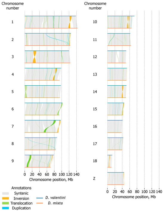
Analyzing the degree of similarity between the assembled genomes of D. mixta and D. valentini is particularly useful in light of possible meiosis disruptions between the genetic material of these species during the emergence of their parthenogenetic offspring. To search for the differences between the D. mixta and D. valentini assemblies, we performed whole-genome alignment with LAST (Supplementary Figure S4) and conducted synteny analysis (Figure 1).
Figure 1.
Synteny between homologous chromosomes (vertical axis) of D. mixta (blue horizontal lines) and D. valentini (orange horizontal lines). Syntenic regions are connected with gray curved lines. Structural rearrangements are represented as curved arcs: orange arcs—inversions; green arcs—translocations; blue arcs—duplications.
The assemblies shared high structural similarity, except for several rearrangements. For instance, a number of inversions are evident in the aligned chromosomes, e.g., the ones located in chromosomes 3 and 14 (Supplementary Figure S4). Synteny analysis between homologous chromosomes in the assembled Darevskia genomes additionally confirmed large-scale structural conservation. However, multiple inversions and translocations were also detected (Figure 1). Both LAST alignment and synteny analysis revealed the absence of large intrachromosomal genome duplications. The Z chromosomes of the analyzed species were highly syntenic, despite several small inversions.
4. Discussion
Parthenogenesis is a rare phenomenon in vertebrate animals [58]. Among amniotes, unisexuality was first identified within the group of Caucasian rock lizards [59]. Within the genus Darevskia, parthenogenetic lineages arose several times upon interspecific hybridization between sexual species [32,60]. Until now, the Darevskia lizard species have represented one of the most well-studied reptile systems in biogeography, ecology, and population and molecular genetics, but not at the whole-genome level [1]. Of the 40 described Darevskia species, seven reproduce parthenogenetically and share the same history of parthenogenesis emergence [1,61]. For two parthenogenetic species, D. armeniaca and D. dahli, D. mixta is an exclusive maternal ancestor; for five parthenospecies, D. armeniaca, D. unisexualis, D. uzzelli, D. sapphirina, and D. bendimahiensis, D. valentini is an exclusive paternal ancestor [6]. In this study, we focused on D. mixta and D. valentini—the parental species for unisexual D. armeniaca. According to Murphy et al. [6], one of the possible reasons limiting the ability of Darevskia species to form parthenogenetic hybrids is associated with the sex chromosome system. Therefore, D. mixta and D. valentini represent the first Darevskia model system to study the origins of parthenogenesis at the genomic level. To uncover the molecular aspects of parthenogenesis, genome assemblies of both parthenogenetic species and their corresponding parents are essential. The comparative analysis between the obtained assemblies can reveal genomic features underlying the parthenogenetic mode of reproduction in Darevskia. We assembled the genome sequences of D. mixta and D. valentini and performed an initial synteny analysis between them. Scaffolding to the chromosome level was performed using the L. agilis genome assembly as a reference. The choice of L. agilis is supported by the highly conserved karyotype within Lacertidae. Srikulnath et al. documented linkage homology between the L. agilis genome and other squamates [57], while Spangenberg et al. directly demonstrated chromosomal homology between parthenogenetic Darevskia species and their parental lineages using cytogenetic methods [62]. Given this karyotype conservation, contigs of D. mixta and D. valentini were assigned to chromosomes of L. agilis. However, since the chromosome-level assemblies presented in this study are inferred from conserved synteny, contig placement should be empirically validated in future chromatin conformation studies. Therefore, the genomes of D. mixta and D. valentini constructed here should be considered reference-scaffolded chromosome-level assemblies.
The final genome sequences of D. mixta and D. valentini have a size of ~1.4 Gb. The assembly of male D. mixta comprises 19 scaffolds (18 autosomes and the Z chromosome) and nine unplaced contigs, with an N50 of 91.1 Mb and an L50 of 7. For the final assembly of female D. valentini, we achieved generally similar statistics—20 scaffolds (18 autosomes, the Z and W chromosomes) and 18 unplaced contigs, an N50 of 97.5 Mb, and an L50 of 7. Thus, a complete haploid set of chromosomes was obtained for both genomes.
The generated genome assemblies of D. mixta and D. valentini were searched for repetitive sequences. Reptile genomes are characterized by the high content of repeats, including transposable elements (TEs) [63]. In the assemblies of both Darevskia species, ~42% of the sequence was occupied by repeats. The observed values fall within the range of multicopy sequence percentages described in literature data for reptiles [64,65]. Since Squamata exhibit high lineage specificity of TEs [66,67,68], future deeper analysis of repeats in the genomes of D. mixta and D. valentini and their parthenogenetic progenitor may be useful for tracing the emergence of the formed hybrid genome [25]. However, the current lineages of the chosen Darevskia species might have significantly diverged from their ancestral populations that participated in hybridization events. Therefore, the general genome evolution of D. mixta and D. valentini should also be considered in such an analysis.
In addition to repeat search, gene annotation was performed for the assembled Darevskia genomes. Both assemblies contained ~41 thousand genes and ~63 thousand transcripts. These numbers correspond to literature data on reptile gene content predicted with an ab initio method. In Lacerta viridis and Lacerta bilineata, 38–55 thousand coding sequences and predicted proteins were annotated using ab initio methods and refined to a final number of 22,100–22,500 gene models, each supported with RNA-Seq data during the curation step [69]. A similar pattern was observed for other closely related species Podarcis raffoneii and Podarcis muralis, where ab initio predictions were combined with transcript and protein evidence to produce final annotations of approximately 24,600 and 20,900 protein-coding genes, respectively [70,71]. D. mixta and D. valentini annotations are based on raw ab initio predictions without the post-processing steps that retain only transcript-supported genes and merge the redundant models. To partially address this, we mapped the kidney, liver, brain, and ovary transcriptomic data from Ochkalova et al. [31] to the annotated D. valentini assembly. About 13 thousand predicted genes had CPM values above 0.5, while ~85% of transcriptomic reads were mapped uniquely. Thus, about a third of the D. valentini annotation was supported by transcriptomic reads. However, since transcriptomic data covered only four tissue types, the fraction of supported genes is likely an underestimate. No transcriptomic data currently exist for D. mixta, so its annotation could not be assessed in this way. Nevertheless, the obtained annotated chromosome-level assemblies already provide a useful resource. While the coding sequence count requires refinement, the underlying gene models contain protein-coding sequences and can be processed in further research on Darevskia.
The chromosomal structure of the assembled D. mixta and D. valentini genomes was investigated. Most species from the family Lacertidae have 38 acrocentric chromosomes (ploidy of 2n = 38) and a ZZ/ZW system of sex determination, with heterogametic females [72,73,74]. The W chromosome in lacertid lizards is highly degenerated [75]. A large ratio of its sequence belongs to repetitive regions. Repeat accumulation is clearly visible in the plot of alignment between the female D. valentini and L. agilis assemblies (chromosome W—NC_046330.1, L. agilis) (Supplementary Figure S2). Meanwhile, the Z sex chromosomes of the analyzed Lacertidae species are structurally conserved and show a similar repeat content to that of the autosomes. The identified striking differences between the structures of the Z and W chromosomes highlight the degenerative state of the latter, confirming the previous observations on lacertid species [75].
Not only sex chromosomes have a significant impact on animal reproduction, but the whole karyotype structure [76]. Darevskia hybrid lineages inherited complete haploid chromosome sets from their ancestral species [77]. Presumably, it is the incompatibility of the parental karyotypes that led to the switch to parthenogenesis in mature hybrids [55]. The meiosis of a cell containing two divergent genomes could undergo a number of deviations. Therefore, parthenogenesis might have evolved as a mechanism to prevent hybrid sterility. To assess genomic differences between the chromosomal structure of D. mixta and D. valentini, we referred to the obtained whole-genome alignment data (Supplementary Figure S4). The alignment plot demonstrated high structural resemblance between both genome assemblies. Synteny analysis also showed high collinearity between the corresponding chromosomes of the two Darevskia species (Figure 1). Our results are concordant with genomic analyses of Aspidoscelis reptiles that gave rise to the hybrid unisexual lineages [25]. In addition, whole-genome alignment data confirmed the similarity between the Darevskia assemblies and that of L. agilis (Supplementary Figures S2 and S4).
Despite structural conservation, all three genome assemblies bore a number of variations. Synteny analysis revealed multiple inversions and translocations between the homologous chromosomes. Genomic variations were identified in other parthenogenetic species [29,30,78,79], where hybridization between divergent but karyotypically similar species precedes the switch to unisexuality. Therefore, we assume that the observed divergent regions between the genome assemblies of Darevskia species might be associated with meiosis disruption in their hybrids. Comparison with a quality genome sequence of the unisexual progenitor may provide even more information on genomic regions possibly associated with the transition to parthenogenesis.
5. Conclusions
The transition to the parthenogenetic mode of reproduction is a rare phenomenon, yet its molecular genetic basis in Darevskia remains unknown. To identify the underlying molecular mechanisms of this switch, comprehensive and high-quality genomic resources should be created. In this study, we obtained reference-scaffolded chromosome-level genome assemblies of D. mixta and D. valentini—the parental species for parthenogenetic D. armeniaca. The assembled genomes are structurally similar, but include multiple inversions and translocations. The revealed architecture, repetitive landscape, and structural variation in these assemblies form a solid foundation for future research on parthenogenesis, sex chromosome evolution, and population genomics in the genus Darevskia. The created platform is useful in comparing the genomes of sexual and parthenogenetic Darevskia lizards, studying sex-determination loci and possible structural variants associated with reproductive mode, and analyzing gene expression and molecular pathways related to parthenogenesis.
Supplementary Materials
The following supporting information can be downloaded at https://www.mdpi.com/article/10.3390/ani16203311/s1. Table S1: Darevskia mixta contig positions in the reference assembly of Lacerta agilis; Table S2: Darevskia valentini contig positions in the reference assembly of Lacerta agilis; Figure S1: Whole-genome alignment of the obtained unscaffolded (a) D. mixta genome assembly (vertical axis) to the genome assembly of L. agilis (NCBI GCA_009819535.1) (horizontal axis) and (b) D. valentini genome assembly (vertical axis) to the genome assembly of L. agilis (NCBI GCA_009819535.1) (horizontal axis); Figure S2: Whole-genome alignment of the final (a) D. mixta genome assembly (vertical axis) to the genome assembly of L. agilis (NCBI GCA_009819535.1) (horizontal axis) and (b) D. valentini genome assembly (vertical axis) to the genome assembly of L. agilis (NCBI GCA_009819535.1) (horizontal axis); Figure S3: Assembly and analysis workflow for Darevskia genomes; Figure S4: Whole-genome alignment between the final assemblies of D. mixta (vertical axis) and D. valentini (horizontal axis).
Author Contributions
Conceptualization, A.P.R., M.S.A. and A.A.D.; methodology, A.A.D., E.M.D. and E.A.I.; software, E.M.D. and E.A.I.; validation, E.M.D. and A.A.D.; formal analysis, E.M.D., E.A.I.; investigation, E.M.D. and E.A.I.; resources, V.I.K., A.A.V., A.E.G. and I.A.M.; data curation, E.M.D., E.A.I., V.I.K. and I.A.M.; writing—original draft preparation, A.P.R., V.I.K., A.A.V., E.M.D. and E.A.I.; writing—review and editing, A.P.R., V.I.K., A.A.V., A.E.G., M.S.A., A.A.D., E.M.D., E.A.I. and I.A.M.; visualization, E.M.D. and E.A.I.; supervision, A.A.D. and A.P.R.; project administration, A.P.R.; funding acquisition, A.P.R. All authors have read and agreed to the published version of the manuscript.
Funding
This research was funded by the Russian Science Foundation (RSF) Research Project 25-14-00028.
Institutional Review Board Statement
The animal study protocol was approved by the Ethics Committee of the Moscow State University (Permit Number: 24-01).
Informed Consent Statement
Not applicable.
Data Availability Statement
The original data presented in the study are openly available in the NCBI database under the BioProject accession number PRJNA1396639.
Acknowledgments
Experiments on isolation and characterization of DNA samples were performed using the equipment of the Center for Precision Genome Editing and Genetic Technologies for Biomedicine, IGB RAS.
Conflicts of Interest
The authors declare no conflicts of interest.
References
- Arakelyan, M.; Spangenberg, V.; Petrosyan, V.; Ryskov, A.; Kolomiets, O.; Galoyan, E. Evolution of parthenogenetic reproduction in Caucasian rock lizards: A review. Curr. Zool. 2023, 69, 128–135. [Google Scholar] [CrossRef] [Scilit] [PubMed]
- Dedukh, D.; Altmanová, M.; Petrosyan, R.; Arakelyan, M.; Galoyan, E.; Kratochvíl, L. Premeiotic endoreplication is the mechanism of obligate parthenogenesis in rock lizards of the genus Darevskia. Biol. Lett. 2024, 20, 20240182. [Google Scholar] [CrossRef] [Scilit] [PubMed]
- Petrosyan, V.G.; Osipov, F.A.; Bobrov, V.V.; Dergunova, N.N.; Kropachev, I.I.; Danielyan, F.D.; Arakelyan, M.S. New records and geographic distribution of the sympatric zones of unisexual and bisexual rock lizards of the genus Darevskia in Armenia and adjacent territories. Biodivers. Data J. 2020, 8, e56030. [Google Scholar] [CrossRef] [Scilit] [PubMed]
- Girnyk, A.E.; Vergun, A.A.; Semyenova, S.K.; Guliaev, A.S.; Arakelyan, M.S.; Danielyan, F.D.; Martirosyan, I.A.; Murphy, R.W.; Ryskov, A.P. Multiple interspecific hybridization and microsatellite mutations provide clonal diversity in the parthenogenetic rock lizard Darevskia armeniaca. BMC Genom. 2018, 19, 979. [Google Scholar] [CrossRef] [Scilit] [PubMed]
- Fu, J.; Murphy, R.W.; Darevsky, I.S. Toward the phylogeny of Caucasian rock lizards: Implications from mitochondrial DNA gene sequences (Reptilia: Lacertidae). Zool. J. Linn. Soc. 1997, 120, 463–477. [Google Scholar] [CrossRef]
- Murphy, R.W.; Fu, J.; Macculloch, R.D.; Darevsky, I.S.; Kupriyanova, L.A. A fine line between sex and unisexuality: The phylogenetic constraints on parthenogenesis in lacertid lizards. Zool. J. Linn. Soc. 2000, 130, 527–549. [Google Scholar] [CrossRef] [Scilit]
- Spangenberg, V.; Arakelyan, M.; Simanovsky, S.A.; Dombrovskaya, Y.; Khachatrian, E.R.; Kolomiets, O. Tendency towards clonality: Deviations of meiosis in parthenogenetic Caucasian rock lizards. Biol. Reprod. 2025, 113, 387–396. [Google Scholar] [CrossRef] [Scilit] [PubMed]
- Alföldi, J.; Di Palma, F.; Grabherr, M.; Williams, C.; Kong, L.; Mauceli, E.; Russell, P.; Lowe, C.B.; Glor, R.E.; Jaffe, J.D.; et al. The genome of the green anole lizard and a comparative analysis with birds and mammals. Nature 2011, 477, 587–591. [Google Scholar] [CrossRef] [Scilit] [PubMed]
- Vonk, F.J.; Casewell, N.R.; Henkel, C.V.; Heimberg, A.M.; Jansen, H.J.; McCleary, R.J.R.; Kerkkamp, H.M.E.; Vos, R.A.; Guerreiro, I.; Calvete, J.J.; et al. The king cobra genome reveals dynamic gene evolution and adaptation in the snake venom system. Proc. Natl. Acad. Sci. USA 2013, 110, 20651–20656. [Google Scholar] [CrossRef] [Scilit] [PubMed]
- Liu, Y.; Zhou, Q.; Wang, Y.; Luo, L.; Yang, J.; Yang, L.; Liu, M.; Li, Y.; Qian, T.; Zheng, Y.; et al. Gekko japonicus genome reveals evolution of adhesive toe pads and tail regeneration. Nat. Commun. 2015, 6, 10033. [Google Scholar] [CrossRef] [Scilit] [PubMed]
- Xiong, Z.; Li, F.; Li, Q.; Zhou, L.; Gamble, T.; Zheng, J.; Kui, L.; Li, C.; Li, S.; Yang, H.; et al. Draft genome of the leopard gecko, Eublepharis macularius. GigaScience 2016, 5, s13742-016-0151-4. [Google Scholar] [CrossRef] [Scilit] [PubMed]
- Yin, W.; Wang, Z.-J.; Li, Q.-Y.; Lian, J.-M.; Zhou, Y.; Lu, B.-Z.; Jin, L.-J.; Qiu, P.-X.; Zhang, P.; Zhu, W.-B.; et al. Evolutionary trajectories of snake genes and genomes revealed by comparative analyses of five-pacer viper. Nat. Commun. 2016, 7, 13107. [Google Scholar] [CrossRef] [Scilit] [PubMed]
- Deakin, J.E.; Edwards, M.J.; Patel, H.; O’Meally, D.; Lian, J.; Stenhouse, R.; Ryan, S.; Livernois, A.M.; Azad, B.; Holleley, C.E.; et al. Anchoring genome sequence to chromosomes of the central bearded dragon (Pogona vitticeps) enables reconstruction of ancestral squamate macrochromosomes and identifies sequence content of the Z chromosome. BMC Genom. 2016, 17, 447. [Google Scholar] [CrossRef] [Scilit] [PubMed]
- Gao, J.; Li, Q.; Wang, Z.; Zhou, Y.; Martelli, P.; Li, F.; Xiong, Z.; Wang, J.; Yang, H.; Zhang, G. Sequencing, de novo assembling, and annotating the genome of the endangered Chinese crocodile lizard Shinisaurus crocodilurus. GigaScience 2017, 6, gix041. [Google Scholar] [CrossRef] [Scilit] [PubMed]
- Çilingir, F.G.; A’Bear, L.; Hansen, D.; Davis, L.R.; Bunbury, N.; Ozgul, A.; Croll, D.; Grossen, C. Chromosome-level genome assembly for the Aldabra giant tortoise enables insights into the genetic health of a threatened population. Gigascience 2022, 11, giac090. [Google Scholar] [CrossRef] [Scilit] [PubMed]
- Morrill, B.H.; Bessell, I.S.; Pirro, S. The complete genome sequence of Morelia viridis, the green tree python. Biodivers. Genomes 2022, 2022, 18–19. [Google Scholar] [CrossRef] [Scilit] [PubMed]
- Pinto, B.J.; Gamble, T.; Smith, C.H.; Keating, S.E.; Havird, J.C.; Chiari, Y. The revised reference genome of the leopard gecko (Eublepharis macularius) provides insight into the considerations of genome phasing and assembly. bioRxiv 2023. [Google Scholar] [CrossRef] [Scilit] [PubMed]
- Wang, Q.; He, F.; Huang, R.-Y.; Yang, X.; Yang, D.; Ngatia, J.N.; Gong, Y.; Xu, Y.; Huang, S.; Liu, H. Draft genome of the oriental garden lizard (Calotes versicolor). Front. Genet. 2023, 14, 1091544. [Google Scholar] [CrossRef] [Scilit] [PubMed]
- Hunt, D.R.; Allen, H.; Martin, T.G.; Feghali, S.N.; Chuong, E.B.; Leinwand, L.A. Genome report: First whole genome assembly of Python regius (ball python), a model of extreme physiological and metabolic plasticity. G3 Genes Genomes Genet. 2025, 15, jkaf197. [Google Scholar] [CrossRef] [Scilit] [PubMed]
- Hoffman, J.J.; Burbrink, F.T.; Pyron, R.A.; Raxworthy, C.J. Telomere-to-telomere reference genome of the common five-lined skink, Plestiodon fasciatus (Squamata: Scincidae). bioRxiv 2025. [Google Scholar] [CrossRef] [Scilit]
- Richmond, J.Q.; Escalona, M.; Marimuthu, M.P.A.; Nguyen, O.; Sacco, S.; Beraut, E.; Toffelmier, E.; Karin, B.R.; Cooper, R.D.; Fisher, R.N.; et al. A chromosome-level reference genome assembly for Gilbert’s skink Plestiodon gilberti. J. Hered. 2025, 117, 115–125. [Google Scholar] [CrossRef] [Scilit] [PubMed]
- Patel, H.R.; Alreja, K.; Reis, A.L.M.; Chang, J.K.; Chew, Z.A.; Jung, H.; Hammond, J.M.; Deveson, I.W.; Ruiz-Herrera, A.; Marin-Gual, L.; et al. A telomere to telomere phased genome assembly and annotation for the Australian central bearded dragon Pogona vitticeps. bioRxiv 2025. [Google Scholar] [CrossRef] [Scilit]
- Du, Y.; Zhu, X.-M.; Yao, Y.-T.; Kang, L.-P.; Lin, C.-X.; Guo, K.; Wang, X.-F.; Qu, Y.-F.; Ji, X. Chromosome-scale genome assembly and annotation of the water monitor lizard, Varanus salvator. Sci. Data 2026, 13, 594. [Google Scholar] [CrossRef] [Scilit] [PubMed]
- Duan, M.; Yang, S.; Li, X.; Tang, X.; Cheng, Y.; Luo, J.; Wang, J.; Song, H.; Wang, Q.; Zhu, G.X. Chromosome-level genome assembly and annotation of the Rhabdophis nuchalis (Hubei keelback). Sci. Data 2024, 11, 850. [Google Scholar] [CrossRef] [Scilit] [PubMed]
- Barley, A.J.; Ho, D.V.; Baumann, P.; Wang, I.J.; Shaffer, H.B.; Fisher, R.N.; Gray, L.N.; Krabbenhoft, T.J.; Espinoza, R.E.; Escalona, M.; et al. Speciation genomics in the tiger whiptail lizards (Aspidoscelis tigris complex). Genome Biol. Evol. 2025, 17, evaf218. [Google Scholar] [CrossRef] [Scilit] [PubMed]
- Ho, D.V.; Odell, A.; Tormey, D.; Deimler, N.; Patterson, V.; Tsuchiya, D.; Klabacka, R.L.; Schnittker, R.R.; Baumann, D.P.; Neaves, W.B. Ancestral chromosome-level assemblies reveal posthybridization genome evolution in the New Mexico whiptail lizard (Aspidoscelis neomexicanus). Genome Biol. Evol. 2025, 17, evaf228. [Google Scholar] [CrossRef] [Scilit] [PubMed]
- Ho, D.V.; Tormey, D.; Odell, A.; Newton, A.A.; Schnittker, R.R.; Baumann, D.P.; Neaves, W.B.; Schroeder, M.R.; Sigauke, R.F.; Barley, A.J.; et al. Post-meiotic mechanism of facultative parthenogenesis in gonochoristic whiptail lizard species. eLife 2024, 13, e97035. [Google Scholar] [CrossRef] [Scilit] [PubMed]
- Lu, Y.; Bierbach, D.; Ormanns, J.; Warren, W.C.; Walter, R.B.; Schartl, M. Fixation of allelic gene expression landscapes and expression bias pattern shape the transcriptome of the clonal Amazon molly. Genome Res. 2021, 31, 372–379. [Google Scholar] [CrossRef] [Scilit] [PubMed]
- Lv, Y.; Wu, W.; Ren, J.-L.; Fujita, M.K.; Song, M.; Wang, Z.; Jiang, K.; Jiang, D.; Yan, C.; Peng, C. Genomic insights into evolution of parthenogenesis and triploidy in the flowerpot snake. Sci. Adv. 2025, 11, eadt6477. [Google Scholar] [CrossRef] [Scilit] [PubMed]
- Warren, W.C.; García-Pérez, R.; Xu, S.; Lampert, K.P.; Chalopin, D.; Stöck, M.; Loewe, L.; Lu, Y.; Kuderna, L.; Minx, P.; et al. Clonal polymorphism and high heterozygosity in the celibate genome of the Amazon molly. Nat. Ecol. Evol. 2018, 2, 669–679. [Google Scholar] [CrossRef] [Scilit] [PubMed]
- Ochkalova, S.; Korchagin, V.; Vergun, A.; Urin, A.; Zilov, D.; Ryakhovsky, S.; Girnyk, A.; Martirosyan, I.; Zhernakova, D.V.; Arakelyan, M.; et al. First genome of rock lizard Darevskia valentini involved in formation of several parthenogenetic species. Genes 2022, 13, 1569. [Google Scholar] [CrossRef] [Scilit] [PubMed]
- Freitas, S.; Rocha, S.; Campos, J.; Ahmadzadeh, F.; Corti, C.; Sillero, N.; Ilgaz, Ç.; Kumlutaş, Y.; Arakelyan, M.; Harris, D.J.; et al. Parthenogenesis through the ice ages: A biogeographic analysis of Caucasian rock lizards (genus Darevskia). Mol. Phylogenetics Evol. 2016, 102, 117–127. [Google Scholar] [CrossRef] [Scilit] [PubMed]
- Sambrook, J.; Fritsch, E.F.; Manitas, T. Molecular Cloning: A Laboratory Manual, 2nd ed.; Cold Spring Harbor Laboratory Press: Cold Spring Harbor, NY, USA, 1989; Volume 3, 1659p. [Google Scholar]
- Shen, W.; Le, S.; Li, Y.; Hu, F. SeqKit: A cross-platform and ultrafast toolkit for FASTA/Q file manipulation. PLoS ONE 2016, 11, e0163962. [Google Scholar] [CrossRef] [Scilit] [PubMed]
- Bolger, A.M.; Lohse, M.; Usadel, B. Trimmomatic: A flexible trimmer for Illumina sequence data. Bioinformatics 2014, 30, 2114–2120. [Google Scholar] [CrossRef] [Scilit] [PubMed]
- Cheng, H.; Jarvis, E.D.; Fedrigo, O.; Koepfli, K.-P.; Urban, L.; Gemmell, N.J.; Li, H. Haplotype-resolved assembly of diploid genomes without parental data. Nat. Biotechnol. 2022, 40, 1332–1335. [Google Scholar] [CrossRef] [Scilit] [PubMed]
- Rautiainen, M.; Nurk, S.; Walenz, B.P.; Logsdon, G.A.; Porubsky, D.; Rhie, A.; Eichler, E.E.; Phillippy, A.M.; Koren, S. Telomere-to-telomere assembly of diploid chromosomes with Verkko. Nat. Biotechnol. 2023, 41, 1474–1482. [Google Scholar] [CrossRef] [Scilit] [PubMed]
- Wood, D.E.; Lu, J.; Langmead, B. Improved metagenomic analysis with Kraken 2. Genome Biol. 2019, 20, 257. [Google Scholar] [CrossRef] [Scilit] [PubMed]
- Walker, B.J.; Abeel, T.; Shea, T.; Priest, M.; Abouelliel, A.; Sakthikumar, S.; Cuomo, C.A.; Zeng, Q.; Wortman, J.; Young, S.K.; et al. Pilon: An integrated tool for comprehensive microbial variant detection and genome assembly improvement. PLoS ONE 2014, 9, e112963. [Google Scholar] [CrossRef] [Scilit] [PubMed]
- Simão, F.A.; Waterhouse, R.M.; Ioannidis, P.; Kriventseva, E.V.; Zdobnov, E.M. BUSCO: Assessing genome assembly and annotation completeness with single-copy orthologs. Bioinformatics 2015, 31, 3210–3212. [Google Scholar] [CrossRef] [Scilit] [PubMed]
- Gurevich, A.; Saveliev, V.; Vyahhi, N.; Tesler, G. QUAST: Quality assessment tool for genome assemblies. Bioinformatics 2013, 29, 1072–1075. [Google Scholar] [CrossRef] [Scilit] [PubMed]
- Alonge, M.; Soyk, S.; Ramakrishnan, S.; Wang, X.; Goodwin, S.; Sedlazeck, F.J.; Lippman, Z.B.; Schatz, M.C. RaGOO: Fast and accurate reference-guided scaffolding of draft genomes. Genome Biol. 2019, 20, 224. [Google Scholar] [CrossRef] [Scilit] [PubMed]
- Rhie, A.; Walenz, B.P.; Koren, S.; Phillippy, A.M. Merqury: Reference-free quality, completeness, and phasing assessment for genome assemblies. Genome Biol. 2020, 21, 245. [Google Scholar] [CrossRef] [Scilit] [PubMed]
- Danecek, P.; Bonfield, J.K.; Liddle, J.; Marshall, J.; Ohan, V.; Pollard, M.O.; Whitwham, A.; Keane, T.; McCarthy, S.A.; Davies, R.M.; et al. Twelve years of SAMtools and BCFtools. GigaScience 2021, 10, giab008. [Google Scholar] [CrossRef] [Scilit] [PubMed]
- Flynn, J.M.; Hubley, R.; Goubert, C.; Rosen, J.; Clark, A.G.; Feschotte, C.; Smit, A.F. RepeatModeler2 for automated genomic discovery of transposable element families. Proc. Natl. Acad. Sci. USA 2020, 117, 9451–9457. [Google Scholar] [CrossRef] [Scilit] [PubMed]
- Tarailo-Graovac, M.; Chen, N. Using RepeatMasker to identify repetitive elements in genomic sequences. Curr. Protoc. Bioinform. 2009, 25, 4.10.11–14.10.14. [Google Scholar] [CrossRef] [Scilit] [PubMed]
- Gabriel, L.; Brůna, T.; Hoff, K.J.; Ebel, M.; Lomsadze, A.; Borodovsky, M.; Stanke, M. BRAKER3: Fully automated genome annotation using RNA-seq and protein evidence with GeneMark-ETP, AUGUSTUS, and TSEBRA. Genome Res. 2024, 34, 769–777. [Google Scholar] [CrossRef] [Scilit] [PubMed]
- Dainat, J. AGAT: Another Gff Analysis Toolkit to Handle Annotations in Any GTF/GFF Format, Version v1.4.2; Zenodo: Geneva, Switzerland, 2020. [Google Scholar] [CrossRef] [Scilit]
- Krasnov, G.S.; Dmitriev, A.A.; Kudryavtseva, A.V.; Shargunov, A.V.; Karpov, D.S.; Uroshlev, L.A.; Melnikova, N.V.; Blinov, V.M.; Poverennaya, E.V.; Archakov, A.I.; et al. PPLine: An automated pipeline for SNP, SAP, and splice variant detection in the context of proteogenomics. J. Proteome Res. 2015, 14, 3729–3737. [Google Scholar] [CrossRef] [Scilit] [PubMed]
- Garrison, E.; Marth, G. Haplotype-based variant detection from short-read sequencing. arXiv 2012. [Google Scholar] [CrossRef] [Scilit]
- Goel, M.; Sun, H.; Jiao, W.-B.; Schneeberger, K. SyRI: Finding genomic rearrangements and local sequence differences from whole-genome assemblies. Genome Biol. 2019, 20, 277. [Google Scholar] [CrossRef] [Scilit] [PubMed]
- Li, H. Minimap2: Pairwise alignment for nucleotide sequences. Bioinformatics 2018, 34, 3094–3100. [Google Scholar] [CrossRef] [Scilit] [PubMed]
- Li, H.; Handsaker, B.; Wysoker, A.; Fennell, T.; Ruan, J.; Homer, N.; Marth, G.; Abecasis, G.; Durbin, R.; Subgroup, G.P.D.P. The Sequence Alignment/Map format and SAMtools. Bioinformatics 2009, 25, 2078–2079. [Google Scholar] [CrossRef] [Scilit] [PubMed]
- Goel, M.; Schneeberger, K. plotsr: Visualizing structural similarities and rearrangements between multiple genomes. Bioinformatics 2022, 38, 2922–2926. [Google Scholar] [CrossRef] [Scilit] [PubMed]
- Kupriyanova, L. Cytogenetic and genetic trends in the evolution of unisexual lizards. Cytogenet. Genome Res. 2009, 127, 273–279. [Google Scholar] [CrossRef] [Scilit] [PubMed]
- Stöck, M.; Dedukh, D.; Reifová, R.; Lamatsch, D.K.; Starostová, Z.; Janko, K. Sex chromosomes in meiotic, hemiclonal, clonal and polyploid hybrid vertebrates: Along the ‘extended speciation continuum’. Philos. Trans. R. Soc. B Biol. Sci. 2021, 376, 20200103. [Google Scholar] [CrossRef] [Scilit] [PubMed]
- Srikulnath, K.; Matsubara, K.; Uno, Y.; Nishida, C.; Olsson, M.; Matsuda, Y. Identification of the linkage group of the Z sex chromosomes of the sand lizard (Lacerta agilis, Lacertidae) and elucidation of karyotype evolution in lacertid lizards. Chromosoma 2014, 123, 563–575. [Google Scholar] [CrossRef] [Scilit] [PubMed]
- Galis, F.; van Alphen, J.J.M. Parthenogenesis and developmental constraints. Evol. Dev. 2020, 22, 205–217. [Google Scholar] [CrossRef] [Scilit] [PubMed]
- Darevsky, I.S. Natural parthenogenesis in some subspecies of the rock lizard (Lacerta saxicola Eversmann), distributed in Armenia. Rep. USSR Acad. Sci. 1958, 122, 730–732. [Google Scholar]
- Freitas, S.N.; Harris, D.J.; Sillero, N.; Arakelyan, M.; Butlin, R.K.; Carretero, M.A. The role of hybridisation in the origin and evolutionary persistence of vertebrate parthenogens: A case study of Darevskia lizards. Heredity 2019, 123, 795–808. [Google Scholar] [CrossRef] [Scilit] [PubMed]
- Martirosyan, I.A.; Odegov, D.O.; Galoyan, E.A.; Arakelyan, M.S.; Vergun, A.A.; Girnyk, A.E.; Korchagin, V.I.; Ryskov, A.P. Origin and diversity of parthenogenetic Darevskia lizards and their parental species through microsatellite marker analysis. J. Innov. Solut. Eco-Environ. Sustain. 2025, 1, 084. [Google Scholar] [CrossRef] [Scilit]
- Spangenberg, V.; Kolomiets, O.; Stepanyan, I.; Galoyan, E.; de Bello Cioffi, M.; Martynova, E.; Martirosyan, I.; Grishaeva, T.; Danielyan, F.; Al-Rikabi, A.; et al. Evolution of the parthenogenetic rock lizard hybrid karyotype: Robertsonian translocation between two maternal chromosomes in Darevskia rostombekowi. Chromosoma 2020, 129, 275–283. [Google Scholar] [CrossRef] [Scilit] [PubMed]
- Ahmad, S.F.; Singchat, W.; Jehangir, M.; Panthum, T.; Srikulnath, K. Consequence of paradigm shift with repeat landscapes in reptiles: Powerful facilitators of chromosomal rearrangements for diversity and evolution. Genes 2020, 11, 827. [Google Scholar] [CrossRef] [Scilit] [PubMed]
- Ahmad, S.F.; Singchat, W.; Panthum, T.; Srikulnath, K. Impact of repetitive DNA elements on snake genome biology and evolution. Cells 2021, 10, 1707. [Google Scholar] [CrossRef] [Scilit] [PubMed]
- Pasquesi, G.I.M.; Adams, R.H.; Card, D.C.; Schield, D.R.; Corbin, A.B.; Perry, B.W.; Reyes-Velasco, J.; Ruggiero, R.P.; Vandewege, M.W.; Shortt, J.A.; et al. Squamate reptiles challenge paradigms of genomic repeat element evolution set by birds and mammals. Nat. Commun. 2018, 9, 2774. [Google Scholar] [CrossRef] [Scilit] [PubMed]
- Gable, S.M.; Bushroe, N.A.; Mendez, J.M.; Wilson, A.; Pinto, B.J.; Gamble, T.; Tollis, M. Differential conservation and loss of chicken repeat 1 (CR1) retrotransposons in squamates reveal lineage-specific genome dynamics across reptiles. Genome Biol. Evol. 2024, 16, evae157. [Google Scholar] [CrossRef] [Scilit] [PubMed]
- Piskurek, O.; Austin, C.C.; Okada, N. Sauria SINEs: Novel short interspersed retroposable elements that are widespread in reptile genomes. J. Mol. Evol. 2006, 62, 630–644. [Google Scholar] [CrossRef] [Scilit] [PubMed]
- Petraccioli, A.; Guarino, F.M.; Kupriyanova, L.; Mezzasalma, M.; Odierna, G.; Picariello, O.; Capriglione, T. Isolation and characterization of interspersed repeated sequences in the common lizard, Zootoca vivipara, and their conservation in Squamata. Cytogenet. Genome Res. 2019, 157, 65–76. [Google Scholar] [CrossRef] [Scilit] [PubMed]
- Kolora, S.R.R.; Weigert, A.; Saffari, A.; Kehr, S.; Walter Costa, M.B.; Spröer, C.; Indrischek, H.; Chintalapati, M.; Lohse, K.; Doose, G. Divergent evolution in the genomes of closely related lacertids, Lacerta viridis and L. bilineata, and implications for speciation. Gigascience 2019, 8, giy160. [Google Scholar] [CrossRef] [Scilit] [PubMed]
- Gabrielli, M.; Benazzo, A.; Biello, R.; Ancona, L.; Fuselli, S.; Iannucci, A.; Balacco, J.; Mountcastle, J.; Tracey, A.; Ficetola, G.F.; et al. A high-quality reference genome for the critically endangered Aeolian wall lizard, Podarcis raffonei. J. Hered. 2023, 114, 279–285. [Google Scholar] [CrossRef] [Scilit] [PubMed]
- Andrade, P.; Pinho, C.; Pérez i de Lanuza, G.; Afonso, S.; Brejcha, J.; Rubin, C.-J.; Wallerman, O.; Pereira, P.; Sabatino, S.J.; Bellati, A.; et al. Regulatory changes in pterin and carotenoid genes underlie balanced color polymorphisms in the wall lizard. Proc. Natl. Acad. Sci. USA 2019, 116, 5633–5642. [Google Scholar] [CrossRef] [Scilit] [PubMed]
- Rovatsos, M.; Vukić, J.; Mrugała, A.; Suwala, G.; Lymberakis, P.; Kratochvíl, L. Little evidence for switches to environmental sex determination and turnover of sex chromosomes in lacertid lizards. Sci. Rep. 2019, 9, 7832. [Google Scholar] [CrossRef] [Scilit] [PubMed]
- Capula, M.; Nascetti, G.; Capanna, E. Chromosome uniformity in Lacertidae: New data on four Italian species. Amphibia-Reptilia 1982, 3, 207–212. [Google Scholar] [CrossRef] [Scilit]
- Gomez-Garrido, J.; Cruz, F.; Alioto, T.S.; Feiner, N.; Uller, T.; Gut, M.; Sanchez Escudero, I.; Tavecchia, G.; Rotger, A.; Otalora Acevedo, K.E. Chromosome-level genome assembly of Lilford’s wall lizard, Podarcis lilfordi (Günther, 1874) from the Balearic Islands (Spain). DNA Res. 2023, 30, dsad008. [Google Scholar] [CrossRef] [Scilit] [PubMed]
- Rovatsos, M.; Vukić, J.; Kratochvíl, L. Mammalian X homolog acts as sex chromosome in lacertid lizards. Heredity 2016, 117, 8–13. [Google Scholar] [CrossRef] [Scilit] [PubMed]
- Lukhtanov, V.A.; Dincă, V.; Friberg, M.; Vila, R.; Wiklund, C. Incomplete sterility of chromosomal hybrids: Implications for karyotype evolution and homoploid hybrid speciation. Front. Genet. 2020, 11, 583827. [Google Scholar] [CrossRef] [Scilit] [PubMed]
- Spangenberg, V.; Arakelyan, M.; Galoyan, E.; Matveevsky, S.; Petrosyan, R.; Bogdanov, Y.; Danielyan, F.; Kolomiets, O. Reticulate evolution of the rock lizards: Meiotic chromosome dynamics and spermatogenesis in diploid and triploid males of the genus Darevskia. Genes 2017, 8, 149. [Google Scholar] [CrossRef] [Scilit] [PubMed]
- Trifonov, V.A.; Paoletti, A.; Caputo Barucchi, V.; Kalinina, T.; O’Brien, P.C.; Ferguson-Smith, M.A.; Giovannotti, M. Comparative chromosome painting and NOR distribution suggest a complex hybrid origin of triploid Lepidodactylus lugubris (Gekkonidae). PLoS ONE 2015, 10, e0132380. [Google Scholar] [CrossRef] [Scilit] [PubMed]
- Manríquez-Morán, N.L.; Cruz, F.R.M.-d.l.; Murphy, R.W. Genetic variation and origin of parthenogenesis in the Aspidoscelis cozumela complex: Evidence from mitochondrial genes. Zool. Sci. 2014, 31, 14–19. [Google Scholar] [CrossRef] [Scilit] [PubMed]

Disclaimer/Publisher’s Note: The statements, opinions and data contained in all publications are solely those of the individual author(s) and contributor(s) and not of MDPI and/or the editor(s). MDPI and/or the editor(s) disclaim responsibility for any injury to people or property resulting from any ideas, methods, instructions or products referred to in the content. |
© 2026 by the authors. Licensee MDPI, Basel, Switzerland. This article is an open access article distributed under the terms and conditions of the Creative Commons Attribution (CC BY) license.