


Simple Summary
Antimicrobial resistance is a challenge to One Health. This study aimed to analyze the antimicrobial resistance profile of Staphylococcus spp. isolates from the oral cavities of non-human primates and wild felids kept in captivity at a zoo in southern Brazil. More than half of the Staphylococcus spp. isolates originated from non-human primates, and S. aureus was identified in only a small percentage (12.9%) of the isolates, all of which came from non-human primates. A large percentage of isolates exhibited multidrug resistance and elevated MAR indices, highlighting the occurrence of antimicrobial resistance among captive wild mammals. Thus, the data from this study suggest the circulation of multidrug-resistant Staphylococcus spp. among captive wild animals and demonstrate that monitoring antimicrobial resistance is of utmost importance for One Health.
Abstract
Antimicrobial resistance in Staphylococcus spp. poses a challenge to One Health due to the potential for transmission among humans, animals, and the environment. This study investigated antimicrobial resistance patterns in Staphylococcus spp. isolates from the oral cavities of non-human primates and wild felids kept in captivity at a zoo in southern Brazil. Samples were collected from Sapajus apella and wild felids (Leopardus pardalis, Leopardus guttulus, Herpailurus yagouaroundi, Leopardus wiedii, Panthera tigris, Puma concolor, Panthera onca). Bacterial isolation and identification were performed using phenotypic methods, followed by antimicrobial susceptibility testing and molecular diagnosis for the detection of Staphylococcus aureus and the mecA gene. Of the Staphylococcus spp. isolates, 64.5% originated from non-human primates and 35.5% from wild felids. S. aureus was identified in 12.9% of the isolates, all from non-human primates. Azithromycin, erythromycin, and sulfamethoxazole-trimethoprim showed 90.3% resistance. Additionally, 80.6% of the isolates were multidrug-resistant (MDR), and 90.3% demonstrated a multiple antibiotic resistance (MAR) index, suggesting possible exposure to antimicrobial selective pressure. These findings demonstrate the circulation of multidrug-resistant Staphylococcus spp. in captive animals and reinforce the importance of antimicrobial resistance surveillance from a One Health perspective.
1. Introduction
Antimicrobial resistance in Staphylococcus species is a global One Health concern, as this pathogen moves between the environment, animals, and humans [1]. Infections caused by these resistant pathogens are associated with therapeutic failure, increased morbidity, and public health impact [2,3,4]. The potential for the transmission of resistance genes across different interfaces requires intense surveillance and intervention [5].
Although bacteria of the genus Staphylococcus are frequently found colonizing the skin and mucous membranes of humans and various animal species [6,7], certain species, such as Staphylococcus aureus, have attracted medical interest due to their high virulence, ability to acquire antimicrobial resistance, and potential for transmission between humans and animals, a matter of relevance to animal production and global public health [2,4,6,7].
Wild animals have been described as potential reservoirs of Staphylococcus aureus capable of transmission to humans [8]. The presence of this pathogen in wild animals has been linked to increasing contact with human populations, facilitated by anthropogenic activities (indiscriminate use of antibiotics in medicine and livestock farming, and soil and water contamination) [9]. In captivity, this interaction is even more intense due to frequent contact with keepers, veterinarians, and visitors. Monitoring wild animal species is relevant as part of epidemiological surveillance [5].
Human interference in the wild environment poses a potential One Health risk [10,11]. This situation is relevant to wild species with significant ecological appeal. Non-human primates play an important biological role in forest regeneration by acting as seed dispersers, while wild felids perform fundamental functions within the ecosystem’s food web as predators, helping to maintain ecological balance [12,13]. However, due to anthropogenic activities, they can act as potential carriers of resistant bacteria, such as Staphylococcus spp., which are commonly found in human infections. These bacteria can adapt to different hosts and acquire resistance through mutations or horizontal gene transfer, favoring the emergence and dissemination of resistant phenotypes [4,8].
In this context, wild animals can act as sentinels, providing important information on potential public health risks due to the possibility of pathogen transmission, and given that antimicrobial resistance in mammals can vary according to taxonomy and trophic source [10,14].
Despite concerns regarding the transmission of multidrug-resistant microorganisms, studies on the oral colonization by Staphylococcus spp. and antimicrobial resistance in captive wild mammals, such as non-human primates and felids, in Brazilian zoos remain limited.
In this context, the surveillance of multidrug-resistant microorganisms in wild animals kept in zoos plays a strategic role, given that these environments foster a continuous interface among wildlife, humans, and the environment. Frequent exposure of animals to direct or indirect contact with visitors, keepers, and technical staff can facilitate the circulation and spread of resistant bacteria, reinforcing the need for continuous microbiological monitoring from a One Health perspective. Thus, the objective of this study was to investigate antimicrobial resistance patterns in Staphylococcus spp. isolates recovered from the oral cavity of non-human primates and wild felids kept in captivity at a municipal zoo in southern Brazil.
2. Materials and Methods
2.1. Ethical Aspects
The research was approved by the Chico Mendes Institute for Biodiversity Conservation (ICMBio) under protocol number 82499-1 and also by the Ethics Committee on Research Involving Animal Experimentation of the Paranaense University (UNIPAR) under protocol number 39559.
2.2. Sample and Collection Site
Sample collection was carried out at a municipal zoo in southern Brazil. The zoo animals included in the study originated from rescue and seizure operations carried out by environmental agencies, such as Water and Land Institute (IAT), Environmental Police, Brazilian Institute of the Environment and Renewable Natural Resources (IBAMA), and the Wildlife Support Center (CAFS). Only non-human primates and wild felids that had been at the zoo for more than a year were included in the study. Samples were collected from non-human primates (Sapajus apella) aged 5 to 23 years and wild felids, such as: Leopardus pardalis (aged 11 to 12 years), Herpailurus yagouaroundi (aged 5 to 9 years), Leopardus guttulus (aged 3 to 7 years), Leopardus wiedii (aged 10 to 11 years), Panthera tigris (aged 16 years), Puma concolor (aged 5 to 11 years), and Panthera onca (aged 10 to 12 years).
2.3. Animal Restraint
The capture and handling procedures for these animals were carried out by properly trained veterinarians and biologists from the zoo’s staff, in accordance with the specific species. The containment of wild felids and non-human primates was carried out using drugs, administered through dart shots using a blowgun, according to Andrade et al. [15].
2.4. Oral Sample Collection
Samples were collected during routine physical examinations using a sterile swab moistened with the transport medium; the swab was inserted into the animal’s oral cavity, pressed against the tissue with a rotating motion, then placed in the transport medium, kept refrigerated on ice, and immediately taken to the laboratory for processing.
2.5. Microbiological Diagnosis
The swabs were individually placed into tubes containing 3 mL of Brain Heart Infusion (BHI) medium and incubated at 37 °C for 24 h. After this period, the obtained cultures were streaked onto plates containing Mannitol Salt Agar and incubated at 37 °C for 24 h for the isolation of aerobic Gram-positive bacteria. Subsequently, each isolate was subjected to analysis of macroscopic characteristics, Gram staining, and biochemical tests [16]. The Gram-positive, catalase-positive cocci were subjected to the coagulase test for classification as coagulase-positive Staphylococcus or coagulase-negative Staphylococcus.
2.6. Conventional Antibacterial Susceptibility Tests
Antimicrobial susceptibility testing was performed using the agar disk diffusion method in accordance with the criteria established by the Clinical and Laboratory Standards Institute [17,18,19,20]. The antibiotics most used in both veterinary and human medical practice against staphylococcal infections served as the inclusion criteria for the antibiotics. Thus, the following antibiotics were tested: amikacin (30 μg), azithromycin (15 μg), ciprofloxacin (5 μg), cefepime (30 μg), cefoxitin (30 μg), erythromycin (15 μg), ertapenem (10 μg), gentamicin (10 μg), imipenem (10 μg), and trimethoprim-sulfamethoxazole (25 μg).
Previously isolated colonies were inoculated into BHI medium and incubated overnight to obtain bacterial growth for subsequent analyses. The bacterial inoculum was standardized according to the 0.5 McFarland scale, and the bacterial suspension was inoculated onto plates containing Mueller-Hinton agar using a swab. Subsequently, antimicrobial-impregnated discs were placed, and the plates were incubated (37 °C/18–24 h). The zones of inhibition were measured (mm), and the results obtained were recorded. The S. aureus ATCC 25923 strain was used for quality control.
The multiple antibiotic resistance (MAR) index was calculated for each isolate as the ratio (MAR = p/q) between the number of antimicrobials to which the isolate showed resistance (p) and the total number of antimicrobials tested (q) [21]. Considering it a source with frequent antibiotic exposure, an MAR greater than 0.20 was used [21,22].
2.7. Molecular Identification of Staphylococcus aureus and Detection of the mecA Gene
Genomic DNA was extracted from the bacterial suspension of the selected samples using the PureLink Genomic DNA Mini Kit (Invitrogen, Carlsbad, CA, USA), according to the manufacturer’s instructions. The extracted DNA was stored at −20 °C until the molecular analyses were performed.
Molecular identification of Staphylococcus aureus was performed by polymerase chain reaction (PCR) using primers Sa442-1 (5′-AAT CTT TGT CGG TAC ACG ATA TTC TTC ACG-3′; positions 5 to 34) and Sa442-2 (5′-CGT AAT GAG ATT TCA GTA GAT AAT ACA ACA-3′; positions 83 to 112). The reactions were performed in a thermal cycler, with adaptations of the parameters described by Martineau et al. [23], consisting of an initial denaturation at 93 °C for 3 min, followed by 40 cycles of denaturation at 95 °C for 1 s and annealing-extension at 55 °C for 30 s. The amplified products were subjected to electrophoresis on a 2% agarose gel stained with GelRed (Uniscience, Fremont, CA, USA) and visualized under ultraviolet light. Thus, the presence of a single band at 241 bp, when compared to the 100 bp molecular marker, was considered indicative of the presence of Staphylococcus aureus.
Detection of the mecA gene was also performed by PCR, but using the primers mecA1 (AAA ATC GAT GGT AAA GGT TGG) and mecA2 (AGT TCT GCA GTA CCG GAT TTG). The reactions were performed in a thermal cycler, using the adapted parameters described by Murakami et al. [24], with an initial denaturation at 94 °C for 5 min, followed by 40 cycles of denaturation at 94 °C for 30 s, primer annealing at 55.5 °C for 30 s, and extension at 72 °C for 1 min. The amplified products were analyzed by 2% agarose gel electrophoresis stained with GelRed (Uniscience, Fremont, CA, USA); the presence of a single band at 533 bp, when compared to a 100 bp molecular marker, was considered positive for the detection of the mecA gene.
Internationally recognized reference strains were used in all reactions performed: a positive control (S. aureus ATCC 33591) and a negative control (autoclaved ultrapure water).
2.8. Statistical Analysis
Statistical analyses were performed using R software (version 4.4.3). Initially, a descriptive analysis of the data was performed, determining the absolute and relative frequencies of antimicrobial resistance, along with the respective 95% confidence intervals (95% CI) using the Wilson method. The multiple antibiotic resistance (MAR) index was calculated for each isolate. The normality of MAR values was assessed using the Shapiro–Wilk test, and, as the assumptions of normality were met, the means of the mammal groups were compared using the t-test. To visualize the antimicrobial susceptibility profiles of the isolates, a heatmap was generated; the isolates were ordered in descending order based on the total number of resistances, and the antimicrobials were hierarchically clustered to identify similarity patterns [25]. Associations between the categorical variables, bacterial groups (CoPS and CoNS), multidrug resistance (MDR), molecular identification of Staphylococcus aureus, and animal groups (non-human primates and wild felids), were evaluated using Pearson’s chi-square test with Yates’ correction or Fisher’s exact test, as appropriate. Comparisons of antimicrobial resistance frequencies between non-human primates and wild felids were performed using Fisher’s exact test. To account for multiple comparisons across the ten antimicrobials evaluated, the Bonferroni correction was applied, resulting in an adjusted significance threshold of p < 0.005. These comparisons were considered exploratory due to the limited sample size. The significance level adopted was 5% (p < 0.05), except for the antimicrobial resistance comparisons, for which the Bonferroni-adjusted threshold was applied.
3. Results
It was possible to collect 37 samples: 22 from tufted capuchins (Sapajus apella) and 16 from wild felids, comprising one tiger (Panthera tigris), four pumas (Puma concolor), four southern tigrinas (Leopardus guttulus), three ocelots (Leopardus pardalis), one jaguar (Panthera onca), two jaguarundis (Herpailurus yagouaroundi), and a margay (Leopardus wiedii).
Of 37 mammal samples collected at the zoo, 83.7% (31/37) of the isolates were phenotypically identified as Staphylococcus spp. Of the total samples identified as Staphylococcus spp. from mammals, 35.5% (11/31) were isolated from felids and 64.5% (20/31) from non-human primates.
Furthermore, 61.3% (19/31) were identified as coagulase-negative staphylococci (CoNS), with 42.1% (8/19) isolated from felids and 57.9% (11/19) from non-human primates. While 38.7% (12/31) of the isolates were identified as coagulase-positive Staphylococcus (CoPS), 25% (3/12) were isolated from felids and 75% (9/12) from non-human primates. Despite the numerical difference, there was no statistically significant difference between the mammal groups (felids and non-human primates; p > 0.05; p = 0.4516; Fisher’s test) regarding the frequency of CoPS and/or CoNS isolates.
The resistance profile of the mammalian isolates is presented in Table 1. Regarding the prevalence of resistance, 56.2% of the feline isolates were resistant to antibiotics, whereas 65.5% of the non-human primate isolates were resistant to the antibiotics tested. There was no statistically significant difference in the overall resistance profile among the mammal groups (p > 0.05; p = 0.1165; Chi-square test with Yates’ correction).
Table 1.
Bacterial resistance profile of isolates from samples collected from the oral cavity of mammals (non-human primates and wild felids) at a zoo in southern Brazil.
Furthermore, Table 1 shows that 80.6% (25/31) of the isolates were classified as multidrug-resistant (MDR), that is, resistant to at least three or more classes of antimicrobials. Non-human primate isolates classified as MDR represent 80% (16/20) of the total samples in that group, a figure very similar to that observed in the felid group, where 81.8% (9/11) of the isolates are MDR (p > 0.05; p = 1; Fisher’s exact test).
The multiple antimicrobial resistance (MAR) index ranged from 0.1 to 1 (mean ± standard deviation: 0.58 ± 0.22). Consequently, the majority of bacterial isolates (90.3%; 28/31) exhibited a MAR index greater than 0.20. The highest MAR value (1) was isolated from a Leopardus pardalis, while the lowest (0.1) was isolated from a Puma concolor. The MAR was similar between the mammal groups (p > 0.05; p = 0.3; t-test), exceeding 0.2 for 90% (18/20) of non-human primate isolates (mean ± standard deviation of 0.52 ± 0.26) and 81.8% (9/11) of wild felid isolates (mean ± standard deviation of 0.62 ± 0.25; Table 1).
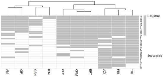
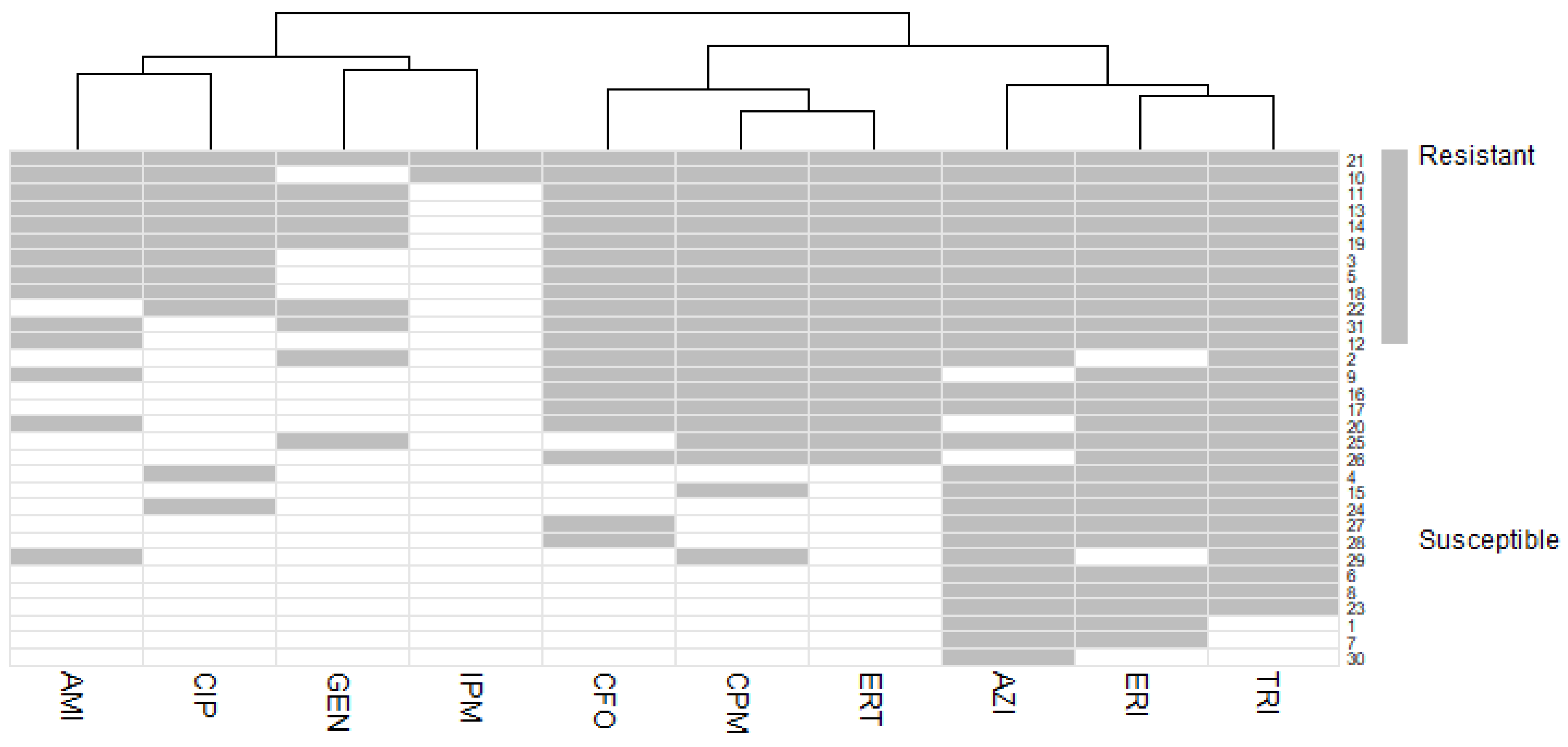
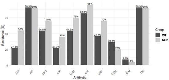
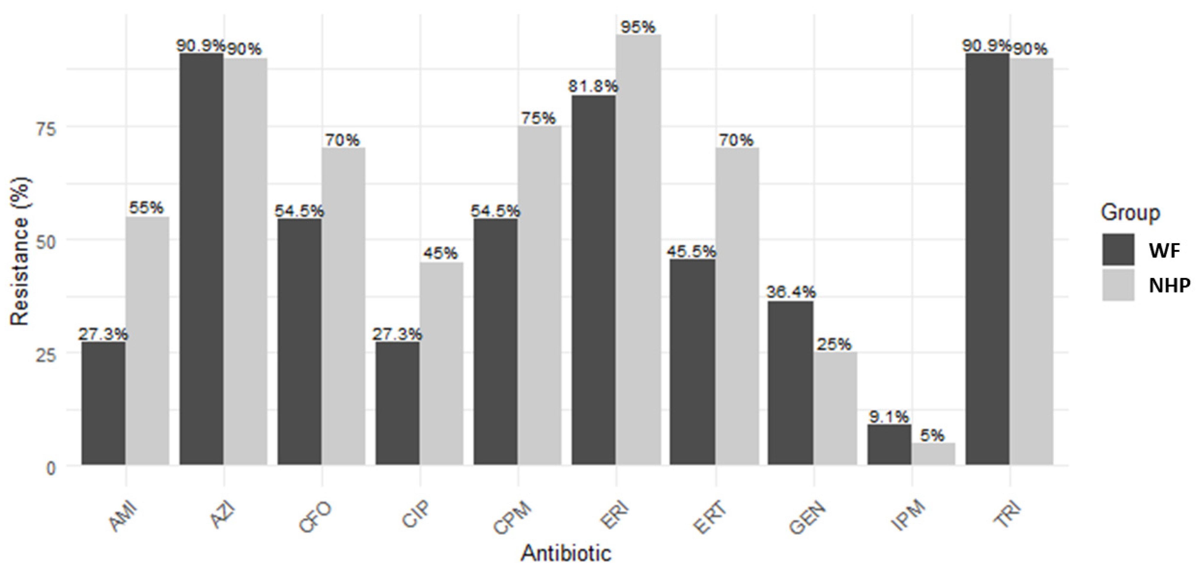
There was similarity between the resistance patterns of Azithromycin, Erythromycin and Sulfamethoxazole-Trimethoprim, all with 90.3% (28/31; 95%CI:75.1-96.7) of the isolates classified as resistant. Meanwhile, the most effective antibiotic for the isolates was imipenem, showing only 6.5% (2/31; 95% CI: 1.8–20.7) of resistant isolates (Figure 1). Although differences in antimicrobial resistance frequencies were observed between non-human primates and felids, none of the comparisons reached statistical significance after Bonferroni correction for multiple comparisons (adjusted p-values = 1.000) (Figure 2).
Figure 1.
Heat map of antimicrobial susceptibility profiles of bacterial isolates. Legend: The rows represent the isolates, ordered from most resistant to most sensitive; gray cells indicate resistance, and white cells indicate susceptibility; AMI—Amikacin, AZI—Azithromycin, CIP—Ciprofloxacin, CFO—Cefoxitin, CPM—Cefepime, ERI—Erythromycin, ERT—Ertapenem, GEN—Gentamicin, IPM—Imipenem, TRI—Sulfamethoxazole-Trimethoprim. Source: Prepared by the authors.
Figure 2.
Resistance frequency of bacterial isolates to antibiotics and mammal group (wild felids and non-human primates). AMI—Amikacin (p = 0.258), AZI—Azithromycin (p = 1), CIP—Ciprofloxacin (p = 0.451), CFO—Cefoxitin (p = 0.452), CPM—Cefepime (p = 0.423), ERI—Erythromycin (p = 0.281), ERT—Ertapenem (p = 0.255), GEN—Gentamicin (p = 0.683), IPM—Imipenem (p = 1), TRI—Sulfamethoxazole-Trimethoprim (p = 1), WF—Wild Felines, NHP—Non-human primates. No statistically significant differences were detected between groups after Bonferroni correction (adjusted significance threshold: p < 0.005). Source: Prepared by the authors.
In addition to the phenotypic identification of isolates belonging to the genus Staphylococcus, Staphylococcus aureus was identified by PCR in 12.9% (4/31) of the isolates; these isolates originated from non-human primate samples (Table 1). The mecA gene was not detected in any of the isolates analyzed.
4. Discussion
Zoos are fundamental to wildlife conservation and the promotion of animal welfare, as they facilitate environmental education, conservation, and scientific research programs that help raise public awareness about the importance of biodiversity. Furthermore, the interaction among visitors, veterinarians, keepers, and animals makes this environment strategic for epidemiological surveillance, as it aids in detecting zoonotic risks and reinforces the understanding of One Health.
This study demonstrated a high prevalence of Staphylococcus spp. in the oral cavity of various wild mammals kept in captivity, including coagulase-positive isolates and Staphylococcus aureus. These data demonstrate that these animals can act as important reservoirs of the genus Staphylococcus in captive environments.
Staphylococcus spp. are opportunistic pathogens commonly found colonizing the skin and mucous membranes of humans and domestic animals [4,16]. However, the presence of pathogenic bacteria in captive animals is becoming increasingly frequent, linked to their proximity to humans and domestic animals; these animals can develop infectious diseases due to poor nutrition, lack of hygiene, management shortcomings, and pre-existing conditions [26,27].
Non-human primates (NHPs) play a fundamental role in ecosystem health, contributing to tropical forest regeneration through seed dispersal or acting as prey and predators within food webs [12,28]. However, due to the lack of food in these habitats, proximity to humans and domestic animals is becoming increasingly frequent. Furthermore, dietary changes can drive alterations in the oral microbiome of non-human primates, so much so that lower bacterial diversity is observed in captive individuals compared to those living in the wild [29,30]. Staphylococcus spp. are commonly found in the oral cavity of Sapajus apella [31]. In addition to this, similar pathogens have been found in the oral cavity of humans and NHPs [32]. These findings reinforce the potential of non-human primates (NHPs) as reservoirs and potential carriers of microorganisms resistant to various antimicrobials, given that they inhabit and move through diverse ecosystems, including forest, rural, and urban environments. This ecological interface facilitates the circulation and dissemination of resistant bacteria among wildlife, domestic animals, humans, and the environment, highlighting the role of these animals in the dynamics of antimicrobial resistance from a One Health perspective [22,23].
The high prevalence (83.7%) of isolates identified as Staphylococcus spp. in captive mammals, as demonstrated in this study, highlights the relevance of this pathogen to One Health; this finding aligns with those of De Alcântara et al. [7], who identified Staphylococcus spp. in 70.6% of samples from captive wild felids and canids, indicating that these microorganisms are part of the oral microbiota of wild felids. Other studies further show that large captive wild felids can also harbor antimicrobial-resistant Staphylococcus spp [13]. However, when the animal is immunosuppressed, the bacterial load tends to increase, resulting in an infection associated with the potential for antimicrobial resistance; if not adequately treated, this can pose a risk to the animal’s health [33]. Furthermore, captive animals typically have a history of stress due to complications preceding their life in the zoo; they also suffer from dietary changes or are immunocompromised, and these conditions can significantly influence the composition of the normal microbiota [34,35,36]. Animals with microbiomes in a state of dysbiosis associated with stress, intensive management, poor hygiene, or therapeutic interventions may be susceptible to infections by potentially resistant microorganisms [35,37]. Furthermore, the handling of captive animals by keepers may facilitate pathogen transmission through contact with feces or via accidental bites or scratches [38].
Another important factor to consider is the origin of the animals being evaluated. The mammals included in this study were rescued by environmental agencies following situations of vulnerability, such as wildlife trafficking, vehicle collisions, and other incidents requiring their removal from the natural environment. Typically, these animals arrive at zoos in a debilitated state due to the stressful conditions of intensive handling during capture, restraint, and transport, potentially exhibiting immunosuppression, nutritional imbalances, and a need for therapeutic interventions. These conditions can alter the microbiota, favoring dysbiosis and increasing the likelihood of colonization by pathogenic microorganisms that are potentially resistant to antimicrobials [34,35,36,37]. Thus, the high frequency of Staphylococcus spp. in the present study may stem not only from the characteristic microbiota of these animals but also from the clinical and environmental conditions to which they were subjected before and during their time in captivity.
In the present study, 38.7% of the isolates were identified as coagulase-positive staphylococci (CoPS), while only 12.9% were confirmed as Staphylococcus aureus, all of which were collected from non-human primates. The low frequency of S. aureus suggests that the species is not the primary colonizer of the oral cavity of the mammals evaluated. The predominance of coagulase-negative staphylococci (CoNS) observed in the present study is consistent with the findings of Sobreira et al. [30], who reported a higher prevalence of CoNS in wild non-human primates. A similar pattern is observed in domestic animals, such as swine [39,40]. CoNS are part of the normal microbiota of the skin and mucous membranes, playing a fundamental role in limiting colonization and invasion by opportunistic pathogens [41,42].
The majority of isolates from non-human primates (65.5%) and wild felids (56.2%) in this study exhibited resistance to the tested antibiotics, antibiotics commonly used in clinical practice to treat bacterial infections in both animals and humans. Although these groups of mammals differ in taxonomy and ecology, there was no significant difference between their antimicrobial resistance profiles. This suggests that these animals may be exposed to similar sources of resistant microorganisms while in captivity, possibly through contact with keepers, veterinarians, and the sharing of the same environment [6].
Furthermore, 80.6% of all isolates were resistant to three or more classes of antibiotics, being classified as multidrug-resistant (MDR) [8]. Meanwhile, 90.3% of the isolates exhibited a multiple antimicrobial resistance (MAR) index greater than 0.20, with a mean of 0.58, a value considered indicative of an origin in environments subject to high selective pressure from antimicrobials [21]. Similar results were reported by Sineke et al. [43], who found multidrug-resistant Staphylococcus aureus with a mean multiple-antibiotic resistance index of 0.50 in intensive pig farming in South Africa. Furthermore, all isolates identified as Staphylococcus aureus in the present study exhibited MAR values greater than 0.2. Both methods are used to characterize multiple antibiotic resistance profiles, and although the results are similar, the MAR method provides a quantitative measure of individual and group resistance, yielding more detailed results than the MDR method [44]. Thus, in the present study, the Leopardus pardalis isolate exhibited the highest MAR value, suggesting greater exposure to sources of selection for antibiotic-resistant bacteria, whereas the Puma concolor isolate showed the lowest index. Although both isolates originated from felids, there was no significant difference in mean MAR values between non-human primates and wild felids, indicating that the groups may be subject to the same level of exposure to resistant Staphylococcus spp. in the captive environment. Similar mean MAR values were found in a study involving swine [43].
The major issue regarding the presence of resistant Staphylococcus spp. in captive wild animals is antimicrobial resistance, which increases mortality rates due to the difficulty of providing adequate treatment; this necessitates meticulous oversight and continuous health monitoring of captive animals given the risk of transmission. Saad, Salama, and Ibrahim [13] also observed high antibiotic resistance in Staphylococcus spp. isolated from fecal samples of captive African lions from Egypt’s national circus, revealing the potential zoonotic risk posed by these bacteria isolated from lions.
The majority of the antibiotics tested, such as azithromycin, erythromycin, sulfamethoxazole-trimethoprim, cefepime, cefoxitin, and ertapenem, proved ineffective against Staphylococcus spp. isolates. Meanwhile, the antibiotic that demonstrated the greatest efficacy against the bacterial isolates was Imipenem, with 93.3% of the isolates being susceptible. These data corroborate the findings of Sobreira et al. [30], who identified erythromycin resistance in Staphylococcus spp. isolates from the oral cavity of Sapajus libidinosus capuchin monkeys in northeastern Brazil. Furthermore, studies by Saad, Salama, and Ibrahim [13] also demonstrated resistance to cefepime and cefoxitin in Staphylococcus spp. isolated from fecal samples of captive African lions from the Egyptian National Circus.
The differences in antimicrobial resistance observed across various studies may be related to the diverse origins of the animals, which involve distinct management practices, the genetic diversity of bacterial populations, and, above all, the selective pressure exerted by antimicrobial use [45,46].
The prevalence of methicillin-resistant Staphylococcus aureus (MRSA) isolates has been reported as low in wild animals, such as non-human primates and felids, although their detection and investigation are of sanitary significance [27,47]. In the present study, there was no molecular evidence of resistance related to the presence of the mecA gene, but other mechanisms associated with resistance in Staphylococcus spp. were not investigated. Indeed, isolate resistance may also have been mediated by alternative mechanisms, such as homologous genes like mecC; for instance, research by Loncaric et al. [48] demonstrated that oxacillin-resistant Staphylococcus spp. isolates from wild animals did not test positive for the mecA gene via conventional PCR, yet all tested positive for the mecC gene via specific PCR. Other factors, such as the overproduction of β-lactamases, modifications to penicillin-binding proteins, and even biofilm formation, can significantly influence and increase antimicrobial resistance [49,50,51]. Thus, the absence of the mecA gene does not rule out the important epidemiological role of these isolates.
Despite not being widely exposed to antimicrobials like other animal groups, wild mammals kept in captivity have been shown to act as potential reservoirs for Staphylococcus spp., including methicillin-resistant Staphylococcus aureus (MRSA) [27]. Thus, the increased presence of these bacteria in the oral cavities of these animals may be linked not only to the therapeutic use of antimicrobials in captive animals but, more importantly, to frequent contact with humans, such as keepers, veterinarians, and handling practices, as well as to diet, water, or even environmental contamination. This situation is further aggravated by the risk of accidents involving animal handlers through contact with excrement or via bites or scratches, as demonstrated by Devaux et al. [38], who described the transmission of Staphylococcus spp. to humans via non-human primate bites.
Studies demonstrate that changes in the environment and diet of wild animals can influence the composition of the gut microbiota, comparing free-ranging animals with those in captivity, and that the latter may face a higher risk of developing infectious diseases [52,53]. Thus, antimicrobial resistance is a serious One Health issue; however, the situation is further exacerbated when it comes to antibiotic resistance in wild and captive animals, due to the lack of monitoring and information [10,11,54].
Infections caused by resistant microorganisms are difficult to resolve and often increase morbidity and mortality rates in humans and animals. Thus, the findings reinforce the need for periodic monitoring of antimicrobial resistance and measures to control the spread of resistant Staphylococcus in captive environments, in order to protect animals, keepers, veterinarians, and zoo visitors.
Study Limitations
Although this study provides important information on antimicrobial resistance in Staphylococcus spp. isolates from captive wild mammals, some limitations should be considered. The study was conducted at a single zoo, with a relatively small number of isolates and unequal representation of animal species. The small sample size may have limited the statistical power to detect differences in antimicrobial resistance between groups. The grouping of different wild felid species may have introduced heterogeneity, and the absence of statistically significant differences should not be interpreted as evidence of equivalence between groups. Furthermore, the cross-sectional design, the absence of environmental and human samples, and the lack of systematic data on prior antimicrobial exposure in this study make it difficult to determine possible transmission events. Molecular identification at the species level was limited, and molecular assessment was restricted to the presence of the mecA gene. Therefore, future research should include a larger number of zoos and bacterial isolates, despite the limitations associated with the restraint and handling of wild animals. In addition to investigating other mechanisms that determine resistance (such as mecC), virulence factors and biofilm formation, future research should explore potential alternatives for the prevention and treatment of staphylococcal infections in captive animals. These new analyses could aid in a better understanding of the epidemiology of these pathogens, in addition to contributing to strategies for the prevention and control of microbial resistance within the One Health context.
5. Conclusions
This study demonstrated a high prevalence of Staphylococcus spp. (83.7%) in the oral cavity of captive wild mammals, with a predominance of coagulase-negative staphylococci and the identification of Staphylococcus aureus in 12.9% of the isolates, exclusively from non-human primates. Furthermore, more than half of the isolates (56.2% from wild felids and 65.5% from non-human primates) showed resistance to the tested antimicrobials; 80.6% were classified as multidrug-resistant, and 90.3% exhibited a multiple antimicrobial resistance index indicative of an origin in environments under high selective pressure from antimicrobials, demonstrating the widespread circulation of bacteria across different organisms and environments. These findings highlight the potential of captive wild animals as reservoirs of Staphylococcus spp. multidrug-resistant to conventional antimicrobials, suggesting potential transmission across the human–animal-environment interface. In this context, this One Health interface requires continuous epidemiological surveillance and biosecurity measures, given that interactions among animals, caretakers, veterinarians, and zoo visitors can facilitate exposure to resistant microorganisms and the risk of zoonotic transmission, with implications for animal production and public health.
Author Contributions
Conceptualization, F.G.C.B. and D.D.G.; methodology, D.D.G., L.N.B., Z.C.G., R.P.J. and D.V.B.; software, F.G.C.B. and D.V.B.; validation, D.D.G., L.N.B., Z.C.G., R.P.J. and D.V.B.; formal analysis, F.G.C.B., D.D.G., L.N.B., Z.C.G., R.P.J. and D.V.B.; investigation, F.G.C.B., A.C.S.A., H.S.T., J.F.d.A., K.A.J.R., H.M.d.S.O., H.d.O.M., V.Y.T., B.P.B.d.S., R.N.R., W.A.S. and G.C.C.S.; resources, D.D.G., L.N.B., Z.C.G., R.P.J. and D.V.B.; data curation, F.G.C.B., A.C.S.A., H.S.T., J.F.d.A., K.A.J.R., H.M.d.S.O., H.d.O.M., V.Y.T., B.P.B.d.S., R.N.R., W.A.S. and G.C.C.S.; writing—original draft preparation, F.G.C.B. and D.D.G.; writing—review and editing, F.G.C.B. and D.D.G.; visualization, F.G.C.B., A.C.S.A., H.S.T., J.F.d.A., K.A.J.R., H.M.d.S.O., H.d.O.M., V.Y.T., B.P.B.d.S., R.N.R., W.A.S. and G.C.C.S.; supervision, D.D.G., L.N.B., Z.C.G., R.P.J. and D.V.B.; project administration, D.D.G.; funding acquisition, D.D.G., L.N.B., Z.C.G., R.P.J. and D.V.B. All authors have read and agreed to the published version of the manuscript.
Funding
This research was funded by CNPq (Call PQ 2023-310953/2023-5) and UNIPAR.
Institutional Review Board Statement
The research was approved by the Chico Mendes Institute for Biodiversity Conservation (ICMBio) under protocol number 82499-1 (06/01/2022) and also by the Ethics Committee on Research Involving Animal Experimentation of the Paranaense University (UNIPAR) under protocol number 39559 (06/24/2022).
Informed Consent Statement
Not applicable.
Data Availability Statement
The original contributions presented in this study are included in the article. Further inquiries can be directed to the corresponding author.
Acknowledgments
The authors thank the Municipal Zoo for its ongoing collaboration and UNIPAR and CNPq (Call PQ 2023-310953/2023-5) for funding this research. The authors have reviewed and edited the output and take full responsibility for the content of this publication.
Conflicts of Interest
The authors declare no conflicts of interest.
References
- Aires-de-Sousa, M. Methicillin-Resistant Staphylococcus aureus among Animals: Current Overview. Clin. Microbiol. Infect. 2017, 23, 373–380. [Google Scholar] [CrossRef] [Scilit] [PubMed]
- Touaitia, R.; Mairi, A.; Ibrahim, N.A.; Basher, N.S.; Idres, T.; Touati, A. Staphylococcus aureus: A Review of the Pathogenesis and Virulence Mechanisms. Antibiotics 2025, 14, 470. [Google Scholar] [CrossRef] [Scilit] [PubMed]
- Galfo, V.; Tiseo, G.; Riccardi, N.; Falcone, M. Therapeutic Drug Monitoring of Antibiotics for Methicillin-Resistant Staphylococcus aureus Infections: An Updated Narrative Review for Clinicians. Clin. Microbiol. Infect. 2025, 31, 194–200. [Google Scholar] [CrossRef] [Scilit] [PubMed]
- Idrees, M.; Sawant, S.; Karodia, N.; Rahman, A. Staphylococcus aureus Biofilm: Morphology, Genetics, Pathogenesis and Treatment Strategies. Int. J. Environ. Res. Public Health 2021, 18, 7602. [Google Scholar] [CrossRef] [Scilit] [PubMed]
- Cao, H.; Bougouffa, S.; Park, T.-J.; Lau, A.; Tong, M.-K.; Chow, K.-H.; Ho, P.-L. Sharing of Antimicrobial Resistance Genes between Humans and Food Animals. mSystems 2022, 7, e00775-22. [Google Scholar] [CrossRef] [Scilit] [PubMed]
- Heaton, C.J.; Gerbig, G.R.; Sensius, L.D.; Patel, V.; Smith, T.C. Staphylococcus aureus Epidemiology in Wildlife: A Systematic Review. Antibiotics 2020, 9, 89. [Google Scholar] [CrossRef] [Scilit] [PubMed]
- de Alcântara, L.P.; Santana, J.A.; Clark Xavier, R.G.; Tinoco, H.P.; Coelho, C.M.; dos Santos, D.O.; Santos, R.L.; Nogueira de Carvalho, M.P.; Silveira Silva, R.O. Antimicrobial Susceptibility of Staphylococcus Spp. Isolated from Felids and Canids in Belo Horizonte Zoo, Brazil. J. Zoo Wildl. Med. 2023, 54, 584–592. [Google Scholar] [CrossRef] [Scilit] [PubMed]
- Martínez-Seijas, C.; Mascarós, P.; Lizana, V.; Martí-Marco, A.; Arnau-Bonachera, A.; Chillida-Martínez, E.; Cardells, J.; Selva, L.; Viana, D.; Corpa, J.M. Genomic Characterization of Staphylococcus aureus in Wildlife. Animals 2023, 13, 1064. [Google Scholar] [CrossRef] [Scilit] [PubMed]
- Boto, L.; Pineda, M.; Pineda, R. Potential Impacts of Horizontal Gene Transfer on Human Health and Physiology and How Anthropogenic Activity Can Affect It. FEBS J. 2019, 286, 3959–3967. [Google Scholar] [CrossRef] [Scilit] [PubMed]
- Smoglica, C.; Carcagnì, A.; Angelucci, S.; Di Tana, F.; Marsilio, F.; López-Olvera, J.R.; Di Francesco, C.E. Systematic Review and Meta-Analysis of Antimicrobial Resistant Bacteria in Free-Ranging Wild Mammals. BMC Vet. Res. 2025, 21, 150. [Google Scholar] [CrossRef] [Scilit] [PubMed]
- Doyle, C.; Wall, K.; Fanning, S.; McMahon, B.J. Making Sense of Sentinels: Wildlife as the One Health Bridge for Environmental Antimicrobial Resistance Surveillance. J. Appl. Microbiol. 2025, 136, lxaf017. [Google Scholar] [CrossRef] [Scilit] [PubMed]
- Sengupta, A.; McConkey, K.R.; Radhakrishna, S. Primates, Provisioning and Plants: Impacts of Human Cultural Behaviours on Primate Ecological Functions. PLoS ONE 2015, 10, e0140961. [Google Scholar] [CrossRef] [Scilit] [PubMed]
- Saad, E.; Salama, A.; Ibrahim, G. Bacterial Screening of Sporadic Cases of Captive African Lions (Panthera Leo) at the Egyptian National Circus. Egypt. Acad. J. Biol. Sci. G. Microbiol. 2023, 15, 105–116. [Google Scholar] [CrossRef] [Scilit]
- García-Fernández, A.J.; Espín, S.; Gómez-Ramírez, P.; Martínez-López, E.; Navas, I. Wildlife Sentinels for Human and Environmental Health Hazards in Ecotoxicological Risk Assessment. In Ecotoxicological QSARs, Methods in Pharmacology and Toxicology; Springer US: New York, NY, USA, 2020; pp. 77–94. [Google Scholar] [CrossRef] [Scilit]
- Andrade, A.C.d.S.; Bonato, F.G.C.; dos Santos, I.C.; Iukava, L.K.; Charnovski, S.G.D.; da Silva, B.P.B.; Suci Junior, E.M.; Pretto-Giordano, L.G.; Piau Junior, R.; Ribeiro, R.N.; et al. Presence of Leptospira Spp. in Nonhuman Primates and Wild Felids from a Brazilian Zoo. Vector-Borne Zoonotic Dis. 2025, 25, 553–557. [Google Scholar] [CrossRef] [Scilit] [PubMed]
- Koneman, E.W.; Koneman, E. Diagnóstico Microbiológico. Texto y Atlas Color; Editorial Médica Panamericana: Buenos Aires, Argentina, 1999. [Google Scholar]
- CLSI. Performance Standards for Antimicrobial Disk and Dilution Susceptibility Tests for Bacteria Isolated From Animals, 5th ed.; Clinical and Laboratory Standards Institute: Wayne, PA, USA, 2020. [Google Scholar]
- CLSI M100; Performance Standards for Antimicrobial Susceptibility Testing, 33th ed. Clinical and Laboratory Standards Institute: Wayne, PA, USA, 2023.
- CLSI. Methods for Antimicrobial Dilution and Disk Susceptibility Testing of Infrequently Isolated or Fastidious Bacteria, 3rd ed.; Clinical and Laboratory Standards Institute, Ed.; Clinical and Laboratory Standards Institute: Wayne, PA, USA, 2015. [Google Scholar]
- CLSI. Methods for Dilution Antimicrobial Susceptibility Tests for Bacteria That Grow Aerobically, 11th ed.; Clinical Laboratory Standards Institute: Wayne, PA, USA, 2018. [Google Scholar]
- Krumperman, P.H. Multiple Antibiotic Resistance Indexing of Escherichia Coli to Identify High-Risk Sources of Fecal Contamination of Foods. Appl. Environ. Microbiol. 1983, 46, 165–170. [Google Scholar] [CrossRef] [Scilit] [PubMed]
- Thenmozhi, S.; Rajeswari, P.; Kumar, B.S.; Saipriyanga, V.; Kalpana, M. Multi-Drug Resistant Patterns of Biofilm Forming Aeromonas Hydrophila from Urine Samples. Int. J. Pharm. Sci. Res. 2014, 5, 2908. [Google Scholar]
- Martineau, F.; Picard, F.J.; Roy, P.H.; Ouellette, M.; Bergeron, M.G. Species-Specific and Ubiquitous-DNA-Based Assays for Rapid Identification of Staphylococcus Aureus. J. Clin. Microbiol. 1998, 36, 618–623. [Google Scholar] [CrossRef] [Scilit] [PubMed]
- Murakami, K.; Minamide, W.; Wada, K.; Nakamura, E.; Teraoka, H.; Watanabe, S. Identification of Methicillin-Resistant Strains of Staphylococci by Polymerase Chain Reaction. J. Clin. Microbiol. 1991, 29, 2240–2244. [Google Scholar] [CrossRef] [Scilit] [PubMed]
- Cave, R.; Misra, R.; Chen, J.; Wang, S.; Mkrtchyan, H.V. Whole Genome Sequencing Revealed New Molecular Characteristics in Multidrug Resistant Staphylococci Recovered from High Frequency Touched Surfaces in London. Sci. Rep. 2019, 9, 9637. [Google Scholar] [CrossRef] [Scilit] [PubMed]
- Radhouani, H.; Poeta, P.; Gonçalves, A.; Pacheco, R.; Sargo, R.; Igrejas, G. Wild Birds as Biological Indicators of Environmental Pollution: Antimicrobial Resistance Patterns of Escherichia coli and Enterococci Isolated from Common Buzzards (Buteo Buteo). J. Med. Microbiol. 2012, 61, 837–843. [Google Scholar] [CrossRef] [Scilit] [PubMed]
- Abdullahi, I.N.; Fernández-Fernández, R.; Juárez-Fernández, G.; Martínez-álvarez, S.; Eguizábal, P.; Zarazaga, M.; Lozano, C.; Torres, C. Wild Animals Are Reservoirs and Sentinels of Staphylococcus aureus and MRSA Clones: A Problem with “One Health” Concern. Antibiotics 2021, 10, 1556. [Google Scholar] [CrossRef] [Scilit] [PubMed]
- Chapman, C.A.; Bicca-Marques, J.C.; Dunham, A.E.; Fan, P.; Fashing, P.J.; Gogarten, J.F.; Guo, S.; Huffman, M.A.; Kalbitzer, U.; Li, B.; et al. Primates Can Be a Rallying Symbol to Promote Tropical Forest Restoration. Folia Primatol. 2020, 91, 669–687. [Google Scholar] [CrossRef] [Scilit] [PubMed]
- Sawaswong, V.; Praianantathavorn, K.; Chanchaem, P.; Khamwut, A.; Kemthong, T.; Hamada, Y.; Malaivijitnond, S.; Payungporn, S. Comparative Analysis of Oral-Gut Microbiota between Captive and Wild Long-Tailed Macaque in Thailand. Sci. Rep. 2021, 11, 14280. [Google Scholar] [CrossRef] [Scilit] [PubMed]
- Sobreira, E.d.A.; Braga, C.A.d.S.B.; Rahal, S.C.; Ribeiro, E.L.; Vasconcelos, L.S.N.d.O.L.; Carneiro, L.C.; Teixeira, C.R.; de Almeida, C.C.D.; Haddad Júnior, V. Oral Microbiota and Their Antibiotic Susceptibility in Free-Living Monkeys in Goiás State, Brazil: Repercussions for Injuries in Humans. Rev. Soc. Bras. Med. Trop. 2019, 52, 20180294. [Google Scholar] [CrossRef] [Scilit] [PubMed]
- Pinheiro, L.C.R.; Junior, E.P.C.S.; do Nascimento, G.V.; Quessada, A.M.; Abreu, A.R.S.; Camapum, J.L.R.; de Morais, N.M.B.; Silva, F.A.d.N.; Belettini, S.T.; Rodrigues, M.C. Zoonotic Potential of Oral Microbiota of Capuchins Monkeys (Sapajus Libidinosus). Res. Soc. Dev. 2020, 9, e1229108182. [Google Scholar] [CrossRef] [Scilit]
- Miranda, B.P.; da Silva, A.F.; Ascenção, J.d.C.; Santos, H.L.C. An Overview of the Microbiota of the Oral Cavity of Humans and Non-Human Primates with Periodontal Disease: Current Issues and Perspectives. Arch. Oral Biol. 2025, 172, 106121. [Google Scholar] [CrossRef] [Scilit] [PubMed]
- Mohammadzadeh, P.; Shahri, N.; Momeni, B. Revolutionizing Oronasal Fistula Treatment in Felis Silvestris: Integrating Bioengineered Solutions and Microbiome Insights for Enhanced Healing. Open Vet. J. 2025, 15, 4775. [Google Scholar] [CrossRef] [Scilit] [PubMed]
- Wachtman, L.M.; Mansfield, K.G. Opportunistic Infections in Immunologically Compromised Nonhuman Primates. ILAR J. 2008, 49, 191–208. [Google Scholar] [CrossRef] [Scilit] [PubMed]
- Lima, V.F.S.; Ramos, R.A.N.; Giannelli, A.; Schettino, S.C.; Galina, A.B.; De Oliveira, J.C.P.; Meira-Santos, P.O.; Alves, L.C. Zoonotic Parasites in Wild Animals Such as Carnivores and Primates That Are Traded Illegally in Brazil. Braz. J. Vet. Med. 2021, 43, e113720. [Google Scholar] [CrossRef] [Scilit] [PubMed]
- Rivera, A.; Zambrano-Mila, M.S.; Orlando, S.A.; Jiménez Valenzuela, F.; Sanchez, E.; Calderon, J.; González, M.; Rodriguez-Pazmiño, A.S.; Marzal, A.; Molineros, E.; et al. A First Insight into the Occurrence of Leptospira, Brucella and Coxiella burnetii Infections in Wild Mammals Rescued from Illegal Trade in Ecuador: A Proxy for One Health Conservation Policies. One Health 2025, 20, 101045. [Google Scholar] [CrossRef] [Scilit] [PubMed]
- da Costa, M.R.; Diana, A. A Systematic Review on the Link between Animal Welfare and Antimicrobial Use in Captive Animals. Animals 2022, 12, 1025. [Google Scholar] [CrossRef] [Scilit] [PubMed]
- Devaux, C.A.; Mediannikov, O.; Medkour, H.; Raoult, D. Infectious Disease Risk Across the Growing Human-Non Human Primate Interface: A Review of the Evidence. Front. Public Health 2019, 7, 305. [Google Scholar] [CrossRef] [Scilit] [PubMed]
- dos Santos, I.C.; Barbosa, L.N.; Grossi, G.D.; de Paula Ferreira, L.R.; Ono, J.M.; Martins, L.A.; Alberton, L.R.; Gonçalves, D.D. Presence of Staphylococcus Spp. Carriers of the MecA Gene in the Nasal Cavity of Piglets in the Nursery Phase. Res. Vet. Sci. 2023, 155, 51–55. [Google Scholar] [CrossRef] [Scilit] [PubMed]
- Low, J.; Tu, H.; Elbadawey, M.; Wayes, A.; Jakubovics, N.; Choo, S.W.; Yee, W.W. Genomic Insights into the Diversity, Antibiotic Resistance, and Virulence Potential of Staphylococci Isolated from Pediatric Patients with Chronic Otitis Media with Effusion (COME). PeerJ 2026, 14, e20782. [Google Scholar] [CrossRef] [Scilit] [PubMed]
- Lean, S.S.; Wetthasinghe, L.; Sim, K.S.; Chong, H.X.; Sharfudeen, S.I.B.; Chen, Y.W.; Lim, Y.Y.; Teoh, H.K.; Yeo, C.C.; Ng, H.F. Genomic Characterisation of Nasal Isolates of Coagulase-Negative Staphylococci from Healthy Medical Students Reveals Novel Staphylococcal Cassette Chromosome Mec Elements. Sci. Rep. 2025, 15, 21770. [Google Scholar] [CrossRef] [Scilit] [PubMed]
- Parlet, C.P.; Brown, M.M.; Horswill, A.R. Commensal Staphylococci Influence Staphylococcus aureus Skin Colonization and Disease. Trends Microbiol. 2019, 27, 497–507. [Google Scholar] [CrossRef] [Scilit] [PubMed]
- Sineke, N.; Asante, J.; Amoako, D.G.; Abia, A.L.K.; Perrett, K.; Bester, L.A.; Essack, S.Y. Staphylococcus aureus in Intensive Pig Production in South Africa: Antibiotic Resistance, Virulence Determinants, and Clonality. Pathogens 2021, 10, 317. [Google Scholar] [CrossRef] [Scilit] [PubMed]
- Savaşan, N.; Çakir, N.; Akartaş, L.; Çoklu, A.; Direnci, İ.; Direnci, Ç.A.; Karşılaştırılması, T.; Yöntemsel, B.; Analizi, E. Comparison of Definitions of Multiple Drug Resistance to Antibiotics and Its Alternative Multiple Antibiotic Resistance and Their Methodological Critical Analysis. Mediterr. J. Infect. Microb. Antimicrob. 2019, 8, 34. [Google Scholar] [CrossRef] [Scilit]
- Bennani, H.; Mateus, A.; Mays, N.; Eastmure, E.; Stärk, K.D.C.; Häsler, B. Overview of Evidence of Antimicrobial Use and Antimicrobial Resistance in the Food Chain. Antibiotics 2020, 9, 49. [Google Scholar] [CrossRef] [Scilit] [PubMed]
- Mediouni, M.; Diallo, A.B.; Makarenkov, V. Quantifying Antimicrobial Resistance in Food-Producing Animals in North America. Front. Microbiol. 2025, 16, 1542472. [Google Scholar] [CrossRef] [Scilit] [PubMed]
- Silveira, D.R.; de Moraes, T.P.; Kaefer, K.; Bach, L.G.; Barbosa, A.d.O.; Moretti, V.D.; de Menezes, P.Q.; de Medeiros, U.S.; da Silva, T.T.; Bandarra, P.M.; et al. MRSA and Enterobacteria of One Health Concern in Wild Animals Undergoing Rehabilitation. Res. Soc. Dev. 2021, 10, e34810111809. [Google Scholar] [CrossRef] [Scilit]
- Loncaric, I.; Kübber-Heiss, A.; Posautz, A.; Stalder, G.L.; Hoffmann, D.; Rosengarten, R.; Walzer, C. Characterization of Methicillin-Resistant Staphylococcus Spp. Carrying the MecC Gene, Isolated from Wildlife. J. Antimicrob. Chemother. 2013, 68, 2222–2225. [Google Scholar] [CrossRef] [Scilit] [PubMed]
- Keseru, J.S.; Gál, Z.; Barabás, G.; Benko, I.; Szabó, I. Investigation of β-Lactamases in Clinical Isolates of Staphylococcus aureus for Further Explanation of Borderline Methicillin Resistance. Chemotherapy 2005, 51, 300–304. [Google Scholar] [CrossRef] [Scilit] [PubMed]
- Dabhi, M.; Patel, R.; Shah, V.; Soni, R.; Saraf, M.; Rawal, R.; Goswami, D. Penicillin-Binding Proteins: The Master Builders and Breakers of Bacterial Cell Walls and Its Interaction with β-Lactam Antibiotics. J. Proteins Proteom. 2024, 15, 215–232. [Google Scholar] [CrossRef] [Scilit]
- Aboelnaga, N.; Elsayed, S.W.; Abdelsalam, N.A.; Salem, S.; Saif, N.A.; Elsayed, M.; Ayman, S.; Nasr, M.; Elhadidy, M. Deciphering the Dynamics of Methicillin-Resistant Staphylococcus aureus Biofilm Formation: From Molecular Signaling to Nanotherapeutic Advances. Cell Commun. Signal. 2024, 22, 188. [Google Scholar] [CrossRef] [Scilit] [PubMed]
- Schmartz, G.P.; Rehner, J.; Schuff, M.J.; Molano, L.A.G.; Becker, S.L.; Krawczyk, M.; Tagirdzhanov, A.; Gurevich, A.; Francke, R.; Müller, R.; et al. Exploring Microbial Diversity and Biosynthetic Potential in Zoo and Wildlife Animal Microbiomes. Nat. Commun. 2024, 15, 8263. [Google Scholar] [CrossRef] [Scilit] [PubMed]
- Huang, X.; Li, H.; Zhang, L.; Zhang, X.; Cheng, S.; Yan, Y.; Yang, W.; Meng, B.; Wang, Z.; Zhao, J.; et al. Comparative Analysis of Gut Microbiota Between Wild and Captive Guizhou Snub-Nosed Monkey (Rhinopithecus brelichi). Ecol. Evol. 2024, 14, e70690. [Google Scholar] [CrossRef] [Scilit] [PubMed]
- Kim, M.; Kim, M.; Yeo, Y.G.; Lee, Y.T.; Han, J.I. Antimicrobial Resistance of Commensal Escherichia coli and Enterococcus faecalis Isolated from Clinically Healthy Captive Wild Animals in Seoul Zoo. Front. Vet. Sci. 2024, 10, 1283487. [Google Scholar] [CrossRef] [Scilit] [PubMed]

Disclaimer/Publisher’s Note: The statements, opinions and data contained in all publications are solely those of the individual author(s) and contributor(s) and not of MDPI and/or the editor(s). MDPI and/or the editor(s) disclaim responsibility for any injury to people or property resulting from any ideas, methods, instructions or products referred to in the content. |
© 2026 by the authors. Licensee MDPI, Basel, Switzerland. This article is an open access article distributed under the terms and conditions of the Creative Commons Attribution (CC BY) license.