


Simple Summary
The greater amberjack (Seriola dumerili) is an important farmed fish that often becomes sick from harmful bacteria, causing serious losses in aquaculture. In this study, we examined a disease-causing bacterium taken from sick fish and confirmed that it can trigger illness in healthy fish. We also studied how the fish reacts after infection by looking at changes in gene activity in its immune tissues. The results show that many genes related to defense and inflammation become more active. These findings help us better understand how the fish fights infection and may support improved disease control in aquaculture.
Abstract
The greater amberjack (Seriola dumerili) is an economically important marine species that is prone to bacterial infections, resulting in high mortality rates and substantial economic losses. In this study, a virulent bacterial strain, Vh-2, was isolated from diseased greater amberjack and identified as Vibrio harveyi. Experimental infections caused high mortality and severe splenic damage characterized by tissue necrosis, abnormal pigment deposition, cellular disintegration, and extensive immune cell infiltration. A virulence gene analysis revealed that Vh-2 harbored multiple virulence-associated genes such as toxR, toxS, vhpA, vhpB, vhhA, vhhB, luxR, and pap6. Antibiotic susceptibility testing demonstrated ampicillin resistance but sensitivity to ceftriaxone, florfenicol, and meropenem. Transcriptomic profiling of infected spleens identified 396 differentially expressed genes (DEGs) compared to the control group, of which 293 were upregulated and 103 were downregulated. A functional enrichment analysis indicated that these genes were primarily involved in cell cycle regulation, DNA repair, metabolic processes, and immune-related pathways. These findings enhance our understanding of V. harveyi pathogenesis and immune responses of S. dumerili and provide new insights into the prevention and control of V. harveyi infections in marine fish.
1. Introduction
Vibrio spp. are known to infect aquatic animals, resulting in significant economic losses to the aquaculture industry [1]. Among these, V. harveyi is a Gram-negative, halophilic, facultative anaerobic bacterium commonly found in warm marine environments and is opportunistically pathogenic in fish and crustaceans. Early symptoms of V. harveyi infection in marine fish include lethargy, reduced feeding, abnormal swimming behavior, and skin ulceration, occasionally accompanied by tail rot, exophthalmia, and abdominal distension or ascites [2,3]. In severe cases, V. harveyi can cause splenic necrosis, liver inflammation, and bacterial invasion of internal organs. In V. harveyi-infected gilthead seabream (Sparus aurata), hemosiderin deposits and melanomacrophage centers have been observed in the spleen [4]. Vibrio harveyi demonstrates significant pathogenic diversity as strains differ in their virulence gene profiles. It harbors key virulence genes, including rtxD, acfD, toxR, luxS, iron acquisition-related genes (sitA, sitB, sitC, and sitD), and the heat-stable cytotoxic enterotoxin gene ast [5]. A pathogenic strain isolated from diseased fish possessed the conserved regulator toxR and its deletion resulted in increased biofilm formation, highlighting its crucial role in colonization and virulence [6].
Transcriptome analysis, a commonly used approach to assess host–pathogen interaction mechanisms, effectively reveals genes involved in immune responses [7,8]. A comparative transcriptomic study of Japanese pufferfish (Takifugu rubripes) revealed differential gene expression profiles in the liver and spleen following V. harveyi infection, highlighting the activation of multiple immune-related pathways [9]. Similarly, a transcriptome analysis identified a broad range of immune genes involved in the response of spotted sea bass (Lateolabrax maculatus) to Edwardsiella tarda infection [10]. Compared to the conventional digital gene expression (DGE) analysis, the RNA-Seq approach is significantly advantageous as it can detect low-abundance transcripts, identify variable splicing events, and reveal novel genes [11]. Because of the efficiency of transcriptome analysis in identifying immune markers, it establishes a theoretical foundation for the optimization of aquaculture disease prevention and control. The fish spleen serves as a primary immune organ rich in lymphocytes, macrophages, and granulocytes, and is essential for innate and adaptive immunity, such as cytokine production and antigen presentation [12,13]. Its primary function in bacterial resistance highlights the necessity of transcriptome analysis to explore spleen immune mechanisms and provide a molecular basis for disease prevention.
The greater amberjack (Seriola dumerili) is a warm-water marine fish globally distributed in tropical and subtropical seas [14]. Due to its rapid growth rate, high quality meat, and adaptability to net-pen aquaculture habitats, it has become an economically important aquaculture species [15]. Over the past decade, aquaculture production of greater amberjack has increased significantly due to an increased consumer demand for high-quality products and the continuous increase in its market value [16]. However, increased stocking density and pollution have caused regular outbreaks of various bacterial diseases, which have considerably impacted aquaculture development [17,18].
The pathogenesis of V. harveyi and the immune response of greater amberjack to infection are not well known. Therefore, this study aims to investigate the pathogenic mechanisms of this bacterium isolated from diseased S. dumerili by bacterial culture, morphological observation, and 16S rDNA gene sequencing, and explore the host immune response mechanisms through transcriptomic analysis of the spleen. This study will provide new insights into the prevention and control of V. harveyi infection in aquaculture farms.
2. Materials and Methods
2.1. Fish Collection and Inspection
The moribund greater amberjacks were collected from the aquaculture farm on Donghai Island, Zhanjiang, China. A microscopic examination was conducted on fresh gill filaments, mucus, and internal organs (liver, spleen, kidney, and intestine) to detect the presence of parasites.
Healthy greater amberjacks (n = 20) from 55–60 cm in length and 2000–2800 g in weight were attained from the aquaculture farm on Donghai Island. The fish were transported to the Guangdong Southern Marine Science and Engineering Laboratory (Zhanjiang, China) for the artificial infection analyses.
2.2. Isolation and Identification of Bacteria
Moribund fish were aseptically dissected to collect spleen samples for bacterial isolation. Tissue homogenates were streaked onto 2216E agar plates (Hopebio, Qingdao, China) and incubated at 30 °C for 24–48 h. Dominant colonies with uniform morphology were sub-cultured on fresh 2216E agar plates to obtain pure isolates for further identification. Genomic DNA was extracted using the TIANGEN Bacterial DNA Kit (TIANGEN Biotech, Beijing, China) following the manufacturer’s instructions, and amplification of the 16S rDNA gene was carried out using the primers listed in Table S1. A PCR was performed in a 25 μL reaction system containing 12.5 μL of 2× Taq PCR Master Mix (Vazyme, Nanjing, China), 1 μL of each primer, 2 μL of template DNA, and 8.5 μL of nuclease-free water. The PCR included an initial denaturation at 95 °C for 5 min followed by 30 cycles of 95 °C for 30 s, annealing at 56 °C for 30 s, extension at 72 °C for 1 min, and a final extension at 72 °C for 7 min. The PCR products were purified and sequenced by Sangon Biotech Co., Ltd. (Sangon, Shanghai, China), sequence alignment was performed using BLAST tool at the National Center for Biotechnology Information (https://blast.ncbi.nlm.nih.gov/Blast.cgi, accessed on 16 November 2025), and phylogenetic trees were constructed using the neighbor-joining method in MEGA 7.0 with 1000 bootstrap replicates [19]. Bacterial characteristics were determined by Gram staining, phenotypic identification was performed using Vibrio-specific biochemical test kits (Hangzhou Microbial Reagent Co., Ltd., Hangzhou, China), and taxonomic classification was based on Bergey’s Manual of Systematic Bacteriology (2nd edition) and Bacteria and Fungi from Fish and Other Aquatic Animals: A Practical Identification Manual [20,21].
2.3. Transmission Electron Microscopy
To observe the morphological structure of the bacterial isolates, negative staining and transmission electron microscopy (TEM) were performed. No chemical fixation was performed prior to negative staining. To collect the cells, 1 mL of bacterial culture in the logarithmic growth phase was centrifuged at 10,000× g for 10 min at 4 °C. The resulting pellet was washed twice with 0.1 M phosphate-buffered saline (PBS) and resuspended. A drop of this bacterial suspension was placed onto a carbon-coated copper grid and left to stand for 5–10 min, excess liquid was removed with filter paper, and the grid was negatively stained with 2% phosphotungstic acid (pH 7.0) for 1 min. The sample was then blotted dry and subsequently air-dried. TEM observation and image acquisition were carried out using a JEOL JEM-1400 transmission electron microscope (JEOL Ltd., Akishima, Tokyo, Japan) at an accelerating voltage of 80 kV.
2.4. Virulence Genetic Testing
For this experiment, eight virulence-related genes were selected in V. harveyi, including toxR and toxS (components of a conserved two-component regulatory system), the quorum sensing master regulator luxR, the hemolysin genes vhhA and vhhB, the metalloproteinase genes vhpA and vhpB, and the zinc metalloproteinase gene pap6. These genes were chosen based on their established roles in virulence regulation, tissue damage, and the availability of validated primers for PCR amplification. Specifically, toxR and toxS (components of a conserved two-component regulatory system) and luxR are conserved regulators involved in the expression of virulence factors, while vhhA, vhhB, vhpA, vhpB, and pap6 have been implicated in host tissue damage and pathogenicity in V. harveyi [22,23,24]. The PCR reaction system was 25 µL and the corresponding primers used for gene amplification are listed in Table S1. The amplification procedure was pre-denaturation at 95 °C for 3 min, denaturation at 95 °C for 15 s, annealing at 50 °C for 15 s, extension at 72 °C for 2 min for 35 cycles, and a final extension at 72 °C for 5 min. The products were visualized via 1% agar gel electrophoresis.
2.5. Drug Sensitivity Tests
The antibiotic susceptibility testing of V. harveyi was performed using the disk diffusion method on 2216E agar plates, following the Clinical and Laboratory Standards Institute (CLSI) guidelines. The bacterial strain was cultured in 2216E broth at 28 °C with orbital shaking at 180 rpm until the logarithmic growth phase was reached. The bacterial suspension was adjusted to a turbidity equivalent to 0.5 McFarland standard and uniformly spread onto the 2216E agar plates using sterile cotton swabs. Eighteen commercial antibiotic disks (Hangzhou Microbiology Reagent Co., Ltd., Hangzhou, China) including ampicillin (10 μg), ceftriaxone (30 μg), ceftazidime (30 μg), cefotaxime (30 μg), meropenem (10 μg), imipenem (10 μg), gentamicin (10 μg), amikacin (30 μg), streptomycin (10 μg), tetracycline (30 μg), doxycycline (30 μg), chloramphenicol (30 μg), florfenicol (30 μg), ciprofloxacin (5 μg), enrofloxacin (5 μg), norfloxacin (10 μg), trimethoprim-sulfamethoxazole (25 μg), and nitrofurantoin (300 μg) were placed on the agar plates and incubated at 30 °C for 24 h. The diameters of inhibition zones were measured using a digital caliper (mm) and antibacterial susceptibility was interpreted based on the CLSI criteria [25].
2.6. Experimental Infections and Sampling
Healthy greater amberjacks (n = 20) were randomly assigned into experimental and control groups, with ten fish in each group. Vibrio harveyi was cultured in a 2216E medium at 28 °C and 180 rpm until the logarithmic growth phase was reached. The bacterial culture was then centrifuged at 4000× g for 10 min at 4 °C, and the resulting pellet was washed twice with sterile PBS. It was then resuspended and adjusted to a final concentration of 1.0 × 10⁸ CFU/mL using PBS. The fish in the experimental group were intraperitoneally injected with 0.1 mL of the bacterial suspension, while those in the control group received 0.1 mL of sterile PBS. After injection, all fish were maintained under identical aquaculture conditions, with a water temperature of 30.10 ± 0.21 °C and a salinity of 30.46 ± 0.26 ppt, and were monitored regularly. At 48 h after injection, spleens from fish in both groups were collected aseptically to prevent contamination. Each sample was halved with one part fixed in 4% paraformaldehyde and the other stored in liquid nitrogen for RNA-Seq analysis.
2.7. Histopathological Observations
The fixed spleen samples were processed through a graded ethanol series for dehydration. The tissues were embedded in paraffin and sectioned with a thickness of 4 μm using a rotary microtome and were then stained with hematoxylin and eosin, mounted using neutral balsam, and air-dried. Histopathological changes were observed under a light microscope (Olympus CX23, Olympus Corporation, Tokyo, Japan) at magnifications of 100× and 400× to evaluate tissue damage.
2.8. Transcriptome Sequencing and Analysis
Total RNA was extracted using Trizol (Vazyme, Nanjing, China). RNA degradation and DNA contamination were assessed using 1% agarose gel electrophoresis. RNA purity and concentration were measured using a Nanodrop2000 spectrophotometer (Thermo Fisher Scientific, Waltham, MA, USA) and RNA integrity was evaluated by the Agilent 2100 bioanalyzer (Agilent Technologies, Santa Clara, CA, USA). The cDNA libraries were prepared using the Hieff NGS® Ultima dual-mode mRNA Library Prep Kit (Yeasen, Shanghai, China) and the quality was assessed using the DNA 1000 assay kit (Agilent Technologies, Santa Clara, CA, USA). Subsequently, cDNA libraries were generated and sequenced on the Illumina Hiseq 4000 platform by Gene Denovo Biotechnology Co., Ltd. (Guangzhou, China).
Quality control of the raw data was performed using fastp (version 0.18.0). Clean reads were obtained by removing all A bases, reads containing adapters, and those with a N content ratio greater than 10%. These clean reads were then aligned to the rRNA database using Bowtie2 (version 2.2.8) to remove rRNA contamination reads. The clean reads were compared to the fish reference genome using HISAT2 (version 2.1.0), and after alignment, the transcripts were assembled using StringTie (version 1.3.1). The raw sequence data have been submitted to the NCBI Sequence Read Archive (SRA) with accession number PRJNA135468.
2.9. Differential Expression and Enrichment Analysis
The expression level of each gene was quantified using HTSeq and expression was presented as the number of fragments per kilobase per million (FPKM). The differentially expressed gene (DEG) analysis was performed using DESeq2, with the screening criteria set with q < 0.05 and log2 fold change (FC) > 1. All genes were functionally annotated by DIAMOND using the gene ontology (GO) knowledge base and the Kyoto Encyclopedia of Genes and Genomes (KEGG) database. Subsequently, an enrichment analysis was performed on the DEGs to identify GO terms and KEGG pathways, which were considered significantly enriched using a threshold of q < 0.05.
2.10. Real-Time Quantitative PCR (RT-qPCR) Validation
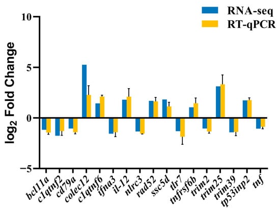
To validate the reliability of the RNA-Seq data, 17 DEGs were selected for RT-qPCR analysis, including eight up-regulated genes—collectin sub-family member 12 (colec12), C1q and tumor necrosis factor related protein 6 (c1qtnf6), interleukin 12 (il-12), DNA repair protein RAD52 (rad52), scavenger receptor cysteine-rich domain-containing protein 5D (ssc5d), tumor necrosis factor receptor superfamily member 6b (tnfrsf6b), tripartite motif containing 25 (trim25), and tumor protein p53 inducible nuclear protein 2 (tp53inp2)-and nine down-regulated genes—B-cell CLL/lymphoma 11A (bcl11a), C1q and tumor necrosis factor related protein 2 (c1qtnf2), cluster of differentiation 79A (cd79a), interferon alpha 3-like (ifna3), NLR family CARD domain containing 3 (nlrc3), toll-like receptor 7 (tlr7), tripartite motif containing 2 (trim2), tripartite motif containing 39 (trim39), and tumor necrosis factor (tnf). β-actin was used as the control. The RT-qPCR was performed on a BIO-RAD CFX96 real-time PCR system using Vazyme SYBR® Green qPCR Master Mix (Vazyme, Nanjing, China). Each 10 μL reaction contained 5 μL of 2× SYBR Green Master Mix, 0.4 μL of each primer, 1 μL of cDNA template, and 3.2 μL nuclease-free water. The cycling conditions were initial denaturation at 95 °C for 3 min followed by 40 cycles of 95 °C for 10 s, annealing at 58 °C for 30 s, and extension at 72 °C for 30 s. A melting curve analysis was performed from 65 °C to 95 °C in 0.5 °C increments every 5 s to verify specificity. The relative expression levels were calculated using the 2−ΔΔCT method. Each sample was run in triplicate to ensure reproducibility.
2.11. Data Analysis
All statistical analyses were performed using IBM SPSS Statistics (version 27.0). All data are presented as mean ± SD and all graphs were generated using GraphPad Prism (version 9.0).
3. Results
3.1. Morphological and Biochemical Characteristics of Bacteria
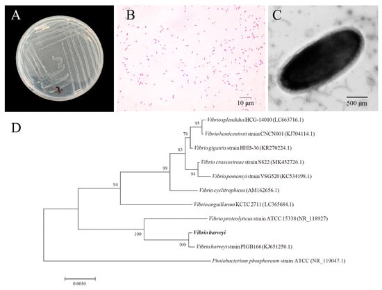
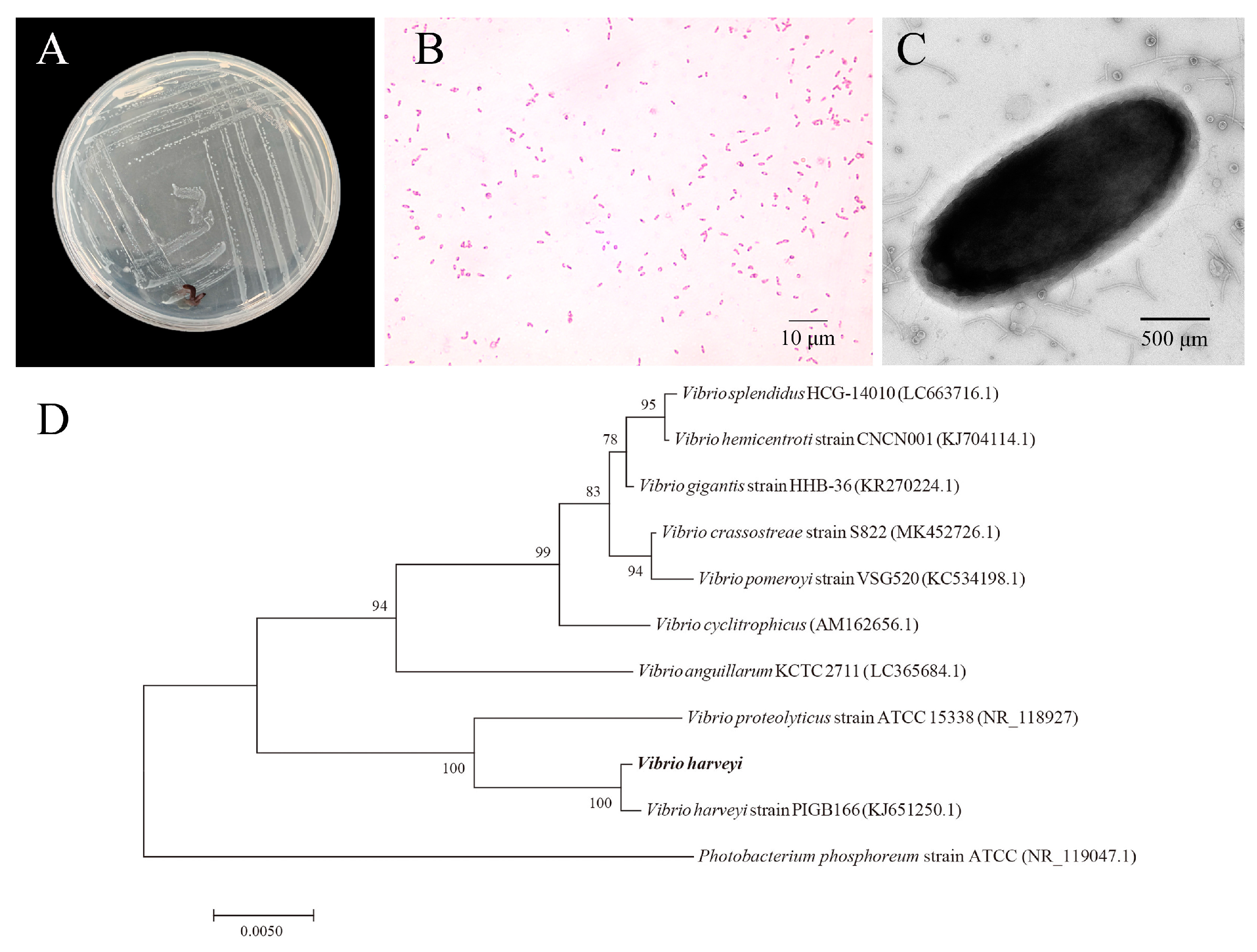
For microscopic examination, no parasites were found in the moribund Seriola dumerili collected from the aquaculture farm on Donghai Island. The bacteria colonies were circular, milky-white, and semi-translucent with a smooth, moist surface, a distinct central elevation, and well-defined boundaries (Figure 1A). Microscopic observation revealed that the cells were Gram-negative and short rod-shaped (Figure 1B) which was confirmed through the presence of short, slightly curved rod-shaped cells using TEM observation (Figure 1C). Biochemical assays indicated that the bacteria were unable to grow in peptone water without added salt, indicating a requirement for saline water. It exhibited moderate halotolerance, with optimal growth occurring at NaCl concentrations of 3–6%, while growth was inhibited at concentrations above 8%. The strain exhibited positive reactions for mannose utilization, lysine decarboxylase activity, and arginine dihydrolase activity. In contrast, it tested negative for glucose and sucrose fermentation, arabinose and inositol metabolism, Voges-Proskauer test, and indole production using the indocyanine substrate (Table 1).
Figure 1.
Morphological and phylogenetic characterization of V. harveyi strain Vh-2. (A) Colony morphology on 2216E agar showing circular, milky-white, semi-translucent colonies with a smooth, moist surface, distinct central elevation, and well-defined boundaries. (B) Gram staining revealing Gram-negative, short, rod-shaped cells (scale = 10 μm). (C) TEM image revealing short, slightly curved, rod-shaped cells (scale = 500 nm). (D) 16S rDNA gene sequence phylogenetic tree with Photobacterium phosphoreum as the outgroup tightly clustering strain Vh-2 with V. harveyi strain PIGB166 (bootstrap value = 100%), both of which were distinct from other Vibrio species (scale = 0.0050).
Table 1.
Physiological and biochemical characteristics of V. harveyi strain Vh-2.
3.2. Sequence Analysis of the 16S rDNA Gene
The 16S rDNA gene sequence of the isolated strain was 1379 bp in length. A BLAST analysis showed that the closest match was V. harveyi strain PIGB166 (KJ651250.1) with 99.85% sequence identity. Therefore, the isolate was identified as V. harveyi and was designated as Vh-2 in this study. A phylogenetic analysis, with Photobacterium phosphoreum used as the outgroup (Figure 1D), revealed that Vh-2 clustered tightly with V. harveyi strain PIGB166 with a bootstrap value of 100%, indicating a high degree of similarity. It was clearly separated from other Vibrio spp. including V. proteolyticus, V. anguillarum, and V. splendidus, further supporting the identification as V. harveyi.
3.3. Detection of Virulence Genes and Drug Sensitivity
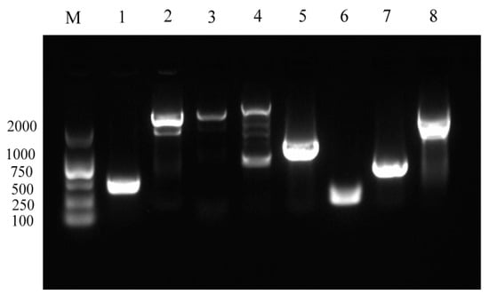
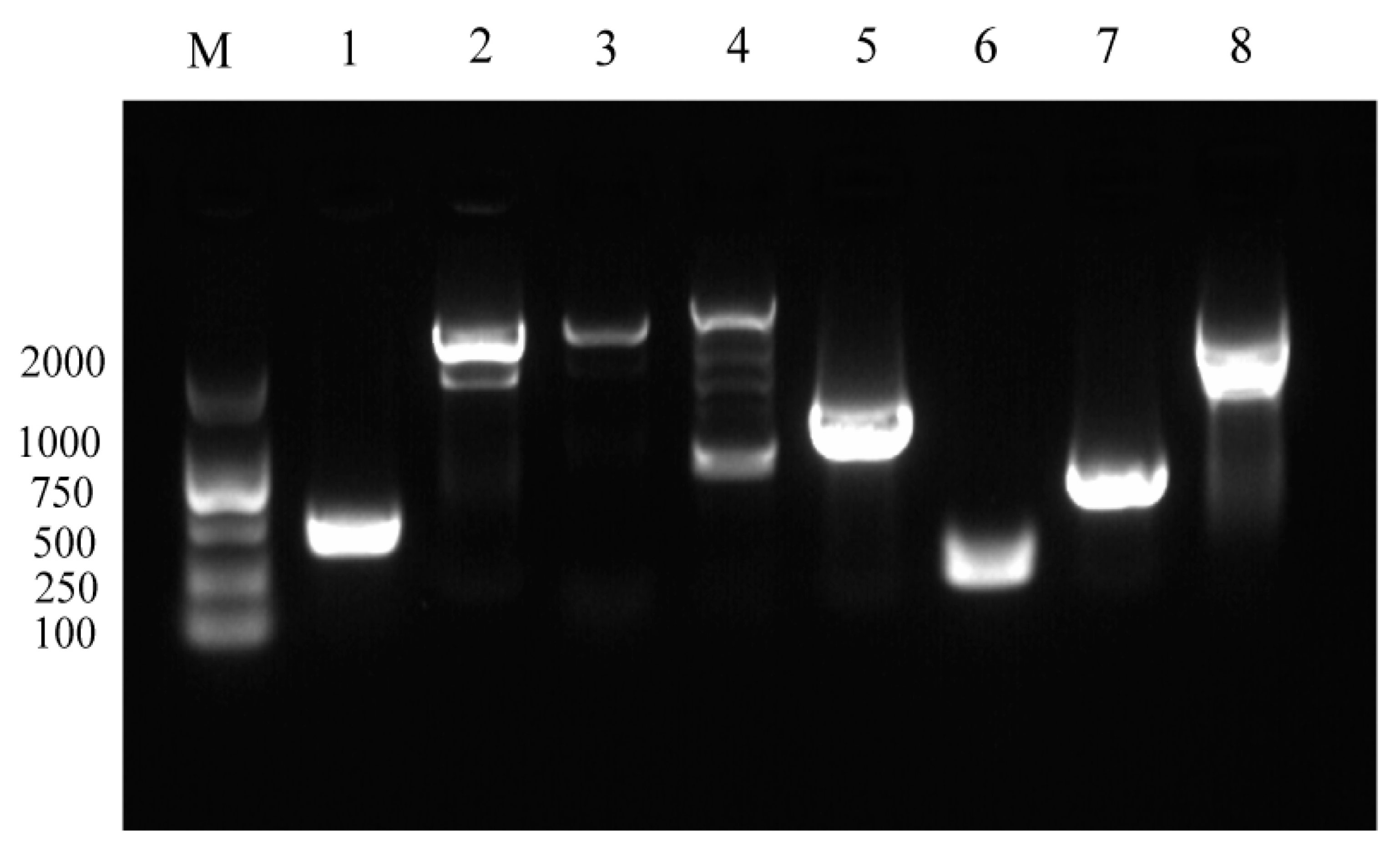
Virulence gene profiling of Vh-2 demonstrated the presence of eight virulence-associated genes, namely toxR, toxS, vhpA, vhpB, vhhA, vhhB, luxR, and pap6 (Figure 2). A total of 29 antibiotics were tested for their activity against the V. harveyi strain Vh-2 (Table 2). The strain exhibited susceptibility (S) to ceftriaxone, florfenicol, and meropenem. Intermediate (I) was observed to cefepime, cefoxitin, azithromycin, chloramphenicol, nitrofurantoin, levofloxacin, and ofloxacin. Conversely, the strain was resistant (R) to the remaining 19 antibiotics, including ampicillin, amoxicillin, piperacillin, ampicillin/sulbactam, ceftazidime, erythromycin, norfloxacin, enrofloxacin, ciprofloxacin, metronidazole, streptomycin, kanamycin, gentamicin, amikacin, tetracycline, vancomycin, polymyxins, trimethoprim-sulfamethoxazole, and imipenem.
Figure 2.
Virulence gene profiling of V. harveyi strain Vh-2 by PCR amplification. Lane M: DNA marker, lanes 1–8: amplification products of the eight virulence-associated genes, including toxR (1), toxS (2), vhpA (3), vhpB (4), vhhA (5), vhhB (6), luxR (7), and pap6 (8).
Table 2.
Results of the antimicrobial susceptibility tests of V. harveyi strain Vh-2.
3.4. Histopathological Analyses
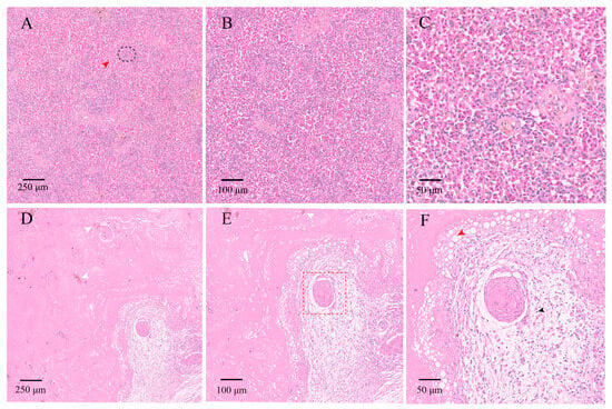
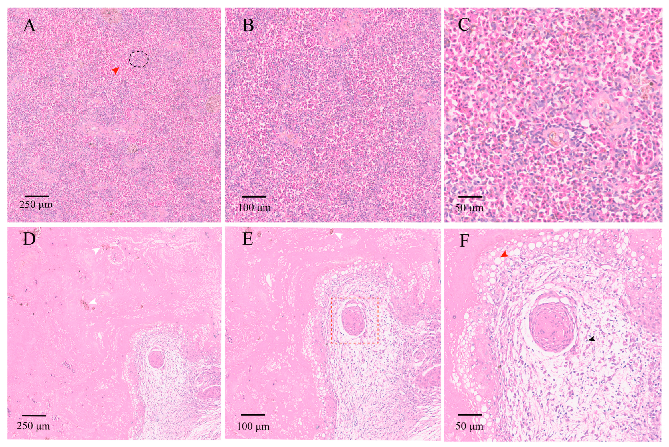
In the control group, the architecture of the spleens was intact with clearly defined red and white pulp and uniform staining. No congestion, hemorrhage, or inflammatory infiltration was observed. Observation under a 100× showed that white pulp was evenly distributed (Figure 3A) and observation under a 200× indicated normal lymphoid follicles (Figure 3B). The splenocytes had intact, round nuclei without vacuolation or degeneration when observed under a 400× (Figure 3C). In the experimental group, extensive lesions were evident on the spleen samples. The diffuse necrosis and pigment deposition (white arrowhead) were visible under a 100× (Figure 3D) and marked structural disorganization, cell loss, and pigment accumulation (white arrowhead) were observed under a 200× (Figure 3E). Furthermore, perivascular lymphocyte infiltration (black arrowhead), prominent cytoplasmic vacuolation (red arrowhead), and nuclear fragmentation were observed under a 400× (Figure 3F).
Figure 3.
Histological changes in the spleen of S. dumerili (H&E staining). (A–C) Control group: (A) 100× (scale = 250 μm), intact spleen structure with clearly defined red and white pulp, Red arrow indicates red pulp; black circle highlights white pulp, (B) 200× (scale = 100 μm), normal lymphoid follicles and uniform tissue staining, (C) 400× (scale = 50 μm), splenocytes with intact, round nuclei and no pathological alterations. (D–F) Experimental group: (D) 100× (scale = 250 μm), diffuse necrotic areas with pigment deposition (white arrowhead), (E) 200× (scale = 100 μm), disrupted tissue architecture, extensive necrosis, and pigment accumulation (white arrowhead), red frame highlights granulomatous lesions, (F) 400× (scale = 50 μm), perivascular lymphocyte infiltration (black arrowhead) and marked cytoplasmic vacuolation (red arrowhead), with nuclear fragmentation.
3.5. Transcriptome Sequencing and Assembly
The quantity of raw reads obtained for all samples ranged from 60.22 to 67.53 million, which decreased to 59.64 to 62.68 million clean reads for the control group (CS1–CS4) and 58.43 to 66.97 million clean reads for the experimental group (PS1–PS4) after quality filtering (Table 3). The Q20 values for all samples were up to 97.75%, the Q30 values were more than 93.58%, and the GC content was stable within the range of 47.76–48.43%, indicative of high-quality sequencing data. The construction of the transcriptome was, therefore, successful and suitable for subsequent analysis.
Table 3.
Summary of sequencing data and quality control for the RNA-Seq samples.
3.6. Identification of DEGs and Enrichment Analysis
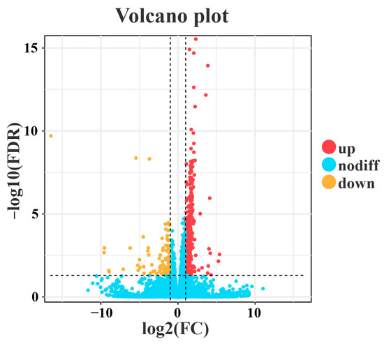
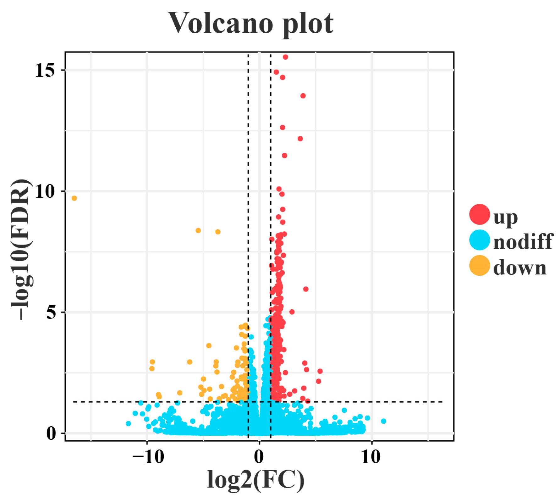
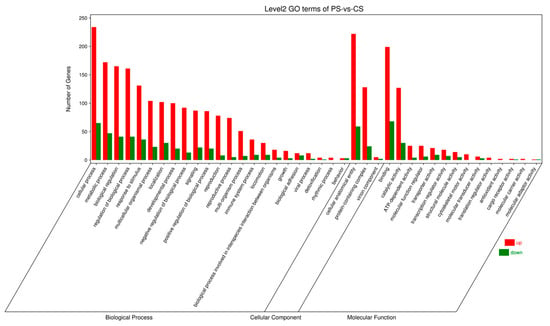
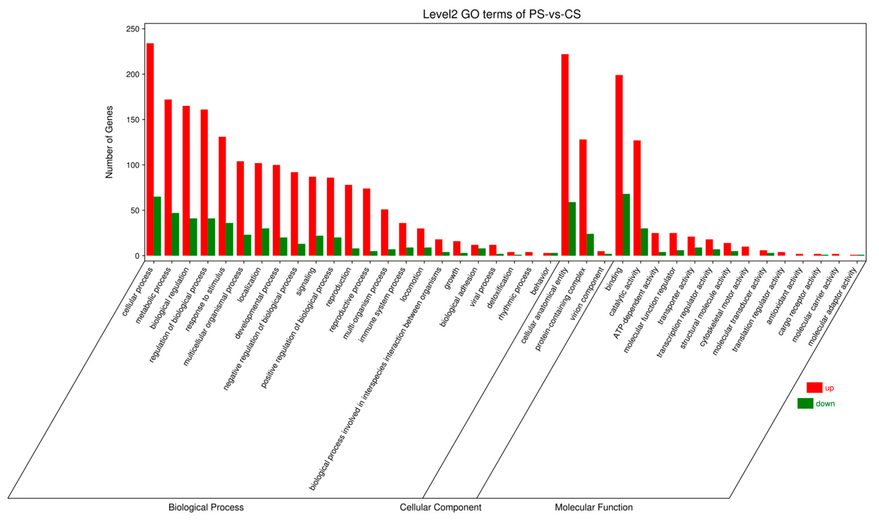
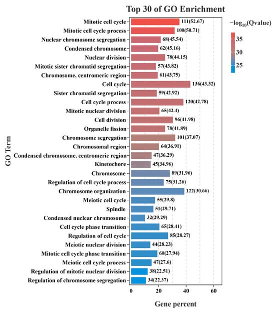
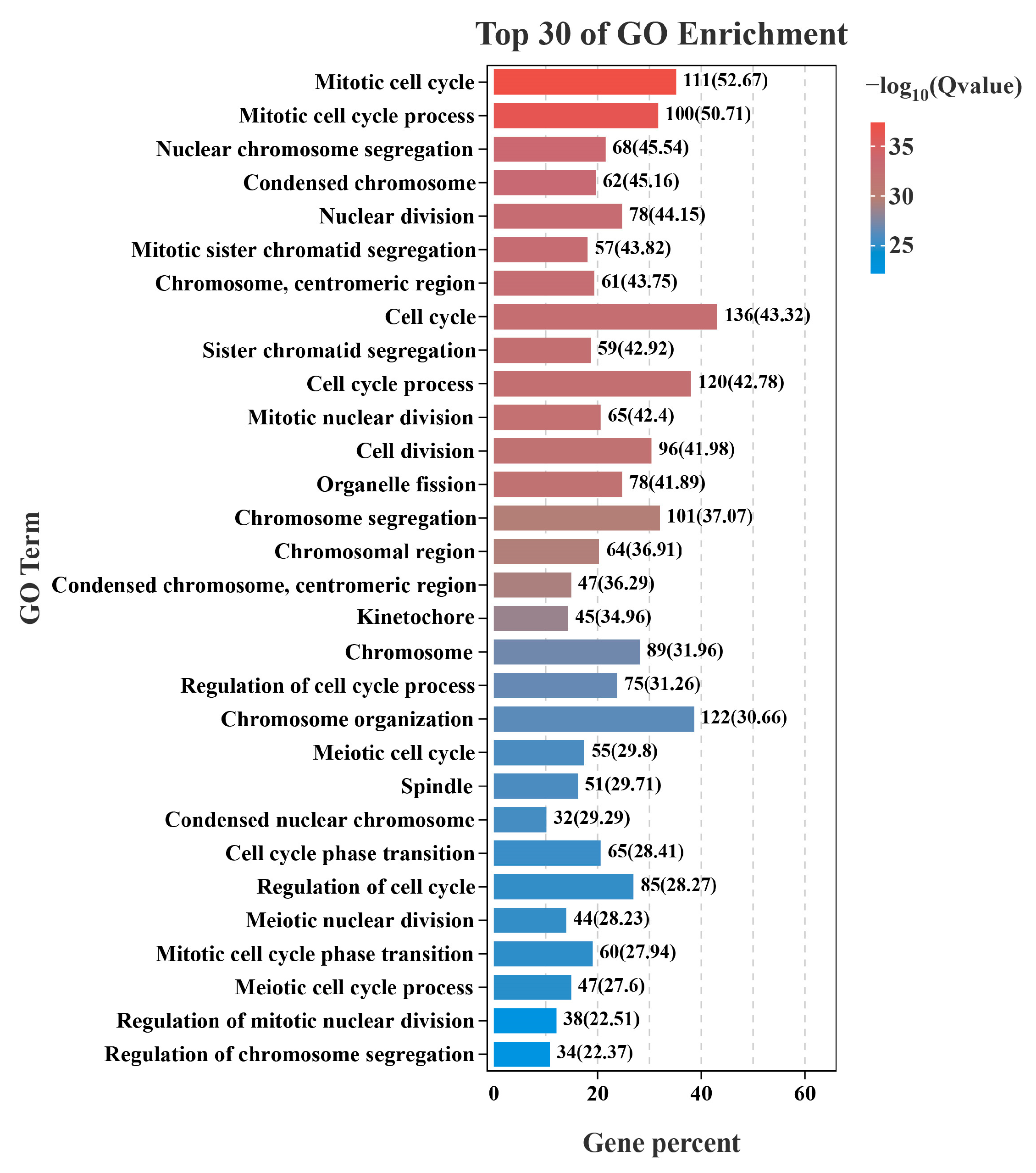
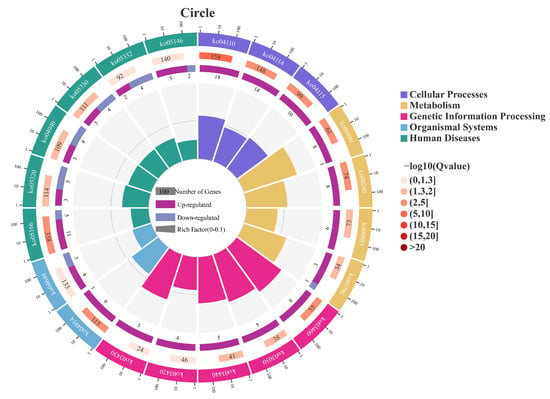
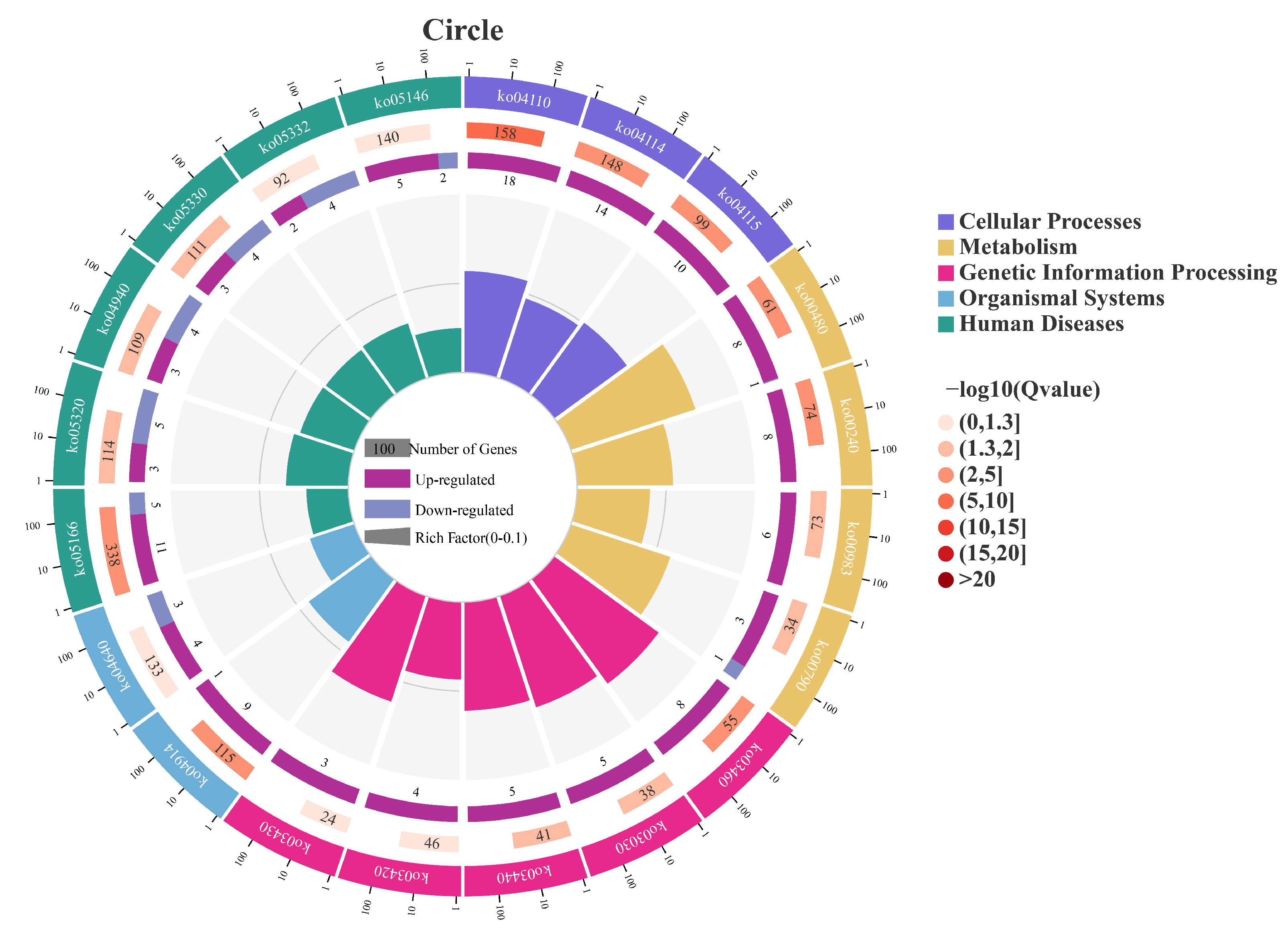
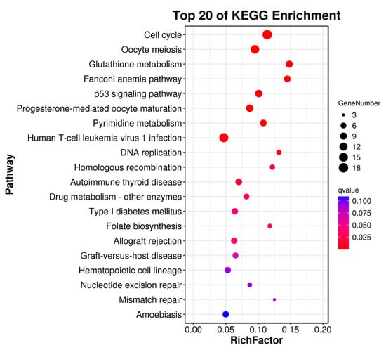
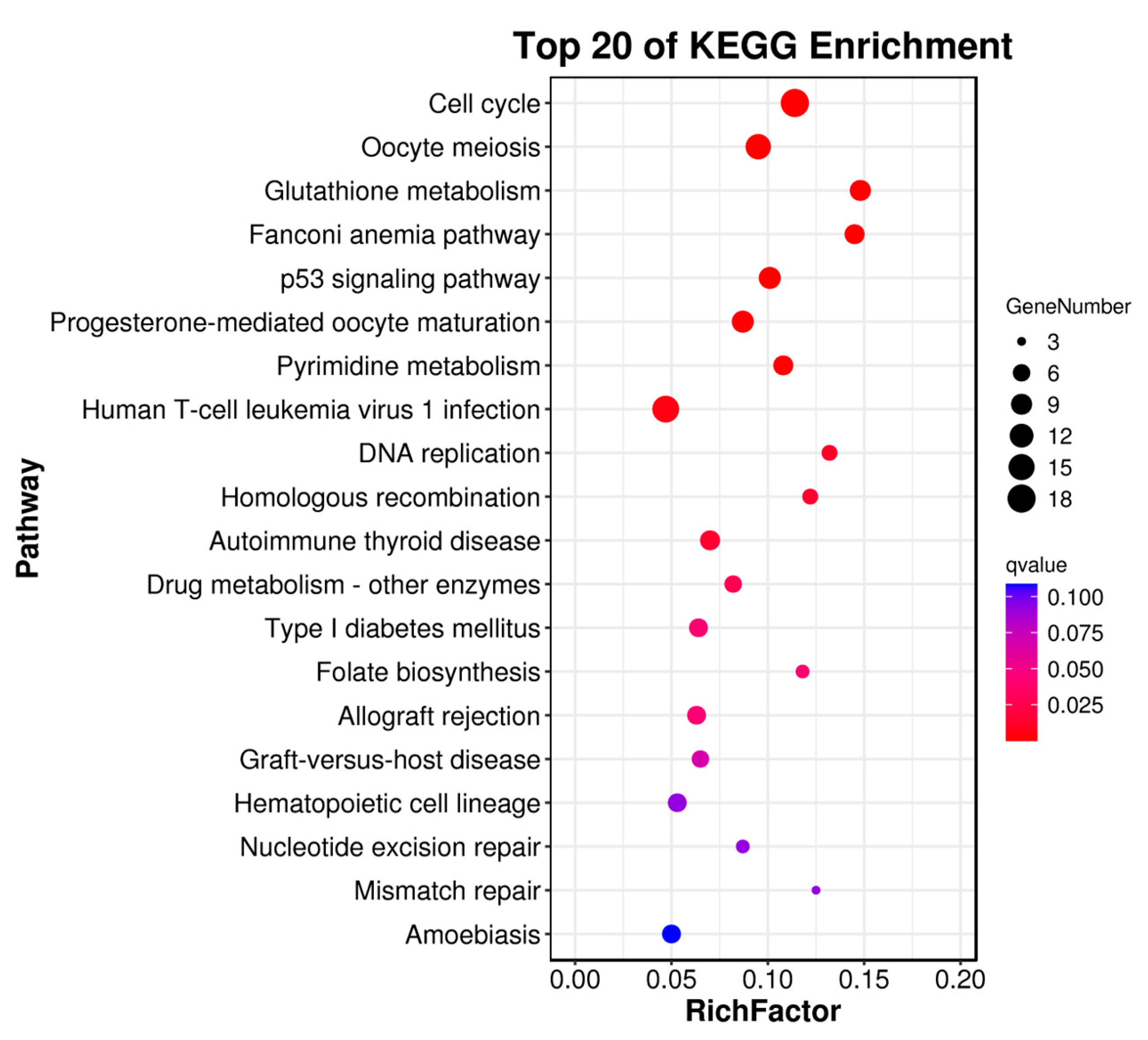
Through RNA sequencing, a total of 396 DEGs, including 293 upregulated genes and 103 downregulated genes, were obtained (Figure 4). Among these, the top 10 up- and down-regulated DEGs are displayed in Table S2. Subsequently, the GO and KEGG pathways were enriched and analyzed. The GO enrichment analysis assigned the DEGs to 1112 functional terms, including 845 terms related to biological processes, 146 terms to cellular components, and 121 terms to molecules functions (Figure 5). Significantly enriched biological process terms included mitotic cell cycle (GO:0000278), mitotic cell cycle process (GO:1903047), nuclear chromosome segregation (GO:0098813), and nuclear division (GO:0000280). Furthermore, during cellular processes, DEGs were predominantly enriched in condensed chromosome (GO:0000793), chromosome, centromeric region (GO:0000775), chromosomal region (GO:0098687), and condensed chromosome, centromeric region (GO:0000779). In the context of molecular processes microtubule binding (GO:0008017), tubulin binding (GO:0015631), ATP binding (GO:0005524), and adenyl nucleotide binding (GO:0030554) were particularly salient (Figure 6). After functional annotation of DEGs, a total of 150 genes were annotated in the KEGG database, mainly involved in cell cycle regulation, DNA repair, metabolic processes, and immune responses (Figure 7). KEGG enrichment analysis revealed that these genes were predominantly enriched in the Cell cycle, Oocyte meiosis, Glutathione metabolism, Fanconi anemia pathway, and p53 signaling pathway (Figure 8).
Figure 4.
Volcano plot of DEGs between naturally infected and healthy S. dumerili. Blue dots represent genes with no significant differential expression, while red and yellow dots denote significantly upregulated and downregulated genes, respectively. The x-axis shows the log2 fold-change, and the y-axis shows −log10 of statistical significance (q-value). Vertical dashed lines indicate the fold-change threshold (|log2FC| > 1), and the horizontal dashed line indicates the significance threshold (q-value < 0.05).
Figure 5.
Level 2 Gene Ontology (GO) terms enriched in PS vs. CS. Red and green bars represent the number of up-regulated and down-regulated differentially expressed genes (DEGs), respectively.
Figure 6.
Top 30 enriched GO terms of DEGs ranked by the proportion of annotated genes in S. dumerili. The x-axis represents the percentage of DEGs annotated to each GO term (gene ratio). The y-axis lists the GO terms ranked by this percentage. Bar length indicates the gene ratio, and bar color represents the significance of enrichment (−log10(q-value)).
Figure 7.
Circular representation of KEGG pathway enrichment in PS vs. CS. Outer circle: the top 20 enriched pathways with a scale indicating the number of DEGs. Second circle: the number of DEGs in the background gene set for each pathway (longer bars indicate higher counts) and q-value (darker red indicates smaller q-value). Third circle: proportion of up-regulated (deep purple) and down-regulated (light purple) DEGs in each pathway. Inner circle: Rich Factor (number of DEGs in the pathway divided by total genes in the pathway), with grid lines at 0.1 intervals.
Figure 8.
Top 20 significantly enriched KEGG pathways of DEGs in S. dumerili. The x-axis represents the gene ratio (proportion of DEGs mapped to each pathway), the y-axis lists the KEGG pathway names, bubble size indicates the number of DEGs in each pathway, and bubble color represents the significance of enrichment (−log10 (q-value)).
3.7. Verification of DEGs by RT-qPCR
To validate the reliability of the RNA-Seq data, the transcriptional levels of 17 selected DEGs were detected by RT-qPCR. These genes covered functional pathways such as immune recognition, signal transduction, inflammatory regulation, and cell repair. The RT-qPCR validation results were highly consistent with the RNA-Seq data, thus confirming the reliability of the transcriptomics sequencing data (Figure 9).
Figure 9.
Validation and comparison of candidate gene expression levels by comparing the RNA-Seq and RT-qPCR results. The x-axis shows the gene names, and the y-axis represents the log2 fold change in expression, where the blue and yellow bars (with SD) indicate RNA-Seq and RT-qPCR data, respectively.
4. Discussion
Greater amberjack aquaculture farms have recently been affected by frequent outbreaks of bacterial diseases, with V. harveyi being the predominant pathogen. To investigate the pathogenic mechanism of V. harveyi and the immune response mechanism of greater amberjack to V. harveyi, we characterized the pathogenic features of the V. harveyi Vh-2 strain and explored the transcriptomic host-disease responses of the greater amberjack to V. harveyi infection. Many DEGs were identified, predominantly involved in immune defense mechanisms, inflammatory responses, and cellular apoptosis.
Through physiological and biochemical characterization and 16S rDNA sequence comparison, the strain isolated from diseased greater amberjack spleen tissue was identified as V. harveyi, which has been widely reported as a primary pathogen in marine fish species [26]. The Vh-2 strain exhibited typical physiological and biochemical halophilic characteristics, growing well under 3% and 6% NaCl conditions, but unable to grow in a salt-free environment, demonstrating its salt dependence as a marine halophilic bacterium [27]. Additionally, the strain exhibited positive reactions with lysine and arginine decarboxylase, indicating its capacity for amino acid metabolism, corresponding to the general characteristics of the Vibrio genus [28]. The isolated strain failed to ferment glucose, sucrose, arabinose, and inositol, but tested positive for mannose, reflecting individual variability in carbon source utilization [29]. Additionally, Vibrio variants may have unstable or even completely negative sugar fermentation capabilities, which may be related to their adaptability to various hosts and environments [30]. The antimicrobial susceptibility patterns of Vh-2 generally align with β-lactam and macrolide resistance and florfenicol and meropenem susceptibility of V. harveyi from southern China [31]. Such patterns may be shaped by regional antibiotic practices or strain-specific resistance gene mutations [32,33,34]. This multi-drug resistance phenotype is consistent with trends reported in isolates from diverse geographic regions, which often retain common resistance to protein-synthesis inhibitors despite genotypic variations [30].
The virulence factors of V. harveyi have been classified and divided into two categories: widely conserved typical genes (luxR, toxR, vhpA, chiA, SP, vhh, vhml and vhs) and atypical genes with variable distribution (zot, toxRVc, tcpA, ctxA, hlyA, flaC, tdh, trh and vvh) [35]. In addition to some typical virulence genes (toxR, luxR, vhh, vhp), our study also identified toxS and pap6. Previous studies have demonstrated that toxS functions alongside toxR, stabilizing it and preventing degradation, thereby ensuring proper regulation of virulence gene expression [36]. Althuohg we did not perform sequencing analysis on the virulence gene amplicons, previous literature indicates that the selected core regulatory genes (e.g., toxR and luxR) are highly conserved across V. harveyi and Harveyi clade strains; the hemolysin genes vhhA/vhhB may exhibit copy duplication in highly virulent strains (with 98.8% homology), whereas the metalloproteases vhpA/vhpB/pap6 share conserved structural domains, occasionally resulting in non-specific amplification products during PCR (as observed in the vhpB lane), and no significant SNPs leading to amino acid changes have been reported [22,35,37]. Specifically, the pap6 gene, originally cloned from V. harveyi AP6, encodes a zinc-dependent metalloprotease capable of degrading gelatin, fibronectin, and type IV collagen, suggesting its potential role in host tissue damage and infection [37]. Histopathology aids in assessing structural damage to organs post-infection and findings have revealed significant structural damage to greater amberjack spleen (extensive cellular necrosis, red blood cell exudation, and increased inflammatory cells) infected with V. harveyi. This is consistent with histopathological splenic lesions reported in other fish species, such as groupers and sea bass, infected with Vibrio spp., which often displayed red blood cell extravasation and lymphocyte aggregation, hallmarks of the spleen’s dual role in blood filtration and inflammatory regulation [38,39].
Innate immunity constitutes the first line of defense against microbial infections [40], which recognizes various pathogen-associated molecular patterns via specific pattern-recognition receptors such as Toll-like receptors (TLR), RIG-I-like receptors (RLR), and NOD-like receptors (NLR) [41]. Enrichment of TLR, RLR, and NLR pathways, along with differential expression of tlr7 (downregulated), trim25 (upregulated), and nlrc3 (downregulated), indicates that pathogen recognition and subsequent innate immune signaling were actively modulated in S. dumerili following V. harveyi infection. The endosomal pattern-recognition receptor tlr7 detects single-stranded RNA and activates MyD88-IRF7-dependent type I interferon (IFN) production [42]. In this study, we found that tlr7 was downregulated in S. dumerili in response to V. harveyi infection, similar to observations in vivo during porcine epidemic diarrhea virus infection where tlr7 downregulation resulted in the inhibition of NF-kappa B signaling and reduced TNF-α expression. It was recently reported that TLR7 expression is downregulated in the livers of patients infected with hepatitis C, which may be associated with immune negative feedback mechanisms or virus-mediated immune evasion [43]. In contrast, TRIM25 acts as a key E3 ubiquitin ligase that mediates K63-linked ubiquitination of RIG-I, enhancing antiviral signaling [44]. It has been suggested that common carp (Cyprinus carpio) RIG-I and TRIM25 effectively inhibit spring viremia of carp virus replication by inducing phosphorylation of TBK1, IRF3, and p65 [45]. Similarly, during nervous necrosis virus infection in red spotted grouper (Epinephelus akaara), zebrafish TRIM25 acted as an enhancer of the RLR signaling pathway [46]. Our results suggest that the RLR signaling pathway in S. dumerili may play an important role in host immune defense against V. harveyi. As nlrc3 expression was downregulated in S. dumerili after V. harveyi infection, this suggests a release of negative immune regulation. The negative regulatory role of NLRC3-like may provide valuable insights into how homeostasis is maintained and the causes of autoimmune and inflammatory diseases [47]. In teleosts, NLRC3-like proteins can inhibit NOD1-RIP2 or STING pathways, exerting both suppressive and activating effects on the regulation of pathogen-induced inflammation [48,49]. Downregulation of nlrc3 may relieve this inhibition, enhancing pro-inflammatory responses, promoting bacterial elimination, and limiting pathogen spread [50]. These changes suggest that an innate immune response played an essential role in great amberjacks when infected with V. harveyi.
Transcriptomic analysis revealed pronounced upregulation of pro-inflammatory cytokines, notably il-12 and tnf-α. Cytokine–cytokine receptor interactions and JAK–STAT pathways were significantly enriched, with il-12 and tnf-α showing notable expression changes. While essential for recruiting immune cells to the infection site and eliminating bacteria [51,52], cytokines from the IL-12 family are recognized for their critical roles in cancer, infections, and inflammatory conditions by regulating both innate and adaptive immunity [53]. IL-12, comprising IL-12p35 and IL-12p40, is a multifunctional cytokine involved in inflammation regulation and the bacterial immune response [54]. Seriola dumerili il-12 was upregulated after V. harveyi infection, consistent with its role in promoting Th1 polarization and macrophage activation in teleosts [55]. LPS, polyI:C, IFN-γ, and IL-1β can trigger IL-12 production in rainbow trout (Oncorhynchus mykiss) macrophages [56]. Recombinant IL-12 has been shown to stimulate peripheral blood lymphocyte proliferation and induce tnf-α expression in groupers, suggesting a conserved role in pro-inflammatory and cell-mediated immunity [57]. As a central mediator of inflammatory responses, tnf-α in fish have shown increased expression during the initial phases of infection [58]. In turbot (Scophthalmus maximus) infected with Aeromonas salmonicida and rainbow trout infected with Yersinia ruckeri, there were significant increases in tnf-α transcripts [59,60,61]. Conversely, our study shows that tnf-α expression in S. dumerili was downregulated after V. harveyi infection. Moreover, gene expression kinetics in flounder were studied using a challenge model that simulated both acute and chronic Nocardia seriolae infections, revealing a significant increase in tnf-α during the early stages of acute infection, followed by an unexpected downregulation [62]. Based on this, we can assume that the V. harveyi infection in the great amberjack was chronic, which may represent a regulatory counterbalance to prevent excessive tissue damage during systemic infection.
It is important to note the limitations of our study. The single time-point analysis only captures a snapshot of the dynamic host–pathogen interactions. Future studies should incorporate a time-series analysis to delineate the kinetic progression of the immune responses in fish. Furthermore, the functional role of the candidate genes identified here, such as il-12, requires validation through in vivo functional assays.
5. Conclusions
In conclusion, this study systematically described the pathogenic mechanism of V. harveyi and screened the primary DEGs and pathways involved in pathogen recognition, inflammation, and immune response to V. harveyi infection in the greater amberjack. These findings have improved our knowledge of the immune responses of greater amberjack to V. harveyi and will assist in producing effective vaccines, treatments, and disease-resistant strains.
Supplementary Materials
The following supporting information can be downloaded at: https://www.mdpi.com/article/10.3390/ani16020184/s1, Table S1: Primers sequences used in this study; Table S2: Top ten up and down-regulated DEGs in the spleen of Seriola dumerili following Vibrio harveyi infection.
Author Contributions
Conceptualization, C.Z.; Methodology, R.Z. and X.Z.; Validation, K.L. and H.T.; Formal analysis, R.Z.; Investigation, J.H., K.L. and H.T.; Resources, K.L., H.T. and C.Z.; Writing—original draft, R.Z.; Writing—review & editing, R.Z., J.H., X.Z., Y.H. and C.Z.; Visualization, R.Z. and Y.H.; Supervision, C.Z.; Project administration, C.Z.; Funding acquisition, C.Z. All authors have read and agreed to the published version of the manuscript.
Funding
This study was supported by Guangdong Ocean University (030301022301) and Southern Marine Science and Engineering Guangdong Laboratory (Zhanjiang) (ZJW2023-10 and ZJW-2022-05).
Institutional Review Board Statement
All experiment procedures were conducted strictly following the ethical review requirements for laboratory animals at Guangdong Ocean University (Ethics Approval Number: 2023090504, approved on 5 May 2023) and followed the relevant guidelines of the National Institutes of Health’s ‘Guide for the Care and Use of Laboratory Animals’.
Informed Consent Statement
Not applicable.
Data Availability Statement
The data that support the findings of this study are available upon reasonable request. The raw reads used in this article have been deposited into the Sequence Read Archive (SRA) of the NCBI database under BioProject accession number: PRJNA135468.
Acknowledgments
We acknowledge all funders of this work.
Conflicts of Interest
The authors declare that the research was conducted in the absence of any commercial or financial relationships that could be construed as a potential conflict of interest.
References
- Pang, H.; Chang, Y.; Zheng, H.; Tan, H.; Zhou, S.; Zeng, F.; Hoare, R.; Monaghan, S.J.; Wang, N.; Ding, Y. A live attenuated strain of HY9901ΔvscB provides protection against Vibrio alginolyticus in pearl gentian grouper (♀Epinephelus fuscoguttatus × ♂Epinephelus lanceolatu). Aquaculture 2022, 546, 737353. [Google Scholar] [CrossRef] [Scilit]
- Minami, T.; Iwata, K.; Shimahara, Y.; Yuasa, K. Vibrio harveyi Infection in Farmed Greater Amberjack Seriola dumerili. Fish Pathol. 2016, 51, 1–7. [Google Scholar] [CrossRef] [Scilit]
- Zhang, X.-H.; He, X.; Austin, B. Vibrio harveyi: A serious pathogen of fish and invertebrates in mariculture. Mar. Life Sci. Technol. 2020, 2, 231–245. [Google Scholar] [CrossRef] [Scilit] [PubMed]
- Firmino, J.; Furones, M.D.; Andree, K.B.; Sarasquete, C.; Ortiz-Delgado, J.B.; Asencio-Alcudia, G.; Gisbert, E. Contrasting outcomes of Vibrio harveyi pathogenicity in gilthead seabream, Sparus aurata and European seabass, Dicentrachus labrax. Aquaculture 2019, 511, 734210. [Google Scholar] [CrossRef] [Scilit]
- Darshanee Ruwandeepika, H.A.; Sanjeewa Prasad Jayaweera, T.; Paban Bhowmick, P.; Karunasagar, I.; Bossier, P.; Defoirdt, T. Pathogenesis, virulence factors and virulence regulation of vibrios belonging to the Harveyi clade. Rev. Aquac. 2012, 4, 59–74. [Google Scholar] [CrossRef] [Scilit]
- Zhang, Y.; Deng, Y.; Feng, J.; Hu, J.; Chen, H.; Guo, Z.; Gao, R.; Su, Y. ToxR modulates biofilm formation in fish pathogen Vibrio harveyi. Lett. Appl. Microbiol. 2022, 74, 288–299. [Google Scholar] [CrossRef] [Scilit]
- Gao, D.; Lei, W.; Wang, C.; Ni, P.; Cui, X.; Huang, X.; Ye, S. RNA-Sequencing Analysis of the Spleen and Gill of Takifugu rubripes in Response to Vibrio harveyi Infection. Front. Vet. Sci. 2022, 8, 813988. [Google Scholar] [CrossRef] [Scilit]
- Zhang, X.; Wang, S.; Chen, S.; Chen, Y.; Liu, Y.; Shao, C.; Wang, Q.; Lu, Y.; Gong, G.; Ding, S.; et al. Transcriptome analysis revealed changes of multiple genes involved in immunity in Cynoglossus semilaevis during Vibrio anguillarum infection. Fish Shellfish Immunol. 2015, 43, 209–218. [Google Scholar] [CrossRef] [Scilit]
- Peng, H.; Yang, B.; Li, B.; Cai, Z.; Cui, Q.; Chen, M.; Liu, X.; Yang, X.; Jiang, C. Comparative transcriptomic analysis reveals the gene expression profiles in the liver and spleen of Japanese pufferfish (Takifugu rubripes) in response to Vibrio harveyi infection. Fish Shellfish Immunol. 2019, 90, 308–316. [Google Scholar] [CrossRef] [Scilit]
- Sun, Z.; Li, X.; Zhang, Q.; Wang, W.; Wang, H.; Pan, T.; Gao, Q. Transcriptome Analysis of Immune Response Against Edwardsiella tarda Infection in Spotted Sea Bass (Lateolabrax maculatus). Fishes 2025, 10, 153. [Google Scholar] [CrossRef] [Scilit]
- Qian, X.; Ba, Y.; Zhuang, Q.; Zhong, G. RNA-Seq technology and its application in fish transcriptomics. OMICS 2014, 18, 98–110. [Google Scholar] [CrossRef] [Scilit] [PubMed]
- Castro, R.; Jouneau, L.; Pham, H.-P.; Bouchez, O.; Giudicelli, V.; Lefranc, M.-P.; Quillet, E.; Benmansour, A.; Cazals, F.; Six, A.; et al. Teleost Fish Mount Complex Clonal IgM and IgT Responses in Spleen upon Systemic Viral Infection. PLoS Pathog. 2013, 9, e1003098. [Google Scholar] [CrossRef] [Scilit] [PubMed]
- Uribe, C.; Folch, H.; Enriquez, R.; Moran, G. Innate and adaptive immunity in teleost fish: A review. Veterinární Medicína 2011, 56, 486–503. [Google Scholar] [CrossRef] [Scilit]
- Long, H.; Tian, Y.; Zhang, D.; Li, L.; Hao, R.; Li, H.; Wang, C.; Ru, X.; Deng, Q.; Huang, Y.; et al. Synergistic integration of transcriptomics and metabolomics analyses provides novel insights into the mechanisms underlying unsynchronized growth of greater amberjack (Seriola dumerili). Comp. Biochem. Physiol. Part D Genom. Proteom. 2025, 54, 101441. [Google Scholar] [CrossRef] [Scilit]
- Yang, J.; Ru, X.; Huang, Y.; Wu, J.; Yang, T.; Chen, P.; Li, J.; Zhu, K.; Zhu, C. Transcriptomic Analysis Provides New Insights into Oocyte Growth and Maturation in Greater Amberjack (Seriola dumerili). Animals 2025, 15, 333. [Google Scholar] [CrossRef] [Scilit]
- Ru, X.; Huang, Y.; Shi, H.; Peng, Y.; Hao, R.; Yang, T.; Zhu, K.; Li, G.; Zhu, C. RNA-seq of hypo- and hyper-salinity stress-response transcriptome in the liver of greater amberjack (Seriola dumerili) juveniles. Aquac. Rep. 2023, 29, 101498. [Google Scholar] [CrossRef] [Scilit]
- Castillo, D.; D’Alvise, P.; Middelboe, M.; Gram, L.; Liu, S.; Kalatzis, P.G.; Kokkari, C.; Katharios, P. Draft Genome Sequences of the Fish Pathogen Vibrio harveyi Strains VH2 and VH5. Genome Announc. 2015, 3, e01062-15. [Google Scholar] [CrossRef] [Scilit]
- Rigos, G.; Katharios, P.; Kogiannou, D.; Cascarano, C.M. Infectious diseases and treatment solutions of farmed greater amberjack Seriola dumerili with particular emphasis in Mediterranean region. Rev. Aquac. 2021, 13, 301–323. [Google Scholar] [CrossRef] [Scilit]
- Kumar, S.; Stecher, G.; Tamura, K. MEGA7: Molecular Evolutionary Genetics Analysis Version 7.0 for Bigger Datasets. Mol. Biol. Evol. 2016, 33, 1870–1874. [Google Scholar] [CrossRef] [Scilit]
- Buller, N.B. Bacteria and Fungi from Fish and Other Aquatic Animals: A Practical Identification Manual; Centre for Agriculture and Bioscience International (CABI): Boston, MA, USA, 2010; Volume 28, p. 627. [Google Scholar]
- Garrity, G.M.; Brenner, D.J.; Krieg, N.R.; Staley, J.T. Bergey’s Manual of Systematic Bacteriology, 2nd ed.; Springer: New York, NY, USA, 2005. [Google Scholar]
- Zhang, X.H.; Meaden, P.G.; Austin, B. Duplication of Hemolysin Genes in a Virulent Isolate of Vibrio harveyi. Appl. Environ. Microbiol. 2001, 67, 3161–3167. [Google Scholar] [CrossRef] [Scilit]
- Kumar, S.; Kumar, C.B.; Rajendran, V.; Abishaw, N.; Anand, P.S.S.; Kannapan, S.; Nagaleekar, V.K.; Vijayan, K.K.; Alavandi, S.V. Delineating virulence of Vibrio campbellii: A predominant luminescent bacterial pathogen in Indian shrimp hatcheries. Sci. Rep. 2021, 11, 15831. [Google Scholar] [CrossRef] [Scilit] [PubMed]
- Yang, A.; Li, W.; Tao, Z.; Ye, H.; Xu, Z.; Li, Y.; Gao, Y.; Yan, X. Vibrio harveyi isolated from marine aquaculture species in eastern China and virulence to the large yellow croaker (Larimichthys crocea). J. Appl. Microbiol. 2021, 131, 1710–1721. [Google Scholar] [CrossRef] [Scilit] [PubMed]
- Weinstein, M.P.; Lewis, J.S. The Clinical and Laboratory Standards Institute Subcommittee on Antimicrobial Susceptibility Testing: Background, Organization, Functions, and Processes. J. Clin. Microbiol. 2020, 58, e01864-19. [Google Scholar] [CrossRef] [PubMed]
- Austin, B.; Zhang, X.-H. Vibrio harveyi: A significant pathogen of marine vertebrates and invertebrates. Lett. Appl. Microbiol. 2006, 43, 119–124. [Google Scholar] [CrossRef] [Scilit]
- Montánchez, I.; Kaberdin, V.R. Vibrio harveyi: A brief survey of general characteristics and recent epidemiological traits associated with climate change. Mar. Environ. Res. 2020, 154, 104850. [Google Scholar] [CrossRef] [Scilit]
- Kang, H.; Yu, Y.; Liao, M.; Wang, Y.; Yang, G.; Zhang, Z.; Li, B.; Rong, X.; Wang, C. Physiology, metabolism, antibiotic resistance, and genetic diversity of Harveyi clade bacteria isolated from coastal mariculture system in China in the last two decades. Front. Mar. Sci. 2022, 9, 932255. [Google Scholar] [CrossRef] [Scilit]
- Triga, A.; Smyrli, M.; Katharios, P. Pathogenic and Opportunistic Vibrio spp. Associated with Vibriosis Incidences in the Greek Aquaculture: The Role of Vibrio harveyi as the Principal Cause of Vibriosis. Microorganisms 2023, 11, 1197. [Google Scholar] [CrossRef] [Scilit]
- Pavlinec, Ž.; Zupičić, I.G.; Oraić, D.; Lojkić, I.; Fouz, B.; Zrnčić, S. Biochemical and molecular characterization of three serologically different Vibrio harveyi strains isolated from farmed Dicentrarchus labrax from the Adriatic Sea. Sci. Rep. 2022, 12, 7309. [Google Scholar] [CrossRef] [Scilit]
- Zhu, Z.M.; Dong, C.F.; Weng, S.P.; He, J.G. The high prevalence of pathogenic Vibrio harveyi with multiple antibiotic resistance in scale drop and muscle necrosis disease of the hybrid grouper, Epinephelus fuscoguttatus (♀) × E. lanceolatus (♂), in China. J. Fish Dis. 2018, 41, 589–601. [Google Scholar] [CrossRef] [Scilit]
- Teo, J.W.P.; Suwanto, A.; Poh, C.L. Novel β-Lactamase Genes from Two Environmental Isolates of Vibrio harveyi. Antimicrob. Agents Chemother. 2000, 44, 1309–1314. [Google Scholar] [CrossRef] [Scilit]
- Wu, Z.; Wu, Y.; Gao, H.; He, X.; Yao, Q.; Yang, Z.; Zhou, J.; Ji, L.; Gao, J.; Jia, X.; et al. Identification and whole-genome sequencing analysis of Vibrio vulnificus strains causing pearl gentian grouper disease in China. BMC Microbiol. 2022, 22, 200. [Google Scholar] [CrossRef] [Scilit] [PubMed]
- Yan, L.; Liu, D.; Wang, X.-H.; Wang, Y.; Zhang, B.; Wang, M.; Xu, H. Bacterial plasmid-mediated quinolone resistance genes in aquatic environments in China. Sci. Rep. 2017, 7, 40610. [Google Scholar] [CrossRef] [Scilit] [PubMed]
- Ruwandeepika, H.A.D.; Defoirdt, T.; Bhowmick, P.P.; Shekar, M.; Bossier, P.; Karunasagar, I. Presence of typical and atypical virulence genes in vibrio isolates belonging to the Harveyi clade: Presence of atypical genes in Harveyi clade. J. Appl. Microbiol. 2010, 109, 888–899. [Google Scholar] [CrossRef] [Scilit] [PubMed]
- Almagro-Moreno, S.; Root, M.Z.; Taylor, R.K. Role of ToxS in the proteolytic cascade of virulence regulator ToxR in Vibrio cholerae. Mol. Microbiol. 2015, 98, 963–976. [Google Scholar] [CrossRef] [Scilit]
- Teo, J.W.P.; Zhang, L.H.; Poh, C.L. Cloning and characterization of a metalloprotease from Vibrio harveyi strain AP6. Gene 2003, 303, 147–156. [Google Scholar] [CrossRef] [Scilit]
- He, Y.; Wang, E.; Wang, K.; Wang, J.; Fan, W.; Chen, D.; Yang, Q. Morphology of the Spleen in Oreochromis niloticus: Splenic Subregions and the Blood-Spleen Barrier. Animals 2021, 11, 2934. [Google Scholar] [CrossRef] [Scilit]
- Manchanayake, T.; Salleh, A.; Amal, M.N.A.; Yasin, I.S.M.; Zamri-Saad, M. Pathology and pathogenesis of Vibrio infection in fish: A review. Aquac. Rep. 2023, 28, 101459. [Google Scholar] [CrossRef] [Scilit]
- Alavi Darazam, I.; Shokouhi, S.; Pourhoseingholi, M.A.; Naghibi Irvani, S.S.; Mokhtari, M.; Shabani, M.; Amirdosara, M.; Torabinavid, P.; Golmohammadi, M.; Hashemi, S.; et al. Role of interferon therapy in severe COVID-19: The COVIFERON randomized controlled trial. Sci. Rep. 2021, 11, 8059. [Google Scholar] [CrossRef] [Scilit]
- Creagh, E.M.; O’Neill, L.A.J. TLRs, NLRs and RLRs: A trinity of pathogen sensors that co-operate in innate immunity. Trends Immunol. 2006, 27, 352–357. [Google Scholar] [CrossRef] [Scilit]
- Palti, Y. Toll-like receptors in bony fish: From genomics to function. Dev. Comp. Immunol. 2011, 35, 1263–1272. [Google Scholar] [CrossRef] [Scilit]
- El Tawdy, A.; Rashed, L. Downregulation of TLR-7 receptor in hepatic and non-hepatic patients with lichen planus. Int. J. Dermatol. 2012, 51, 785–789. [Google Scholar] [CrossRef] [Scilit]
- Martín-Vicente, M.; Medrano, L.M.; Resino, S.; García-Sastre, A.; Martínez, I. TRIM25 in the Regulation of the Antiviral Innate Immunity. Front. Immunol. 2017, 8, 1187. [Google Scholar] [CrossRef] [Scilit]
- Liu, R.; Li, H.; Liu, X.; Liang, B.; Qi, Y.; Meng, F.; Yang, G.; Shan, S. TRIM25 inhibits spring viraemia of carp virus replication by positively regulating RIG-I signaling pathway in common carp (Cyprinus carpio L.). Fish Shellfish Immunol. 2022, 127, 306–317. [Google Scholar] [CrossRef] [Scilit] [PubMed]
- Jin, Y.; Jia, K.; Zhang, W.; Xiang, Y.; Jia, P.; Liu, W.; Yi, M. Zebrafish TRIM25 Promotes Innate Immune Response to RGNNV Infection by Targeting 2CARD and RD Regions of RIG-I for K63-Linked Ubiquitination. Front. Immunol. 2019, 10, 2805. [Google Scholar] [CrossRef] [Scilit] [PubMed]
- Shiau, C.E.; Monk, K.R.; Joo, W.; Talbot, W.S. An anti-inflammatory NOD-like receptor is required for microglia development. Cell Rep. 2013, 5, 1342–1352. [Google Scholar] [CrossRef] [Scilit] [PubMed]
- Chang, M.X.; Xiong, F.; Wu, X.M.; Hu, Y.W. The expanding and function of NLRC3 or NLRC3-like in teleost fish: Recent advances and novel insights. Dev. Comp. Immunol. 2021, 114, 103859. [Google Scholar] [CrossRef] [Scilit]
- Wang, C.; Xu, J.; Zhang, Y.; Yan, D.; Si, L.; Chang, L.; Li, T. Regulation of NF-κB signaling by NLRC (NLRC3-like) gene in the common carp (Cyprinus carpio). Fish Shellfish Immunol. 2024, 146, 109416. [Google Scholar] [CrossRef] [Scilit]
- Gao, C.; Cai, X.; Lymbery, A.J.; Ma, L.; Li, C. The evolution of NLRC3 subfamily genes in Sebastidae teleost fishes. BMC Genom. 2023, 24, 683. [Google Scholar] [CrossRef] [Scilit]
- Miller, F.C.; Coburn, P.S.; Huzzatul, M.M.; LaGrow, A.L.; Livingston, E.; Callegan, M.C. Targets of immunomodulation in bacterial endophthalmitis. Prog. Retin. Eye Res. 2019, 73, 100763. [Google Scholar] [CrossRef] [Scilit]
- Vareille, M.; Kieninger, E.; Edwards, M.R.; Regamey, N. The Airway Epithelium: Soldier in the Fight against Respiratory Viruses. Clin. Microbiol. Rev. 2011, 24, 210–229. [Google Scholar] [CrossRef] [Scilit]
- Tait Wojno, E.D.; Hunter, C.A.; Stumhofer, J.S. The Immunobiology of the Interleukin-12 Family: Room for Discovery. Immunity 2019, 50, 851–870. [Google Scholar] [CrossRef] [Scilit] [PubMed]
- Gao, H.; Li, K.; Ai, K.; Geng, M.; Cao, Y.; Wang, D.; Yang, J.; Wei, X. Interleukin-12 induces IFN-γ secretion and STAT signaling implying its potential regulation of Th1 cell response in Nile tilapia. Fish Shellfish Immunol. 2023, 140, 108974. [Google Scholar] [CrossRef] [Scilit] [PubMed]
- Zou, J.; Secombes, C.J. The Function of Fish Cytokines. Biology 2016, 5, 23. [Google Scholar] [CrossRef] [Scilit] [PubMed]
- Wang, T.; Husain, M.; Hong, S.; Holland, J.W. Differential expression, modulation and bioactivity of distinct fish IL-12 isoforms: Implication towards the evolution of Th1-like immune responses. Eur. J. Immunol. 2014, 44, 1541–1551. [Google Scholar] [CrossRef] [Scilit]
- Tsai, J.-L.; Priya, T.A.J.; Hu, K.-Y.; Yan, H.-Y.; Shen, S.-T.; Song, Y.-L. Grouper interleukin-12, linked by an ancient disulfide-bond architecture, exhibits cytokine and chemokine activities. Fish Shellfish Immunol. 2014, 36, 27–37. [Google Scholar] [CrossRef] [Scilit]
- Li, Y.; Xiao, T.; Zou, J. Fish TNF and TNF receptors. Sci. China Life Sci. 2021, 64, 196–220. [Google Scholar] [CrossRef] [Scilit]
- Coscelli, G.; Bermúdez, R.; Ronza, P.; Losada, A.P.; Quiroga, M.I. Immunohistochemical study of inducible nitric oxide synthase and tumour necrosis factor alpha response in turbot (Scophthalmus maximus) experimentally infected with Aeromonas salmonicida subsp. salmonicida. Fish Shellfish Immunol. 2016, 56, 294–302. [Google Scholar] [CrossRef] [Scilit]
- Wenger, M.; Shved, N.; Akgül, G.; Caelers, A.; Casanova, A.; Segner, H.; Eppler, E. Developmental oestrogen exposure differentially modulates IGF-I and TNF-α expression levels in immune organs of Yersinia ruckeri-challenged young adult rainbow trout (Oncorhynchus mykiss). Gen. Comp. Endocrinol. 2014, 205, 168–175. [Google Scholar] [CrossRef] [Scilit]
- Wiens, G.D.; Vallejo, R.L. Temporal and pathogen-load dependent changes in rainbow trout (Oncorhynchus mykiss) immune response traits following challenge with biotype 2 Yersinia ruckeri. Fish Shellfish Immunol. 2010, 29, 639–647. [Google Scholar] [CrossRef] [Scilit]
- Tanekhy, M.; Matsuda, S.; Itano, T.; Kawakami, H.; Kono, T.; Sakai, M. Expression of cytokine genes in head kidney and spleen cells of Japanese flounder (Paralichthys olivaceus) infected with Nocardia seriolae. Vet. Immunol. Immunopathol. 2010, 134, 178–183. [Google Scholar] [CrossRef] [Scilit]

Disclaimer/Publisher’s Note: The statements, opinions and data contained in all publications are solely those of the individual author(s) and contributor(s) and not of MDPI and/or the editor(s). MDPI and/or the editor(s) disclaim responsibility for any injury to people or property resulting from any ideas, methods, instructions or products referred to in the content. |
© 2026 by the authors. Licensee MDPI, Basel, Switzerland. This article is an open access article distributed under the terms and conditions of the Creative Commons Attribution (CC BY) license.