


Abstract
Plant–microbe interactions shape plant metabolism and rhizosphere processes, yet how root metabolic states are coupled to exudation remains unclear. Here, we show that inoculation with Serratia marcescens (NJ2D) and Funneliformis mosseae (BJ04), particularly in combination, enhances biomass (p < 0.005) and alleviates oxidative stress in Liquidambar formosana. Metabolomic analyses revealed a concerted remodeling of root primary metabolism, characterized by shifts in amino acids, organic acids, and sugars, alongside consistent enrichment of pentose and glucuronate interconversions. Concomitantly, root exudation was restructured, with increased release of carbon-rich metabolites. Notably, trehalose declined in both roots and exudates, indicating reduced osmoprotective demand and reallocation of metabolic resources. Together, these findings demonstrate that microbial inoculation reshapes root metabolism and exudation patterns in L. formosana, linking plant physiological responses with rhizosphere processes and potentially strengthening plant–microbe feedbacks.
1. Introduction
Liquidambar formosana Hance is a deciduous tree species widely distributed in temperate regions [1]. It possesses diverse medicinal properties and contains a range of bioactive compounds, particularly terpenoids, flavonoids, and phenolic acids, which are responsible for its pharmacological activities [2]. In addition, L. formosana has been identified as a promising natural pigment resource and has attracted increasing attention for its potential applications in the pharmaceutical and cosmetic industries [3]. Owing to its medicinal, economic, ecological, and ornamental value, L. formosana represents an important species with considerable prospects for sustainable utilization and industrial development. However, in recent decades, Asia has experienced significant climate change, characterized by increasing acid deposition [4], rising temperatures, and more frequent water-related stresses such as drought [5,6]. These environmental changes pose substantial challenges to the growth and survival of L. formosana. Furthermore, its productivity is constrained by additional factors, including soil nutrient deficiencies [7]. Given the growing demand for forest resources, ecological services, and bio-based products, there is an urgent need to better understand the adaptive responses and cultivation strategies of L. formosana under changing environmental conditions.
Interactions between plants and beneficial soil microorganisms play a central role in regulating plant metabolism, growth, and stress adaptation, with important implications for ecosystem functioning and sustainable agriculture [8,9,10]. Arbuscular mycorrhizal fungi (AMF) form intimate symbioses with plant roots, enhancing the acquisition of mineral nutrients and water in exchange for photosynthetically derived carbon, thereby improving plant growth and tolerance to abiotic and biotic stresses [11,12,13,14,15,16]. In parallel, plant growth-promoting rhizobacteria (PGPR), including Serratia marcescens, colonize the rhizosphere and phyllosphere and enhance plant performance through multiple mechanisms, such as phosphate solubilization, regulation of reactive oxygen species (ROS) homeostasis, siderophore production, and modulation of plant immune responses [17,18,19,20,21]. Despite substantial progress in understanding the individual contributions of AMF and PGPR to plant growth and stress responses, how these functionally distinct microbial partners interact to coordinately regulate plant metabolism remains poorly resolved. In particular, the extent to which AMF-PGPR co-inoculation drives metabolic reprogramming in host roots and alters root exudation patterns is largely unknown. Root exudates represent a primary interface between plants and the rhizosphere microbiome, functioning as both carbon sources and signaling molecules; changes in their composition can reshape microbial community assembly and establish feedbacks that influence plant physiological performance [22,23,24].
This study examines the combined effects of AMF and S. marcescens inoculation on L. formosana. By integrating phenotypic assessments with untargeted metabolomic profiling of root tissues and root exudates, we aim to elucidate the biochemical mechanisms underlying this tripartite interaction. Specifically, we seek to identify microbial-mediated metabolic pathways that enhance plant growth and stress resilience, thereby providing a mechanistic foundation for the sustainable cultivation and industrial utilization of this economically important tree species.
2. Materials and Methods
2.1. Microbial Strains
Serratia marcescens NJ2D, a plant growth-promoting bacterium, was isolated from the rhizosphere soil of 50-year-old Liquidambar formosana Hance in Nanjing, Jiangsu, China, and deposited in the China Center for Type Culture Collection (CCTCC; accession no. M 2012261). The arbuscular mycorrhizal fungus Funneliformis mosseae (BJ04) was provided by the Institute of Plant Nutrition and Resources, Beijing Academy of Agriculture and Forestry Sciences, China.
2.2. Plant Materials and Experimental Treatments
Surfaces of uniform, undamaged seeds of L. formosana were sterilized by immersion in 70% (v/v) ethanol for 2 min, followed by 3% (w/v) NaClO for 10 min, rinsed thoroughly with sterile distilled water, and then air-dried. Germinated seedlings were aseptically transferred to MS medium [25] solidified with agar in tissue culture vessels. Cultures were maintained in a growth chamber under a 16 h light/8 h dark photoperiod at 21 °C and 70% relative humidity.
Four treatments were established: (i) non-inoculated control (CK); (ii) inoculation with NJ2D alone (+NJ2D); (iii) inoculation with BJ04 alone (+BJ04); and (iv) co-inoculation with NJ2D and BJ04 (NJ2D_BJ04). Each treatment comprised 10 replicates, with a total of 40 tissue culture vessels arranged in a completely randomized design.
2.3. Plant Biomass and Root Colonization
One month after inoculation, L. formosana seedlings were harvested to evaluate plant biomass and AMF colonization. The seedlings were first weighed to determine their biomass. Subsequently, root samples were collected, and root segments approximately 1.0–1.5 cm in length were excised. Mycorrhizal colonization was assessed using the trypan blue staining method (0.05%, w/v) as previously described [26]. Three biological replicates were included for each treatment.
A green fluorescent protein (GFP)-tagged strain of S. marcescens was constructed to visualize and quantify bacterial colonization in L. formosana roots following established protocols [27,28]. The GFP plasmid was introduced into electrocompetent cells at the logarithmic growth phase via electroporation, and transformants were selected on LB plates supplemented with kanamycin (50 µg/mL) and verified by fluorescence microscopy (ZEISS LSM 900, Carl Zeiss Microscopy GmbH, Jena, Germany) (excitation 488 nm; emission 510–530 nm). The labeled strain was cultured to an OD600 = 0.6 and adjusted to 1 × 10⁸ CFU mL−1 for inoculation, and seedling roots were immersed in the suspension for 30 min. At 30 days post-inoculation, roots were gently washed three times with sterile phosphate-buffered saline (PBS) and examined using confocal laser scanning microscopy (ZEISS LSM 900, Carl Zeiss Microscopy GmbH, Jena, Germany; excitation 488 nm). Z-stack images were acquired for three-dimensional reconstruction to resolve bacterial distribution on the rhizoplane and within root tissues, and colonization density was quantified based on fluorescence intensity using ImageJ (version 1.53) with standardized thresholding. At least three biological replicates were included for each treatment.
2.4. Antioxidant Enzyme Activity Assays
Antioxidant enzyme activities were measured in L. formosana seedlings one month after microbial inoculation. Fresh seedlings (approximately 0.5 g) were rinsed, flash-frozen in liquid nitrogen, ground to a fine powder, and homogenized in 2.5 mL of cold 100 mM borate buffer (pH 8.8) containing 2% (w/v) polyvinylpyrrolidone (PVP). The homogenate was centrifuged at 11,304× g for 13 min at 4 °C, and the supernatant was used as the crude enzyme extract. Peroxidase (POD) activity was determined according to Yu et al. [29] by monitoring guaiacol oxidation in the presence of H2O2 at 470 nm, with one unit defined as the amount of enzyme causing an absorbance increase of 0.01 min−1 at 25 °C. Phenylalanine ammonia-lyase (PAL) activity was measured following Konappa et al. [30] by quantifying trans-cinnamic acid formation from L-phenylalanine at 290 nm. Superoxide dismutase (SOD) activity was assayed according to Lu et al. [31] based on the inhibition of nitroblue tetrazolium (NBT) photoreduction, with one unit defined as the amount of enzyme required to achieve 50% inhibition at 560 nm. Lipid peroxidation was estimated as malondialdehyde (MDA) content using the thiobarbituric acid (TBA) method described by Sun et al. [32], with absorbance measured at 532 nm and corrected at 600 nm. All enzyme activities and MDA content were expressed on a fresh weight basis, and measurements were performed with three biological replicates.
2.5. Sample Collection for Metabolomic Analysis
At 1 month post-inoculation, roots of L. formosana were excised into small segments, transferred into cryogenic tubes, immediately flash-frozen in liquid nitrogen, and transported to the laboratory. For each treatment, three independent biological replicates were harvested and analyzed. Samples were then stored at −80 °C until further analysis.
Approximately 100 mg of L. formosana root tissue was weighed into 1.5 mL polypropylene microtubes for sequential extraction of primary metabolites. Five steel beads were added, and samples were frozen in liquid nitrogen for 5 min, followed by homogenization at 70 Hz for 1 min using a high-throughput tissue grinder. Methanol (400 μL, pre-cooled to −20°C) was added, and samples were vortexed for 30 s. Ribitol (60 μL, 0.2 mg mL−1 in methanol) was then added as an internal standard, followed by vortexing for 30 s. Samples were sonicated at room temperature for 30 min, after which chloroform (750 μL, −20°C) and deionized water (1400 μL, 4 °C) were added and mixed thoroughly. After centrifugation (14,000 rpm, 4 °C, 10 min), 1 mL of the supernatant was transferred to a new tube and dried under vacuum. The residue was derivatized with methoxyamine in pyridine (60 μL, 15 mg mL−1) at 37°C for 120 min, followed by addition of BSTFA (60 μL, 1% TMCS) and incubation at 37°C for 90 min. After centrifugation (12,000 rpm, 4 °C, 10 min), the supernatant was transferred to GC vials for GC-MS analysis.
GC-MS analysis was performed using an HP-5MS capillary column (Agilent J&W Scientific, Folsom, CA, USA, 30 m × 250 μm i.d., 0.25 μm film thickness; 5% phenyl/95% methylpolysiloxane), with helium as the carrier gas at a constant flow rate of 1 mL min−1. Samples (1 μL) were injected in split mode (20:1). The injector, transfer line, and ion source temperatures were set at 280 °C, 150 °C, and 230 °C, respectively. The oven temperature was initially held at 60 °C for 2 min, ramped to 300 °C at 10 °C min−1, and maintained for 5 min. Mass spectra were acquired in full-scan mode over an m/z range of 35–750 [33,34].
2.6. Root Exudate Extraction and Analysis
Plantlets were cultured on MS agar medium as described above. After 1 month, whole L. formosana plantlets were removed from the medium and transferred to sterile distilled water for overnight incubation to collect root exudates. The exudate solution was subsequently freeze-dried and sent to Panomics (Bionovogene, Suzhou, China) for analysis.
Samples were thawed at 4 °C. An aliquot (100 μL) was transferred to a 1.5 mL microcentrifuge tube, mixed with 400 μL methanol, and vortexed for 60 s. Nonadecylic acid (60 μL, 0.2 mg mL−1 in methanol) and d4-alanine (60 μL, 10 mM in methanol) were added as internal standards, followed by vortexing for 60 s. Samples were centrifuged (12,000 rpm, 4 °C, 10 min), and the supernatant was transferred to a new tube and dried under vacuum. The residue was derivatized with methoxyamine in pyridine (60 μL, 15 mg mL−1) at 37 °C for 120 min, followed by addition of BSTFA (60 μL, 1% TMCS) and incubation at 37 °C for 90 min. After centrifugation (12,000 rpm, 4 °C, 10 min), the supernatant was transferred to GC vials for GC-MS analysis.
Samples were analyzed by GC–MS under the conditions described above [35,36].
2.7. Statistical Analyses
Data analysis was performed using SPSS 19.0 (IBM, Armonk, NY, USA). One-way ANOVA was used to evaluate differences in metabolite abundance among treatments in root tissues and rhizosphere exudates of L. formosana. Two-way ANOVA was conducted using NJ2D inoculation and BJ04 inoculation as fixed factors to assess their main and interaction effects on plant growth and physiological traits. Differential metabolites were identified using the combined thresholds of variable importance in projection (VIP) ≥ 1.0, fold change (FC) ≥ 1.5 or ≤0.667, and p < 0.05. Principal component analysis (PCA), heatmap visualization, and hierarchical clustering analyses were performed in R (software version 4.5.3) (R Core Team, Vienna, Austria), whereas box plots were generated using SigmaPlot version 15.0 (Systat Software Inc., San Jose, CA, USA).
3. Results
3.1. Plant Growth Parameters and Analysis of Mycorrhization
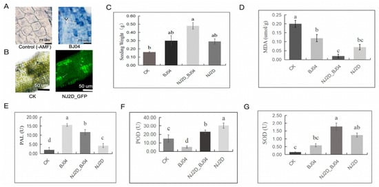
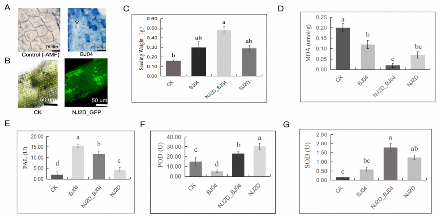
Arbuscular mycorrhizal fungi (AMF) successfully colonized the roots of L. formosana seedlings following BJ04 inoculation (Figure 1A). Consistently, fluorescence imaging confirmed effective root colonization by NJ2D, with strong green signals detected on the rhizoplane and within internal tissues, indicating stable bacterial establishment (Figure 1B). To assess plant growth-promoting effects, L. formosana seedlings were subjected to NJ2D, BJ04, and combined (NJ2D_BJ04) treatments. Notably, NJ2D_BJ04 significantly increased seedling biomass compared with the control (CK; p < 0.005) (Figure 1C). All inoculation treatments markedly reduced malondialdehyde (MDA) levels, with decreases of 74.00%, 35.00%, and 90.00% under NJ2D, BJ04, and NJ2D_BJ04, respectively, indicating alleviation of oxidative damage (Figure 1D). Correspondingly, antioxidant responses were enhanced, as phenylalanine ammonia-lyase (PAL) activity increased by 170.00% (NJ2D), 700.00% (BJ04), and 500.00% (NJ2D_BJ04) relative to the control (Figure 1E), while peroxidase (POD) activity rose by 146.15% (NJ2D) and 83.33% (NJ2D_BJ04) relative to the control (Figure 1F). Superoxide dismutase (SOD) activity was also significantly elevated in NJ2D and NJ2D_BJ04 treatments (p < 0.005) (Figure 1G). Significant interaction effects between NJ2D and BJ04 were detected only for PAL activity (p < 0.05), while no significant interaction effects were observed for seedling biomass, MDA, POD, or SOD (Table S1).
Figure 1.
Colonization and growth-promoting effects of NJ2D and AMF on L. formosana seedlings. (A) AMF root colonization; (B) NJ2D root colonization (fluorescence imaging); (C) biomass accumulation; (D) MDA content; (E) PAL activity; (F) POD activity; (G) SOD activity. The arrow indicates the colonization site of GFP-labelled NJ2D in the root tissue. Different lowercase letters indicate significant differences among treatments at p < 0.05.
3.2. Metabolic Profiles in L. formosana Roots
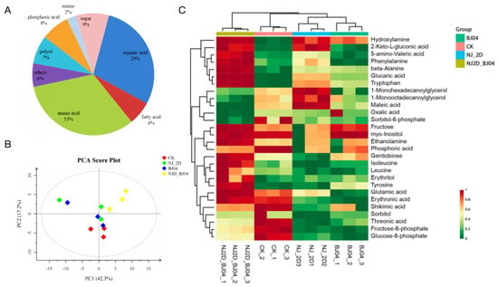
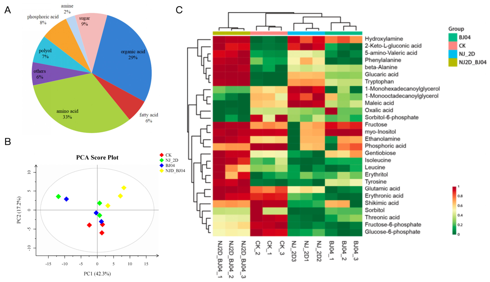
A total of 81 metabolites were identified by gas chromatography–mass spectrometry (GC-MS) and comprised predominantly primary metabolites, including amino acids (33%), organic acids (29%), sugars (9%), phosphoric acids (8%), polyols (7%), fatty acids (6%), and amines (2%). Amino acids, organic acids, sugars, and phosphoric acids together constituted the dominant metabolic classes (Figure 2A). Principal component analysis (PCA) revealed pronounced metabolic divergence among treatments, capturing both global variation and group-specific signatures. The PCA score plot (Figure 2B) showed clear and consistent separation among CK, NJ2D, BJ04, and NJ2D_BJ04 samples, with PC1 and PC2 explaining 42.3% and 17.2% of the total variance, respectively. Notably, the distinct clustering patterns indicate that microbial inoculation imposes a strong and differential metabolic imprint on root tissues. Collectively, these results suggest that NJ2D and BJ04, individually or in combination, drive a coordinated reconfiguration of root-derived primary metabolism, which may act as a key regulatory interface mediating downstream physiological and molecular responses in L. formosana.
Figure 2.
Microbial modulation of root metabolite profiles in L. formosana. (A) Metabolite classification; (B) PCA score plot; (C) heatmap of differential metabolites.
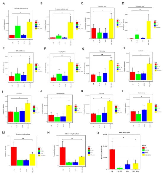
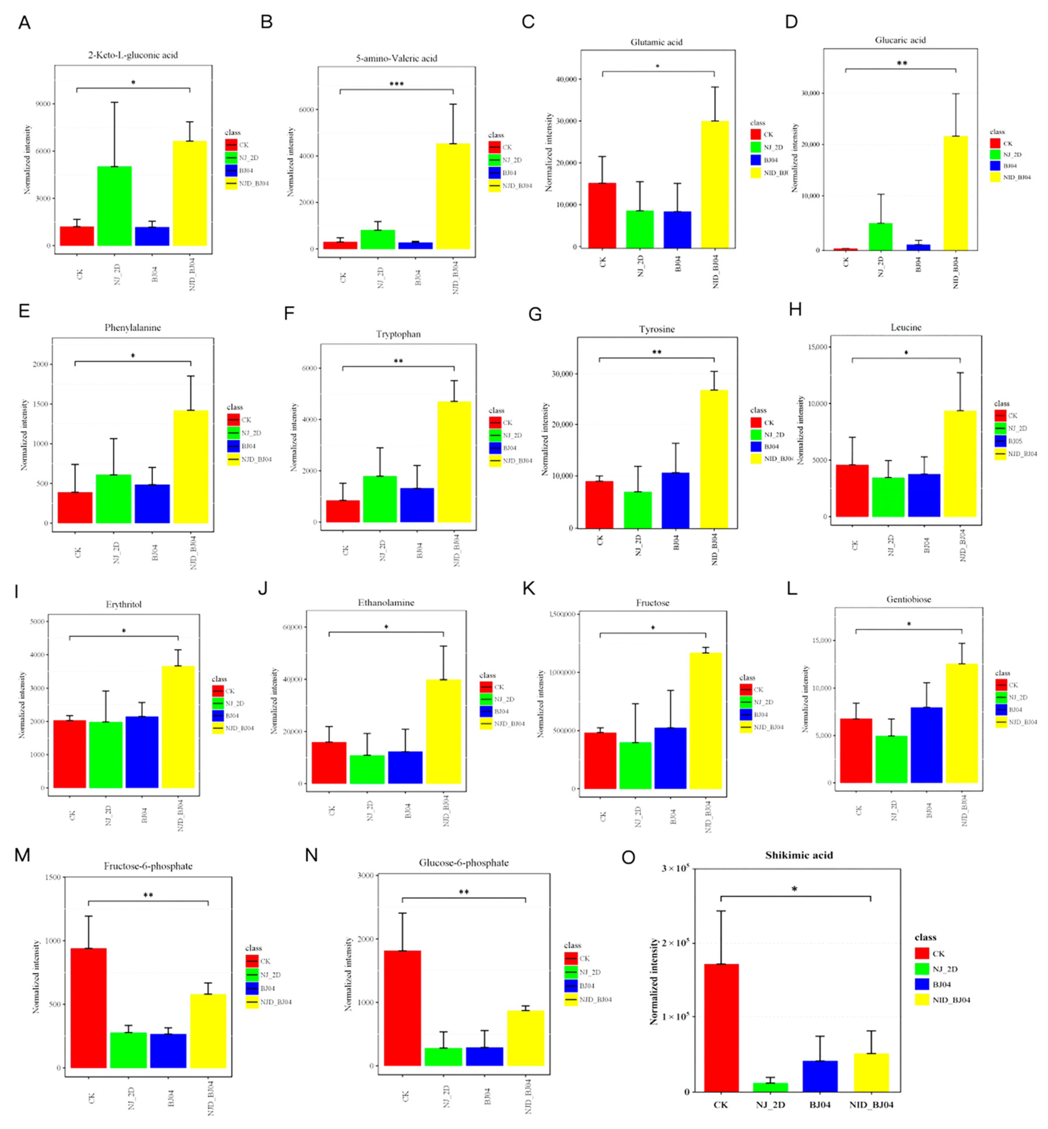
Heatmap clustering further highlighted pronounced treatment-specific metabolic reprogramming (Figure 2C). Relative to the CK, NJ2D inoculation led to a marked reduction in several metabolites, including shikimic acid, threonic acid, fructose-6-phosphate, erythronic acid, sorbitol, and glucose-6-phosphate. In contrast, NJ2D significantly increased the accumulation of glucaric acid, 2-keto-L-gluconic acid, hydroxylamine, 1-monohexadecanoylglycerol, 1-monooctadecanoylglycerol, maleic acid, phenylalanine, and tryptophan (Figure 2C). BJ04 treatment exhibited a distinct metabolic signature, characterized by elevated levels of hydroxylamine, tryptophan, fructose, tyrosine, and β-alanine compared with the CK. Notably, the combined NJ2D_BJ04 treatment induced a broader and more pronounced metabolic shift, with significant increases in 5-aminovaleric acid, glucaric acid, tryptophan, tyrosine, β-alanine, gentiobiose, fructose, glutamic acid, isoleucine, ethanolamine, erythritol, 2-keto-L-gluconic acid, and phenylalanine (p < 0.05; Figure 3). Conversely, shikimic acid and glucose-6-phosphate were consistently reduced in the NJ2D_BJ04 treatment relative to the CK (p < 0.05; Figure 3).
Figure 3.
Significantly altered LC-MS metabolites in L. formosana roots following NJ2D and BJ04 inoculation. (A–O) Relative abundance of key metabolites, including 2-keto-L-gluconic acid, 5-aminovaleric acid, glutamic acid, glucaric acid, phenylalanine, tryptophan, tyrosine, leucine, erythritol, ethanolamine, fructose, gentiobiose, fructose-6-phosphate, glucose-6-phosphate, and shikimic acid. Significance levels are indicated as *** p < 0.001, ** p < 0.01, * p < 0.05.
3.3. Differentially Abundant Metabolite KEGG Pathways
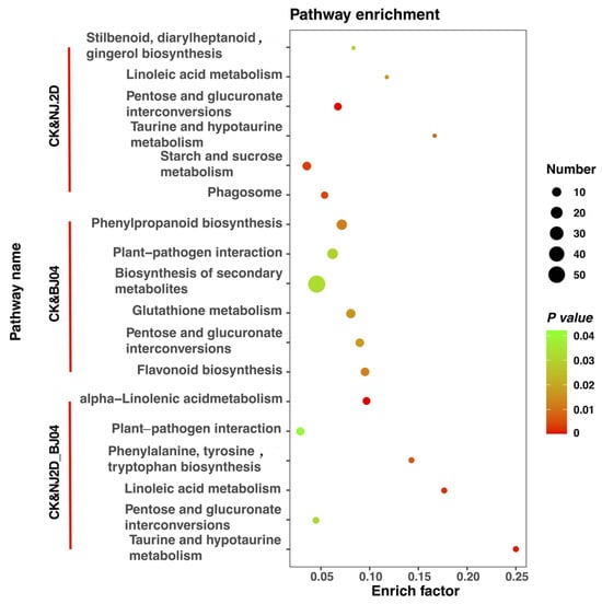
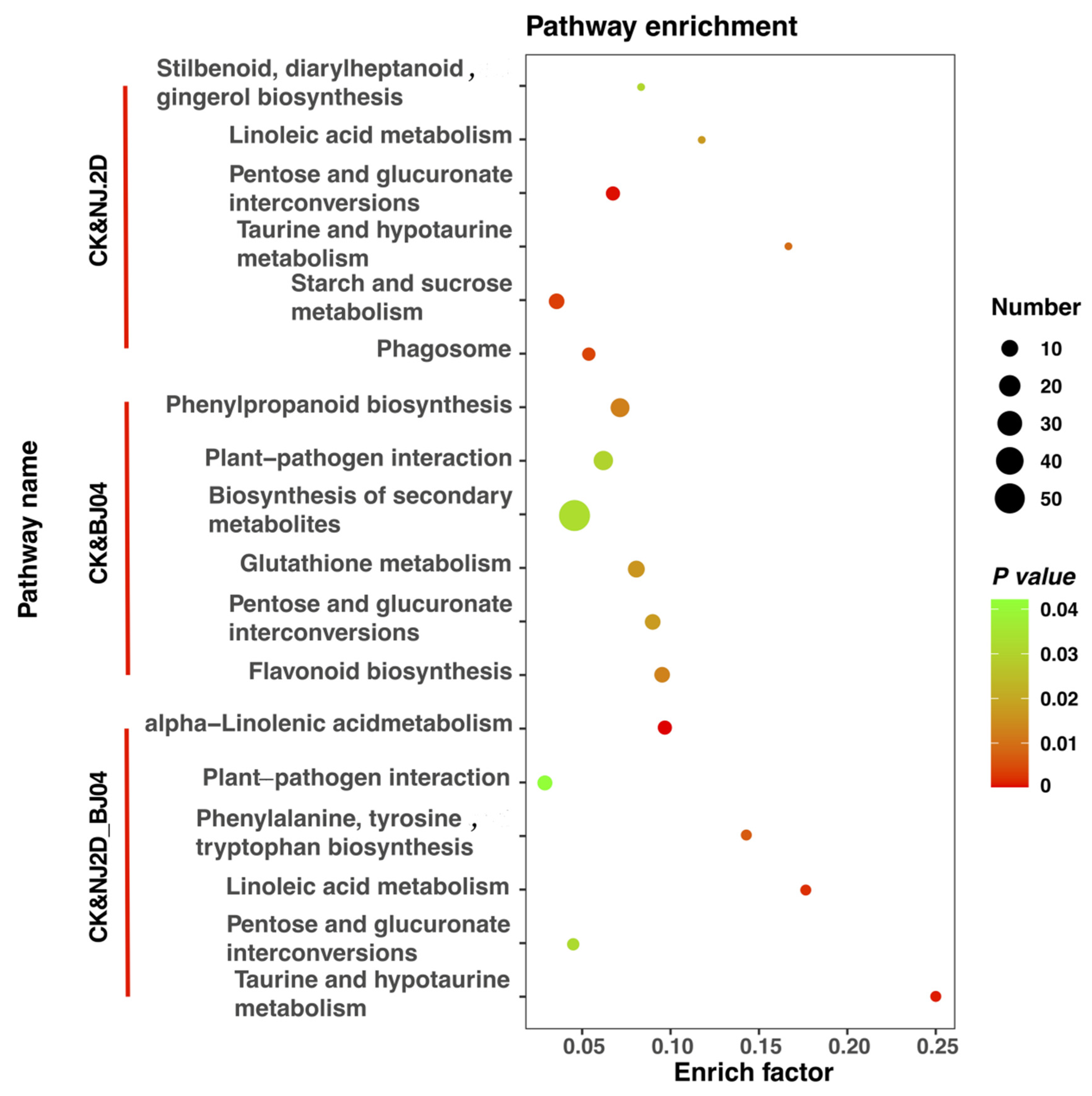
All metabolites were significantly impacted by NJ2D, BJ04, and NJ2D_BJ04 treatments, which were mapped to biological pathways using the KEGG database. The most differentiated metabolites were selected for comparative analysis (Figure 4). In the CK vs. NJ2D group, key pathways included stilbenoid, diarylheptanoid, and gingerol biosynthesis; linoleic acid metabolism; pentose and glucuronate interconversions; taurine and hypotaurine metabolism; starch and sucrose metabolism; and phagosome. In the CK vs. BJ04 group, pathways included phenylpropanoid biosynthesis; plant–pathogen interaction; biosynthesis of secondary metabolites; glutathione metabolism; pentose and glucuronate interconversions; and flavonoid biosynthesis. In the CK vs. NJ2D_BJ04 group, the most notable pathways were alpha-linolenic acid metabolism; plant–pathogen interaction; phenylalanine, tyrosine, and tryptophan biosynthesis; linoleic acid metabolism; pentose and glucuronate interconversions; and taurine and hypotaurine metabolism (Figure 4). Notably, the pentose and glucuronate interconversions pathway was common across all comparisons.
Figure 4.
KEGG pathway enrichment reveals microbiome-driven metabolic reprogramming in L. formosana roots. Dot size indicates the number of enriched metabolites and color denotes significance (redder = lower p values; p < 0.05). Comparisons include CK vs. NJ2D, BJ04, and NJ2D_BJ04.
3.4. Analysis of Root Exudate Components in L. formosana
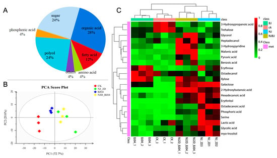
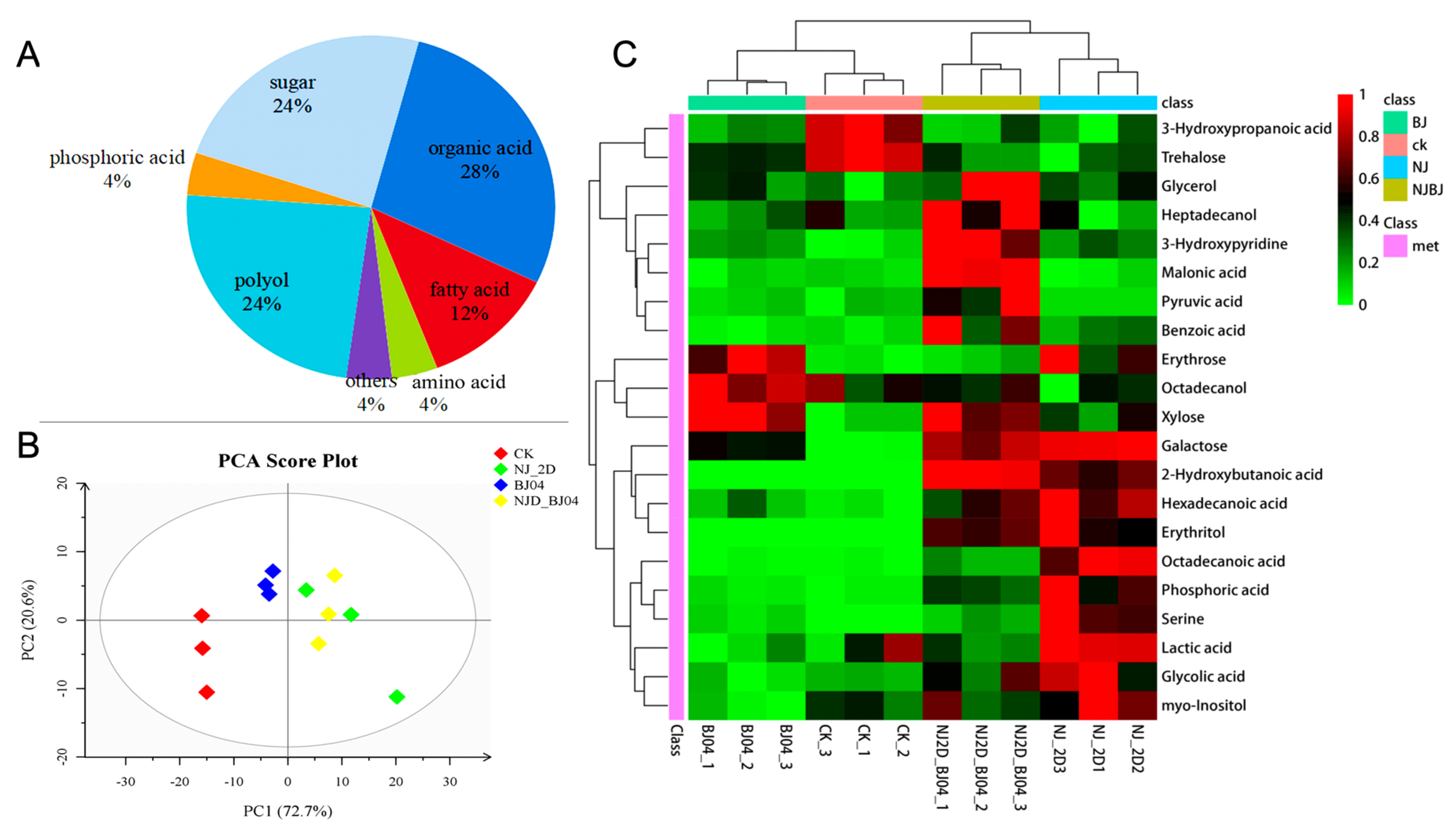
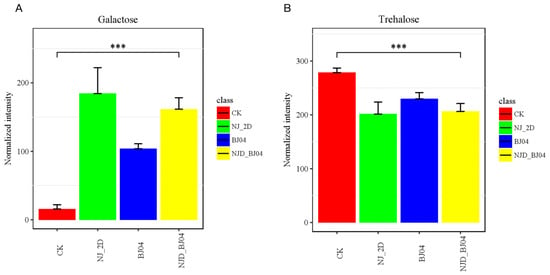
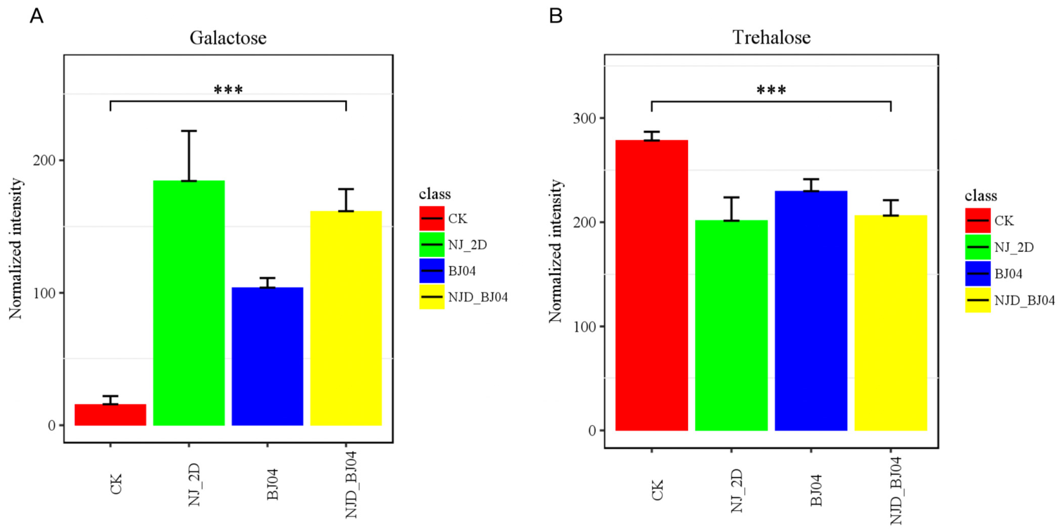
The root exudates of L. formosana were predominantly composed of organic acids (28%), sugars (24%), and polyols (24%), followed by fatty acids (12%), amino acids (4%), phosphoric acid (4%), and other minor compounds (Figure 5A), indicating a carbon-rich exudation profile. Principal component analysis (PCA) revealed a clear separation among treatments, with PC1 and PC2 explaining 72.7% and 20.6% of the total variance, respectively (Figure 5B), suggesting that microbial inoculation substantially reshaped the root exudate composition. Consistently, heatmap clustering highlighted pronounced treatment-specific shifts in metabolite profiles (Figure 5C). NJ2D inoculation markedly increased the accumulation of carbohydrates (e.g., galactose), organic acids (e.g., 2-hydroxybutanoic acid, lactic acid, glycolic acid), fatty acids (e.g., hexadecanoic acid, octadecanoic acid), and polyols (e.g., erythritol, myo-inositol), suggesting enhanced carbon allocation and metabolic activity in the rhizosphere. BJ04 treatment preferentially enriched sugars and sugar alcohol-related metabolites, including erythrose, galactose, and xylose, indicating a shift toward carbohydrate-dominated exudation patterns. Notably, co-inoculation (NJ2D_BJ04) induced a broader and more pronounced metabolic reprogramming, characterized by significant increases in heptadecanol, 3-hydroxypyridine, malonic acid, xylose, galactose, 2-hydroxybutanoic acid, and erythritol (p < 0.05; Figure 5C and Figure 6). These results collectively indicate that the microbial consortium may exert a synergistic effect in modulating primary metabolism. Furthermore, trehalose accumulation was significantly reduced in all inoculated treatments compared with the CK (p < 0.05; Figure 6), suggesting a reduced requirement for osmoprotective metabolites. This decline may reflect an improved physiological status of the host or microbial modulation of primary metabolism, which may be associated with altered stress adaptation and microbe–host signaling processes. Overall, these results demonstrate that microbial inoculation, particularly co-inoculation, drives a coordinated reconfiguration of root exudate composition, likely contributing to rhizosphere metabolic interactions and plant–microbe feedback regulation.
Figure 5.
Microbial synergy reshapes root exudate chemistry in L. formosana. (A) Classification of root exudates; (B) PCA of exudate profiles; (C) heatmap of differential metabolites.
Figure 6.
Microbial regulation of carbohydrate allocation in L. formosana root exudates. (A) Galactose; (B) trehalose. Significance levels are indicated as *** p < 0.001.
3.5. Functional Pathway Analysis of Differentially Abundant Root Exudates
The KEGG database was used to map differentially accumulated metabolites (DAMs) from the NJ2D vs. CK, BJ04 vs. CK, and NJ2D_BJ04 vs. CK comparisons of metabolic pathways. Although only a small number of DAMs were assigned to individual pathways, several functionally relevant categories were identified. Notably, starch and sucrose metabolism (ko00500) was the only pathway shared across all treatments (Table 1). Given the limited number of mapped metabolites, these results should be interpreted as indicative of targeted metabolic adjustments rather than robust pathway-level enrichment. Nevertheless, the recurrent involvement of carbohydrate metabolism suggests a potential role in mediating treatment-induced changes in root exudation.
Table 1.
KEGG pathway mapping of differentially accumulated root exudate metabolites.
3.6. Linking Root Metabolic Shifts to Rhizosphere Exudation
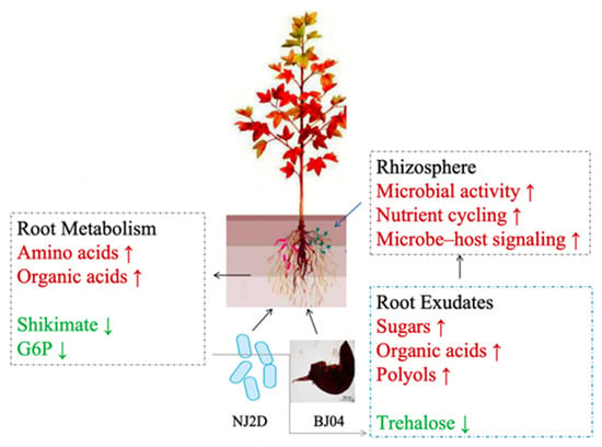
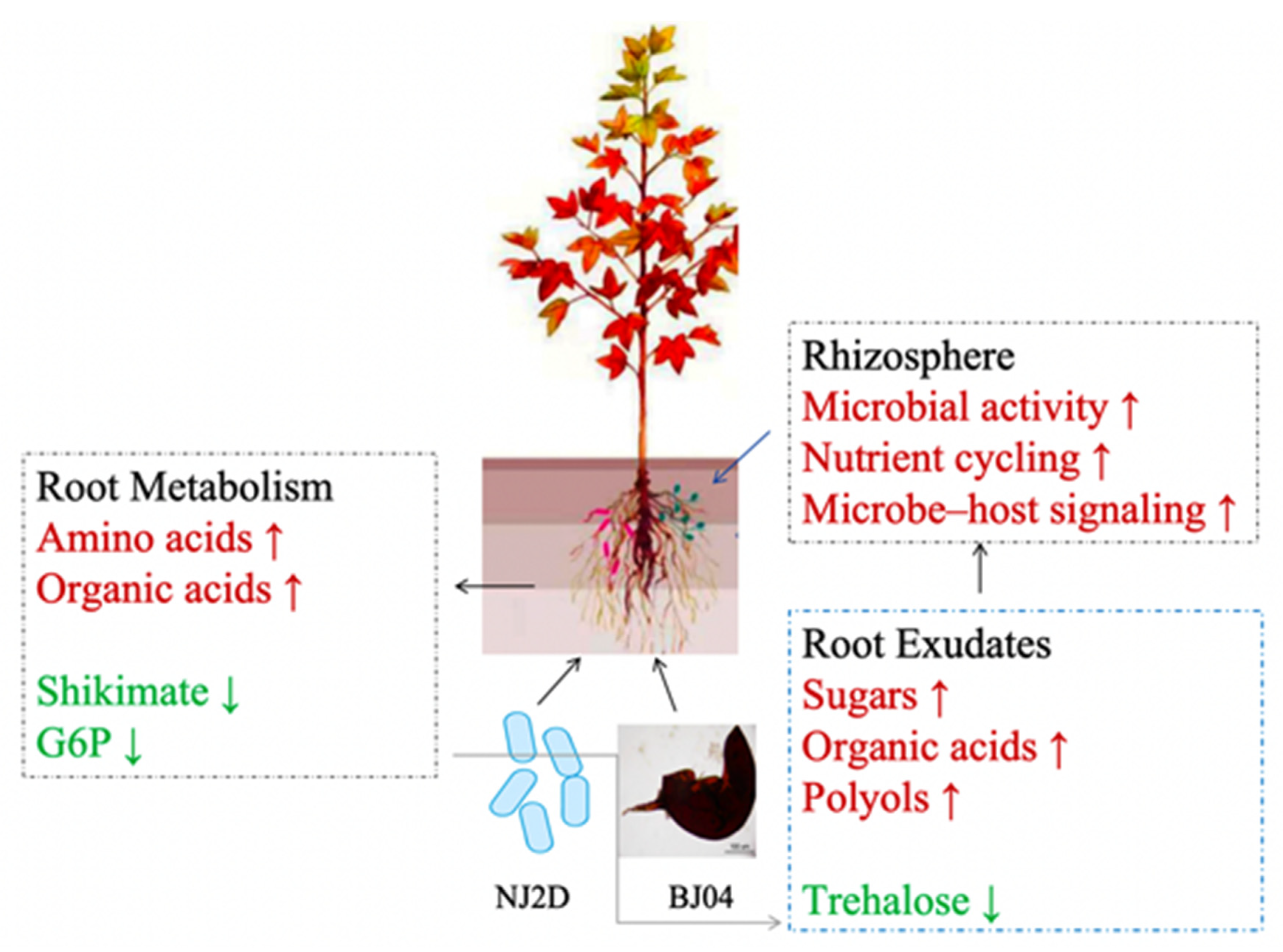
Root tissues were characterized by a high proportion of amino acids (33%) and organic acids (29%), while root exudates displayed a carbon-enriched profile comprising organic acids (28%), sugars (24%), and polyols (24%) (Figure 2A and Figure 5A). Principal component analysis indicated that microbial treatments induced stronger metabolic shifts in exudates (93.3% variance explained) compared to tissues (59.5%) (Figure 2B and Figure 5B). Coordinated accumulations of tryptophan, phenylalanine, erythritol, and 2-keto-L-gluconic acid were detected in both compartments under inoculation. In contrast, shikimic acid and glucose-6-phosphate were reduced only in tissues, while trehalose declined exclusively in exudates (Figure 7), suggesting independent regulatory mechanisms. Together, these results reveal a coordinated albeit compartment-specific metabolic landscape shaped by microbial inoculation.
Figure 7.
Conceptual model illustrating microbial consortium-driven reprogramming of root metabolism and exudation in L. formosana. Red and green arrows indicate changes in abundance: red arrows represent increase (↑), and green arrows represent decrease (↓).
4. Discussion
Arbuscular mycorrhizal fungi (AMF) and plant growth-promoting rhizobacteria (PGPR) are key components of the soil microbiota, and both single and combined inoculations have been widely reported to enhance plant growth [37,38,39,40]. Consistent with these findings, our results showed that all inoculation treatments significantly increased the aboveground biomass of L. formosana seedlings compared with the control. Notably, co-inoculation resulted in greater increases than single inoculation, with the NJ2D_BJ04 treatment exhibiting the highest values across most measured indices. This pattern indicates a synergistic rather than additive interaction between NJ2D and BJ04, potentially driven by functional complementarity. AMF is known to enhance nutrient acquisition, particularly phosphorus, whereas PGPR can stimulate root development and nutrient mobilization. Previous studies have identified Pseudomonas spp. and Bacillus spp. as bacteria that facilitate AMF colonization and function [39,41]. In line with this, NJ2D may facilitate AMF colonization and function, thereby enhancing BJ04-mediated growth promotion, although further studies are required to confirm the underlying mechanisms.
Reactive oxygen species (ROS) act as central regulators of plant metabolism and development, with their homeostasis tightly controlled to balance signaling and oxidative damage [42]. Plants mitigate excessive ROS through an antioxidant defense system involving enzymes such as POD, PAL, and SOD, while malondialdehyde (MDA) reflects the extent of membrane lipid peroxidation [43]. Increasing evidence indicates that AMF can modulate this redox balance by enhancing antioxidant capacity, thereby reducing ROS accumulation while stabilizing cellular processes [44,45,46,47]. Consistent with this, our results showed that NJ2D, BJ04, and NJ2D_BJ04 treatments significantly decreased MDA content and increased POD, PAL, and SOD activities in L. formosana roots. These coordinated changes suggest an improved redox homeostasis, which may facilitate metabolic reprogramming toward more efficient resource allocation and cellular functioning. Notably, the stronger responses under co-inoculation indicate a synergistic effect, potentially amplifying redox-mediated metabolic adjustments and thereby contributing to enhanced plant growth.
Following the observed increases in biomass and antioxidant enzyme activities, metabolomic profiling was conducted to elucidate the mechanisms underlying growth promotion. Microbial inoculation significantly altered the metabolic composition of L. formosana, with multiple differential metabolites (p < 0.05) showing positive associations with plant growth (Figure 3). Among these, amino acids were prominently enriched, suggesting a central role in mediating plant responses. Amino acids are essential for protein synthesis, serve as precursors for phytohormones, and contribute to stress resistance, thereby supporting plant growth and development [48,49]. Consistently, amino-acid-based biostimulants have been shown to enhance crop yield and quality [50]. In this study, key amino acids, such as tryptophan and tyrosine, may promote cell division and elongation through their roles in hormone biosynthesis and metabolic regulation, while also acting as precursors of secondary metabolites involved in defense and signaling [51,52]. Additionally, phenylalanine is linked to lignin biosynthesis and anthocyanin production [53].
It should be noted that the collection of root exudates via overnight incubation in sterile distilled water may induce physiological stress, potentially altering the composition and abundance of released metabolites. Therefore, while the observed metabolomic changes provide valuable insights into microbial-mediated growth promotion, the exudate profiles may not fully reflect in situ conditions. Collectively, these results suggest that NJ2D and BJ04 facilitate growth by modulating amino acid metabolism, thereby enhancing metabolic capacity and developmental processes in L. formosana.
Root exudation plays a central role in mediating plant–environment interactions [54]. These exudates comprise diverse compounds, including sugars, organic acids, amino acids, and phenolics, which function in nutrient exchange and chemical signaling [55]. Through such signaling processes, plants and plant growth-promoting microorganisms (PGPM) establish dynamic interactions that influence mutual responses and rhizosphere functioning [56]. In this study, galactose levels were significantly elevated under NJ2D, BJ04, and NJ2D_BJ04 inoculation compared with the CK. Beyond its role as a carbon source, galactose contributes to cell wall biosynthesis and the formation of storage oligosaccharides, thereby supporting plant growth and stress tolerance [57,58]. Moreover, galactose has been implicated in mediating plant–microbe interactions by promoting bacterial colonization, as reported for Bacillus velezensis SQR9 [59]. Consistent with this, our findings suggest that increased galactose exudation may facilitate the establishment of NJ2D and BJ04, thereby reinforcing plant–microbe interactions and contributing to enhanced growth performance in L. formosana.
5. Conclusions
Co-inoculation with Serratia marcescens NJ2D and Funneliformis mosseae BJ04 maximized Liquidambar formosana growth via coordinated regulation of redox homeostasis, primary metabolism, and root exudation. Enhanced antioxidant capacity mitigated oxidative stress, while systemic reprogramming of amino acid, organic acid, and carbohydrate metabolism supported biomass accumulation. Parallel shifts in exudate composition revealed tight coupling between root metabolic state and rhizosphere processes. These findings demonstrate that microbial synergism functionally links root metabolic reprogramming to exudation dynamics, providing a mechanistic basis for plant growth promotion and highlighting the potential of synthetic microbial consortia as sustainable enhancers of tree productivity and ecosystem resilience.
Supplementary Materials
The following supporting information can be downloaded at: https://www.mdpi.com/article/10.3390/microorganisms14061346/s1, Table S1. Two-way ANOVA examining the effects of NJ2D, BJ04 and their interaction on physiological traits of L. formosana.
Author Contributions
Conceptualization, J.S., F.-M.C. and J.-H.W.; methodology, J.S., F.-M.C., J.-H.W. and L.-Q.Z.; validation, all authors; formal analysis, W.G., J.-W.L., Y.-Q.F., J.-X.R. and X.W.; writing—original draft preparation, J.S. and F.-M.C.; writing—review and editing, F.-M.C., J.-W.L. and C.-Y.L.; supervision, L.-Q.Z.; funding acquisition, J.S. All authors have read and agreed to the published version of the manuscript.
Funding
This research was funded by the Scientific Research Foundation for the Introduction of Talented Scholars (RK23097).
Institutional Review Board Statement
Not applicable.
Informed Consent Statement
Not applicable.
Data Availability Statement
The original contributions presented in this study are included in the article. Further inquiries can be directed to the corresponding author.
Conflicts of Interest
The authors declare no conflicts of interest.
References
- Bean, W.J.; Taylor, G. Trees and Shrubs Hardy in the British Isles; John Murray: Philadelphia, PA, USA, 1976. [Google Scholar]
- Chinese Pharmacopoeia Commission. Pharmacopoeia of the People’s Republic of China 2015; China Medical Science Press: Beijing, China, 2017.
- Li, X.; Liu, X.; Xioang, D.; Lin, W.; Lin, T.; Shi, Y.; Xie, J.; Yang, Y. Impact of litterfall addition and exclusion on soil respiration in Cunninghamia lanceolata plantation and secondary Castanopsis carlesii forest in mid-subtropical China. Chin. J. Plant Ecol. 2016, 40, 447–457. [Google Scholar] [CrossRef] [Scilit]
- Anza, M.; Garbisu, C.; Salazar, O.; Epelde, L.; Alkorta, I.; Martínez-Santos, M. Acidification alters the functionality of metal polluted soils. Appl. Soil Ecol. 2021, 163, 103920. [Google Scholar] [CrossRef] [Scilit]
- Zachos, J.; Pagani, M.; Sloan, L.; Thomas, E.; Billups, K. Trends, rhythms, and aberrations in global climate 65 Ma to present. Science 2001, 292, 686–693. [Google Scholar] [CrossRef] [Scilit]
- Guo, Z.T.; Sun, B.; Zhang, Z.S.; Peng, S.Z.; Xiao, G.Q.; Ge, J.Y.; Hao, Q.Z.; Qiao, Y.S.; Liang, M.Y.; Liu, J.F.; et al. A major reorganization of Asian climate by the early Miocene. Clim. Past 2008, 4, 153–174. [Google Scholar] [CrossRef] [Scilit]
- Burton, P.J.; Bazzaz, F.A. Tree Seedling Emergence on Interactive Temperature and Moisture Gradients and in Patches of Old-Field Vegetation. Am. J. Bot. 1991, 78, 131–149. [Google Scholar] [CrossRef] [Scilit]
- Ahmad, M.; Pataczek, L.; Hilger, T.H.; Zahir, Z.A.; Hussain, A.; Rasche, F.; Schafleitner, R.; Solberg, S. Perspectives of Microbial Inoculation for Sustainable Development and Environmental Management. Front. Microbiol. 2018, 9, 2992. [Google Scholar] [CrossRef] [Scilit] [PubMed]
- Kumar, B.L.; Gopal, D.V. Effective role of indigenous microorganisms for sustainable environment. 3 Biotech 2015, 5, 867–876. [Google Scholar] [CrossRef] [Scilit]
- Vaxevanidou, K.; Christou, C.; Kremmydas, G.F.; Georgakopoulos, D.G.; Papassiopi, N. Role of indigenous arsenate and iron(III) respiring microorganisms in controlling the mobilization of arsenic in a contaminated soil sample. Bull. Environ. Contam. Toxicol. 2015, 94, 282–288. [Google Scholar] [CrossRef] [Scilit] [PubMed]
- Fang, X.; Lee, X.; Twagirayezu, G.; Cheng, H.; Lu, H.; Huang, S.; Deng, L.; Ji, B. A Critical Review of the Effectiveness of Biochar Coupled with Arbuscular Mycorrhizal Fungi in Soil Cadmium Immobilization. J. Fungi 2024, 10, 182. [Google Scholar] [CrossRef] [Scilit] [PubMed]
- Shi, Z.; Li, K.; Zhu, X.; Wang, F. The worldwide leaf economic spectrum traits are closely linked with mycorrhizal traits. Fungal Ecol. 2020, 43, 100877. [Google Scholar] [CrossRef] [Scilit]
- Basiru, S.; Ait Si Mhand, K.; Elfermi, R.; Khatour, I.; Errafii, K.; Legeay, J.; Hijri, M. Enhancing chickpea growth through arbuscular mycorrhizal fungus inoculation: Facilitating nutrient uptake and shifting potential pathogenic fungal communities. Mycorrhiza 2024, 35, 1. [Google Scholar] [CrossRef] [Scilit] [PubMed]
- Pozo, M.J.; Azcón-Aguilar, C. Unraveling mycorrhiza-induced resistance. Curr. Opin. Plant Biol. 2007, 10, 393–398. [Google Scholar] [CrossRef] [Scilit]
- Zhang, H.; Cheng, H.; Twagirayezu, G.; Zhang, F.; Shi, Y.; Luo, C.; Yan, F.; Wang, Z.; Xing, D. Arbuscular Mycorrhizal Fungi Adjusts Root Architecture to Promote Leaf Nitrogen Accumulation and Reduce Leaf Carbon–Nitrogen Ratio of Mulberry Seedlings. Forests 2023, 14, 2448. [Google Scholar] [CrossRef] [Scilit]
- Riaz, M.; Kamran, M.; Fang, Y.; Wang, Q.; Cao, H.; Yang, G.; Deng, L.; Wang, Y.; Zhou, Y.; Anastopoulos, I.; et al. Arbuscular mycorrhizal fungi-induced mitigation of heavy metal phytotoxicity in metal contaminated soils: A critical review. J. Hazard. Mater. 2021, 402, 123919. [Google Scholar] [CrossRef] [Scilit] [PubMed]
- Niu, H.; Sun, Y.; Zhang, Z.; Zhao, D.; Wang, N.; Wang, L.; Guo, H. The endophytic bacterial entomopathogen Serratia marcescens promotes plant growth and improves resistance against Nilaparvata lugens in rice. Microbiol. Res. 2022, 256, 126956. [Google Scholar] [CrossRef] [Scilit]
- Saikia, J.; Sarma, R.K.; Dhandia, R.; Yadav, A.; Bharali, R.; Gupta, V.K.; Saikia, R. Alleviation of drought stress in pulse crops with ACC deaminase producing rhizobacteria isolated from acidic soil of Northeast India. Sci. Rep. 2018, 8, 3560, Erratum in Sci. Rep. 2018, 8, 7000. https://doi.org/10.1038/s41598-018-25174-5. [Google Scholar] [CrossRef] [Scilit]
- Mahdi, I.; Hafidi, M.; Allaoui, A.; Biskri, L. Halotolerant Endophytic Bacterium Serratia rubidaea ED1 Enhances Phosphate Solubilization and Promotes Seed Germination. Agriculture 2021, 11, 224. [Google Scholar] [CrossRef] [Scilit]
- Nordstedt, N.P.; Jones, M.L. Genomic Analysis of Serratia plymuthica MBSA-MJ1: A Plant Growth Promoting Rhizobacteria That Improves Water Stress Tolerance in Greenhouse Ornamentals. Front. Microbiol. 2021, 12, 653556. [Google Scholar] [CrossRef] [Scilit] [PubMed]
- Ahmad, H.M.; Fiaz, S.; Hafeez, S.; Zahra, S.; Shah, A.N.; Gul, B.; Aziz, O.; Mahmood-Ur-Rahman Fakhar, A.; Rafique, M.; Chen, Y.; et al. Plant Growth-Promoting Rhizobacteria Eliminate the Effect of Drought Stress in Plants: A Review. Front. Plant Sci. 2022, 13, 875774. [Google Scholar] [CrossRef] [Scilit]
- Tian, B.; Pei, Y.; Huang, W.; Ding, J.; Siemann, E. Increasing flavonoid concentrations in root exudates enhance associations between arbuscular mycorrhizal fungi and an invasive plant. ISME J. 2021, 15, 1919–1930. [Google Scholar] [CrossRef] [Scilit]
- Li, J.; Li, Q.; Wang, M.; Xu, S.; Zhang, D. Crop rotation-driven changes in secondary metabolites of potato rhizosphere soil exert stronger regulation on soil microbial community. Front. Microbiol. 2026, 17, 1768797. [Google Scholar] [CrossRef] [Scilit]
- Yue, H.; Yue, W.; Jiao, S.; Kim, H.; Lee, Y.H.; Wei, G.; Song, W.; Shu, D. Plant domestication shapes rhizosphere microbiome assembly and metabolic functions. Microbiome 2023, 11, 70. [Google Scholar] [CrossRef] [Scilit]
- Murashige, T.; Skoog, F. A revised medium for rapid growth and bio assays with tobacco tissue cultures. Physiol. Plant. 1962, 15, 473–497. [Google Scholar] [CrossRef] [Scilit]
- Song, J.; Han, Y.; Bai, B.; Jin, S.; He, Q.; Ren, J. Diversity of arbuscular mycorrhizal fungi in rhizosphere soils of the Chinese medicinal herb Sophora flavescens Ait. Soil Tillage Res. 2019, 195, 104423. [Google Scholar] [CrossRef] [Scilit]
- Yeh, E.; Gustafson, K.; Boulianne, G.L. Green fluorescent protein as a vital marker and reporter of gene expression in Drosophila. Proc. Natl. Acad. Sci. USA 1995, 92, 7036–7040. [Google Scholar] [CrossRef] [Scilit] [PubMed]
- Mathis, R.; Van Gijsegem, F.; De Rycke, R.; D’Haeze, W.; Van Maelsaeke, E.; Anthonio, E.; Van Montagu, M.; Holsters, M.; Vereecke, D. Lipopolysaccharides as a communication signal for progression of legume endosymbiosis. Proc. Natl. Acad. Sci. USA 2005, 102, 2655–2660. [Google Scholar] [CrossRef] [Scilit]
- Yu, J.; Tang, X.X.; Zhang, P.Y.; Tian, J.Y.; Cai, H.J. Effects of CO2 Enrichment on Photosynthesis, Lipid Peroxidation and Activities of Antioxidative Enzymes of Platymonas subcordiformis Subjected to UV-B Radiation Stress. J. Integr. Plant Biol. 2004, 46, 682–690. [Google Scholar]
- Konappa, N.M.; Maria, M.; Uzma, F.; Krishnamurthy, S.; Nayaka, S.C.; Niranjana, S.R.; Chowdappa, S. Lactic acid bacteria mediated induction of defense enzymes to enhance the resistance in tomato against Ralstonia solanacearum causing bacterial wilt. Sci. Hortic. 2016, 207, 183–192. [Google Scholar] [CrossRef] [Scilit]
- Lu, X.H.; Sun, D.Q.; Mo, Y.W.; Xi, J.G.; Sun, G.M. Effects of post-harvest salicylic acid treatment on fruit quality and anti-oxidant metabolism in pineapple during cold storage. J. Hortic. Sci. Biotechnol. 2010, 85, 454–458. [Google Scholar] [CrossRef] [Scilit]
- Sun, D.; Lu, X.; Hu, Y.; Li, W.; Hong, K.; Mo, Y.; Cahill, D.; Xie, J. Methyl jasmonate induced defense responses increase resistance to Fusarium oxysporum f. sp. cubense race 4 in banana. Sci. Hortic. 2013, 164, 484–491. [Google Scholar] [CrossRef] [Scilit]
- Lisec, J.; Schauer, N.; Kopka, J.; Willmitzer, L.; Fernie, A.R. Gas chromatography mass spectrometry–based metabolite profiling in plants. Nat. Protoc. 2006, 1, 387–396, Erratum in Nat. Protoc. 2015, 10, 1457. https://doi.org/10.1038/nprot0915-1457a. [Google Scholar] [CrossRef] [Scilit]
- Sangster, T.; Major, H.; Plumb, R.; Wilson, A.J.; Wilson, I.D. A pragmatic and readily implemented quality control strategy for HPLC-MS and GC-MS-based metabonomic analysis. Analyst 2006, 131, 1075–1078. [Google Scholar] [CrossRef] [Scilit]
- Dunn, W.B.; Broadhurst, D.; Begley, P.; Zelena, E.; Francis-McIntyre, S.; Anderson, N.; Brown, M.; Knowles, J.D.; Halsall, A.; Haselden, J.N. Procedures for large-scale metabolic profiling of serum and plasma using gas chromatography and liquid chromatography coupled to mass spectrometry. Nat. Protoc. 2011, 6, 1060–1083. [Google Scholar] [CrossRef] [Scilit] [PubMed]
- Want, E.J.; Wilson, I.D.; Gika, H.; Theodoridis, G.; Plumb, R.S.; Shockcor, J.; Holmes, E.; Nicholson, J.K. Global metabolic profiling procedures for urine using UPLC–MS. Nat. Protoc. 2010, 5, 1005–1018. [Google Scholar] [CrossRef] [Scilit] [PubMed]
- Mathimaran, N.; Jegan, S.; Thimmegowda, M.N.; Prabavathy, V.R.; Yuvaraj, P.; Kathiravan, R.; Sivakumar, M.N.; Manjunatha, B.N.; Bhavitha, N.C.; Sathish, A.; et al. Intercropping Transplanted Pigeon Pea With Finger Millet: Arbuscular Mycorrhizal Fungi and Plant Growth Promoting Rhizobacteria Boost Yield While Reducing Fertilizer Input. Front. Sustain. Food Syst. 2020, 4, 88. [Google Scholar] [CrossRef] [Scilit]
- Sagar, A.; Rathore, P.; Ramteke, P.W.; Ramakrishna, W.; Reddy, M.S.; Pecoraro, L. Plant Growth Promoting Rhizobacteria, Arbuscular Mycorrhizal Fungi and Their Synergistic Interactions to Counteract the Negative Effects of Saline Soil on Agriculture: Key Macromolecules and Mechanisms. Microorganisms 2021, 9, 1491. [Google Scholar] [CrossRef] [Scilit]
- Cai, X.; Zhao, H.; Liang, C.; Li, M.; Liu, R. Effects and Mechanisms of Symbiotic Microbial Combination Agents to Control Tomato Fusarium Crown and Root Rot Disease. Front. Microbiol. 2021, 12, 629793. [Google Scholar] [CrossRef] [Scilit]
- Abkenar, M.; Mozafari, H.; Karimzadeh, K.; Rajabzadeh, F.; Azimi, R. Arbuscular Mycorrhizal Fungi (AMF) and Plant Growth-promoting Rhizobacteria (PGPR) as an Alternative to Mineral Fertilizers to Improve the Growth, Essential Oil Profile, and Phenolic Content of Satureja macrantha L. Gesunde Pflanz. 2023, 76, 347–356. [Google Scholar] [CrossRef] [Scilit]
- Toljander, J.F.; Artursson, V.; Paul, L.R.; Jansson, J.K.; Finlay, R.D. Attachment of different soil bacteria to arbuscular mycorrhizal fungal extraradical hyphae is determined by hyphal vitality and fungal species. FEMS Microbiol. Lett. 2006, 254, 34–40. [Google Scholar] [CrossRef] [Scilit]
- Yan, L.; Riaz, M.; Wu, X.; Du, C.; Liu, Y.; Lv, B.; Jiang, C. Boron inhibits aluminum-induced toxicity to citrus by stimulating antioxidant enzyme activity. J. Environ. Sci. Health C Environ. Carcinog. Ecotoxicol. Rev. 2018, 36, 145–163. [Google Scholar] [CrossRef] [Scilit]
- Guo, B.; Liang, Y.C.; Zhu, Y.G.; Zhao, F.J. Role of salicylic acid in alleviating oxidative damage in rice roots (Oryza sativa) subjected to cadmium stress. Environ. Pollut. 2007, 147, 743–749. [Google Scholar] [CrossRef] [Scilit] [PubMed]
- Molina, A.S.; Lugo, M.A.; Pérez Chaca, M.V.; Vargas-Gil, S.; Zirulnik, F.; Leporati, J.; Ferrol, N.; Azcón-Aguilar, C. Effect of Arbuscular Mycorrhizal Colonization on Cadmium-Mediated Oxidative Stress in Glycine max (L.) Merr. Plants 2020, 9, 108. [Google Scholar] [CrossRef] [Scilit]
- Xiao, Y.; Liu, C.; Hu, N.; Wang, B.; Zheng, K.; Zhao, Z.; Li, T. Contributions of ectomycorrhizal fungi in a reclaimed poplar forest (Populus yunnanensis) in an abandoned metal mine tailings pond, southwest China. J. Hazard. Mater. 2023, 448, 130962. [Google Scholar] [CrossRef] [Scilit] [PubMed]
- Zhao, W.; Chen, Z.; Yang, X.; Sheng, L.; Mao, H.; Zhu, S. Integrated transcriptomics and metabolomics reveal key metabolic pathway responses in Pistia stratiotes under Cd stress. J. Hazard. Mater. 2023, 452, 131214. [Google Scholar] [CrossRef] [Scilit] [PubMed]
- Zhu, Q.; Xu, P.; Lei, L.; Jing, Y. Transcriptome analysis reveals decreased accumulation and toxicity of Cd in upland rice inoculated with arbuscular mycorrhizal fungi. Appl. Soil Ecol. 2022, 177, 104501. [Google Scholar] [CrossRef] [Scilit]
- Rai, V.K. Role of Amino Acids in Plant Responses to Stresses. Biol. Plant. 2002, 45, 481–487. [Google Scholar] [CrossRef] [Scilit]
- Teixeira, W.F.; Fagan, E.B.; Soares, L.H.; Soares, J.N.; Reichardt, K.; Neto, D.D. Seed and Foliar Application of Amino Acids Improve Variables of Nitrogen Metabolism and Productivity in Soybean Crop. Front. Plant Sci. 2018, 9, 396. [Google Scholar] [CrossRef] [Scilit]
- Popko, M.; Michalak, I.; Wilk, R.; Gramza, M.; Chojnacka, K.; Górecki, H. Effect of the New Plant Growth Biostimulants Based on Amino Acids on Yield and Grain Quality of Winter Wheat. Molecules 2018, 23, 470. [Google Scholar] [CrossRef] [Scilit]
- Maeda, H.; Dudareva, N. The shikimate pathway and aromatic amino Acid biosynthesis in plants. Annu. Rev. Plant Biol. 2012, 63, 73–105. [Google Scholar] [CrossRef] [Scilit]
- Hare, P.D.; Cress, W.A. Metabolic implications of stress-induced proline accumulation in plants. Plant Growth Regul. 1997, 21, 79–102. [Google Scholar] [CrossRef] [Scilit]
- Duttagupta, I.; Ghosh, K.C.; Sinha, S. Chapter 2—Synthetic Studies Toward Nonribosomal Peptides. In Studies in Natural Products Chemistry; Elsevier: Amsterdam, The Netherlands, 2016; Volume 48, pp. 29–64. [Google Scholar] [CrossRef] [Scilit]
- Yousef-Yousef, M.; García-Espinoza, F.; García del Rosal, M.J.; Quesada-Moraga, E. Guardians within: Entomopathogenic fungi-driven antibiosis and compensatory growth combines to reduce the damage of herbivores to melon plants. J. Pest Sci. 2024, 98, 291–308. [Google Scholar] [CrossRef] [Scilit]
- Bais, H.P.; Weir, T.L.; Perry, L.G.; Gilroy, S.; Vivanco, J.M. The role of root exudates in rhizosphere interactions with plants and other organisms. Annu. Rev. Plant Biol. 2006, 57, 233–266. [Google Scholar] [CrossRef] [Scilit]
- Afzal, M.Y.; Das, B.K.; Valappil, V.T.; Scaria, J.; Brözel, V.S. Root exudate compounds change the bacterial community in bulk soil. Rhizosphere 2024, 30, 100885. [Google Scholar] [CrossRef] [Scilit]
- Gangl, R.; Tenhaken, R. Raffinose Family Oligosaccharides Act As Galactose Stores in Seeds and Are Required for Rapid Germination of Arabidopsis in the Dark. Front. Plant Sci. 2016, 7, 1115. [Google Scholar] [CrossRef] [Scilit]
- Santra, H.K.; Banerjee, D. Drought alleviation efficacy of a galactose rich polysaccharide isolated from endophytic Mucor sp. HELF2: A case study on rice plant. Front. Microbiol. 2023, 13, 1064055. [Google Scholar] [CrossRef] [Scilit]
- Liu, Y.; Feng, H.; Fu, R.; Zhang, N.; Du, W.; Shen, Q.; Zhang, R. Induced root-secreted D-galactose functions as a chemoattractant and enhances the biofilm formation of Bacillus velezensis SQR9 in an McpA-dependent manner. Appl. Microbiol. Biotechnol. 2020, 104, 785–797. [Google Scholar] [CrossRef] [Scilit] [PubMed]

Disclaimer/Publisher’s Note: The statements, opinions and data contained in all publications are solely those of the individual author(s) and contributor(s) and not of MDPI and/or the editor(s). MDPI and/or the editor(s) disclaim responsibility for any injury to people or property resulting from any ideas, methods, instructions or products referred to in the content. |
© 2026 by the authors. Licensee MDPI, Basel, Switzerland. This article is an open access article distributed under the terms and conditions of the Creative Commons Attribution (CC BY) license.