


Abstract
It is widely accepted in prokaryotic systematics that a 95–96% ANI (average nucleotide identity) value is equivalent to 70% dDDH (digital DNA–DNA hybridization) value in prokaryotic systematics. However, we recently found that a 70% dDDH value was equivalent to an approximately 96.7% ANIm value in the genus Micromonospora based on a correlation analysis between dDDH and ANIm from a total of 1770 pairs of type Micromonospora strains (60 type strains). Therefore, we proposed that 96.7% ANIm (ANI based on the MUMmer algorithm) value could act as the threshold value in delineating Micromonospora species. Meanwhile, the taxonomic status of an actinobacterial strain HUAS LYJ1T, isolated from the rhizosphere soil of Camellia oleifera, was determined by using a polyphasic method. A 16S rRNA gene sequence analysis indicated that strain HUAS LYJ1T shared the highest similarity to Micromonospora wenchangensis CCTCC AA 2012002T (99.3%). Phylogenetic trees based on 16S rRNA gene and whole-genome sequences demonstrated that strain HUAS LYJ1T was most closely related to M. wenchangensis CCTCC AA 2012002T. However, the ANIm value between them was 95.77%, below the 96.7% cut-off point recommended above; the dDDH value between them was 63.2%, also far below the 70% threshold value in delineating bacterial species. Based on these molecular data, as well as phenotypic and chemotaxonomical features, it is concluded that strain HUAS LYJ1T represents a novel Micromonospora species, for which the name Micromonospora cynarisoli sp. nov. is proposed. In addition, it was found that the ANIm and dDDH values of Micromonospora haikouensis DSM 45626T, Micromonospora harpali NEAU-JC6T and Micromonospora oryzae DSM 102119T were 97.21–97.86% and dDDH 73.9–79.6%, respectively, above the 96.6% ANIm and 70% dDDH threshold value in delineating Micromonospora species. Consequently, according to rule 42 of the International Committee on Systematics of Prokaryote Code, we propose that M. harpali Fang et al. 2015 and M. oryzae Kittiwongwattana et al. 2015 are later heterotypic synonyms of M. haikouensis Xie et al. 2012.
1. Introduction
In microbial taxonomy, genomic methods have become crucial in the identification of novel species, with algorithms such as Average Nucleotide Identity (ANI) and digital DNA–DNA hybridization (dDDH) being widely employed to delineate species boundaries. The currently accepted threshold values in delineating species are ANI of 95–96% [1] and dDDH of 70% [2]. In other words, those strains, sharing an ANI of ≥95–96% or ≥dDDH of 70%, belong to the same species; those strains, sharing an ANI of <95–96% or <dDDH of 70%, belong to the different species. However, a growing body of research suggests that these two metrics may yield inconsistent results across different taxonomic groups, leading to conflicting taxonomic conclusions [3,4].
Micromonospora is a significant source of biologically active natural products [5,6,7,8] and an ideal model for studying microbial species boundaries. Our preliminary analysis revealed that the ANIm (ANI based on the MUMmer algorithm) value between M. arida LB32T and M. saelicesensis DSM 44871T is 96.33%, which is above the conventional threshold for species delineation. However, their dDDH value was only 68.4%, below the 70% threshold. This discrepancy highlights the uncertainties in applying current classification frameworks to this genus. Consequently, it remains to be investigated whether systematic deviations exist between ANIm and dDDH values across taxonomic units, and how the current thresholds might be revised to achieve more consistent species delineation.
To address these questions, this study aims to systematically evaluate the taxonomic relationships among several species within the genus Micromonospora using comparative genomic methods, with a focus on analyzing the compatibility and discrepancies between ANIm and dDDH algorithms in species demarcation. Additionally, a polyphasic taxonomic approach was applied to characterize a novel strain HUAS LYJ1T and to reassess the phylogenetic relationships among Micromonospora haikouensis DSM 45626T, Micromonospora oryzae DSM 102119T, and Micromonospora harpali NEAU-JC6T, thereby contributing to a refined taxonomic framework for the genus.
2. Materials and Methods
2.1. Relationship Between ANIm and dDDH in the Genus Micromonospora
In order to obtain an accurate relationship between dDDH and ANI, we downloaded the genomes of 135 type Micromonospora strains (all validly published under the ICNP) from the GenBank database (as of Jan 2026), and then performed a quality analysis of these genomes. Only those genomes whose quality meets the criteria of >90% completeness and <5% contamination [9] can be used for subsequent analyses. Genome completeness and contamination were evaluated using CheckM (version 1.2.2) with the lineage_wf workflow. The analysis was run with the following parameters: lineage wf-t8-xfasta-tab table. The lineage wf automatically infers the appropriate marker set for each genome based on its phylogenetic lineage, ensuring accurate completeness and contamination estimates. The t8 option enables parallel processing to accelerate computation, x fasta specifies the input file format, and tab table generates a tab-delimited output for easy downstream processing. Genomes with completeness >90% and contamination <5% were retained for subsequent analyses, following the minimum quality standards recommended for high-quality bacterial genomes [10]. These thresholds ensure that only sufficiently complete and minimally contaminated genomes are included, reducing the risk of phylogenetic artifacts introduced by low-quality assemblies. Finally, a total number of 60 Micromonospora genomes with validly published names were obtained and their quality analysis and GenBank assembly of genomes is shown in Supplementary Table S1.
The calculation of the ANIm and dDDH values were done using the JSpeciesWS online service described by Richter et al. [11] and the Genome-to-Genome Distance Calculating described by Meier-Kolthoff et al. [12], respectively. The correlation analysis between ANIm and dDDH values was carried out according to the methods of Hu et al [3].
2.2. Evaluation of the Taxonomic Status of Strain HUAS LYJ1T
2.2.1. Isolation of Strain HUAS LYJ1T and Source of the Type Strain
The rhizosphere soil of Camellia oleifera was collected from Taoyuan County, Hunan Province, China (28°24′24″ N, 10°51′47″ E). The soil samples were placed indoors and air-dried at room temperature. Then, the dilution plate method was used to obtain the microbial isolates on modified Gauze’s synthetic medium [13] which contained (g/L): carboxy methycellulose-Na 5.0 g, KNO3 1.0 g, MgSO47H2O 0.5 g, NaCl 0.5 g, FeSO47H2O 0.01 g, K2HPO43H2O 0.5 g, agar 15.0 g, H2O 1.0 L, pH7.2, supplemented with 2.0–3.0 mL of K2Cr2O7 solution (1.775 g/L) in a 100 mL medium (All chemicals and reagents are from Hunan province, China). After 7 days of incubation at 28 °C, all Micromonospora-like colonies were selected and purified on Gauze’s synthetic medium [14]. One of the Micromonospora-like strains, strain HUAS LYJ1T, was maintained using a vacuum freeze-drying technology for long-term preservation. The type strains Micromonospora wenchangensis CCTCC AA 2012002T and Micromonospora mangrovi CCTCC AA 2012012T were purchased from China Center for Type Culture Collection (CCTCC). The type strain M. oryzae DSM 102119T was purchased from the DSMZ-German Collection of Microorganisms and Cell Cultures GmbH (DSMZ). The type strain M. harpali NEAU-JC6T was provided by Professor W.S. Xiang as a gift from Northeast Agricultural University. M. wenchangensis CCTCC AA 2012002T was cultured under the same conditions for comparative analyses.
2.2.2. Genome Sequencing and Phylogeny
For primary identification [15,16], four genomic DNAs were sequenced and assembled by the Nanopore PromethION sequencing system and SOAPnuke (version 2.1.2), which was carried out by Wuhan Benagen Technology Co., Ltd. (Wuhan city, Huibei Province, China). The full-length 16S rRNA gene sequences of strain HUAS LYJ1T were extracted from whole-genome sequences. A similarity search of 16S rRNA gene sequences was carried out using the EzTaxon server (www.ezbiocloud.net/eztaxon) [17] (July 2025). The nearly full-length 16S rRNA gene sequence of strain HUAS LYJ1T was directly sequenced using an automated DNA sequencing system (ABI 3730XL; Applied Biosystems), carried by the Sangon Biotech (Shanghai, China). The whole-genome sequencing of strain HUAS LYJ1T, M. mangrovi CCTCC AA 2012012T, M. oryzae DSM 102119T and M. harpali NEAU-JC6T was carried out using a Nanopore PromethION sequencing system, and then high-quality reads were assembled with SOAPnuke (Version: 2.1.2) by Wuhan Benagen Technology Co., Ltd. (Wuhan city, Hubei province, China). Phylogenetic trees based on 16S rRNA gene sequences were inferred using MEGA software (version, 11.0) [18] with the neighbor-joining (NJ) [19], maximum-likelihood (ML) [20], and maximum-parsimony (MP) [21] methods. The topology of phylogenetic trees was evaluated using 1000-replicate bootstrap analysis [22]. Phylogenomic tree was constructed using the Type (Strain) Genome Server [23]. Tree was inferred with FastME 2.1.6.1 [24] from GBDP (Genome Blast Distance Phylogeny) distances calculated from genome sequences. The branch lengths are scaled in terms of GBDP distance formula d5, which is a key computational framework for microbial classification. It calculates pairwise genome distances to generate both phylogenetic trees and standard digital DNA-DNA hybridization (dDDH) values used for species delineation. Branch support in GBDP trees is statistically evaluated through pseudo-bootstrap analysis, as cited. The numbers above branches are GBDP pseudo-bootstrap support values >60% from 100 replications, with an average branch support of 96.0%. The tree was rooted at the midpoint [25].
2.2.3. Phenotypic Characteristics
After being grown on Reasoner’ 2A (R2A) [26] agar using the insert method at 28 °C for 14 days, morphological features were observed by light microscopy (NE620, Ningbo Yongxin Optics CO., LTD, Changsha City, Hunan Province, China) and scanning electron microscope (JSM-6610LV, Japan), respectively. After 21 days at 28 °C, cultural characteristics were observed on Gauze’s synthetic medium, R2A and ISP 2–7(International Streptomyces Project.) series media [27]; and then the color of aerial mycelium, substrate mycelium and soluble pigments was determined with the Color Standard and Color Nomenclature (basically includes all colors as positive controls) [28]. Sole carbon and nitrogen sources, a minimal medium (per liter: 1.0 g (NH4)2SO4, 1.0 g KH2PO4, 0.5 g MgSO4·7H2O, 0.5 g NaCl, pH 7.0), was used as the basal medium. Carbon sources were added at 0.5% (w/v); nitrogen sources were added at 0.1% (w/v). The medium without any carbon or nitrogen source served as a negative control. Cultures were incubated at 30 °C for 48 h with shaking (180 rpm). The effects of different temperatures (4–45 °C), pH values (pH 2.0–12.0 at intervals of 1.0 pH units) and NaCl concentrations (0–15%, w/v) on growth, and the other phenotypic characteristics experiments, were tested as described by Xu et al. [29].
Enzyme-activity tests were carried out using API ZYM strips purchased in Changsha City, Hunan Province (bioMérieux, France), according to the manufacturer’s instructions.
2.2.4. Chemotaxonomical Characteristics
Cells of strain HUAS LYJ1T and the reference strain M. wenchangensis CCTCC AA 2012002T were grown in trypticase soy broth (TSB) on a rotary shaker for 5 days at 28 °C, and then were collected by centrifugation and washed three times with sterile water, freeze-dried by using a vacuum freeze-drying apparatus. Cellular fatty acids component analysis was carried out by the Marine Culture Collection of China (MCCC) (Xiamen City) (http://www.midi-inc.com/). The cell phospholipids were extracted, and then examined by two-dimensional thin-layer chromatography (TLC) and identified by four different chromogens, including 10% ethanolic molybdophosphoric acid, ninhydrin, anise aldehyde and molybdenum blue reagent [30]. The plate dotted with sample was subjected to two-dimensional development, with the first solvent of chloroform–methanol–water (65:25:4, v/v/v) followed by the second solvent of chloroform–methanol–acetic acid–water (80:18:12:5, v/v/v/v). The isomers of diaminopimelic acid (DAP) in the cell wall, whole-cell sugars in the whole-cell hydrolysates and dominant menaquinones were analyzed as described previously [30]. Strain HUAS LYJ1T and strain M. wenchangensis CCTCC AA 2012002T were inoculated into R2A liquid medium, cultured at an appropriate temperature and 120 rpm/min for about a week, centrifuged to collect the cells, washed twice with distilled water and centrifuged to retain the cells; then, the cells were lyophilized until they were completely dry. Then, the cells were used for MALDI-TOF MS (Matrix-Assisted Laser Desorption Ionization–Time-of-Flight Mass Spectrometry) analysis, carried out by China Center of Industrial Culture Collection (CICC, China) according to the protocol of the Methods for the Identification of Cultured Microorganisms Using MALDI-TOF MS (GB/T 33682-2017).
3. Results and Discussion
3.1. Relationship Between ANIm and dDDH in the Genus Micromonospora
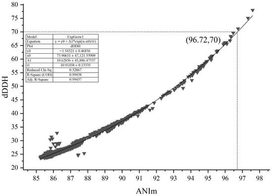
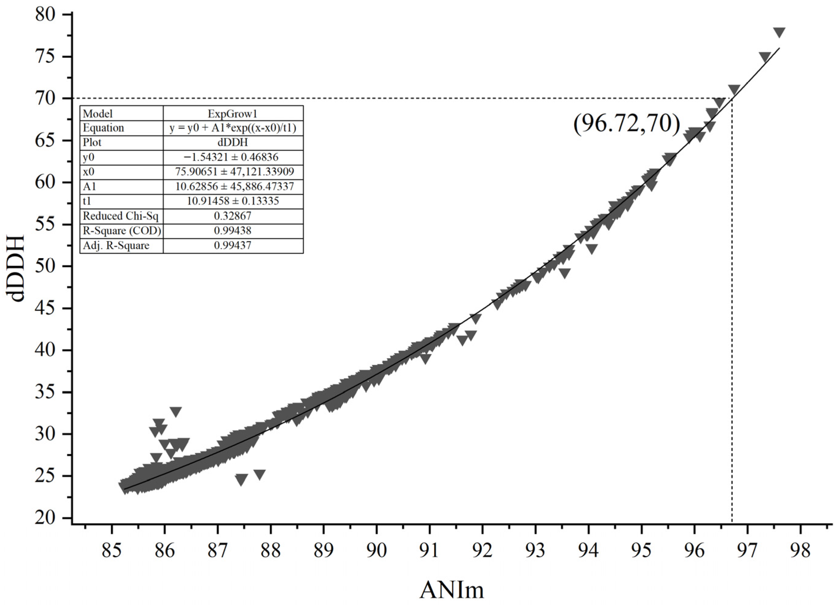
To resolve the contradictory phenomenon mentioned in the introduction, the ANIm and dDDH values of 60 type Micromonospora strains were selected for correlation analysis with a total of 1770 datasets by calculating pairwise between the strains (Table S2). As shown in Figure 1, the dDDH value showed an extremely high correlation (R2 = 0.99438) with the ANIm value based on an exponential regression model. However, a 70% dDDH value was not equivalent to a 95–96% ANIm value, but to an approximately 96.72% ANIm value. At present, in addition to ANIm, ANIb (based on the OrthoANIu algorithm) is also often used for evaluating genomic correlations between two strains. Similarly, in this genus, a 70% dDDH value did not correspond to a 95–96% ANIb value, but to an approximately 96.15% ANIb (Figure S1), and the dDDH value showed an extremely high correlation (R2 = 0.99245) with the ANIm value based on an exponential regression model (The R2 values are presented only as descriptive measures of goodness-of-fit for the exponential regression models; no statistical significance (p-values) is inferred due to the non-independence of pairwise comparisons). It can be seen that an ANI value of 95–96% does not correspond to a dDDH value of 70% in the genus Micromonospora. As the R2 in the ANIm fitting results is higher (0.99438), we recommend that the 96.7% ANIm value could act as a threshold in delineating Micromonospora species.
Figure 1.
The correlation between ANIm and dDDH from the 1770 pairs of Micromonospora species. ANIm (Average Nucleotide Identity based on MUMmer alignment) and dDDH (digital DNA–DNA hybridization) are genome-based metrics for species delineation. dDDH values ≥70% are generally considered the threshold for assigning two genomes to the same prokaryotic species.
To evaluate the impact of threshold selection, we varied the ANI cutoff from 95.0% to 97.5%. At the universal lower bound (95.0%), the maximum ANI between strain HUAS LYJ1T and its closest relative (95.05%) exceeded the threshold, which would imply conspecificity. However, at any threshold ≥95.5% (including our proposed genus-specific threshold of 96.7%), the strain falls below the cutoff and is recognized as a novel species. As the 95% threshold is known to be suboptimal for Micromonospora (as supported by our pairwise analysis of all type strains), we advocate a data-driven threshold of 96.7%. Under this recommended threshold, the novel species assignment is robust.
The present study focused solely on Micromonospora; therefore, the proposed genus-specific threshold should not be extrapolated to other genera without independent validation. Ongoing work in our laboratory is examining additional actinobacterial genera to assess whether similar adjustments are needed, but those results will be reported elsewhere.
3.2. Evaluation of the Taxonomic Status of Strain HUAS LYJ1T
Morphologically, strain HUAS LYJ1T produced non-motile single spores with a smooth surface on well-developed, extensively branched substrate hyphae (Figure S2) on R2A agar medium at 28 °C after incubation for 14 days, but does not produce aerial hyphae. Chemotaxonomically, the cell wall contains meso-diaminopimelic acid as a diagnostic amino acid. Detailed phenotypic and chemotaxonomical characteristics were provided in the species description. These data indicated that strain HUAS LYJ1T represented a member of the genus Micromonospora [30,31].
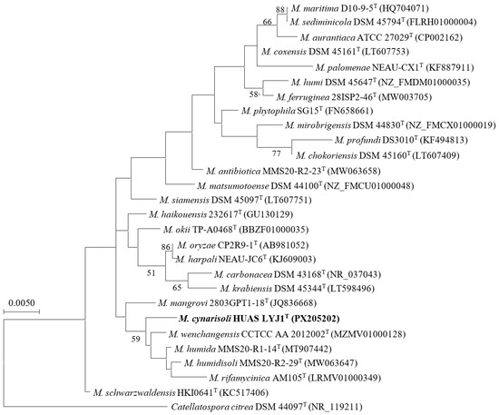
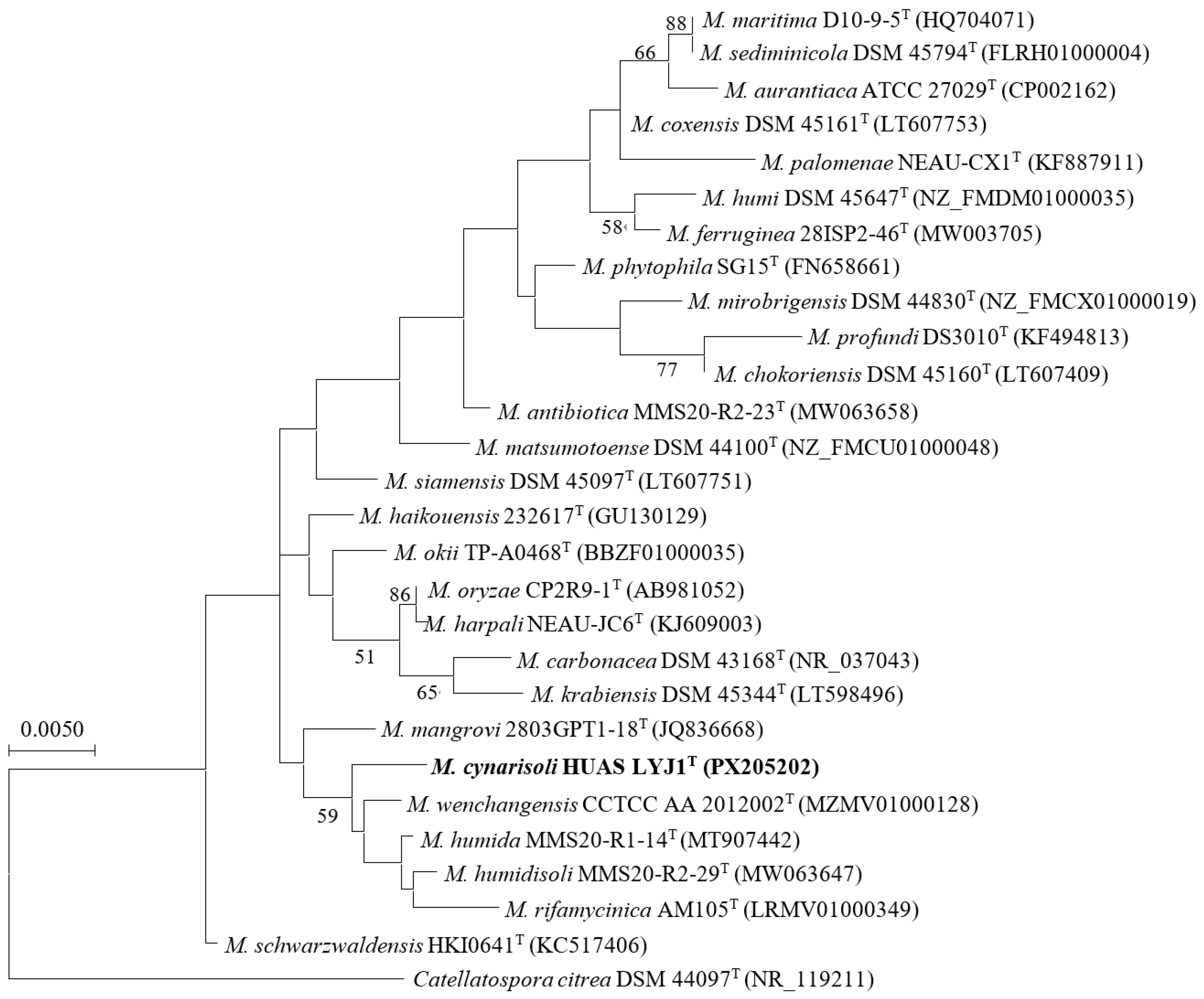
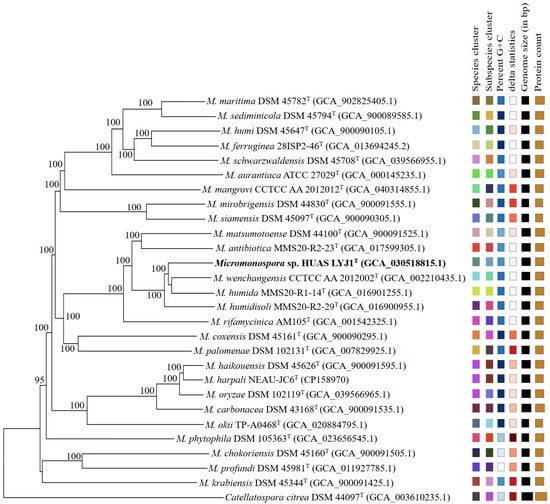
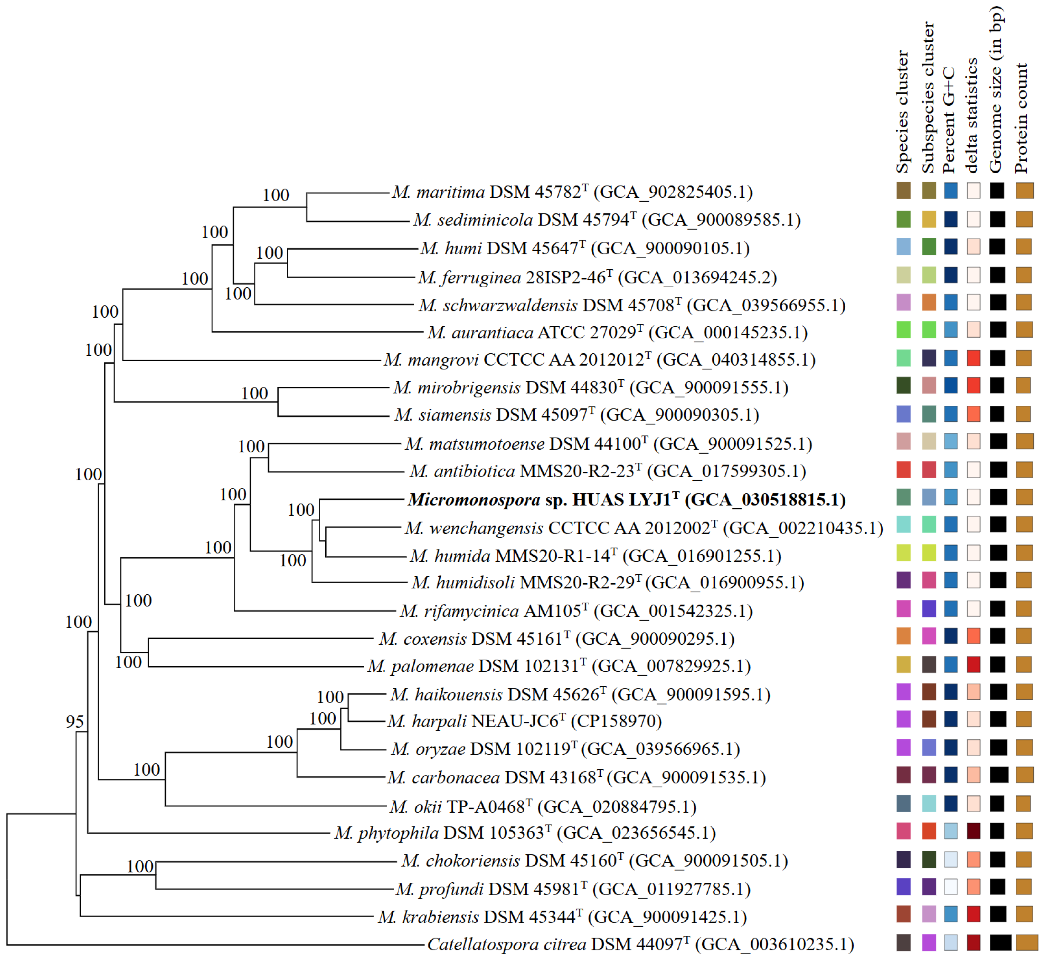
At the molecular level, full-length 16S rRNA gene sequence (1514 nt) analysis showed that strain HUAS LYJ1T belonged to the genus Micromonospora and shared highest similarity to M. wenchangensis CCTCC AA 2012002T (99.3%). Phylogenetic trees based on 16S rRNA gene sequences demonstrated that the evolutionary neighbor of strain HUAS LYJ1T was M. wenchangensis CCTCC AA 2012002T (Figure 2, Figures S3 and S4). This result was also confirmed by a phylogenomic tree (Figure 3), suggesting that these two strains were most related to each other. However, the ANIm value between strain HUAS LYJ1T and M. wenchangensis CCTCC AA 2012002T was 95.77%, below the 96.7% cut-off point recommended above; the dDDH value between these two strains was 63.2%, also far below the 70% threshold value in delineating bacterial species [6]. These data indicated that strain HUAS LYJ1T was distinct from M. wenchangensis CCTCC AA 2012002T and should represent a new Micromonospora species, as was also further confirmed by the differences in phenotypic features, chemotaxonomical features and protein fingerprinting between strain HUAS LYJ1T and M. wenchangensis CCTCC AA 2012002T (Table 1, Table S3 and Figure S5). The protein fingerprints of strain HUAS LYJ1T and its closest relative M. wenchangensis CCTCC AA 2012002T were analyzed by MALDI-TOF MS (Figure S6). The two strains showed distinct peak patterns across the mass range of 2000–12,000 m/z. Strain HUAS LYJ1T exhibited major peaks at [e.g., 3450, 5230, 7810] m/z (Figure S6a), whereas M. wenchangensis displayed prominent peaks at [e.g., 3210, 6050] m/z (Figure S6b), with clear differences in both peak positions and relative intensities. These distinct protein profiles provide additional phenotypic evidence supporting the classification of HUAS LYJ1T as a novel species. In addition, the ANIm and dDDH values between strain HUAS LYJ1T and all other type strains, which exhibited 16S rRNA gene sequence similarities of ≥98.7% to strain HUAS LYJ1T, were 86.68–95.05% and 26.5–58.9% (Table 2), respectively, much less than the 96.6% and 70% threshold values recommended in delineating Micromonospora species and bacterial species.
Figure 2.
Maximum-likelihood phylogenetic tree based on 16S rRNA gene sequences (1514 bp) showing the relationship between strain HUAS LYJ1T and the type strains of selected species of the genus Micromonospora. Catellatospora citrea DSM 44097T was used as an outgroup. Bootstrap percentages over 50% derived from 1000 replications are shown at the nodes. Bar, 0.005 substitutions per site.
Figure 3.
Phylogenomic tree of strain HUAS LYJ1T and related type strains. The branch lengths are scaled in terms of GBDP distance formula d5. The numbers above branches are GBDP pseudo-bootstrap support values >60% from 100 replications, with an average branch support of 96.0%. The color scale on the right represents the range of pairwise GBDP d5 distances among all strains. The gradient from light to dark corresponds to increasing genetic distance (lighter = closer; darker = more distant).
Table 1.
Differential features between strain HUAS LYJ1T and M. wenchangensis CCTCC AA 2012002T.
Table 2.
Comparative genotypic analysis between strain HUAS LYJ1T and the closest related type strains.
In conclusion, based on phenotypic, chemotaxonomic and genotypic data, strain HUAS LYJ1T represents a novel species of the genus Micromonospora, for which the name Micromonospora cynarisoli sp. nov. is proposed.
3.3. Taxonomic Relationship Among M. oryzae, M. harpali and M. haikouensis
According to LPSN (List of Prokaryotic names with Standing in Nomenclature), the genus Micromonospora includes over 120 species with a validly published and correct name. Of all these species, M. haikouensis DSM 45626T, isolated from mangrove sediment sample, was first described as a separate genomic species by Xie et al. in 2012 [32] and approved in the same year [33]. In 2015, M. harpali NEAU-JC6T from a beetle and M. oryzae DSM 102119T from the root internal tissues of upland rice were reported as two different genomic species [34,35] and approved in 2015 and 2016 [36,37], respectively. However, in the present work, the clustering pattern in phylogenomic tree revealed that M. haikouensis DSM 45626T, M. harpali NEAU-JC6T and M.oryzae DSM 102119T should belong to the same genomic species (Figure 3), as was also confirmed by the higher ANIm (97.21–97.86%) and dDDH (73.9–79.6%) values among these three strains (Table 3). Although there were some obvious differences in the phenotypic and chemotaxonomic features among these three strains (Table 4), these differences are most likely due to adaptive selection pressures in their respective ecological niches [38], such as plant endophytes [39], rhizosphere soil [40], marine sediments [41], and so on. In the genus Micromonospora, numerous studies have shown that many phenotypic traits are highly plastic. For example, traits directly related to ecological adaptation, such as plant colonization ability and interaction effects, have been shown to vary significantly depending on the strain source and the host environment.
Table 3.
ANIm and dDDH values among M. oryzae DSM 102119T, M. haikouensis DSM 45626T and M. harpali NEAU-JC6T.
Table 4.
Differential features among Micromonospora haikouensis DSM 45626T, Micromonospora harpali NEU-JC6T and Micromonospora oryzae CP2R9-1T.
Given the environmental dependence of phenotypic traits, genome consistency is considered a more fundamental and reliable criterion for defining species in modern microbial taxonomy. Metrics based on whole-genome sequences, such as average nucleotide identity (ANI) and digital DNA-DNA hybridization (dDDH), provide stable, quantifiable measures of evolutionary relationships that are not affected by environmental factors. In this study, the relevant strains showed ANIm values of 97.21–97.86% and dDDH values of 73.9–79.6%, far above the recognized species thresholds (typically 96% ANI and 70% dDDH), providing strong evidence for classifying them as the same species. Therefore, we consider that the robust genomic homology is sufficient to support merging the strains, which exhibit limited phenotypic differences that may be driven by ecological niches, into a single taxonomic unit. Moreover, we re-examined the phenotypic data and found no single or combined trait that consistently separates the merged strains into distinct groups. The observed variations are quantitative and strain-specific, as is commonly seen within many bacterial species (e.g., in the genus Micromonospora).
Consequently, based on all these data and rule 42 of the International Committee on Systematics of Prokaryote Code [42], we propose that M. harpali [34] and M. oryzae [35] are later heterotypic synonyms of M. haikouensis [32].
4. Conclusions
4.1. The Relationship Between Average Nucleotide Identity and dDDH Values in the Genus Micromonospora
A 70% dDDH value was not equivalent to a 95–96% ANIm value, but to an approximately 96.7% ANIm value in the genus Micromonospora.
4.2. Description of Micromonospora cynarisoli sp. nov.
Micromonospora cynarisoli (cy.na.ri.so’li N.L. fem. n. Cynara, the scientific name of the botanical genus to which Cynara scolymus belongs; L. neut. n. solum, soil; N.L. gen. neut. n. cynarisoli, to indicate the isolation of the type strain from rhizosphere soil around the roots of the globe artichoke, Cynara scolymus)
Gram-stain-positive and strictly aerobic. Grows well on Reasoner’2A, Gauze’s synthetic No. 1 medium and ISP 3 to ISP 7. Aerial mycelium was not produced; substrate hyphae were well-developed and produced single spores with a smooth surface. A seashell pink diffusible pigment was produced on ISP 7, but no diffusible pigments were produced on Reasoner’s 2A medium, Gauze’s synthetic No. 1 medium and ISP media 2 to 6. Melanin was not produced on any of the media tested. Growth occurred at a temperature range of 10–40 °C (optimum 28 °C), a pH range of 6.0–12.0 (optimum 7.0), and a NaCl concentration range of 0–3.0% (w/v) (optimum 1.0%). Positive for milk solidification, milk peptonement, starch, nitrate reduction, gelatin liquefaction, and hydrolysis of aesculin and Tween (20, 40, 60). Negative for H2S production, starch hydrolysis and Tween 80 hydrolysis. In the API ZYM test, positive for cystine arylamidase, esterase (C4), esterase lipase (C8), α-galactosidase and α-glucosidase, leucine arylamidase, lipase (C14), naphtol-AS-BI-phosphohydrolase, trypsin, valine arylamidase; but negative for acid phosphatase, alkaline phosphatase, chymotrypsin, β-glucuronidase, α-mannosidase, β-fucosidase, β-galactosidase, β-glucosidase and N-acetyl-β-glucosaminidase. Utilizes d-galactose, d-raffinose, d-glucose as sole carbon sources, but not l-arabinose, l-rhamnose, inositol, mannitol, d-sorbitol, d-trehalose, xylitol, cellose, d-ribose, d-fructose and d-xylose. Positive for utilization of l-arginine, l-aspartic acid, creatine, l-cysteine, l-lysine, l-methionine, l-serine, and l-threonine as sole nitrogen sources, but negative for l-histidine and l-tyrosine. The major menaquinones are MK-9 (H2), MK-9 (H6), MK-10 and MK-10 (H2). The cell wall contains Meso-diaminopimelic acid, aspartic acid and glycine; whole-cell hydrolysates are glucose and xylose. The major fatty acids are iso-C15:0, iso-C16:0, C18:0 10-methyl (TBSA), C18:1ω9c, iso-C16:1 G, C17:010-methyl and Sum In Feature 9. The polar lipids are diphosphatidylglycerol (DPG), phospholipids (PLS), phosphatidyl ethanolamine (PE), phospholipids of unknown structure containing glucosamine (NPG), phosphatidylinositol (PI), phosphatidylinositol mannosides (PIM).
The type strain, strain HUAS LYJ1T (=MCCC 1K08697T = JCM 36306T), the DNA G + C content of the genome sequence, consisting of 7.52 Mbp, is 72.5%. The NCBI accession number for the full-length 16S rRNA gene sequence extracted from the whole genome and the genome sequence of strain HUAS LYJ1T are PX205202 and CP130426.1, respectively.
4.3. Emended Description of Micromonospora haikouensis
Later heterotypic synonyms: Micromonospora harpali Fang et al. 2015 (Approved Lists 2015) and Micromonospora oryzae Kittiwongwattana et al. 2015 (Approved Lists 2016).
The description is as before [32] with the following modification. The DNA G + C content of the type-strain genome is 73.8%, its approximate size 7.58 Mbp, its GenBank accession NZ_FMCW00000000.
The type strain is DSM 45626T (=232617T = CCTCC AA 2011012T). Sequence data that support the findings of this study have been deposited in the National Center for Biotechnology Information with the primary accession code SRR32251829.
Supplementary Materials
The following supporting information can be downloaded at: https://www.mdpi.com/article/10.3390/microorganisms14050981/s1. Figure S1 The correlation between ANIb and dDDH from the 1770 pairs of Micromonospora species. Figure S2 Optical micrograph (a) and scanning electron micrograph (b) of strain HUAS LYJ1T grown on Reasoner’ 2A medium at 28 °C after incubation for 14 days. Figure S3 Neighbor-joining phylogenetic tree based on 16S rRNA gene sequences (1514 bp) showing the relationship between selected species of the genus Micromonospora. Figure S4 Maximum-parsimony phylogenetic tree based on 16S rRNA gene sequences (1514 bp) showing the relationship between selected species of the genus Micromonospora. Figure S5 Polar lipids composition of strain HUAS LYJ1T (1) and M. wenchangensis strain CCTCC AA 2012002T (2). Figure S6 MALDI-TOF MS protein fingerprint of strain HUAS LYJ1T (a) and M. wenchangensis strain CCTCC AA 2012002T (b). Table S1 Quality analysis and GenBank assembly of genomes of Micromonospora species in this work (60 species). Table S2 ANI and dDDH values of 1770 pairs of type Micromonospora strains. Table S3 Cultural characteristics between strain HUAS LYJ1T and M. wenchangensis strain CCTCC AA 2012002T.
Author Contributions
K.L.: Supervision, Writing—review and editing, Writing—original draft, Data curation; L.F.: Data curation, Writing—original draft, Writing–review and editing; P.L.: Supervision, Methodology, Writing—review and editing; Y.Q.: Data curation, Investigation; W.L.: Funding acquisition, Resources; J.G.: Methodology, Project administration, Conceptualization. All authors have read and agreed to the published version of the manuscript.
Funding
This work was supported by the Postdoctoral Fellowship Program of CPSF under Grant Number (GZC20251753), the National Natural Science Foundation of China (62472168) and the Hunan Provincial Natural Science Foundation of China and Xiangtan Science and Technology Bureau (2022JJ50125).
Institutional Review Board Statement
Not applicable.
Informed Consent Statement
Not applicable.
Data Availability Statement
Data will be made available on request.
Conflicts of Interest
The authors declare that the research was conducted in the absence of any commercial or financial relationships that could be construed as a potential conflict of interest.
Abbreviations
| MCCC | Marine Culture Collection of China |
| JCM | the Japan Collection of Microorganisms |
| CCTCC | China Center for Type Culture Collection |
| ISP | International Streptomyces Project |
References
- Richter, M.; Rosselló-Móra, R. Shifting the genomic gold standard for the prokaryotic species definition. Proc. Natl. Acad. Sci. USA 2009, 106, 19126–19131. [Google Scholar] [CrossRef] [Scilit]
- Auch, A.F.; von Jan, M.; Klenk, H.P.; Göker, M. Digital DNA-DNA hybridization for microbial species delineation by means of genome-to-genome sequence comparison. Stand. Genom. Sci. 2010, 2, 117–134. [Google Scholar] [CrossRef] [Scilit]
- Hu, S.R.; Li, K.Q.; Zhang, Y.F.; Wang, Y.F.; Fu, L.; Gao, J. New insights into the threshold values of multi-locus sequence analysis, average nucleotide identity and digital DNA–DNA hybridization in delineating Streptomyces species. Front. Microbiol. 2022, 13, 910277. [Google Scholar] [CrossRef] [Scilit]
- Deng, A.H.; Fu, L.; Zheng, Y.X.; Tang, T.; Mo, P.; Gao, J. New insights into the relationship between the average nucleotide identity and the digital DNA-DNA hybridization values in the genus Amycolatopsis and Amycolatopsis cynarae sp. nov., a novel actinobacterium from the rhizosphere soil of Cynara scolymus and proposal of Amycolatopsis niigatensis as a synonym of Amycolatopsis echigonensis based on comparative genomic analysis. Front. Microbiol. 2024, 15, 1359021. [Google Scholar] [CrossRef] [Scilit]
- Zhang, Y.Y.; Su, J.Q.; Liao, H.; Breed, M.F.; Yao, H.F.; Zhu, Y.G. Increasing antimicrobial resistance and potential human bacterial pathogens in an invasive land snail driven by urbanization. Environ. Sci. Technol. 2023, 57, 7273−7284. [Google Scholar] [CrossRef] [Scilit] [PubMed]
- Demain, A.L.; Sanchez, S. Microbial drug discovery: 80 years of progress. J. Antibiot. 2009, 62, 5–16. [Google Scholar] [CrossRef] [Scilit]
- Tiwari, K.; Gupta, R.K. Rare actinomycetes: A potential storehouse for novel antibiotics. Crit. Rev. Biotechnol. 2012, 32, 108–132. [Google Scholar] [CrossRef] [Scilit]
- Parra, J.; Beaton, A.; Seipke, R.F.; Wilkinson, B.; Hutchings, M.I.; Duncan, K.R. Antibiotics from rare actinomycetes, beyond the genus Streptomyces. Curr. Opin. Microbiol. 2023, 76, 102385. [Google Scholar] [CrossRef] [Scilit] [PubMed]
- Parks, D.H.; Imelfort, M.; Skennerton, C.T.; Hugenholtz, P.; Tyson, G.W. CheckM: Assessing the quality of microbial genomes recovered from isolates, single cells, and metagenomes. Genome Res. 2015, 25, 1043–1055. [Google Scholar] [CrossRef] [Scilit] [PubMed]
- Bowers, R.; Kyrpides, N.; Stepanauskas, R. Minimum information about a single amplified genome (MISAG) and a metagenome-assembled genome (MIMAG) of bacteria and archaea. Nat. Biotechnol. 2017, 35, 725–731. [Google Scholar] [CrossRef] [Scilit] [PubMed]
- Richter, M.; Rosselló-Móra, R.; Glöckner, F.O.; Peplies, J. JSpeciesWS: A web server for prokaryotic species circumscription based on pairwise genome comparison. Bioinformatics 2016, 32, 929–931. [Google Scholar] [CrossRef] [Scilit]
- Meier-Kolthoff, J.P.; Auch, A.F.; Klenk, H.P.; Göker, M. Genome sequence-based species delimitation with confidence intervals and improved distance functions. BMC Bioinform. 2013, 14, 60. [Google Scholar] [CrossRef] [Scilit]
- Mo, P.; Yu, Y.Z.; Zhao, J.R.; Gao, J. Streptomyces xiangtanensis sp. nov., isolated from a manganese-contaminated soil. Antonie Van Leeuwenhoek 2017, 110, 297–304. [Google Scholar] [CrossRef] [Scilit] [PubMed]
- Atlas, R.M. Handbook of Microbiological Media; Parks, L.C., Ed.; CRC Press: Boca Raton, FL, USA, 1993. [Google Scholar]
- Weisburg, W.G.; Barns, S.M.; Pelletier, D.A.; Lane, D.J. 16S ribosomal DNA amplification for phylogenetic study. J. Bacteriol. 1991, 173, 697–703. [Google Scholar] [CrossRef] [Scilit] [PubMed]
- Lane, D.J. 16S/23S rRNA sequencing. In Nucleic Acid Techniques in Bacterial Systematics; Stackebrandt, E., Goodfellow, M., Eds.; Wiley: New York, NY, USA, 1991; pp. 115–175. [Google Scholar]
- Chalita, M.; Kim, Y.O.; Park, S.; Oh, H.S.; Cho, J.H.; Moon, J.; Baek, N.; Chun, J.; Moon, C.; Lee, K.; et al. EzBioCloud: A genome-driven database and platform for microbiome identification and discovery. Int. J. Syst. Evol. Microbiol. 2024, 74, 006421. [Google Scholar] [CrossRef] [Scilit]
- Tamura, K.; Stecher, G.; Kumar, S. MEGA11: Molecular Evolutionary Genetics Analysis Version 11. Mol. Biol. Evol. 2021, 38, 3022–3027. [Google Scholar] [CrossRef] [Scilit]
- Saitou, N.; Nei, M. The neighbor-joining method: A new method for reconstructing phylogenetic trees. Mol. Biol. Evol. 1987, 4, 406–425. [Google Scholar] [CrossRef] [Scilit]
- Felsenstein, J. Evolutionary trees from DNA sequences: A maximum likelihood approach. J. Mol. Evol. 1981, 17, 368–376. [Google Scholar] [CrossRef] [Scilit]
- Kluge, A.G.; Farris, F.S. Quantitative phyletics and the evolution of anurans. Syst. Biol. 1969, 18, 1–32. [Google Scholar] [CrossRef] [Scilit]
- Felsenstein, J. Confidence limit on phylogenies: An approach using the bootstrap. Evolution 1985, 39, 783–791. [Google Scholar] [CrossRef] [Scilit] [PubMed]
- Meier-Kolthoff, J.P.; Sardà Carbasse, J.; Peinado-Olarte, R.L.; Göker, M. TYGS and LPSN: A database tandem for fast and reliable genome-based classification and nomenclature of prokaryotes. Nucleic Acid. Res. 2022, 50, D801–D807. [Google Scholar] [CrossRef] [Scilit]
- Vincent, L.; Richard, D.; Olivier, G. FastME 2.0: A comprehensive, accurate, and fast distance-based phylogeny inference program. Mol. Biol. Evol. 2015, 32, 2798–2800. [Google Scholar] [CrossRef] [Scilit]
- Farris, J.S. Estimating phylogenetic trees from distance matrices. Am. Nat. 1972, 106, 645–668. [Google Scholar] [CrossRef] [Scilit]
- Reasoner, D.J.; Geldreich, E.E. A new medium for the enumeration and subculture of bacteria from potable water. Appl. Environ. Microbiol. 1985, 49, 1–7. [Google Scholar] [CrossRef] [Scilit]
- Shirling, E.B.; Gottlieb, D. Methods for characterization of Streptomyces species. Int. J. Syst. Bacteriol. 1966, 16, 313–340. [Google Scholar] [CrossRef] [Scilit]
- Ridgway, R. Color Standards and Color Nomenclature; plate I–LII; The Author: Washington, DC, USA, 1912; pp. 1–43. [Google Scholar]
- Xu, L.H.; Li, W.J.; Liu, Z.H.; Jiang, C.L. Actinomycete Systematic Principle, Methods and Practice; Science Press: Beijing, China, 2007. (In Chinese) [Google Scholar]
- Lechevalier, M.P.; Lechevalier, H. Chemical composition as a criterion in the classification of aerobic actinomycetes. Int. J. Syst. Evol. Microbiol. 1970, 20, 435–443. [Google Scholar] [CrossRef] [Scilit]
- Stackebrandt, E.; Rainey, F.A.; Ward-Rainey, N.L. Proposal for a new hierarchic classification system, Actinobacteria classis nov. Int. J. Syst. Bacteriol. 1997, 47, 479–491. [Google Scholar] [CrossRef] [Scilit]
- Xie, Q.Y.; Qu, Z.; Lin, H.P.; Li, L.; Hong, K. Micromonospora haikouensis sp. nov., isolated from mangrove soil. Antonie Van Leeuwenhoek 2012, 101, 649–655. [Google Scholar] [CrossRef] [Scilit] [PubMed]
- Euzeby, J.P. Validation list no. 148. List of new names and new combinations previously effectively, but not validly, published. Int. J. Syst. Evol. Microbiol. 2012, 62, 2549–2554. [Google Scholar] [CrossRef] [Scilit]
- Fang, B.Z.; Liu, C.X.; Guan, X.J.; Song, J.; Zhao, J.W.; Liu, H.; Li, C.; Ning, W.; Wang, X.; Xiang, W. Two new species of the genus Micromonospora: Micromonospora palomenae sp. nov. and Micromonospora harpali sp. nov. isolated from the insects. Antonie Van Leeuwenhoek 2015, 108, 141–150. [Google Scholar] [CrossRef] [Scilit] [PubMed]
- Kittiwongwattana, C.; Thanaboripat, D.; Laosinwattana, C.; Koohakan, P.; Parinthawong, N.; Thawai, C. Micromonospora oryzae sp. nov., isolated from roots of upland rice. Int. J. Syst. Evol. Microbiol. 2015, 65, 3818–3823. [Google Scholar] [CrossRef] [Scilit] [PubMed]
- Oren, A.; Garrity, G.M. Validation list no. 165. List of new names and new combinations previously effectively, but not validly, published. Int. J. Syst. Evol. Microbiol. 2015, 65, 2777–2783. [Google Scholar] [CrossRef] [Scilit]
- Oren, A.; Garrity, G.M. Notification list. Notification that new names and new combinations have appeared in volume 65, part 11 of the IJSEM. Int. J. Syst. Evol. Microbiol. 2016, 66, 527–529. [Google Scholar] [CrossRef] [Scilit]
- Ara, I.; Kudo, T. Two new species of the genus Micromonospora: Micromonospora chokoriensis sp. nov. and Micromonospora coxensis sp. nov., isolated from sandy soil. J. Gen. Appl. Microbiol. 2007, 53, 29–37. [Google Scholar] [CrossRef] [Scilit] [PubMed]
- Carro, L.; Oren, A. Descriptions of Micromonospora grosourdyae nom. nov., Micromonospora sonchi comb. nov. and Micromonospora thawaii sp. nov. to resolve problems with the taxonomy and nomenclature of strains named Micromonospora endophytica. Int. J. Syst. Evol. Microbiol. 2022, 72, 005628. [Google Scholar] [CrossRef] [Scilit] [PubMed]
- Long, P.L.; Fu, L.; Xiao, Y.; Gao, J. Micromonospora cathayae sp. nov., isolated from the rhizosphere soil of Cathaya argyrophylla. Int. J. Syst. Evol. Microbiol. 2024, 74, 006332. [Google Scholar] [CrossRef] [Scilit] [PubMed]
- Veyisoglu, A.; Carro, L.; Cetin, D.; Guven, K.; Spröer, C.; Pötter, G.; Klenk, H.P.; Sahin, N.; Goodfellow, M. Micromonospora profundi sp. nov., isolated from deep marine sediment. Int. J. Syst. Evol. Microbiol. 2016, 66, 4735–4743. [Google Scholar] [CrossRef] [Scilit]
- Oren, A.; Arahal, D.R.; Göker, M.; Moore, E.R.B.; Rossello-Mora, R.; Sutcliffe, I.C. International Code of Nomenclature of Prokaryotes. Prokaryotic Code (2022 Revision). Int. J. Syst. Evol. Microbiol. 2022, 73, 005585. [Google Scholar] [CrossRef] [Scilit] [PubMed]

Disclaimer/Publisher’s Note: The statements, opinions and data contained in all publications are solely those of the individual author(s) and contributor(s) and not of MDPI and/or the editor(s). MDPI and/or the editor(s) disclaim responsibility for any injury to people or property resulting from any ideas, methods, instructions or products referred to in the content. |
© 2026 by the authors. Licensee MDPI, Basel, Switzerland. This article is an open access article distributed under the terms and conditions of the Creative Commons Attribution (CC BY) license.