


Abstract
Streptococcus canis is an opportunistic pathogen that colonises the mucosal surfaces and skin of its host. Though predominantly a veterinary pathogen affecting cats and dogs, S. canis has also been identified as the causative agent in severe human disease. IdeC is a secreted cysteine protease of S. canis that has a high specificity for IgG, cleaving at the hinge region. We show here that the protein binds back to the surface of the bacteria. Additionally, the protein contains a conserved Arg-Gly-Asp (RGD) motif, the minimal peptide sequence required for integrin binding. Several bacterial proteins containing RGD motifs have been implicated in adhesion and invasion of host cells. This RGD motif along with the ability of IdeC to bind back to the bacterial surface after secretion is the basis for this study into a potential secondary function of IdeC in adhesion and/or invasion. We used protein-coated latex beads to investigate the interaction of IdeC with epithelial and endothelial cells and, further, the extent to which the RGD motif is involved in this interaction by utilising an RGD->RGE recombinant protein. We also report here that the deletion of IdeC in S. canis results in a significant reduction in invasion into epithelial cells.
1. Introduction
Streptococcus canis (S. canis) is a Gram-positive, ß-haemolytic, opportunistic pathogen [1] belonging to Lancefield group G or C [2]. Though originally thought to affect only dogs and cows, S. canis is a multi-host pathogen, having been isolated from a broad range of animals, including foxes, mink and rabbits. Nevertheless, cats and dogs are the predominant hosts [1,3,4]. S. canis colonises the mucosal surfaces and skin of the host, but is also an opportunistic pathogen. Clinical presentations include infections of the mucosa and skin, septicaemia, endocarditis, and necrotising fasciitis [1].
Though S. canis has been isolated from a wide range of hosts, it is not as well studied as other streptococcal species. However, based on the zoonotic potential of this pathogen, along with the acquisition of antimicrobial resistance (AMR) that has now been shown, it is increasingly recognised as a potential threat [5,6,7]. The virulence factors of S. canis are not well characterised, although some have been described, including the S. canis M Protein, SCM [8,9,10], and an arginine deiminase system [11].
Clinical manifestation suggests that similar virulence factors may exist in S. canis as in Streptococcus pyogenes (S. pyogenes). Previous studies describe genome comparisons as a starting point for investigating possible virulence factors of S. canis [6,12]. Cysteine proteases have been identified as potent virulence factors in many bacteria, including various streptococcal species. IdeS (also called Mac-1) is a cysteine protease of S. pyogenes that has been demonstrated to cleave human immunoglobulin G (IgG), thereby inhibiting phagocytosis [13,14]. In our previous work we described the cysteine protease IdeC (Immunoglobulin G-degrading enzyme of S. canis), identified based on a sequence similarity to IdeS [15]. IdeC is a highly specific endopeptidase that cleaves feline, canine and human IgG in the hinge region. IdeC also contains an Arg-Gly-Asp (RGD) motif [15].
The RGD motif is an amino acid motif involved in integrin binding [16]. This motif is found in many proteins involved in mediating cell-to-cell interaction [17]. The engagement of extracellular matrix (ECM) proteins such as fibronectin, vitronectin and fibrinogen with integrins is primarily mediated by RGD motifs present within the ECM proteins [18]. Integrin interaction with ECM proteins results in the activation of intracellular signalling pathways via recruitment of the signalling molecules to the integrin engagement/clustering site [19]. This binding and subsequent clustering of integrins has been demonstrated to be necessary for the activation of integrin signalling as well as invasion into cells by bacteria [20]. In proteins such as human fibronectin, the RGD motif has been revealed to be located on an outward-facing loop, making it easily accessible to a single integrin molecule [21]. The position of the RGD motif, its 3D structure, flanking residues, etc., are factors in the interaction that may occur between the RGD motif and integrin receptors. Several bacterial proteins have been shown to have a role in human infection mediated by an RGD motif, for example, CagL of Helicobacter pylori [22], Pertactin of Bordetella pertussis [23], and SfbI/F1 of S. pyogenes [24,25]. Indeed, the IdeS protein that shares a high sequence identity with IdeC also contains the RGD motif, which has been demonstrated to mediate integrin interaction [26].
This study aims to determine whether the previously described cysteine protease IdeC is a ‘moonlighting’ protein with a second role, in adhesion or invasion. The IgG-cleaving properties of IdeC have been described, but a secondary function will be investigated here based on the presence of an integrin-binding RGD motif and the ability of the protein to bind to the bacterial surface after secretion.
2. Materials and Methods
2.1. Bacterial Strains and Growth Conditions
The Streptococcus canis strains G361 [27] and Sc957 (isolated from skin infection in a canine, during routine diagnostics at Freie Universität Berlin, Germany) were routinely grown in Todd–Hewitt broth (THB) at 37 °C. Strains carrying the pGhost9 plasmid or its derivatives were grown in the same medium supplemented with 200 µg/mL erythromycin at 28 °C. The Escherichia coli strain M15 pREP was grown at 37 °C in lysogeny broth (LB) supplemented with 25 µg/mL kanamycin. After transformation with the pQE30 plasmid, M15 pREP pQE30 was grown in LB supplemented with 100 µg/mL ampicillin and 25 µg/mL kanamycin.
2.2. Analysis of IdeC Amino Acid Sequence
The sequence for IdeC was taken from the NCBI entry, ‘Immunoglobulin G-endopeptidase (IdeS)/Mac [Streptococcus canis FSL Z3-227]’, accession: EIQ82374. The program SignalP 6.0 (https://services.healthtech.dtu.dk/services/SignalP-6.0/ (accessed on 11 June 2024)) was used to analyse the protein for the presence of a signal sequence and locate it.
2.3. Recombinant Protein Expression and Purification
Genomic DNA was extracted from an overnight bacterial culture using the QIAGEN QIAamp DNA Mini Kit. The IdeC protein was amplified from gDNA using the IdeC-sigseq BamH1 (5′-GCTGGATCCGACAACATCGA-3′) and IdeC HindIII (5′-GGGAAGCTTTTAGTTTGATGCC-3′) PCR primers. The PCR product was analysed by agarose gel electrophoresis, on a 1.5% gel. The PCR product was then purified using the QIAGEN QIAquick PCR Purification Kit. The purified PCR product and the pQE30 plasmid were then digested using the BamHI and HindIII restriction enzymes; digestion was carried out at 37 °C for 1 h. The digested PCR product was ligated into the digested plasmid using T4 ligase at 4 °C overnight. The pQE30_IdeC plasmid was then transformed into the E. coli strain M15 pREP. The M15 pREP pQE30_IdeC was grown to OD600 = 0.5 at 29 °C, IPTG was added to a final concentration of 1 M, and the culture was grown for a further 4 h at 29 °C. Cells were harvested by centrifugation and lysed using a 1 mg/mL lysozyme solution. The protein was then purified by nickel chromatography on Protino Ni-TED 1000 columns. Proteins were run on 15% SDS gel to confirm that purification was successful. Thermo Scientific Slide-A-Lyzer Dialysis Cassettes with a 10,000 molecular-weight cutoff were used to exchange the elution buffer, so proteins could be stored in 1 × PBS.
2.4. Preparation of Recombinant Proteins
To prepare a recombinant IdeC protein (rIdeC) with an amino acid replacement of the RGD to RGE, inverse PCR was performed on the pQE30_IdeC construct using the PCR primers RGEfor (5′-CAGGTATTTACAAGAGGAGAACAAAGTAAACTATTA-3′) and RGErev (5′-TTTACTTTGTTCTCCTCTTGTAAATACCTGATCAAA-3′). The PCR product was purified and digested with the Dpn1 restriction enzyme for 1 h at 37 °C, before ligation with T4 ligase. pQE30IdeCRGE was transformed into M15 pREP, and the protein was purified as described above.
2.5. IgG Cleavage Assay
To demonstrate the IgG cleavage abilities of IdeCRGE, 3 µg of purified rIdeCRGE was incubated with 3 µg purified feline IgG (Jackson Immuno Research, Westgrove, PA, USA) for 4 h at 37 °C. 4× Laemmli sample buffer with dithiothreitol (DTT) was added to the reaction mix and it was denatured at 95 °C for 10 min. The reaction mix was run on 15% SDS gel to visualise results. Precision Plus Protein™ All Blue Pre-stained Protein Standard was used to determine the molecular weight of bands (BioRad, Hercules, CA, USA, catalogue number 1610373).
2.6. Preparation of the IdeC-Coated Latex Beads
For experiments involving protein-coated beads, SIGMA (Sigma-Aldrich, Saintlouis, MO, USA) fluorescent carboxyl-modified latex beads or SIGMA latex beads were used. In total, 108 beads were washed 3 times in 1 × PBS, and then coated with 1000 µg protein overnight at 4 °C, with rotation. In the case of carboxyl-modified beads, EDAC was used as a chemical linker at a final concentration of 0.15–2.6 mM. Finally, beads were washed three times in 1 × PBS. Successful coating of the beads was confirmed by SDS-Page.
2.7. Cell Culture and Bead Internalisation Assay
All cell lines used in this study (Table 1) were incubated at 37 °C and 5% CO2. The MEF cell lines were kindly gifted by Dr. Markus Keller of the Friedrich-Loeffler Institut. The A549 cells were provided by Dr. Thomas Meyer of the Max Planck Institute for Medical Research.
Table 1.
Sources of cell types used in this study.
Bead assays were carried out as previously described by Kaur et al. [29]; cells were seeded onto coverslips in 24-well plates and grown to ~80% confluency. Latex beads (Sigma-Aldrich, Saintlouis, MO, USA) were coated in protein by incubation with purified recombinant protein overnight, rotating at 4 °C. Beads were then washed three times with 1 × PBS. Protein-coated beads were suspended in endothelial cell culture medium and added to cell monolayers at an excess of 100 beads per cell. Cells were incubated with beads at 37 °C, 5% CO2, for various time periods (30 min, 1 h, 2 h). After incubation, monolayers were washed 6 times with 1 × PBS before fixing with 4% paraformaldehyde. To ensure non-specific binding of beads did not occur, bovine serum albumin (BSA)-coated latex beads were used in the same assay as a control.
2.8. Fluorescence Staining and Confocal Microscopy
Coverslips with fixed cells were first blocked for 30 min with PBS containing 5% foetal calf serum (FCS). For visualisation of cell nuclei and actin cytoskeletons, DAPI and Phalloidin were used, respectively. In monolayers treated with fluorescent beads, no further staining was performed. For monolayers treated with non-fluorescent beads, extracellular beads were stained using rabbit polyclonal anti-IdeC IgG followed by anti-rabbit Alexa Fluor 488-conjugated IgG. Mounted samples were viewed and analysed using the Leica DMI6000 B microscope with a 63× objective lens, and adherent/associated beads were counted. Bead counts were performed as follows: 6 fields of view (FOVs) per slide were examined. FOVs were selected based on a similar number/distribution of cells, and then the number of beads in clusters, the number of single beads and the number of clusters were recorded per FOV.
All images were deconvolved using Leica Application Suite 4,0. Images were brightened identically using IrfanView 64-bit software.
2.9. Preparation of 957ΔideC Strain
To prepare an IdeC deleted strain, the ideC gene of the S. canis strain 957, along with the upstream and downstream regions, was amplified using the PCR primers IdeCup (5′-CCCCCATGGCACCTGATAATATCGCCTC-3′) and IdeCdw (5′-GGGCCATGGACGAGCATCATTACTGGG-3′). The plasmid pGhost9::ISS1 was amplified using the PCR primers pGhost9-dISS1 F (5′-GGGCCATGGGCTCCTTGGAAGCTGTCAG-3′) and pGhost9-dISS1 R (5′-CCCCCATGGGGTACCCAATTCGCCCTATA-3′). Both PCR products were purified and digested with Nco1 restriction enzyme, before being ligated using T4 ligase at 4 °C overnight. pGhost9_ideCup+dw was transformed into E. coli EC101 using the BIO-RAD MicroPulser (1.8 kV). The pGhost9_ideCup+dw construct was purified from E. coli EC101 using QIAGEN QIAprep Spin Miniprep Kit and the ideC gene was removed by inverse PCR using the primers IdeCdel-fw (5′-GGGGATATCAGAGCCCAAGACATGCT-3′) and IdeCdel-rv (5′-CCCCTATAGATGCTGGACTTAGGCAT-3‘). The PCR product was purified and digested with EcoRV before ligation with T4 ligase. The pGhost9ΔideC construct was transformed into E. coli EC101. The pGhost9ΔideC construct was purified from EC101 and transformed via electroporation into 957 using the BIO-RAD MicroPulser (1.8 kV).
To induce allelic replacement, a single colony of 957 pGhost9ΔideC was inoculated into THB 200 µg/mL erythromycin and grown overnight at 28 °C. This culture was sub-cultivated and grown to OD600 0.2 at 28 °C; this culture was left at 37 °C overnight. A serial dilution was performed on the culture; it was plated on Todd–Hewitt agar (THA) containing 200 µg/mL erythromycin and incubated overnight at 28 °C. A single colony was inoculated into THB without erythromycin and grown overnight at 28 °C. The culture was serially diluted and plated on THA without erythromycin and grown overnight at 37 °C. Single colonies were picked and tested on THA with and without erythromycin at 37 °C. Colonies that were erythromycin-sensitive were checked for the presence of the ideC gene using the IdeCup and IdeCdw PCR primers.
2.10. Bacterial Infection Assay
Bacterial infections were performed in the A549 cell line. Cells were seeded in 24-well plates and grown to 90% confluence. S. canis strains were grown to mid-log phase in THB (OD600 ~ 0.3). Monolayers were washed 3 times with PBS before cell culture media was replaced with media containing S. canis at a multiplicity of infection (MOI) of 1:10 (10 bacteria for each cell). Bacterial inoculates were standardised based on absorbance at OD600, CFU counts were used to confirm bacterial counts, and cultures were vortexed prior to application to monolayers to disrupt any bacterial aggregates. The plate was incubated at 37 °C and 5% CO2 for 2 h to allow for bacterial adhesion. Monolayers were washed six times to remove non-adherent bacteria. After washing, 0.01% Triton X-100 was added to lyse cells. Cell lysates were serially diluted in PBS, plated on THA and incubated overnight to count the colony-forming units (CFU).
To determine levels of internalised bacteria, steps were performed as above, but extracellular bacteria were inactivated after removal of non-adherent bacteria by further incubation of 40 min at 37 °C, 5% CO2, in DMEM containing 150 µg/mL erythromycin. Afterwards, wells were washed 6 times with PBS and then lysed with 0.01% Triton X-100. As above, cell lysates were serially diluted, plated on THA and grown overnight to allow CFU determination.
2.11. Electron Microscopy
For the labelling of rebound IdeC on the S. canis bacterial surface, the protocol by Bergmann et al. 2017 was applied [30]. In short, S. canis cells were washed with 1 × PBS, incubated with a 1:50 dilution in PBS of protein A-purified rabbit IgG protein against IdeC (~75 µg IgG protein) for 1 h at 37 °C. After several washing steps with PBS, bound IgG was visualised by incubation with protein A-gold/protein G-gold nanoparticles (ratio 1:1, 15 nm in size) for 30 min at 37 °C. As a control, specific IdeC, IgG antibodies were incubated with IdeC protein before incubation with S. canis. After washing with PBS and TE buffer (10 mM TRIS, 2 mM EDTA, pH 6.9), samples were placed onto butvar-coated copper grids and air-dried. Grids were attached to adhesive carbon tape on aluminium stubs and samples were examined in a Zeiss Merlin (Oberkochen, Germany), applying the Everhart-Thornley HESE2 secondary electron detector at an acceleration voltage of 10 kV. Images were taken with the Zeiss SmartSEM software version 5.05.
2.12. Bright-Field Microscopy
Liquid cultures of Sc957 and Sc957∆ideC were viewed and analysed using the Leica DMI6000 B microscope with a 63× objective lens.
2.13. Statistical Analysis
All statistical analyses were performed using GraphPad Prism 10.4.2. For bead interaction assays and infection assays, an unpaired two-tailed t-test was used to assess the significance of results. In addition to this, a Mann–Whitney test was employed to ensure significance as data was not tested for normality and equal variance. All assays were performed in triplicate with statistics based on at least three biological replicates.
3. Results
3.1. Sequence and Structural Characteristics of IdeC
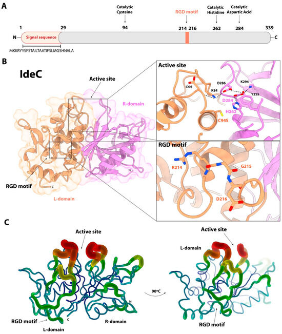
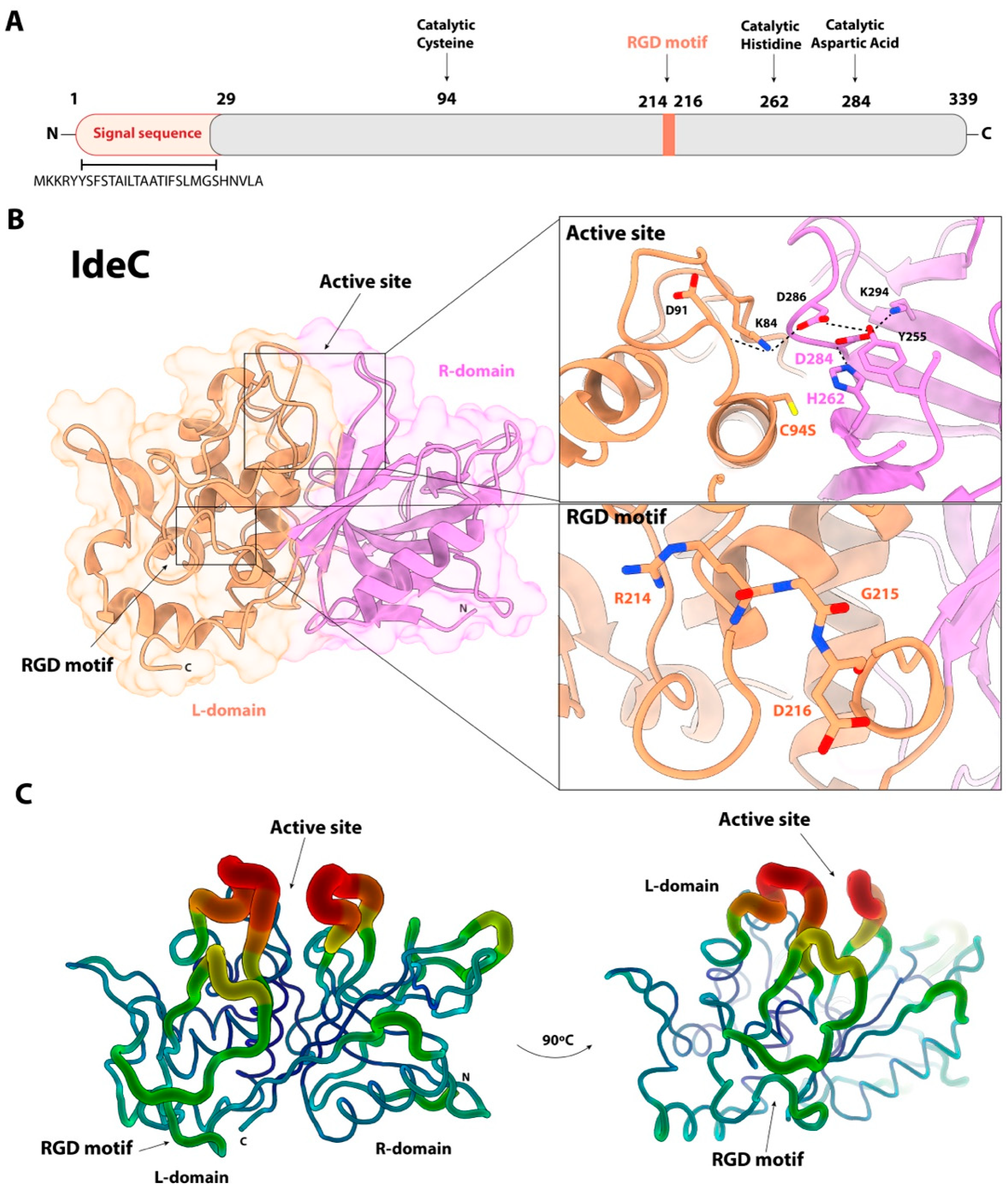
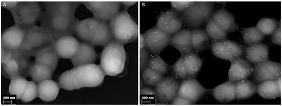
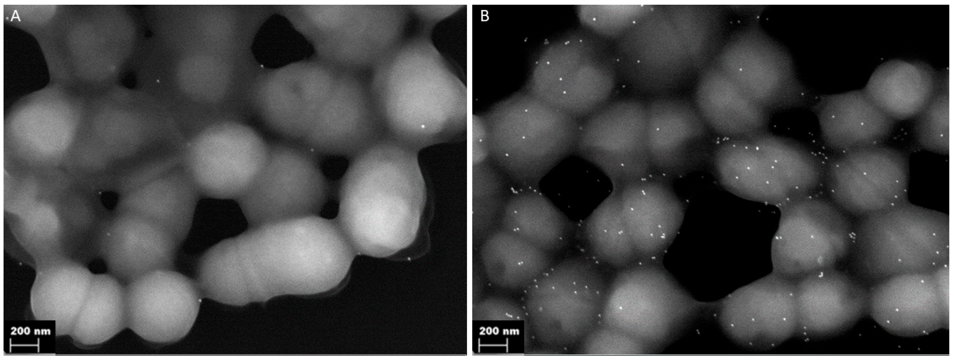
The catalytic properties of IdeC have been investigated in a prior study. We characterised the IgG-cleaving abilities of the protein and determined IdeC to be a species-specific protease cleaving only the IgG of dogs, cats and humans [15]. Here, we investigated the amino acid sequence of IdeC further in order to assess whether there is evidence for a secondary function for the protein. Sequence analysis of the ideC gene, using SignalP 6.0, allowed identification of a signal sequence, indicating IdeC is a secreted protein. The sequence MKKRYYSFSTAILTAATIFSLMGSHNVLA was predicted to be the protein’s signal sequence, with the cleavage site located between the Ala29 and Asp30 residues (Figure 1, Supplementary Figure S1). Accordingly, we have classified the amino acids 1–29 as the predicted signal sequence (Figure 1A). Despite being a secreted protein, electron microscopy of S. canis using rabbit α-IdeC and α-rabbit-gold-labelled antibodies revealed that IdeC is surface localised (Figure 2).
Figure 1.
Structure of the IdeC Protein. (A) Schematic representation of IdeC; marked are the predicted N-terminal signal sequence (determined using SignalP 6.0 Server), the RGD motif (amino acids 214–216) and the catalytic triad (Cys94, His262 and Asp284). (B) The crystal structure of the IdeC protein (PDB code 9HB1) is displayed in both surface and cartoon representations, with the active site and RGD motif highlighted by black arrows. On the right, a detailed view illustrates the IdeC active site and the RGD motif in greater detail. Relevant residues are depicted as capped sticks and labelled. Polar interactions are represented as dotted lines. (C) B-factor putty representation of the complete IdeC structure. Regions with lower B-factors are depicted as narrower tubes in shades of blue, indicating highly ordered and tightly packed areas. Wider tubes coloured from orange to red correspond to higher B-factors, highlighting the greater flexibility of the channel that the active site and RGD motif constitute.
Figure 2.
Surface localisation of IdeC. Gold-labelled antibodies detect secreted IdeC on the bacterial surface. (A) Binding of gold-labelled antibody without pre-incubation with IdeC-specific antibody served as negative control. (B) Binding of gold-labelled antibody after pre-incubation of S. canis bacteria with IdeC-specific antibody.
Based on comparison to the group A streptococcal IdeS protein [13], the amino acids Cys94, His224 and Asp284 were predicted to act as the catalytic triad of the protein. The cys94 residue was then confirmed to be essential for IgG cleavage activity in our previous study [15]. Additionally, an RGD motif was present at position 214–216 (Arg214, Gly215, Asp216) (Figure 1A).
IdeS has been described as resembling the typical 3D structure of the papain superfamily of cysteine proteases, though they do not share significantly similar amino acid sequences [31]. We have previously reported the structural and functional characterisation of IdeC, which also belongs to the Papain-like cysteine peptidase superfamily and is highly structurally, and sequentially, similar to IdeS [15]. The three-dimensional structure of the IdeC cysteine protease presents the active site located at the interface of the left (L-) and right (R-) domains of the protein (Figure 1B), with the catalytic triad (Cys94, His262 and Asp284) involving residues from the two domains (Figure 1A) [15]. Relevant to this study is the position of the RGD motif in the 3D structure of the protein (Figure 1B). The motif is located in the L-domain, positioned away from the catalytic centre and solvent-exposed [15]. It is worth mentioning that the RGD motif is exposed in a region that, together with the active site, presents a high mobility as observed by the distribution of B-factors in the crystal structure (Figure 1C). In this position, the motif could be involved in the recruitment of substrates to the catalytic active site, or interaction with proteins involved in other functions.
The presence of the RGD motif together with the surface localisation of the IdeC protein provides the basis for this study investigating a possible role in adhesion and/or invasion for IdeC.
3.2. Replacing the RGD Motif of IdeC with an RGE Motif Does Not Affect Its Enzymatic Activity
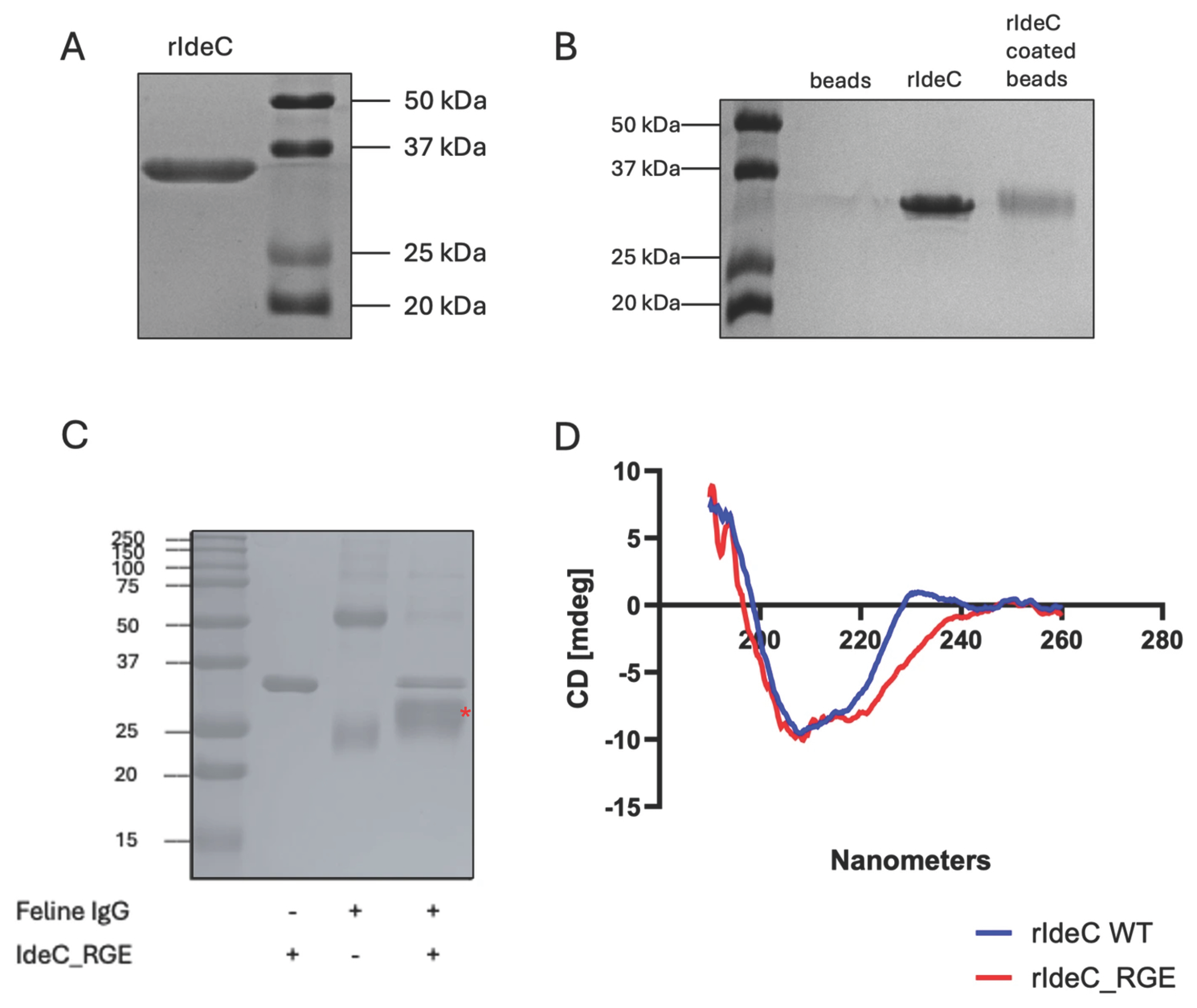
Two recombinant IdeC proteins were produced for this study: rIdeC wild-type (WT) and rIdeCRGE. The recombinant IdeC proteins all consist of residues 30–339 of the IdeC protein, i.e., the protein without the predicted signal sequence, and are ~35 kDa in size (Figure 3A). A single-nucleotide change in the sequence of rIdeCRGE resulted in a glutamic acid replacing the aspartic acid at position 216, resulting in an ‘RGE’ in the place of the RGD motif found at position 214–216. The RGD-to-RGE change has been demonstrated to interfere with integrin-binding without causing structural changes [32]. rIdeC and rIdeCRGE are both capable of IgG cleavage. The cleavage of IgG by IdeC WT has been previously documented [15] and Figure 3C shows that incubation of rIdeCRGE with feline IgG results in an additional band of ~24 kDa which is produced during the breakdown of the heavy chain of the IgG. This indicates that the amino acid change (Asp216Glu) had no effect on the protein’s enzymatic activity. Comparison of the circular dichroism (CD) spectra of both proteins reveals no difference in 3D structure caused by the amino acid change (Figure 3D).
Figure 3.
Recombinant expression of IdeC proteins. (A) Recombinant IdeC protein visualised as ~35 kDa protein on 15% SDS-PAGE gel. (B) SDS-Page detection confirmed that latex beads were successfully coated with rIdeC. (C) Feline IgG cleavage assay with rIdeCRGE shows that changing the RGD amino acid motif to a RGE motif still results in IgG cleavage. The cleavage fragment of ~24 kDa is indicated with a red *. (D) There is no significant difference in the CD spectra of rIdeC and rIdeCRGE.
3.3. IdeC Interacts with Endothelial and Epithelial Cells
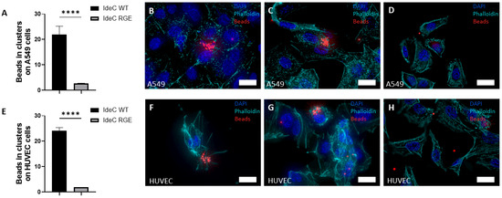
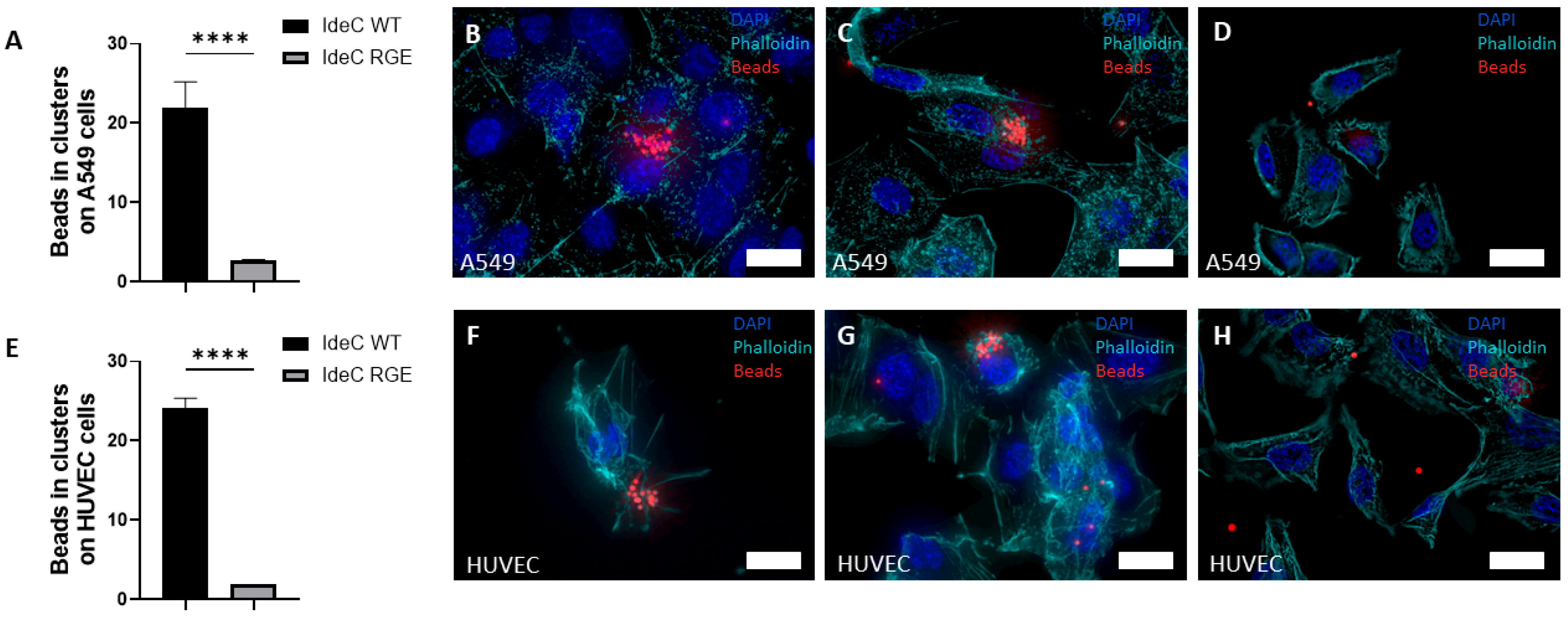
To further define functionalities of IdeC with regard to interaction with eukaryotic cells, we coated fluorescent latex beads with recombinant protein (Figure 3B). Beads were then applied to cell monolayers to investigate the interaction of IdeC with host cells. The fluorescent beads emitted light that was detectable in all fluorescent channels, making differential staining impossible, so internalisation could not be determined by using this assay. However, latex beads coated with WT IdeC showed increased interaction with epithelial monolayers of human adenocarcinomic alveolar basal epithelial cells (A549 cells) (Figure 4A–D) and with human umbilical vein endothelial cells (HUVEC) (Figure 4E–H), compared to BSA-coated beads. rIdeC-coated beads formed clusters in association with both epithelial and endothelial cells; these clusters could be observed 1 h after application of beads but were more common and consisted of more beads at the 2- and 3-h time points post-incubation. The average number of beads per cell in cell-associated clusters was 21 beads in association with the A549 cells and 24 beads with HUVEC (Figure 4A,E).
Figure 4.
rIdeC-coated beads interact with endothelial and epithelial cells in an RGD-dependent manner. Fluorescent beads were coated with purified recombinant protein and interaction with A549 and HUVECs was microscopically determined after immunofluorescence staining was used; the cells were incubated with 100-fold excess of beads. Graphs demonstrate the average number of beads in clusters associated with A549 (A) and HUVECs (E). Representative images of bead clusters in A549 (B,C) and HUVECs (F,G). Representative images of A549 (D) and HUVECs (H) with IdeCRGE-coated beads. White scale bars indicate 10 µm. Error bars were based on standard deviation from the mean. Statistical significance was assessed using an unpaired two-tailed T test and a Mann–Whitney test. p-value of 0.0001 is indicated by ****.
When these experiments were repeated using rIdeCRGE-coated beads, single beads and coupled beads were observed in association with host cells but bead clustering was not observed (Figure 4D,H). Using both the t-test and Mann–Whitney test, it was determined that both A549 and HUVECs displayed increased interaction with latex beads coated in rIdeC compared to rIdeCRGE (p < 0.0001).
3.4. Integrin αVβ3 Mediates IdeC-Uptake
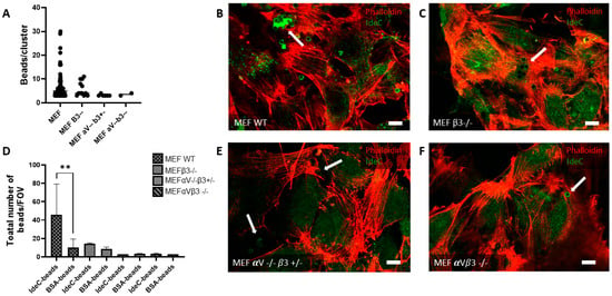
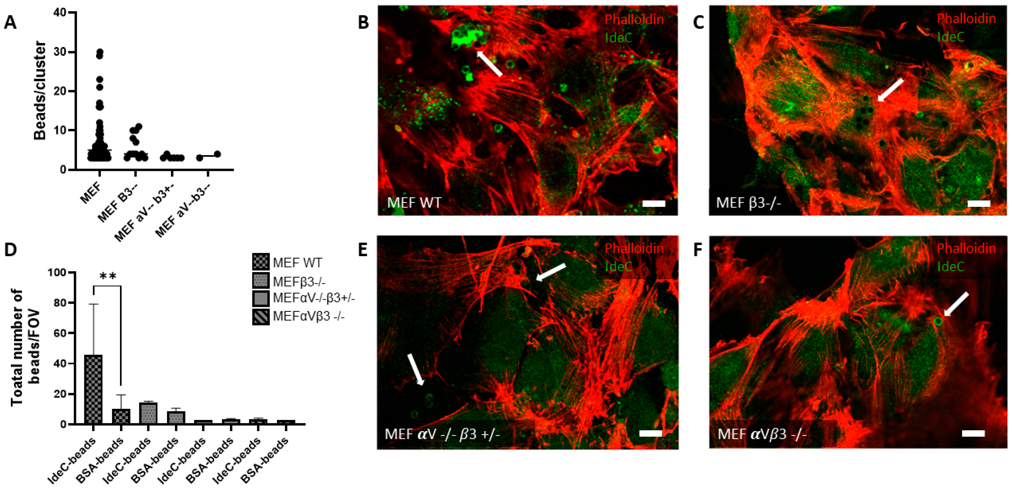
Results of the cell culture interaction analyses determined that rIdeC-coated beads interact with endothelial and epithelial cells in an RGD-motif-dependent manner. As the RGD motif is notable for being an integrin-binding domain, the function of integrin receptors in the IdeC–cell interaction should be further characterised. For this aim, rIdeC-coated beads were incubated with a series of mouse embryonic fibroblast (MEF) cell lines, which were used for these experiments, with either single- or double-subunit deletions for the integrin αVβ3. Cell lines used were the WT MEF cells along with MEF β3 -/-, MEF αV -/-β3 +/- and MEF αVβ3 -/-. In the WT MEF cells, we observed the same clustering of rIdeC-coated beads (Figure 5A) as was demonstrated in A549 and HUVECs (Figure 4).
Figure 5.
Expression of integrin αVβ3 facilitates increased interaction of rIdeC-coated beads with MEF cells. rIdeC-coated latex beads were applied to MEF monolayers on glass slips. Monolayers were incubated for 3 h before slides were immunostained for imaging. (A) Graphical presentation of average number of beads per cluster on MEF cells. (D) Graph showing total number of beads interacting with cells, per field of view. The only cell type with a significant difference in interaction between rIdeC-coated beads and BSA-coated beads was the MEF wild-type. (B,C,E,F) Representative microscopic image demonstrating MEF cells visualised with phalloidin (red) and rIdeC-coated beads (green); images show interactions with MEF WT cells (B), β3-integrin-subunit-deleted MEF cells (C), αV -/- β3 +/- integrin-deleted MEF cells (E), and on αVβ3-integrin-deleted cells (F). White arrows point to beads or clusters of beads; white scale bars indicate 10 µm. Error bars were based on the standard deviation from the mean. Statistical significance was assessed using both an unpaired two-tailed T test and a Mann–Whitney test. A p-value of 0.001 is indicated by **.
The size and frequency of these clusters were reduced in the β3 subunit deletion (Figure 5C), and further reduced in the MEF αV -/-β3 +/- and MEF αV -/-β3 -/- double-subunit deletion (Figure 5E,F). Figure 5A shows the range in size of bead clusters observed in each of the cell lines; the WT MEF cells are the only cells where clusters with more than 10 beads were observed. Figure 5D shows a side-by-side comparison of rIdeC-coated beads and BSA-coated beads for each cell line. The only cell line where there is a significant difference in the interaction of rIdeC-coated beads compared to BSA-coated beads is the MEF with WT integrin expression.
3.5. IdeC Plays a Role During S. canis Infection
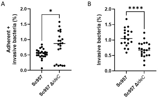
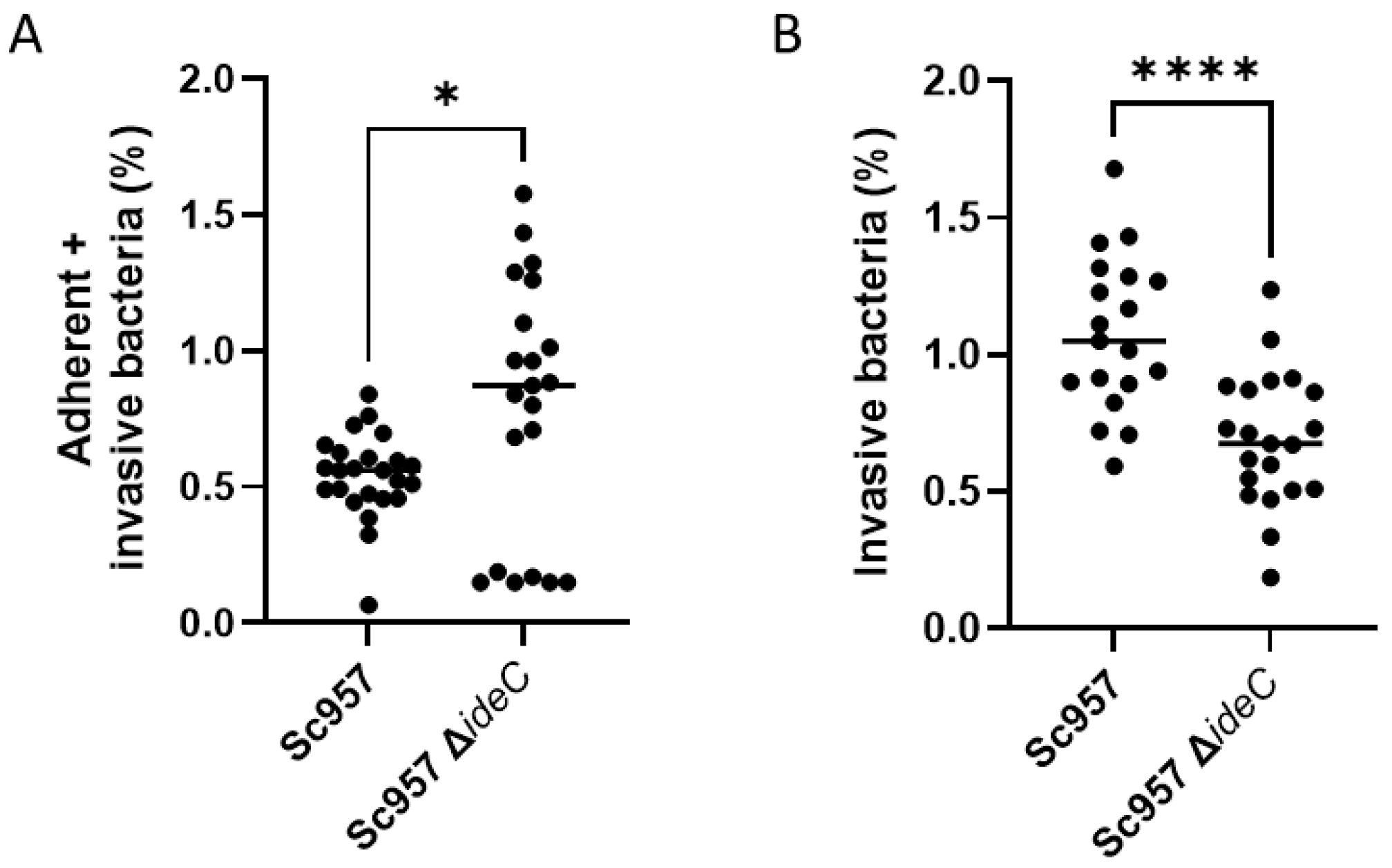
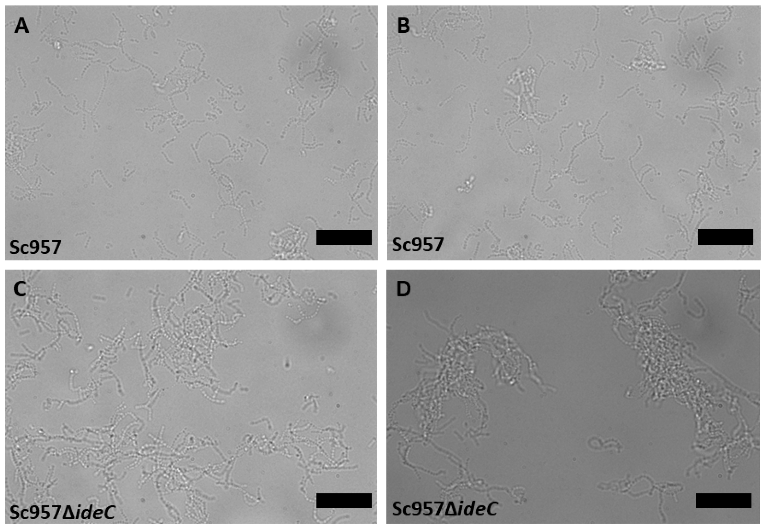
The preceding results demonstrate that IdeC can facilitate adhesion and uptake of latex beads into HUVECs and A549 and MEF cells. However, to state concretely whether IdeC plays an active role in cell culture infection, an ideC gene-deleted S. canis mutant named Sc957ΔideC was generated. There was no significant difference in growth rate between the WT and the deletion mutant (Supplemental Figure S2), although in liquid culture, the Sc957ΔideC strain appeared to aggregate more than the WT. In cell culture infection analyses with human epithelial A549 cells, a significant reduction in the percentage of internalised Sc957ΔideC bacteria was observed when compared to infection with the WT strain (Figure 6B).
Figure 6.
Function of IdeC in bacterial adhesion and invasion of A549 cells. A549 cells were infected with wild-type Sc957 and Sc957ΔideC at an MOI of 10. (A) Adhesion samples: After 2 h of bacterial infection, cells were lysed, and the CFU/mL as a percentage of the CFU/mL of the inoculum was calculated. (B) Invasion samples: After 2 h of bacterial infection, cells were incubated for a further 45 min in medium supplemented with erythromycin before being treated as described previously. Graphs show data from three biological replicates. Statistical significance was assessed using an unpaired two-tailed t-test and a Mann–Whitney test; a p-value of 0.05 was considered significant and indicated by *, and a p-value of 0.0001 is indicated by ****.
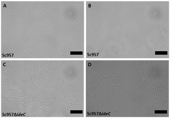
However, significantly more ΔideC streptococci interacted with A549 than WT S. canis overall (Figure 6A). There was also a much broader range of results when considering the mutant, compared to the WT. It is possible that this broader range of results is due to the increased aggregation displayed by the ΔideC mutant, so microscopy was employed to visualise any differences in the interactions/appearance of the bacteria (Figure 7). This confirmed that Sc957ΔideC displays increased self-interaction among bacteria (Figure 7C,D), compared to the WT (Figure 7A,B). This increased bacterial interaction observed in the mutant may be responsible for the formation of bacterial aggregates.
Figure 7.
Representative light microscopy images displaying differences in bacterial aggregation of Sc957 and the Sc957ΔideC. (A,B) Images of Sc957 wild-type. (C,D) Images of Sc957ΔideC. Black scale bar signifies 10 µm.
4. Discussion
Characterisation of virulence determinants in S. canis has thus far been limited, although there have been studies that have identified genes in S. canis that are homologues of virulence genes reported in other bacteria [5,6]. In our previous study, we have identified IdeC as a cysteine protease that cleaves feline, canine and human IgG [15]. The protein sequence of IdeC is highly similar to that of IdeS of S. pyogenes and the protein is predicted to function in accordance with the mechanism described by Von Pawel-Rammingen and colleagues—in short, as a secreted protein that cleaves IgG that is bound to the bacterial surface via M-like proteins or other surface antigens [13]. Among the similarities shared by IdeS and IdeC are the putative catalytic triad and the presence of an RGD motif (Figure 1). The presence of this motif is a feature of proteins that interact with integrins, including a variant of streptococcal pyrogenic exotoxin B (SpeB) and also S. pyogenes, a cysteine protease that has been shown to directly interact with cell surface integrins as well as to degrade ECM proteins [33,34].
‘Moonlighting proteins’ are proteins that exhibit more than one biological function, often involved in one function as a secreted protein, and another when attached to the cell wall [35]. The first ‘moonlighting’ protein identified was a S. pyogenes glyceraldehyde 3-phosphate dehydrogenase (GAPDH) with multiple-bingeing activity, which is essential for virulence [36,37]. Another important streptococcal ‘moonlighting’ protein is enolase, with roles in glycolysis and also in plasminogen binding [38]. We have described IdeC here as a secreted protein that can bind to the bacterial surface after secretion (Figure 1 and Figure 2). Since its function as a secreted IgG protease has been described previously, here we focus on a possible second/’moonlighting’ function it may perform as a surface-bound protein. For this purpose, the previously reported protein structure was re-examined. The crystal structure revealed the location of the RGD motif of the protein, positioned on an accessible, outward-facing loop (Figure 1B). Other streptococcal proteins have been described with RGD motifs that have been proven to interact with integrins via the RGD residues; for example, the mSpeB2 variant of SpeB interacts with the integrin αVβ3 and the IdeS/Mac-1 variant Mac5005 binds both αVβ3 and αIIbβ3 integrins [26,39]. In both proteins, the RGD is located on a loop, in SpeB on a surface loop near the C-terminal protease domain [40] and in IdeS on a loop in the N-terminal domain [41]. The positioning of the RGD on a loop removed from the catalytic centre of the protein suggests it is possible for the protein to interact with cell surface proteins while maintaining its IgG-cleaving action.
A surface localised protein with an integrin-binding site is an interesting candidate to investigate for a role in adhesion to or invasion of eukaryotic cells, which can serve as a prerequisite for colonisation and tissue distribution. Streptococcal infections usually begin with the interaction of bacteria with host cells via adhesins [42]. Stockbauer et al. reported that though the SpeB variant containing the RGD motif was not exclusively capable of causing invasive disease, M-types with the binding site were frequent originators of severe disease. Notably, the M1 S. pyogenes serotype expressed the RGD-containing variant of SpeB [39]. The M1 serotype is strongly associated with invasive disease [43]. We specifically show here that the integrin αVβ3 interacts with IdeC, and can facilitate the RGD-dependent interaction of IdeC with host cells (Figure 4 and Figure 5). The integrin αVβ3 was chosen here as it had previously been shown to interact with streptococcal proteins containing the RGD motif [26,39], and is present in both A549 and HUVECs; however, this does not mean that αVβ3 is the only integrin capable of interaction with IdeC. The αV subunit has specifically been shown to promote Streptococcus agalactiae invasion of epithelial cells [44]. Indeed, Figure 5C shows that deletion of only the β3 subunit is not sufficient to interfere with IdeC binding to MEF cells; the binding of IdeC with other αV subunit-containing integrins could therefore be an interesting avenue to pursue in clarifying the mechanism of IdeC.
Though we show here that IdeC is capable of interacting with host cells, it is not expected to be the only factor mediating adhesion and invasion of S. canis to host cells. We see in Figure 6 that there is still adherence to and invasion into A549 cells by bacteria when ideC is deleted. Indeed, deletion of ideC has different implications for adherence and invasion. Figure 6A shows almost a doubling in the combined adhesive and invasive fractions in the ΔideC mutant, while the invasion alone (Figure 6B) is reduced significantly. We can infer from this that the increase we see in the adherent and invasive fractions combined is mainly due to an increase in external adherence only.
From this it seems that the deletion of ideC promotes adhesion. However, when we consider the increased aggregation exhibited by the ideC-negative strain (Figure 7), it is possible that much of the observed increase in ‘adherence’ is actually due to increased aggregation exhibited by this strain; i.e., the deletion of ideC results in increased interaction with other bacteria and not increased adhesion to the host cell. It is possible that the decrease in invasion observed in the ΔideC strain results from a decrease in bacteria actually interacting with the A549 cells or an inability of the cells to take up large aggregates of bacteria. It has been demonstrated in S. pyogenes that bacterial aggregation inhibits uptake by phagocytic cells [45]. When we consider that the RGD–integrin interaction has been demonstrated to be crucial for the integrin clustering that leads to signal activation, allowing invasion of pathogens into host cells [19,20], it is also possible that the absence of the RGD motif of IdeC limits the invasive ability of the bacteria despite their ability to adhere to host cells via an IdeC-independent mechanism.
The aggregation phenotype demonstrated in Figure 7 cannot be conclusively explained based on this study, though possible mechanisms can be suggested. It is possible that the absence of IdeC results in the increased availability of another surface protein of S. canis, which is responsible for the binding of IdeC to the bacterial surface after secretion. Such a protein could theoretically form bonds with other surface-associated proteins when not bound to IdeC, resulting in aggregation. Recent work in S. pyogenes has demonstrated that S. pyogenes nuclease A (SpnA) is involved in IdeS binding to the bacterial surface, although it was determined that it was not the sole binding partner of IdeS on the bacterial surface [46]. This may indicate that IdeC also has multiple binding partners responsible for its surface association, and these may be capable of interaction. The M-like protein of S. canis is a plasminogen-binding protein that is covalently bound to the cell surface [8]. M proteins are antiphagocytic proteins that bind to a number of different host proteins, e.g., fibrinogen [47]. SCM has been shown to form homophilic interactions with itself that can cause bacterial aggregates [4]. It is possible that IdeC regulates the amount of surface-exposed SCM. IdeC-specific gene deletion may remove regulation of SCM, leading to increased levels of SCM on the bacterial surface. A deeper investigation into the interactions of IdeC with the bacterial surface should be carried out to explain the aggregation phenotype, as well as the ability of IdeC to bind back to the bacterial surface after secretion (Figure 2); these interactions could be reliant on one surface protein like ScM or involve several.
Moreover, results depicted in Figure 6 demonstrate that a greater number of WT S. canis bacteria are in the invasive-only fraction compared to the combined fraction of adherent and invasive bacteria. As these fractions are measured 40 min apart, this may indicate that S. canis is replicating within the host cell after invasion. When bacteria invade host cells they are initially taken up into membrane-bound phagosomes. Some bacteria have evolved strategies to escape these phagosomes and survive intracellularly. As a consequence, they can multiply and potentially spread to adjacent cells [48]. In this respect, other streptococci have been demonstrated to propagate successfully intracellularly and this has been linked to immune evasion and persistent infection. For example, S. pyogenes is able to modulate the human macrophage via the M1 protein to promote persistence within vacuoles [49]. In line with this, another study reported that S. pyogenes was shown to evade the host immune response by surviving within phagocytic cells [50].
5. Conclusions
In conclusion, in addition to the formerly described role of IdeC as an IgG-cleaving protease [15], the internalisation into epithelial and endothelial cells might represent a second important immune evasion mechanism. The presented data provides strong evidence that IdeC does indeed play a role in internalisation of S. canis into host cells, thereby representing a ‘moonlighting’ function important for the pathophysiology of S. canis. Furthermore, the cellular uptake into host cells depends on the interaction of the RGD motif with integrins on the host cell surface, representing a pivotal molecular pathomechanism mediated by the cysteine protease IdeC of S. canis. This discovery is especially significant given how few virulence determinants have been described in S. canis, and how little we understand about the dissemination of S. canis throughout the host that can lead to invasive disease.
This study provides important evidence for a function of IdeC outside of IgG cleavage; however, it also highlights several avenues where further research is required to fully understand the role this protein plays during infection. Notably, the interaction of IdeC with other proteins, including, as discussed above, other integrins and proteins present on the bacterial cell surface, should be explored in further work.
Supplementary Materials
Supplementary figures and raw data are openly available at figshare. Supplementary figures are available under DOI: 10.6084/m9.figshare.30306004 (https://figshare.com/articles/figure/Supplementary_Figures/30306004, accessed on 12 April 2026) [51]. Raw data, including bead and bacterial count numbers associated with bead association and S. canis infection assays, are available under DOI: 10.6084/m9.figshare.30305872 (https://figshare.com/articles/dataset/Supplementary_raw_data/30305872, accessed on 12 April 2026) [52].
Author Contributions
M.F. conceived the study, provided support and revised the manuscript. S.W. performed experiments, analysed the data and wrote the manuscript. S.B. provided support and revised the manuscript. M.K. revised the manuscript. M.R. performed SEM. A.G.-Á. and J.H. performed protein structural analysis. All authors have read and agreed to the published version of the manuscript.
Funding
This work was supported by the German Research Foundation (DFG) (grant FU 1027/3-1 to S.W. and M.F. and FU 1027/5-1 to M.F.). The work in Spain was supported by grant PID2023-153118OB-I00 funded by MCIN/AEI/10.13039/501100011033 to J.A.H.
Data Availability Statement
The raw data (including bead and bacterial count numbers associated with bead association and S. canis infection assays) presented in the study are openly available in FigShare at DOI: https://doi.org/10.6084/m9.figshare.30305872.
Acknowledgments
We would like to thank Nina Janze for excellent technical assistance. We would like to acknowledge M. van der Linden (National Reference Centre for Streptococci, Aachen, Germany) for providing Streptococcus canis strain G361. We are also grateful to Markus Keller (Federal Research Institute for Animal Health, Greifswald) and Thomas Meyer (Max Planck Institute) for the provision of the MEF and A549 cell lines, respectively. We are also grateful to Suvrat Chowdhary for help with circular dichroism analysis. Antje Lapschies, Institute of Microbiology and Epizootics, is greatly acknowledged for her help with the IdeC cleavage assays. Data from this manuscript was presented at the 76th Annual Meeting of the DGHM [53]. The publication of this article was funded by Freie Universität Berlin.
Conflicts of Interest
The authors declare no conflicts of interest.
References
- Pagnossin, D.; Smith, A.; Oravcová, K.; Weir, W. Streptococcus canis, the underdog of the genus. Vet. Microbiol. 2022, 273, 109524. [Google Scholar] [CrossRef] [Scilit]
- Katsburg, M.; Brombach, J.; Hanke, D.; Aubry, E.; Lübke-Becker, A.; Fulde, M. New variant strain of Streptococcus canis with Lancefield group C isolated from canine otitis externa. Vet. Microbiol. 2023, 285, 109869. [Google Scholar] [CrossRef] [Scilit] [PubMed]
- Devriese, L.A.; Hommez, J.; Kilpper-Bälz, R.; Schleifer, K.-H. Streptococcus canis sp. nov.: A Species of Group G Streptococci from Animals. Int. J. Syst. Evol. Microbiol. 1986, 36, 422–425. [Google Scholar] [CrossRef] [Scilit]
- Nerlich, A.; Lapschies, A.-M.; Kohler, T.P.; Cornax, I.; Eichhorn, I.; Goldmann, O.; Krienke, P.; Bergmann, S.; Nizet, V.; Hammerschmidt, S.; et al. Homophilic protein interactions facilitate bacterial aggregation and IgG-dependent complex formation by the Streptococcus canis M protein SCM. Virulence 2019, 10, 194–206. [Google Scholar] [CrossRef] [Scilit]
- Pagnossin, D.; Weir, W.; Smith, A.; Fuentes, M.; Coelho, J.; Oravcova, K. Streptococcus canis genomic epidemiology reveals the potential for zoonotic transfer. Microb. Genom. 2023, 9, 000974. [Google Scholar] [CrossRef] [Scilit]
- Richards, V.P.; Zadoks, R.N.; Bitar, P.D.P.; Lefébure, T.; Lang, P.; Werner, B.; Tikofsky, L.; Moroni, P.; Stanhope, M.J. Genome characterization and population genetic structure of the zoonotic pathogen, Streptococcus canis. BMC Microbiol. 2012, 12, 293. [Google Scholar] [CrossRef] [Scilit] [PubMed]
- Galpérine, T.; Cazorla, C.; Blanchard, E.; Boineau, F.; Ragnaud, J.-M.; Neau, D. Streptococcus canis infections in humans: Retrospective study of 54 patients. J. Infect. 2007, 55, 23–26. [Google Scholar] [CrossRef] [Scilit]
- Fulde, M.; Rohde, M.; Hitzmann, A.; Preissner, K.T.; Nitsche-Schmitz, D.P.; Nerlich, A.; Chhatwal, G.S.; Bergmann, S. SCM, a novel M-like protein from Streptococcus canis, binds (mini)-plasminogen with high affinity and facilitates bacterial transmigration. Biochem. J. 2011, 434, 523–535. [Google Scholar] [CrossRef] [Scilit]
- Yoshida, H.; Goto, M.; Fukushima, Y.; Maeda, T.; Tsuyuki, Y.; Takahashi, T. Intracellular Invasion Ability and Associated Microbiological Characteristics of Streptococcus canis in Isolates from Japan. Jpn. J. Infect. Dis. 2021, 74, 129–136. [Google Scholar] [CrossRef] [Scilit]
- Fulde, M.; Rohde, M.; Polok, A.; Preissner, K.T.; Chhatwal, G.S.; Bergmann, S. Cooperative plasminogen recruitment to the surface of Streptococcus canis via M protein and enolase enhances bacterial survival. mBio 2013, 4, e00629-12. [Google Scholar] [CrossRef] [Scilit] [PubMed]
- Hitzmann, A.; Bergmann, S.; Rohde, M.; Chhatwal, G.; Fulde, M. Identification and characterization of the arginine deiminase system of Streptococcus canis. Vet. Microbiol. 2013, 162, 270–277. [Google Scholar] [CrossRef] [Scilit]
- DeWinter, L.M.; Prescott, J.F. Relatedness of Streptococcus canis from canine streptococcal toxic shock syndrome and necrotizing fasciitis. Can. J. Vet. Res. 1999, 63, 90–95. [Google Scholar]
- von Pawel-Rammingen, U.; Johansson, B.P.; Björck, L. IdeS, a novel streptococcal cysteine proteinase with unique specificity for immunoglobulin G. EMBO J. 2002, 21, 1607–1615. [Google Scholar] [CrossRef] [Scilit]
- Lei, B.; DeLeo, F.R.; Hoe, N.P.; Graham, M.R.; Mackie, S.M.; Cole, R.L.; Liu, M.; Hill, H.R.; Low, D.E.; Federle, M.J.; et al. Evasion of human innate and acquired immunity by a bacterial homolog of CD11b that inhibits opsonophagocytosis. Nat. Med. 2001, 7, 1298–1305. [Google Scholar] [CrossRef] [Scilit]
- Walsh, S.; Lapschies, A.-M.; Miguel-Ruano, V.; Batuecas, M.T.; Acebrón-Ávalos, I.; Kohler, T.P.; Hammerschmidt, S.; Eichhorn, I.; Hermoso, J.A.; Fulde, M. Structural and functional characterization of IdeC, a novel IgG-specific protease of Streptococcus canis. Infect. Immun. 2025, 93, e00248-25. [Google Scholar] [CrossRef] [Scilit]
- Wickham, T.J.; Mathias, P.; Cheresh, D.A.; Nemerow, G.R. Integrins αvβ3 and αvβ5 promote adenovirus internalization but not virus attachment. Cell 1993, 73, 309–319. [Google Scholar] [CrossRef] [Scilit] [PubMed]
- Bonsor, D.A.; Pham, K.T.; Beadenkopf, R.; Diederichs, K.; Haas, R.; Beckett, D.; Fischer, W.; Sundberg, E.J. Integrin engagement by the helical RGD motif of the Helicobacter pylori CagL protein is regulated by pH-induced displacement of a neighboring helix. J. Biol. Chem. 2015, 290, 12929–12940. [Google Scholar] [CrossRef] [Scilit]
- Bellis, S.L. Advantages of RGD peptides for directing cell association with biomaterials. Biomaterials 2011, 32, 4205–4210. [Google Scholar] [CrossRef] [Scilit]
- Fincham, V.J.; James, M.; Frame, M.C.; Winder, S.J. Active ERK/MAP kinase is targeted to newly forming cell-matrix adhesions by integrin engagement and v-Src. Embo J. 2000, 19, 2911–2923. [Google Scholar] [CrossRef] [Scilit] [PubMed]
- Isberg, R.R.; Barnes, P. Subversion of integrins by enteropathogenic Yersinia. J. Cell Sci. 2001, 114, 21–28. [Google Scholar] [CrossRef] [Scilit] [PubMed]
- Leahy, D.J.; Aukhil, I.; Erickson, H.P. 2.0 A crystal structure of a four-domain segment of human fibronectin encompassing the RGD loop and synergy region. Cell 1996, 84, 155–164. [Google Scholar] [CrossRef] [Scilit] [PubMed]
- Kwok, T.; Zabler, D.; Urman, S.; Rohde, M.; Hartig, R.; Wessler, S.; Misselwitz, R.; Berger, J.; Sewald, N.; König, W.; et al. Helicobacter exploits integrin for type IV secretion and kinase activation. Nature 2007, 449, 862–866. [Google Scholar] [CrossRef] [Scilit]
- Leininger, E.; Roberts, M.; Kenimer, J.G.; Charles, I.G.; Fairweather, N.; Novotny, P.; Brennan, M.J. Pertactin, an Arg-Gly-Asp-containing Bordetella pertussis surface protein that promotes adherence of mammalian cells. Proc. Natl. Acad. Sci. USA 1991, 88, 345–349. [Google Scholar] [CrossRef] [Scilit]
- Molinari, G.; Rohde, M.; Guzman, C.A.; Chhatwal, G.S. Two distinct pathways for the invasion of Streptococcus pyogenes in non-phagocytic cells. Cell. Microbiol. 2000, 2, 145–154. [Google Scholar] [CrossRef] [Scilit]
- Ozeri, V.; Rosenshine, I.; Mosher, D.F.; Fässler, R.; Hanski, E. Roles of integrins and fibronectin in the entry of Streptococcus pyogenes into cells via protein F1. Mol. Microbiol. 1998, 30, 625–637. [Google Scholar] [CrossRef] [Scilit]
- Lei, B.; DeLeo, F.R.; Reid, S.D.; Voyich, J.M.; Magoun, L.; Liu, M.; Braughton, K.R.; Ricklefs, S.; Hoe, N.P.; Cole, R.L.; et al. Opsonophagocytosis-inhibiting mac protein of group a streptococcus: Identification and characteristics of two genetic complexes. Infect. Immun. 2002, 70, 6880–6890. [Google Scholar] [CrossRef] [Scilit] [PubMed]
- Eichhorn, I.; van der Linden, M.; Jarek, M.; Fulde, M. Draft Genome Sequence of Zoonotic Streptococcus canis Isolate G361. Genome Announc. 2017, 5, e00967-17. [Google Scholar] [CrossRef] [Scilit] [PubMed]
- Schmidt, K.; Keller, M.; Bader, B.L.; Korytář, T.; Finke, S.; Ziegler, U.; Groschup, M.H. Integrins modulate the infection efficiency of West Nile virus into cells. J. Gen. Virol. 2013, 94, 1723–1733. [Google Scholar] [CrossRef] [Scilit]
- Kaur, S.J.; Nerlich, A.; Bergmann, S.; Rohde, M.; Fulde, M.; Zähner, D.; Hanski, E.; Zinkernagel, A.; Nizet, V.; Chhatwal, G.S.; et al. The CXC chemokine-degrading protease SpyCep of Streptococcus pyogenes promotes its uptake into endothelial cells. J. Biol. Chem. 2010, 285, 27798–27805. [Google Scholar] [CrossRef] [Scilit]
- Bergmann, S.; Eichhorn, I.; Kohler, T.P.; Hammerschmidt, S.; Goldmann, O.; Rohde, M.; Fulde, M. SCM, the M Protein of Streptococcus canis Binds Immunoglobulin G. Front. Cell. Infect. Microbiol. 2017, 7, 80. [Google Scholar] [CrossRef] [Scilit]
- Wenig, K.; Chatwell, L.; von Pawel-Rammingen, U.; Björck, L.; Huber, R.; Sondermann, P. Structure of the streptococcal endopeptidase IdeS, a cysteine proteinase with strict specificity for IgG. Proc. Natl. Acad. Sci. USA 2004, 101, 17371–17376. [Google Scholar] [CrossRef] [Scilit]
- Ruoslahti, E.; Pierschbacher, M.D. New perspectives in cell adhesion: RGD and integrins. Science 1987, 238, 491–497. [Google Scholar] [CrossRef] [Scilit]
- Kapur, V.; Topouzis, S.; Majesky, M.W.; Li, L.-L.; Hamrick, M.R.; Hamill, R.J.; Patti, J.M.; Musser, J.M. A conserved Streptococcus pyogenes extracellular cysteine protease cleaves human fibronectin and degrades vitronectin. Microb. Pathog. 1993, 15, 327–346. [Google Scholar] [CrossRef] [Scilit]
- Lukomski, S.; Burns, E.H.; Wyde, P.R.; Podbielski, A.; Rurangirwa, J.; Moore-Poveda, D.K.; Musser, J.M. Genetic inactivation of an extracellular cysteine protease (SpeB) expressed by Streptococcus pyogenes decreases resistance to phagocytosis and dissemination to organs. Infect. Immun. 1998, 66, 771–776. [Google Scholar] [CrossRef] [Scilit]
- Jeffery, C.J. Protein moonlighting: What is it, and why is it important? Philos. Trans. R. Soc. B Biol. Sci. 2018, 373, 20160523. [Google Scholar] [CrossRef] [Scilit]
- Pancholi, V.; Fischetti, V.A. A major surface protein on group A streptococci is a glyceraldehyde-3-phosphate-dehydrogenase with multiple binding activity. J. Exp. Med. 1992, 176, 415–426. [Google Scholar] [CrossRef] [Scilit] [PubMed]
- Jin, H.; Agarwal, S.; Pancholi, V. Surface export of GAPDH/SDH, a glycolytic enzyme, is essential for Streptococcus pyogenes virulence. mBio 2011, 2, e00068-11. [Google Scholar] [CrossRef] [Scilit] [PubMed]
- Pancholi, V.; Fischetti, V.A. Alpha-enolase, a novel strong plasmin(ogen) binding protein on the surface of pathogenic streptococci. J. Biol. Chem. 1998, 273, 14503–14515. [Google Scholar] [CrossRef] [Scilit]
- Stockbauer, K.E.; Magoun, L.; Liu, M.; Burns, E.H.; Gubba, S.; Renish, S.; Pan, X.; Bodary, S.C.; Baker, E.; Coburn, J.; et al. A natural variant of the cysteine protease virulence factor of group A Streptococcus with an arginine-glycine-aspartic acid (RGD) motif preferentially binds human integrins alphavbeta3 and alphaIIbbeta3. Proc. Natl. Acad. Sci. USA 1999, 96, 242–247. [Google Scholar] [CrossRef] [Scilit]
- Kagawa, T.F.; Cooney, J.C.; Baker, H.M.; McSweeney, S.; Liu, M.; Gubba, S.; Musser, J.M.; Baker, E.N. Crystal structure of the zymogen form of the group A Streptococcus virulence factor SpeB: An integrin-binding cysteine protease. Proc. Natl. Acad. Sci. USA 2000, 97, 2235–2240. [Google Scholar] [CrossRef] [Scilit] [PubMed]
- Agniswamy, J.; Nagiec, M.J.; Liu, M.; Schuck, P.; Musser, J.M.; Sun, P.D. Crystal Structure of Group A Streptococcus Mac-1: Insight into Dimer-Mediated Specificity for Recognition of Human IgG. Structure 2006, 14, 225–235. [Google Scholar] [CrossRef] [Scilit] [PubMed]
- Nobbs, A.H.; Jenkinson, H.F.; Everett, D.B. Generic determinants of Streptococcus colonization and infection. Infect. Genet. Evol. 2015, 33, 361–370. [Google Scholar] [CrossRef] [Scilit]
- Stevens, D.L. Invasive streptococcal infections. J. Infect. Chemother. 2001, 7, 69–80. [Google Scholar] [CrossRef] [Scilit]
- De Gaetano, G.V.; Pietrocola, G.; Romeo, L.; Galbo, R.; Lentini, G.; Giardina, M.; Biondo, C.; Midiri, A.; Mancuso, G.; Venza, M.; et al. The Streptococcus agalactiae cell wall-anchored protein PbsP mediates adhesion to and invasion of epithelial cells by exploiting the host vitronectin/α(v) integrin axis. Mol. Microbiol. 2018, 110, 82–94. [Google Scholar] [CrossRef] [Scilit]
- Frick, I.M.; Mörgelin, M.; Björck, L. Virulent aggregates of Streptococcus pyogenes are generated by homophilic protein-protein interactions. Mol. Microbiol. 2000, 37, 1232–1247. [Google Scholar] [CrossRef] [Scilit]
- Frick, I.M.; Happonen, L.; Wrighton, S.; Nordenfelt, P.; Björck, L. IdeS, a secreted proteinase of Streptococcus pyogenes, is bound to a nuclease at the bacterial surface where it inactivates opsonizing IgG antibodies. J. Biol. Chem. 2023, 299, 105345. [Google Scholar] [CrossRef] [Scilit]
- Oehmcke, S.; Shannon, O.; Mörgelin, M.; Herwald, H. Streptococcal M proteins and their role as virulence determinants. Clin. Chim. Acta 2010, 411, 1172–1180. [Google Scholar] [CrossRef] [Scilit]
- Greco, R.; De Martino, L.; Donnarumma, G.; Conte, M.; Seganti, L.; Valenti, P. Invasion of cultured human cells by Streptococcus pyogenes. Res. Microbiol. 1995, 146, 551–560. [Google Scholar] [CrossRef] [Scilit]
- Hertzén, E.; Johansson, L.; Wallin, R.; Schmidt, H.; Kroll, M.; Rehn, A.P.; Kotb, M.; Mörgelin, M.; Norrby-Teglund, A. M1 protein-dependent intracellular trafficking promotes persistence and replication of Streptococcus pyogenes in macrophages. J. Innate Immun. 2010, 2, 534–545. [Google Scholar] [CrossRef] [Scilit] [PubMed]
- Medina, E.; Goldmann, O.; Toppel, A.W.; Chhatwal, G.S. Survival of Streptococcus pyogenes within host phagocytic cells: A pathogenic mechanism for persistence and systemic invasion. J. Infect. Dis. 2003, 187, 597–603. [Google Scholar] [CrossRef] [Scilit] [PubMed]
- Walsh, S.G.-Á.A.; Garay-Álvarez, A.; Rohde, M.; Keller, M.; Hermoso, J. The role of the RGD motif of the IdeC protein in Streptococcus canis—Supplementary Figures. 2025. Available online: https://www.dghm.org/wp-content/uploads/2024/06/DGHM_VAAM2024_Abstracts.pdf (accessed on 12 April 2026).
- Walsh, S.; Garay-Álvarez, A.; Rohde, M.; Keller, M.; Hermoso, J. The role of the RGD motif of the IdeC protein in Streptococcus canis—Supplementary Raw Data. 2025. Available online: https://www.dghm.org/wp-content/uploads/2024/06/DGHM_VAAM2024_Abstracts.pdf (accessed on 12 April 2026).
- Walsh, S.; Lapschies, A.; Rohde, M.; Bergmann, S.; Fulde, M. The role of the RGD motif of streptococcal protein IdeC in Streptococcus canis infection. In Proceedings of the 76th Annual Meeting of the DGHM, Würzburg, Germany, 2–5 June 2024. [Google Scholar]
Disclaimer/Publisher’s Note: The statements, opinions and data contained in all publications are solely those of the individual author(s) and contributor(s) and not of MDPI and/or the editor(s). MDPI and/or the editor(s) disclaim responsibility for any injury to people or property resulting from any ideas, methods, instructions or products referred to in the content. |
© 2026 by the authors. Licensee MDPI, Basel, Switzerland. This article is an open access article distributed under the terms and conditions of the Creative Commons Attribution (CC BY) license.