


Abstract
As a representative next-generation probiotic, Akkermansia muciniphila (A. muciniphila) produces a variety of functional proteins that play critical roles in the prevention and treatment of multiple diseases, including metabolic disorders, inflammatory diseases, neurological disorders, and cancer. This review summarizes the disease-associated proteins of A. muciniphila reported to date, including the outer membrane proteins Amuc_1100 and Amuc_1098, as well as the secreted proteins P9 (Amuc_1631), P5, Amuc_1409, Amuc_1434, and Amuc_2109. These proteins exert their biological effects by activating multiple signaling pathways, such as Toll-like receptor 2 (TLR2), ICAM-2, and Wnt/β-catenin, thereby regulating physiological processes including glucagon-like peptide-1 (GLP-1) secretion, serotonin biosynthesis, lipid metabolism, and intestinal stem cell proliferation. This review provides a theoretical foundation and future perspectives for in-depth research investigation and clinical application of A. muciniphila disease-related proteins.
1. Introduction
Akkermansia muciniphila (A. muciniphila) is a Gram-negative, strictly anaerobic bacterium that widely colonizes the mucus layer of the cecum and colon in vertebrates, where it utilizes mucin as its sole source of carbon and nitrogen [1]. First isolated from human fecal samples by Derrien et al. in 2004 [2], A. muciniphila was the first successfully cultured gut bacterium within the phylum Verrucomicrobia. In recent years, owing to its prominent roles in enhancing host metabolic function, maintaining immune homeostasis, and promoting intestinal health, A. muciniphila has been widely recognized as one of the most promising next-generation probiotics.
A. muciniphila is a key intestinal symbiont that regulates host metabolism and intestinal immunity, and its physiological functions are closely associated with obesity, type 2 diabetes, inflammatory bowel disease and other diseases. Most traditional studies have focused on model strains, whereas recent phylogenomic evidence has revealed that A. muciniphila is not a genetically homogeneous group but exhibits substantial strain-level diversity within the species. According to phylogeny and pan-genome analyses, A. muciniphila can be divided into several phylogenetic groups. Distinct functional differentiation exists among different strains in terms of genome composition, mucin-degrading capacity, oxygen tolerance, and outer membrane protein structure. Such diversity directly determines the intestinal colonization advantage, metabolic regulatory efficiency, and immunomodulatory activity of each strain, demonstrating typical strain-specific effects. Currently, there are approximately 215 registered strains and 234 isolates in public databases. However, only 5 Akkermansia strains (Akkermansia muciniphila ATCC BAA-835T; AM06; AKM Lab-01®; YL-44; KGMB01888) have been formally approved and universally recognized as having completed a comprehensive and systematic evaluation [3,4].
Strain-specific variability further complicates interpretation, as certain human-derived A. muciniphila isolates exert protective effects in DSS models while others exacerbate colitis, or fail to demonstrate efficacy [5]. This heterogeneity underscores that A. muciniphila cannot be universally categorized as “probiotic” or “pathobiont” but rather represents a microbial “opportunist” whose pathogenic or protective potential is exquisitely sensitive to the tripartite interplay of host genetics, immune status, and environmental factors (diet, inflammation, microbial community context) [6].
With advances in microbiome research, it has become increasingly clear that the health benefits of A. muciniphila are not solely attributable to the presence of live bacteria, but are also mediated by functional proteins produced by this microorganism. These proteins play central roles in host–microbe interactions by directly interacting with host cells or modulating the intestinal microenvironment. In particular, the landmark study by Plovier et al. in 2017 [7], which identified the outer membrane protein Amuc_1100, marked a turning point in the field and triggered growing interest in the functional protein repertoire of A. muciniphila. Since then, an increasing number of disease-associated proteins [8,9] have been identified and characterized [10,11], providing important mechanistic insights into the health-promoting effects of A. muciniphila and offering new opportunities for therapeutic development.
This review summarizes current advances in the identification and functional characterization of disease-associated proteins derived from A. muciniphila, analyzes their molecular mechanisms of action, evaluates their therapeutic potential across different disease contexts, and discusses their prospects for clinical translation, with the aim of providing a reference for future research and industrial applications.
2. Discovery and Functions of Disease-Associated Proteins from A. muciniphila
Currently identified disease-associated proteins from A. muciniphila include outer membrane proteins (Amuc_1100 [7] and Amuc_1098) and secreted proteins (P9 [8], Amuc_1409 [11], and P5 [12]). These proteins play prominent roles in regulating glucose and lipid metabolism, enhancing intestinal barrier function, suppressing inflammatory responses, and promoting neuroprotective effects (Figure 1).
Figure 1.
Timeline of discovery and functions of disease-associated proteins (Amuc_1100 [7,13]; Amuc_1098 [14]; Amuc_1434 [9]; Amuc_1409 [11]; Amuc_2109 [10]; P9 [8]; P5 [12]) from A. muciniphila.
2.1. Amuc_1100: A Multifunctional Outer Membrane Protein
Amuc_1100 is the most extensively studied protein derived from A. muciniphila and has been renamed the pili-associated signaling (PAS) protein [15] (Table 1). Research on A. muciniphila outer membrane proteins began in 2017, when Plovier et al. first identified Amuc_1100 and demonstrated that it is one of the most abundantly expressed proteins on the bacterial outer membrane [7]. This discovery provided the first direct evidence that the health benefits of A. muciniphila can be mediated by specific protein components, representing a milestone in the field (Figure 1). Amuc_1100 was shown to be a heat-stable, pilus-like protein that retains biological activity and health-promoting effects even after pasteurization.
Table 1.
Functions of disease-associated proteins from Akkermansia muciniphila.
Subsequent studies further elucidated the molecular characteristics of Amuc_1100 (Figure 1). Structural analyses revealed that its extracellular domain consists of four antiparallel β-strands and four α-helices, forming a core structure composed of two “αββ” motifs, which share structural similarity with type IV pili and type II secretion system proteins [13]. Interestingly, although Amuc_1100 exists as a monomer in solution, it forms trimers in the crystalline state, suggesting that its variable oligomerization may be associated with functional regulation.
As an outer membrane protein with a molecular weight of approximately 33 kDa, Amuc_1100 exerts pleiotropic biological functions, playing critical roles in metabolic regulation [16], immune modulation [17], and intestinal protection [18].
2.1.1. Amuc_1100 and Metabolic Regulation
Amuc_1100 effectively ameliorates metabolic disturbances induced by a high-fat diet (HFD), including excessive weight gain and systemic inflammation [19]. It promotes lipolysis, adipose tissue browning, and energy expenditure by activating the AC3/PKA/HSL signaling pathway (Figure 2) and upregulates related functional genes. During the differentiation of 3T3-L1 preadipocytes, Amuc_1100 treatment significantly reduces lipid droplet formation [16]. and fat accumulation while stimulating the expression of browning markers.
Figure 2.
Schematic diagram of Molecular Mechanisms of Disease-Associated Proteins from A. muciniphila (A) Signaling cascades in intestinal L cells and downstream systemic metabolic effects. GLP-1 secretory pathways: (1) P9 binds to ICAM-2 and synergizes with IL-6 from immune cells to activate the PLC-Ca2+-CREM axis, promoting GLP-1 release; (2) P5 activates FFAR-2 (a G protein-coupled receptor, GPCR) to induce GLP-1 synthesis; (3) Amuc_1100 engages TLR2 to augment GLP-1 secretion. Systemic physiological effects of GLP-1: Circulating GLP-1 exerts pleiotropic metabolic effects, including inducing satiety in the brain, improving insulin resistance and glucose intolerance in the pancreas, and promoting thermogenesis and energy expenditure in brown adipose tissue (BAT). Inflammatory suppression pathway: Amuc_1100 alleviates HFD-induced NAFLD by downregulating nucleotide-binding oligomerization NLRP3, TLR4, NF-κB, and pro-inflammatory cytokines (TNF-α, IL-6). (B) In adipocytes, Amuc_1100 activates the TLR2–MyD88–PI3K/Akt–AC3 cascade, elevating cAMP to activate PKA, which phosphorylates HSL (Ser 660) to drive lipolysis. (C) Amuc_1434 downregulates Muc2 expression in CRC cells to inhibit cancer cell adhesion, upregulates p53 and activates the TRAIL-mediated apoptotic pathway, thereby suppressing tumor proliferation, promoting cancer cell apoptosis, and ultimately blocking CRC progression. (D) Amuc_1409 binds to E-cadherin on intestinal epithelial cells, induces dissociation of the E-cadherin/β-catenin complex, activates the Wnt/β-catenin signaling pathway, and promotes intestinal stem cell (ISC) proliferation and gut epithelial regeneration. (E) Amuc_2109 reshapes the intestinal flora, downregulates pro-inflammatory factors (TNF-α, IL-6) and NLRP3 inflammasome expression, upregulates tight junction proteins, enhances intestinal barrier function, and maintains intestinal flora homeostasis. Arrows indicate upregulation (↑) or downregulation (↓) of gene/protein expression or biological activity.
In addition, Amuc_1100 activates Toll-like receptor 2 (TLR2) to enhance intestinal barrier function [20,21]. and interacts with bile acid signaling [22,23] to reduce body weight and lipid accumulation in mice [24,25]. Amuc_1100 also improves HFD-induced non-alcoholic fatty liver disease by downregulating pro-inflammatory factors and modulating gut microbiota homeostasis [26] (Figure 1).
2.1.2. Amuc_1100 and Immune Regulation
Amuc_1100 primarily exerts its immunomodulatory effects through activation of TLR2. It has been shown to directly bind human TLR2 (hTLR2), leading to activation of downstream NF-κB signaling (Figure 1). This activation promotes the production of the anti-inflammatory cytokine IL-10 while suppressing the expression of pro-inflammatory cytokines, including TNF-α, IL-1β, IFN-γ, and IL-6 [27]. In murine models of acute pancreatitis, pretreatment with Amuc_1100 significantly alleviates pancreatic injury and reduces serum amylase and lipase levels, effects that are associated with inhibition of NF-κB signaling and reduced inflammatory cell infiltration.
2.1.3. Amuc_1100 and Intestinal Protection
Amuc_1100 improves intestinal barrier function through multiple mechanisms [28]: activating CREBH to regulate tight junction protein expression, promoting goblet cell proliferation and mucin secretion, and modulating intestinal stem cell (ISC) activity via the Wnt/β-catenin signaling pathway [7,29] (Figure 1).
2.1.4. Additional Regulatory Functions of Amuc_1100
Amuc_1100 also regulates neurotransmitter metabolism. It has been shown to enhance the expression of tryptophan hydroxylase 1 (Tph1), the rate-limiting enzyme for serotonin (5-hydroxytryptamine, 5-HT) synthesis in enterochromaffin cells, while reducing the expression of the serotonin reuptake transporter (SERT) in intestinal epithelial cells, resulting in increased levels of 5-HT in both the gut and circulation [30] (Figure 1). This finding reveals a novel mechanism by which A. muciniphila influences nervous system function via modulation of the gut–brain axis [27].
2.2. P9: A Key Factor Regulating GLP-1 Secretion
The discovery of secreted proteins from A. muciniphila has largely benefited from advances in proteomic technologies. In 2021, Yoon et al. [8] identified an 84 kDa secreted protein from A. muciniphila culture supernatants using fast protein liquid chromatography combined with liquid chromatography-mass spectrometry, and named it P9, encoded by the gene Amuc_1631. The identification of P9 filled a major gap in the study of A. muciniphila secreted proteins and demonstrated that, in addition to outer membrane proteins, A. muciniphila can exert health-promoting effects through secreted factors (Table 1).
P9 belongs to the peptidase S41A family and represents the second A. muciniphila-derived functional protein to be extensively characterized after Amuc_1100 [8]. Its most prominent biological activity is the robust induction of glucagon-like peptide-1 (GLP-1) secretion, highlighting its considerable therapeutic potential for the treatment of diabetes and obesity (Figure 1).
P9 induces GLP-1 secretion by binding to its specific receptor intercellular adhesion molecule-2 (ICAM-2) and activating downstream signaling pathways [8]. Notably, the GLP-1–inducing effect of P9 is stronger than that of short-chain fatty acids (SCFAs) and is mediated through a distinct mechanism, indicating unique biological properties.
Beyond GLP-1 induction, P9 also enhances thermogenesis in brown adipose tissue. In HFD-induced obese mice, oral administration of P9 for eight weeks significantly reduces body weight gain, fat mass, and food intake, while increasing brown adipose tissue mass and the expression of thermogenesis-related genes [8]. Indirect calorimetry analyses reveal a reduced respiratory quotient and increased fatty acid oxidation in P9-treated mice, indicating a metabolic shift toward lipid utilization.
The metabolic effects of P9 are dependent on interleukin-6 (IL-6) [8]. In IL-6 knockout mice, the beneficial effects of P9 on glucose homeostasis are completely abolished, accompanied by downregulation of ICAM-2 expression. These findings suggest that IL-6 is a critical mediator of P9 activity and may participate in regulating ICAM-2 expression, forming a positive IL-6–ICAM-2–P9 feedback loop [8].
To facilitate industrial application, P9 has been successfully heterologously expressed in Lactococcus lactis. Recombinant Lactococcus lactis NZP9 secretes P9 that stimulates GLP-1 secretion in NCI-H716 cells more effectively than the native A. muciniphila BAA-835 strain [31], laying the foundation for the development of P9-based probiotic formulations.
2.3. Amuc_1409: A Novel Regulator of Intestinal Stem Cells
In 2024, Kang et al. [11] identified another important secreted protein, Amuc_1409, in the cell-free supernatant of A. muciniphila cultures (Table 1). Amuc_1409 is the most abundant protein in the A. muciniphila secretome and is stably expressed under various culture conditions. Western blotting and nLC-MS/MS analyses revealed that Amuc_1409 is predominantly present in the supernatant, with minor localization in the outer membrane (OM) and periplasmic space (PP). This distribution suggests that Amuc_1409 may be transiently retained during transport from the periplasm to the outer membrane via a non-classical secretion pathway (Figure 1).
Amuc_1409 is a ~40 kDa protein with distinct biological functions, primarily promoting intestinal health through regulation of intestinal stem cell (ISC) activity [11]. It exerts this effect by binding directly to the extracellular domain of E-cadherin and activating the Wnt/β-catenin signaling pathway. Co-immunoprecipitation experiments confirmed that Amuc_1409 disrupts the interaction between E-cadherin and β-catenin in HT-29 cells.
Functionally, Amuc_1409 significantly enhances ISC proliferation and intestinal regeneration [11]. In intestinal organoid cultures, Amuc_1409 treatment markedly increases organoid size and branching in both mouse and human models and upregulates proliferation markers such as Ki67, ASCL2, and OLFM4. In vivo, Amuc_1409 protects mice from radiation- or 5-fluorouracil (5-FU)–induced intestinal injury, extending survival after radiation exposure from 14 to 18 days and alleviating 5-FU–induced weight loss, diarrhea, and histopathological damage.
Notably, Amuc_1409 also exhibits anti-aging effects. In aged mouse models, oral administration of Amuc_1409 for 15 weeks restores age-related declines in ISC number and function, increases ISC and proliferation marker expression in the small intestine, and enhances budding efficiency and branching complexity in aged intestinal organoids [11]. These findings suggest that Amuc_1409 may serve as a novel therapeutic candidate for preventing and treating age-associated intestinal dysfunction [32].
2.4. Other Bioactive Proteins with Disease-Modulating Functions
In addition to the major proteins described above, A. muciniphila produces several other bioactive proteins with disease-modulating potential. Proteomic analyses have identified additional proteins with putative functions. For instance, P5 has been shown to induce GLP-1 secretion, although with lower potency than P9 [12] (Table 1).
Amuc_1434 is an aspartic protease [33] that suppresses the viability of human colorectal cancer LS174T cells through tumor necrosis factor–related apoptosis-inducing ligand (TRAIL)-mediated apoptotic pathways [9], suggesting a potential role in colorectal cancer prevention and therapy (Table 1).
Amuc_2109, a β-N-acetylhexosaminidase, protects mice from dextran sulfate sodium (DSS)-induced colitis by enhancing intestinal barrier integrity and modulating gut microbiota composition (Table 1). Qian et al. [10] demonstrated that Amuc_2109 ameliorates DSS-induced colitis through four mechanisms: repairing the intestinal barrier, inhibiting oxidative stress, blocking inflammatory signaling pathways, and regulating the gut microbiota.
In 2024, another outer membrane protein, Amuc_1098, was identified. According to patent information, Amuc_1098 is annotated as a PilQ protein derived from type IV pili and localized to the outer membrane of A. muciniphila. Its gene cluster enriched in membrane and cell envelope proteins [14]. Similarly to Amuc_1100, Amuc_1098 exhibits anti-inflammatory properties and alleviates clinical symptoms in murine models of acute pancreatitis [32], reducing systemic inflammation and inflammatory cytokine secretion from pancreatic acinar cells (Table 1).
In addition, the A. muciniphila genome contains more than 300 putative protein-coding genes (approximately 11%) involved in mucin degradation, although the functions of most of these proteins remain poorly characterized [34]. These proteins may play important roles in host-microbe interactions and warrant further investigation (Figure 1).
Interestingly, components of the A. muciniphila cell envelope also exhibit immunomodulatory activity. In 2022, Bae et al. [35] reported that a15:0-i15:0 phosphatidylethanolamine activates immune signaling via the TLR2-TLR1 heterodimer, preferentially inducing a specific subset of inflammatory cytokines. Although its activity is lower than that of known natural or synthetic TLR2 agonists, low-level stimulation of the TLR2-TLR1 pathway may help reset activation thresholds and maintain immune signaling homeostasis.
3. Molecular Mechanisms of Disease-Associated Proteins from A. muciniphila
3.1. Interactions with Host Receptors
Specific binding of A. muciniphila proteins to host receptors represents the initial step in the execution of their biological functions. To date, several key receptors have been identified, including TLR2, intercellular adhesion molecule-2 (ICAM-2), and E-cadherin. These receptors not only mediate the direct effects of A. muciniphila proteins but also participate in the regulation of downstream signaling cascades.
TLR2 is the primary receptor for Amuc_1100, and their interaction exhibits a high degree of specificity. Structural studies by Wang et al. [13] demonstrated that the extracellular domain of Amuc_1100 forms a stable complex with the ligand-binding domain of TLR2. Notably, the oligomeric state of Amuc_1100 influences its binding affinity for TLR2, with dimeric Amuc_1100 exhibiting stronger TLR2 binding than the monomeric form. This finding suggests that modulation of Amuc_1100 oligomerization may represent an effective strategy for enhancing its biological activity (Figure 2).
ICAM-2 has been identified as the specific receptor for P9, a discovery with important biological implications. ICAM-2 belongs to the immunoglobulin superfamily [36] and is traditionally regarded as a cell adhesion molecule involved in leukocyte trafficking and T cell activation. The identification of ICAM-2 as a P9 receptor revealed a previously unrecognized signaling function. Yoon et al. [8] demonstrated that P9 binds ICAM-2 in a dose-dependent manner, and that ICAM-2–derived peptides or neutralizing antibodies significantly inhibit P9-induced GLP-1 secretion (Figure 2).
E-cadherin serves as the receptor for Amuc_1409, and this interaction displays unique molecular characteristics. Amuc_1409 selectively binds to the extracellular domain of E-cadherin, but not its intracellular domain. This selective interaction induces E-cadherin internalization and β-catenin release, thereby activating the Wnt signaling pathway [11]. Importantly, other A. muciniphila proteins, such as P9 and Amuc_1100, do not bind E-cadherin, underscoring the high specificity of this interaction.
Beyond these identified receptors, A. muciniphila proteins may interact with additional, as-yet-uncharacterized host receptors. For example, during receptor screening for P9, kinectin 1 (KTN1) was identified as a potential interacting protein [8]. These findings suggest that A. muciniphila proteins may exert pleiotropic effects through interactions with multiple host receptors.
3.2. Activation and Regulation of Signaling Pathways
The activation and modulation of key intracellular signaling pathways constitute the central mechanisms by which A. muciniphila proteins exert their biological effects. Major pathways identified to date include the TLR2 signaling pathway [37], ICAM-2–associated signaling [36], the Wnt/β-catenin pathway [38], and the AC3/PKA/HSL pathway [39]. These pathways intersect to form a complex regulatory network that collectively mediates the health-promoting effects of A. muciniphila proteins.
3.2.1. TLR2 Signaling Pathway
The TLR2 signaling pathway is the principal route through which Amuc_1100 exerts its biological effects. Amuc_1100 directly binds TLR2, leading to activation of downstream NF-κB and MAPK signaling cascades. This activation upregulates protective tight junction proteins, including occludin, ZO-1, and claudins-1/3/5/8, while suppressing the expression of the leaky tight junction protein claudin-2 [40]. In addition, Amuc_1100 activates the CREBH–miR-143/145 axis to enhance insulin-like growth factor-1 (IGF-1) signaling and promote intestinal epithelial repair [28]. Concurrently, downregulation of cannabinoid receptor 1 (CB1) reduces intestinal permeability, thereby improving systemic metabolic and inflammatory states [7].
In enterochromaffin cells, TLR2 activation by Amuc_1100 leads to upregulation of tryptophan hydroxylase 1 (Tph1), the rate-limiting enzyme for serotonin synthesis [40]. In intestinal epithelial cells, the same signaling pathway drives alterations in tight junction protein expression and enhances barrier integrity. Notably, the TLR2-binding activity of Amuc_1100 is concentration-dependent, suggesting that dosage may critically influence its biological outcomes.
3.2.2. ICAM-2 Signaling Pathway
The ICAM-2 signaling pathway is essential for the biological activity of P9 (Figure 2). The binding of P9 to ICAM-2 triggers a cascade of intracellular signaling events, including activation of phospholipase C (PLC), intracellular Ca2+ release, and phosphorylation of cAMP response element-binding protein (CREB) [8]. These signaling events ultimately lead to robust GLP-1 secretion and activation of brown adipose tissue thermogenesis. Interestingly, ICAM-2 has traditionally been regarded as a cell adhesion molecule; studies on P9 represent the first evidence that ICAM-2 can function as a GPCR-like signaling molecule.
3.2.3. Wnt/β-Catenin Signaling Pathway
The Wnt/β-catenin signaling pathway constitutes the core mechanism by which Amuc_1409 regulates intestinal stem cell (ISC) activity. By binding to E-cadherin and promoting dissociation of the E-cadherin/β-catenin complex, Amuc_1409 induces β-catenin nuclear translocation and activation of Wnt target gene transcription [11]. This mechanism not only promotes ISC proliferation and intestinal regeneration but also possibly influences ISC fate determination (Figure 2).
3.2.4. AC3/PKA/HSL Pathway
The AC3/PKA/HSL pathway is a critical mechanism underlying Amuc_1100-mediated lipolysis (Figure 2). Zheng et al. [16] demonstrated that Amuc_1100 upregulates adenylate cyclase 3 (AC3) expression, increases intracellular cAMP levels, activates protein kinase A (PKA), and ultimately induces phosphorylation and activation of hormone-sensitive lipase (HSL) at Ser660. This signaling cascade accelerates triglyceride hydrolysis and promotes lipid catabolism.
3.3. Cell-Type-Specific Effects
Proteins derived from A. muciniphila exert distinct effects on different host cell types, and this cell-type specificity constitutes a fundamental basis for their multifunctional biological activities. In intestinal enteroendocrine L cells [41], both P9 and Amuc_1100 are capable of inducing glucagon-like peptide-1 (GLP-1) secretion, albeit through different mechanisms. P9 primarily acts via the ICAM-2–Ca2+–CREB signaling axis, whereas Amuc_1100 appears to regulate GLP-1 synthesis mainly through activation of the TLR2–NF-κB pathway [8] (Figure 2).
In intestinal epithelial cells, A. muciniphila proteins predominantly influence barrier integrity and cellular proliferation. Amuc_1100 upregulates the expression of tight junction proteins such as occludin, claudin-5, and zonula occludens-1 (ZO-1) [7], while downregulating the permeability-associated protein claudin-2. These changes strengthen intercellular junctions and reduce the translocation of harmful luminal substances. In addition, Amuc_1409 promotes epithelial cell proliferation and migration, thereby accelerating intestinal repair following injury [11].
In immune cells, A. muciniphila proteins exhibit pronounced anti-inflammatory activities. Amuc_1100 has been shown to modulate macrophage polarization by promoting the differentiation of anti-inflammatory M2 macrophages while suppressing the activation of pro-inflammatory M1 macrophages [42]. In T cells, Amuc_1100 facilitates differentiation toward Th2 and regulatory T (Treg) cells, while inhibiting Th1 and Th17 cell differentiation, collectively contributing to an overall anti-inflammatory immune profile [43].
In adipocytes, Amuc_1100 displays unique metabolic regulatory properties. In 3T3-L1 preadipocytes, Amuc_1100 suppresses the expression of adipogenesis-related genes while enhancing the expression of genes associated with lipolysis and adipose tissue browning [16]. This dual action results in reduced lipid accumulation and increased energy expenditure, providing a mechanistic rationale for its potential application in obesity treatment.
3.4. Protein Interaction Networks
During the execution of their biological functions, A. muciniphila proteins not only interact with host receptors but may also form complex interaction networks with other A. muciniphila proteins or host proteins. Such network effects may amplify or fine-tune the biological activity of individual proteins, resulting in synergistic or antagonistic outcomes.
Among A. muciniphila proteins, Amuc_1100 and P9 appear to exhibit synergistic effects. Under certain experimental conditions, combined administration of Amuc_1100 and P9 induces stronger GLP-1 secretion than either protein alone. This synergy may be attributed to activation of distinct signaling pathways: Amuc_1100 primarily signals through TLR2, whereas P9 acts via ICAM-2, with convergence of these pathways leading to signal integration and amplification. In addition, P5 has also been reported to synergize with these proteins; Niu et al. [12] demonstrated that P5 enhances GLP-1 synthesis and secretion by activating G protein–coupled receptor (GPCR) signaling.
At the level of host protein networks, the effects of A. muciniphila proteins involve multiple interconnected signaling pathways. For example, P9-induced GLP-1 secretion not only depends on ICAM-2 but also requires the involvement of IL-6 [8]. IL-6 functions both as a downstream mediator of P9 activity and as a potential regulator of ICAM-2 expression, thereby influencing cellular sensitivity to P9 and forming a complex regulatory feedback loop.
4. Applications of A. muciniphila Bioactive Proteins in Diseases
4.1. Metabolic Diseases
Proteins derived from A. muciniphila have demonstrated substantial potential in the prevention and treatment of metabolic diseases, particularly type 2 diabetes mellitus (T2DM) and obesity (Table 2). Description of the study by Depommier et al. [44] has shown that pasteurized A. muciniphila can significantly improve insulin resistance and reduce fasting blood glucose and glycated hemoglobin levels. In a randomized, double-blind, placebo-controlled trial, 40 overweight or obese insulin-resistant individuals received a daily oral dose of 1 × 10¹⁰ CFU of viable or pasteurized A. muciniphila for three months. The results indicated that the pasteurized A. muciniphila group exhibited significant reductions in plasma insulin levels, insulin resistance, and total cholesterol, accompanied by decreases in body weight, fat mass, and hip circumference. In addition, Wang et al. [45] confirmed that pasteurized A. muciniphila and its heat-stable outer membrane protein Amuc_1100 attenuate placental inflammation by regulating placental macrophage polarization, thereby improving glucose metabolic disorders and insulin resistance in mice with gestational diabetes mellitus.
Table 2.
The mechanisms and effects of active proteins from A. muciniphila in metabolic diseases.
4.1.1. Obesity and Insulin Resistance
In the context of obesity, Amuc_1100 exerts its effects through multiple mechanisms. First, it activates the AC3/PKA/HSL signaling pathway (Figure 2), thereby promoting lipolysis. Second, it induces the browning of white adipose tissue, leading to increased energy expenditure. Third, it regulates the secretion of appetite-related hormones, such as glucagon-like peptide-1 (GLP-1), resulting in reduced food intake [16]. In high-fat diet (HFD)-induced obese mouse models, oral administration of Amuc_1100 significantly attenuated body weight gain, reduced fat mass and hepatic lipid accumulation, and improved glucose tolerance and insulin sensitivity. In addition, A. muciniphila treatment was shown to decrease plasma levels of inflammatory cytokines, including TNF-α and IL-6, improve endothelial function, and lower blood pressure. It also modulated lipid metabolism by reducing total cholesterol, triglycerides, and low-density lipoprotein cholesterol levels, while increasing high-density lipoprotein cholesterol [46].
Beyond direct administration, Zhang et al. [47] anchored Amuc_1100 on the surface of Lactococcus lactis ZHY1 and demonstrated that the recombinant strain AM-ZHY1 reduced hepatic lipid accumulation in zebrafish by inhibiting lipogenesis and significantly decreasing serum levels of aspartate aminotransferase (AST) and alanine aminotransferase (ALT). Notably, the expression of lipolysis-related genes such as ATGL and UCP2 was not significantly altered, suggesting that AM-ZHY1 primarily regulates hepatic lipid accumulation by suppressing lipid synthesis and absorption rather than enhancing lipid breakdown.
The mechanism of the P9 protein in diabetes treatment is particularly well defined. Studies have demonstrated that P9 strongly induces GLP-1 secretion, a key therapeutic target in current diabetes management [8]. Unlike conventional GLP-1 receptor agonists, P9 stimulates GLP-1 secretion by activating intercellular adhesion molecule-2 (ICAM-2) rather than directly binding to the GLP-1 receptor, which may help avoid receptor desensitization. In addition, P9 promotes thermogenesis in brown adipose tissue, thereby increasing energy expenditure and providing added therapeutic value for obesity-associated diabetes (Figure 2).
In addition to P9, P5 enhances GLP-1 synthesis and secretion by activating G protein–coupled receptor (GPCR) signaling pathways, with free fatty acid receptor 2 (FFAR2) identified as a key target mediating its physiological activity [12] (Figure 2). These findings further support the broad application of protein-enriched A. muciniphila (P-AKK) in the food industry and provide a solid theoretical basis for its development as a functional or bioactive food ingredient.
Given that pasteurized A. muciniphila [48] and Amuc_1100 [7] also exert beneficial effects on glycemic regulation, P9 is not the sole mediator through which A. muciniphila modulates glucose homeostasis. However, the precise mechanisms by which P9 stimulates GLP-1 secretion in L cells, as well as whether P9 is specific to A. muciniphila, remain unclear. Moreover, it is still unknown whether P9-induced IL-6 production during GLP-1 stimulation promotes intestinal inflammation or how P9 influences intestinal barrier function (Figure 2).
4.1.2. Non-Alcoholic Fatty Liver Disease
Non-alcoholic fatty liver disease (NAFLD) [49] is a chronic liver disease and a hepatic manifestation of metabolic syndrome. Reduced abundance of A. muciniphila has been observed in obese mouse models with NAFLD [50]. The development of NAFLD is closely associated with intestinal barrier dysfunction and dysregulation of the gut-liver axis [51]. Qu et al. [26] demonstrated that Amuc_1100 alleviates HFD-induced NAFLD by downregulating nucleotide-binding oligomerization domain-like receptor protein 3 (NLRP3), TLR4 (Figure 2), NF-κB, and pro-inflammatory cytokines, while also restoring gut microbiota homeostasis.
4.2. Inflammatory Diseases
4.2.1. Inflammatory Bowel Disease
Inflammatory bowel disease (IBD) [52], including ulcerative colitis and Crohn’s disease, is characterized by chronic and relapsing intestinal inflammation. A. muciniphila proteins have shown significant therapeutic potential in IBD, primarily through anti-inflammatory effects, immune regulation, and protection of the intestinal barrier. Wang et al. [53] reported that A. muciniphila and its protein components markedly ameliorated dextran sulfate sodium (DSS)-induced colitis in mice, as evidenced by reduced colon shortening, improved histopathological damage, and decreased inflammatory cell infiltration (Table 3).
Table 3.
The mechanisms and effects of active proteins from A. muciniphila in Inflammatory diseases.
Mechanistically, Amuc_1100 exerts anti-inflammatory effects via activation of TLR2. It promotes the production of the anti-inflammatory cytokine IL-10 while suppressing the expression of pro-inflammatory cytokines, including TNF-α, IFN-γ, IL-1β, IL-6, IL-18, and IL-33 [53]. Flow cytometric analysis further revealed that Amuc_1100 treatment significantly reduced the number of CD16/32+ macrophages [55] and CD8+ cytotoxic T lymphocytes in the colon [56], as well as CD16/32+ macrophages in the spleen and mesenteric lymph nodes [53].
Regarding intestinal barrier repair, Kang et al. identified the active protein Amuc_1409 using nLC-MS/MS analysis and demonstrated that it promotes intestinal stem cell proliferation by activating the E-cadherin/β-catenin pathway [11], thereby repairing intestinal barrier damage caused by radiotherapy, chemotherapy, or aging. Additionally, the β-galactosidase Amuc_1686 functions as an altruistic enzyme that is regulated by phospholipids and avoids degradation of the mucus layer covering apoptotic epithelial cells, facilitating epithelial regeneration and barrier repair [57]. In inflamed regions, the β-N-acetylhexosaminidase Amuc_2109 improves intestinal barrier integrity and alleviates DSS-induced colitis by downregulating NLRP3 and pro-inflammatory cytokines [10]. Furthermore, the MbpA-like adhesin Amuc_1620 promotes A. muciniphila adhesion to N-acetylglucosamine residues in mucin, thereby facilitating intestinal barrier repair and improving colitis [58].
4.2.2. Pancreatitis
Beyond IBD, A. muciniphila proteins have shown therapeutic potential in other inflammatory diseases. In acute pancreatitis models, Amuc_1100 pretreatment significantly attenuated pancreatic injury and reduced serum amylase [59] and lipase [27] levels (Table 3). These protective effects are mediated by inhibition of the NF-κB signaling pathway, reduced inflammatory cytokine expression, decreased infiltration of Ly6C+ macrophages and neutrophils, and modulation of gut microbiota composition and tryptophan metabolism. Microbiota analysis revealed that Amuc_1100 pretreatment reduced the abundance of Bacteroidetes [60], Proteobacteria [61], Desulfobacterota [62], and Campylobacterota [63], while increasing the proportions of Firmicutes [64] and Actinobacteria [65].
4.2.3. Hepatic Inflammation
In studies of alcoholic liver disease [66], pasteurized A. muciniphila and Amuc_1100 were shown to ameliorate hepatic inflammation and steatosis by modulating gut microbiota composition and host metabolism (Table 3). In 2024, Cheng et al. [54] demonstrated that these interventions reduce intestinal permeability, alleviate endotoxemia [67], suppress hepatic inflammatory responses, and improve lipid metabolism, providing new therapeutic insights for alcoholic liver disease.
4.2.4. Other Inflammatory Conditions
The relative abundance of A. muciniphila is significantly reduced in patients with sepsis [68]. Supplementation with live A. muciniphila or its fermentation products markedly decreased sepsis-induced mortality. Xie et al. [69] identified a novel tripeptide, Arg-Lys-His (RKH), produced by live A. muciniphila using metabolomic analysis. In mouse models, RKH significantly reduced sepsis-induced inflammatory cell activation and excessive production of pro-inflammatory cytokines, thereby protecting against sepsis-related mortality and organ damage. Mechanistic studies revealed that RKH directly binds to TLR4 and blocks TLR4 signaling in immune cells, suggesting that it may function as a novel endogenous TLR4 antagonist. Upon successful clinical translation, RKH could represent a promising therapeutic candidate for the treatment of lethal sepsis (Table 3).
In addition, Mulhall et al. [70] reported that A. muciniphila is deficient in patients with periodontitis, and supplementation with A. muciniphila or Amuc_1100 alleviated Porphyromonas gingivalis–induced experimental periodontitis. Amuc_1100 was further shown to improve experimental periodontitis by promoting macrophage M2 polarization [42] and upregulating IL-10 expression [71].
4.3. Neurological Diseases: Alzheimer’s Disease, Parkinson’s Disease, and Depression
The concept of the “microbiota–gut–brain axis” was proposed [72] more than a decade ago, and accumulating evidence [73] has linked gut microbiota dysbiosis to the pathogenesis of central nervous system (CNS) disorders. This paradigm has provided novel insights into disease mechanisms, biomarker discovery, and potential therapeutic strategies for debilitating neurological diseases (Table 4).
A. muciniphila proteins influence neurological disorders through multiple mechanisms. For instance, Amuc_1100 modulates the serotonin (5-hydroxytryptamine, 5-HT) pathway, which is of particular relevance to depression [74]. Studies have shown that Amuc_1100 promotes 5-HT synthesis in enterochromaffin cells while reducing its reuptake, thereby increasing 5-HT levels in the intestine and circulation. Given that approximately 90% of 5-HT is produced in the gut, this regulation may affect CNS function via the gut–brain axis.
In Alzheimer’s disease (AD) models [75,76], supplementation with A. muciniphila improved spatial learning and memory deficits in APP/PS1 mice and delayed neuropathological progression [77]. Mechanistically, A. muciniphila reduced cerebral β-amyloid (Aβ) deposition, improved glucose metabolism, and protected blood–brain barrier integrity, although the specific active proteins involved remain to be identified.
In models of depression and anxiety, A. muciniphila and its protein components exhibit notable antidepressant and anxiolytic activities [74]. In chronic unpredictable mild stress (CUMS)-induced depressive mouse models, oral administration of Amuc_1100 significantly alleviated depression-like behaviors. These effects were associated with increased expression of 5-HT and brain-derived neurotrophic factor (BDNF) [75,78] in the hippocampus and reduced hippocampal inflammatory cytokine levels. Beyond the wild-type protein, structurally modified variants of Amuc_1100 also influence host physiology. In 2022, Cheng et al. [79] reported that deletion of the N-terminal 80 amino acids of Amuc_1100 generated Amuc_1100Δ80, which exhibited stronger binding affinity to TLR2 than the wild-type protein. Amuc_1100Δ80 activated tryptophan hydroxylase 1 (Tph1) to promote 5-HT synthesis by interacting with TLR2 and reshaping gut microbiota composition, thereby activating the downstream 5-HTR1A–CREB–BDNF signaling pathway. This process suppressed inflammation and hyperactivation of the hypothalamic–pituitary–adrenal axis and alleviated depressive symptoms in mice. In antibiotic-induced anxiety and depression-like behaviors, Amuc_1100 mitigated these effects by modulating the BDNF/TrkB signaling pathway, increasing glial fibrillary acidic protein (GFAP) [80] expression, and reducing c-Fos [81] expression in the hippocampus [82].
Furthermore, A. muciniphila has been shown to improve cognitive function, potentially through its effects on neural plasticity. The abundance of A. muciniphila declines in aging humans and mice, whereas supplementation alleviates aging-associated cognitive decline via the microbiota–gut–brain axis. He et al. [83] demonstrated that Amuc_1100 improves age-related cognitive impairment by regulating L-arginine metabolism [84], attenuating oxidative stress, and restoring intestinal barrier function.
Table 4.
The mechanisms and effects of active proteins from A. muciniphila in neurological diseases.
4.4. Cancer: Immunotherapy and Tumor Prevention
Cancer immunotherapy represents a major breakthrough in oncology, and A. muciniphila proteins have shown remarkable potential in enhancing immunotherapeutic efficacy (Table 5). Notably, the mechanisms underlying their antitumor effects are multifaceted.
In colorectal cancer, previous studies have shown that A. muciniphila enhances the activity of CD8+ T cells and M1 macrophages, improves the efficacy of PD-1 blockade therapy, and suppresses tumor growth [85]. Meng et al. [86] reported that Amuc_1434, a member of the aspartic protease family, regulates intestinal mucus layer thickness by degrading mucin 2 (Muc2) secreted by LS174T colorectal cancer cells, thereby contributing to restoration of the intestinal barrier. Amuc_1434 arrested the LS174T cell cycle at the G0/G1 phase and upregulated tumor suppressor p53, inhibiting cell proliferation. It also suppressed LS174T cell viability [9] via the TRAIL-mediated apoptotic pathway by increasing mitochondrial reactive oxygen species (ROS) levels and decreasing mitochondrial membrane potential, ultimately promoting apoptosis. These findings suggest a role for Amuc_1434 in controlling colorectal cancer progression. In addition, the acetyltransferase Amuc_2172 contained in extracellular vesicles derived from A. muciniphila reprograms the tumor microenvironment by inducing HSP70 secretion and enhancing cytotoxic T lymphocyte–associated immune responses, thereby inhibiting colorectal cancer progression [87].
In lung adenocarcinoma [88], Amuc_1100 enhances antitumor immunity by modulating the immune microenvironment and inhibiting immune evasion. Xu et al. [89] demonstrated that Amuc_1100 significantly accelerated recruitment of CD8+ T cells to tumors and increased granzyme B [90] and IFN-γ levels, thereby enhancing CD8+ T cell cytotoxicity. Concurrently, Amuc_1100 suppressed tumor immune evasion by reducing PD-L1 expression through inhibition of the JAK–STAT signaling pathway. Moreover, Amuc_1100 epigenetically regulated RNF181 acetylation and ATG7 ubiquitination to activate CD8+ T cell–mediated antitumor immunity [91]. Specifically, Amuc_1100 promoted acetylation of the RNF181 promoter, upregulated its expression, mediated ubiquitin-dependent degradation of ATG7, and ultimately downregulated PD-L1 expression, thereby enhancing CD8+ T cell antitumor activity.
Beyond direct antitumor effects, A. muciniphila proteins also contribute to cancer prevention [92]. By modulating gut microbiota composition, reducing carcinogen production, and strengthening intestinal barrier function [93], Amuc_1100 triggers the activation of TLR2 signaling, which not only upregulates the expression of tight junction components (occludin, claudin-1 and ZO-1) to reduce intestinal mucosal permeability and limit systemic translocation of endotoxins and carcinogens [7], but also augments the antitumor effector function of CD8+ T cells [89]. By reinforcing intestinal barrier integrity and boosting immune surveillance, Amuc_1100 thereby impedes colitis-associated colorectal cancer (CAC) tumorigenesis. In addition, the RKH tripeptide interacts directly with TLR4 to inhibit pro-inflammatory signaling, thereby alleviating the risk of chronic inflammation-driven tumor development [69].
Table 5.
The mechanisms and effects of active proteins from A. muciniphila in cancer.
5. Clinical Translational Prospects and Industrial Development
5.1. Progress in Clinical Trials and Safety Evaluation
Currently, clinical trials targeting A. muciniphila–derived proteins are being actively conducted across multiple countries and regions, with a primary focus on the prevention and treatment of metabolic diseases. It is important to emphasize that the current human clinical evidence regarding A. muciniphila is still in the preliminary stage. The most representative human study to date is an exploratory proof-of-concept trial conducted in Belgium, which randomly assigned 40 overweight or obese insulin-resistant subjects to three groups. Two intervention groups received a daily oral dose of 1 × 10¹⁰ CFU of viable or pasteurized A. muciniphila for a 3-month intervention period, with a relatively small number of completers in each arm (approximately 32 subjects actually completed the trial) [94].
The preliminary results of this trial demonstrated that, compared with the placebo group, supplementation with pasteurized A. muciniphila was associated with a significant reduction in plasma insulin levels, insulin resistance, and total cholesterol. In addition, it could decrease leukocyte counts and circulating lipopolysaccharide levels, as well as moderately reduce body weight, fat mass, and hip circumference. Importantly, 3-month continuous supplementation with pasteurized A. muciniphila significantly reduced the activity of dipeptidyl peptidase-4 (DPP-4), a finding that may be potentially beneficial for diabetes management. Notably, despite the improvements in metabolic indicators in this cohort, no significant changes were observed in plasma GLP-1 levels in the trial.
The aforementioned exploratory trial indicated that both live and pasteurized A. muciniphila exhibited preliminary favorable safety and tolerability profiles during the 3-month intervention period, with no serious adverse events reported. Hematological and biochemical analyses showed no adverse effects on liver or kidney function or routine blood parameters. Notably, supplementation with A. muciniphila did not significantly alter the overall structure of the gut microbiota, suggesting preliminary good ecological safety. It should be noted that these safety results are based on a small sample size and short intervention duration, and long-term safety data remain to be supplemented.
5.2. Product Development and Market Prospects
Translational research on A. muciniphila has driven the development of related products, with formulations primarily categorized into live bacterial preparations, pasteurized bacterial formulations, and protein extracts. Pasteurized A. muciniphila preparations, in particular, have garnered increasing attention due to their superior stability, safety profile, and consistent biological efficacy—advantages that address key challenges in probiotic delivery and application, laying a foundation for industrial translation.
Against this backdrop, industrial interest in translating A. muciniphila research into practical applications has intensified, as evidenced by verifiable industry actions. In June 2025, Danone acquired The Akkermansia Company (TAC), a Belgium-based entity specializing in next-generation probiotics, to strengthen its capabilities in gut health and microbiome research. This acquisition serves as a clear signal of growing investment and resource allocation by major food and beverage corporations in the translational potential of A. muciniphila, aligning with the expanding scientific recognition of its role in metabolic and gut health [95].
5.3. Regulatory Status (EU/UK/US)
The majority of current studies are based on in vitro experiments and animal models, and their relevance to human physiology remains to be fully validated. Notably, the European Food Safety Authority (EFSA) issued a scientific opinion in 2021 supporting the safety of pasteurized A. muciniphila as a novel food under specific conditions, rather than a general approval [96].
In the EU, EFSA’s 2021 scientific opinion defines the initial safe use of pasteurized A. muciniphila for adults (excluding pregnant/lactating women) in food supplements and special medical foods, with a confirmed safe daily dose of 3.4 × 10¹⁰ cells/day (equivalent to 34 billion TFU). Extensions of use approved in 2025–2026 include adolescents aged 12 to <14 years (up to 2.1 × 10¹⁰ cells/day) and 14 to <18 years (up to 3.0 × 10¹⁰ cells/day), while safety in pregnant and lactating women and children under 12 years old remains unestablished [97,98]. In the UK, the Food Standards Agency (FSA) and Food Standards Scotland (FSS) approved pasteurized A. muciniphila as a novel food in 2023 for use in food supplements and special medical foods for individuals aged 12 years and above, with a maximum assessed dose of 4 × 10¹⁰ cells/day [99]. In the US, the Food and Drug Administration (FDA) has acknowledged New Dietary Ingredient (NDI) submissions for pasteurized A. muciniphila, with approved doses of 1.0 × 10¹⁰ cells/day (NDI 1363) and 3.0 × 10¹⁰ cells/day (NDI 1438) for dietary supplement use [100,101].
Moreover, some studies have administered doses of Amuc_1100 as high as 100 μg per day in mice, exceeding physiological concentrations in the normal gut. Future research should investigate potential dose-dependent effects of individual proteins across different diseases and determine appropriate therapeutic dosing regimens aligned with established regulatory dose limits where applicable.
6. Controversies
The relationship between A. muciniphila and colitis remains one of the most intriguing paradoxes in gut microbiome research, with findings oscillating dramatically between protective and pathogenic roles, contingent upon host context, microbial factors, and environmental conditions. This controversy extends beyond colitis to encompass infection susceptibility, colorectal cancer development, and even metabolic and neurological disorders, highlighting the bacterium’s chameleonic nature in host health and disease.
In conventional DSS-induced colitis models, A. muciniphila supplementation consistently demonstrates protective effects, attributed to upregulation of tight junction proteins (ZO-1, Occludin) for gut barrier repair, mitigation of endoplasmic reticulum stress, and promotion of regulatory T cell (Treg) responses. Luo et al.’s comprehensive review [102] further corroborates that live A. muciniphila enhances tight junction protein expression, reinforcing intestinal barrier integrity. Similarly, the A. muciniphila surface protein Amuc_1100 recapitulates these protective effects, suggesting structural components may mediate therapeutic potential independent of viable bacteria.
However, this beneficial profile undergoes a striking reversal in genetically susceptible hosts. In IL-10 knockout mice, a model of spontaneous colitis, monocolonization with A. muciniphila exacerbates inflammation, thinning the mucus layer and elevating pro-inflammatory cytokines. This phenotype highlights how A. muciniphila’s defining mucin-degrading activity transforms from a symbiotic adaptation to a pathological liability when host anti-inflammatory capacity is compromised. The host protein Intelectin-1 (ITLN1) introduces another layer of complexity: Grant et al. [6] demonstrated that ITLN1, overexpressed in ulcerative colitis (UC) patients, specifically binds A. muciniphila and recruits it to the epithelial surface, intensifying immune activation and tissue damage in inflammatory environments. This host-mediated “hijacking” converts A. muciniphila from a potential commensal to a pro-inflammatory accomplice, dependent on disease context.
The controversy extends to infection dynamics, where A. muciniphila’s role shifts with dietary fiber availability [103]. In the presence of adequate fiber, A. muciniphila produces protective metabolites like the tripeptide RKH [69], which blocks TLR4 signaling to prevent lethal sepsis, and Amuc_1100, which safeguards against Salmonella-induced liver injury. Conversely, fiber deprivation triggers A. muciniphila’s “mucus-eating” behavior, compromising the intestinal barrier and facilitating lethal Citrobacter rodentium infection—a phenotype reversed by A. muciniphila depletion. Restoring fiber reverses this effect, with A. muciniphila then correlating with reduced pathogen burden, underscoring nutrition as a critical determinant of its functional identity.
In CRC, A. muciniphila presents another contradictory profile [104]. While some studies report A. muciniphila enrichment in CRC tissues and mouse models, implying potential procarcinogenic activity via mucus barrier degradation, others demonstrate tumor-suppressive effects through extracellular vesicles (EVs) and the acetyltransferase Amuc_2172 [87]. The most consistent finding emerges in cancer immunotherapy, where A. muciniphila abundance correlates with improved response to PD-1 checkpoint inhibitors across multiple malignancies, with fecal microbiota transplantation studies confirming A. muciniphila’s capacity to restore treatment sensitivity by modulating systemic immune responses.
Beyond intestinal inflammation, A. muciniphila’s controversial roles persist in metabolic and neurological diseases. In metabolic disorders, evidence consistently supports A. muciniphila’s beneficial impact, with both live and pasteurized A. muciniphila improving insulin sensitivity, reducing cholesterol, and limiting fat accumulation through GLP-1 secretion and fatty acid metabolism regulation. In contrast, neurological conditions like Parkinson’s disease and multiple sclerosis present conflicting associations: A. muciniphila often enriches in patient microbiomes, with in vitro studies suggesting potential to induce α-synuclein aggregation or pro-inflammatory responses, yet some reports link higher A. muciniphila levels to reduced disability in multiple sclerosis, proposing a compensatory protective mechanism. The role of A. muciniphila in Parkinson’s disease (PD) [76] appears to be complex [105]. While some studies report reduced abundance of A. muciniphila in fecal samples from patients with Parkinson’s disease, others have observed significant increases [106], potentially reflecting differences in disease stage, medication use, or individual variability [107]. Nevertheless, given its ability to modulate gut microbiota composition, enhance intestinal barrier function, and reduce inflammation, A. muciniphila may exert beneficial effects on PD symptoms.
Resolving these controversies is imperative for translational applications, as indiscriminate A. muciniphila supplementation could yield adverse outcomes in susceptible individuals. Future research must clarify context-dependent mechanisms, identify therapeutic strains, and develop precision approaches that account for host-specific factors to harness A. muciniphila’s benefits while mitigating risks in colitis and beyond.
7. Perspectives and Outlook
Disruption of gut homeostasis has been recognized as a common pathological basis of systemic diseases, while the gut–brain axis serves as a central communication network linking the intestine and the brain and plays a critical role in disease onset and progression. Within the gut microbiota, A. muciniphila occupies a pivotal position. Accumulating evidence indicates that this species profoundly influences multiple aspects of host health, including metabolic regulation, inflammatory responses, and the pathogenesis of neuropsychiatric disorders. Through functions such as enhancement of intestinal barrier integrity, efficient mucin degradation, and production of short-chain fatty acids, A. muciniphila has established itself as a key determinant of gut health and beyond, making it a representative next-generation probiotic. The diverse functional proteins produced by A. muciniphila exhibit remarkable potential for applications in human health. Nevertheless, despite these promising findings, significant challenges remain in translating basic research into practical clinical applications.
The majority of current studies are based on in vitro experiments and animal models, and their relevance to human physiology remains to be fully validated. Although A. muciniphila–derived active proteins have demonstrated efficacy across a wide range of diseases, potential mechanistic links among these conditions remain incompletely understood. For example, emerging evidence suggests associations between A. muciniphila and neurodegenerative or neuropsychiatric disorders, and Amuc_1100 has shown therapeutic promise in depression and anxiety [108]. However, its role in Parkinson’s disease and the underlying mechanisms require further clarification.
This review summarizes current advances in the study of disease-associated proteins derived from A. muciniphila and provides an in-depth analysis of their molecular mechanisms and clinical translational prospects. The main conclusions are as follows:
First, regarding protein discovery and identification, the disease-associated proteins of A. muciniphila reported to date include outer membrane proteins Amuc_1100, and Amuc_1098, as well as secreted proteins such as P9 (Amuc_1631), P5, Amuc_1409, Amuc_1434, and Amuc_2109. These proteins have been successfully identified using proteomic approaches, and their biological functions have been validated in multiple in vitro and in vivo models. In particular, in-depth investigations of Amuc_1100, P9, and Amuc_1409 have provided critical insights into the mechanisms underlying A. muciniphila–mediated host regulation.
Second, at the molecular level, A. muciniphila proteins exert their effects by activating multiple signaling pathways, including TLR2, ICAM-2, Wnt/β-catenin, and AC3/PKA/HSL. These pathways interact to form a complex regulatory network that enables A. muciniphila proteins to participate in metabolic regulation, immune modulation, and intestinal protection. Notably, the receptor specificity and mechanisms of action differ among individual proteins, offering opportunities for the development of precision therapeutic strategies.
Third, with respect to disease applications, A. muciniphila proteins have demonstrated therapeutic efficacy across a broad spectrum of conditions, including metabolic diseases (obesity, insulin resistance, and NAFLD), inflammatory diseases (IBD, colitis, and pancreatitis), neurological disorders (Alzheimer’s disease and depression), and cancer (immunotherapy enhancement and tumor prevention). This wide disease coverage highlights their value as multifunctional therapeutic molecules. Both live and pasteurized A. muciniphila exhibited preliminary good safety and tolerability during the 3-month intervention period, with no serious adverse events reported. It had no adverse effects on liver and kidney function or routine blood parameters, and supplementation with A. muciniphila did not significantly alter the overall structure of the gut microbiota, suggesting preliminary good ecological safety. It should be noted that these safety results are based on a small sample size and short intervention duration, and long-term safety data remain to be supplemented.
Looking ahead, substantial opportunities remain for advancing research and applications of A. muciniphila proteins. Future priorities include: (i) identification and characterization of additional functional proteins to expand the A. muciniphila proteomic landscape; (ii) elucidation of synergistic interactions among proteins to enable multi-target combination therapies; (iii) strengthening clinical translational research, particularly personalized therapeutic strategies for specific diseases; (iv) advancing industrial technologies, including efficient production, stabilization, and quality control; and (v) enhancing international collaboration to establish globally harmonized standards and accelerate worldwide deployment of A. muciniphila–based technologies.
In summary, research on disease-associated proteins derived from A. muciniphila opens new avenues for improving human health, with broad prospects in disease prevention and treatment. With continued scientific advances and technological innovation, therapeutic strategies based on A. muciniphila proteins are poised to bring transformative benefits to human health.
Funding
This review was supported by The National Key R&D Program of China (No. 2020YFA0906800, 2020YFA0907900), Scientific Research Projects during the Construction Phase of Research Institutes in Shangyu District, Shaoxing.
Institutional Review Board Statement
Not applicable.
Informed Consent Statement
Not applicable.
Data Availability Statement
No new data were created or analyzed in this study.
Conflicts of Interest
The authors declare no conflict of interest.
References
- Zhang, T.; Li, Q.; Cheng, L.; Buch, H.; Zhang, F. Akkermansia muciniphila is a promising probiotic. Microb. Biotechnol. 2019, 12, 1109–1125. [Google Scholar] [CrossRef] [Scilit]
- Derrien, M.; Vaughan, E.E.; Plugge, C.M.; de Vos, W.M. Akkermansia muciniphila gen. nov., sp. nov., a human intestinal mucin-degrading bacterium. Int. J. Syst. Evol. Microbiol. 2004, 54, 1469–1476. [Google Scholar] [CrossRef] [Scilit]
- Tsifintaris, M.; Kiousi, D.E.; Repanas, P.; Kamarinou, C.S.; Kavakiotis, I.; Galanis, A. Probio-Ichnos: A Database of Microorganisms with In Vitro Probiotic Properties. Microorganisms 2024, 12, 1955. [Google Scholar] [CrossRef] [Scilit]
- Tarracchini, C.; Viglioli, M.; Lugli, G.A.; Mancabelli, L.; Fontana, F.; Alessandri, G.; Turroni, F.; Ventura, M.; Milani, C. The Integrated Probiotic Database: A genomic compendium of bifidobacterial health-promoting strains. Microbiome Res. Rep. 2022, 1, 9. [Google Scholar] [CrossRef] [Scilit]
- Tingler, A.M.; Engevik, M.A. Breaking down barriers: Is intestinal mucus degradation by Akkermansia muciniphila beneficial or harmful? Infect. Immun. 2025, 93, e00503-24. [Google Scholar] [CrossRef] [Scilit] [PubMed]
- Grant, E.T.; Monzel, E.; Desai, M.S. Navigating the duality of Akkermansia muciniphila. Nat. Microbiol. 2026, 11, 20–30. [Google Scholar] [CrossRef] [Scilit] [PubMed]
- Plovier, H.; Everard, A.; Druart, C.; Depommier, C.; Van Hul, M.; Geurts, L.; Chilloux, J.; Ottman, N.; Duparc, T.; Lichtenstein, L.; et al. A purified membrane protein from Akkermansia muciniphila or the pasteurized bacterium improves metabolism in obese and diabetic mice. Nat. Med. 2017, 23, 107–113. [Google Scholar] [CrossRef] [Scilit]
- Yoon, H.S.; Cho, C.H.; Yun, M.S.; Jang, S.J.; You, H.J.; Kim, J.; Han, D.; Cha, K.H.; Moon, S.H.; Lee, K.; et al. Akkermansia muciniphila secretes a glucagon-like peptide-1-inducing protein that improves glucose homeostasis and ameliorates metabolic disease in mice. Nat. Microbiol. 2021, 6, 563–573. [Google Scholar] [CrossRef] [Scilit] [PubMed]
- Meng, X.; Zhang, J.; Wu, H.; Yu, D.; Fang, X. Akkermansia muciniphila Aspartic Protease Amuc_1434* Inhibits Human Colorectal Cancer LS174T Cell Viability via TRAIL-Mediated Apoptosis Pathway. Int. J. Mol. Sci. 2020, 21, 9. [Google Scholar]
- Qian, K.; Chen, S.; Wang, J.; Sheng, K.; Wang, Y.; Zhang, M. A β-N-acetylhexosaminidase Amuc_2109 from Akkermansia muciniphila protects against dextran sulfate sodium-induced colitis in mice by enhancing intestinal barrier and modulating gut microbiota. Food Funct. 2022, 13, 2216–2227. [Google Scholar] [CrossRef] [Scilit]
- Kang, E.-J.; Kim, J.-H.; Kim, Y.E.; Lee, H.; Jung, K.B.; Chang, D.-H.; Lee, Y.; Park, S.; Lee, E.-Y.; Lee, E.-J.; et al. The secreted protein Amuc_1409 from Akkermansia muciniphila improves gut health through intestinal stem cell regulation. Nat. Commun. 2024, 15, 2983. [Google Scholar] [CrossRef] [Scilit]
- Niu, H.; Zhou, M.; Ji, A.; Zogona, D.; Wu, T.; Xu, X. Molecular Mechanism of Pasteurized Akkermansia muciniphila in Alleviating Type 2 Diabetes Symptoms. J. Agric. Food Chem. 2024, 72, 13083–13098. [Google Scholar] [CrossRef] [Scilit]
- Wang, J.; Xiang, R.; Wang, R.; Zhang, B.; Gong, W.; Zhang, J.; Zhang, M.; Wang, M. The variable oligomeric state of Amuc_1100 from Akkermansia muciniphila. J. Struct. Biol. 2020, 212, 107593. [Google Scholar] [CrossRef] [Scilit]
- Lin, W.; Wang, L.J.; Zhang, R.L.; Li, L.; Zhang, X.Y.; Xiao, L.X.; Chen, P.Y. Akkermansia muciniphila Outer Membrane Protein Amuc_1098 in the Preparation of Drugs or Health Food for Preventing and Treating Inflammatory Diseases; State Intellectual Property Office of the People’s Republic of China: Beijing, China, 2024.
- Segers, A.; De Vos, W.M. Mode of action of Akkermansia muciniphila in the intestinal dialogue: Role of extracellular proteins, metabolites and cell envelope components. Microbiome Res. Rep. 2023, 2, 6. [Google Scholar] [CrossRef] [Scilit]
- Zheng, X.; Huang, W.; Li, Q.; Chen, Y.; Wu, L.; Dong, Y.; Huang, X.; He, X.; Ou, Z.; Peng, Y. Membrane Protein Amuc_1100 Derived from Akkermansia muciniphila Facilitates Lipolysis and Browning via Activating the AC3/PKA/HSL Pathway. Microbiol. Spectr. 2023, 11, e04323-22. [Google Scholar] [CrossRef] [Scilit] [PubMed]
- Wu, X.; Yu, D.; Ma, Y.; Fang, X.; Sun, P. Function and therapeutic potential of Amuc_1100, an outer membrane protein of Akkermansia muciniphila: A review. Int. J. Biol. Macromol. 2025, 308, 142442. [Google Scholar] [CrossRef] [Scilit] [PubMed]
- Faghfuri, E.; Gholizadeh, P. The role of Akkermansia muciniphila in colorectal cancer: A double-edged sword of treatment or disease progression? Biomed. Pharmacother. 2024, 173, 116416. [Google Scholar] [CrossRef] [Scilit]
- Deng, S.S.; Chen, J.; Liu, X.; Dong, X.Y.; Zhang, X.L.; Wang, G.Q. [Protective Effects of Akkermansia muciniphila and Amuc_1100 Protein on Rats on High-Fat Diet Combined with Streptozotocin Injection]. Sichuan Da Xue Xue Bao Yi Xue Ban 2022, 53, 83–91. (In Chinese) [Google Scholar] [CrossRef] [Scilit] [PubMed] [PubMed Central]
- Acciarino, A.; Diwakarla, S.; Handreck, J.; Bergola, C.; Sahakian, L.; McQuade, R.M. The role of the gastrointestinal barrier in obesity-associated systemic inflammation. Obes. Rev. 2024, 25, e13673. [Google Scholar] [CrossRef] [Scilit] [PubMed]
- Hamilton, M.K.; Boudry, G.; Lemay, D.G.; Raybould, H.E. Changes in intestinal barrier function and gut microbiota in high-fat diet-fed rats are dynamic and region dependent. Am. J. Physiol. Gastrointest. Liver Physiol. 2015, 308, G840–G851. [Google Scholar] [CrossRef] [Scilit]
- Sun, L.; Cai, J.; Gonzalez, F.J. The role of farnesoid X receptor in metabolic diseases, and gastrointestinal and liver cancer. Nat. Rev. Gastroenterol. Hepatol. 2021, 18, 335–347. [Google Scholar] [CrossRef] [Scilit]
- Molinaro, A.; Wahlström, A.; Marschall, H.-U. Role of Bile Acids in Metabolic Control. Trends Endocrinol. Metab. 2018, 29, 31–41. [Google Scholar] [CrossRef] [Scilit]
- Collins, S.L.; Stine, J.G.; Bisanz, J.E.; Okafor, C.D.; Patterson, A.D. Bile acids and the gut microbiota: Metabolic interactions and impacts on disease. Nat. Rev. Microbiol. 2023, 21, 236–247. [Google Scholar] [CrossRef] [Scilit]
- Song, Z.; Chen, J.; Ji, Y.; Yang, Q.; Chen, Y.; Wang, F.; Wu, Z. Amuc attenuates high-fat diet-induced metabolic disorders linked to the regulation of fatty acid metabolism, bile acid metabolism, and the gut microbiota in mice. Int. J. Biol. Macromol. 2023, 242, 124650. [Google Scholar] [CrossRef] [Scilit]
- Qu, D.; Chen, M.; Zhu, H.; Liu, X.; Cui, Y.; Zhou, W.; Zhang, M. Akkermansia muciniphila and its outer membrane protein Amuc_1100 prevent high-fat diet-induced nonalcoholic fatty liver disease in mice. Biochem. Biophys. Res. Commun. 2023, 684, 149131. [Google Scholar] [CrossRef] [Scilit]
- Wang, L.; Jin, Y.; Pei, W.; Li, J.; Zhang, R.; Wang, J.; Lin, W. Amuc_1100 pretreatment alleviates acute pancreatitis in a mouse model through regulating gut microbiota and inhibiting inflammatory infiltration. Acta Pharmacol. Sin. 2024, 45, 570–580. [Google Scholar] [CrossRef] [Scilit]
- Wade, H.; Pan, K.; Duan, Q.; Kaluzny, S.; Pandey, E.; Fatumoju, L.; Saraswathi, V.; Wu, R.; Harris, E.; Su, Q. Akkermansia muciniphila and its membrane protein ameliorates intestinal inflammatory stress and promotes epithelial wound healing via CREBH and miR-143/145. J. Biomed. Sci. 2023, 30, 38. [Google Scholar] [CrossRef] [Scilit]
- Ma, X.; Li, M.; Zhang, Y.; Xu, T.; Zhou, X.; Qian, M.; Yang, Z.; Han, X. Akkermansia muciniphila identified as key strain to alleviate gut barrier injury through Wnt signaling pathway. eLife 2025, 12, RP92906. [Google Scholar] [CrossRef] [Scilit] [PubMed]
- Wang, J.; Xu, W.; Wang, R.; Cheng, R.; Tang, Z.; Zhang, M. The outer membrane protein Amuc_1100 of Akkermansia muciniphila promotes intestinal 5-HT biosynthesis and extracellular availability through TLR2 signalling. Food Funct. 2021, 12, 3597–3610. [Google Scholar] [CrossRef] [Scilit]
- Di, W.; Zhang, Y.; Zhang, X.; Han, L.; Zhao, L.; Hao, Y.; Zhai, Z. Heterologous expression of P9 from Akkermansia muciniphila increases the GLP-1 secretion of intestinal L cells. World J. Microbiol. Biotechnol. 2024, 40, 199. [Google Scholar] [CrossRef] [Scilit]
- Xie, J.; Du, L.; Lu, Y.; Guo, X.; Zhou, X.; Tong, Y.; Shen, B.; Yu, X.; Guo, F.; Yu, H. Akkermansia muciniphila Alleviates Severe Acute Pancreatitis via Amuc1409-Ube2k-Foxp3 Axis in Regulatory T Cells. Adv. Sci. 2025, 12, e04214. [Google Scholar] [CrossRef] [Scilit]
- Hu, L.; Guan, C.; Zhao, Y.; Chai, R.; Zhang, W.; Bai, R. Identification of the enzyme activity of human Demodex aspartic protease and its function to hydrolyse host macromolecules and skin cell proteins. Int. J. Biol. Macromol. 2024, 283, 137291. [Google Scholar] [CrossRef] [Scilit]
- Vergalito, F.; Bagnoli, D.; Maiuro, L.; Pannella, G.; Palombo, V.; Testa, B.; Coppola, F.; Di Marco, R.M.A.; Tremonte, P.; Lombardi, S.J.; et al. Akkermansia muciniphila: New insights into resistance to gastrointestinal stress, adhesion, and protein interaction with human mucins through optimised in vitro trials and bioinformatics tools. Front. Microbiol. 2024, 15, 1462220. [Google Scholar] [CrossRef] [Scilit]
- Bae, M.; Cassilly, C.D.; Liu, X.; Park, S.-M.; Tusi, B.K.; Chen, X.; Kwon, J.; Filipčík, P.; Bolze, A.S.; Liu, Z.; et al. Akkermansia muciniphila phospholipid induces homeostatic immune responses. Nature 2022, 608, 168–173. [Google Scholar] [CrossRef] [Scilit]
- Vignarajah, M.; Wood, A.; Nelmes, E.; Subburayalu, J.; Herre, J.; Nourshargh, S.; Summers, C.; Chilvers, E.; Farahi, N. Regulation of ICAM-1 in human neutrophils. J. Leukoc. Biol. 2024, 116, 901–908. [Google Scholar] [CrossRef] [Scilit]
- Jiang, S.; Xu, X.; Deng, C.; Tian, J.; Wang, X.; Liu, L.; Liu, X.; Zhou, K.; He, W.; Yang, Y.; et al. Pleiotropic role of TLR2-mediated signaling in the protection of psoralidin against sepsis-induced acute lung injury. Phytomedicine 2025, 144, 156443. [Google Scholar] [CrossRef] [Scilit]
- Zhao, Z.; Cui, T.; Wei, F.; Zhou, Z.; Sun, Y.; Gao, C.; Xu, X.; Zhang, H. Wnt/β-Catenin signaling pathway in hepatocellular carcinoma: Pathogenic role and therapeutic target. Front. Oncol. 2024, 14, 1367364. [Google Scholar] [CrossRef] [Scilit] [PubMed]
- Li, Z.; Yang, P.; Liang, Y.; Xia, N.; Li, Y.; Su, H.; Pan, H. Effects of liraglutide on lipolysis and the AC3/PKA/HSL pathway. DIABETES Metab. Syndr. Obes. Targets Ther. 2019, 12, 1697–1703. [Google Scholar] [CrossRef] [Scilit]
- Liu, S.; Zhao, S.; Cheng, Z.; Ren, Y.; Shi, X.; Mu, J.; Ge, X.; Dai, Y.; Li, L.; Zhang, Z. Akkermansia muciniphila Protects Against Antibiotic-Associated Diarrhea in Mice. Probiotics Antimicrob. Proteins 2024, 16, 1190–1204. [Google Scholar] [CrossRef] [Scilit] [PubMed]
- Movahednasab, M.; Dianat-Moghadam, H.; Khodadad, S.; Nedaeinia, R.; Safabakhsh, S.; Ferns, G.; Salehi, R. GLP-1-based therapies for type 2 diabetes: From single, dual and triple agonists to endogenous GLP-1 production and L-cell differentiation. Diabetol. Metab. Syndr. 2025, 17, 60. [Google Scholar] [CrossRef] [Scilit]
- Mulhall, H.; DiChiara, J.M.; Deragon, M.; Iyer, R.; Huck, O.; Amar, S. Akkermansia muciniphila and Its Pili-Like Protein Amuc_1100 Modulate Macrophage Polarization in Experimental Periodontitis. Infect. Immun. 2020, 89, e00500-20. [Google Scholar] [CrossRef] [Scilit]
- Zheng, M.; Han, R.; Yuan, Y.; Xing, Y.; Zhang, W.; Sun, Z.; Liu, Y.; Li, J.; Mao, T. The role of Akkermansia muciniphila in inflammatory bowel disease: Current knowledge and perspectives. Front. Immunol. 2023, 13, 1089600. [Google Scholar] [CrossRef] [Scilit]
- Depommier, C.; Everard, A.; Druart, C.; Plovier, H.; Van Hul, M.; Vieira-Silva, S.; Falony, G.; Raes, J.; Maiter, D.; Delzenne, N.M.; et al. Supplementation with Akkermansia muciniphila in overweight and obese human volunteers: A proof-of-concept exploratory study. Nat. Med. 2019, 25, 1096–1103. [Google Scholar] [CrossRef] [Scilit]
- Wang, Y.; Zhu, Y.; Cui, Y.; Fang, J.; Zhong, H.; Shi, Y.; Liu, L.; Cui, X. Pasteurized Akkermansia muciniphila ameliorates insulin resistance by reducing placental inflammation in GDM mouse model. Reprod. Biol. 2025, 25, 101073. [Google Scholar] [CrossRef] [Scilit]
- Niu, H.; Zhou, M.; Zogona, D.; Xing, Z.; Wu, T.; Chen, R.; Cui, D.; Liang, F.; Xu, X. Akkermansia muciniphila: A potential candidate for ameliorating metabolic diseases. Front. Immunol. 2024, 15, 1370658. [Google Scholar] [CrossRef] [Scilit]
- Zhang, F.; Yang, Y.; Zhang, Z.; Yao, Y.; Xia, R.; Gao, C.; Du, D.; Hu, J.; Ran, C.; Liu, Z.; et al. Surface-Displayed Amuc_1100 From Akkermansia muciniphila on Lactococcus lactis ZHY1 Improves Hepatic Steatosis and Intestinal Health in High-Fat-Fed Zebrafish. Front. Nutr. 2021, 8, 726108. [Google Scholar] [CrossRef] [Scilit]
- Depommier, C.; Van Hul, M.; Everard, A.; Delzenne, N.; De Vos, W.; Cani, P. Pasteurized Akkermansia muciniphila increases whole-body energy expenditure and fecal energy excretion in diet-induced obese mice. Gut Microbes 2020, 11, 1231–1245. [Google Scholar] [CrossRef] [Scilit] [PubMed]
- Lu, Y.; Nie, L.; Yang, X.; Zhao, Z.; Wang, Y.; Wu, Q.; Zhou, X. Lingguizhugan Decoction in the Treatment of Non-Alcoholic Fatty Liver Disease: A Systematic Review and Meta-Analysis. Endocr. Metab. Immune Disord. Drug Targets 2025, 25, 1060–1072. [Google Scholar] [CrossRef] [Scilit] [PubMed]
- Aron-Wisnewsky, J.; Vigliotti, C.; Witjes, J.; Le, P.; Holleboom, A.G.; Verheij, J.; Nieuwdorp, M.; Clément, K. Gut microbiota and human NAFLD: Disentangling microbial signatures from metabolic disorders. Nat. Rev. Gastroenterol. Hepatol. 2020, 17, 279–297. [Google Scholar] [CrossRef] [Scilit]
- Ma, H.-D.; Wang, Y.-H.; Chang, C.; Gershwin, M.E.; Lian, Z.-X. The intestinal microbiota and microenvironment in liver. Autoimmun. Rev. 2015, 14, 183–191. [Google Scholar] [CrossRef] [Scilit] [PubMed]
- Centanni, L.; Cicerone, C.; Fanizzi, F.; D’Amico, F.; Furfaro, F.; Zilli, A.; Parigi, T.; Peyrin-Biroulet, L.; Danese, S.; Allocca, M. Advancing Therapeutic Targets in IBD: Emerging Goals and Precision Medicine Approaches. Pharmaceuticals 2025, 18, 78. [Google Scholar] [CrossRef] [Scilit]
- Wang, L.; Tang, L.; Feng, Y.; Zhao, S.; Han, M.; Zhang, C.; Yuan, G.; Zhu, J.; Cao, S.; Wu, Q.; et al. A purified membrane protein from Akkermansia muciniphila or the pasteurised bacterium blunts colitis associated tumourigenesis by modulation of CD8 + T cells in mice. Gut 2020, 69, 1988–1997. [Google Scholar] [CrossRef] [Scilit]
- Cheng, J.; Lei, Z.; Fang, C.; Jia, W.; Xu, Y. Pasteurized Akkermansia muciniphila and its outer membrane protein Amuc_1100 alleviate alcoholic liver disease through modulating gut microbiota and host metabolism. Food Biosci. 2024, 59, 104072. [Google Scholar] [CrossRef] [Scilit]
- Angyal, A.; Szekeres, Z.; Balogh, P.; Neer, Z.; Szarka, E.; Virag, V.; Medgyesi, D.; Prechl, J.; Sarmay, G. CD16/32-specific biotinylated 2.4G2 single-chain Fv complexed with avidin-FITC enhances FITC-specific humoral immune response in vivo in a CD16-dependent manner. Int. Immunol. 2010, 22, 71–80. [Google Scholar] [CrossRef] [Scilit]
- Lai, G.; Li, X.; Liu, S.; Zhao, L.; Xie, M.; Huang, L.; Zhang, W.; Bao, W.; Lin, Y.; Chen, X.; et al. Characterization of CD8+ T Cells in large yellow croaker Larimichthys crocea and their dynamics in response to Cryptocaryon irritans infection. Fish Shellfish Immunol. 2025, 166, 110596. [Google Scholar] [CrossRef] [Scilit]
- Kosciow, K.; Deppenmeier, U. Characterization of a phospholipid-regulated β-galactosidase from Akkermansia muciniphila involved in mucin degradation. Microbiologyopen 2019, 8, e00796. [Google Scholar] [CrossRef] [Scilit] [PubMed]
- Smith, T.; Sundarraman, D.; Melancon, E.; Desban, L.; Parthasarathy, R.; Guillemin, K. A mucin-regulated adhesin determines the spatial organization and inflammatory character of a bacterial symbiont in the vertebrate gut. Cell Host Microbe 2023, 31, P1371–P1385.E6. [Google Scholar] [CrossRef] [Scilit] [PubMed]
- Yang, S.; Li, W.; Guo, D.; Lin, C.; Xu, J.; Li, Y.; Jian, M.; Wang, M.; Zhiqiang, C. Amylase-producing lung adenocarcinoma with an anaplastic lymphoma kinase fusion mutation: A case report. Lab. Med. 2025, 56, 782–785. [Google Scholar] [CrossRef] [Scilit] [PubMed]
- Matsushita, M.; Fujita, K.; Motooka, D.; Hatano, K.; Hata, J.; Nishimoto, M.; Banno, E.; Takezawa, K.; Fukuhara, S.; Kiuchi, H.; et al. Firmicutes in Gut Microbiota Correlate with Blood Testosterone Levels in Elderly Men. World J. Mens Health 2022, 40, 517–525. [Google Scholar] [CrossRef] [Scilit]
- Mena-Portales, J.; Pérez-Llano, Y.; Vásquez-Dávila, M.; Cantillo, T.; Manzanero-Medina, G.; Sánchez-Carbente, M.; Batista-García, R. Fungi and Myxomycetes of the Tehuacán-Cuicatlán Biosphere Reserve, Mexico: A Comprehensive Review and Future Perspectives. IUBMB Life 2025, 77, e70034. [Google Scholar] [CrossRef] [Scilit]
- Ahmad, A.; Zamzami, M.A.; Ahmad, V.; Al-Thawadi, S.; Akhtar, M.S.; Khan, M.J. Bacterial Biological Factories Intended for the Desulfurization of Petroleum Products in Refineries. Fermentation 2023, 9, 211. [Google Scholar] [CrossRef] [Scilit]
- Zhu, Y.; Chen, B.; Zhang, X.; Akbar, M.T.; Wu, T.; Zhang, Y.; Zhi, L.; Shen, Q. Exploration of the Muribaculaceae Family in the Gut Microbiota: Diversity, Metabolism, and Function. Nutrients 2024, 16, 2660. [Google Scholar] [CrossRef] [Scilit] [PubMed]
- Dong, J.; Deng, Q.; Chen, M.; Chen, D.; Zhou, C.; Zhao, C. Molecular phylogeny and morphology reveal four new wood-inhabiting fungi of Asterostroma and Radulomyces (Basidiomycota) from Southwestern China. MycoKeys 2025, 112, 35–58. [Google Scholar] [CrossRef] [Scilit] [PubMed]
- Zhang, J.; Luo, S.; Wang, Y.; Yin, Y.; Li, Y.; Zhao, W.; Zheng, S.; Xu, G.; Ma, H.; Cao, P.; et al. Distribution and Potential Metabolic Functions of Soil Actinobacteria in Degraded Alpine Grassland on the Northern Tibetan Plateau. Microorganisms 2025, 13, 2230. [Google Scholar] [CrossRef] [Scilit] [PubMed]
- Arab, J.P.; Díaz, L.A.; Rehm, J.; Im, G.; Arrese, M.; Kamath, P.S.; Lucey, M.R.; Mellinger, J.; Thiele, M.; Thursz, M.; et al. Metabolic dysfunction and alcohol-related liver disease (MetALD): Position statement by an expert panel on alcohol-related liver disease. J. Hepatol. 2025, 82, 744–756. [Google Scholar] [CrossRef] [Scilit]
- Kong, J.; Yang, J.; He, C.; Zhou, B.; Fang, S.; Salinas, M.; Mohabbat, A.B.; Bauer, B.A.; Wang, X. Regulation of endotoxemia through the gut microbiota: The role of the Mediterranean diet and its components. APMIS 2024, 132, 948–955. [Google Scholar] [CrossRef] [Scilit]
- Mahmoodpoor, A.; Ghaemi, R.; Shadvar, K.; Saghaleini, S.; Rahnemayan, S.; Shamekh, A.; Sanaie, S. Effect of A Probiotic Preparation on Gut Microbiota in Critically Ill Septic Patients Admitted to Intensive Care Unit: A Pilot Randomized Controlled Trial. Pharm. Sci. 2023, 29, 175–181. [Google Scholar] [CrossRef] [Scilit]
- Xie, S.; Li, J.; Lyu, F.; Xiong, Q.; Gu, P.; Chen, Y.; Chen, M.; Bao, J.; Zhang, X.; Wei, R.; et al. Novel tripeptide RKH derived from Akkermansia muciniphila protects against lethal sepsis. Gut 2024, 73, 78–91. [Google Scholar] [CrossRef] [Scilit]
- Mulhall, H.; DiChiara, J.; Huck, O.; Amar, S. Pasteurized Akkermansia muciniphila reduces periodontal and systemic inflammation induced by Porphyromonas gingivalis in lean and obese mice. J. Clin. Periodontol. 2022, 49, 717–729. [Google Scholar] [CrossRef] [Scilit]
- Xu, Y.; Wang, K.; Dai, Y.; Yang, W.; Ru, X.; Li, W.; Feng, H.; Zhu, G.; Hu, Q.; Chen, Y. Peripheral cytokine interleukin-10 alleviates perihematomal edema after intracerebral hemorrhage via interleukin-10 receptor/JAK1/STAT3 signaling. CNS Neurosci. Ther. 2024, 30, e14796. [Google Scholar] [CrossRef] [Scilit]
- Cryan, J.; O’Mahony, S. The microbiome-gut-brain axis: From bowel to behavior. Neurogastroenterol. Motil. 2011, 23, 187–192. [Google Scholar] [CrossRef] [Scilit]
- Sherwin, E.; Dinan, T.; Cryan, J. Recent developments in understanding the role of the gut microbiota in brain health and disease. Ann. N. Y. Acad. Sci. 2018, 1420, 5–25. [Google Scholar] [CrossRef] [Scilit]
- Cheng, R.; Xu, W.; Wang, J.; Tang, Z.; Zhang, M. The outer membrane protein Amuc_1100 of Akkermansia muciniphila alleviates the depression-like behavior of depressed mice induced by chronic stress. Biochem. Biophys. Res. Commun. 2021, 566, 170–176. [Google Scholar] [CrossRef] [Scilit]
- Edmunds, K.J.; Pandos, A.A.; Hoang, I.; Mamlouk, G.M.; Motovylyak, A.; Lose, S.R.; Asthana, S.; Stremlau, M.; Johnson, S.C.; Van Praag, H.; et al. BDNF expression mediates verbal learning and memory in women in a cohort enriched with risk for Alzheimer’s disease. Alzheimers Dement. Diagn. Assess. Dis. Monit. 2025, 17, e70062. [Google Scholar] [CrossRef] [Scilit] [PubMed]
- Zoila, F.; De Stefano, M.I.; Sgobbio, A.; Panaro, M.A.; Maffione, A.B.; Antonucci, L.; Benameur, T.; Massaro, M.; Frota Gaban, S.V.; Filannino, F.M.; et al. Yoga for Neurodegenerative Disorders: Therapeutic Effects, Mechanisms, and Applications in Alzheimer’s and Parkinson’s Disease. Sports 2025, 13, 458. [Google Scholar] [CrossRef] [Scilit] [PubMed]
- Ou, Z.; Deng, L.; Lu, Z.; Wu, F.; Liu, W.; Huang, D.; Peng, Y. Protective effects of Akkermansia muciniphila on cognitive deficits and amyloid pathology in a mouse model of Alzheimer’s disease. Nutr. Diabetes 2020, 10, 12. [Google Scholar] [CrossRef] [Scilit]
- Numakawa, T.; Kajihara, R. The Role of Brain-Derived Neurotrophic Factor as an Essential Mediator in Neuronal Functions and the Therapeutic Potential of Its Mimetics for Neuroprotection in Neurologic and Psychiatric Disorders. Molecules 2025, 30, 848. [Google Scholar] [CrossRef] [Scilit] [PubMed]
- Cheng, R.; Zhu, H.; Sun, Y.; Hang, T.; Zhang, M. The modified outer membrane protein Amuc_1100 of Akkermansia muciniphila improves chronic stress-induced anxiety and depression-like behavior in mice. Food Funct. 2022, 13, 10748–10758. [Google Scholar] [CrossRef] [Scilit]
- Li, S.; Jiao, F.; Li, X.; Xu, Z.; Hu, T.; Liang, X.; Wu, J.; Wang, J.; Zuo, C.; Tang, Y. Plasma GFAP and NfL associate with cerebral glucose metabolism in putative brain-first and body-first Parkinson’s disease subtypes. npj Park. Dis. 2025, 11, 54. [Google Scholar] [CrossRef] [Scilit]
- Cruz-Mendoza, F.; Jauregui-Huerta, F.; Aguilar-Delgadillo, A.; García-Estrada, J.; Luquin, S. Immediate Early Gene c-fos in the Brain: Focus on Glial Cells. Brain Sci. 2022, 12, 687. [Google Scholar] [CrossRef] [Scilit]
- Sun, Y.; Zhu, H.; Cheng, R.; Tang, Z.; Zhang, M. Outer membrane protein Amuc_1100 of Akkermansia muciniphila alleviates antibiotic-induced anxiety and depression-like behavior in mice. Physiol. Behav. 2023, 258, 114023. [Google Scholar] [CrossRef] [Scilit]
- He, J.; Hou, T.; Wang, Q.; Wang, Q.; Jiang, Y.; Chen, L.; Xu, J.; Qi, Y.; Jia, D.; Gu, Y.; et al. L-arginine metabolism ameliorates age-related cognitive impairment by Amuc_1100-mediated gut homeostasis maintaining. Aging Cell 2024, 23, e14081. [Google Scholar] [CrossRef] [Scilit]
- Ramos-Rodríguez, C.; Rojas-Gomez, A.; Santos-Calderón, L.A.; Ceruelo, S.; Ríos, L.; Ueland, P.M.; Fernandez-Ballart, J.D.; Salas-Huetos, A.; Murphy, M.M. The l-Arginine pathway may act as a mediator in the association between impaired one-carbon metabolism and hypertension. Biochimie 2025, 230, 86–94. [Google Scholar] [CrossRef] [Scilit] [PubMed]
- Routy, B.; Le Chatelier, E.; Derosa, L.; Duong, C.; Alou, M.; Daillère, R.; Fluckiger, A.; Messaoudene, M.; Rauber, C.; Roberti, M.; et al. Gut microbiome influences efficacy of PD-1-based immunotherapy against epithelial tumors. Science 2018, 359, 91–97. [Google Scholar] [CrossRef] [Scilit] [PubMed]
- Meng, X.; Wang, W.; Lan, T.; Yang, W.; Yu, D.; Fang, X.; Wu, H. A Purified Aspartic Protease from Akkermansia muciniphila Plays an Important Role in Degrading Muc2. Int. J. Mol. Sci. 2019, 21, 72, Erratum in Int. J. Mol. Sci. 2021, 22, 3122.. [Google Scholar] [CrossRef] [Scilit]
- Jiang, Y.; Xu, Y.; Zheng, C.; Ye, L.; Jiang, P.; Malik, S.; Xu, G.; Zhou, Q.; Zhang, M. Acetyltransferase from Akkermansia muciniphila blunts colorectal tumourigenesis by reprogramming tumour microenvironment. Gut 2023, 72, 1308–1318. [Google Scholar] [CrossRef] [Scilit]
- Zheng, H.; Tan, J.; Qin, F.; Zheng, Y.; Yang, X.; Qin, X.; Liao, H. Analysis of cancer-associated fibroblasts related genes identifies COL11A1 associated with lung adenocarcinoma prognosis. BMC Med. Genom. 2024, 17, 97. [Google Scholar] [CrossRef] [Scilit]
- Xu, Y.; Tan, X.; Yang, Q.; Fang, Z.; Chen, W. Akkermansia muciniphila outer membrane protein regulates recruitment of CD8+ T cells in lung adenocarcinoma and through JAK-STAT signalling pathway. Microb. Biotechnol. 2024, 17. [Google Scholar] [CrossRef] [Scilit]
- Ki, M.S.; Shin, J.H.; Sung, M.D.; Chang, S.; Leem, A.Y.; Lee, S.H.; Park, M.S.; Kim, Y.S.; Chung, K.S. Association Between Plasma Granzyme B Levels, Organ Failure, and 28-Day Mortality Prediction in Patients with Sepsis. J. Clin. Med. 2025, 14, 1461. [Google Scholar] [CrossRef] [Scilit]
- Xu, Y.; Qian, T.; Pan, H.; Gu, Y.; Xu, M.; Bai, C.; Chen, W. Akkermansia muciniphila-derived outer membrane protein Amuc_1100 promotes acetylation of RING Finger Protein 181 promoter and mediates Autophagy Related 7 ubiquitination to activate CD8+T cells in lung adenocarcinoma. Int. J. Biol. Macromol. 2025, 330, 147773. [Google Scholar] [CrossRef] [Scilit]
- Liu, Y.; Li, Z.; Lee, S.; Chen, S.; Li, F. Akkermansia muciniphila: Promises and pitfallsfor next-generation beneficial microorganisms. Arch. Microbiol. 2025, 207, 76. [Google Scholar] [CrossRef] [Scilit]
- Chi, L.; Ke, C.; Wu, H.; Liu, I.; Huang, C.; Lin, C. Novel TLR2 agonist Amuc_C derived from Akkermansia muciniphila exhibits potent anti-tumor activity in colorectal cancers. Anim. Cells Syst. 2025, 29, 628–642. [Google Scholar] [CrossRef] [Scilit]
- Everard, A.; Belzer, C.; Geurts, L.; Ouwerkerk, J.; Druart, C.; Bindels, L.; Guiot, Y.; Derrien, M.; Muccioli, G.; Delzenne, N.; et al. Cross-talk between Akkermansia muciniphila and intestinal epithelium controls diet-induced obesity. Proc. Natl. Acad. Sci. USA 2013, 110, 9066–9071. [Google Scholar] [CrossRef] [Scilit] [PubMed]
- MicrobiomePost. Gut to Lung: How a New Akkermansia muciniphila Strain is Reshaping Respiratory Health. MicrobiomePost, 8 January 2025.
- European Food Safety Authority (EFSA); Anastassiadou, M.; Bellisai, G.; Bernasconi, G.; Brancato, A.; Carrasco Cabrera, L.; Ferreira, L.; Giner, G.; Greco, L.; Jarrah, S. Scientific opinion on the safety of Akkermansia muciniphila (strain DSM 22959) as a novel food. EFSA J. 2021, 19, 6805. [Google Scholar]
- EFSA Panel on Nutrition, Novel Foods and Food Allergens (NDA). Scientific opinion on the extension of use of Akkermansia muciniphila (strain DSM 22959) as a novel food to adolescents aged 12 to <18 years. EFSA J. 2025, 23, 7892. [Google Scholar]
- EFSA Panel on Nutrition, Novel Foods and Food Allergens (NDA). Scientific opinion on the safety of Akkermansia muciniphila (strain DSM 22959) in adolescents aged 12 to <14 years. EFSA J. 2026, 24, 8105. [Google Scholar]
- Food Standards Agency (FSA); Food Standards Scotland (FSS). Novel Food Approval: Pasteurized Akkermansia muciniphila (Strain DSM 22959); FSA Official Publication: London, UK, 2023. Available online: https://science.food.gov.uk/article/125531-safety-assessment-on-pasteurised-akkermansia-muciniphila-used-as-a-food-supplement-rp1468 (accessed on 27 March 2026).
- HealthBiome Inc. New Dietary Ingredient (NDI) Notification for Pasteurized Akkermansia muciniphila HB05P (HB05P®) (FDA-2024-S-0023-0082); U.S. Food and Drug Administration: Silver Spring, MD, USA, 2024. Available online: https://www.regulations.gov/document/FDA-2024-S-0023-0082 (accessed on 27 March 2026).
- ADM Deerland Probiotics and Enzymes, Inc. New Dietary Ingredient Notification (NDIN) for Akkermansia muciniphila (Whole Cell) (FDA-2024-S-0023); U.S. Food and Drug Administration: Silver Spring, MD, USA, 2024. Available online: https://www.regulations.gov/document/FDA-2024-S-0023-0013 (accessed on 27 March 2026).
- Luo, Y.; Lan, C.; Li, H.; Ouyang, Q.; Kong, F.; Wu, A.; Ren, Z.; Tian, G.; Cai, J.; Yu, B.; et al. Rational consideration of Akkermansia muciniphila targeting intestinal health: Advantages and challenges. npj Biofilms Microbiomes 2022, 8, 81. [Google Scholar] [CrossRef] [Scilit] [PubMed]
- Wolter, M.; Grant, E.T.; Boudaud, M.; Pudlo, N.A.; Pereira, G.V.; Eaton, K.A.; Martens, E.C.; Desai, M.S. Diet-driven differential response of Akkermansia muciniphila modulates pathogen susceptibility. Mol. Syst. Biol. 2024, 20, 596–625. [Google Scholar] [CrossRef] [Scilit]
- Soheilipour, M.; Noursina, A.; Nekookhoo, M.; Malekpour, E.; Mirzaei, S.A.; Shafieyoun, M.H.; Rouzbahani, H.; Chitsazi, M.R.; Safari, F. The pathobiont role of Akkermansia muciniphila in colorectal cancer: A systematic review. BMC Gastroenterol. 2025, 25, 702. [Google Scholar] [CrossRef] [Scilit]
- Xi, J.; Tang, C.; Xiao, X.; Chen, M.; Zou, Q.; Zhou, X. Neuroprotective effects of hypoactive Akkermansia muciniphila in MPTP-induced mouse models of Parkinson’s disease. Microbiol. Spectr. 2025, 13, e03379-24. [Google Scholar] [CrossRef] [Scilit] [PubMed]
- Bedarf, J.; Hildebrand, F.; Coelho, L.; Sunagawa, S.; Bahram, M.; Goeser, F.; Bork, P.; Wüllner, U. Functional implications of microbial and viral gut metagenome changes in early stage L-DOPA-naive Parkinson’s disease patients. Genome Med. 2017, 9, 39, Erratum in Genome Med. 2017, 9, 61.. [Google Scholar] [CrossRef] [Scilit]
- Wang, W.; Shi, S.; Su, M.; Li, Y.; Yao, X.; Jiang, J.; Yao, W.; Qin, X.; Wang, Z.; Tang, C. Akkermansia muciniphila Akk11 Supplementation Attenuates MPTP-Induced Neurodegeneration by Inhibiting Microglial NLRP3 Inflammasome. Probiotics Antimicrob. Proteins 2025, 17, 2348–2361. [Google Scholar] [CrossRef] [Scilit] [PubMed]
- Lei, W.; Cheng, Y.; Gao, J.; Liu, X.; Shao, L.; Kong, Q.; Zheng, N.; Ling, Z.; Hu, W. Akkermansia muciniphila in neuropsychiatric disorders: Friend or foe? Front. Cell. Infect. Microbiol. 2023, 13, 1224155. [Google Scholar] [CrossRef] [Scilit] [PubMed]
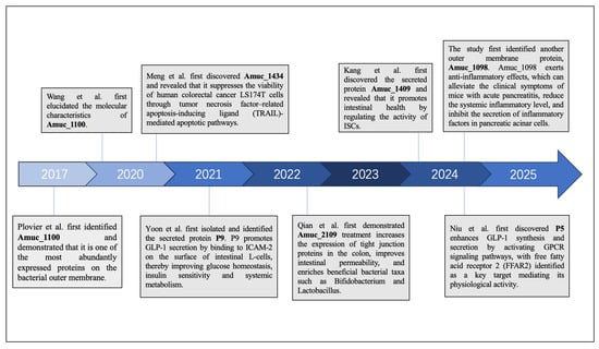
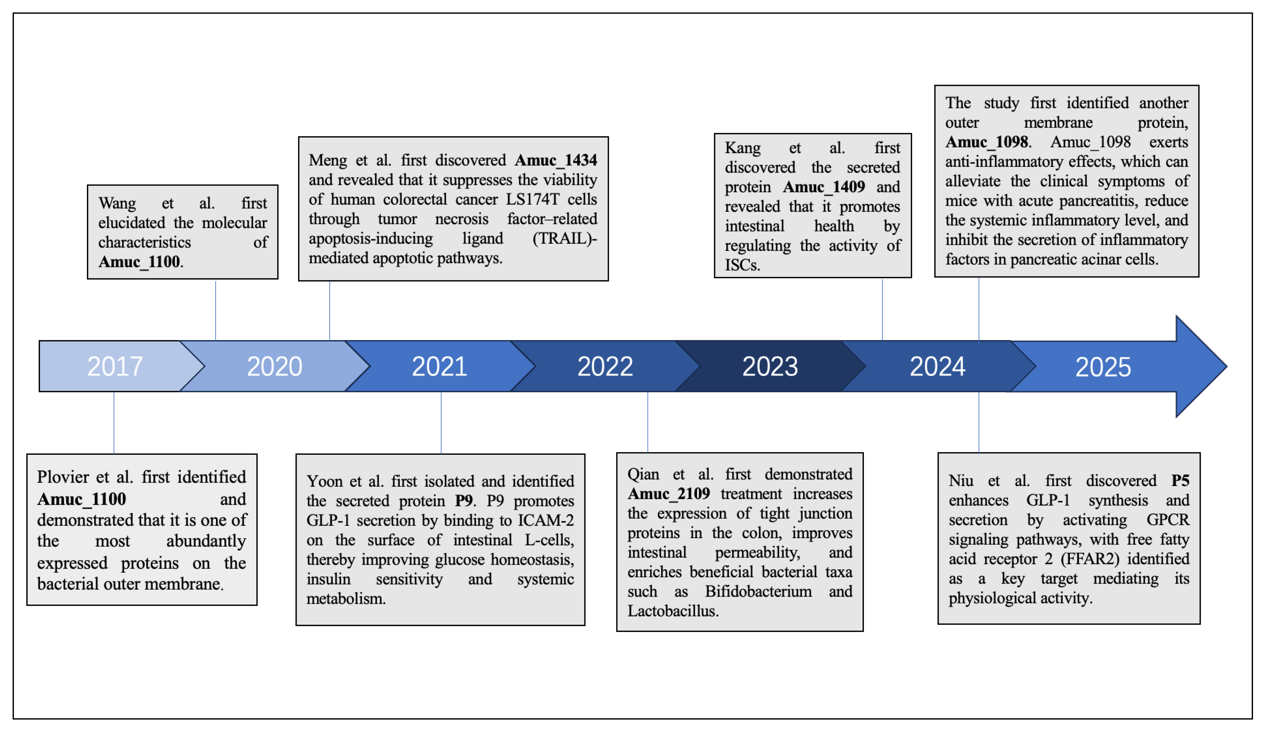
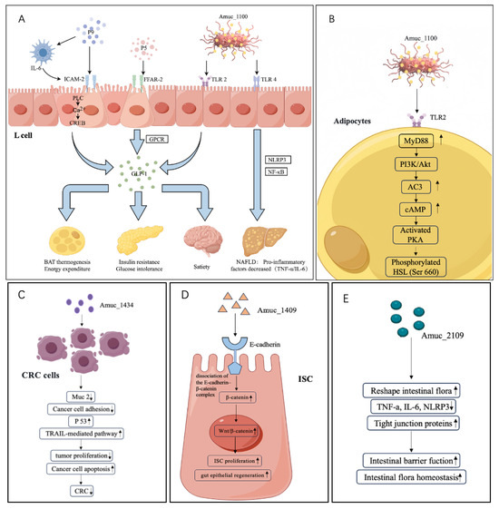
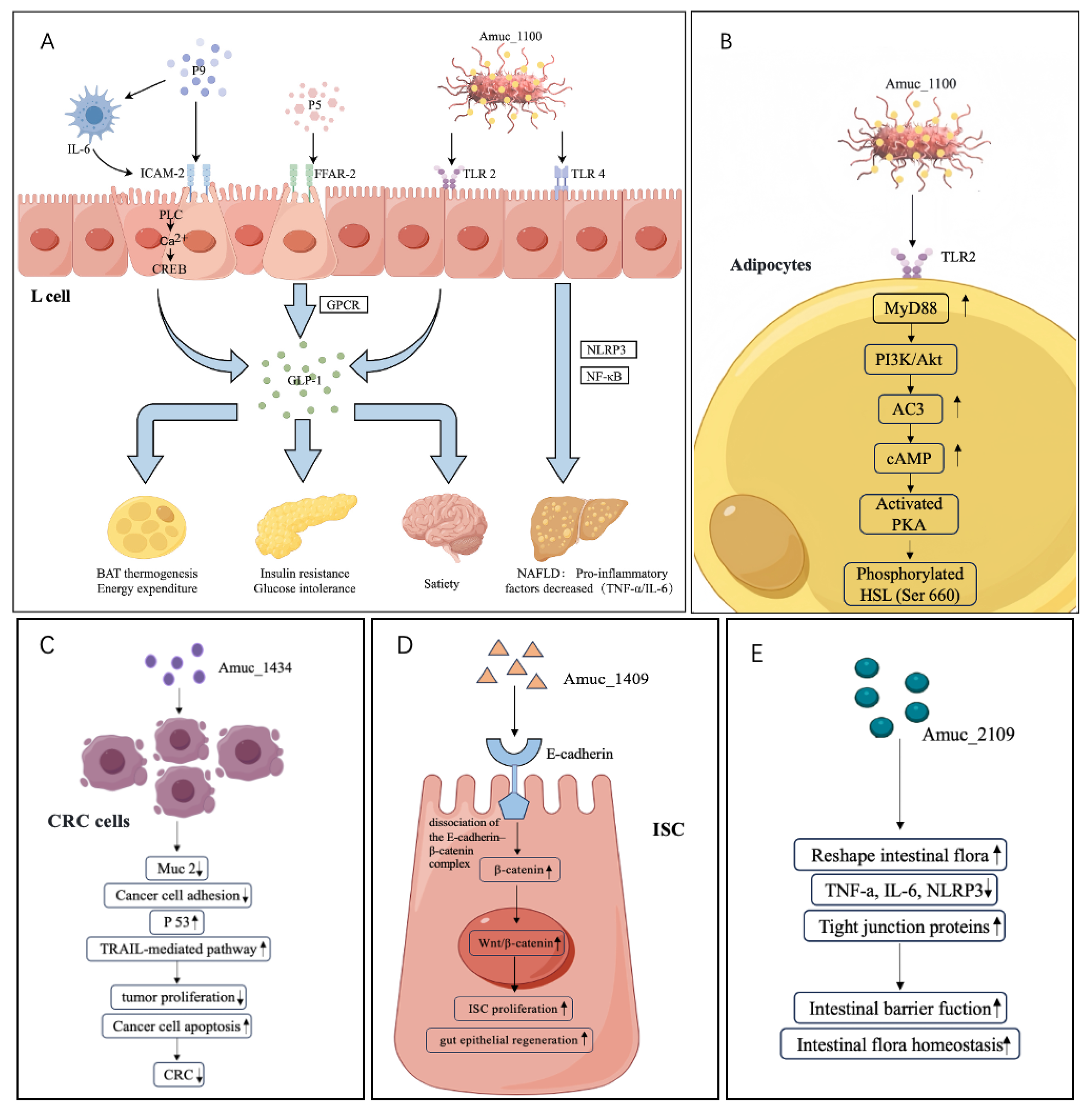

Disclaimer/Publisher’s Note: The statements, opinions and data contained in all publications are solely those of the individual author(s) and contributor(s) and not of MDPI and/or the editor(s). MDPI and/or the editor(s) disclaim responsibility for any injury to people or property resulting from any ideas, methods, instructions or products referred to in the content. |
© 2026 by the authors. Licensee MDPI, Basel, Switzerland. This article is an open access article distributed under the terms and conditions of the Creative Commons Attribution (CC BY) license.