


Abstract
Growing Angus steers (n = 20) were blocked by weight and randomly assigned to one of two treatment groups: Control group (CON, n = 10) fed a feedlot ration ad libitum, or a ruminally protected hydrogenated fat-embedded calcium gluconate (HFCG) treatment group (HFCG, n = 10), which was fed the control ration top-dressed at 16 g/head/day for 55 days. During the slaughter process, digesta samples were collected from the cecum, colon, and rectum. Acetate concentrations were greater in the cecal and rectal digesta of steers (p ≤ 0.05) in the HFCG treatment group. Propionate concentrations were greater in the cecal, colonic, and rectal (p ≤ 0.05) digesta of steers in the HFCG treatment group. Butyrate concentrations were greater (p = 0.098) in the colon digesta of steers in the HFCG treatment group; however, they were not different in the cecal and rectal digesta. To determine the microbial composition within each section of the hindgut, DNA was extracted, and 16S rRNA gene sequencing was performed. Data were analyzed using a General Linear Model with dietary treatment as the main effect. Species richness in the cecum, colon, and rectum was not different between treatments. Erysipelotrichaceae, Peptostreptococcaceae, and Atopobiaceae abundances were increased (p ≤ 0.05) in the cecal bacterial community of steers in the HFCG group, while a significant decrease (p ≤ 0.05) in Rikenellaceae and Muribaculaceae abundances was recorded within the same bacterial community. In the colon bacterial community of steers in the HFCG group, Ruminococcaceae and Muribaculaceae abundances were elevated (p ≤ 0.05), while there was a reduction (p ≤ 0.05) in Lachnospiraceae, Erysipelotrichaceae, Atopobiaceae, and Peptostreptococcaceae abundances. Paeniclostridium, Romboutsia, and Turicibacter abundances were increased (p ≤ 0.05) in the cecal bacterial community of steers in the HFCG group, while there was a decrease (p ≤ 0.05) in Rikenellaceae_RC9 _gut_group abundance within the same bacterial community. In the colon microbiota of steers in the HFCG group, Turicibacter abundance was decreased (p ≤ 0.05). Supplementing growing steers with HFCG impacted some members of the bacterial populations, which have important roles in gut homeostasis and health, along with the formation of beneficial end-products in the gastrointestinal tract.
1. Introduction
Ruminant animals have a gastrointestinal tract (GIT) that is colonized by a complex ecosystem of bacteria, fungi, protozoa, and archaea, which degrade feedstuffs, enabling the animal to harvest energy and protein for growth [1,2,3]. In particular, the rumen microbial ecosystem provides the ruminant animal the ability to harvest energy from forages and other feedstuffs, yet this adaptability comes at the cost of lower feed efficiency than monogastric animals [4,5]. While cattle productivity primarily depends on the symbiotic microbial fermentation of feedstuffs in the rumen, a secondary fermentation occurs in the hindgut (e.g., cecum and colon), which provides further metabolizable energy to the animal, and the fecal microbiota activity has been linked with improving the feed efficiency of cattle [6,7,8]. The dietary energy produced from the hindgut fermentation in the form of volatile fatty acids (VFAs, e.g., acetate, butyrate, and propionate) contributes to the energy available for ruminants to utilize for maintenance, growth, and production [5,9,10]. Among these predominant VFAs that have beneficial effects for animals, butyrate has received particular attention from researchers. Butyrate is a vital VFA produced in the GIT via microbial fermentation of dietary carbohydrates that are ingested by the host and is associated with a healthy gut in the host [2,11,12].
In ruminants, butyrate is mainly produced in the rumen, where it is absorbed rapidly; however, it is also present in high concentrations in the cecum and colon due to secondary hindgut fermentation [11,12]. Butyrate is the preferred energy source for colonocytes, accounting for approximately 70% of the total energy consumption of the colonocyte [12]. The anti-inflammatory effect of butyrate provides a benefit to the host animal by minimizing the energy directed to the fight against diseases; therefore, it frees up that energy for host growth and development [13,14]. Moreover, butyrate stimulates mucus production, increases cell proliferation in the GIT, strengthens the intestinal barrier by enhancing tight junction assembly, and affects nutrient, mineral, and water absorption from the intestinal lumen [12,15]. The use of butyrate as a dietary supplement in animals has been limited due to its volatility and offensive odor; however, butyrate salts (e.g., calcium, sodium, magnesium, or potassium salts), and encapsulated products as well as butyrate precursors have been developed and investigated to increase butyrate supply in the GIT of animals [11,12].
Gluconate is produced by a variety of microorganisms (e.g., bacteria and fungi) through the dehydrogenation of glucose via glucose oxidase, which naturally occurs in plants, fruits, wine, honey, and meat [16,17,18,19]. It has desirable characteristics for use in foods and feeds because it is odorless, non-volatile, non-corrosive, non-toxic, and soluble in water [16,17,18,20]. Classified as generally recognized as safe (GRAS) by the United States Food and Drug Administration (FDA), gluconate and its alkali salts (e.g., calcium gluconate and sodium gluconate) are used as bio-based additives in food and medicines [16,17,18,20]. When fed to monogastric species, gluconate has demonstrated a prebiotic effect, including escaping ruminal and abomasal degradation and small intestinal absorption and being available for microbial fermentation in the hindgut [21,22]. Gluconate enhanced animal performance through improving average daily gain [23,24], which was associated with changes in microbial VFA production in the host hindgut because gluconate is identified as a precursor for butyrate synthesis [25,26]; however, evidence in ruminant species in this regard is still scarce. Moreover, the majority of gluconate-fermenting bacteria in the hindgut are lactic acid bacteria (LAB) that produce mainly lactate, which is in turn converted to butyrate by acid—utilizing butyrate-producing bacteria [21,22,25,26]. This relationship between LAB and acid—utilizing butyrate-producing bacteria, and the fermentation pattern of gluconate in the ruminant hindgut, has yet to be elucidated.
In the first published study conducted in ruminant species, calcium gluconate supplementation increased milk fat yield in lactating dairy cattle [27]. The method of calcium gluconate delivery demonstrated a differential impact on animal performance, with post-ruminal infusion of calcium gluconate increasing milk fat yield and energy-corrected milk yield in lactating dairy cattle [28,29]; however, when directly dosed in the rumen, gluconate addition reduced weight gain and milk yield [29]. Supplementing dairy cattle with a hydrogenated fat-embedded calcium gluconate (HFCG; designed to reach the hindgut for calcium gluconate delivery) increased or tended to increase milk fat yield and both fat- and energy-corrected milk yield [30,31,32]. Another recent study has confirmed the effectiveness of HFCG supplementation at increasing milk yield, protein content and yield, and lactose yield in lactating cattle [33]. Feeding HFCG to growing lambs does not increase butyrate concentration in the intestine, the absorptive surface area, functionality, or size of the GIT [34]. In addition, while both post-ruminal infusion of calcium butyrate and HFCG affected the molar proportion of acetate and propionate in the colon, only post-ruminal infusion of calcium butyrate stimulated ruminal short-chain fatty acid (SCFA) absorption for growing beef heifers [35]. There is little information on the utilization of gluconate in the hindgut of ruminant animals, and the impact of gluconate supplementation on the VFA profile and microbiota in the hindgut of ruminant animals is not fully understood. Therefore, the objective of the study was to evaluate the impact of feeding a ruminally protected hydrogenated fat-embedded calcium gluconate supplement (HFCG; a prebiotic targeting the hindgut) on hindgut microbiota and VFA profile using Angus beef steers as a gut model. We hypothesized that feeding HFCG to Angus beef steers would modulate the hindgut bacterial community and enhance VFA production in the hindgut.
2. Materials and Methods
2.1. Experimental Design
All animal care followed guidelines approved by the University of Georgia’s Animal Care and Use Committee (UGA AUP A2020 02-017-Y1-A0). The study started on April 14 and ended on 5 June 2020. Angus steers with a starting weight of approximately 448 kg (9.6 kg SEM) that were calved and raised at the Eatonton Beef Research Unit, Eatonton, GA, USA, were utilized in the study. All steers (n = 20) were reared in a pasture-based system from 9 months of age until enrollment in the present study at 13 months of age, before being transported to the feedlot in Eatonton. Upon arrival at the feedlot, steers were acclimated to the Calan gate system (Calan Broadbent Feeding System; American Calan, Northwood, NH, USA) and to the control feedlot finishing rations fed (Table 1) during the adaptation phase (−49 d), before being fed either the control (CON; n = 10) ration or HFCG [Selko® Lactibute, Micronutrients, Indianapolis, IN, USA; a ruminally protected hydrogenated fat-embedded calcium gluconate] treatment ration (HFCG; n = 10) for 55 or 62 days based on slaughter group. The current study utilized a completely randomized design. Steers in each treatment were blocked by weight and randomly assigned to two slaughter groups. 6 steers from the CON group and 4 steers from the HFCG group were slaughtered during the first slaughter date (d 56), and the alternate 4 steers from the CON group and 6 steers from the HFCG group were slaughtered on the second slaughter date (d 63). The two slaughter weeks were required due to COVID limitations on the processing facility procedures. All steers were fed their respective treatment rations until the day prior to slaughter. All steers always had ad libitum access to water.
Table 1.
Ration composition fed daily to both treatment groups (HFCG and CON; n = 10/treatment) of growing Angus steers and Nutrient Analysis (determined by Cumberland Valley Analytical Services, Waynesboro, PA, USA) of mixed ration fed to both treatment groups. The calcium gluconate supplement (16 g/hd/d) was added as a top-dress to the HFCG ration daily for each steer in the group. Values are presented on a DM basis. Steer rations were fed from d −49 through d 55 or 62.
2.2. Rations: Control and HFCG Treatments
The ration (Table 1) was prepared at a commercial feed mill (Godfrey’s Feed, Madison, GA, USA) and delivered to the Eatonton Beef Research Facility. Each day, the HFCG supplement was top-dressed (16 g/hd/d) and mixed by hand in each HFCG group steer’s Calan feed bunk. Steers were fed ad libitum with a target of 10% refusals.
2.3. Gastrointestinal Sample Collection and pH Measurements
At slaughter, digesta contents from each hindgut compartment (e.g., cecum, colon, and rectum) were aseptically collected (two subsamples per compartment, 5–10 g/subsample) for analysis and stored in sterile 40 mL plastic tubes. Additionally, digesta pH from each section was measured immediately at the site using an Orion pH meter (Thermo Fisher Scientific, Waltham, MA, USA) and was frozen at −20 °C until later analysis. Each sample was aseptically transported on ice from the Meat Science Teaching and Research Laboratory to the pilot laboratory (less than 10 min away from the sample collection site).
2.4. Volatile Fatty Acid (VFA) Analysis in Hindgut Digesta
Concentrations of VFA in gastrointestinal digesta from each sampled hindgut compartment were analyzed [36]. Briefly, 3 g of each sample was placed into 15 mL conical tubes and diluted with 9 mL of distilled water, and mixed prior to subsampling. Tubes were vortexed for 30 s, and 2.0 mL of the mixture was transferred to centrifuge tubes, which were then centrifuged at 10,000× g for 10 min; 1 mL of the resulting supernatant was aliquoted into new centrifuge tubes, along with 0.2 mL of a meta-phosphoric acid solution (25% weight/volume), and samples were frozen overnight. Samples were thawed and centrifuged at 10,000× g for 10 min. The supernatant was transferred into polypropylene tubes and mixed with ethyl acetate in a 2:1 ratio of ethyl acetate to supernatant. Tubes were vortexed for 15 s and allowed to settle for 5 min before 0.5 mL of the supernatant was transferred to screw-thread vials for VFA analysis using a Shimadzu GC-2010 Plus gas chromatograph (Shimadzu Corporation, Kyoto, Japan) equipped with a flame ionization detector and a capillary column (Zebron ZB-FFAP; 30 m × 0.32 mm × 0.25 μm; Phenomenex Inc., Torrance, CA, USA). The sample injection volume was set as 1.0 μL, and helium was used as the carrier gas. The column temperature was initially set at 110 °C and gradually increased to 200 °C. Injector and detector temperatures were set at 250 and 350 °C, respectively. The samples’ peak heights were compared to standards to determine the concentrations of VFAs in the digesta samples.
2.5. DNA Extraction and Sequencing
Deoxyribonucleic acid (DNA) was extracted from the GIT samples following previously described procedures, which used a combination of mechanical and enzymatic methods to optimize DNA extraction [36,37,38]. Briefly, this procedure uses 0.33 g of sample placed in 2-mL Lysing Matrix E tubes (MP Biomedicals LLC, Irvine, CA, USA), which are homogenized in a FastPrep 24 Instrument (MP Biomedicals LLC, Irvine, CA, USA) to disrupt the cells. Enzymatic inhibition was achieved by using InhibitEX Tablets (QIAGEN, Venlo, The Netherlands), and DNA elution and purification were conducted using an automated robotic workstation (QIAcube; QIAGEN, Venlo, The Netherlands). Determination of DNA concentration and purity in the resulting eluate was performed spectrophotometrically using the Synergy H4 Hybrid Multi-Mode Microplate Reader in conjunction with the Take3 Micro-Volume Plate (BioTek Instruments Inc., Winooski, VT, USA). Samples with a minimum concentration of 10 ng/μL of DNA were frozen at −20 °C until shipping. Samples that failed to meet these requirements were rejected and subjected to a new DNA extraction cycle.
2.6. DNA Library Preparation and Bioinformatics Analysis
Extracted DNA samples were transported frozen to LC Sciences (Houston, TX, USA) for 16S rRNA gene sequencing of the V3–V4 hypervariable regions. Library preparation included PCR replications using the forward: S-D-Bact-0341-b-S-17 (5′-CCTACGGGNGGCWGCAG-3′); and reverse: S-D-Bact-0785-a-A-21 (5′-GACTACHVGGGTATCTAATCC-3′) primer pair [36]. The amplified DNA was then sequenced using an Illumina MiSeq instrument (Illumina Inc., San Diego, CA, USA). Demultiplexed paired-end sequences were imported into QIIME 2 Version 2024.1 [39] for further processing. Non-biological nucleotides were removed, and sequences were denoised, dereplicated, and chimera-filtered using the DADA2 plugin 12 January 2024 access [40]. Taxonomic classifications were assigned to the sequences using a pre-trained Naïve Bayes classifier based on the SILVA database [37], and reads were classified by taxon using the fitted classifier. Samples underwent rarefication to 4780 sequences per sample prior to computing alpha diversity. The following alpha diversity indices were computed: Observed Amplicon Sequence Variants (ASV), Chao1 Index, Shannon Diversity Index, Simpson Diversity Index, and Good’s Coverage Index. The relative bacterial abundance was quantified at the phylum, family, and genus levels.
2.7. Statistical Analysis
Statistical analysis was conducted using MINITAB® Statistical Software (2021). The study was a completely randomized design using the individual steer as the experimental unit. An ANOVA (General Linear Model; GLM) was constructed for VFA concentrations as well as microbiome results (individual microbial taxa and alpha diversity indices) and included treatment as the main effect. Least square means were separated using the Fisher LSD Method. Significance was declared at p ≤ 0.05, and tendency was determined as 0.05 < p ≤ 0.10.
3. Results
3.1. VFA Concentrations and pH in Hindgut Digesta
Acetate concentrations were greater in the cecal (79.7 mM vs. 61.8 mM; p = 0.01) and rectal (52.7 mM vs. 32.0 mM; p = 0.02) digesta of steers in the HFCG group (Table 2). There was a tendency (p = 0.08) towards an increased acetate concentration (56.4 mM vs. 35.6 mM) in the colon digesta of steers in the HFCG group (Table 2). Propionate concentrations were greater in the cecal (28.97 mM vs. 17.86 mM; p = 0.02), colon (21.2 mM vs. 11.3 mM; p ≤ 0.05), and rectal (18.6 mM vs. 9.09 mM; p = 0.01) digesta of steers in the HFCG group (Table 2). There was a tendency towards a greater butyrate concentration (9.13 mM vs. 4.9 mM; p = 0.098) in the colon digesta of steers in the HFCG group; however, butyrate concentrations in the cecal (14.1 mM vs. 13.1 mM) and rectal (6.82 mM vs. 6.85 mM) digesta were not different between the groups (Table 2). Isobutyrate concentrations were not different between groups in any of the digesta collected from the hindgut. In both the cecal and rectal digesta of steers in the HFCG group, total VFA concentrations were (p = 0.01 and p = 0.02, respectively) greater than those of steers in the CON group (125.41 mM vs. 95.55 mM and 79.97 mM vs. 49.22 mM, respectively; Table 2). There was a tendency (p = 0.07) towards a greater total VFA concentration in the colon digesta of steers in the HFCG group (98.29 mM vs. 58.97 mM). The ratio of acetate to propionate (A:P) in the cecal, colon, or rectal digesta of all steers was not significantly different between treatment groups. Mean pH of hindgut digesta at slaughter is presented in Table 3. Cecal digesta pH in the CON group was significantly greater when compared to the cecal digesta pH in the HFCG group (6.32 vs. 5.95; p = 0.03). Colon digesta pH tended to be greater in the CON group (6.31 vs. 5.97; p = 0.07). No significant difference between treatments was observed in the rectal digesta pH (6.28 vs. 6.11; p = 0.39).
Table 2.
Volatile fatty acid concentrations ([VFA], mM) in the digesta from each hindgut section of steers (n = 10/treatment) from the HFCG or CON groups. The calcium gluconate supplement (16 g/hd/d) was added as a top-dressing to each HFCG group ration daily. Individual p-values for treatment effect on VFA concentrations are indicated for each hindgut section below each column.
Table 3.
Digesta pH in the cecum, colon, and rectum of steers (n = 10/treatment) from the CON or HFCG groups. The calcium gluconate supplement (16 g/hd/d) was added as a top-dressing to each HFCG group ration daily. Individual p-values for treatment effect on digesta pH are indicated for each hindgut section within each row.
3.2. Bacterial Richness and Diversity
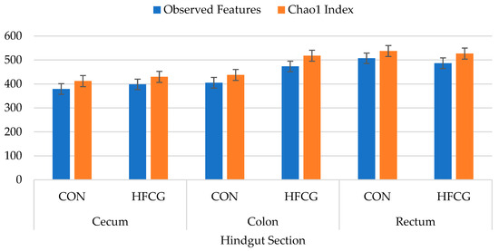
The alpha diversity indices used in this study were: Number of observed features (ASV), Chao1 Index, Shannon Diversity Index, Simpson Diversity Index, and Good’s Coverage Index. Although we observed numerical differences, there were no significant differences between treatment groups in species richness (in either the Chao1 Index or the number of observed features) of bacterial communities in the cecum, colon, or rectum (Figure 1). The greatest numbers of Observed Features and the Chao1 Index were recorded in the rectum bacterial community of steers in the CON group (Figure 1, Supplementary Table S1). Shannon and Simpson diversity indices were numerically greater in all bacterial communities in all hindgut compartments of steers in the HFCG group (Supplementary Table S1).
Figure 1.
Effect of HFCG treatment on bacterial richness (indicated by Observed Features [ASV] and Chao1 Index) in the cecum, colon, and rectum digesta of steers (n = 10/treatment) from CON or HFCG groups. The calcium gluconate supplement (16 g/hd/d) was added as a top-dressing to each HFCG group ration daily.
3.3. Hindgut Microbiota Composition at Phylum Level
Firmicutes, Bacteroidetes, Actinobacteria, Spirochaetes, and Proteobacteria were the predominant phyla in the bacterial community of sampled hindgut sections (Table 4). Firmicutes and Actinobacteria abundances were increased (p = 0.007 and p = 0.012, respectively), and Bacteroidetes abundance was decreased (p = 0.004) in the cecal microbiota of steers in the HFCG group compared to the CON group. Actinobacteria abundance was decreased (p = 0.018), and Bacteroidetes abundance tended to increase (p = 0.075) in the colon microbiota of steers in the HFCG group compared to the CON group. No significant difference was observed between treatment groups in the abundance of the predominant phyla in the rectum; however, Spirochaetes abundance tended (p = 0.074) to increase in the rectum microbiota of steers in the HFCG group. Firmicutes:Bacteroidetes ratio was elevated (p ≤ 0.05) in the cecal microbiota and decreased (p = 0.008) in the colon microbiota of steers in the HFCG group.
Table 4.
The predominant phyla in the cecum, colon, and rectum digesta of steers (n = 10/treatment) from the CON or HFCG groups. The calcium gluconate supplement (16 g/hd/d) was added as a top-dressing to each HFCG group ration daily. Individual p-values for treatment effect on predominant bacterial phyla are indicated for each hindgut section below each column.
3.4. Hindgut Microbiota Composition at Family Level
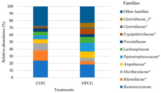
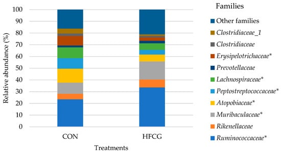
Microbial composition at the family level for the cecum, colon, and rectum of all steers is presented in detail in Supplementary Table S2. Prevotellaceae, Lachnospiraceae, Ruminococcaceae, Erysipelotrichaceae, Atopobiaceae, Rikenellaceae, Muribaculaceae, Clostridiaceae, Clostridiaceae_1, and Peptostreptococcaceae were some of the predominant families in the bacterial community of sampled hindgut compartments of all steers (Figure 2 and Figure 3). The abundance of Erysipelotrichaceae, Peptostreptococcaceae, Clostridiaceae, Clostridiaceae_1, and Atopobiaceae was increased (p = 0.003, p = 0.011, p = 0.001, p = 0.009, p = 0.001, and p = 0.012, respectively) in the cecal bacterial community of steers in the HFCG group (Figure 2). A significant decrease (p = 0.002 and p = 0.021, respectively) in the abundance of Rikenellaceae and Muribaculaceae was recorded within the same bacterial community (Figure 2). Additionally, Spirochaetaceae abundance tended to decrease (p = 0.069) in the cecal bacterial community of steers in the HFCG group (Supplementary Table S2). The abundance of Ruminococcaceae and Muribaculaceae was elevated (p = 0.019 and p = 0.004, respectively) in the colon bacterial community of steers in the HFCG group (Figure 3), while there was a reduction (p = 0.033, p = 0.015, p = 0.023, and p = 0.002, respectively) in the abundances of Lachnospiraceae, Erysipelotrichaceae, Atopobiaceae, and Peptostreptococcaceae within the same bacterial community (Figure 3). Furthermore, Rikenellaceae abundance was increased (p = 0.045) in the rectal bacterial community of steers in the HFCG group (Supplementary Table S2).
Figure 2.
Effect of HFCG treatment on the cecal microbiota of steers (n = 10/treatment from CON or HFCG groups) at the family level. The calcium gluconate supplement (16 g/hd/d) was added as a top-dressing to each HFCG group ration daily. (*) refers to the statistical significance of p ≤ 0.05.
Figure 3.
Effect of HFCG treatment on the colonic microbiota of steers (n = 10/treatment from CON or HFCG groups) at the family level. The calcium gluconate supplement (16 g/hd/d) was added as a top-dressing to each HFCG group ration daily. (*) refers to the statistical significance of p ≤ 0.05.
3.5. Hindgut Microbiota Composition at Genus Level
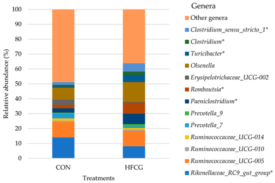
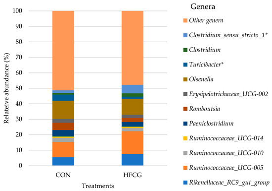
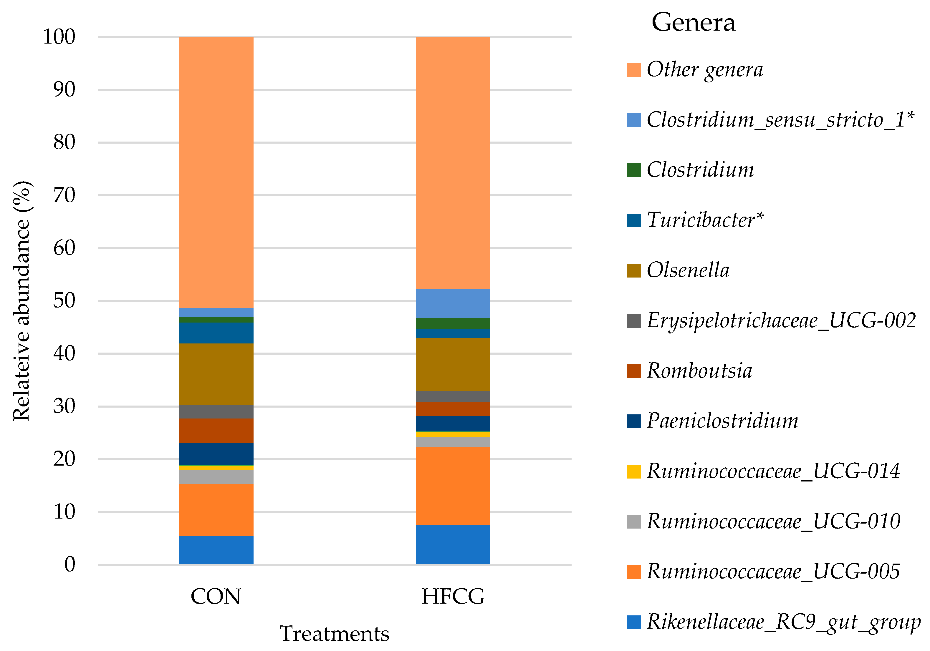
The lowest taxonomic level at which the samples in this study were classified was the genus level, and the predominant bacterial genera detected in the cecum, colon, and rectum of all steers are presented in detail in Supplementary Table S3. Resolution in this study was not sufficient to assign ASVs consistently at the species level. Ruminococcaceae_UCG-005, Ruminococcaceae_UCG-010, Ruminococcaceae_UCG-014, Rikenellaceae_RC9 _gut_group, Paeniclostridium, Romboutsia, Erysipelotrichaceae_UCG-002, Olsenella, Turicibacter, Prevotella_7, and Prevotella_9 were found to be the predominant genera in the cecum and colon microbiota of all steers (Figure 4 and Figure 5). The abundance of Paeniclostridium, Romboutsia, and Turicibacter was increased (p = 0.007, p = 0.005, and p = 0.001, respectively) in the cecal bacterial community of steers in the HFCG group, while there was a decrease (p = 0.022) in the abundance of Rikenellaceae_RC9 _gut_group within the same bacterial community (Figure 4). Additionally, Clostridium_sensu_stricto_1 and Clostridium abundances were elevated (p = 0.019 and p = 0.012, respectively) in the cecal bacterial community of steers in the HFCG group (Figure 4). In the colon microbiota of steers in the HFCG group, Turicibacter and Clostridium_sensu_stricto_1 abundances were decreased (p = 0.014 and p = 0.037, respectively; Figure 5).
Figure 4.
Effect of HFCG treatment on the cecal microbiota of steers (n = 10/treatment from CON or HFCG groups) at the genus level. The calcium gluconate supplement (16 g/hd/d) was added as a top-dressing to each HFCG group ration daily. (*) refers to the statistical significance of p ≤ 0.05.
Figure 5.
Effect of HFCG treatment on the colonic microbiota of steers (n = 10/treatment from CON or HFCG groups) at the genus level. The calcium gluconate supplement (16 g/hd/d) was added as a top-dressing to each HFCG group ration daily. (*) refers to the statistical significance of p ≤ 0.05.
4. Discussion
Cattle productivity heavily depends on the microbial fermentation of feedstuffs in the rumen; however, a secondary microbial fermentation occurs in the hindgut, which has been associated with improving the feed efficiency of cattle [6,7,41]. Substrates that are partially digested by enzymes in the rumen and small intestine are further broken down in the cecum and colon by a specialized consortia of microorganisms that is mostly comprised of bacteria [2,9,42,43]. Additional dietary energy derived from this hindgut microbial fermentation in the form of VFAs (e.g., acetate, butyrate, and propionate) is considered a significant contributor to energy availability in ruminants throughout all stages of production [3,9,43]. Among these major VFAs, butyrate is a crucial one as it (i) has an anti-inflammatory effect on the host [13,14], (ii) stimulates mucus production, (iii) strengthens the epithelial structural integrity in the GIT, (iv) affects the dynamics of nutrient, mineral, and water absorption in the GIT [12,15], and (v) is the preferred energy source for colonic epithelial cells, accounting for approximately 70% of the total energy consumption of the colonocytes [2,11,12]. In ruminant species, although butyrate is mainly produced in the rumen (approximately 8–10 mM), it is also found in high concentrations in the lumen of the large intestine, especially in the cecum and colon, due to the secondary hindgut fermentation [12]. Butyrate salts and encapsulated products, as well as butyrate precursors, have been developed and investigated to increase butyrate supply in the GIT of animals [11,12].
Gluconate is a precursor for butyrate synthesis [25,26] and has demonstrated a prebiotic effect when fed to monogastric species. Gluconate can survive the gastrointestinal passage and be available for microbial fermentation in the hindgut [21,22] and enhance animal performance through improving average daily gain [23,24]. Improved ADG was associated with changes in microbial VFA production in the host hindgut [25,26]. Unfortunately, there is a lack of information on the utilization of gluconate in the hindgut of ruminant animals, and the impact of gluconate supplementation on the hindgut VFA profile and microbiota in ruminants is not well understood.
Increasing use of Next Generation Sequencing (NGS) technologies has enabled researchers to gain more insight into the members and activities of the gastrointestinal microbiota of ruminants [7,44,45]. Therefore, understanding the rumen microbiota has garnered interest because of its substantial impact on animal health and productivity; however, the microbiota harbored in the remainder of the GIT has not received the same interest. Each compartment of the ruminant GIT deals with different fermentable substrates and presents different environmental conditions; therefore, the microbiota composition within each compartment changes from one to another. In general, the use of prebiotic compounds in ruminants has been challenging due to the diverse catabolic activity of ruminal microbially-produced carbohydrate active enzymes (CAZymes); however, protecting carbohydrates from microbial degradation can pass these compounds to the hindgut [44,46,47].
4.1. Cecal Microbiota Composition and VFA Profile
Acetate, propionate, as well as total VFA concentrations were significantly greater in the cecum of steers in the HFCG group in the present study (Table 2). Additionally, cecal digesta pH of steers in the CON group was significantly greater than that of steers in the HFCG group (Table 3). These results suggest that the microbial fermentation of gluconate (as a prebiotic source) by the cecum microbiota might have resulted in increased concentrations of certain VFAs and a lower pH in the cecal digesta of steers in the HFCG group in the current study.
Gluconate has been described as a precursor for butyrate synthesis in the hindgut of non-ruminant animals [22,26]. The effect of gluconate on in vitro growth response and metabolism of swine cecal microflora and on animal growth performance, intestinal wall morphology, and intestinal microflora was investigated [24]. Compared with the control group, gluconate tended to improve the average daily gain (ADG) of the piglets, yet cecal morphology and microbiota did not show any difference among treatments [24]. After 24 h of in vitro cecal fermentation, acetate, propionate, and butyrate concentrations were increased by gluconate [24]. Digesta pH in the small intestine and cecum as well as bacterial populations in both cecum and rectum, digesta of piglets were not affected by sodium gluconate supplementation [23]. Increasing the dietary supply of HFCG to growing lambs linearly increased the proportion of acetate in cecal digesta and linearly decreased the proportion of propionate in both cecal and colonic digesta [35]; however, it did not affect the butyrate concentrations in the hindgut. In the present study, calcium gluconate supplementation to the ration of steers did not increase butyrate concentration in the cecal digesta; instead, it elevated acetate and propionate concentrations in the cecum of steers in the HFCG group.
Firmicutes, Bacteroidetes, and Actinobacteria phyla were predominant in the cecum microbiota of all steers in the current study, corroborating previous studies where these phyla were commonly found in the hindgut microbiota of varied ruminant species under different experimental settings [8,10,43,48]. The abundance of Firmicutes and Actinobacteria was elevated while Bacteroidetes was decreased in the cecal microbiota of steers in the HFCG group compared to the CON treatment group; therefore, the Firmicutes to Bacteroidetes ratio was increased in the cecal microbiota, and the F:B ratio has been linked to increased dietary energy capture in mice [49,50,51] (Figure 2). The abundance of Erysipelotrichaceae, Peptostreptococcaceae, and Atopobiaceae (belonging to Actinobacteria) was significantly increased in the cecal bacterial community of steers in the HFCG group. A significant decrease in the abundance of Rikenellaceae and Muribaculaceae was recorded within the same bacterial community.
In the present study, Lachnospiraceae was abundant in the cecal digesta of steers regardless of the ration composition, and its abundance was numerically increased (from 5.12 to 8.28%) in the HFCG group. The abundant detection of Lachnospiraceae in the cecum of ruminant animals is a common finding reported by previous studies [6,9,42]. Several members of Lachnospiraceae are able to ferment pectin and break down other complex polysaccharides to VFAs (e.g., acetate, butyrate, and propionate) in the GIT environment, providing energy for the host [42,52,53]. Increased concentrations of acetate and propionate in the cecum of steers in the HFCG group can be associated with the (numerical) increase in the abundance of Lachnospiraceae. However, the exact role of Lachnospiraceae in the hindgut of ruminant species warrants further investigation.
Our data revealed that Peptostreptococcaceae was predominant in the cecal digesta regardless of the ration composition, and its abundance was increased (from 5.33 to 12.25%) due to the HFCG supplementation. Previous studies reported that Peptostreptococcaceae was also abundant in the cecum microbiota of ruminant animals investigated under different experimental conditions [54,55]. Some members of this family have been reported to play a vital role in feed digestion and are known as hyper-ammonia-producing bacteria [54,56]. However, the exact role of Peptostreptococcaceae in the GIT ecosystem(s), particularly in the cecum of ruminants, still warrants further investigation.
At the genus level, Romboutsia and Paeniclostridium represented the Peptostreptococcaceae family, and their abundances were also increased (from 2.36 to 7.47% and from 2.93 to 7.01%, respectively) in the cecal microbiota of steers in the HFCG group. Romboutsia as a genus has been linked to a healthy GIT mucosa in humans and functions as a microbial biomarker of polyps and/or colorectal cancer; therefore, its presence or elevated populations in the GIT may be considered beneficial to prevent intestinal dysbiosis [57]. Romboutsia species were also reported to be non-pathogenic and commensal in the GIT and capable of producing VFAs such as formate and acetate in the GIT of animals and of fermenting single amino acids [58,59,60]. It has been reported that Romboutsia may function as a candidate microbial biomarker of intestinal dysbiosis, as well as of coccidiosis in chickens, as its abundance in the chicken cecum declined post-infection caused by Eimeria tenella [60].
The addition of HFCG increased the crude fat content of the basal rations fed [31]. The impact of dietary fat on the abundance of Erysipelotrichaceae in the GIT of monogastric animals has been reported [61,62]. An increase in the abundance of this family was observed in the cecum and colon [61] microbiota of mice fed a high-fat diet. Additionally, a greater Erysipelotrichaceae abundance was reported in the cecal microbial community of steers with better average daily gain as well as dry matter intake [37]. The increase (from 1.52 to 7.44%) in the abundance of Erysipelotrichaceae in the cecal microbiota of steers in the HFCG group in our study may be attributed to the physical characteristics of the dietary supplement. At the genus level, Turicibacter and Erysipelotrichaceae_UCG-002 represented the Erysipelotrichaceae family in the cecal microbiota of steers in the HFCG group (Figure 4). In particular, Turicibacter abundance was increased (from 0.93 to 4.98%; p > 0.05) at the expense of Erysipelotrichaceae_UCG-002 and other members of the Erysipelotrichaceae family. Turicibacter represents the primary lactate producers in the large intestine [63] and was positively correlated with butyric acid production in rats fed high-fat diets [64].
Clostridiaceae and Clostridiaceae_1 were other predominant bacterial families detected in the cecal microbiota of all steers, and their abundances were increased (from 1.17 to 2.33% and from 0.52 to 6.15%, respectively) due to the calcium gluconate supplementation (Supplementary Table S2). Clostridiaceae is a functionally diverse family comprised of members that are mostly commensal in the GIT and have important roles in the digestion of carbohydrates or proteins, altering their fermentation pathway depending on nutrient availability [64,65]. At the genus level, Clostridium and Clostridium_sensu_stricto_1 represented the Clostridiaceae family, and their abundances were also increased (from 1.05 to 2.09% and from 1.73 to 5.52%, respectively) in the cecal microbiota of steers in the HFCG group (Supplementary Table S3). In animal as well as human GIT, Clostridium species are able to digest starch or indigestible/complex polysaccharides, and VFAs (e.g., acetate, propionate, and butyrate) that they produce provide benefits to the host by being used as energy sources by epithelial cells in the GIT [64,66].
4.2. Colonic Microbiota Composition and VFA Profile
The main function of the colon in ruminants is to absorb water from the digesta moving through it as well as vitamins produced by the microbiota harbored in this GIT compartment or originating from the digesta [3,9,52]. Moreover, the colon absorbs and utilizes the VFAs, especially the butyrate, generated from the post-ruminal degradation of cellulose, starch, and other partially digested feedstuffs [2,31]. In the present study, elevated concentrations of VFAs and a lower pH in the colon digesta can be attributed to the microbial fermentation of gluconate as a prebiotic agent in the colon. In particular, butyrate concentration tended to increase in the colon digesta of steers in the HFCG group, corroborating previous studies reporting gluconate as a butyrate precursor in the large intestine of non-ruminant animals [22,25,26]. While butyrate differences were not statistically significant, the role of butyrate on gut health/integrity has been increasingly demonstrated to play biologically impactful roles in animal and human health [11,15].
Increasing the dietary supply of HFCG to growing lambs linearly decreased the proportion of propionate in colonic digesta; however, it did not affect the butyrate concentrations [34]. While both post-ruminal infusion of calcium butyrate and HFCG affected the molar proportion of acetate and propionate in the colon, only post-ruminal infusion of calcium butyrate stimulated ruminal SCFA absorption for growing beef heifers [35]. Compared to the control group, both calcium butyrate and calcium gluconate treatments reduced the proportion of acetate and increased the proportion of propionate in the colonic digesta [35]. Acetate, butyrate, and total VFA concentrations tended to increase, and propionate concentration was significantly greater in the colon digesta of steers in the HFCG group (Table 2), and colon digesta pH tended to be greater in the CON group compared to the HFCG group (Table 3) in the present study. In the colon microbiota of steers in the HFCG group, the abundance of Firmicutes and Actinobacteria decreased while Bacteroidetes abundance tended to increase (Table 4).
Bacteroidetes in the GIT generally produce butyrate, which is an end-product of colon fermentation and plays a vital role in the gut health of the host [67]. Butyrate and other VFAs produced by the Bacteroidetes in the GIT are absorbed by the host, helping the host to gain energy from varied carbohydrate sources [67]. In the current study, acetate, butyrate, and propionate concentrations were greater (though not always reaching statistical significance), and total VFA concentration tended to increase in the colon digesta of steers in the HFCG group. Increased concentrations of VFAs in the colon digesta may be associated with the increase in the abundance of Bacteroidetes, as the breakdown of a variety of polysaccharides in the hindgut is the main biological function of this phylum as symbionts [67]. It is widely known that butyrate produced by the hindgut bacteria is a major energy source for the colon epithelium and regulates enterocyte proliferation and differentiation; thus, butyrate, as well as other VFAs, is vital to the GIT epithelium integrity and homeostasis [68,69]. Bacteroidetes was represented by Prevotellaceae, Rikenellaceae, and Muribaculaceae in the colon microbiota of steers regardless of the ration composition. Among these families in the colon microbiota of steers, Muribaculaceae abundance was significantly elevated in the HFCG group. Muribaculaceae was positively correlated with intramuscular fat deposition (i.e., marbling) in Angus steers [7]. In the present study, steers marbled well due to their diets earlier in their lives; however, the relationship with increased Muribaculaceae presence and/or abundance remains potentially important, as marbling is an economically important variable to beef producers.
At the family level, Ruminococcaceae (belonging to Firmicutes) abundance was significantly increased. In the current study, the elevated level of Ruminococcaceae can be correlated with increased VFA concentrations in the colon digesta, as this family has been reported as a major butyrate producer in the GIT [70]. The abundance of Actinobacteria was increased in the cecal microbiota of steers in the HFCG group, while there was a decrease in the abundance of this phylum in the colon microbiota. At the family level, the same trend was detected regarding Atopobiaceae abundance. Steers had a greater abundance of Atopobiaceae in the cecum microbiota but a lower abundance of this family in their colon microbiota. Certain Actinobacteria members, such as Atopobiaceae, are known as the producers of a variety of bioactive metabolites (e.g., VFAs) that are beneficial for the host’s gut health [71].
4.3. Rectal Microbiota Composition and VFA Profile
The digesta moving through the large intestine is exposed to a series of fermentative reactions along the GIT and reaches its final destination, which is the rectum that is connected to the anus, from where the remaining digesta/material is excreted as feces into the environment [2,3,48]. Bacteria found in feces have important roles in livestock farming, including nutrient cycling, producing gaseous compounds, and acting as a source for zoonotic pathogenic microorganisms as well as antibiotic-resistant bacteria [48]. In the current study, no significant difference was observed between treatment groups in the abundance of the predominant phyla. At the family level, Rikenellaceae abundance was increased in the rectal bacterial community of steers in the HFCG group (Supplementary Table S2).
Acetate, propionate, and total VFA concentrations were significantly greater in the rectal (fecal) digesta of steers in the HFCG group (Table 3); however, butyrate concentration and digesta pH were not different between the groups (Table 4). Similarly, supplementing dairy cattle with HFCG did not affect the pH or concentrations of acetate, propionate, or butyrate in the fecal digesta [31]. However, post-ruminal calcium gluconate infusion increased the concentration of fecal isobutyric acid in lactating dairy cattle [28].
4.4. Bacterial Richness and Diversity
Gut health is still not a clear term and is often described via a proxy measure: bacterial population richness and diversity. The host animal often depends on the energy produced by the microbial fermentation of feedstuff; therefore, any perturbation in bacterial populations can lead to changes in the predominant VFA (including lactate) produced and their gastrointestinal concentrations. Analysis of alpha diversity is a common approach to assess differences between biological samples, summarizing the structure of an ecological community with respect to its richness (e.g., number of taxonomic groups), diversity, and evenness (i.e., abundance distribution of the groups) or all [69,72]. The alpha diversity indices used in this study to obtain a comprehensive representation of all species in the samples are the following: Observed Features, Chao1 Index, Shannon Diversity Index, Simpson Diversity Index, and Good’s Coverage Index. Although there was a numerical difference, no significant difference was observed between treatment groups in species richness (in either the Chao1 Index or the number of Observed Features) of bacterial communities in the cecum, colon, or rectum. The greatest numbers of Observed Features and the Chao1 Index were recorded in the rectum bacterial community of steers in the CON group. Shannon and Simpson diversity indices, which are measures of species richness and evenness [73], were numerically greater in all bacterial communities in all GITs of steers in the HFCG group. Good’s coverage index provides an estimation of what percent of the total species is represented in a sample [72]. There was no difference in Good’s Coverage Index in the hindgut bacterial community of steers in the CON or HFCG group. The average Good’s Coverage for all samples was greater than 99%, indicating that the sequencing depth was adequate for a proper bacterial community analysis [72].
5. Conclusions
Supplementing the ration of growing steers with HFCG enhanced VFA production in the hindgut and had some impacts on the hindgut microbial ecosystem. HFCG supplementation led to shifts in bacterial composition through impacting populations of several bacterial genera (e.g., Paeniclostridium, Romboutsia, and Turicibacter), several of which have been reported to have vital roles in gut homeostasis and health and to play a role in the formation of end products that benefit the host animal’s energetic status (e.g., butyrate and propionate). Results provide novel insights into the potential of rumen—protected gluconate-based additives to modulate the hindgut microbiota in ruminants, a relatively underexplored site of fermentation. Although these results support the hypothesis that HFCG impacts hindgut microbial dynamics in favor of beneficial fermentative processes, further research is warranted to quantify corresponding changes in Volatile Fatty Acid concentrations (especially in butyrate, which is linked with gut integrity), monitor long-term impacts on growth performance, and evaluate possible interactions with host physiology.
Supplementary Materials
The following supporting information can be downloaded at: https://www.mdpi.com/article/10.3390/microorganisms14040802/s1, Table S1: Bacterial richness and alpha diversity indices of bacterial communities in the cecum, colon, and rectum digesta of steers (n = 10/treatment) from CON or HFCG groups. Table S2: The predominant families from bacterial compositions of the cecum, colon, and rectum digesta of steers (n = 10/treatment) from the CON or HFCG treatment groups. Table S3: The predominant genera in bacterial compositions of the cecum, colon, and rectum of steers (n = 10/treatment) fed CON or HFCG rations. Table S4: The predominant species in bacterial compositions of the cecum, colon, and rectum of steers (n = 10/treatment) fed CON or HFCG rations.
Author Contributions
Conceptualization, T.R.C. and E.E.R.; methodology, T.R.C., E.E.R., T.D.P. and A.M.S.; formal analysis, E.E.R., O.Y.K. and T.R.C.; resources, K.E.G., J.M.L., F.L.F., J.J.B., T.D.P. and R.L.S.; writing—original draft preparation, O.Y.K.; writing—review and editing, T.R.C. and K.E.G.; funding acquisition, T.R.C. All authors have read and agreed to the published version of the manuscript.
Funding
This research was funded by Micronutrients—Selko, who provided the product and aided in study design and editing of the final manuscript.
Institutional Review Board Statement
The animal study protocol was approved by the University of Georgia Animal Care and Use Committee Protocol A2020 02-017-Y1-A1, approved on 6 January 2020.
Informed Consent Statement
Not applicable.
Data Availability Statement
The raw data supporting the conclusions of this article will be made available by the authors on request.
Acknowledgments
We would like to thank Micronutrients—Selko for supporting the research financially and providing the HFCG for the study. We also would like to thank Kip McMillian and the staff at the UGA Eatonton Feedyard for their invaluable help with this study.
Conflicts of Interest
Author Kenneth E. Griswold was employed by the company Micronutrients—Selko. The authors declare that this study received funding from Micronutrients-Selko. The funding sponsors had the following involvement with the study design finalization, and editing of the manuscript. The remaining authors declare no conflicts of interest.
Abbreviations
The following abbreviations are used in this manuscript:
| VFA | Volatile fatty acids |
| GIT | Gastrointestinal tract |
| HFCG | Hydrogenated fat-embedded calcium gluconate |
| SCFA | Short-chain fatty acids |
| GRAS | Generally recognized as safe |
| FDA | Food and Drug Administration |
| LAB | Lactic acid bacteria |
References
- Hungate, R.E. The Rumen and Its Microbes; Academic Press: New York, NY, USA, 1966. [Google Scholar]
- Russell, J.B. Rumen Microbiology and Its Role in Ruminant Nutrition; Department of Microbiology, Cornell University: Ithaca, NY, USA, 2002; p. 120. [Google Scholar]
- Mackie, R.; White, B.; Isaacson, R.E. Gut environment and evolution of mutualistic fermentative digestion. In Gastrointestinal Microbiology; Chapman & Hall Microbiology Series; Springer: Boston, MA, USA, 1997; pp. 13–35. [Google Scholar]
- Yeoman, C.J.; White, B.A. Gastrointestinal tract microbiota and probiotics in production animals. Annu. Rev. Anim. Biosci. 2014, 2, 469–486. [Google Scholar] [CrossRef] [Scilit]
- Sasson, G.; Kruger Ben-Shabat, S.; Seroussi, E.; Doron-Faigenboim, A.; Shterzer, N.; Yaacoby, S.; Berg Miller, M.E.; White, B.A.; Halperin, E.; Mizrahi, I. Heritable bovine rumen bacteria are phylogenetically related and correlated with the cow’s capacity to harvest energy from its feed. Mbio 2017, 8, e00703–e00717. [Google Scholar] [CrossRef] [Scilit]
- Freetly, H.C.; Dickey, A.; Lindholm-Perry, A.K.; Thallman, R.M.; Keele, J.W.; Foote, A.P.; Wells, J.E. Digestive tract microbiota of beef cattle that differed in feed efficiency. J. Anim. Sci. 2020, 98, skaa008. [Google Scholar] [CrossRef] [Scilit] [PubMed]
- Krause, T.R.; Lourenco, J.M.; Welch, C.B.; Rothrock, M.J.; Callaway, T.R.; Pringle, T.D. The relationship between the rumen microbiome and carcass merit in Angus steers. J. Anim. Sci. 2020, 98, skaa287. [Google Scholar] [CrossRef] [Scilit] [PubMed]
- Welch, C.B.; Lourenco, J.M.; Davis, D.B.; Krause, T.R.; Carmichael, M.N.; Rothrock, M.J.; Pringle, T.D.; Callaway, T.R. The impact of feed efficiency selection on the ruminal, cecal, and fecal microbiomes of Angus steers from a commercial feedlot. J. Anim. Sci. 2020, 98, skaa230. [Google Scholar] [CrossRef] [Scilit] [PubMed]
- O’Hara, E.; Neves, A.L.A.; Song, Y.; Guan, L.L. The Role of the Gut Microbiome in Cattle Production and Health: Driver or Passenger? Annu. Rev. Anim. Biosci. 2020, 8, 199–220. [Google Scholar] [CrossRef] [Scilit]
- Popova, M.; McGovern, E.; McCabe, M.S.; Martin, C.; Doreau, M.; Arbre, M.; Meale, S.J.; Morgavi, D.P.; Waters, S.M. The structural and functional capacity of ruminal and cecal microbiota in growing cattle was unaffected by dietary supplementation of linseed oil and nitrate. Front. Microbiol. 2017, 8, 937. [Google Scholar] [CrossRef] [Scilit]
- Bedford, A.; Gong, J. Implications of butyrate and its derivatives for gut health and animal production. Anim. Nutr. 2018, 4, 151–159. [Google Scholar] [CrossRef] [Scilit]
- Guilloteau, P.; Martin, L.; Eeckhaut, V.; Ducatelle, R.; Zabielski, R.; Immerseel, F.V. From the gut to the peripheral tissues: The multiple effects of butyrate. Nutr. Res. Rev. 2010, 23, 366–384. [Google Scholar] [CrossRef] [Scilit]
- Frankenfield, D.C.; Ashcraft, C.M. Estimating energy needs in nutrition support patients. J. Parenter. Enter. Nutr. 2011, 35, 563–570. [Google Scholar] [CrossRef] [Scilit]
- Zebeli, Q.; Metzler-Zebeli, B.U. Interplay between rumen digestive disorders and diet-induced inflammation in dairy cattle. Res. Vet. Sci. 2012, 933, 1099–1108. [Google Scholar] [CrossRef] [Scilit]
- Yan, H.; Ajuwon, K.M. Butyrate modifies intestinal barrier function in IPEC-J2 cells through a selective upregulation of tight junction proteins and activation of the Akt signaling pathway. PLoS ONE 2017, 12, e0179586. [Google Scholar] [CrossRef] [Scilit] [PubMed]
- Anastassiadis, S.; Morgunov, I.G. Gluconic acid production. Recent Pat. Biotechnol. 2007, 1, 167–180. [Google Scholar] [CrossRef] [Scilit]
- Ramachandran, S.; Fontanille, P.; Pandey, A.; Larroche, C. Gluconic acid: Properties, applications, and microbial production. Food Technol. Biotechnol. 2006, 44, 185–195. [Google Scholar]
- Ma, Y.; Li, B.; Zhang, X.; Wang, C.; Chen, W. Production of gluconic acid and its derivatives by microbial fermentation: Process improvement based on integrated routes. Front. Bioeng. Biotechnol. 2022, 10, 864787. [Google Scholar] [CrossRef] [Scilit]
- Cañete-Rodriguez, A.M.; Santos-Duenas, I.M.; Jimenez-Hornero, J.E.; Ehrenreich, A.; Liebl, W.; Garcia-Garcia, I. Gluconic acid: Properties, production methods and applications—An excellent opportunity for agro-industrial by-products and waste bio-valorization. Process Biochem. 2016, 51, 1891–1903. [Google Scholar] [CrossRef] [Scilit]
- Pal, P.; Kumar, R.; Banerjee, S. Manufacture of gluconic acid: A review towards process intensification for green production. Chem. Eng. Process. Process Intensif. 2016, 104, 160–171. [Google Scholar] [CrossRef] [Scilit]
- Asano, T.; Yuasa, K.; Kunugita, K.; Teraji, T.; Mitsuoka, T. Effects of gluconic acid on human faecal bacteria. Microb. Ecol. Health Dis. 1994, 7, 247–256. [Google Scholar] [CrossRef] [Scilit]
- Asano, T.; Yuasa, K.; Yoshimura, Y.; Takenawa, S.; Fukuba, H. Digestion, absorption and intestinal residue of various gluconic acids in rats. J. Jpn. Soc. Nutr. Food Sci. 1997, 50, 287–294. [Google Scholar] [CrossRef] [Scilit]
- Poeikhampha, T.; Bunchasak, C. Comparative effects of sodium gluconate, mannan oligosaccharide and potassium diformate on growth performances and small intestinal morphology of nursery pigs. Asian-Australas. J. Anim. Sci. 2011, 24, 844–850. [Google Scholar] [CrossRef] [Scilit]
- Biagi, G.; Piva, A.; Moschini, M.; Vezzali, E.; Roth, F.X. Effect of gluconic acid on piglet growth performance, intestinal microflora, and intestinal wall morphology. J. Anim. Sci. 2006, 84, 370–378. [Google Scholar] [CrossRef] [Scilit]
- Tsukahara, T.; Hashizume, K.; Koyama, H.; Ushida, K. Stimulation of butyrate production through the metabolic interaction among lactic acid bacteria, Lactobacillus acidophilus, and lactic acid-utilizing bacteria, Megasphaera elsdenii, in porcine cecal digesta. Anim. Sci. J. 2006, 77, 454–461. [Google Scholar] [CrossRef] [Scilit]
- Tsukahara, T.; Koyama, H.; Okada, M.; Ushida, K. Stimulation of butyrate production by gluconic acid in batch culture of pig cecal digesta and identification of butyrate-producing bacteria. J. Nutr. 2002, 132, 2229–2234. [Google Scholar] [CrossRef] [Scilit]
- Emery, R.S.; Smith, C.K.; Lewis, T.R.; Hate, J.D.; Brown, J.L.D. Effect of a modified sulfite waste liquor and of calcium gluconate on milk production. J. Dairy Sci. 1960, 43, 1643–1647. [Google Scholar] [CrossRef] [Scilit]
- Doelman, J.; McKnight, L.L.; Carson, M.; Nichols, K.; Waterman, D.F.; Metcalf, J.A. Postruminal infusion of calcium gluconate increases milk fat production and alters fecal volatile fatty acid profile in lactating dairy cows. J. Dairy Sci. 2019, 102, 1274–1280. [Google Scholar] [CrossRef] [Scilit]
- McKnight, L.L.; Doelman, J.; Carson, M.; Waterman, D.F.; Metcalf, J.A. Feeding and postruminal infusion of calcium gluconate to lactating dairy cows. Can. J. Anim. Sci 2019, 99, 563–569. [Google Scholar] [CrossRef] [Scilit]
- Seymour, D.J.; McKnight, L.L.; Carson, M.; Sanz-Fernandez, M.V.; Daniel, J.B.; Metcalf, J.A.; Martín-Tereso, J.; Doelman, J. Effect of hydrogenated fat-embedded calcium gluconate on lactation performance in dairy cows. Can. J. Anim. Sci. 2022, 102, 518–527. [Google Scholar] [CrossRef] [Scilit]
- Seymour, D.J.; Sanz-Fernandez, M.V.; Daniel, J.B.; Martín-Tereso, J.; Doelman, J. Effects of supplemental calcium gluconate embedded in a hydrogenated fat matrix on lactation, digestive, and metabolic variables in dairy cattle. J. Dairy Sci. 2021, 104, 7845–7855. [Google Scholar] [CrossRef] [Scilit] [PubMed]
- Seymour, D.J.; Winia, P.A.; Uittenbogaard, G.; Carson, M.; Doelman, J. Supplementation of hydrogenated fat-embedded calcium gluconate improves milk fat content and yield in multiparous Holstein dairy cattle. J. Dairy Res. 2023, 90, 2–4. [Google Scholar] [CrossRef] [Scilit]
- Sanz-Fernandez, M.V.; Seymour, D.J.; Daniel, J.B.; Doelman, J.; Martín-Tereso, J. Effects of hydrogenated fat-embedded calcium gluconate on lactation performance in multiple commercial dairy herds. Anim. Open Space 2023, 2, 100048. [Google Scholar] [CrossRef] [Scilit]
- Watanabe, D.H.M.; Doelman, J.; Steele, M.A.; Guan, L.L.; Seymour, D.J.; Metcalf, J.A.; Penner, G.B. Effect of feeding calcium gluconate embedded in a hydrogenated fat matrix on feed intake, gastrointestinal fermentation and morphology, intestinal brush border enzyme activity and blood metabolites in growing lambs. J. Anim. Sci. 2022, 100, skac205. [Google Scholar] [CrossRef] [Scilit] [PubMed]
- Watanabe, D.H.; Doelman, J.; Steele, M.A.; Guan, L.L.; Seymour, D.J.; Penner, G.B. A comparison of post-ruminal provision of Ca-gluconate and Ca-butyrate on growth performance, gastrointestinal barrier function, short-chain fatty acid absorption, intestinal histology, and brush-border enzyme activity in beef heifers. J. Anim. Sci. 2023, 101, skad050. [Google Scholar] [CrossRef] [Scilit] [PubMed]
- Lourenco, J.M.; Kieran, T.J.; Seidel, D.S.; Glenn, T.C.; Silveira, M.F.D.; Callaway, T.R.; Stewart, R.L., Jr. Comparison of the ruminal and fecal microbiotas in beef calves supplemented or not with concentrate. PLoS ONE 2020, 15, e0231533. [Google Scholar] [CrossRef] [Scilit]
- Quast, C.; Pruesse, E.; Yilmaz, P.; Gerken, J.; Schweer, T.; Yarza, P.; Peplies, J.; Glöckner, F.O. The SILVA ribosomal RNA gene database project: Improved data processing and web-based tools. Nucl. Acids. Res. 2014, 41, D590–D596. [Google Scholar]
- Rothrock, M.J., Jr.; Hiett, K.L.; Gamble, J.; Caudill, A.C.; Cicconi-Hogan, K.M.; Caporaso, J.G. A hybrid DNA extraction method for the qualitative and quantitative assessment of bacterial communities from poultry production samples. J. Vis. Exp. 2014, 7, e52161. [Google Scholar] [CrossRef] [Scilit] [PubMed]
- Bolyen, E.; Rideout, J.R.; Dillon, M.R.; Bokulich, N.A.; Abnet, C.C.; Al-Ghalith, G.A.; Alexander, H.; Alm, E.J.; Arumugam, M.; Asnicar, F.; et al. Reproducible, interactive, scalable and extensible microbiome data science using QIIME 2. Nat. Biotechnol. 2019, 37, 852–857. [Google Scholar]
- Callahan, B.J.; McMurdie, P.J.; Rosen, M.J.; Han, A.W.; Johnson, A.J.A.; Holmes, S.P. DADA2: High-resolution sample inference from Illumina amplicon data. Nat. Methods 2016, 13, 581–583. [Google Scholar] [CrossRef] [Scilit]
- Ferrell, C.L. Energy metabolism. In The Ruminant Animal: Digestive Physiology and Nutrition; Church, D.C., Ed.; Prentice Hall: Englewood Cliffs, NJ, USA, 1988. [Google Scholar]
- Myer, P.R.; Smith, T.P.; Wells, J.E.; Kuehn, L.A.; Freetly, H.C. Rumen microbiome from steers differing in feed efficiency. PLoS ONE 2015, 10, e0129174. [Google Scholar] [CrossRef] [Scilit]
- Salgado-Flores, A.; Hagen, L.H.; Ishaq, S.L.; Zamanzadeh, M.; Wright, A.D.G.; Pope, P.B.; Sundset, M.A. Rumen and cecum microbiomes in reindeer (Rangifer tarandus tarandus) are changed in response to a lichen diet and may affect enteric methane emissions. PLoS ONE 2016, 11, e0155213. [Google Scholar] [CrossRef] [Scilit]
- Henderson, G.; Cox, F.; Ganesh, S.; Jonker, A.; Young, W.; Janssen, P.H.; Abecia, L.; Angarita, E.; Aravena, P.; Arenas, G.N.; et al. Rumen microbial community composition varies with diet and host, but a core microbiome is found across a wide geographical range. Sci. Rep. 2015, 5, 14567. [Google Scholar] [CrossRef] [Scilit]
- Seshadri, R.; Leahy, S.C.; Attwood, G.T.; Teh, K.H.; Lambie, S.C.; Cookson, A.L.; Eloe-Fadrosh, E.A.; Pavlopoulos, G.A.; Hadjithomas, M.; Varghese, N.J.; et al. Cultivation and sequencing of rumen microbiome members from the Hungate1000 Collection. Nat. Biotechnol. 2018, 36, 359–375. [Google Scholar] [CrossRef] [Scilit]
- Pitta, D.W.; Pinchak, W.E.; Indugu, N.; Vecchiarelli, B.; Sinha, R.; Fulford, J.D. Metagenomic Analysis of the Rumen Microbiome of Steers with Wheat-Induced Frothy Bloat. Front. Microbiol. 2016, 7, 689. [Google Scholar] [CrossRef] [Scilit]
- Wang, L.; Zhang, G.; Xu, H.; Xin, H.; Zhang, Y. Metagenomic Analyses of Microbial and Carbohydrate-Active Enzymes in the Rumen of Holstein Cows Fed Different Forage-to-Concentrate Ratios. Front. Microbiol. 2019, 10, 649. [Google Scholar] [CrossRef] [Scilit]
- Durso, L.M.; Miller, D.N.; Schmidt, T.B.; Callaway, T. Tracking bacteria through the entire gastrointestinal tract of a beef steer. Agric. Environ. Lett. 2017, 2, 170016. [Google Scholar] [CrossRef] [Scilit]
- Ley, R.E.; Bäckhed, F.; Turnbaugh, P.J.; Lozupone, C.A.; Knight, D.R.; Gordon, J.I. Obesity alters gut microbial ecology. Proc. Natl. Acad. Sci. USA 2005, 102, 11070–11075. [Google Scholar] [CrossRef] [Scilit]
- Turnbaugh, P.J.; Hamady, M.; Yatsunenko, T.; Cantarel, B.L.; Duncan, A.; Ley, R.E.; Sogin, M.L.; Jones, W.J.; Roe, B.A.; Affourtit, J.P.; et al. A core gut microbiome in obese and lean twins. Nature 2009, 457, 480–484. [Google Scholar] [CrossRef] [Scilit]
- Turnbaugh, P.J.; Ley, R.E.; Mahowald, M.A.; Magrini, V.; Mardis, E.R.; Gordon, J.I. An obesity-associated gut microbiome with increased capacity for energy harvest. Nature 2006, 444, 1027–1031. [Google Scholar] [CrossRef] [Scilit] [PubMed]
- Bach, A.; López-García, A.; González-Recio, O.; Elcoso, G.; Fàbregas, F.; Chaucheyras-Durand, F.; Castex, M. Changes in the rumen and colon microbiota and effects of live yeast dietary supplementation during the transition from the dry period to lactation of dairy cows. J. Dairy Sci. 2019, 102, 6180–6198. [Google Scholar] [CrossRef] [Scilit] [PubMed]
- Rosenberg, E.; DeLong, E.F.; Lory, S.; Stackebrandt, E.; Thompson, F. (Eds.) The family Lachnospiraceae. In The Prokaryotes; Springer: Berlin/Heidelberg, Germany, 2014; pp. 197–201. [Google Scholar]
- Mao, S.; Zhang, M.; Liu, J.; Zhu, W. Characterizing the bacterial microbiota across the gastrointestinal tracts of dairy cattle: Membership and potential function. Sci. Rep. 2015, 5, 16116. [Google Scholar] [CrossRef] [Scilit] [PubMed]
- Bergmann, G.T. Microbial community composition along the digestive tract in forage-and grain-fed bison. BMC Vet. Res. 2017, 13, 253. [Google Scholar] [CrossRef] [Scilit]
- Paster, B.J.; Russell, J.B.; Yang, C.M.J.; Chow, J.M.; Woese, C.R.; Tanner, R. Phylogeny of the ammonia-producing ruminal bacteria Peptostreptococcus anaerobius, Clostridium sticklandii, and Clostridium aminophilum sp. nov. Int. J. Syst. Bacteriol. 1993, 43, 107–110. [Google Scholar]
- Mangifesta, M.; Mancabelli, L.; Milani, C.; Gaiani, F.; de’Angelis, N.; de’Angelis, G.L.; Sinderen, D.V.; Ventura, M.; Turroni, F. Mucosal microbiota of intestinal polyps reveals putative biomarkers of colorectal cancer. Sci. Rep. 2018, 8, 13974. [Google Scholar] [CrossRef] [Scilit]
- Gerritsen, J. The genus Romboutsia: Genomic and Functional Characterization of Novel Bacteria Dedicated to Life in the Intestinal Tract. Ph.D. Thesis, Wageningen University, Wageningen, The Netherlands, 2015. [Google Scholar]
- Yang, Q.; Stewart, S.N.; Zhang, G. Gut Microbiome and Poultry Health. In Gut Microbiota, Immunity, and Health in Production Animals; Kogut, M.H., Zhang, G., Eds.; Springer: Cham, Switzerland, 2022; pp. 69–84. [Google Scholar] [CrossRef] [Scilit]
- Chen, H.L.; Zhao, X.Y.; Zhao, G.X.; Huang, H.B.; Li, H.R.; Shi, C.W.; Yang, W.T.; Jiang, Y.L.; Wang, J.Z.; Ye, L.P.; et al. Dissection of the cecal microbial community in chickens after Eimeria tenella infection. Parasit. Vectors 2020, 13, 56. [Google Scholar] [CrossRef] [Scilit] [PubMed]
- Fleissner, C.K.; Huebel, N.; El-Bary, M.M.A.; Loh, G.; Klaus, S.; Blaut, M. Absence of intestinal microbiota does not protect mice from diet-induced obesity. Br. J. Nutr. 2010, 104, 919–929. [Google Scholar] [CrossRef] [Scilit] [PubMed]
- Kaakoush, N.O. Insights into the role of Erysipelotrichaceae in the human host. Front. Cell. Infect. Microbiol. 2015, 5, 84. [Google Scholar] [CrossRef] [Scilit]
- Ikeda-Ohtsubo, W.; Brugman, S.; Warden, C.H.; Rebel, J.M.; Folkerts, G.; Pieterse, C.M. How can we define “optimal microbiota?”: A comparative review of structure and functions of microbiota of animals, fish, and plants in agriculture. Front. Nutr. 2018, 5, 90, Correction in Front. Nutr. 2018, 5, 113. [Google Scholar] [CrossRef] [Scilit] [PubMed]
- Zhong, Y.; Nyman, M.; Fåk, F. Modulation of gut microbiota in rats fed high-fat diets by processing whole-grain barley to barley malt. Mol. Nutr. Food Res. 2015, 59, 2066–2076. [Google Scholar] [CrossRef] [Scilit]
- The family Clostridiaceae, other genera. In The Prokaryotes; Rosenberg, E., DeLong, E.F., Lory, S., Stackebrandt, E., Thompson, F., Eds.; Springer: Berlin/Heidelberg, Germany, 2014; pp. 67–73. [Google Scholar]
- Lopetuso, L.R.; Scaldaferri, F.; Petito, V.; Gasbarrini, A. Commensal Clostridia: Leading players in the maintenance of gut homeostasis. Gut Pathog. 2013, 5, 23. [Google Scholar] [CrossRef] [Scilit]
- Thomas, F.; Hehemann, J.H.; Rebuffet, E.; Czjzek, M.; Michel, G. Environmental and gut bacteroidetes: The food connection. Front. Microbiol. 2011, 2, 93. [Google Scholar] [CrossRef] [Scilit]
- Sanders, D.J.; Inniss, S.; Sebepos-Rogers, G.; Rahman, F.Z.; Smith, A.M. The role of the microbiome in gastrointestinal inflammation. Biosci. Rep. 2021, 41, BSR20203850. [Google Scholar] [CrossRef] [Scilit]
- Usuda, H.; Okamoto, T.; Wada, K. Leaky Gut: Effect of Dietary Fiber and Fats on Microbiome and Intestinal Barrier. Int. J. Mol. Sci. 2021, 22, 7613. [Google Scholar] [CrossRef] [Scilit]
- Biddle, A.; Stewart, L.; Blanchard, J.; Leschine, S. Untangling the genetic basis of fibrolytic specialization by Lachnospiraceae and Ruminococcaceae in diverse gut communities. Diversity 2013, 5, 627–640. [Google Scholar] [CrossRef] [Scilit]
- Anandan, R.; Dharumadurai, D.; Manogaran, G.P. An introduction to Actinobacteria. In Actinobacteria-Basics and Biotechnological Applications; Dhanasekaran, D., Jiang, Y., Eds.; Intechopen: Rijeka, Croatia, 2016; pp. 3–37. [Google Scholar] [CrossRef] [Scilit]
- Lourenco, J.M.; Welch, C.B. Using microbiome information to understand and improve animal performance. Ital. J. Anim. Sci. 2022, 21, 899–913. [Google Scholar] [CrossRef] [Scilit]
- Kim, B.R.; Shin, J.; Guevarra, R.; Lee, J.H.; Kim, D.W.; Seol, K.H.; Lee, J.H.; Kim, H.B.; Isaacson, R. Deciphering diversity indices for a better understanding of microbial communities. J. Microbiol. Biotechnol. 2017, 12, 2089–2093. [Google Scholar] [CrossRef] [Scilit] [PubMed]

Disclaimer/Publisher’s Note: The statements, opinions and data contained in all publications are solely those of the individual author(s) and contributor(s) and not of MDPI and/or the editor(s). MDPI and/or the editor(s) disclaim responsibility for any injury to people or property resulting from any ideas, methods, instructions or products referred to in the content. |
© 2026 by the authors. Licensee MDPI, Basel, Switzerland. This article is an open access article distributed under the terms and conditions of the Creative Commons Attribution (CC BY) license.