


Abstract
Avibacterium paragallinarum (Av. paragallinarum), the causative agent of infectious coryza, imposes substantial economic burdens on the poultry industry by inducing growth retardation in broilers and reducing egg production in laying hens by up to 40%. Disease control is hindered by the limited efficacy of available vaccines and the increasing prevalence of antibiotic resistance—challenges that are exacerbated by the pathogen’s capacity to form biofilms, which facilitate bacterial persistence and enhance drug tolerance. To systematically elucidate the genetic determinants underlying biofilm formation in Av. Paragallinarum, we constructed a high-density random mutant library using mini-Tn5 transposon mutagenesis, comprising 3106 individual mutants. Phenotypic screening via crystal violet staining identified 188 mutants displaying altered biofilm-forming capacity relative to the wild-type strain, including 172 with enhanced and 16 with reduced biofilm formation. Sequencing of transposon insertion sites in these mutants revealed 105 disrupted genes involved in diverse biological pathways, including amino acid metabolism, quorum sensing, and transmembrane transport. A representative subset of eight mutants was selected for detailed phenotypic characterization. Their biofilm phenotypes were consistent with the initial screening results; certain mutants exhibited markedly enhanced biofilm formation (e.g., Tn-2206), whereas others, including Tn-1504, Tn-2428, and Tn-2859, showed significant reductions in biofilm production. Notably, these three biofilm-deficient mutants—harboring disruptions in a TonB-dependent receptor (Tn-1504), a GntP family permease (Tn-2428), and a hypothetical protein (Tn-2859)—displayed drastically attenuated virulence in vitro. Compared with the wild-type strain, these mutants exhibited reductions in cytotoxicity (up to 66.38%), cell adhesion (up to 50.68%), and invasive capacity, while maintaining normal growth kinetics. These findings indicate that the identified genes may play crucial roles in biofilm-associated virulence and highlight Tn-1504, Tn-2428, and Tn-2859 as promising candidates for the development of live attenuated vaccines. Collectively, this study provides a comprehensive genetic foundation for the rational design of novel anti-biofilm strategies against Av. paragallinarum.
1. Introduction
Av. paragallinarum, a Gram-negative bacterium, is the causative agent of infectious coryza (IC), a prevalent respiratory disease in chickens that leads to substantial economic losses in the poultry industry worldwide. The disease was first described in 1920, and the etiological agent was subsequently isolated in 1932. IC is clinically characterized by facial swelling, nasal discharge, and respiratory distress, which collectively result in growth retardation in broilers and a marked reduction (10–40%) in egg production in laying hens [1,2,3,4,5]. Av. paragallinarum is classified into three major serovars (A, B, and C) and exhibits a global distribution, with confirmed reports from numerous countries, including China, the United States, and South Africa [6,7,8,9,10,11,12,13]. Although several virulence factors—such as lipopolysaccharide, hemagglutinin, and fimbriae—have been identified [14,15,16], the comprehensive pathogenic mechanisms of Av. paragallinarum, particularly those underlying chronic infection and evasion of host defenses, have remained incompletely elucidated.
Current control strategies primarily involve vaccination and antibiotic treatment. However, vaccine efficacy is frequently constrained by the lack of robust cross-protection among different serovars, and the escalating challenge of antimicrobial resistance further complicates disease management [6,11,13,17,18]. This situation underscores the urgent need to elucidate novel virulence determinants that could serve as targets for the development of more effective vaccines or alternative therapeutic interventions.
Biofilm formation is a critical factor contributing to bacterial persistence, antibiotic recalcitrance, and evasion of host immune clearance in numerous pathogens. Biofilms are structured communities of bacteria encased in a self-produced extracellular polymeric substance (EPS) matrix, a complex hydrogel composed of polysaccharides, proteins, extracellular DNA, and lipids, which can account for over 90% of the biofilm’s dry mass [19,20,21,22,23]. This matrix functions as a formidable physical barrier, conferring mechanical stability and drastically enhancing antibiotic tolerance—often by more than 100-fold—by impeding antimicrobial penetration [24,25,26,27,28,29]. Beyond conferring drug resistance, the biofilm mode of growth serves as a key amplifier of bacterial virulence [30]. It facilitates immune evasion through multiple mechanisms: the sheer size of biofilm aggregates can hinder phagocytosis by immune cells such as neutrophils, and the EPS matrix can mask surface-associated bacterial structures, thereby preventing recognition by the host immune system [31,32,33]. Moreover, the altered metabolic state of bacteria within biofilms promotes the emergence of persister cells, which are central to the establishment of chronic and recurrent infections [34,35,36]. The formation of these complex structures is orchestrated by intricate regulatory networks involving quorum sensing, two-component systems, and other signaling pathways [37,38,39].
Notably, Av. paragallinarum has been demonstrated to form biofilms [40,41], suggesting that this phenotype likely constitutes a crucial component of its pathogenicity and may contribute to bacterial persistence as well as the challenges associated with disease control. Despite the recognized importance of biofilm formation, systematic genetic investigations aimed at identifying the specific genes governing this process in Av. paragallinarum remain limited. A recent study by Guo et al. [42] constructed a random transposon mutant library comprising approximately 3000 mutants and identified 17 genes associated with impaired biofilm formation, thereby providing the initial genetic blueprint for biofilm development in this pathogen. However, this pioneering work focused exclusively on biofilm-deficient mutants, leaving the regulatory roles of genes whose disruption enhances biofilm formation largely unexplored. A comprehensive understanding of the genetic circuitry governing biofilm formation—including both positive and negative regulators—is therefore essential for fully elucidating the pathogenic mechanisms of Av. paragallinarum and for identifying novel targets for therapeutic intervention.
To address this knowledge gap, the present study aimed to systematically identify the genetic determinants involved in biofilm formation in Av. paragallinarum, with the objective of not only validating known biofilm-associated genes but also discovering novel regulatory elements. By comprehensively elucidating both positive and negative regulators of biofilm formation and the complex regulatory network they constitute, and by further exploring the intrinsic link between this network and bacterial virulence, we seek to provide new insights into the pathogenic mechanisms of this organism. To achieve this objective, we employed a random transposon mutagenesis approach to construct a high-throughput, whole-genome mutant library [43,44,45,46,47]. Through phenotypic screening, we isolated mutants exhibiting either enhanced or reduced biofilm-forming capacity and systematically evaluated their virulence-associated traits, including adhesion, invasion, and cytotoxicity. This strategy aims to establish a more comprehensive genetic map of biofilm formation in Av. paragallinarum, thereby laying a theoretical foundation for a deeper understanding of its pathogenesis and for the development of novel strategies to control infectious coryza.
2. Materials and Methods
2.1. Strains, Cells, and Culture Conditions
The bacterial strains and plasmids used in this study are listed in Table 1. Av. paragallinarum was cultured in tryptic soy broth (TSB) at 37 °C, 180 rpm, or on TSA solid medium containing 1.5% agar at 37 °C under 5% CO2 atmosphere. Antibiotics were added as needed at the following concentrations: tetracycline hydrochloride (20 µg/mL) and erythromycin (10 µg/mL). DF-1 cells were cultured in Dulbecco’s Modified Eagle Medium (DMEM, Thermo Fisher Scientific, Waltham, MA, USA) supplemented with 10% fetal bovine serum (FBS) at 37 °C under 5% CO2 conditions.
Table 1.
Bacterial strains and plasmids.
2.2. Construction of Av. paragallinarum Transposon Mutant Library
A random transposon mutant library of Av. paragallinarum was constructed via triparental conjugation. Escherichia coli strain S17 harboring the transposon plasmid pEVS170 served as the donor strain, Av. paragallinarum (strain JZIC-005) as the recipient strain, and E. coli harboring plasmid pRK2013 as the helper strain. All three strains were cultured to the logarithmic growth phase. Bacterial pellets were collected, washed with sterile 10 mM MgSO4, and mixed at a ratio of donor:recipient:helper of 2:2:1. The mixture was spotted onto TSA plates and incubated upright at 37 °C under 5% CO2 for 24 h. Subsequently, the bacterial growth was scraped off, resuspended in 10 mM MgSO4, and plated onto TSA screening plates containing tetracycline hydrochloride (20 µg/mL) and erythromycin (10 µg/mL). Plates were incubated at 37 °C under 5% CO2 until transconjugants appeared. Potential mutants were confirmed by PCR amplification of the erythromycin resistance gene on the mini-Tn5 transposon and the Av. paragallinarum-specific HagA gene, while ensuring the absence of the kanamycin resistance gene from the pEVS170 plasmid. Confirmed mutant strains were stored at −80 °C in a solution of 25% glycerol. (Primer sequences are shown in Table 2).
Table 2.
Primers used in this study.
2.3. Phenotypic Screening of Biofilm Formation in Mutant Library
Biofilm-forming capacity was assessed using the crystal violet staining method [48]. PCR-confirmed mutant strains and the wild-type strain were cultured in tryptic soy broth (TSB) to an optical density at 600 nm (OD600) of 0.6 ± 0.02. Subsequently, 20 μL of each bacterial suspension was inoculated into 180 μL of fresh TSB per well in 96-well polystyrene microtiter plates. Each strain was tested in triplicate. Plates were incubated at 37 °C under 5% CO2 and 45% humidity for 24 h. To monitor growth kinetics and account for potential growth-dependent effects on biofilm quantification, OD600 was also measured at 14 and 24 h post-inoculation. Following incubation, the culture medium was discarded, and non-adherent bacteria were removed by gently rinsing the wells three times with sterile phosphate-buffered saline (PBS). After air-drying the plates, 200 μL of 0.1% (w/v) crystal violet solution was added to each well and incubated for 20 min at room temperature. Excess stain was then discarded, and the wells were gently rinsed with PBS. Following complete drying, 200 μL of absolute ethanol was added to each well to solubilize the bound crystal violet, and the plates were incubated for 10 min. The absorbance at 595 nm (OD595) was measured using a microplate reader. Mutants exhibiting significantly altered biofilm-forming capacity in the initial screen (p < 0.05) were subjected to secondary and tertiary screening rounds to confirm phenotypic stability. Only those mutants displaying significant changes consistently across all three screening rounds (primary, secondary, and tertiary) were selected as candidate strains for subsequent analyses.
Statistical analyses were performed using GraphPad Prism version 9.5 (GraphPad Software, San Diego, CA, USA). Data are presented as mean ± standard deviation. Homogeneity of variances was assessed using the Brown–Forsythe test; however, owing to the limited sample size (n = 3 per group), the results of this test should be interpreted with caution. Comparisons among multiple groups were conducted using one-way analysis of variance (ANOVA) followed by Dunnett’s multiple comparisons test to evaluate differences between each experimental group and the control group. A p-value < 0.05 was considered statistically significant.
2.4. Transposon Insertion Site Identification and Mutant Gene Function Analysis
To minimize the risk of phenotypic alterations arising from secondary mutations or multicopy transposon insertions, all candidate strains were subjected to at least two rounds of single-colony purification prior to genomic DNA extraction and subsequent TAIL-PCR analysis. Moreover, during the analysis of TAIL-PCR products, mutants yielding a single, distinct band were prioritized, whereas those producing excessive bands were excluded to avoid potential confounding effects from multiple insertion sites in downstream experiments.
Thermal asymmetric interlaced PCR (TAIL-PCR) was employed to amplify the genomic regions flanking the transposon insertion sites. Three nested sequence-specific primers with high annealing temperatures (R1, R2, R3; see Table 2) were used in successive PCR rounds in conjunction with degenerate primers (LAD1 and LAC). In the primary round, PCR was performed using primer LAD1 and the specific primer R1, with mutant genomic DNA as the template. The resulting product was diluted and used as the template for the secondary round with primers LAC and R2. The secondary PCR product was further diluted and subjected to a tertiary round of amplification with primers LAC and R3. Products from the secondary and tertiary rounds were resolved by agarose gel electrophoresis, purified, and subjected to Sanger sequencing. The obtained flanking sequences were aligned against the reference genome of Av. paragallinarum strain C-AP4 (NCBI accession No. CP113955.1) using BLASTn (https://blast.ncbi.nlm.nih.gov, accessed on 28 February 2026) to identify the disrupted genes. Lists of identified genes were subsequently subjected to functional enrichment analyses, including Gene Ontology (GO), Kyoto Encyclopedia of Genes and Genomes (KEGG), and Clusters of Orthologous Groups (COG) classification. Visualization of the functional annotation results was performed using GraphPad Prism version 9.5.0.
2.5. Growth Curve Assay
The growth kinetics of the wild-type strain and selected mutants (Tn-441, Tn-483, Tn-1216, Tn-1504, Tn-1706, Tn-2206, Tn-2428, and Tn-2859) were determined. Overnight cultures were diluted to an OD600 of 0.6 and then subcultured at a 1:100 dilution into 50 mL of fresh TSB in a shaking incubator at 37 °C and 180 rpm. The OD600 was measured hourly for 14 h.
2.6. Adhesion Assay
Bacterial adhesion to host cells was assessed using chicken embryo fibroblast (DF-1) cells. DF-1 cells were seeded into 24-well plates and grown to approximately 70–80% confluency. Bacterial suspensions were added at a multiplicity of infection (MOI) of 100. After a 2-h incubation at 37 °C under 5% CO2, the bacterial suspension was removed, and monolayers were gently washed three times with sterile PBS to remove non-adherent bacteria. Cells were lysed with 1 mL of 0.5% Triton X-100 for 10 min. Lysates were serially diluted and plated on TSA supplemented with NAD and chicken serum to enumerate viable colony-forming units (CFU). Data were expressed as mean ± standard deviation. Homogeneity of variances was assessed using the Brown–Forsythe test; however, due to the small sample size (n = 2 per group), the results of this test should be interpreted with caution. All data are presented as mean ± SD (n = 2). Comparisons with wild-type (WT) were performed using one-way ANOVA followed by Dunnett’s multiple comparison test. p < 0.05 was considered statistically significant.
2.7. Invasion Assay
Cell invasion capability was evaluated using a gentamicin protection assay. DF-1 cells were infected as described for the adhesion assay. After the 2-h infection and PBS washes, fresh medium containing 25 µg/mL gentamicin was added and incubated for 1 h to kill extracellular bacteria. Cells were then lysed with 0.5% Triton X-100, and intracellular bacteria were quantified by plating as described above. Data were expressed as mean ± standard deviation. Homogeneity of variances was assessed using the Brown–Forsythe test; however, due to the small sample size (n = 2 per group), the results of this test should be interpreted with caution. All data are presented as mean ± SD (n = 2). Comparisons with wild-type (WT) were performed using one-way ANOVA followed by Dunnett’s multiple comparison test. p < 0.05 was considered statistically significant.
2.8. Cytotoxicity Assay
Cytotoxicity was assessed by measuring lactate dehydrogenase (LDH) release. DF-1 cells were seeded in a 96-well plate and incubated overnight. Bacterial suspensions were added at an MOI of 1000. To ensure contact, plates were centrifuged at 1000× g for 5 min before incubation at 37 °C under 5% CO2 for 4 h. Supernatants were collected, and LDH activity was measured using the CytoTox96® Non-Radioactive Cytotoxicity Assay kit (Promega, Madison, WI, USA) according to the manufacturer’s instructions. Each condition was tested in quadruplicate. Cytotoxicity was expressed as a percentage relative to the maximum LDH release control (cells lysed with Triton X-100). Data were expressed as mean ± standard deviation. Homogeneity of variances was assessed using the Brown–Forsythe test; however, due to the small sample size (n = 3 per group), the results of this test should be interpreted with caution. All data are presented as mean ± SD (n = 3). Comparisons with wild-type (WT) were performed using one-way ANOVA followed by Dunnett’s multiple comparison test. p < 0.05 was considered statistically significant.
2.9. Statistical Analysis
Data analysis and visualization were performed using GraphPad Prism 9.5.0 software. Statistical significance was determined by one-way analysis of variance (ANOVA) with appropriate post hoc tests, as indicated in the figure legends. Significance levels are denoted as follows: ns, not significant; * p < 0.05; ** p < 0.01; *** p < 0.001; **** p < 0.0001.
3. Results
3.1. Identification of Mutant Strains
PCR results indicate that all mutant strains harbored an erythromycin resistance gene (Erm) of approximately 484 bp and an Av. paragallinarum-specific gene (HagA) of approximately 510 bp, while no kanamycin resistance gene (Kan) was detected. This indicates erythromycin resistance in mutant strains arises from random integration of the mini-Tn5 transposon element within the pEVS170 vector system, rather than autonomous plasmid transfer. This molecular feature confirms the successful construction of the transposon mutant library. The vast majority of mutant strains screened from dual-resistance medium were transposon insertion-positive random mutants, with only a few exceptions. A total of 3106 random mutant strains were identified and designated Tn1-Tn3106.
3.2. Screening of Mutant Strains for Altered Biofilm-Forming Capacity in Av. paragallinarum
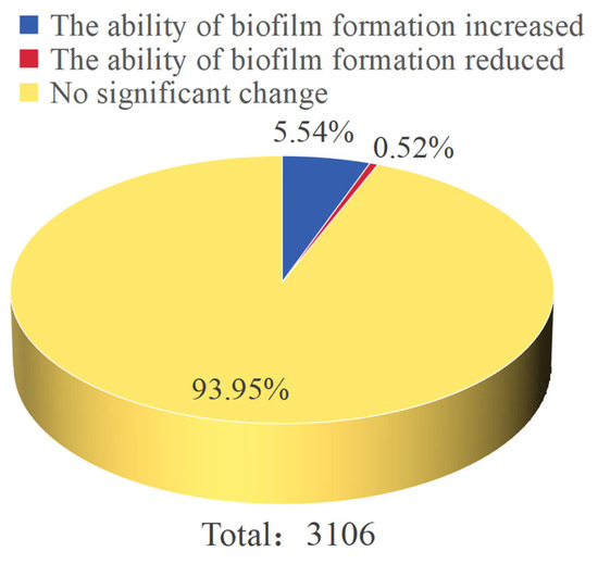
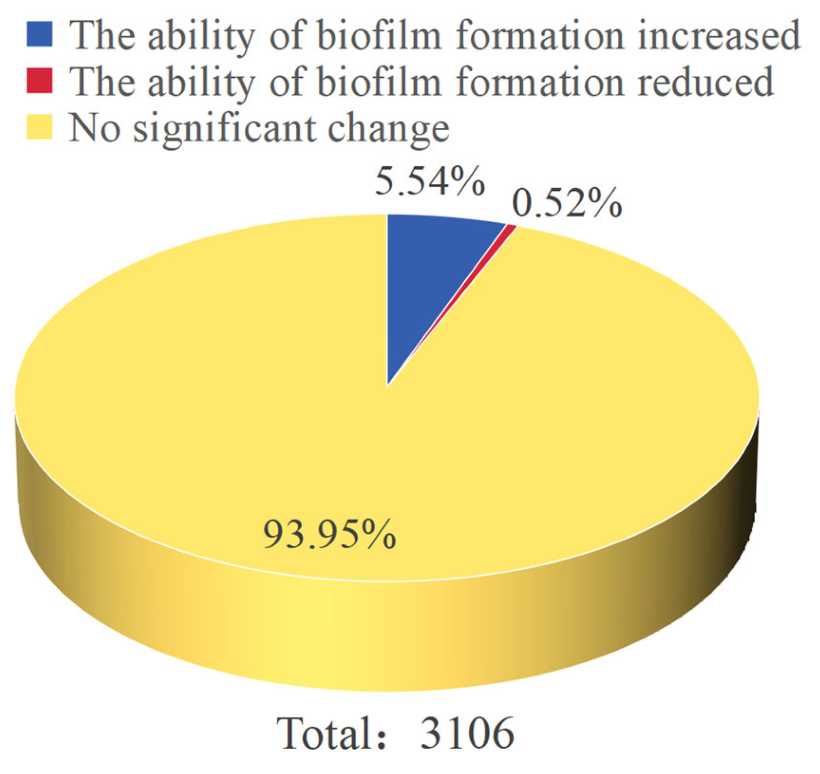
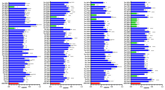
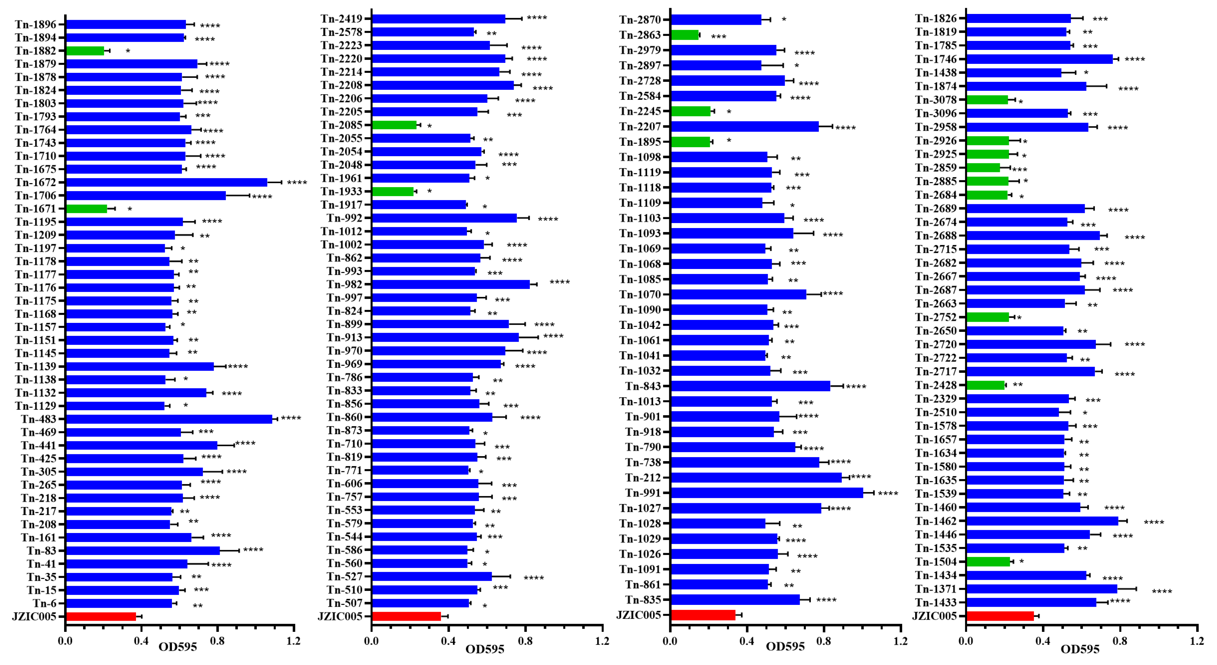
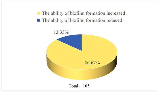
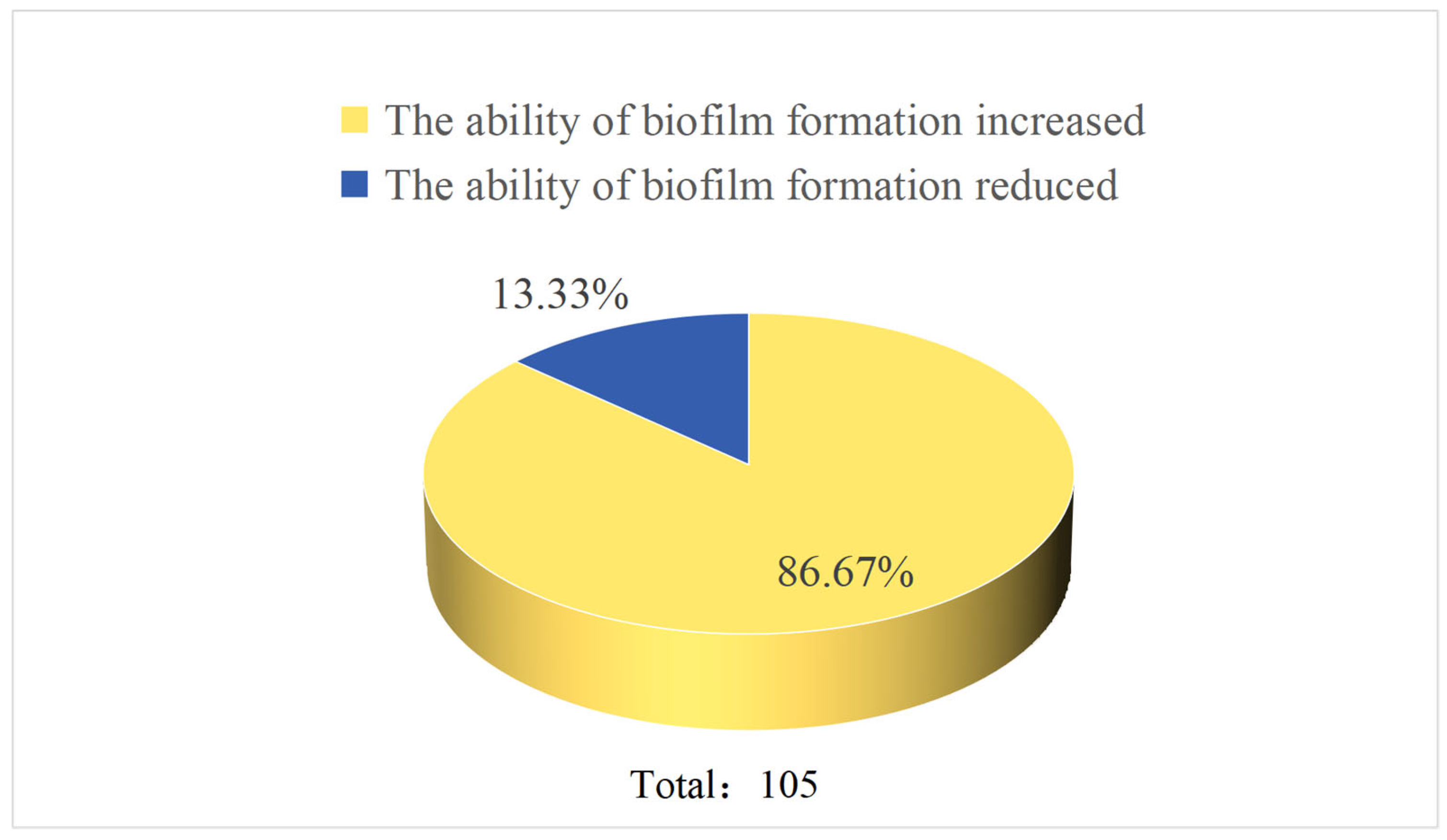
Biofilm formation capacity was evaluated for all 3106 transposon mutant strains via crystal violet staining. The results indicated that 2918 mutants (93.94%) displayed no significant difference in biofilm formation compared to the wild-type strain. In contrast, 172 mutants (5.54%) showed significantly enhanced biofilm formation, while 16 mutants (0.52%) exhibited significantly reduced biofilm formation (Figure 1). Further analysis revealed that the biofilm phenotypes varied in degree among the different mutant strains. Partial statistical results of the OD595 measurements are summarized in Figure 2. Mutants with significantly reduced biofilm formation included Tn-1504, Tn-2752, Tn-2684, etc. (p < 0.05); Tn-2428 (p < 0.01); Tn-2859 (p < 0.001). Mutant strains with significantly increased biofilm formation included Tn-1438, Tn-2510 (p < 0.05); Tn-1535, Tn-1539, Tn-1635, etc. (p < 0.01); Tn-1578, Tn-2329, Tn-2715, etc. (p < 0.001); and Tn-1371, Tn-1433, Tn-1462, etc. (p < 0.0001).
Figure 1.
Proportion of mutants with increased or decreased biofilm formation.
Figure 2.
Analysis of biofilm formation among 3106 transposon mutants. Following random transposon mutagenesis, 188 mutant strains exhibited significantly altered biofilm-forming capacity. Biofilm biomass was quantified by measuring the absorbance at 595 nm (OD595) of ethanol-solubilized crystal violet. Data are presented as mean ± SD, with each assay representing an independent biofilm formation experiment. Statistical significance compared to the control group was determined by one-way ANOVA with Dunnett’s post hoc test (* p < 0.05, ** p < 0.01, *** p < 0.001, **** p < 0.0001 relative to the wild-type control). Quantitative results for selected mutants are shown.
3.3. Identification of Transposon Insertion Sites in Mutant Strains with Altered Biofilm Phenotypes
Given the random nature of transposon insertion, multiple insertions can occur within the same gene. After mapping the insertion sites and removing duplicates, 105 non-redundant genes were identified from 188 mutant strains that showed significant alterations in biofilm formation (Table 3). Of these, 19 genes were annotated as encoding putative or hypothetical proteins. Fourteen unique genes were linked to reduced biofilm formation, whereas 91 were associated with enhanced biofilm formation (Figure 3). The difference between the number of mutant strains (188) and the number of unique genes identified (105) indicates that certain genes were independently mutated in multiple strains.
Table 3.
Characteristics of 188 Av. paragallinarum transposon mutants with significantly altered biofilm-forming capacity used in this study.
Figure 3.
Proportion of genes with increased or decreased biofilm formation.
3.4. Bioinformatics Analysis of Proteins Encoded by Inserted Genes
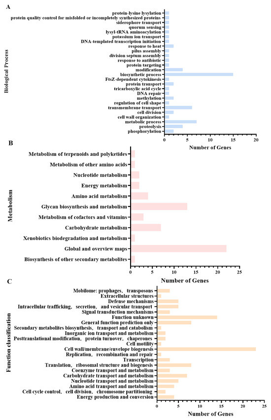
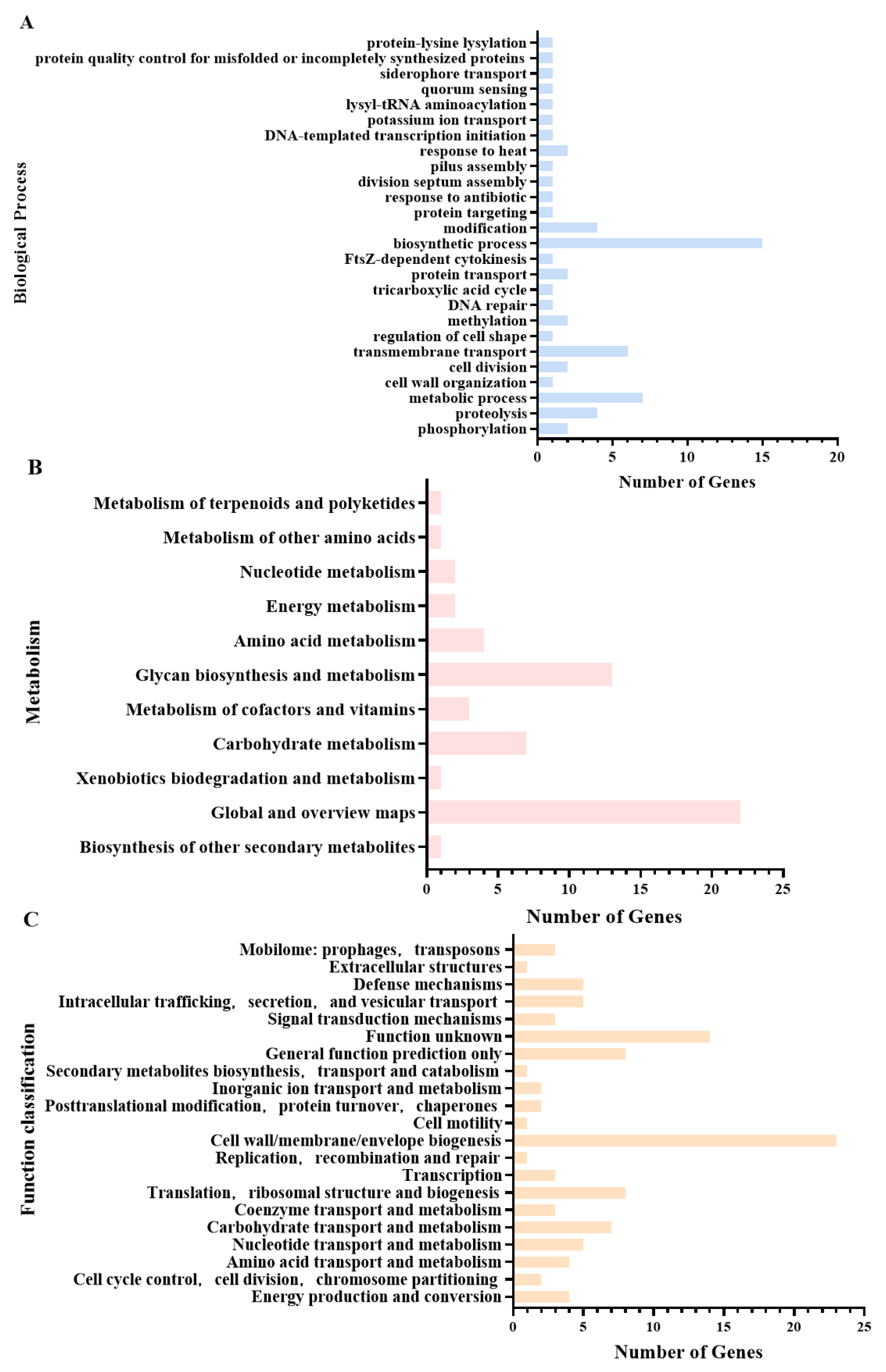
Gene Ontology (GO) enrichment analysis revealed that the proteins encoded by the genes interrupted by transposon insertions were primarily involved in biosynthesis and metabolism (22 genes), transmembrane transport (6 genes), proteolysis and transport (4 genes), phosphorylation or methylation (4 genes), cell division (2 genes), and ciliary assembly (1 gene) (Figure 4A). Kyoto Encyclopedia of Genes and Genomes (KEGG) pathway analysis further indicated that these genes predominantly participate in sugar biosynthesis and metabolism (13 genes), carbohydrate metabolism (7 genes), and amino acid, cofactor, and vitamin metabolism (7 genes), among other metabolic pathways (Figure 4B). Clusters of Orthologous Groups (COG) functional classification demonstrated that the most abundant categories among these protein-coding genes were cell wall/membrane/envelope biogenesis (23 genes); translation, ribosomal structure, and biogenesis (8 genes); carbohydrate transport and metabolism (7 genes); and intracellular trafficking, secretion, and vesicular transport (5 genes), in addition to other functional classes (Figure 4C). Based on the integration of quantitative biofilm screening data and functional annotation results, a systematic screening of 188 biofilm-associated mutant strains was conducted in this study. The selection criteria were as follows: First, to ensure the reliability of the genetic background, all candidate mutant strains were verified to produce a single band by TAIL-PCR and confirmed to exhibit stable phenotypes through three rounds of independent screening. On this basis, to comprehensively cover biofilm-associated phenotypes and functions, both biofilm-enhanced and biofilm-deficient strains were selected, with priority given to mutants carrying insertions in genes known to be involved in biofilm formation. Additionally, to explore potential novel regulatory factors, two mutants carrying insertions in genes encoding hypothetical proteins were specifically selected. Ultimately, a total of eight representative mutant strains—Tn-441, Tn-483, Tn-1216, Tn-1504, Tn-1706, Tn-2206, Tn-2428, and Tn-2859—were selected for subsequent studies. Detailed information on these mutants is provided in Table 4.
Figure 4.
Gene Ontology (GO) enrichment analysis, Kyoto Encyclopedia of Genes and Genomes (KEGG) pathway enrichment analysis, and Clusters of Orthologous Groups (COG) classification were performed on the 105 genes harboring transposon insertions. Bar lengths indicate the number of genes assigned to each respective term, pathway, or category. (A) GO enrichment analysis of the 105 genes identified with insertion mutations; (B) KEGG pathway enrichment analysis of the 105 genes identified with insertion mutations; (C) COG functional classification of the 105 genes identified with insertion mutations.
Table 4.
Characteristics of eight key biofilm mutants.
3.5. Growth Curves of Biofilm-Related Mutant Strains
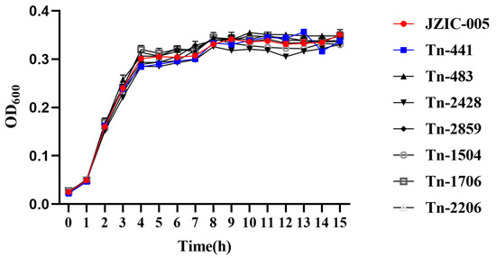
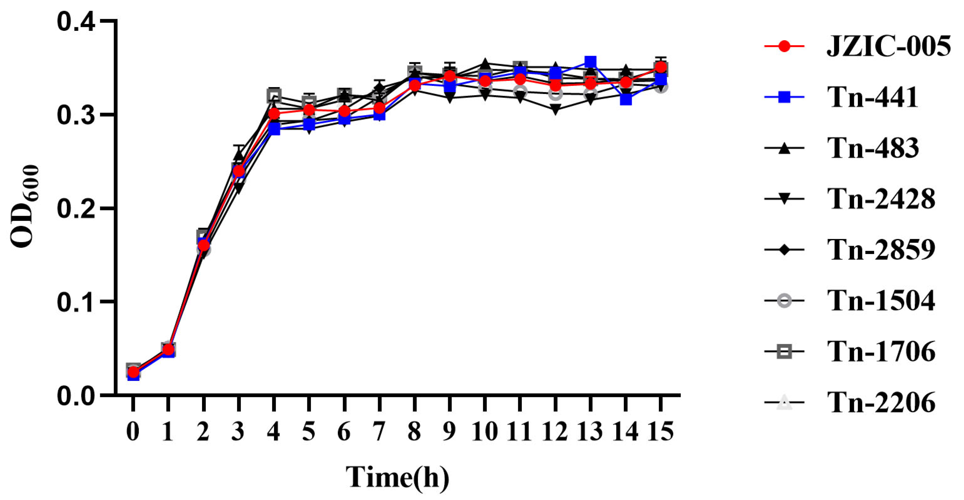
Growth curves were measured for the wild-type strain JZIC-005 and the transposon insertion mutant strains Tn-441, Tn-483, Tn-1216, Tn-1504, Tn-1706, Tn-2206, Tn-2428, and Tn-2859. Comparative analysis revealed that all mutant strains exhibited growth kinetics highly similar to those of the wild-type strain (Figure 5), indicating that the observed alterations in biofilm-forming capacity are not attributable to differences in growth ability.
Figure 5.
Analysis of bacterial growth curves indicated that the wild-type strain and the mutant strains exhibited highly similar growth kinetics. This figure shows the growth curves of the wild-type strain and its mutants cultured with shaking at 37 °C in TSB medium. The OD600 was measured at 1-h intervals. All data are presented as mean ± SD (n = 3).
3.6. Adhesion and Invasion Capabilities of Biofilm-Related Mutants on DF-1 Cells
The adhesion and invasion capabilities of the wild-type strain JZIC-005 and the eight biofilm-related mutant strains were evaluated using DF-1 cells.
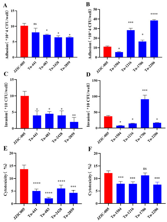
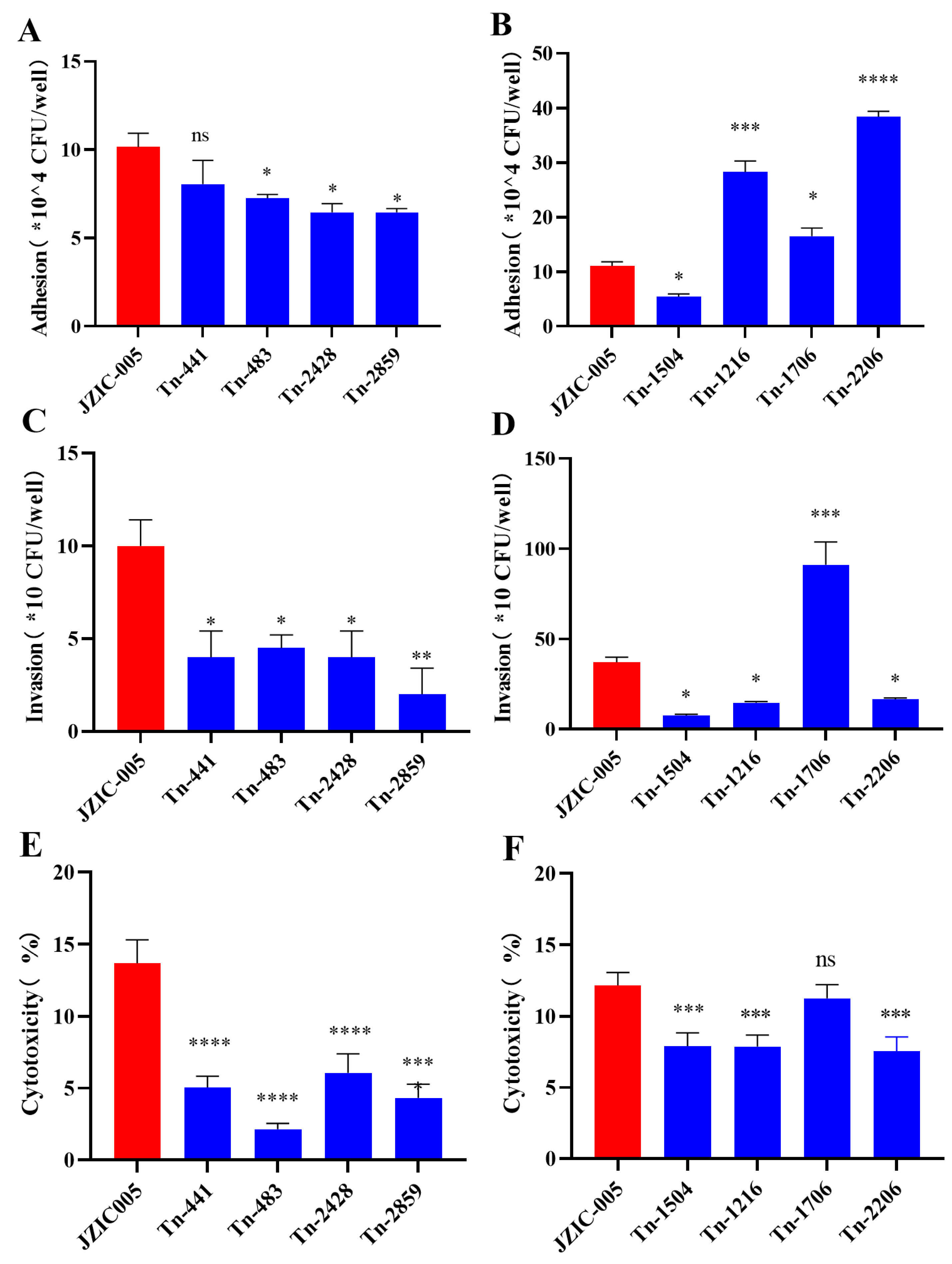
Adhesion assays revealed significant differences compared to the wild type. The adhesion capacity of mutants Tn-483, Tn-1504, Tn-2428, and Tn-2859 was significantly decreased (p < 0.05). Mutant Tn-1504 exhibited the most pronounced reduction, with a 50.68% lower adhesion rate, followed by Tn-2428 and Tn-2859. In contrast, mutants Tn-1216 (p < 0.001), Tn-1706 (p < 0.05), and Tn-2206 (p < 0.0001) showed significantly enhanced adhesion. Notably, the adhesion rate of Tn-2206 increased by 247.51%. No significant difference (p > 0.05) was observed for mutant Tn-441 (Figure 6A,B). These findings indicate that transposon insertion into genes OW731_03300, OW731_11000, and lsrk enhances bacterial adhesion to host cells, whereas insertion into OW731_01055, OW731_10585, OW731_03180, and OW731_09840 impairs this ability.
Figure 6.
Adhesion, invasion, and cytotoxicity capabilities of mutant strains Tn-483, Tn-2428, and Tn-2859. Compared to the wild-type strain, the adhesive and invasive capabilities, as well as the cytotoxicity, of the three mutant strains were significantly reduced. (A,B) Bacterial adhesion assay. DF-1 cells were infected with the wild-type and mutant strains at a multiplicity of infection (MOI) of 100 for 2 h, and adherent bacteria were counted. Data are presented as the relative number of adhered bacteria compared to the wild-type. All data are presented as mean ± SD (n = 2). (C,D) Bacterial invasion assay. DF-1 cells were infected with the wild-type and mutant strains at an MOI of 100 for 2 h. Extracellular bacteria were killed by gentamicin treatment to quantify the number of invading bacteria. Data are presented as the relative number of invading bacteria compared to the wild-type. All data are presented as mean ± SD (n = 2). (E,F) Cytotoxicity assay. DF-1 cells were infected with the wild-type and mutant strains at an MOI of 1000 for 4 h. Cytotoxicity was measured using a lactate dehydrogenase (LDH) release assay and expressed as a percentage relative to the maximum LDH release control. All data are presented as mean ± SD (n = 3). Statistical significance compared to the wild-type strain was determined by one-way ANOVA followed by Dunnett’s multiple comparison test. ns, not significant; * p < 0.05, ** p < 0.01, *** p < 0.001, and **** p < 0.0001.
Invasion assays revealed that several mutants exhibited significant reductions in the number of bacteria invading DF-1 cells compared to the wild type. These included Tn-441, Tn-483, Tn-1504, Tn-1216, Tn-2206, and Tn-2428 (p < 0.05), as well as Tn-2859 (p < 0.01). The most pronounced defect was observed in mutant Tn-2859, which showed an 80% decrease in invasion, followed by Tn-1504 and Tn-2206. In contrast, mutant Tn-1706 (p < 0.001) demonstrated a significantly enhanced capacity to infect DF-1 cells (Figure 6C, D). Collectively, these results suggest that the genes OW731_05445, OW731_01055, OW731_03300, OW731_10585, lsrk, OW731_03180, and OW731_09840 promote cellular invasion by Av. paragallinarum, while the OW731_11000 gene appears to attenuate this process.
3.7. Cytotoxicity Assay of Biofilm Defect Mutant Strains Against DF-1 Cells
The cytotoxicity of the biofilm mutants was evaluated using a lactate dehydrogenase (LDH) release assay. Compared to the wild-type strain, mutants Tn-441, Tn-483, Tn-1216, Tn-1504, Tn-2206, Tn-2428, and Tn-2859 exhibited significantly reduced toxicity toward DF-1 cells. The most pronounced decrease was observed for Tn-2859, which showed 66.38% lower toxicity relative to the wild type, followed by Tn-441 and Tn-2428. In contrast, no significant difference in cytotoxicity was detected for mutant Tn-1706 (p > 0.05) (Figure 6E,F). These results indicate that transposon insertion into the genes OW731_05445, OW731_01055, OW731_03300, OW731_10585, lsrk, OW731_03180, and OW731_09840 reduces the virulence of Av. paragallinarum.
4. Discussion
In this study, we performed a systematic, genome-wide screen for genes involved in biofilm formation in Av. paragallinarum. By integrating transposon mutagenesis with phenotypic screening, we identified a complex genetic network underlying this critical virulence-associated trait and constructed a comprehensive genetic landscape encompassing both positive and negative regulators of biofilm formation. Using mini-Tn5 transposon mutagenesis, we successfully generated a mutant library comprising 3106 individual mutants, which theoretically covers approximately 1.2-fold of the protein-coding sequences in the reference genome CP113955.1 (3106/2592), thereby achieving near-saturation mutagenesis. Initial screening via crystal violet staining identified 188 mutants exhibiting significantly altered biofilm-forming capacity relative to the wild-type strain, including 172 mutants with enhanced and 16 with reduced biofilm formation. Subsequent TAIL-PCR coupled with sequencing analysis successfully identified 105 non-redundant genes disrupted by transposon insertions. The combination of large-scale saturating mutagenesis with systematic phenotype–genotype association analysis strongly supports the comprehensiveness and reliability of the findings presented in this study.
In this study, mutants Tn-483, Tn-2428, and Tn-2859 exhibited reduced biofilm formation, along with diminished adhesion to and invasion of DF-1 cells, as well as attenuated cytotoxicity. Among these, mutant strain Tn-2428, which carries a transposon insertion in the gluconate permease (GntP)-encoding gene (OW731_03180), displayed comprehensive attenuation of the aforementioned virulence-related phenotypes, suggesting that disruption of gluconate metabolism resulting from gntP inactivation may underlie this pleiotropic defect. In E. coli, GntP is involved in glucuronate transport and is regulated by UxuR; deletion of uxuR impairs biofilm formation [49,50,51]. Moreover, gntP deletion in E. coli reduces intestinal colonization capacity [52]. Based on these findings, we hypothesize that disruption of this sugar transporter in Av. paragallinarum may alter metabolic flux, which may in turn interfere with biofilm development and virulence expression through effects on energy homeostasis, precursor availability, or signaling molecules. Future metabolomic studies to quantify intracellular gluconate levels could test this hypothesis.
The autoinducer-2 (AI-2) quorum-sensing (QS) system is a crucial intercellular communication mechanism that regulates collective bacterial behaviors, including biofilm formation and virulence expression [38,53,54,55]. Within this pathway, the LsrK kinase plays a pivotal role by phosphorylating internalized AI-2, thereby activating downstream signaling cascades that coordinate pathogenicity traits [56,57,58]. In the present study, the lsrK transposon mutant Tn-2206 exhibited a distinctive phenotypic profile, characterized by markedly enhanced biofilm formation and host cell adhesion, yet concurrently reduced cellular invasion and cytotoxicity. This decoupling of phenotypes—in which colonization potential is increased while virulence capacity is diminished—mirrors observations in Klebsiella pneumoniae, where deletion of the lsrCD gene downregulates lsrK expression and yields analogous outcomes [59]. The mechanistic basis for this phenomenon may stem from a dual effect: the primary disruption of the LsrK-mediated QS cascade likely attenuates the expression of acute virulence factors required for invasion and cytotoxicity, while concurrently dysregulating feedback loops within the QS network, leading to compensatory overexpression of genes that promote biofilm maturation and adhesion [57,58]. This dissociation between biofilm-mediated persistence and invasive damage aligns closely with the principles of the “anti-virulence” strategy, which seeks to disarm pathogens rather than eliminate them. Collectively, our findings suggest that LsrK may represent a promising target for the development of quorum-sensing inhibitors against Av. paragallinarum, although further functional validation is warranted.
The TonB protein functions as an energy-transducing component in the iron acquisition systems of Gram-negative bacteria and plays an essential role in the transmembrane transport of iron complexes [60,61]. Iron availability serves as an environmental signal that influences biofilm architecture; sufficient iron can activate the expression of biofilm-associated genes, whereas iron limitation may suppress the production of extracellular polymeric substances, resulting in structurally fragile biofilms [62,63]. Consistent with this, TonB-deficient mutants in other pathogens, such as Riemerella anatipestifer and E. coli, exhibit impaired biofilm formation and reduced colonization capacity [64,65]. In the present study, the Tn-1504 mutant, which carries a transposon insertion in the tonB gene (OW731_10585), displayed similar pleiotropic phenotypic alterations, including reduced biofilm formation, diminished adhesion to and invasion of host cells, and attenuated cytotoxicity. These observations suggest that in Av. paragallinarum, the TonB system may not only participate in iron acquisition but also influence biofilm matrix synthesis and the expression of additional virulence determinants, potentially through iron-dependent regulatory pathways.
Interestingly, mutations affecting surface structures such as fimbriae can lead to a dissociation between biofilm formation and acute virulence. The TonB system itself has been shown to influence the production of adhesins, including type IV pili, in other organisms [66]. For instance, disruption of genes involved in type IV pilus biogenesis—such as apfA in Actinobacillus pleuropneumoniae or pilU in Pseudomonas aeruginosa—has been reported to enhance biofilm formation while simultaneously diminishing invasive virulence [67,68]. In the present study, the Tn-1216 mutant, which carries a transposon insertion in a gene (OW731_03300) implicated in fimbrial biogenesis in Av. paragallinarum, phenocopied this dissociation: biofilm formation and adhesion were enhanced, whereas invasion and cytotoxicity were reduced. These observations suggest that surface appendages such as fimbriae in Av. paragallinarum may possess distinct, separable functions in surface colonization versus host cell invasion and damage delivery.
In addition to metal acquisition and surface structures, another metabolically relevant mutant, Tn-1706, which carries an insertion in a gene encoding a GDP-L-fucosyltransferase (OW731_11000), exhibited enhanced biofilm formation, adhesion, and invasion, whereas cytotoxicity remained unchanged. This phenotypic profile contrasts with previous reports in which fucose availability suppressed biofilm formation in Campylobacter jejuni and mutation of a homologous fucosyltransferase attenuated virulence in Pectobacterium carotovorum [69,70]. Such divergence may suggest a distinct role for fucose metabolism or fucosylated substrates in regulating biofilm formation in Av. paragallinarum, or alternatively, may reflect polar effects on the expression of downstream genes within the same operon.
We also characterized mutants carrying disruptions in genes encoding hypothetical proteins. Tn-2859 (OW731_09840) exhibited a coordinated reduction in biofilm formation, adhesion, invasion, and cytotoxicity. In contrast, Tn-441 (OW731_05445) and Tn-483 (OW731_01055) displayed a decoupled phenotype: while virulence-associated traits (invasion and cytotoxicity) were attenuated, biofilm formation was enhanced. These distinct phenotypic profiles suggest that these uncharacterized proteins may be involved in regulating the balance between a biofilm-associated lifestyle and an invasive state in Av. paragallinarum; however, their specific roles warrant further investigation.
In summary, this study has preliminarily identified a set of genes potentially involved in the regulation of biofilm formation in Av. paragallinarum. We have constructed a preliminary and more comprehensive phenotypic landscape linking specific genetic disruptions—affecting nutrient acquisition (TonB), surface structure biogenesis (fimbriae), and metabolism-related genes (GntP, fucosyltransferase)—to alterations in biofilm development and virulence expression. Notably, three mutant strains, Tn-1504, Tn-2428, and Tn-2859, exhibited attenuated virulence in vitro in the absence of apparent growth defects, suggesting their potential as live attenuated vaccine candidates. It should be emphasized that the transposon insertion mutagenesis employed in this study may exert polar effects on downstream genes; therefore, future construction of in-frame deletion and complementation strains is warranted to confirm the specific functions of each gene. Future research directions emerging from this study include evaluating the in vivo protective efficacy of these candidate strains, as well as exploring the therapeutic potential of newly identified targets—such as the LsrK quorum-sensing kinase and the GntP permease—in the development of anti-biofilm or anti-virulence strategies against infectious coryza.
Author Contributions
W.J. and X.H. designed this study. B.F., Q.S. and Y.S. conducted the experiments and interpreted the data. B.F., Q.S. and W.J. drafted the initial manuscript. W.S. and J.Z. reviewed the manuscript and provided important suggestions. W.J. and Q.S. revised and proofread the manuscript. All authors have read and agreed to the published version of the manuscript.
Funding
This study was funded by the National Key Research and Development Programs of China (Grant No. 2023YFD1800602-2) and Fujian Foreign Cooperation Project (Grant No. 2025I0030-1).
Institutional Review Board Statement
Not applicable.
Informed Consent Statement
Not applicable.
Data Availability Statement
The data presented in this study are available in a public repository (DOI: 10.57760/sciencedb.30806).
Acknowledgments
We gratefully acknowledge the financial support provided by the National Key Research and Development Programs of China (Grant No. 2023YFD1800602-2) and Fujian Foreign Cooperation Project (Grant No. 2025I0030-1). Furthermore, we sincerely express our profound gratitude to the laboratory instructors and fellow students for their invaluable support of this research.
Conflicts of Interest
The authors declare no conflicts of interest.
References
- Chen, L.; Sun, J.; Hu, J.; Tian, Y.; Du, P.; Guo, Q.; Yang, C.; Zhang, Q.; Feng, S.; Liao, M. Identification and characterization of biosynthetic loci of lipooligosaccharide and capsular polysaccharide in Avibacterium paragallinarum. Vet. Microbiol. 2024, 299, 110317. [Google Scholar] [CrossRef] [Scilit]
- Blackall, P.J.; Soriano-Vargas, E. Infectious Coryza and Related Bacterial Infections. In Diseases of Poultry; Wiley: Hoboken, NY, USA, 2020; pp. 890–906. [Google Scholar] [CrossRef] [Scilit]
- Sakamoto, R.; Baba, S.; Ushijima, T.; Kino, Y.; Honda, T.; Mizokami, H.; Sakaguchi, M. Development of a recombinant vaccine against infectious coryza in chickens. Res. Vet. Sci. 2013, 94, 504–509. [Google Scholar] [CrossRef] [Scilit] [PubMed]
- De Blieck, L. The treatment of coryza infectiosa gallinarum type II (Nelson) with streptomycin. Vet. Rec. 1950, 62, 787. [Google Scholar] [CrossRef] [Scilit]
- Wang, Y.; Si, M.; Qiu, J.; Chong, X.; Yang, B.; Chu, M.; Liang, Y.; Cheng, W.; Zhang, H.; Chen, X.; et al. Prevalence, risk factors, and regional insights of infectious coryza among poultry populations in China during 1993–2024: A meta-analysis. J. Vet. Med. Sci. 2025, 88, 201–209. [Google Scholar] [CrossRef] [Scilit]
- Page, L.A. Haemophilus infections in chickens. I. Characteristics of 12 Haemophilus isolates recovered from diseased chickens. Am. J. Vet. Res. 1962, 23, 85–95. [Google Scholar]
- Tan, D.H.; Ou, S.C.; Shien, J.H.; Huang, S.W.; Hsieh, M.K.; Chang, P.C. Serotypes and Hemagglutinin Gene Sequences of Avibacterium paragallinarum Isolated in Taiwan. Avian Dis. 2020, 64, 197–202. [Google Scholar] [CrossRef] [Scilit]
- Soriano, V.E.; Téllez, G.; Hargis, B.M.; Newberry, L.; Salgado-Miranda, C.; Vázquez, J.C. Typing of Haemophilus paragallinarum strains by using enterobacterial repetitive intergenic consensus-based polymerase chain reaction. Avian Dis. 2004, 48, 890–895. [Google Scholar] [CrossRef] [Scilit]
- Crispo, M.; Blackall, P.; Khan, A.; Shivaprasad, H.L.; Clothier, K.; Sentíes-Cué, C.G.; Cooper, G.; Blakey, J.; Pitesky, M.; Mountainspring, G.; et al. Characterization of an Outbreak of Infectious Coryza (Avibacterium paragallinarum) in Commercial Chickens in Central California. Avian Dis. 2019, 63, 486–494. [Google Scholar] [CrossRef] [Scilit] [PubMed]
- Luna-Castrejón, L.P.; Buter, R.; Pantoja-Nuñez, G.I.; Acuña-Yanes, M.; Ceballos-Valenzuela, K.; Talavera-Rojas, M.; Salgado-Miranda, C.; Heuvelink, A.; de Wit, S.; Soriano-Vargas, E.; et al. Identification, HPG2 Sequence Analysis, and Antimicrobial Susceptibility of Avibacterium paragallinarum Isolates Obtained from Outbreaks of Infectious Coryza in Commercial Layers in Sonora State, Mexico. Avian Dis. 2021, 65, 95–101. [Google Scholar] [CrossRef] [Scilit]
- El-Gazzar, M.; Gallardo, R.; Bragg, R.; Hashish, A.; Sun, H.L.; Davison, S.; Feberwee, A.; Huberman, Y.; Skein, T.; Coertzen, A.; et al. Avibacterium paragallinarum, the Causative Agent of Infectious Coryza: A Comprehensive Review. Avian Dis. 2025, 68, 362–379. [Google Scholar] [CrossRef] [Scilit] [PubMed]
- Shelkamy, M.M.S.; Hashish, A.; Srednik, M.E.; Chaves, M.; Macedo, N.R.; Gadu, E.; Schmitz-Esser, S.; Zhang, Q.; Wang, C.; Sato, Y.; et al. Prevalence of Nonpathogenic Avibacterium paragallinarum in Naïve-Healthy Layer Flocks Across Multiple States in the United States. Transbound. Emerg. Dis. 2025, 2025, 9994679. [Google Scholar] [CrossRef] [Scilit]
- Deresse, G.; Assefa, E.; Tesfaw, L.; Dufera, D.; Adamu, K.; Akalu, M.; Bayissa, B.; Abayneh, T.; Zewdie, G.; Abebe, A.; et al. Isolation, molecular detection, and sequence analysis of Avibacterium paragallinarum from suspected cases of infectious coryza infected chickens from different areas of Ethiopia, 2022–2024. BMC Microbiol. 2025, 25, 141. [Google Scholar] [CrossRef] [Scilit]
- Hsu, Y.M.; Shieh, H.K.; Chen, W.H.; Shiang, J.H.; Chang, P.C. Immunogenicity and haemagglutination of recombinant Avibacterium paragallinarum HagA. Vet. Microbiol. 2007, 122, 280–289. [Google Scholar] [CrossRef] [Scilit]
- Takagi, M.; Hirayama, N.; Simazaki, T.; Taguchi, K.; Yamaoka, R.; Ohta, S. Purification of hemagglutinin from Haemophilus paragallinarum using monoclonal antibody. Vet. Microbiol. 1993, 34, 191–197. [Google Scholar] [CrossRef] [Scilit]
- Frey, J. RTX Toxins of Animal Pathogens and Their Role as Antigens in Vaccines and Diagnostics. Toxins 2019, 11, 719. [Google Scholar] [CrossRef] [Scilit]
- Guo, M.; Liu, D.; Xu, H.; Zhang, H.; Jin, Y.; Tan, H.; Wu, Y.; Zhang, X. The Protective Efficacy of an Inactivated Vaccine against Avibacterium paragallinarum Field Isolates. Vet. Sci. 2022, 9, 458. [Google Scholar] [CrossRef] [Scilit] [PubMed]
- Gütgemann, F.; Heuvelink, A.; Müller, A.; Churin, Y.; Buter, R.; Jung, A.; Feberwee, A.; Wiegel, J.; Kumm, F.; Braun, A.S.; et al. Recommendation of a standardized broth microdilution method for antimicrobial susceptibility testing of Avibacterium paragallinarum and resistance monitoring. J. Clin. Microbiol. 2024, 62, e0101123. [Google Scholar] [CrossRef] [Scilit] [PubMed]
- Uruén, C.; Chopo-Escuin, G.; Tommassen, J.; Mainar-Jaime, R.C.; Arenas, J. Biofilms as Promoters of Bacterial Antibiotic Resistance and Tolerance. Antibiotics 2020, 10, 3. [Google Scholar] [CrossRef] [Scilit] [PubMed]
- Flemming, H.C.; Wingender, J. The biofilm matrix. Nat. Rev. Microbiol. 2010, 8, 623–633. [Google Scholar] [CrossRef] [Scilit]
- Karygianni, L.; Ren, Z.; Koo, H.; Thurnheer, T. Biofilm Matrixome: Extracellular Components in Structured Microbial Communities. Trends Microbiol. 2020, 28, 668–681. [Google Scholar] [CrossRef] [Scilit]
- Hindieh, P.; Yaghi, J.; Assaf, J.C.; Chokr, A.; Atoui, A.; Tzenios, N.; Louka, N.; Khoury, A.E. Emerging Multimodal Strategies for Bacterial Biofilm Eradication: A Comprehensive Review. Microorganisms 2025, 13, 2796. [Google Scholar] [CrossRef] [Scilit] [PubMed]
- Zhao, A.; Sun, J.; Liu, Y. Understanding bacterial biofilms: From definition to treatment strategies. Front. Cell. Infect. Microbiol. 2023, 13, 1137947. [Google Scholar] [CrossRef] [Scilit]
- Aparna, M.S.; Yadav, S. Biofilms: Microbes and disease. Braz. J. Infect. Dis. Off. Publ. Braz. Soc. Infect. Dis. 2008, 12, 526–530. [Google Scholar] [CrossRef] [Scilit]
- Hall-Stoodley, L.; Stoodley, P. Evolving concepts in biofilm infections. Cell. Microbiol. 2009, 11, 1034–1043. [Google Scholar] [CrossRef] [Scilit]
- Hathroubi, S.; Loera-Muro, A.; Guerrero-Barrera, A.L.; Tremblay, Y.D.N.; Jacques, M. Actinobacillus pleuropneumoniae biofilms: Role in pathogenicity and potential impact for vaccination development. Anim. Health Res. Rev. 2018, 19, 17–30. [Google Scholar] [CrossRef] [Scilit]
- Dramé, I.; Rossez, Y.; Krzewinski, F.; Charbonnel, N.; Ollivier-Nakusi, L.; Briandet, R.; Dague, E.; Forestier, C.; Balestrino, D. FabR, a regulator of membrane lipid homeostasis, is involved in Klebsiella pneumoniae biofilm robustness. mBio 2024, 15, e0131724. [Google Scholar] [CrossRef] [Scilit] [PubMed]
- Wang, H.; Fan, Q.; Gao, S.; Yi, L.; Wang, Y.; Wang, Y. Transposon library screening to identify genes with a potential role in Streptococcus suis biofilm formation. Future Microbiol. 2024, 19, 107–115. [Google Scholar] [CrossRef] [Scilit]
- Høiby, N.; Bjarnsholt, T.; Givskov, M.; Molin, S.; Ciofu, O. Antibiotic resistance of bacterial biofilms. Int. J. Antimicrob. Agents 2010, 35, 322–332. [Google Scholar] [CrossRef] [Scilit]
- Cepas, V.; López, Y.; Muñoz, E.; Rolo, D.; Ardanuy, C.; Martí, S.; Xercavins, M.; Horcajada, J.P.; Bosch, J.; Soto, S.M. Relationship Between Biofilm Formation and Antimicrobial Resistance in Gram-Negative Bacteria. Microb. Drug Resist. 2019, 25, 72–79. [Google Scholar] [CrossRef] [Scilit]
- Herant, M.; Heinrich, V.; Dembo, M. Mechanics of neutrophil phagocytosis: Experiments and quantitative models. J. Cell Sci. 2006, 119, 1903–1913. [Google Scholar] [CrossRef] [Scilit] [PubMed]
- Alhede, M.; Lorenz, M.; Fritz, B.G.; Jensen, P.; Ring, H.C.; Bay, L.; Bjarnsholt, T. Bacterial aggregate size determines phagocytosis efficiency of polymorphonuclear leukocytes. Med. Microbiol. Immunol. 2020, 209, 669–680. [Google Scholar] [CrossRef] [Scilit]
- Yuan, S.; Liu, B.; Quan, Y.; Gao, S.; Zuo, J.; Jin, W.; Shen, Y.; Li, Y.; Wang, Y.; Wang, Y. Streptococcus suis regulates central carbon fluxes in response to environment to balance drug resistance and virulence. Microbiol. Res. 2025, 296, 128157. [Google Scholar] [CrossRef] [Scilit]
- Chou, K.T.; Lee, D.D.; Chiou, J.G.; Galera-Laporta, L.; Ly, S.; Garcia-Ojalvo, J.; Süel, G.M. A segmentation clock patterns cellular differentiation in a bacterial biofilm. Cell 2022, 185, 145–157.e113. [Google Scholar] [CrossRef] [Scilit] [PubMed]
- Yuan, S.; Shen, Y.; Quan, Y.; Gao, S.; Zuo, J.; Jin, W.; Li, R.; Yi, L.; Wang, Y.; Wang, Y. Molecular mechanism and application of emerging technologies in study of bacterial persisters. BMC Microbiol. 2024, 24, 480. [Google Scholar] [CrossRef] [Scilit] [PubMed]
- Flemming, H.C.; Wingender, J.; Szewzyk, U.; Steinberg, P.; Rice, S.A.; Kjelleberg, S. Biofilms: An emergent form of bacterial life. Nat. Rev. Microbiol. 2016, 14, 563–575. [Google Scholar] [CrossRef] [Scilit]
- Ruhal, R.; Kataria, R. Biofilm patterns in gram-positive and gram-negative bacteria. Microbiol. Res. 2021, 251, 126829. [Google Scholar] [CrossRef] [Scilit] [PubMed]
- Zhang, M.; Xue, X.; Li, X.; Wu, Q.; Zhang, T.; Yang, W.; Hu, L.; Zhou, D.; Lu, R.; Zhang, Y. QsvR and OpaR coordinately repress biofilm formation by Vibrio parahaemolyticus. Front. Microbiol. 2023, 14, 1079653. [Google Scholar] [CrossRef] [Scilit]
- van Hoogstraten, S.W.G.; Kuik, C.; Arts, J.J.C.; Cillero-Pastor, B. Molecular imaging of bacterial biofilms-a systematic review. Crit. Rev. Microbiol. 2024, 50, 971–992. [Google Scholar] [CrossRef] [Scilit] [PubMed]
- Wang, Y.P.; Hsieh, M.K.; Tan, D.H.; Shien, J.H.; Ou, S.C.; Chen, C.F.; Chang, P.C. The haemagglutinin of Avibacterium paragallinarum is a trimeric autotransporter adhesin that confers haemagglutination, cell adherence and biofilm formation activities. Vet. Microbiol. 2014, 174, 474–482. [Google Scholar] [CrossRef] [Scilit]
- Guo, M.; Wang, H.; Liu, D.; Bo, Z.; Zhang, C.; Wu, Y.; Zhang, X. Development and evaluation of an attenuated Avibacterium paragallinarum strain as a live vaccine candidate for infectious coryza. Vet. Res. 2025, 56, 115. [Google Scholar] [CrossRef] [Scilit]
- Guo, M.; Wang, H.; Zhang, H.; Bo, Z.; Zhang, C.; Zhang, X.; Wu, Y. Identifcation of the genes involved in biofilm formation of Avibacterium paragallinarum using random transposon mutagenesis. Vet. Microbiol. 2025, 302, 110410. [Google Scholar] [CrossRef] [Scilit]
- Schmitz, M.; Querques, I. DNA on the move: Mechanisms, functions and applications of transposable elements. FEBS Open Bio 2024, 14, 13–22. [Google Scholar] [CrossRef] [Scilit]
- Shapiro, J.A. Molecular model for the transposition and replication of bacteriophage Mu and other transposable elements. Proc. Natl. Acad. Sci. USA 1979, 76, 1933–1937. [Google Scholar] [CrossRef] [Scilit]
- Cain, A.K.; Barquist, L.; Goodman, A.L.; Paulsen, I.T.; Parkhill, J.; van Opijnen, T. A decade of advances in transposon-insertion sequencing. Nat. Rev. Genet. 2020, 21, 526–540. [Google Scholar] [CrossRef] [Scilit]
- Pang, M.; Xie, X.; Dong, Y.; Du, H.; Wang, N.; Lu, C.; Liu, Y. Identification of novel virulence-related genes in Aeromonas hydrophila by screening transposon mutants in a Tetrahymena infection model. Vet. Microbiol. 2017, 199, 36–46. [Google Scholar] [CrossRef] [Scilit]
- Elkatmis, B.; Han, B.; Parween, S.; Kopriva, S.; Hirt, H.; Saad, M.M. Comprehensive Mapping of EZ-Tn5 Transposon Insertion Sites in Pseudomonas argentinensis SA190 Using RATE-PCR. Bio-Protocol 2025, 15, e5389. [Google Scholar] [CrossRef] [Scilit]
- Coffey, B.M.; Anderson, G.G. Biofilm formation in the 96-well microtiter plate. Methods Mol. Biol. 2014, 1149, 631–641. [Google Scholar] [CrossRef] [Scilit]
- Bates Utz, C.; Nguyen, A.B.; Smalley, D.J.; Anderson, A.B.; Conway, T. GntP is the Escherichia coli Fructuronic acid transporter and belongs to the UxuR regulon. J. Bacteriol. 2004, 186, 7690–7696. [Google Scholar] [CrossRef] [Scilit]
- Tutukina, M.N.; Potapova, A.V.; Vlasov, P.K.; Purtov, Y.A.; Ozoline, O.N. Structural modeling of the ExuR and UxuR transcription factors of E. coli: Search for the ligands affecting their regulatory properties. J. Biomol. Struct. Dyn. 2016, 34, 2296–2304. [Google Scholar] [CrossRef] [Scilit]
- Bessonova, T.A.; Fando, M.S.; Kostareva, O.S.; Tutukina, M.N.; Ozoline, O.N.; Gelfand, M.S.; Nikulin, A.D.; Tishchenko, S.V. Differential Impact of Hexuronate Regulators ExuR and UxuR on the Escherichia coli Proteome. Int. J. Mol. Sci. 2022, 23, 8379. [Google Scholar] [CrossRef] [Scilit]
- Sweeney, N.J.; Klemm, P.; McCormick, B.A.; Moller-Nielsen, E.; Utley, M.; Schembri, M.A.; Laux, D.C.; Cohen, P.S. The Escherichia coli K-12 gntP gene allows E. coli F-18 to occupy a distinct nutritional niche in the streptomycin-treated mouse large intestine. Infect. Immun. 1996, 64, 3497–3503. [Google Scholar] [CrossRef] [Scilit]
- Zhao, X.; Yu, Z.; Ding, T. Quorum-Sensing Regulation of Antimicrobial Resistance in Bacteria. Microorganisms 2020, 8, 425. [Google Scholar] [CrossRef] [Scilit]
- Tripathi, S.; Purchase, D.; Govarthanan, M.; Chandra, R.; Yadav, S. Regulatory and innovative mechanisms of bacterial quorum sensing-mediated pathogenicity: A review. Environ. Monit. Assess. 2022, 195, 75. [Google Scholar] [CrossRef] [Scilit]
- Chen, M.; Duan, Y.; Yang, W.; Chen, J.; Wang, Q.; Xu, Y.; Li, X.; Ran, X.; Li, Y. Role of LuxS/AI-2 quorum sensing in Staphylococcus aureus biofilm formation, intestinal damage and pathogenesis in loach (Misgurnus anguillicaudatus). Fish Shellfish Immunol. 2025, 167, 110691. [Google Scholar] [CrossRef] [Scilit]
- Milli, G.; Pellegrini, A.; Listro, R.; Fasolini, M.; Pagano, K.; Ragona, L.; Pietrocola, G.; Linciano, P.; Collina, S. New LsrK Ligands as AI-2 Quorum Sensing Interfering Compounds against Biofilm Formation. J. Med. Chem. 2024, 67, 18139–18156. [Google Scholar] [CrossRef] [Scilit]
- Gatta, V.; Ilina, P.; Porter, A.; McElroy, S.; Tammela, P. Targeting Quorum Sensing: High-Throughput Screening to Identify Novel LsrK Inhibitors. Int. J. Mol. Sci. 2019, 20, 3112. [Google Scholar] [CrossRef] [Scilit]
- Xing, C.; Xu, Y.; Hou, S.; Cao, H.; Ye, Z.; Yao, B.; Guo, J.; Xiao, J.; Zhou, X. Design, Synthesis, and Biological Evaluation of Indole Derivatives Targeting LsrK as AI-2 Quorum Sensing Inhibitors. J. Med. Chem. 2025, 68, 21701–21724. [Google Scholar] [CrossRef] [Scilit]
- De Araujo, C.; Balestrino, D.; Roth, L.; Charbonnel, N.; Forestier, C. Quorum sensing affects biofilm formation through lipopolysaccharide synthesis in Klebsiella pneumoniae. Res. Microbiol. 2010, 161, 595–603. [Google Scholar] [CrossRef] [Scilit]
- Jaskula, J.C.; Letain, T.E.; Roof, S.K.; Skare, J.T.; Postle, K. Role of the TonB amino terminus in energy transduction between membranes. J. Bacteriol. 1994, 176, 2326–2338. [Google Scholar] [CrossRef] [Scilit]
- Lan, P.; Lu, Y.; Fu, Y.; Yu, Y.; Zhou, J. Siderophores and beyond: A comprehensive review of iron acquisition in Klebsiella pneumoniae. Virulence 2025, 16, 2550621. [Google Scholar] [CrossRef] [Scilit]
- Wang, Z.; Yu, H.; Ma, L. Construction and phenotypic study of Pseudomonas aeruginosa inducibly expressing a ferric uptake regulator. Chin. J. Biotechnol. 2021, 37, 3253–3267. [Google Scholar] [CrossRef] [Scilit]
- Song, Y.; Zhang, S.; Ye, Z.; Song, Y.; Chen, L.; Tong, A.; He, Y.; Bao, R. The novel type II toxin-antitoxin PacTA modulates Pseudomonas aeruginosa iron homeostasis by obstructing the DNA-binding activity of Fur. Nucleic Acids Res. 2022, 50, 10586–10600. [Google Scholar] [CrossRef] [Scilit]
- Liao, H.; Cheng, X.; Zhu, D.; Wang, M.; Jia, R.; Chen, S.; Chen, X.; Biville, F.; Liu, M.; Cheng, A. TonB Energy Transduction Systems of Riemerella anatipestifer Are Required for Iron and Hemin Utilization. PLoS ONE 2015, 10, e0127506. [Google Scholar] [CrossRef] [Scilit] [PubMed]
- Parker, A.C.; Seals, N.L.; Baccanale, C.L.; Rocha, E.R. Analysis of Six tonB Gene Homologs in Bacteroides fragilis Revealed That tonB3 is Essential for Survival in Experimental Intestinal Colonization and Intra-Abdominal Infection. Infect. Immun. 2022, 90, e0046921. [Google Scholar] [CrossRef] [Scilit]
- Moumène, A.; Gonzalez-Rizzo, S.; Lefrançois, T.; Vachiéry, N.; Meyer, D.F. Iron Starvation Conditions Upregulate Ehrlichia ruminantium Type IV Secretion System, tr1 Transcription Factor and map1 Genes Family through the Master Regulatory Protein ErxR. Front. Cell. Infect. Microbiol. 2017, 7, 535. [Google Scholar] [CrossRef] [Scilit]
- Lu, Y.C.; Li, M.C.; Chen, Y.M.; Chu, C.Y.; Lin, S.F.; Yang, W.J. DNA vaccine encoding type IV pilin of Actinobacillus pleuropneumoniae induces strong immune response but confers limited protective efficacy against serotype 2 challenge. Vaccine 2011, 29, 7740–7746. [Google Scholar] [CrossRef] [Scilit]
- Gloag, E.S.; Marshall, C.W.; Kubota, N.; Deaver, S.E.; Deshotel, B.; Cooper, V.S.; Wozniak, D.J. Pseudomonas aeruginosa biofilm-deficient mutants undergo parallel adaptation during chronic infection. bioRxiv 2025. [Google Scholar] [CrossRef] [Scilit]
- Dwivedi, R.; Nothaft, H.; Garber, J.; Xin Kin, L.; Stahl, M.; Flint, A.; van Vliet, A.H.; Stintzi, A.; Szymanski, C.M. L-fucose influences chemotaxis and biofilm formation in Campylobacter jejuni. Mol. Microbiol. 2016, 101, 575–589. [Google Scholar] [CrossRef] [Scilit]
- Islam, R.; Brown, S.; Taheri, A.; Dumenyo, C.K. The Gene Encoding NAD-Dependent Epimerase/Dehydratase, wcaG, Affects Cell Surface Properties, Virulence, and Extracellular Enzyme Production in the Soft Rot Phytopathogen, Pectobacterium carotovorum. Microorganisms 2019, 7, 172. [Google Scholar] [CrossRef] [Scilit] [PubMed]

Disclaimer/Publisher’s Note: The statements, opinions and data contained in all publications are solely those of the individual author(s) and contributor(s) and not of MDPI and/or the editor(s). MDPI and/or the editor(s) disclaim responsibility for any injury to people or property resulting from any ideas, methods, instructions or products referred to in the content. |
© 2026 by the authors. Licensee MDPI, Basel, Switzerland. This article is an open access article distributed under the terms and conditions of the Creative Commons Attribution (CC BY) license.