


Abstract
An outbreak of early-onset disease associated with Enterococcus cecorum was investigated in two commercial Ross 308 broiler flocks in southern Bulgaria in November 2025. Clinical signs developed at approximately 20 days of age and included lameness, recumbency and impaired mobility. Reported flock mortality was 2.8% and 3.7% in the respective flocks. Nineteen dead birds were submitted, of which 13 (four from Farm A and nine from Farm B) were suitable for diagnostic examination. Necropsy and histopathology revealed systemic and osteoarticular lesions, including fibrinous pericarditis, perihepatitis, cartilage erosion, osteomyelitis and spondylitis. A total of 62 anatomical-site cultures were examined independently; 42/62 (67.7%) yielded E. cecorum, and pure growth was recorded in 11/13 examined birds (84.6%). Five selected isolates underwent molecular characterization, including sodA-based species confirmation and screening for cpsO and selected virulence-, antimicrobial-resistance- and conjugation-associated genes. Detection of these markers indicates genetic potential but does not establish their expression, phenotypic resistance or primary causation. Because validated species-specific clinical breakpoints were unavailable for the disk-diffusion conditions used, inhibition zones were reported descriptively. The observations support an association between systemic E. cecorum infection and disease in the investigated birds, warranting larger, controlled investigations.
1. Introduction
Enterococcus cecorum (EC) is a Gram-positive, facultative anaerobic coccus, primarily recognised as a commensal organism in the gastrointestinal tract of birds [1]. However, pathogenic strains have been linked to serious health issues, including sepsis, chondronecrosis, osteomyelitis, and vertebral osteoarthritis, particularly in broilers and broiler breeder flocks [2,3]. EC is the main pathogen responsible for vertebral osteoarthritis in broilers [4,5].
Earlier reports indicate that clinical disease has mainly been observed in older broilers, suggesting that early EC infections often remain subclinical until skeletal issues arise [2,6]. In recent years, virulent EC strains have been linked to increased morbidity and mortality in young broiler chickens. Infections caused by these pathogenic strains are associated with higher morbidity rates and significant economic impacts, negatively affecting broiler farm welfare. Additional losses also result from increased slaughterhouse scrapping [7,8].
The transition from intestinal carriage to invasive disease remains incompletely understood. Flock management, host susceptibility, and concurrent infections may contribute to disease development. Although EC may be present in the intestinal microbiota of clinically healthy birds, its isolation from extraintestinal sites in birds with compatible lesions raises suspicion of opportunistic systemic infection. Interpretation should therefore integrate the clinical history, pathological findings, culture results, and potential predisposing factors [3,8]. Molecular markers associated with virulence alone cannot reliably distinguish pathogenic isolates from commensal ones [7].
Members of the genus Enterococcus occupy diverse intestinal and environmental niches and include both commensals and opportunistic pathogens. Antimicrobial resistance among animal-associated enterococci is relevant to One Health surveillance, although the zoonotic significance of E. cecorum itself remains insufficiently characterised. Detection of resistance-associated genes in poultry isolates does not establish phenotypic resistance or transmission to humans [1,6].
In the context of previous reports, this study does not establish a novel pathogenic mechanism or population-level genetic characteristics [7,8,9]. Instead, it provides a descriptive account of early-onset clinical disease and extraintestinal E. cecorum isolation in two Bulgarian broiler flocks, based on clinical, pathological, bacteriological, and exploratory molecular observations. Although reports of pathogenic E. cecorum are increasing, data on early-onset infections and associated antimicrobial resistance profiles in Eastern European broiler systems remain limited.
2. Materials and Methods
2.1. Study Population and Farm Description
The present investigation included two commercial broiler farms in Southern Bulgaria. Farm A consisted of a single building with a capacity of 10,000 broilers. Farm B comprised two buildings with a capacity of 13,000 broilers each, although only one building was populated at the time of the study. The flocks consisted of Ross 308 broilers raised for extended fattening (roasters), with a 60–70-day production cycle and a target slaughter weight of 5–5.5 kg. The chickens from both farms originated from the same hatchery, although the eggs were sourced from at least two parent flocks.
2.2. Flock Management and Prophylaxis
Parent flocks were vaccinated against major avian diseases, including Marek’s disease, infectious bronchitis, Newcastle disease, chicken anaemia, avian encephalomyelitis, and reovirus. The broilers were vaccinated against Newcastle disease, infectious bronchitis, and infectious bursal disease (Gumboro).
2.3. Case Selection and Sampling
The investigation began after reports of increased mortality in mid-November 2025 in both farms. To determine the causes of the elevated morbidity and mortality, Farm B was visited, and a total of 34 affected chickens, aged 22 days, were clinically examined. At Farm A, the owner declined a visit due to biosecurity concerns. A total of 19 birds (7 from Farm A and 12 from Farm B) were obtained from the two farms for diagnostic purposes; they had been found dead during the routine morning inspection of the facilities. After day 21, due to elevated mortality, carcass removal was carried out twice daily. All gross abnormalities were recorded, and representative samples from 13 broilers (4 from Farm A and 9 from Farm B) were subjected to microbiological and histopathological examination. The remaining 6 birds showed signs of decomposition and were therefore excluded from the analysis.
Mortality was recorded using farm recording forms, which documented the number of birds that died each day. The surviving population was updated by subtracting each day’s deaths from the number of birds alive at the start of that day; this became the starting population for the following day. Cumulative mortality (%) was calculated by dividing the total number of deaths during the observation period by the initial flock population, then multiplying by 100.
2.4. Clinical Examination
We conducted the observation only at Farm B by walking through the building and getting all the chickens to stand up. A total of 34 chickens that did not stand up were collected and taken to an auxiliary room in the building for clinical examination.
2.5. Bacterial Isolation
Samples were collected aseptically from each chicken (n = 13), including the heart (n = 13), liver (n = 13), spleen (n = 13), several joints (hip, knee, and intertarsal joints) (n = 7), bone marrow (n = 13), and brain (n = 3). Organ samples were collected after the surface was sterilised with a red-hot spatula. For joint samples, a spray of 70% ethanol was applied; the joints were incised with a sterile scalpel blade, and material was collected using a sterile swab [9]. Bacteria were isolated using liquid enrichment media (Tryptic Soy Broth and Selenite Broth) and solid media (Columbia agar, Blood Agar Base, and MacConkey agar; HiMedia, Mumbai, India). Petri dishes were incubated for up to 48 h at 37 °C under aerobic and microaerophilic conditions. Biochemical identification was performed using VITEK 2 Compact (software version 9.01 and GP card; bioMérieux, Marcy-l’Etoile, France).
2.6. Antibiotic Susceptibility Testing
The susceptibility to various antimicrobials was determined in vitro by the Kirby–Bauer disk diffusion technique on Mueller–Hinton medium (HiMedia, India) enriched with 5% sheep blood [10]. The following antimicrobial disks were used: amoxicillin (10 µg), neomycin (30 µg), chloramphenicol (30 µg), trimethoprim/sulfamethoxazole (25 µg), enrofloxacin (5 µg), and colistin (10 µg) (HiMedia, India); lincomycin/spectinomycin (9/100 µg) and tiamulin (30 µg) (Biolab Zrt., Budapest, Hungary); and doxycycline (30 µg) (Mast Group Ltd., Merseyside, UK). Following incubation for 24 h ± 2 h at 37 °C, inhibition-zone diameters were measured in millimetres. In the absence of validated E. cecorum-specific clinical breakpoints for this testing protocol, measured inhibition-zone diameters were retained as descriptive data and were not classified as susceptible, intermediate or resistant [10,11].
2.7. Histopathological Examination
Samples for histopathology were fixed in 10% neutral-buffered formalin. Bone and joint specimens were then decalcified in 10% buffered ethylenediaminetetraacetic acid (EDTA; pH 7.0–7.4) before routine paraffin processing. Sections (4–5 µm) were stained with haematoxylin and eosin (H&E) and examined by light microscopy.
2.8. Molecular Investigation
To further confirm the isolates’ identification, five of them (2 from Farm A, and 3 from farm B) were subjected to total DNA isolation and PCR amplification with species-specific primers, targeting the superoxide dismutase gene (sodA). For DNA isolation, the bacterial cultures were cultivated in MRS broth (HiMedia, Mumbai, India) at 37 °C for 24 h before genomic DNA extraction. The biomass was harvested by centrifugation at 10,000× g. Total DNA was extracted using the GenElute Bacterial Genomic DNA Kit (Sigma-Aldrich, St. Louis, MO, USA). To improve cell lysis, 2 µL of 1000 units/mg mutanolysin (Merck KGaA, Darmstadt, Germany) was added during the enzyme lysis step. For further confirmation of the species identification, the isolated total DNA was amplified using species-specific primers based on the superoxide dismutase gene (sodA). For that, the following primers were used: CE1 (5′ to 3′) AAACATCATAAAACCTATTTA and CE2 (5′ to 3′) AATGGTGAATCTTGGTTCGCA [12]. Reaction conditions were as follows: initial denaturation at 95 °C for 5 min, followed by 30 cycles of denaturation at 95 °C for 45 s, annealing at 55 °C for 45 s, extension at 72 °C for 45 s, and a final extension at 72 °C for 7 min. PCR products were visualised by 1.5% agarose gel electrophoresis at 100 V for 35 min. The identification of the E. cecorum species was confirmed by the visualisation of a 371 bp fragment. PCR amplification of the cpsO gene, unique to pathogenic E. cecorum isolates, was carried out to confirm the pathogenic potential of the isolates [13,14]. PCR was conducted using cpsO-specific primers (Forward (5′ to 3′): GCGATTGTTCCAAAGGTGTTAG and Reverse (5′ to 3′): AGTTTGAATGGCAAAGCTAATTC). The reaction conditions were as follows: initial denaturation at 95 °C for 5 min, followed by 35 cycles of denaturation at 95 °C for 45 s, annealing at 52 °C for 45 s, extension step at 72 °C for 45 s, and a final extension step at 72 °C for 7 min. PCR products were visualised by 1.5% agarose gel electrophoresis at 100 V for 35 min. To confirm isolates’ potential pathogenicity, the 195 bp cpsO gene fragment was visualised. The cpsO PCR assay was interpreted as detection of a pathogenicity-associated marker, not as direct evidence of gene expression or demonstrated virulence. The five molecularly characterised isolates comprised two from Farm A and three from Farm B.
2.9. Detection of Virulence Genes, Antibiotic Resistance Genes and Genes for Horizontal Genetic Transfer
PCR was carried out for the detection of seven virulence (cylB, esp, gls24, psaA, agg, gelE, and ace), seventeen antibiotic resistance-related genes (ermB, blaZ, aphA3, mefA, catpIP501, aac6′-aph2″, strA, strB, aadA, ant(6), tetM, tetL, tetO, tetK, tetS, tetT and tetW), as well as genes responsible for horizontal gene transfer (cpd, cob, ccf, asa1, asa373, prgB, prgW, Int-tn). The reaction conditions were as follows: initial denaturation at 95 °C for 5 min, followed by 30 cycles of denaturation at 95 °C for 45 s, annealing at a temperature according to primer specificity (Table 1) for 45 s, extension at 72 °C for 45 s, and a final extension step at 72 °C for 7 min. PCR products were visualised by 1.5% agarose gel electrophoresis at 100 V for 35 min.
Table 1.
Primer pair used to identify genes responsible for virulence, antibiotic resistance (ABR), and horizontal gene transfer (HGT).
2.10. Statistical Analysis
Inhibition zone diameters for the 13 tested isolates were summarized using descriptive statistics and are reported as means ± sample standard deviations (SD), together with minimum and maximum values. No inferential comparisons were performed between antimicrobials.
3. Results
3.1. Clinical Findings
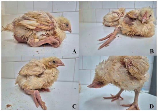
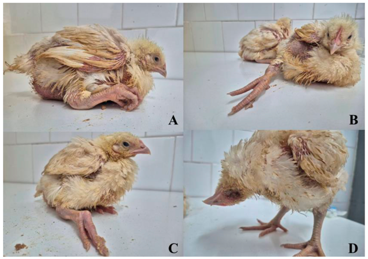
The epidemiological investigation found that about one week before the observed increase in mortality, loose droppings appeared on the litter in both flocks, accompanied by reduced feed intake below expected levels. Clinical signs appeared at around 20 days of age and subsequently progressed rapidly. Affected birds exhibited depression, somnolence, ruffled feathers, and diarrhoea. Lameness was a prominent clinical feature, with birds reluctant to move and adopting a characteristic hock-sitting posture with their legs extended forward. In more severe cases, paraplegia was observed; severely affected individuals were unable to access feeders and drinkers, leading to dehydration, impaired growth, and increased variability in body weight within the flocks (Figure 1). At Farm B, the owner reported three cases of torticollis, but only one bird with torticollis was observed during the visit. Mortality rates increased significantly between days 19 and 25, reaching 2.8% in Farm A and 3.7% in Farm B, compared to a baseline mortality of approximately 0.8%. By day 25, the prevalence of lameness and paralysis reached 2.4% (237/9876) and 2.9% (370/12,760) in the respective flocks. The first detected cases are considered to have occurred on day 20, so the number of live birds on that day is used as the denominator. The total number of birds affected by lameness was counted through day 25, when antibiotic treatment began. Overall, the disease markedly reduced flock performance, as shown by reduced feed intake, growth retardation, and increased heterogeneity in body weight.
Figure 1.
Affected broilers at 22 days of age showed inability to move, dog-sitting posture, paralysis (A–C), torticollis, and severe ocular inflammation (D).
3.2. Gross Pathology
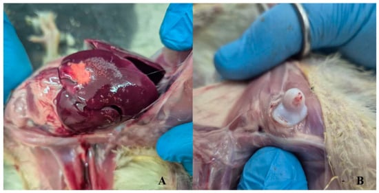
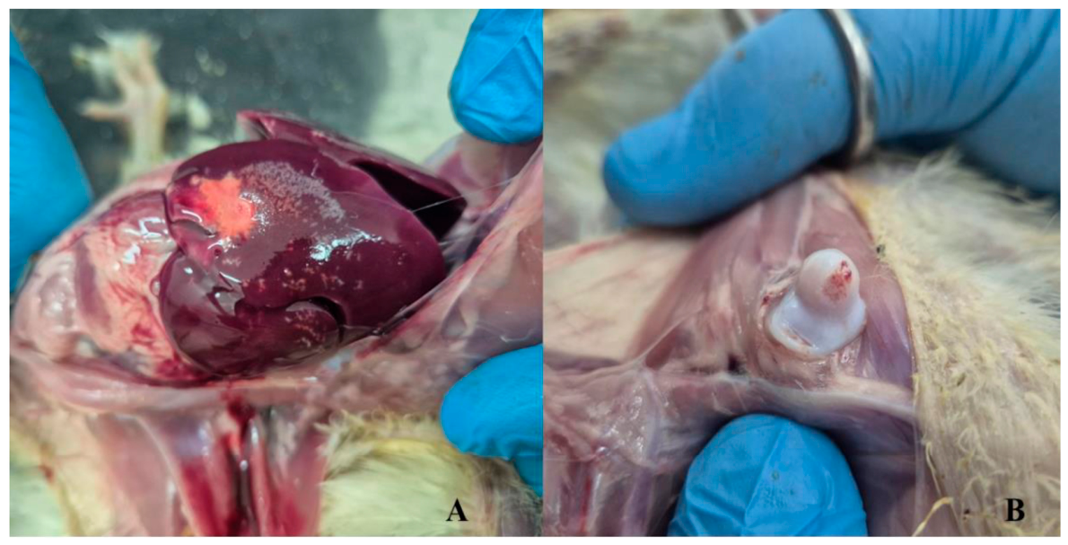
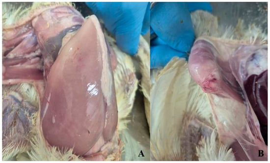
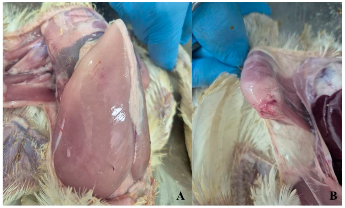
Post-mortem examination revealed fibrinous pericarditis and perihepatitis, multifocal pale hepatic foci consistent with necrosis, and splenomegaly. Early osteoarticular lesions consisted of focal erosion and necrosis of the femoral-head articular cartilage, with associated haemorrhage (Figure 2). Mild airsacculitis and a small volume predominantly thoracic coelomic effusion were present in some birds. Multifocal petechial haemorrhages were observed within the pectoral and pelvic-limb muscles (Figure 3). Examination of the thoracic vertebral column revealed malalignment in the T4–T6 region, accompanied by gross lesions consistent with osteomyelitis and spondylitis.
Figure 2.
Representative gross lesions in affected broilers. (A) Multifocal pale hepatic foci. (B) Focal erosion and loss of femoral-head articular cartilage with adjacent haemorrhage.
Figure 3.
Haemorrhagic lesions in affected broilers. (A) Multifocal petechial haemorrhages within the superficial pectoral muscle. (B) Petechial haemorrhages in the skeletal muscles adjacent to the hock joint.
3.3. Histopathological Findings
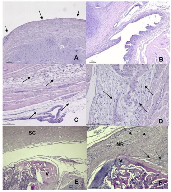
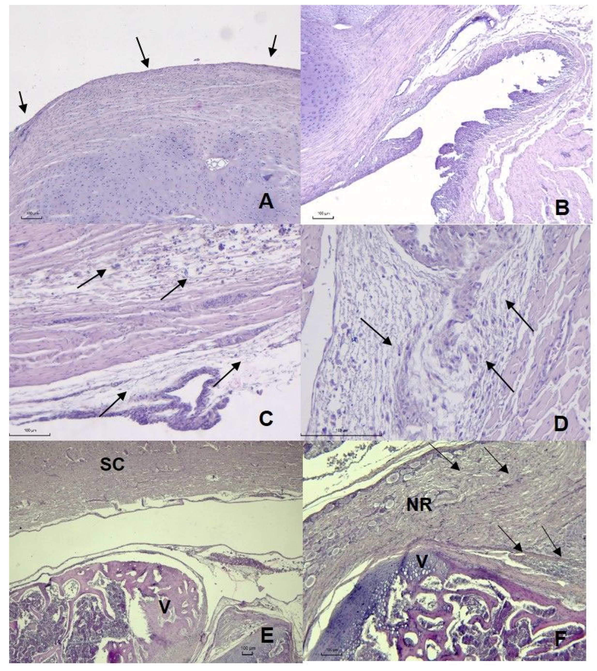
Histologically, the femoral head showed multifocal thinning and loss of the superficial articular cartilage (Figure 4A). The coxofemoral joint exhibited mild synovial-lining hyperplasia (Figure 4B). Periarticular connective tissues were expanded by oedema and mild heterophilic and lymphoplasmacytic inflammation, accompanied by fibroblast proliferation (Figure 4C). Sections of SDFT showed focal collagen-fibre degeneration and disruption, accompanied by oedema and heterophilic and mononuclear inflammation involving both the tendon and its sheath, consistent with tendinitis and tenosynovitis (Figure 4D). Vertebral sections from the T4–T6 region showed inflammation involving the cartilage and vertebral structures, consistent with chondritis and spondylitis. Associated vertebral displacement, consistent with spondylolisthesis, resulted in compression of the spinal cord and adjacent nerve roots. Adjacent neural tissue exhibited mild vacuolation and mononuclear inflammation (Figure 4E,F).
Figure 4.
Histopathological lesion in affected broilers. (A) Femoral-head articular cartilage attenuation and erosion (arrows). (B) Mild coxofemoral synovial hyperplasia (arrows). (C) Oedematous and inflamed periarticular tissue (arrows). (D) SDF tendinitis and tenosynovitis with early fibroplasia (arrows). (E) Spondylolisthesis with chondritis, spondylitis, and neural compression in the free thoracic vertebral region (V—vertebrae; SC—spinal cord) (F) Neuronal vacuolation and mononuclear radiculoneuritis (NR—nerve root, V—vertebrae, and arrows—inflammation and degeneration). H&E; scale bars = 100 µm.
3.4. Microbiological Findings
Bacteriological analysis revealed Gram-positive, catalase-negative cocci forming small (approximately 1 mm), clear to opalescent colonies after 24 h of incubation. Pure cultures were obtained from 11 of 13 birds examined, and isolations from various anatomical sites are shown in Table 2. Concurrent microbiological analysis identified Escherichia coli in two liver samples. However, it was present only as minor growth alongside the dominant E. cecorum isolates. Biochemical methods (Vitek 2 Compact) confirmed E. cecorum identification with a probability of 99% (Table 3).
Table 2.
Isolation of E. cecorum from different anatomical sites.
Table 3.
Biochemical identification of E. cecorum.
No other significant bacterial pathogens were consistently detected. Molecular testing for infectious bursal disease virus, Mycoplasma gallisepticum, Mycoplasma synoviae, and reovirus were negative. Recovery of E. coli from two liver samples and the limited differential work-up preclude comprehensive exclusion of concurrent or predisposing infections.
3.5. Antimicrobial Resistance Profile
One septicemic isolate per chicken was tested for antibiotic susceptibility. All isolates tested from both farms showed a similar profile. Disk diffusion yielded mean inhibition zones of 28 mm for amoxicillin and 23 mm for tiamulin, and smaller zones for the other tested agents (Table 4). These measurements were not assigned clinical susceptible, intermediate, or resistant categories because validated E. cecorum-specific interpretive criteria applicable to the present testing conditions were not established.
Table 4.
Inhibition zone diameters of the tested antimicrobials against the investigated isolates of E. cecorum (n = 13).
3.6. Molecular Findings
The five isolates selected for molecular testing were obtained from the following tissues: A2, spleen; A3, joint; B3, joint; B5, liver; and B8, spleen. Species identification was further confirmed by PCR targeting the sodA gene, with all 5 isolates producing the expected 371 bp amplicon. Amplification of the cpsO gene supported the isolates’ pathogenic potential.
Analysis of the distribution of genes associated with virulence, antimicrobial resistance and horizontal gene transfer among the five tested isolates revealed similar outcomes for all of them. Three of the seven tested virulence determinants (psaA, gelE, and ace) were established, while cylB, esp, agg, and gls24 were not detected. Screening for antimicrobial resistance genes showed multiple tetracycline resistance determinants (tetM, tetK, tetL, tetS, and tetT) and the macrolide resistance gene ermB (Table 5). Genes associated with horizontal gene transfer included the conjugation-related pheromone genes (cpd and ccf), while other tested elements were not detected.
Table 5.
Genetic determinants responsible for virulence, antibiotic resistance (ABR), and horizontal gene transfer (HGT) in the tested isolates. Green indicates positive amplification of the target fragment, and red indicates no amplification of the correct fragment.
3.7. Treatment
The preferred drug, tiamulin (RODOTIUM® 45% water-soluble granules, Huvepharma, Bulgaria), was selected for its lipophilic properties, which enable it to diffuse easily across most tissues, allowing it to reach concentrations in synovial fluid [30]. It was administered in water for 5 days at 50 mg/kg, within the recommended dose range of 30–60 mg/kg [31]. The manufacturer-recommended withdrawal period was 7 days after the final administration of the preparation. The treatment regimen also involved administering a product containing butyric acid glycerol esters, specifically ProPhorce™ SR 130 (Perstorp, Sweden), at a dose of 500 g per tonne of feed, which was continued until the end of the rearing period. Two days after therapy began, no further cases of affected birds were observed.
3.8. Additional Control Measures
Other measures implemented included: drinker management, removal of wet spots near drinkers, additional bedding material, dry disinfectant, and culling of severely affected chickens. Between production cycles, cleaning and disinfection of buildings and equipment, along with insect and rodent control, were recommended.
4. Discussion
Pathogenic E. cecorum strains have emerged as an important cause of septicaemia, locomotor disorders, and rising mortality rates in global commercial broiler production. This study describes the early onset of a septicemic infection on two broiler farms in Bulgaria, potentially associated with a pathogenic strain of E. cecorum.
The clinical signs observed, including depression, diarrhoea, lameness, paralysis, and recumbency, are consistent with previous reports of pathogenic EC infections [8]. Disease onset at approximately 20 days of age supports recent evidence that virulent EC strains may cause systemic infection earlier than traditionally recognised. While vertebral osteomyelitis and spondylitis are frequently reported in older broilers, recent studies indicate that septicemic forms of the disease may occur in younger birds before skeletal lesions become fully developed [7,32]. The pathological findings of fibrinous pericarditis, hepatitis, splenomegaly, and femoral head cartilage erosion were consistent with systemic dissemination of EC. The identification of a single case of vertebral osteomyelitis suggests the infection was detected early, before the development of chronic skeletal lesions. Similar observations have been reported during experimental and field outbreaks of pathogenic EC [33]. Developmental spondylolisthesis (“kinky back”) is an important differential diagnosis for posterior paresis or paralysis in rapidly growing broilers. It is characterised by displacement of the free thoracic vertebra within the T4–T6 region, resulting in angular narrowing of the vertebral canal and spinal cord compression. However, similar neurological signs may occur with spondylosis, vertebral osteomyelitis, and osteochondrosis and are therefore not lesion-specific [34]. In the present outbreak, vertebral malalignment was accompanied by gross osteomyelitis and spondylitis, with microscopic chondritis, spondylitis, and neural compression. Together with the systemic lesions and bacteriological and molecular identification of pathogenic EC, these findings support EC-associated vertebral infection with secondary instability and neural compression rather than uncomplicated developmental spondylolisthesis. Flexor tendinitis and tenosynovitis should also be interpreted as nonspecific lesions. Tendon-sheath inflammation in broilers may accompany bacterial musculoskeletal disease. In contrast, avian reovirus is a well-established cause of arthritis and tenosynovitis involving the hock region and gastrocnemius tendons [35]. The negative reovirus result in the present investigation reduces support for reoviral tenosynovitis but does not, by itself, establish EC as the direct cause of the tendon lesion. Direct culture, PCR, or in situ hybridization of the organism in the affected tendon or tendon sheath would be required for lesion-specific attribution. The predominance of E. cecorum in cultures, together with compatible gross and histopathological lesions, supports its role as the potential bacterial pathogen in this outbreak. These findings are consistent with those of Stalker et al. [36] who reported E. cecorum-associated systemic and osteoarticular disease in broilers, characterised by pericarditis, perihepatitis, osteomyelitis, and arthritis. No additional bacterial pathogens were consistently isolated under the culture conditions used. In contrast, E. coli was detected as countable single colonies in only two samples and was therefore classified as a contaminant. The negative results for infectious bursal disease virus, avian reovirus, Mycoplasma gallisepticum, and Mycoplasma synoviae further support an association between E. cecorum and the disease, although concurrent infections cannot be fully excluded. The potential contributions of concurrent infections, host factors, and husbandry conditions have not been fully evaluated. Moreover, PCR characterization of five selected isolates cannot be extrapolated to all isolates or to the broader poultry population. Molecular analysis confirmed the isolate’s species identity was supported by sodA and a pathogenicity-associated cpsO marker was detected. Although we identified only three virulence-associated genes (ace, gelE, and psaA), previous studies show that pathogenic EC strains often possess relatively few classical enterococcal virulence genes and that no single determinant is consistently associated with pathogenicity [13,37]. The detected genes may nevertheless contribute to bacterial adhesion, tissue invasion, and persistence within the host [34]. All isolates subjected to molecular characterization were also tested using disk diffusion. However, antimicrobial resistance genes and inhibition zone diameters were treated as separate descriptive findings because no isolate-specific genotype–phenotype analysis was performed. Accordingly, these results do not establish associations between individual resistance determinants and phenotypic responses. Laurentie et al. [11] investigated these relationships in E. cecorum by integrating genomic data with disk diffusion and broth microdilution results, demonstrating the value of directly matched molecular and phenotypic data. Further investigation using this approach is needed to assess genotype–phenotype concordance among the present isolates. Interpretation of the antimicrobial susceptibility findings was limited by the absence of validated E. cecorum-specific clinical breakpoints for the disk diffusion conditions used. Laurentie et al. [11] proposed tentative epidemiological cutoff values for selected antimicrobials; however, these distinguish wild-type from non-wild-type populations and do not predict clinical treatment success. Differences in culture media and incubation conditions also limit their direct application to the present results. Accordingly, inhibition-zone diameters were interpreted as quantitative observations rather than definitive. The 23 mm inhibition zone obtained with the 30 µg tiamulin disk demonstrated in vitro inhibitory activity under the conditions tested, but did not establish clinical susceptibility. A reduction in newly affected birds was observed following the administration of tiamulin and ProPhorce™ SR 130. This temporal association suggests a favourable response to intervention; nevertheless, the absence of an untreated comparison group and the concurrent administration of both products prevent attribution of the improvement to tiamulin alone or determination of the independent contribution of ProPhorce™ SR 130.
Even in a small commercial poultry enterprise, timely intervention is essential to limit further morbidity and mortality, reduce economic losses, and maintain the viability of the business. Alongside these economic objectives, reducing the incidence of new clinical cases may improve animal welfare by limiting the number of birds experiencing pain, impaired mobility, and other disease-related issues.
The source of E. cecorum infection in the present outbreak remained undetermined. Although both affected flocks came from the same hatchery, this epidemiological link does not establish a hatchery-associated source, especially since neither the hatchery nor the breeder flocks were investigated. However, exposure before placement on the farm remains plausible. Jung et al. [38] detected E. cecorum in newly hatched chicks and suggested possible hatchery-associated introduction, while noting that further investigation was needed. Importantly, acquisition within the hatchery should be distinguished from true vertical transmission, which Kense and Landman [39] were unable to demonstrate through molecular comparison of isolates from breeder flocks and their affected offspring. Thus, a hatchery-associated source cannot be ruled out, but the present findings do not establish either the source or the transmission route.
5. Conclusions
This pilot investigation documents an association between systemic E. cecorum isolation and early-onset clinical disease with systemic and osteoarticular lesions in two Bulgarian broiler flocks. The presence of selected virulence- and antimicrobial-resistance-associated genes indicates molecular potential but does not demonstrate primary causation, gene expression, phenotypic multidrug resistance or horizontal transfer. The observed field improvement cannot be assigned to a single intervention, and economic losses were not quantified. Broader epidemiological sampling, validated MIC testing, isolate-level genomic analysis and pathogen localization in affected tissues are needed.
6. Limitations
This pilot study was limited to two broiler flocks and a small, diagnostically selected sample, with no healthy controls or longitudinal follow-up. Although the isolation of E. cecorum from extraintestinal sites, together with compatible lesions, supports an association with disease, primary causation cannot be established. The potential contributions of E. coli, other concurrent infections, and predisposing management or host factors cannot be excluded.
Molecular characterization was conducted on only five isolates. Generally, PCR amplifications were interpreted according previously reported data without inclusion of positive or negative controls. Moreover, gene detection does not establish gene expression, functional activity, virulence, or horizontal gene transfer. In addition, no isolate-specific genotype–phenotype comparisons or high-resolution strain typing were performed. Disk diffusion results were interpreted descriptively because validated clinical breakpoints specific to E. cecorum and applicable to the testing conditions were unavailable. Colistin susceptibility could not be reliably assessed because broth microdilution MICs were not determined.
Flock-level estimates relied on farm records. Because treatment and management interventions were implemented concurrently and no untreated comparison group was available, clinical improvement could not be attributed to any single intervention. Economic losses were not quantified.
Author Contributions
Conceptualization, B.B.-M. and P.M.; methodology, M.P., Y.K., and P.H., investigation, B.B.-M. and P.M.; resources, B.B.-M.; P.M., K.D., M.P., Y.K., and P.H.; writing—original draft preparation, B.B.-M., P.M., K.D., M.P., Y.K., and P.H.; writing—review and editing, B.B.-M. and P.M.; visualization, B.B.-M., P.M., K.D., M.P., Y.K., and P.H.; supervision, B.B.-M. and P.M. All authors have read and agreed to the published version of the manuscript.
Funding
The authors sincerely thank Trakia University, Stara Zagora, for APC support.
Institutional Review Board Statement
No birds were experimentally infected or euthanised specifically for research; diagnostic specimens originated from birds found dead during a naturally occurring field event.
Informed Consent Statement
We obtained written informed consent from all farm owners.
Data Availability Statement
The data presented in this study are available upon request from the corresponding author.
Acknowledgments
Conflicts of Interest
The authors declare no conflicts of interest.
Abbreviations
The following abbreviations are used in this manuscript:
| ABR | Antimicrobial resistance |
| DNA | Deoxyribonucleic acid |
| EC | Enterococcus cecorum |
| EDTA | Ethylenediaminetetraacetic acid |
| GP | Gram-positive |
| HGT | Horizontal gene transfer |
| PCR | Polymerase chain reaction |
| SDFT | Superficial digital flexor tendon |
| Tm | Melting temperature |
References
- Mullally, C.A.; Fahriani, M.; Mowlaboccus, S.; Coombs, G.W. Non-faecium non-faecalis enterococci: A review of clinical manifestations, virulence factors, and antimicrobial resistance. Clin. Microbiol. Rev. 2024, 37, e00121–e00123. [Google Scholar] [CrossRef] [Scilit] [PubMed]
- Anthney, A.; Do, A.D.T.; Alrubaye, A.A. Bacterial chondronecrosis with osteomyelitis lameness in broiler chickens and its implications for welfare, meat safety, and quality: A review. Front. Physiol. 2024, 15, 1452318. [Google Scholar] [CrossRef] [Scilit] [PubMed]
- Tessin, J.; Jung, A.; Silberborth, A.; Rohn, K.; Schulz, J.; Visscher, C.; Kemper, N. Detection of Enterococcus cecorum to identify persistently contaminated locations using faecal and environmental samples in broiler houses of clinically healthy flocks. Avian Pathol. 2024, 53, 312–320. [Google Scholar] [CrossRef] [Scilit] [PubMed]
- Souillard, R.; Laurentie, J.; Kempf, I.; Le Caër, V.; Le Bouquin, S.; Serror, P.; Allain, V. Increasing incidence of Enterococcus-associated diseases in poultry in France over the past 15 years. Vet. Microbiol. 2022, 269, 109426. [Google Scholar] [CrossRef] [Scilit] [PubMed]
- Meng, F.; Zhou, D.; Li, R.; Lang, F.; Xiang, B.; Yang, L.; Cheng, Z. Emergence of linezolid-resistant Enterococcus cecorum clade F harbouring optrA/fexA causing enterococcal spondylitis in commercial broilers in China. BMC Microbiol. 2025, 25, 313. [Google Scholar] [CrossRef] [Scilit] [PubMed]
- Alzahrani, O.M.; Fayez, M.; Alswat, A.S.; Alkafafy, M.; Mahmoud, S.F.; Al-Marri, T.; Almuslem, A.; Ashfaq, H.; Yusuf, S. Antimicrobial resistance, biofilm formation, and virulence genes in Enterococcus species from small backyard chicken flocks. Antibiotics 2022, 11, 380. [Google Scholar] [CrossRef] [Scilit] [PubMed]
- Higuita, J.; Arango, M.; Forga, A.; Cortes, D.; Graham, D. An updated review of Enterococcus cecorum infections in poultry. Avian Dis. 2025, 68, 404–411. [Google Scholar] [CrossRef] [Scilit] [PubMed]
- Watson, K.; Arais, L.; Green, S.; O’Kane, P.; Kirchner, M.; Demmers, T.; Commins, C.; Smith, R.; Cordoni, G.; Kyriazakis, I.; et al. Towards the identification of transmission pathways and early detection of Enterococcus cecorum infection in broiler chickens. Poult. Sci. 2024, 103, 104224. [Google Scholar] [CrossRef] [Scilit] [PubMed]
- Suyemoto, M.M.; Walker, G.K.; Taldo, U.; Diveley, K.R.; Borst, L.B. Development and application of optimized isolation methods and diagnostic PCR protocols for the detection of pathogenic Enterococcus cecorum isolated from broiler chickens. Avian Dis. 2025, 68, 427–434. [Google Scholar] [CrossRef] [Scilit] [PubMed]
- Bauer, A.W.; Kirby, W.M.M.; Sherris, J.C.; Turck, M. Antibiotic susceptibility testing by a standard single disk method. Am. J. Clin. Pathol. 1966, 36, 493–496. [Google Scholar] [CrossRef] [Scilit]
- Laurentie, J.; Mourand, G.; Grippon, P.; Furlan, S.; Chauvin, C.; Jouy, E.; Serror, P.; Kempf, I. Determination of epidemiological cutoff values for antimicrobial resistance of Enterococcus cecorum. J. Clin. Microbiol. 2023, 61, e01445-22. [Google Scholar] [CrossRef] [Scilit] [PubMed]
- Jackson, C.R.; Fedorka-Cray, P.J.; Barrett, J.B. Use of a genus- and species-specific multiplex PCR for identification of enterococci. J. Clin. Microbiol. 2004, 42, 3558–3565. [Google Scholar] [CrossRef] [Scilit] [PubMed]
- Combar, D.O.; Rubite, S.; Scott, P.C.; Campbell, B.E.; Van, T.T.H. Comparison of Enterococcus cecorum genomes from broiler chickens with enterococcal spondylitis in Australian farms and strains from other countries. Poult. Sci. 2024, 103, 104356. [Google Scholar] [CrossRef] [Scilit] [PubMed]
- Arango, M.; Mackey, R.; Thornton, J.K.; Manginsay, N.; Pulido-Landinez, M. Differentiation of Pathogenic from Nonpathogenic Enterococcus cecorum Field Isolates Based on the Detection of the cpsO Gene and Its Association with Macroscopic Lesions. Avian Dis. 2026, 70, 183–188. [Google Scholar] [CrossRef] [Scilit] [PubMed]
- McBride, S.M.; Fischetti, V.A.; LeBlanc, D.J.; Moellering, R.C., Jr.; Gilmore, M.S. Genetic diversity among Enterococcus faecalis. PLoS ONE 2007, 2, e582. [Google Scholar] [CrossRef] [Scilit] [PubMed]
- Mannu, L.; Paba, A.; Daga, E.; Comunian, R.; Zanetti, S.; Duprè, I.; Sechi, L.A. Comparison of the incidence of virulence determinants and antibiotic resistance between Enterococcus faecium strains of dairy, animal, and clinical origin. Int. J. Food Microbiol. 2003, 88, 291–304. [Google Scholar] [CrossRef] [Scilit] [PubMed]
- Eaton, T.J.; Gasson, M.J. Molecular screening of Enterococcus virulence determinants and potential for genetic exchange between food and medical isolates. Appl. Environ. Microbiol. 2001, 67, 1628–1635. [Google Scholar] [CrossRef] [Scilit] [PubMed]
- Van Asselt, G.J.; Vliegenthart, J.S.; Petit, P.L.C.; Van de Klundert, J.A.M.; Mouton, R.P. High-level aminoglycoside resistance among enterococci and group A streptococci. J. Antimicrob. Chemother. 1992, 30, 651–659. [Google Scholar] [CrossRef] [Scilit] [PubMed]
- Garofalo, C.; Vignaroli, C.; Zandri, G.; Aquilanti, L.; Bordoni, D.; Osimani, A.; Clementi, F.; Biavasco, F. Direct detection of antibiotic resistance genes in specimens of chicken and pork meat. Int. J. Food Microbiol. 2007, 113, 75–83. [Google Scholar] [CrossRef] [Scilit] [PubMed]
- Aarestrup, F.M.; Agersø, Y.; Gerner-Smidt, P.; Madsen, M.; Jensen, L.B. Comparison of antimicrobial resistance phenotypes and resistance genes in Enterococcus faecalis and Enterococcus faecium from humans in the community, broilers, and pigs in Denmark. Diagn. Microbiol. Infect. Dis. 2000, 37, 127–137. [Google Scholar] [CrossRef] [Scilit] [PubMed]
- Jensen, L.B.; Frimodt-Møller, N.; Aarestrup, F.M. Presence of erm gene classes in Gram-positive bacteria of animal and human origin in Denmark. FEMS Microbiol. Lett. 1999, 170, 151–158. [Google Scholar] [CrossRef] [Scilit]
- Jia, W.; Li, G.; Wang, W. Prevalence and antimicrobial resistance of Enterococcus species: A hospital-based study in China. Int. J. Environ. Res. Public Health 2014, 11, 3424–3442. [Google Scholar] [CrossRef] [Scilit] [PubMed]
- Doherty, N.; Trzcinski, K.; Pickerill, P.; Zawadzki, P.; Dowson, C.G. Genetic diversity of the tet(M) gene in tetracycline-resistant clonal lineages of Streptococcus pneumoniae. Antimicrob. Agents Chemother. 2000, 44, 2979–2984. [Google Scholar] [CrossRef] [Scilit] [PubMed]
- Aminov, R.I.; Garrigues-Jeanjean, N.; Mackie, R.I. Molecular ecology of tetracycline resistance: Development and validation of primers for detection of tetracycline resistance genes encoding ribosomal protection proteins. Appl. Environ. Microbiol. 2001, 67, 22–32. [Google Scholar] [CrossRef] [Scilit] [PubMed]
- Wang, Y.; Dong, J.; Wang, J.; Chi, W.; Wong, A.; Tian, Q.; Hong, Y.; Zhou, X.; Ye, H.; Tian, X.; et al. Assessing the drug resistance profiles of oral probiotic lozenges. J. Oral Microbiol. 2022, 14, 2019992. [Google Scholar] [CrossRef] [Scilit] [PubMed]
- Billström, H.; Lund, B.; Sullivan, Å.; Nord, C.E. Virulence and antimicrobial resistance in clinical Enterococcus faecium. Int. J. Antimicrob. Agents 2008, 32, 374–377. [Google Scholar] [CrossRef] [Scilit] [PubMed]
- Creti, R.; Imperi, M.; Bertuccini, L.; Fabretti, F.; Orefici, G.; Di Rosa, R.; Baldassarri, L. Survey for virulence determinants among Enterococcus faecalis isolated from different sources. J. Med. Microbiol. 2004, 53, 13–20. [Google Scholar] [CrossRef] [Scilit] [PubMed]
- Heaton, M.P.; Handwerger, S. Conjugative mobilization of a vancomycin resistance plasmid by a putative Enterococcus faecium sex pheromone response plasmid. Microb. Drug Resist. 1995, 1, 177–183. [Google Scholar] [CrossRef] [Scilit] [PubMed]
- Jensen, L.B.; Garcia-Migura, L.; Valenzuela, A.J.S.; Løhr, M.; Hasman, H.; Aarestrup, F.M. A classification system for plasmids from enterococci and other Gram-positive bacteria. J. Microbiol. Methods 2010, 80, 25–43. [Google Scholar] [CrossRef] [Scilit] [PubMed]
- Dowling, P.M. Introduction to clinical pharmacology. In Equine Internal Medicine; Reed, S.M., Bayly, W.M., Sellon, D.C., Eds.; Saunders Elsevier: St. Louis, MO, USA, 2009. [Google Scholar]
- Sartini, I.; Vercelli, C.; Łebkowska-Wieruszewska, B.; Lisowski, A.; Fadel, C.; Poapolathep, A.; Dessi, F.; Giorgi, M. Pharmacokinetics and pharmacodynamics of tiamulin after single and multiple oral administrations in geese. Vet. Anim. Sci. 2023, 22, 100317. [Google Scholar] [CrossRef] [Scilit] [PubMed]
- Huang, Y.; Eeckhaut, V.; Goossens, E.; Rasschaert, G.; Van Erum, J.; Roovers, G.; Ducatelle, R.; Antonissen, G.; Van Immerseel, F. Bacterial chondronecrosis with osteomyelitis-related Enterococcus cecorum isolates are genetically distinct from the commensal population and are more virulent in an embryo mortality model. Vet. Res. 2023, 54, 13. [Google Scholar] [CrossRef] [Scilit] [PubMed]
- Dunnam, G.; Thornton, J.K.; Pulido-Landinez, M. Characterization of an emerging Enterococcus cecorum outbreak causing severe systemic disease with concurrent leg problems in a broiler integrator in the southern United States. Avian Dis. 2023, 67, 137–144. [Google Scholar] [CrossRef] [Scilit] [PubMed]
- Musyahadah, U.S.A.; Asnayanti, A.; Do, A.D.T.; Alrubaye, A. Genetic and environmental factors contributing to the pathogenesis of vertebral osteomyelitis caused by Enterococcus cecorum in broiler chicken. Animals 2025, 15, 2327. [Google Scholar] [CrossRef] [Scilit] [PubMed]
- Nour, I.; Mohanty, S.K. Avian reovirus: From molecular biology to pathogenesis and control. Viruses 2024, 16, 1966. [Google Scholar] [CrossRef] [Scilit] [PubMed]
- Stalker, M.J.; Brash, M.L.; Weisz, A.; Ouckama, R.M.; Slavic, D. Arthritis and osteomyelitis associated with Enterococcus cecorum infection in broiler and broiler breeder chickens in Ontario, Canada. J. Vet. Diagn. Investig. 2010, 22, 643–645. [Google Scholar] [CrossRef] [Scilit] [PubMed]
- Dolka, B.; Chrobak-Chmiel, D.; Czopowicz, M.; Szeleszczuk, P. Characterization of pathogenic Enterococcus cecorum from different poultry groups: Broiler chickens, layers, turkeys, and waterfowl. PLoS ONE 2017, 12, e0185199. [Google Scholar] [CrossRef] [Scilit] [PubMed]
- Jung, A.; Petersen, H.; Teske, L.; Rautenschlein, S. Colonization patterns of Enterococcus cecorum in two different broiler production cycles detected with a newly developed quantitative real-time PCR. BMC Microbiol. 2017, 17, 106. [Google Scholar] [CrossRef] [Scilit] [PubMed]
- Kense, M.J.; Landman, W.J.M. Enterococcus cecorum infections in broiler breeders and their offspring: Molecular epidemiology. Avian Pathol. 2011, 40, 603–612. [Google Scholar] [CrossRef] [Scilit] [PubMed]

Disclaimer/Publisher’s Note: The statements, opinions and data contained in all publications are solely those of the individual author(s) and contributor(s) and not of MDPI and/or the editor(s). MDPI and/or the editor(s) disclaim responsibility for any injury to people or property resulting from any ideas, methods, instructions or products referred to in the content. |
© 2026 by the authors. Licensee MDPI, Basel, Switzerland. This article is an open access article distributed under the terms and conditions of the Creative Commons Attribution (CC BY) license.