


Abstract
The fall armyworm, Spodoptera frugiperda, is a globally distributed agricultural pest. Metarhizium robertsii is an entomopathogenic fungus (EPF) with great application potential for the biological control of S. frugiperda. Nevertheless, to date, there have been no reports describing the interactions between M. robertsii and the immune system of S. frugiperda. In the present study, we investigated the pathogenicity of M. robertsii against S. frugiperda and demonstrated that this fungus exhibits high virulence toward the pest. M. robertsii infection induces apoptosis and necrosis in hemocytes of S. frugiperda, which, in turn, reduces circulating hemocyte abundance. Fungal infection also impairs the spreading capacity of plasmatocytes. These adverse effects on hemocytes collectively lead to a marked suppression of host cellular immune responses, including phagocytosis, nodulation, and encapsulation. Meanwhile, M. robertsii infection upregulates the expression of prophenoloxidase genes in hemocytes, resulting in significantly elevated phenoloxidase activity in host hemolymph. Transcription of antimicrobial peptide genes is also increased in the fat body following fungal challenge. Upregulation of these humoral antibacterial response-related genes enhances the antibacterial activity of hemolymph. This study reveals the mechanistic basis underlying immune interactions between M. robertsii and S. frugiperda and deepens our understanding of the pathogenic mechanisms employed by EPF.
1. Introduction
Due to the absence of adaptive immunity, insects depend entirely on their innate immune system for defense against invasive pathogens. The insect innate immune response is functionally divided into cellular and humoral immune branches. Cellular immune reactions, which are primarily mediated by hemocytes, encompass phagocytosis, nodulation, and encapsulation processes [1,2]. In contrast, the humoral immune response is characterized by the synthesis of antimicrobial peptides (AMPs) [3] and the activation of the prophenoloxidase (PPO) cascade. Activation of the PPO cascade further triggers melanization and facilitates the production of toxic reactive intermediates to eliminate pathogens [4]. AMPs are short cationic peptides that elicit antimicrobial functions through embedding within and rupturing microbial membranes to eradicate invasive pathogens. Biosynthesis of these peptides mainly occurs in hemocytes and, at a markedly higher level, the fat body tissue, which rapidly secretes AMPs into hemolymph in response to pathogen exposure [3]. Pattern recognition receptors (PRRs) are indispensable for pathogen identification via the perception of conserved microbial motifs termed pathogen-associated molecular patterns (PAMPs) [5]. This sophisticated immune machinery permits insects to survive amid pathogen-abundant habitats and is presumed to underpin their ecological dominance.
Infection by entomopathogenic fungi (EPF) commences via spore adherence to the insect cuticle; subsequent germination and appressorium formation facilitate cuticular penetration. Once inside the hemocoel, the pathogen undergoes dimorphic switching from filamentous growth to yeast-type hyphal bodies (blastospores) and secretes cytotoxic toxins against the insect host [6]. Prolonged host–fungal co-evolution has equipped EPF with varied virulence strategies to evade and suppress insect immune machinery. A spectrum of adaptive mechanisms protects colonizing fungi against host immune offence. Representative approaches comprise modification or masking of hyphal body surface carbohydrate epitopes to avoid immune recognition [7,8], and the secretion of destruxin, oosporein and other metabolites for immunosuppression [9,10].
The fall armyworm, Spodoptera frugiperda, originates from the tropical and subtropical zones of the Americas. As a severe worldwide agricultural pest, this insect has triggered considerable yield-loss incidents of maize across numerous nations [11]. S. frugiperda first invaded China in 2018 and has since spread to more than 20 provinces [12]. While chemical pesticides have long served as the primary strategy for their management, their extensive application disrupts ecological homeostasis and poses severe threats to human health, thereby driving the urgent demand for sustainable alternative pest control approaches. Microbial biocontrol agents, which rely on natural pest antagonists, have emerged as a promising alternative to chemical pesticides due to their environmental safety and non-toxicity to humans. Among diverse biocontrol resources, EPF represent one of the most valuable candidates for pest management. As a well-characterized EPF, Metarhizium robertsii possesses a broad host spectrum and exhibits high virulence against a wide range of insect pests [13]. Accordingly, this entomopathogenic fungus exhibits prominent application potential in the biological control of S. frugiperda.
In this study, we explored the pathogenicity of M. robertsii against S. frugiperda larvae through conidial immersion and intrahemocoelic injection assays. We further characterized the impacts of M. robertsii conidia on both cellular and humoral immune responses in S. frugiperda. The findings of this study deepen our understanding of host–fungus immune interactions and elucidate the pathogenic mechanisms underlying M. robertsii infection, providing valuable insights for developing strategies to promote fungal pathogenicity. Furthermore, our results establish a theoretical foundation for optimizing the practical application of M. robertsii as an efficient biocontrol agent against S. frugiperda.
2. Materials and Methods
2.1. Fungi, Insects and Infection
The M. robertsii strain ARSEF 2575 was kindly gifted by Anhui Agricultural University. The fungal strain was routinely cultured in our laboratory on potato dextrose agar (PDA) medium containing 20% potato extract, 2% glucose, and 2% agar at 25 ± 1 °C for 10 days. For conidial suspension preparation, fungal conidia were harvested and resuspended in 0.05% Tween-80 solution via shaking. The suspension was subsequently filtered through sterile non-woven fabric to remove residual hyphal debris.
S. frugiperda larvae were reared at 26 ± 1 °C with a 16:8 h light/dark photoperiod. For topical infection assays, third-instar larvae were immersed in conidial suspensions at gradient concentrations of 1 × 10⁷, 1 × 10⁸, and 1 × 10⁹ conidia mL−1 to simulate natural fungal infection, with 28 larvae set for each treatment group. Larvae treated with 0.05% Tween-80 solution served as the blank control. For intrahemocoelic injection assays, sixth-instar larvae were individually injected with 5 μL of conidial suspension containing 1 × 10³, 1 × 10⁴, or 1 × 10⁵ conidia, with 28 biological replicates per group. An equivalent volume of Tween-80 solution was injected as the control. Larval mortality was recorded every 12 h, and survival curves were generated using GraphPad Prism 10.12 (GraphPad Inc., La Jolla, CA, USA) software. Additionally, larvae injected with 1 × 10⁴ conidia per individual were selected for all subsequent immune-related functional assays.
2.2. Nodulation, Encapsulation, Phagocytosis and Plasmatocyte Length Assays
Nodulation, encapsulation, phagocytosis, and plasmatocyte morphological assays were conducted according to previously established protocols [14,15]. For the in vivo nodulation assay, 5 μL of PBS containing 1 × 10⁵ colony-forming units (CFUs) of Escherichia coli was injected into the hemocoel of sixth-instar larvae at 12, 24, and 48 h post-fungal inoculation (hpi). These larvae were dissected 3 h after E. coli challenge, and the formed nodules were visualized and quantified under a stereomicroscope at 10× magnification. Five larvae were analyzed for each treatment.
For the in vivo encapsulation assay, Sephadex DEAE A-25 beads were stained overnight in 1% Congo red solution, washed thoroughly with PBS until the supernatant was clear, and resuspended in PBS to yield a final concentration of 50 beads μL−1. At 12, 24, and 48 hpi, larvae were chilled on ice for 30 min, and each larva was injected with 5 μL of the bead suspension. At 24 h post-injection, larvae were dissected to recover the beads. Recovered beads were washed with PBS and mounted on glass slides. Images were acquired using a differential interference contrast microscope at 40× magnification. The encapsulation index was calculated based on measurements of bead diameter and the thickness of the surrounding hemocyte layer, according to the method of Wang et al. [14]. Five larvae were analyzed for each treatment.
For the in vitro phagocytosis assay, hemolymph (30 μL per larva) was harvested from larvae by clipping their prolegs at 12, 24, and 48 hpi and immediately mixed with 120 μL of insect physiological saline (137.78 mM NaCl, 8.51 mM KCl, 3.87 mM CaCl2, 1 mM MgCl2, and 10 mM HEPES, pH 7.0) supplemented with a trace amount of phenylthiourea (PTU) to inhibit melanization. Fluorescein isothiocyanate (FITC)-labeled E. coli (10 μL) were added, and the mixture was incubated at room temperature for 1 h. Post-incubation, hemocytes were permitted to settle onto glass slides within a humidified chamber and incubated in the dark for 30 min. Slides were gently rinsed with insect saline, fixed in 4% formaldehyde for 10 min, and washed three times with PBS. The proportion of hemocytes that had phagocytosed E. coli was determined by counting 100 hemocytes per slide under an inverted fluorescence microscope. Five larvae were analyzed for each treatment.
For the plasmatocyte length measurement assay, hemolymph was harvested from larvae at 12, 24, and 48 hpi and diluted at a 1:3 ratio with sterile anticoagulant buffer (62 mM NaCl, 100 mM glucose, 30 mM sodium citrate, 26 mM citric acid, 20 mM EDTA; pH 4.6). Hemocytes were allowed to settle and adhere to glass slides for 30 min to form a monolayer, followed by imaging under a phase-contrast microscope. Plasmatocyte length was subsequently quantified using ImageJ 1.54g (Wayne Rasband, National Institutes of Health, Bethesda, MD, USA) software. Five larvae were analyzed for each treatment.
2.3. Hemocyte Counts
Larvae were dissected at 12, 24, and 48 hpi for hemolymph collection. The harvested hemolymph (30 μL per larva) was diluted at a 1:3 ratio with sterile anticoagulant buffer. After gentle mixing, 20 μL aliquots of the cell suspension were loaded onto a hemocytometer for hemocyte enumeration. Granulocytes and plasmatocytes were differentiated based on their distinct morphological characteristics under phase-contrast microscopy. The abundances of total hemocytes, granulocytes, and plasmatocytes were quantified under a phase-contrast microscope. Five larvae were analyzed for each treatment.
2.4. Hemocyte Apoptosis and Necrosis Assays
Hemolymph was harvested from sixth-instar larvae at 12, 24, and 48 hpi and diluted at a 1:3 ratio using sterile anticoagulant buffer. Hemocytes were allowed to adhere and settle on glass slides for 30 min, followed by incubation with YO-PRO-1 and propidium iodide (PI) staining solutions at 37 °C for 5 min in the dark. Stained samples were visualized and imaged using an inverted fluorescence microscope. For quantitative analysis, 100 hemocytes were randomly selected per sample. Cells emitting only green fluorescence were defined as apoptotic, while those displaying red fluorescence were identified as necrotic. Five larvae were analyzed for each treatment. The apoptotic and necrotic rates were calculated according to the following formulas:
Apoptosis rate (%) = (green-fluorescent hemocytes/total hemocytes counted) × 100.
Necrotic rate (%) = (red-fluorescent hemocytes/total hemocytes counted) × 100.
2.5. Measurements of Phenoloxidase and Antibacterial Activity
Phenoloxidase (PO) activity and antibacterial activity were measured following previously established protocols [14,16]. At 12, 24, and 48 hpi, five larvae per group (control and treatment) were chilled on ice for 30 min, and 30 μL of hemolymph was harvested from each individual larva. Hemolymph samples from the five larvae were pooled in a single tube and diluted with pre-chilled anticoagulant buffer at a 1:3 ratio. The diluted hemolymph mixture was centrifuged at 4 °C and 2000 rpm for 10 min; the supernatant was retained, and the pellet was discarded. Next, aliquots (50 μL) of the supernatant were combined with an equal volume of trypsin solution (2 mg mL−1) and incubated at room temperature for 20 min. Fifty μL of L-3,4-dihydroxyphenylalanine substrate solution (3 mg mL−1) was then added to the reaction mixture. Phenoloxidase (PO) activity was evaluated by recording time-dependent changes in absorbance at 490 nm. One unit of PO enzymatic activity was defined as a 0.001 increase in absorbance per minute.
E. coli and Staphylococcus aureus in mid-log phase were harvested by centrifugation and diluted to 100 cells μL−1 for antibacterial activity assays. Briefly, 90 μL of larval plasma was mixed with 10 μL of bacterial suspension. After 1 h of incubation at room temperature, the mixture was spread evenly onto lysogeny broth (LB) agar plates. Following overnight incubation at 37 °C, the CFUs on each plate were enumerated. All PO activity and antibacterial activity assays were performed with five biological replicates.
2.6. Reverse Transcription Quantitative PCR (RT-qPCR)
Total RNA was extracted from the fat bodies or hemocytes of sixth-instar larvae at 12, 24, and 48 hpi using TRIzol Reagent (Invitrogen). First-strand cDNA was reverse-transcribed using the EasyScript cDNA Synthesis SuperMix (TransGen Biotech). Quantitative real-time PCR was performed with TransStart Tip Green qPCR SuperMix (TransGen Biotech). The housekeeping gene Rpl32 was employed as an endogenous reference gene for normalization. All primers used for RT-qPCR are listed in Table 1.
Table 1.
Primer sequences used in RT-qPCR.
2.7. Statistical Analysis
All data are expressed as the mean ± SEM. Statistical analyses were performed using GraphPad Prism software. Student’s t-test was used for two-group comparisons. Pairwise log-rank tests were performed for intergroup survival comparisons, and the Bonferroni correction was applied to adjust p-values for multiple testing (adjusted p < 0.016).
3. Results
3.1. Infection by M. robertsii Decreases S. frugiperda Survival
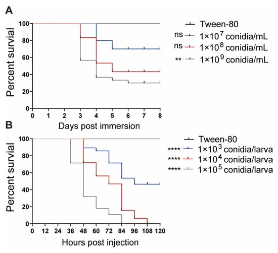
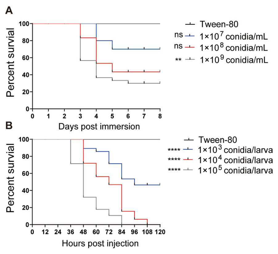
To assess the pathogenicity of M. robertsii against S. frugiperda, third-instar larvae were challenged via conidial immersion, whereas sixth-instar larvae were subjected to intrahemocoelic conidial injection. Larval survival rates in both infection groups were significantly reduced compared with their corresponding control groups (Figure 1). Fungal-induced mortality exhibited a clear dose-dependent increase with elevated conidial concentrations. Specifically, immersion treatment with 1 × 10⁹ conidia mL−1 resulted in a cumulative mortality of 68% within eight days (Figure 1A). For injection assays, dosages of 1 × 10⁵ and 1 × 10⁴ conidia per larva caused 100% mortality at 84 h and 108 h post-inoculation, respectively (Figure 1B). To establish a stable infection condition and obtain sufficient hemolymph for subsequent functional assays, the dosage of 1 × 10⁴ conidia per larva was selected for all follow-up injection experiments.
Figure 1.
Survival curves of S. frugiperda infected with M. robertsii. (A) Survival of third-instar larvae following immersion in different concentrations of M. robertsii conidia. (B) Survival of sixth-instar larvae following injection with different amounts of M. robertsii conidia. Asterisks indicate significant differences (** p < 0.01, **** p < 0.0001).
3.2. M. robertsii Infection Impairs Cellular Immunity
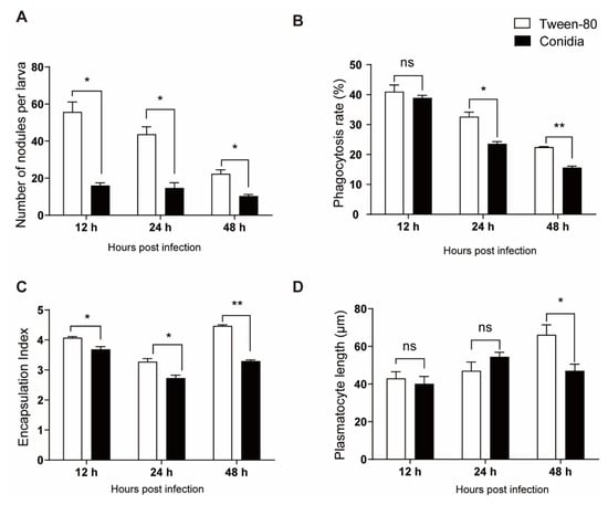
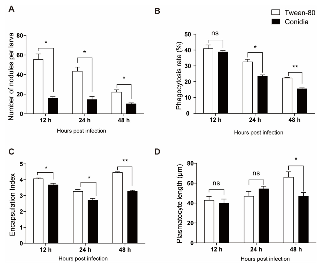
To determine whether M. robertsii modulates host cellular immune responses, we systematically compared nodulation, encapsulation, and phagocytosis activities, as well as plasmatocyte morphological characteristics, between fungal-infected and control larvae. Hemocytic nodulation was induced by E. coli challenge. The number of newly formed nodules was significantly reduced in M. robertsii-infected larvae relative to control larvae at 12, 24, and 48 hpi (Figure 2A). In vitro phagocytosis assays further showed that hemocytic phagocytic capacity against E. coli was suppressed at 24 and 48 hpi, whereas no significant difference was observed at 12 hpi between the two groups (Figure 2B). Sephadex beads were injected to trigger host encapsulation responses against foreign bodies. The encapsulation index was markedly lower in M. robertsii-infected larvae than in control larvae across all detected time points (Figure 2C). Collectively, these results demonstrate that M. robertsii infection substantially impairs host cellular immune functions.
Figure 2.
M. robertsii infection suppresses cellular immune responses in S. frugiperda larvae. Sixth-instar larvae were injected with M. robertsii conidia or Tween-80. Assays of nodulation (A), phagocytosis (B), encapsulation (C) and plasmatocyte spreading (D) were conducted at 12, 24 and 48 hpi. Asterisks indicate significant differences (* p < 0.05, ** p < 0.01).
Given that the morphological transition of plasmatocytes from a rounded to an adherent, spread state is a prerequisite for cellular immune responses [17], we further quantified plasmatocyte length to evaluate cellular spreading behavior. Plasmatocytes from fungal-infected larvae exhibited significantly shorter cell lengths at 48 hpi, while no obvious morphological differences were detected at 12 and 24 hpi (Figure 2D). These findings suggest that M. robertsii infection interferes with plasmatocyte spreading, which may further constrain host cellular immune competence.
3.3. M. robertsii Infection Decreases the Number of Hemocytes
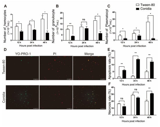
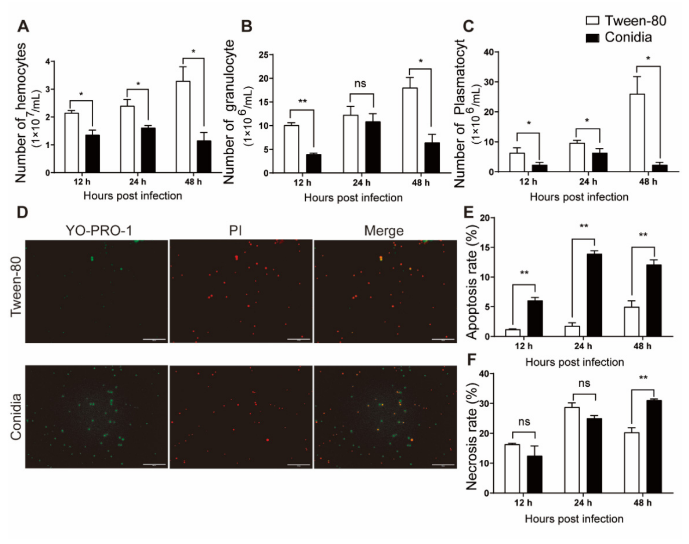
To explore the influence of M. robertsii infection on hemocyte abundance in S. frugiperda, we quantified the total number of circulating hemocytes. The results showed that fungal infection significantly reduced the total hemocyte count in larval hemolymph (Figure 3A). Given that granulocytes and plasmatocytes are the two predominant hemocyte subtypes responsible for insect immune responses [17], we further quantified their respective cell densities. Specifically, granulocyte numbers markedly declined at 12 and 48 hpi, while plasmatocyte abundance was significantly decreased across all detected time points in M. robertsii-infected larvae compared with control groups (Figure 3B,C).
Figure 3.
M. robertsii infection reduces hemocyte abundance by triggering hemocyte apoptosis and necrosis in S. frugiperda larvae. (A–C) Quantification of total hemocytes (A), granulocytes (B), and plasmatocytes (C) at 12, 24, and 48 hpi with M. robertsii conidia. (D) Hemocyte apoptosis and necrosis in control and fungus-infected larvae at 24 hpi were detected via YO-PRO-1/PI double staining. Green fluorescence marks apoptotic cells, and red fluorescence marks necrotic cells. Scale bar = 50 μm. (E,F) Quantitative analyses of apoptotic (E) and necrotic (F) rates. Asterisks indicate significant differences (* p < 0.05, ** p < 0.01).
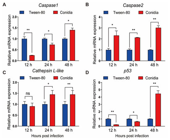
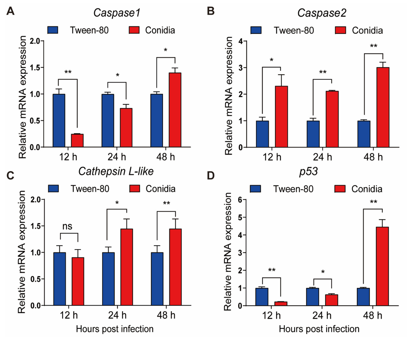
We hypothesized that the fungus might reduce circulating hemocyte abundance by triggering hemocyte apoptosis and necrosis. To test this hypothesis, YO-PRO-1/PI double-staining was performed to quantify the apoptotic and necrotic rates of hemocytes following fungal infection (Figure 3D). YO-PRO-1 staining demonstrated that hemocytes isolated from M. robertsii-infected larvae displayed significantly elevated apoptotic levels at 12, 24, and 48 hpi, while hemocytes from control larvae maintained consistently low apoptosis rates throughout the detection period (Figure 3E). PI staining further showed that hemocyte necrosis was markedly increased at 48 hpi in infected larvae, with no significant differences observed at 12 and 24 hpi (Figure 3F). We further examined the expression profiles of apoptosis-related genes in hemocytes after fungal infection. The results revealed that Caspase-2 was significantly upregulated at all three sampled time points. The transcript level of Cathepsin-like was markedly increased at 24 and 48 hpi. Although Caspase-1 and p53 exhibited significant downregulation at 12 and 24 hpi, their expression was also significantly elevated at 48 hpi (Figure 4). Taken together, these results confirm that M. robertsii infection triggers robust hemocyte apoptosis and necrosis, which ultimately contributes to the decrease in circulating hemocytes in S. frugiperda larvae.
Figure 4.
Transcriptional changes in apoptosis-related genes in hemocytes upon fungal infection. Relative transcript levels of Caspase1 (A), Caspase2 (B), Cathepsin L-like (C), and p53 (D) were detected in larval hemocytes at 12, 24, and 48 hpi. Hemolymph samples were harvested from M. robertsii-infected and control larvae, and gene expression profiles were quantified via RT-qPCR. All expression values were normalized to the internal reference gene Rpl32. Asterisks indicate significant differences (* p < 0.05, ** p < 0.01).
3.4. M. robertsii Infection Enhances Humoral Antibacterial Responses
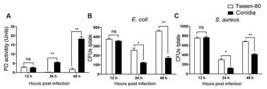
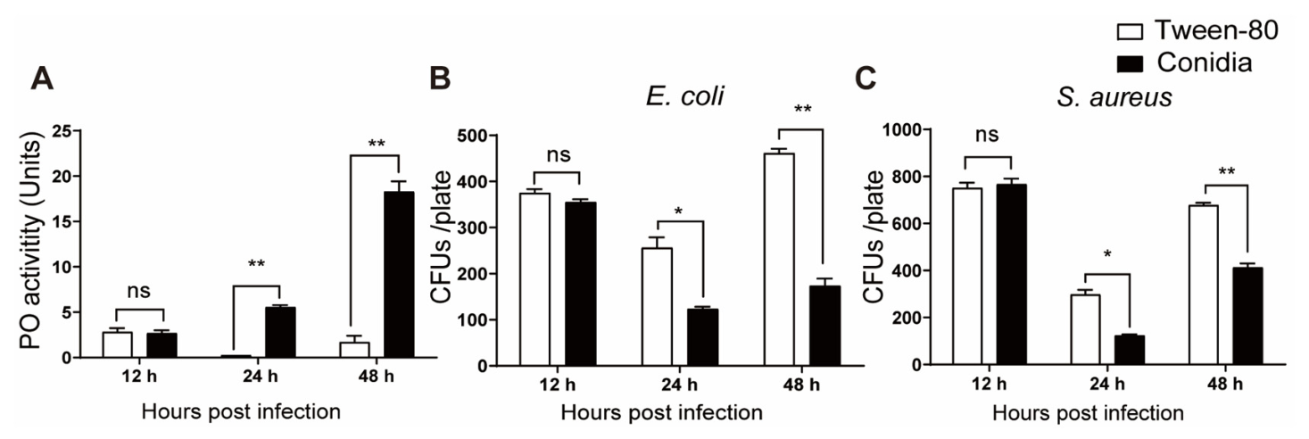
To explore the effects of M. robertsii infection on host humoral antibacterial responses, plasma phenoloxidase (PO) activity and antibacterial capacity were tested. Significantly elevated PO activity was observed in the plasma of M. robertsii-infected larvae at 24 and 48 hpi, while no obvious difference was detected at 12 hpi compared with control larvae (Figure 5A).
Figure 5.
M. robertsii infection enhances humoral antibacterial responses in S. frugiperda larvae. Plasma samples were harvested from sixth-instar larvae injected with M. robertsii conidia or Tween-80 solution and subsequently examined for PO activity (A) and antibacterial activity against E. coli (B) or S. aureus (C). Asterisks indicate significant differences (* p < 0.05, ** p < 0.01).
Plasma antibacterial activities against E. coli and S. aureus remained unaltered at 12 hpi following fungal infection. In contrast, plasma collected from M. robertsii-infected larvae at 24 and 48 hpi yielded significantly fewer bacterial colonies for both tested bacterial strains, demonstrating that fungal infection markedly enhances host plasma antibacterial activity at these two time points.
3.5. M. robertsii Infection Enhances Immune-Related Gene Expression
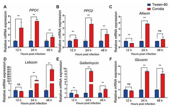
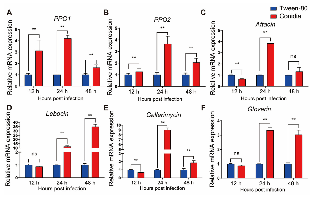
To elucidate the molecular mechanism by which M. robertsii potentiates host humoral antibacterial responses, we further profiled the expression of immune-related genes in larval fat bodies or hemocytes during fungal infection. Both PPO1 and PPO2 transcripts were significantly upregulated across all sampled time points following fungal infection (Figure 6A,B). This finding suggests that the elevated PO activity in larval hemolymph upon M. robertsii challenge stems from the transcriptional induction of PPO genes. The four AMP genes (Gallerimycin, Attacin, Lebocin, and Gloverin) exhibited unaltered or moderately reduced expression at 12 hpi, followed by strong upregulation at 24 and 48 hpi (Figure 6C–F). These results suggest that the enhanced antibacterial activity of hemolymph triggered by fungal infection may arise not only from elevated PO activity, but also from increased abundance of AMPs.
Figure 6.
Transcriptional changes in AMP genes in fat bodies and PPO genes in hemocytes upon fungal infection. Relative transcript levels of PPO1 (A), PPO2 (B), Attacin (C), Lebocin (D), Gallerimycin (E), and Gloverin (F) were detected at 12, 24, and 48 hpi. Hemolymph and fat bodies samples were harvested from M. robertsii-infected and control larvae, and gene expression profiles were quantified via RT-qPCR. All expression values were normalized to the internal reference gene Rpl32. Asterisks indicate significant differences (** p < 0.01).
4. Discussion
The fall armyworm is a highly destructive agricultural pest that infests staple crops including maize, rice, and cotton. Following its recent invasion from the Americas into Africa and Asia, this pest has emerged as a severe and escalating threat to global food security [18,19]. While chemical insecticides currently serve as the dominant strategy for S. frugiperda management, their widespread application poses substantial ecological risks and human health hazards. These drawbacks have stimulated the development of environmentally sustainable alternative control methods, among which EPF-based biological control exhibits great application potential [20]. Elucidating the interactive mechanisms between EPF and insect hosts is indispensable for developing efficient fungal pest control strategies and improving fungal pathogenicity against insect pests.
In the present study, S. frugiperda larval mortality increased in a time- and dose-dependent manner following M. robertsii infection, regardless of whether conidia were delivered via immersion or injection. Consistent with our results, previous studies have demonstrated positive correlations between fungal conidial concentrations and larval mortality in S. frugiperda infected with other EPF, including Beauveria bassiana [21] and M. rileyi [22]. Such dose-dependent larval killing reflects the inherent virulence capacity of M. robertsii. Notably, conidial injection yielded faster and more lethal infection outcomes than immersion treatment. This disparity is primarily attributable to direct hemocoelic delivery, which bypasses the natural cuticular penetration barrier required for topical fungal infection. Conidial injection has been widely adopted in previous studies to characterize host immune responses in S. frugiperda [22]. Considering its higher mortality efficiency and better experimental reproducibility, we selected the injection inoculation method for all subsequent experiments to ensure uniform fungal infection for downstream immune assays. In this study, sixth-instar larvae rather than fourth- or fifth-instar larvae were selected for experiments. The primary reason is that fourth- and fifth-instar larvae have smaller body sizes, and the volume of hemolymph harvested from them is insufficient for immune response assays. In contrast, sixth-instar larvae are larger, enabling the collection of adequate hemolymph for immune response analysis.
Upon entering the hemocoel, M. robertsii confronts the multifaceted immune defense system of S. frugiperda. Prior work has demonstrated that infection by the closely related fungus M. rileyi suppresses cellular immunity and inhibits hemocyte spreading in Helicoverpa armigera [15]. In agreement with these observations, our results revealed that M. robertsii infection compromised multiple key cellular immune processes, including nodulation, encapsulation, and phagocytosis. The diminished spreading capacity of plasmatocytes in fungus-injected larvae may underlie the impairment of encapsulation and nodulation responses, given that the morphological transition of plasmatocytes from a rounded, non-spread phenotype to an adherent, spread morphology is a prerequisite for hemocyte recognition and clearance of invading foreign targets [17]. Nevertheless, our results diverge from a previous report by Liu et al. (2019), which described no detectable influence of M. robertsii on the cellular immunity of S. litura [23]. Interestingly, M. anisopliae triggers host immune activation during cuticular penetration, yet host defenses become suppressed once the fungus successfully colonizes the hemocoel [24]. Collectively, these comparative findings indicate that variable host cellular immune outcomes can arise from differences in host species, fungal strains, or the sampling time point within the infection cycle.
Our results demonstrated that M. robertsii infection markedly reduced the abundance of circulating hemocytes, especially granulocytes and plasmatocytes. Hemocyte depletion is a critical factor underlying impaired cellular immune performance, as hemocyte quantity serves as a core determinant of insect immune capacity and participates in both cellular and humoral immune cascades [17]. In this study, fungal infection significantly triggered hemocyte apoptosis and necrosis, which was further supported by the transcriptional upregulation of multiple apoptosis-related genes in infected hemocytes. These findings indicate that M. robertsii suppresses host cellular immunity by inducing hemocyte apoptosis and cell death.
EPF secrete diverse cytotoxic virulence factors to disrupt host immune homeostasis. For example, ribotoxins produced by Metarhizium fungi induce ribosome dysfunction and apoptotic cell death, thereby exerting potent insecticidal activity [25,26]. Moreover, hemocytes are essential for inducing the expression of AMP genes such as Defensin in the fat body during immune challenge, further highlighting the central role of hemocytes in systemic immune signaling [27]. Consistent with this mechanism, B. bassiana secretes multiple immune-suppressive metabolites, including the nonribosomal peptide beauvericin, cyclic beauverolides, and the 2-pyridone tenellin, all of which can directly inhibit host immune responses [28,29,30].
Based on our findings regarding cellular immunity, we propose that M. robertsii suppresses host cellular immune responses by a combined strategy: reducing the number of host hemocytes and impairing hemocyte spreading capacity. M. robertsii reduces hemocyte abundance by inducing hemocyte apoptosis and cell death. However, the mechanism underlying the impairment of hemocyte spreading capacity remains unclear. Previous studies have demonstrated that EPF disrupt host hemocyte spreading by damaging the cytoskeleton. For instance, M. anisopliae impairs spreading of Galleria mellonella hemocytes by secreting destruxins, secondary metabolites that disrupt the cytoskeleton [31]. Further investigation is required to determine whether M. robertsii impairs the spreading capacity of S. frugiperda hemocytes via cytoskeleton disruption.
In contrast to the suppressed cellular immune response, M. robertsii infection significantly potentiated host humoral antibacterial responses. This conclusion is supported by our observation that M. robertsii infection markedly increases the antibacterial activity of host hemolymph. A similar phenomenon was also detected in our previous studies following M. rileyi infection [22]. To explore the underlying cause of elevated hemolymph antibacterial activity, we examined the transcript levels of several host AMP genes. Our results showed that these genes were significantly upregulated following fungal infection. These findings indicate that the enhanced antibacterial capacity of hemolymph is, at least in part, attributable to increased AMP abundance. The upregulation of AMP expression has also been documented in other fungal infection systems. For instance, in Drosophila melanogaster, the recognition of fungal cell wall β-1,3-glucan by the pattern recognition receptor GNBP3 activates the Toll signaling pathway, thereby robustly inducing the expression of the antifungal peptide Drosomycin [32,33]. M. anisopliae infection elevates the transcript levels of lysozyme and inducible antibacterial peptides in Galleria mellonella [34].
The mechanisms underlying the upregulation of host AMP expression following fungal infection remain poorly understood. Our previous study suggested that M. rileyi may upregulate host AMP expression to eliminate gut-derived bacteria that have escaped into the hemolymph. This strategy prevents these opportunistic bacteria from competing with the fungus for hemolymph nutrients [35]. Whether M. robertsii upregulates AMP gene expression in S. frugiperda through this mechanism remains to be further investigated.
PPO activation constitutes a key regulatory step within the insect melanization cascade. Upon microbial challenge, inactive PPO undergoes proteolytic cleavage to yield PO, the catalytic enzyme responsible for melanin biosynthesis [36]. In the present study, we also assessed the influence of M. robertsii infection on PPO activation, an important humoral immune response of the host. Our results demonstrated that fungal infection resulted in increased PO activity in the hemolymph. These findings suggest that the elevated antibacterial activity of hemolymph following fungal infection may stem, at least in part, from enhanced PO activity. RT-qPCR analysis revealed that the expression of host PPO genes was upregulated after fungal infection. This finding suggests that the fungus may enhance hemolymph PO activity by upregulating host PPO transcription.
The effects of fungal infection on host PPO activation response vary across different host–fungal infection systems. For example, B. bassiana suppresses the host PPO-activating response by secreting oosporein [9]. Our previous studies demonstrated that M. robertsii infection exerted no influence on PPO activation response in H. armigera [22]. By contrast, the present study revealed that M. robertsii infection triggers the PPO activating cascade in S. frugiperda. This increase in PO activity has also been reported in several other infection systems. For example, hemolymph PO activity rises significantly in Locusta migratoria migratorioides following M. anisopliae infection [37]. These findings suggest that host insects may deploy PO to defend against fungal infection. Collectively, these findings indicate that fungal impacts on host PPO-activating responses are dependent on both fungal and host species.
In this study, we used two common bacterial species, E. coli and S. aureus, to evaluate host immune responsiveness following fungal infection. In fact, insects harbor a rich community of symbiotic bacteria, especially gut microbiota [38]. Previous studies have shown that gut bacteria can modulate fungal virulence. For example, infection by B. bassiana increases the abundance of gut bacteria such as Serratia marcescens in S. frugiperda. These gut bacteria attenuate fungal virulence against the host [39]. Studies in mosquitoes have demonstrated that gut bacteria can even translocate into the host hemocoel after fungal infection [40]. Whether the host insect mounts the same immune response against these gut-derived bacteria as it does against E. coli and S. aureus remains to be further investigated.
5. Conclusions
This study investigated the pathogenicity of M. robertsii against S. frugiperda using two inoculation methods: larval immersion in conidial suspensions and direct conidial injection into the larval hemocoel. We further comprehensively evaluated the modulatory effects of M. robertsii infection on the cellular and humoral immune responses of S. frugiperda. Mechanistic analysis revealed prominent alterations in host humoral antibacterial responses following fungal challenge, with key antimicrobial peptide and PPO genes exhibiting significant transcriptional upregulation. Collectively, these findings deepen our understanding of molecular interactions between M. robertsii and its insect host and elucidate the core pathogenic mechanisms of fungal infection. This work provides valuable insights for developing efficient strategies to enhance fungal pathogenicity and accelerate the infection process. Furthermore, our results establish a theoretical foundation for the optimized application of M. robertsii as a promising biocontrol agent for sustainable management of S. frugiperda.
Author Contributions
Conceptualization, M.Z.; methodology, M.Z.; software, M.Z.; validation, M.Z.; formal analysis, M.Z.; investigation, M.Z. and J.W.; resources, J.W. and X.L.; data curation, M.Z.; writing—original draft preparation, M.Z.; writing—review and editing, J.W. and X.L.; visualization, M.Z.; supervision, X.L.; project administration, J.W. and X.L.; funding acquisition, J.W. and X.L. All authors have read and agreed to the published version of the manuscript.
Funding
This work was supported by grants from the National Natural Science Foundation of China (32172489 and 32470517).
Institutional Review Board Statement
Ethical review and approval were waived for this study due to Spodoptera frugiperda is an invertebrate insect pest, and experiments with this species do not fall under the jurisdiction of the Institutional Review Board for animal research.
Informed Consent Statement
Not applicable.
Data Availability Statement
The original contributions presented in this study are included in the article. Further inquiries can be directed to the corresponding author.
Conflicts of Interest
The authors declare no conflicts of interest.
References
- Casanova-Torres, Á.; Goodrich-Blair, H. Immune signaling and antimicrobial peptide expression in lepidoptera. Insects 2013, 4, 320–338. [Google Scholar] [CrossRef] [Scilit] [PubMed]
- Schmidt, O.; Theopold, U.; Strand, M. Innate immunity and its evasion and suppression by hymenopteran endoparasitoids. BioEssays 2001, 23, 344–351. [Google Scholar] [CrossRef] [Scilit] [PubMed]
- Zhou, L.; Meng, G.; Zhu, L.; Ma, L.; Chen, K. Insect antimicrobial peptides as guardians of immunity and beyond: A review. Int. J. Mol. Sci. 2024, 25, 3835. [Google Scholar] [CrossRef] [Scilit] [PubMed]
- Marieshwari, B.N.; Bhuvaragavan, S.; Sruthi, K.; Mullainadhan, P.; Janarthanan, S. Insect phenoloxidase and its diverse roles: Melanogenesis and beyond. J. Comp. Physiol. B 2023, 193, 1–23. [Google Scholar] [CrossRef] [Scilit] [PubMed]
- Lu, Y.; Su, F.; Li, Q.; Zhang, J.; Li, Y.; Tang, T.; Hu, Q.; Yu, X.Q. Pattern recognition receptors in Drosophila immune responses. Dev. Comp. Immunol. 2020, 102, 103468. [Google Scholar] [CrossRef] [Scilit] [PubMed]
- Wang, C.; Wang, S. Insect pathogenic fungi: Genomics, molecular interactions, and genetic improvements. Annu. Rev. Entomol. 2017, 62, 73–90. [Google Scholar] [CrossRef] [Scilit] [PubMed]
- Zhang, Q.; Wei, X.; Fang, W.; Huang, X.; Zhang, X. The secretory protein COA1 enables Metarhizium robertsii to evade insect immune recognition during cuticle penetration. Commun. Biol. 2024, 7, 1220. [Google Scholar] [CrossRef] [Scilit] [PubMed]
- Wang, H.; Lu, Z.; Keyhani, N.O.; Deng, J.; Zhao, X.; Huang, S.; Luo, Z.; Jin, K.; Zhang, Y. Insect fungal pathogens secrete a cell wall-associated glucanase that acts to help avoid recognition by the host immune system. PLoS Pathog. 2023, 19, e1011578. [Google Scholar] [CrossRef] [Scilit] [PubMed]
- Feng, P.; Shang, Y.; Cen, K.; Wang, C. Fungal biosynthesis of the bibenzoquinone oosporein to evade insect immunity. Proc. Natl. Acad. Sci. USA 2015, 112, 11365–11370. [Google Scholar] [CrossRef] [Scilit] [PubMed]
- Wang, J.; Hu, H.; Pang, S.; Yin, X.; Cao, B.; Huang, J.; Xu, X.; Weng, Q.; Hu, Q. Destruxin A inhibits the hemocytin-mediated hemolymph immunity of host insects to facilitate Metarhizium infection. Cell Rep. 2024, 43, 113686. [Google Scholar] [CrossRef] [Scilit] [PubMed]
- Early, R.; González-Moreno, P.; Murphy, S.T.; Day, R. Forecasting the global extent of invasion of the cereal pest Spodoptera frugiperda, the fall armyworm. NeoBiota 2018, 40, 25–50. [Google Scholar] [CrossRef] [Scilit]
- Sun, X.X.; Hu, C.X.; Jia, H.R.; Wu, Q.L.; Shen, X.J.; Zhao, S.Y.; Jiang, Y.Y.; Wu, K.M. Case study on the first immigration of fall armyworm, Spodoptera frugiperda invading into China. J. Integr. Agric. 2021, 20, 664–672. [Google Scholar] [CrossRef] [Scilit]
- Sheng, H.; McNamara, P.J.; St Leger, R.J. Metarhizium: An opportunistic middleman for multitrophic lifestyles. Curr. Opin. Microbiol. 2022, 69, 102176. [Google Scholar] [CrossRef] [Scilit] [PubMed]
- Wang, G.J.; Zhuo, X.R.; Wang, W.W.; Liu, X.S.; Wang, G.X.; Wang, J.L. Molecular characterization of immune responses of Helicoverpa armigera to infection with the mermithid nematode Ovomermis sinensis. BMC Genom. 2019, 20, 161. [Google Scholar] [CrossRef] [Scilit] [PubMed]
- Zhong, K.; Liu, Z.; Wang, J.; Liu, X. The entomopathogenic fungus Nomuraea rileyi impairs cellular immunity of its host Helicoverpa armigera. Arch. Insect Biochem. Physiol. 2017, 96, e21402. [Google Scholar] [CrossRef] [Scilit] [PubMed]
- Qiao, C.; Li, J.; Wei, X.H.; Wang, J.L.; Wang, Y.F.; Liu, X.S. SRP gene is required for Helicoverpa armigera prophenoloxidase activation and nodulation response. Dev. Comp. Immunol. 2014, 44, 94–99. [Google Scholar] [CrossRef] [Scilit] [PubMed]
- Lavine, M.D.; Strand, M.R. Insect hemocytes and their role in immunity. Insect Biochem. Mol. Biol. 2002, 32, 1295–1309. [Google Scholar] [CrossRef] [Scilit] [PubMed]
- Goergen, G.; Kumar, P.L.; Sankung, S.B.; Togola, A.; Tamò, M. First report of outbreaks of the fall armyworm Spodoptera frugiperda (J E smith) (lepidoptera, noctuidae), a new alien invasive pest in west and central Africa. PLoS ONE 2016, 11, e0165632. [Google Scholar] [CrossRef] [Scilit] [PubMed]
- Baloch, M.N.; Fan, J.; Haseeb, M.; Zhang, R. Mapping potential distribution of Spodoptera frugiperda (Lepidoptera: Noctuidae) in Central Asia. Insects 2020, 11, 172. [Google Scholar] [CrossRef] [Scilit] [PubMed]
- Wang, L.; Wang, J.; Zhang, X.; Yin, Y.; Li, R.; Lin, Y.; Deng, C.; Yang, K.; Liu, X.; Wang, Z. Pathogenicity of Metarhizium rileyi against Spodoptera litura larvae: Appressorium differentiation, proliferation in hemolymph, immune interaction, and reemergence of mycelium. Fungal Genet. Biol. 2021, 150, 103508. [Google Scholar] [CrossRef] [Scilit] [PubMed]
- Abd El Azim, A.M.; Khashaba, E.H.K.; El Kady, G.A. Effectiveness study of the dual application of new indigenous entomopathogenic nematode isolate Heterorhabditis taysearae and entomopathogenic fungi Beauveria bassiana against armyworm (Spodoptera frugiperda). Egypt. J. Biol. Pest Control 2024, 34, 41. [Google Scholar] [CrossRef] [Scilit]
- Wang, J.L.; Yang, K.H.; Wang, S.S.; Li, X.L.; Liu, J.; Yu, Y.X.; Liu, X.S. Infection of the entomopathogenic fungus Metarhizium rileyi suppresses cellular immunity and activates humoral antibacterial immunity of the host Spodoptera frugiperda. Pest Manag. Sci. 2022, 78, 2828–2837. [Google Scholar] [CrossRef] [Scilit] [PubMed]
- Liu, S.; Xu, Z.; Wang, X.; Zhao, L.; Wang, G.; Li, X.; Zhang, L. Pathogenicity and in vivo Development of Metarhizium rileyi Against Spodoptera litura (Lepidoptera: Noctuidae) Larvae. J. Econ. Entomol. 2019, 112, 1598–1603. [Google Scholar] [CrossRef] [Scilit] [PubMed]
- Wang, C.; St Leger, R.J. The Metarhizium anisopliae Perilipin homolog MPL1 regulates lipid metabolism, appressorial turgor pressure, and virulence. J. Biol. Chem. 2007, 282, 21110–21115. [Google Scholar] [CrossRef] [Scilit] [PubMed]
- Li, H.; Xia, Y. High cell density fed-batch production of insecticidal recombinant ribotoxin hirsutellin A from Pichia pastoris. Microb. Cell Fact. 2018, 17, 145. [Google Scholar] [CrossRef] [Scilit] [PubMed]
- Yuan, Y.; Huang, W.; Chen, K.; Ling, E. Beauveria bassiana ribotoxin inhibits insect immunity responses to facilitate infection via host translational blockage. Dev. Comp. Immunol. 2020, 106, 103605. [Google Scholar] [CrossRef] [Scilit] [PubMed]
- Brennan, C.A.; Delaney, J.R.; Schneider, D.S.; Anderson, K.V. Psidin is required in Drosophila blood cells for both phagocytic degradation and immune activation of the fat body. Curr. Biol. 2007, 17, 67–72. [Google Scholar] [CrossRef] [Scilit] [PubMed]
- Molnár, I.; Gibson, D.M.; Krasnoff, S.B. Secondary metabolites from entomopathogenic hypocrealean fungi. Nat. Prod. Rep. 2010, 27, 1241–1275. [Google Scholar] [CrossRef] [Scilit] [PubMed]
- Zhang, L.; Yue, Q.; Wang, C.; Xu, Y.; Molnár, I. Secondary metabolites from hypocrealean entomopathogenic fungi: Genomics as a tool to elucidate the encoded parvome. Nat. Prod. Rep. 2020, 37, 1164–1180. [Google Scholar] [CrossRef] [Scilit] [PubMed]
- Zhang, L.; Fasoyin, O.E.; Molnár, I.; Xu, Y. Secondary metabolites from hypocrealean entomopathogenic fungi: Novel bioactive compounds. Nat. Prod. Rep. 2020, 37, 1181–1206. [Google Scholar] [CrossRef] [Scilit] [PubMed]
- Vilcinskas, A.; Matha, V.; Götz, P. Effects of the entomopathogenic fungus Metarhizium anisopliae and its secondary metabolites on morphology and cytoskeleton of plasmatocytes isolated from the greater wax moth, Galleria mellonella. J. Insect Physiol. 1997, 43, 1149–1159. [Google Scholar] [CrossRef] [Scilit] [PubMed]
- Gottar, M.; Gobert, V.; Matskevich, A.A.; Reichhart, J.M.; Wang, C.; Butt, T.M.; Belvin, M.; Hoffmann, J.A.; Ferrandon, D. Dual detection of fungal infections in Drosophila via recognition of glucans and sensing of virulence factors. Cell 2006, 127, 1425–1437. [Google Scholar] [CrossRef] [Scilit] [PubMed]
- Zheng, X.; Li, S.; Si, Y.; Hu, J.; Xia, Y. Locust can detect β-1, 3-glucan of the fungal pathogen before penetration and defend infection via the Toll signaling pathway. Dev. Comp. Immunol. 2020, 106, 103636. [Google Scholar] [CrossRef] [Scilit] [PubMed]
- Schuhmann, B.; Seitz, V.; Vilcinskas, A.; Podsiadlowski, L. Cloning and expression of gallerimycin, an antifungal peptide expressed in immune response of greater wax moth larvae, Galleria mellonella. Arch. Insect Biochem. Physiol. 2003, 53, 125–133. [Google Scholar] [CrossRef] [Scilit] [PubMed]
- Wang, J.L.; Sun, J.; Song, Y.J.; Zheng, H.H.; Wang, G.J.; Luo, W.X.; Li, L.; Liu, X.S. An entomopathogenic fungus exploits its host humoral antibacterial immunity to minimize bacterial competition in the hemolymph. Microbiome 2023, 11, 116. [Google Scholar] [CrossRef] [Scilit] [PubMed]
- Kanost, M.R.; Jiang, H.; Yu, X.Q. Innate immune responses of a lepidopteran insect, Manduca sexta. Immunol. Rev. 2004, 198, 97–105. [Google Scholar] [CrossRef] [Scilit] [PubMed]
- Mullen, L.M.; Goldsworthy, G.J. Immune responses of locusts to challenge with the pathogenic fungus Metarhizium or high doses of laminarin. J. Insect Physiol. 2006, 52, 389–398. [Google Scholar] [CrossRef] [Scilit] [PubMed]
- Han, S.; Akhtar, M.R.; Xia, X. Functions and regulations of insect gut bacteria. Pest Manag. Sci. 2024, 80, 4828–4840. [Google Scholar] [CrossRef] [Scilit] [PubMed]
- Peng, Y.; Wen, S.; Wang, G.; Zhang, X.; Di, T.; Du, G.; Chen, B.; Zhang, L. Reconstruction of gut bacteria in Spodoptera frugiperda infected by Beauveria bassiana affects the survival of host pest. J. Fungi 2023, 9, 906. [Google Scholar] [CrossRef] [Scilit] [PubMed]
- Wei, G.; Lai, Y.; Wang, G.; Chen, H.; Li, F.; Wang, S. Insect pathogenic fungus interacts with the gut microbiota to accelerate mosquito mortality. Proc. Natl. Acad. Sci. USA 2017, 114, 5994–5999. [Google Scholar] [CrossRef] [Scilit] [PubMed]

Disclaimer/Publisher’s Note: The statements, opinions and data contained in all publications are solely those of the individual author(s) and contributor(s) and not of MDPI and/or the editor(s). MDPI and/or the editor(s) disclaim responsibility for any injury to people or property resulting from any ideas, methods, instructions or products referred to in the content. |
© 2026 by the authors. Licensee MDPI, Basel, Switzerland. This article is an open access article distributed under the terms and conditions of the Creative Commons Attribution (CC BY) license.