


Abstract
Collective catering can expose many consumers to foodborne and antimicrobial-resistant bacteria but is poorly monitored in sub-Saharan Africa. This study assessed the microbiological quality of 159 ready-to-eat meals served in university, hospital-adjacent, and industrial canteens in Ouagadougou, Burkina Faso, and the virulence genes and antimicrobial resistance (AMR) of Escherichia coli and Staphylococcus aureus isolates. Genes were detected by PCR and susceptibility by disk diffusion. Under criteria adapted from those for cooked ready-to-eat dishes, 70.4% and 50.3% of meals were unsatisfactory for coagulase-positive staphylococci and E. coli, respectively (38.4% for staphylococci under UK Health Protection Agency guidelines). E. coli isolates carrying astA as the sole virulence gene (n = 50) were recovered from 44 meals (27.7%), and enterotoxigenic E. coli (estIa) from five meals; aggR and pic were not detected. The exfoliative toxin A gene eta was detected by PCR (not sequenced) in 73 S. aureus isolates from 51 meals; none carried mecA or sea–see. Multidrug resistance affected 76.0% of astA-positive E. coli and 73.9% of S. aureus isolates (68.0% and 65.2%, excluding presumptive phenotypes). In E. coli, blaSHV (81.8%), blaCTX-M−1 group (27.3%), and blaTEM (25.5%) predominated. These findings indicate inadequate microbiological control and warrant AMR surveillance across the food chain.
1. Introduction
Foodborne diseases remain a major global public health challenge. The World Health Organization (WHO) estimates that contaminated food causes approximately 600 million illnesses and 420,000 deaths every year, with children under five years of age bearing a disproportionate share of this burden [1]. The WHO African Region has the highest per-capita burden, with an estimated 91 million illnesses and 137,000 deaths annually, most of which are caused by diarrheal disease agents [1,2].
Among the bacterial agents of foodborne illness, Staphylococcus aureus and diarrheagenic Escherichia coli (DEC) are of particular concern. S. aureus produces a wide range of virulence factors, including staphylococcal enterotoxins (SEs), which cause staphylococcal food poisoning; toxic shock syndrome toxin-1 (TSST-1); and exfoliative toxins A and B, which are encoded by eta and etb and cause staphylococcal scalded skin syndrome rather than food poisoning [3,4]. Among the DEC pathotypes, enteroaggregative E. coli (EAEC) and enterotoxigenic E. coli (ETEC) are leading causes of persistent diarrhea and acute watery diarrhea, respectively, in low- and middle-income countries [5,6,7].
The spread of antimicrobial resistance (AMR) among foodborne bacteria adds a further dimension to this risk, and the WHO regards AMR as one of the greatest threats to global health [8]. Ready-to-eat (RTE) foods, including cooked meals served in collective catering, constitute a critical interface through which bacteria carrying resistance determinants can reach large numbers of consumers simultaneously [9]. A meta-analysis of RTE foods in developing countries reported pooled prevalences of 30.2% for S. aureus and 23.8% for E. coli [10].
In Burkina Faso, university restaurants, hospital-adjacent canteens, and industrial-zone canteens serve meals daily to large numbers of students, patients’ relatives, healthcare workers, and employees, yet their microbiological surveillance remains limited. Poor hygiene practices have been documented in university restaurants in Ouagadougou [11] and in street restaurants [12], and multidrug-resistant Salmonella carrying blaTEM and other resistance genes has been recovered from RTE street foods [13,14]. Compaore et al. reported that 11.9% of common foods sold in Burkina Faso were of unsatisfactory microbiological quality [15], and a recent scoping review confirmed that data on resistance genes in African RTE foods remain scarce and geographically uneven [16]. To our knowledge, no study has combined the molecular screening of virulence and resistance determinants in E. coli and S. aureus isolates from collective catering in Burkina Faso.
This study therefore aimed (i) to assess the microbiological quality of RTE meals served in collective catering facilities in Ouagadougou; (ii) to screen E. coli and S. aureus isolates for virulence genes; and (iii) to describe their phenotypic AMR and, for E. coli, the carriage of acquired resistance genes to provide baseline data for food safety and AMR surveillance policies in Burkina Faso. The study was descriptive and was not designed to compare the three types of collective catering.
2. Materials and Methods
2.1. Study Design, Period, and Setting
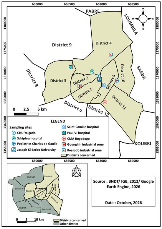
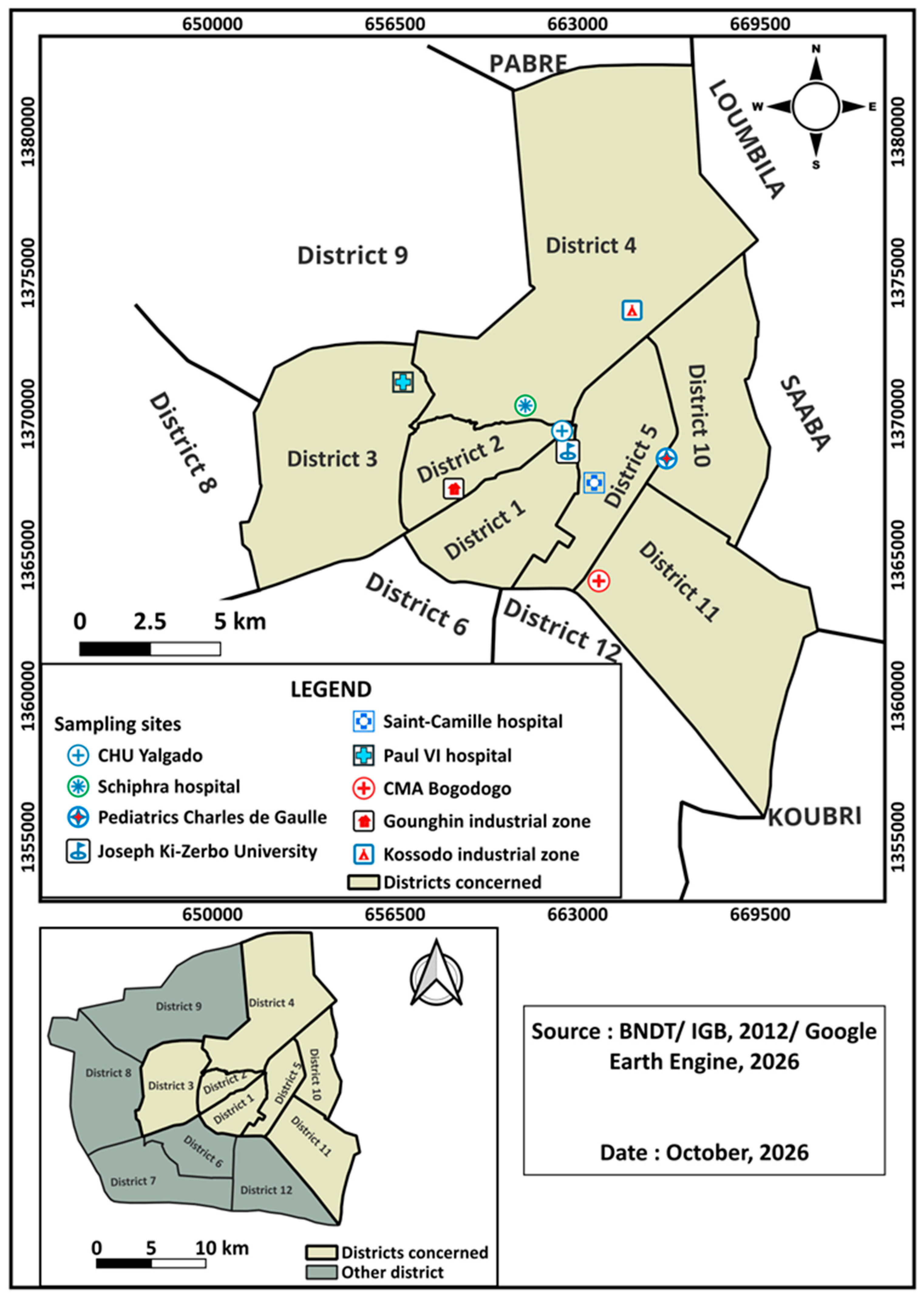
A prospective cross-sectional study was conducted from August 2020 to July 2021 in collective catering establishments in Ouagadougou, Burkina Faso (Figure 1). Three types of collective catering were targeted: (i) university catering (restaurants serving students on the Université Joseph Ki-Zerbo campus); (ii) hospital-adjacent catering (restaurants and canteens near the Hôpital Paul VI, the Schiphra Hospital, the Centre Médical avec Antenne chirurgicale de Bogodogo, the Centre Hospitalier Universitaire Yalgado Ouédraogo, the Hôpital Saint Camille, and the Charles de Gaulle Pediatric Hospital); and (iii) industrial catering (canteens in the Kossodo and Gounghin industrial zones). Each catering establishment was visited once, and a single meal was sampled per establishment during standard meal service hours (10:00–14:00). In total, 159 meals were collected from nine sites: 105 from industrial catering (Kossodo, n = 77; Gounghin, n = 28), 45 from hospital-adjacent catering (Schiphra Hospital, n = 13; Charles de Gaulle Pediatric Hospital, n = 9; Hôpital Saint Camille, n = 9; Centre Hospitalier Universitaire Yalgado Ouédraogo, n = 7; Centre Médical avec Antenne chirurgicale de Bogodogo, n = 6; Hôpital Paul VI, n = 1), and 9 from university catering (Université Joseph Ki-Zerbo). The sites were selected because of their size and the number of consumers served: the Université Joseph Ki-Zerbo is the largest university in Burkina Faso; the hospitals include the largest university hospital centers in Ouagadougou; and Kossodo and Gounghin are the two industrial zones of the city.
Figure 1.
Location of the sampling sites (university, hospital-adjacent, and industrial catering) in Ouagadougou, Burkina Faso. Source: Base Nationale de Données Topographiques (BNDT)/Institut Géographique du Burkina (IGB), 2012; Google Earth Engine, 2026; map prepared with QGIS version 3.30 (QGIS Development Team, Open Source Geospatial Foundation).
2.2. Sample Collection
A total of 159 RTE meal samples (one sample unit per meal) were collected aseptically in sterile containers (300–500 g or mL per sample). The meals comprised cereal-based dishes (n = 63; rice with sauce, riz gras, couscous, pigri, spaghetti, macaroni and other pasta, tô, and donkounou), legume-based dishes (n = 63; beans, plain or with couscous, maize, or attiéké; gonré; and Bambara groundnut dishes), tuber-based dishes (n = 10; attiéké, placali, and boiled yam), dairy products (n = 9; yogurt and dèguè), and other preparations (n = 14; fish soup, meat soup, millet porridge, babenda, and alloco). Samples were transported in an insulated cooler (<4 °C) to the LaBESTA laboratory, Université Joseph Ki-Zerbo, and analyzed within 2 h. Tables S4, S5 and S11 list the origin of each isolate, and Table S13 provides the counts for each sample.
2.3. Enumeration of Thermotolerant Coliforms and Identification of Escherichia coli
Thermotolerant coliforms were enumerated according to NF V08-060:2009 [17], which applies the colony-count technique of ISO 4832:1991 [18] with incubation at 44 °C, except that 100 µL of inoculum was plated. The initial suspension (10−1) was prepared by aseptically adding 25 g (or 25 mL) of each sample to 225 mL of buffered peptone water (BPW; Liofilchem, Roseto degli Abruzzi, Italy) and homogenizing it. Serial decimal dilutions were prepared by transferring 1 mL of the initial suspension into a screw-capped tube containing 9 mL of sterile BPW, and then 1 mL of each dilution into 9 mL of BPW to obtain the next dilution (10−2 and 10−3). A 100 µL aliquot of the initial suspension and of each dilution was placed in the center of a sterile 90 mm Petri dish (one plate per dilution). Approximately 12.5 mL of molten violet red bile lactose (VRBL) agar (Liofilchem, Roseto degli Abruzzi, Italy) was added to each plate and carefully mixed with the inoculum; after solidification, approximately 7.5 mL of the same medium was poured as an overlay to create microaerophilic conditions and prevent the spreading of swarming bacteria such as Proteus. Plates were incubated at 44.0 ± 0.5 °C for 24 h. Characteristic colonies (small, purple-violet colonies with a dark center) were counted as thermotolerant coliforms, and the counts were calculated as described in Section 2.5.
E. coli was identified from the thermotolerant coliform colonies. From each positive sample, five characteristic colonies (or all colonies when fewer than five were present) were picked from the VRBL plates used for counting and subcultured on bromocresol purple (BCP) lactose agar (Liofilchem, Roseto degli Abruzzi, Italy) at 37 °C for 24 h. Colonies that turned the medium from purple to yellow (lactose fermentation) were purified on nutrient agar (Liofilchem, Roseto degli Abruzzi, Italy) and examined by the IMViC tests, including indole production in peptone water (Kovacs’ reagent, Liofilchem, Roseto degli Abruzzi, Italy), the methyl red and Voges–Proskauer reactions in methyl red–Voges–Proskauer (MR-VP) broth (Liofilchem, Roseto degli Abruzzi, Italy), and citrate utilization on Simmons citrate agar (Liofilchem, Roseto degli Abruzzi, Italy), each after incubation at 37 °C for 18–24 h. Isolates with the IMViC profile + + − − (typical E. coli) or − + − − (atypical E. coli) were confirmed using the API 20E system (bioMérieux, Marcy-l’Étoile, France) according to the manufacturer’s instructions. The number of E. coli in each sample was then calculated from the proportion of confirmed colonies (Section 2.5). Confirmed isolates were purified on nutrient agar, stored at −80 °C in brain heart infusion broth (Liofilchem, Roseto degli Abruzzi, Italy) supplemented with 30% (v/v) glycerol, and subsequently analyzed by polymerase chain reaction (PCR) (Section 2.6, Section 2.7 and Section 2.10).
2.4. Enumeration of Coagulase-Positive Staphylococci and Identification of Staphylococcus aureus
Coagulase-positive staphylococci were enumerated according to ISO 6888-1:1999/Amd 1:2003 [19] on Baird–Parker agar supplemented with egg yolk tellurite emulsion (Liofilchem, Roseto degli Abruzzi, Italy), where tellurite reduction produces black colonies and egg yolk proteolysis produces a clear halo. A 100 µL aliquot of the initial suspension and of each decimal dilution prepared as described in Section 2.3 was spread as rapidly as possible over the surface of the medium (one plate per dilution). After the inoculum had dried, the plates were incubated at 37 °C for 24–48 h, and characteristic colonies (black, shiny, and convex, surrounded by a clear halo) were counted; counts were calculated as described in Section 2.5. For confirmation, five characteristic colonies per positive sample (or all colonies when fewer than five were present) were subcultured on DNase agar (Liofilchem, Roseto degli Abruzzi, Italy) and incubated at 37 °C for 18–24 h. The plates were then flooded with 1 N hydrochloric acid and examined after 5 min at room temperature; a clear halo around the colonies (DNA hydrolysis) indicated a positive reaction, whereas an unchanged medium indicated a negative reaction. DNase-positive colonies were further tested for catalase and used the tube coagulase test (rabbit plasma; Liofilchem, Roseto degli Abruzzi, Italy), and catalase- and coagulase-positive colonies were confirmed as S. aureus; species identity was further supported by amplification of femA in the multiplex PCR (Section 2.8). The number of coagulase-positive staphylococci was then calculated from the proportion of confirmed colonies (Section 2.5). Confirmed isolates were purified on Mueller–Hinton (MH) agar (Liofilchem, Roseto degli Abruzzi, Italy), stored at −80 °C in brain heart infusion broth supplemented with 30% (v/v) glycerol, and subsequently analyzed by PCR (Section 2.6 and Section 2.8).
2.5. Calculation of Counts and Microbiological Quality Criteria
Counts were calculated according to ISO 7218:2007 [20]. The number N of thermotolerant coliforms or of coagulase-positive staphylococci per gram (or milliliter) of the sample was calculated from two successive dilutions as N = ΣC/[V(n1 + 0.1n2)d], where ΣC is the sum of the characteristic colonies counted on the retained plates, V is the volume of inoculum per plate (0.1 mL), n1 and n2 are the numbers of plates retained at the first and second dilutions (one plate per dilution), and d is the dilution of the first retained dilution. After confirmation, the number of E. coli or coagulase-positive staphylococci was calculated as a = (b/A) × C, where A is the number of colonies picked for confirmation, b is the number of colonies that met the identification criteria, and C is the count of characteristic colonies (thermotolerant coliforms on VRBL agar or characteristic colonies on Baird–Parker agar); when all the colonies picked are confirmed (b = A), a equals C. With one plate per dilution and 100 µL of inoculum, one colony at the 10−1 dilution corresponds to approximately 9 × 101 colony-forming units (CFU)/g, which was the lowest detectable count; samples without characteristic colonies were reported as not detected.
Burkina Faso has no national microbiological criteria for cooked meals, and the Codex Alimentarius provides principles for establishing such criteria rather than numerical limits [21]. The available criteria, such as those of Regulation (EC) No 2073/2005 [22], were developed for foods produced and consumed in Europe or North America, and none covers traditional dishes such as tô, riz gras, pigri, gonré, or dèguè, which are not consumed in the countries where these criteria were established. We therefore adapted the criteria used for cooked, ready-to-eat dishes. The primary reference was the Luxembourg guidelines for “cooked prepared dishes ready to eat” [23], which are based on Regulation (EC) No 2073/2005, opinions of the French Food Safety Agency, and Quebec standards: coagulase-positive staphylococci, m = 102 CFU/g and M = 103 CFU/g; E. coli, m = 10 CFU/g and M = 102 CFU/g. For thermotolerant coliforms, for which these guidelines set no limit for cooked dishes, m = 102 CFU/g and M = 103 CFU/g were used [24]. For each parameter, meals were classified as satisfactory (≤m), acceptable (>m and ≤M), or unsatisfactory (>M) [23]. Because the lowest detectable count exceeded the E. coli m value, the satisfactory and acceptable classes were pooled for E. coli (≤102 CFU/g). Counts of coagulase-positive staphylococci ≥105 CFU/g, which the Luxembourg guidelines regard as a direct health risk, were also recorded. As a sensitivity analysis, coagulase-positive staphylococci were also interpreted according to the guidelines of the UK Health Protection Agency (HPA) for ready-to-eat foods [25] (unsatisfactory, >104 CFU/g; potentially hazardous, ≥105 CFU/g), with the satisfactory and borderline classes pooled because the lowest detectable count exceeded the HPA satisfactory limit (<20 CFU/g); the HPA limit for E. coli (unsatisfactory, >102 CFU/g) is identical to the M value used here.
2.6. DNA Extraction and General PCR Procedures
Total DNA was extracted by thermal lysis [26]: three colonies from an 18–24 h culture of each isolate were suspended in 500 µL of sterile distilled water, boiled for 10 min, and centrifuged at 10,000 rpm (12,400× g) for 10 min. The supernatant was used as the DNA template. For efficient screening, lysates were pooled in groups of five (100 µL each); isolates from positive pools were retested individually by simplex PCR with the corresponding primers, and only individually confirmed results are reported. All PCR assays were performed in a Techne TC-412 thermocycler (Techne, Cambridge, UK). Each run included the positive control strains indicated in Section 2.7, Section 2.8 and Section 2.10, a negative control strain for the 16-plex PCR (Section 2.7), and a no-template control (nuclease-free water). Amplicons were separated by electrophoresis on 1.5% agarose gels in 1× Tris–acetate–EDTA (TAE) buffer (Cleaver Scientific, Rugby, UK) alongside a 100 bp DNA ladder (Solis BioDyne, Tartu, Estonia), stained with ethidium bromide, visualized under ultraviolet (UV) light, and identified by comparison with the expected amplicon sizes listed in Tables S1–S3. A run was considered valid when each positive control yielded a single band of the expected size and the negative controls yielded no band; otherwise, the run was repeated. An isolate was considered positive for a gene when it yielded a band of the expected size, as determined against the ladder, in the individual (simplex) confirmation reaction.
2.7. Detection of Virulence Genes in Escherichia coli
All confirmed E. coli isolates were screened for 16 target genes using a 16-plex PCR (two 8-plex reactions) adapted from Antikainen et al. [27], with primers described previously [27,28,29,30] (Table S1). Multiplex 1 targeted eae, escV, ent, bfpB, EHEC-hlyA, stx1, stx2, and ipaH; multiplex 2 targeted invE, aggR, pic, astA, elt, estIa, estIb, and uidA (internal control). Each 20 µL reaction contained 4 µL of 5× FIREPol® Master Mix Ready to Load (Solis BioDyne), 0.5 µL of each primer (Genecust, Boynes, France), 3 µL of nuclease-free water, and 5 µL of the DNA template. Cycling conditions were as follows: 98 °C for 30 s; 35 cycles of 98 °C for 30 s, 62.5 °C for 60 s, and 72 °C for 90 s; and a final extension at 72 °C for 10 min. Amplicons were separated on 1.5% agarose gels (130 V, 90 min). Pathotypes were assigned as follows: enteropathogenic E. coli (EPEC), eae and/or escV/ent (typical EPEC if bfpB-positive); Shiga toxin-producing E. coli (STEC), stx1 and/or stx2; enterohemorrhagic E. coli (EHEC), stx together with eae; enteroinvasive E. coli (EIEC), ipaH and/or invE; EAEC, aggR and/or pic (the EAEC markers included in the panel, which did not include aaiC or aatA); and ETEC, elt, estIa, and/or estIb. Reference strains 17-2 (EAEC), E2348/69 (EPEC), EDL933 (EHEC), Shigella flexneri M90T (EIEC/Shigella), and EDL1493 (ETEC) were used as positive controls, and E. coli HB101 was used as a negative control. Because astA, which encodes enteroaggregative heat-stable enterotoxin 1 (EAST1), also occurs in other pathotypes and in commensal E. coli [31,32], isolates in which astA was the only virulence gene detected were not assigned to any pathotype and are referred to as astA-positive E. coli.
2.8. Detection of Staphylococcus aureus Toxin Genes and mecA
All confirmed S. aureus isolates were screened using the multiplex PCR described by Mehrotra et al. [33] for mecA; the classical enterotoxin genes sea, seb, sec, sed, and see; the TSST-1 gene tst; and the exfoliative toxin genes eta and etb, with femA as an internal control (Table S2). Each 20 µL reaction contained 5 µL of genomic DNA, 4 µL of 5× FIREPol® Master Mix Ready to Load (Solis BioDyne), 6 µL of nuclease-free Milli-Q water, and 0.5 µL of each of the 10 primer-pair mixes (each containing the forward and reverse primers, including the femA pair; Table S2). Previously characterized S. aureus strains carrying the target genes, from the LaBESTA strain collection, were used as positive controls, and a no-template control was included in each run. Cycling conditions were as follows: 94 °C for 5 min; 35 cycles of 94 °C for 2 min, 57 °C for 2 min, and 72 °C for 1 min; and a final extension at 72 °C for 7 min. Amplicons were separated by electrophoresis on 1.5% agarose gels. Because the expected eta (93 bp) and sea (102 bp) amplicons differ by only 9 bp, all eta-positive results were confirmed by simplex PCR with the eta primers. The genes encoding the more recently described SEs and SE-like toxins (e.g., seg–sei and selj–selx) were not included in the panel.
2.9. Antimicrobial Susceptibility Testing
The antimicrobial susceptibility of the E. coli isolates carrying virulence genes and of the eta-positive S. aureus isolates was determined by disk diffusion on MH agar with antibiotic disks (Liofilchem, Roseto degli Abruzzi, Italy) and interpreted according to the 2020 recommendations (version 1.0, April 2020) of the Comité de l’Antibiogramme de la Société Française de Microbiologie (CA-SFM) and the European Committee on Antimicrobial Susceptibility Testing (EUCAST) [34]. Inocula were adjusted to a 0.5 McFarland standard (≈1.5 × 10⁸ CFU/mL) using a densitometer (WPA Biowave CO8000, Biochrom, Cambridge, UK). E. coli isolates were tested against 11 agents: ampicillin (AMP, 10 µg), amoxicillin–clavulanic acid (AMC, 20/10 µg), ticarcillin–clavulanic acid (TCC, 75/10 µg), cefoxitin (FOX, 30 µg), ceftriaxone (CRO, 30 µg), cefepime (FEP, 30 µg), imipenem (IPM, 10 µg), ciprofloxacin (CIP, 5 µg), fosfomycin (FOS, 200 µg with 50 µg glucose-6-phosphate), trimethoprim–sulfamethoxazole (SXT, 1.25/23.75 µg), and chloramphenicol (C, 30 µg). S. aureus isolates were tested against 10 agents: penicillin G (P, 1 IU), cefoxitin (FOX, 30 µg), fusidic acid (FA, 10 µg), clindamycin (DA, 2 µg), erythromycin (E, 15 µg), linezolid (LZD, 10 µg), ofloxacin (OFX, 5 µg), chloramphenicol (C, 30 µg), trimethoprim–sulfamethoxazole (SXT, 1.25/23.75 µg), and fosfomycin (FOS, 200 µg). E. coli American Type Culture Collection (ATCC) 25922 and S. aureus ATCC 29213 were used for quality control. Ceftriaxone results were unavailable for 10 E. coli isolates, and 4 of the 73 eta-positive S. aureus isolates were not tested; these isolates were excluded from the corresponding denominators (Table S11). No minimum inhibitory concentrations (MICs) were determined, and no phenotypic extended-spectrum β-lactamase (ESBL), AmpC, or carbapenemase confirmatory tests were performed. The zone diameter breakpoints of CA-SFM/EUCAST 2020 for Enterobacterales and for Staphylococcus spp. were used for E. coli and S. aureus, respectively; for cefoxitin in E. coli, the CA-SFM 2020 breakpoints were applied (S ≥ 19 mm; R < 15 mm). The zone diameter breakpoints, source, and status of each organism–agent combination are given in Table S14. Results were reported as presumptive when disk diffusion was not the recommended method or when resistance required confirmation: (i) in S. aureus, this included cefoxitin resistance in mecA-negative isolates, which was recorded as a discordant phenotype rather than as methicillin resistance, and linezolid and fosfomycin resistance, for which CA-SFM/EUCAST recommend MIC-based confirmation [34]; (ii) in E. coli, this included fosfomycin resistance, because disk diffusion breakpoints for fosfomycin apply only to isolates from uncomplicated urinary tract infections, cefoxitin resistance, which is used mainly as a screening marker for AmpC production, and imipenem resistance, which was not confirmed by MIC determination or carbapenemase testing. Presumptive results are identified as such in the text and are marked in the tables and figures. Multidrug resistance (MDR) was defined as resistance to at least one agent in three or more antimicrobial categories, with categories assigned according to Magiorakos et al. [35] (Table S10). Because the panels covered only 10 of the 17 (E. coli) and 18 (S. aureus) categories proposed by Magiorakos et al., and no aminoglycoside was tested, MDR proportions refer to the categories tested and are not directly comparable with those of studies using broader panels. In line with the CA-SFM/EUCAST 2020 definitions, intermediate results (“susceptible, increased exposure”) were considered susceptible.
2.10. Detection of Acquired Resistance Genes in Escherichia coli
All 55 E. coli isolates carrying a virulence gene (50 astA-positive and 5 ETEC isolates) were screened by simplex PCR for nine acquired resistance genes (Table S3): the β-lactamase genes blaTEM, blaSHV, and blaCTX-M [36,37]; the quinolone resistance genes qnrA, qnrB, qnrC, qnrS, and qepA; and the aminoglycoside acetyltransferase gene aac(6′)-Ib, whose -cr variant also acetylates ciprofloxacin. The last six genes were detected with the primers described by Jacoby et al. [38], which include the qnrC and qepA primers of Kim et al. [39] and the aac(6′)-Ib primers of Park et al. [40]. Each 20 µL reaction contained 5 µL of genomic DNA, 4 µL of 5× FIREPol® Master Mix Ready to Load (Solis BioDyne), 10 µL of nuclease-free Milli-Q water, and 0.5 µL of each primer (forward and reverse; Genecust). The cycling conditions used for each gene are given in Table S3. DNA from previously characterized strains carrying each target gene, from the LaBESTA strain collection, was used as a positive control, and a no-template control was included in each run. After amplification, 10 µL of each product was separated by electrophoresis on 1.5% agarose gels alongside a 100 bp ladder (Solis BioDyne). Because the CTX-M primers are specific for the CTX-M-1 group, blaCTX-M results are reported throughout as blaCTX-M−1 group. Amplicons were not sequenced. Consequently, (i) blaTEM and blaSHV amplicons could not be assigned to extended-spectrum or narrow-spectrum variants; (ii) the aac(6′)-Ib-cr variant could not be distinguished from aac(6′)-Ib; and (iii) the genetic location (plasmid or chromosome) of the genes was not determined. The term “plasmid-mediated quinolone resistance (PMQR) genes” therefore designates the gene families conventionally described as such and does not imply that a plasmid location was demonstrated in our isolates.
2.11. Statistical Analysis
Proportions are reported with their denominators and 95% confidence intervals (CIs), calculated using the Wilson score method; isolates not tested against a given agent were excluded from the denominator for that agent. Differences in the proportion of unsatisfactory samples between food categories were assessed using Pearson’s χ2 test; because some expected counts were <5, p-values were obtained by a Monte Carlo simulation (20,000 replicates). A p-value < 0.05 was considered statistically significant. Resistance proportions are reported at the isolate level. Because up to five colonies per sample were characterized and isolates from the same meal may represent the same strain, the number of positive meals is reported alongside the number of isolates, and MDR was also analyzed at the meal level, as the proportion of positive meals yielding at least one MDR isolate and as the proportion of MDR isolates when only one isolate per meal (the first isolate) was retained. Two further sensitivity analyses were performed: the classification of meals for coagulase-positive staphylococci was repeated using the HPA guidelines [25], and MDR was recomputed after excluding the presumptive results (cefoxitin, linezolid, and fosfomycin in S. aureus; cefoxitin, fosfomycin, and imipenem in E. coli). Analyses were performed in Python 3.11 (SciPy 1.17 and statsmodels 0.15).
3. Results
3.1. Microbiological Quality of Collective Catering Meals
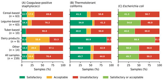
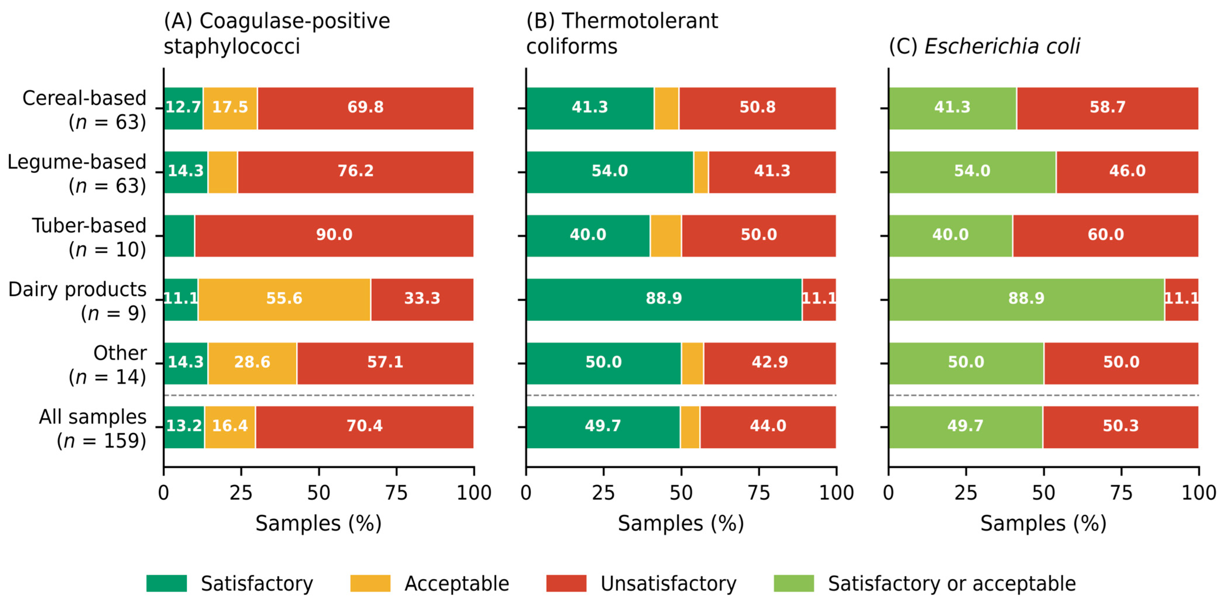
Of the 159 meals analyzed, 112 (70.4%; 95% CI: 62.9–77.0) were unsatisfactory for coagulase-positive staphylococci, 70 (44.0%; 95% CI: 36.5–51.8) for thermotolerant coliforms, and 80 (50.3%; 95% CI: 42.6–58.0) for E. coli (Table 1; Figure 2). Only 21 samples (13.2%; 95% CI: 8.8–19.3) were satisfactory for coagulase-positive staphylococci, and 6 (3.8%) contained ≥105 CFU/g. The proportion of samples unsatisfactory for coagulase-positive staphylococci ranged from 33.3% (dairy products) to 90.0% (tuber-based dishes) and differed significantly between food categories (χ2 = 9.99, df = 4, p = 0.038). Thermotolerant coliforms were detected in 86 samples (54.1%); the proportion of samples unsatisfactory for thermotolerant coliforms ranged from 11.1% (dairy products) to 50.8% (cereal-based dishes) (χ2 = 5.47, df = 4, p = 0.24), and 10 samples (6.3%) were acceptable. In all 86 positive samples, all the colonies picked (up to five per sample) were confirmed as E. coli by the BCP, IMViC, and API 20E tests (b = A), so that E. coli was detected in 86 samples (54.1%) and the E. coli counts were equal to the thermotolerant coliform counts. The proportion of samples unsatisfactory for E. coli (>102 CFU/g) ranged from 11.1% (dairy products) to 60.0% (tuber-based dishes), and the difference between food categories was not statistically significant (χ2 = 8.16, df = 4, p = 0.089). In the sensitivity analysis based on the HPA guidelines, 61 meals (38.4%; 95% CI: 31.2–46.1) were unsatisfactory for coagulase-positive staphylococci (Table S12). The counts obtained for each sample are given in Table S13.
Table 1.
Microbiological quality of meals served in collective catering facilities in Ouagadougou, Burkina Faso (n = 159).
Figure 2.
Distribution of microbiological quality classes by food category for (A) coagulase-positive staphylococci, (B) thermotolerant coliforms, and (C) Escherichia coli. Values are percentages of samples within each category. Classes follow the Luxembourg guidelines for cooked ready-to-eat dishes (coagulase-positive staphylococci, m = 102 CFU/g and M = 103 CFU/g; E. coli, M = 102 CFU/g, with the satisfactory and acceptable classes pooled) and, for thermotolerant coliforms, m = 102 CFU/g and M = 103 CFU/g. The classification according to the HPA guidelines is given in Table S12.
3.2. Virulence Genes of Escherichia coli
Confirmed E. coli isolates were preserved and available for molecular analysis from 60 of the 86 positive samples and from three additional samples (RS 1, HC 4, and HC 6) in which colonies appeared only after the 24 h incubation period used for counting; these three meals were therefore classified as satisfactory, but their isolates were characterized. Isolates from the remaining 26 positive samples were not available for molecular analysis. In total, 149 isolates from these 63 samples were screened by 16-plex PCR, and 55 (36.9%) carried at least one virulence gene; these isolates were recovered from 44 samples (27.7% of all samples; 95% CI: 21.3–35.1) (Table 2; Tables S4 and S11). The most frequent gene was astA, which was detected as the only virulence gene in 50 isolates from 44 samples (27.7%), with the largest numbers from cereal-based (21 samples, 23 isolates) and legume-based (16 samples, 19 isolates) dishes. Because neither aggR nor pic was detected, these isolates were not assigned to a pathotype (Section 2.7). Five isolates from five samples (3.1%; 95% CI: 1.4–7.1) carried estIa and were classified as ETEC; each of these samples also yielded an astA-positive isolate. No EPEC, STEC, EHEC, or EIEC isolates and no isolates carrying aggR or pic were detected.
Table 2.
Positive samples and isolates of Escherichia coli and Staphylococcus aureus carrying virulence genes, by food category.
3.3. Virulence Genes of Staphylococcus aureus
Of the 220 confirmed S. aureus isolates (from 142 samples), 73 (33.2%) were positive for the exfoliative toxin A gene eta by PCR (confirmed by simplex PCR but not by sequencing); these isolates were recovered from 51 samples (32.1% of all samples; 95% CI: 25.3–39.7) across all food categories, with the largest numbers from cereal-based (20 samples, 26 isolates) and legume-based (18 samples, 29 isolates) dishes (Table 2; Tables S5 and S11). None of the 220 S. aureus isolates carried mecA or the classical enterotoxin genes sea–see, and none of the eta-positive isolates carried tst or etb; thus, none could be shown to be enterotoxigenic with the panel used.
3.4. Antimicrobial Resistance of Escherichia coli
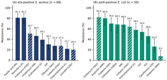
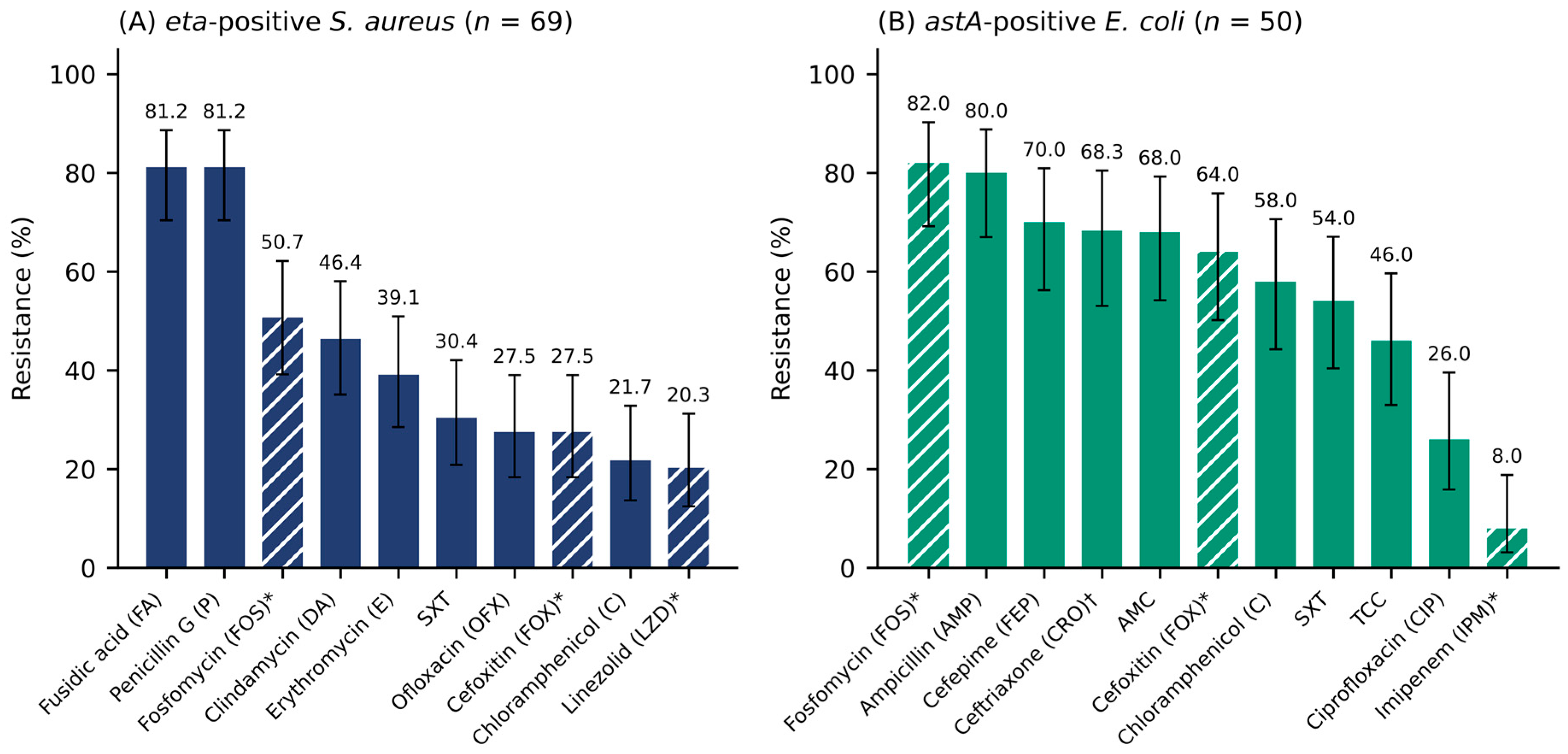
Among the 50 astA-positive E. coli isolates, resistance was most frequent to fosfomycin (41/50, 82.0%; presumptive), ampicillin (40/50, 80.0%), cefepime (35/50, 70.0%), ceftriaxone (28/41 tested, 68.3%), amoxicillin–clavulanic acid (34/50, 68.0%), and cefoxitin (32/50, 64.0%; presumptive), followed by chloramphenicol (29/50, 58.0%), trimethoprim–sulfamethoxazole (27/50, 54.0%), and ticarcillin–clavulanic acid (23/50, 46.0%) (Figure 3B; Table S6). Resistance to ciprofloxacin (13/50, 26.0%) and imipenem (4/50, 8.0%; presumptive) was less frequent. Among the five ETEC isolates, resistance was observed to ampicillin (4/5); amoxicillin–clavulanic acid, trimethoprim–sulfamethoxazole, and fosfomycin (3/5 each; fosfomycin presumptive); cefepime, ticarcillin–clavulanic acid, and cefoxitin (2/5 each; cefoxitin presumptive); chloramphenicol (1/5); and ceftriaxone (1/4 tested). None was resistant to ciprofloxacin or imipenem (Table S7). Overall, 38 of the 50 astA-positive isolates (76.0%; 95% CI: 62.6–85.7) and 3 of the 5 ETEC isolates were MDR, for a total of 41 of the 55 E. coli isolates (74.5%; 95% CI: 61.7–84.2). MDR astA-positive isolates were resistant to a median of 7.5 of the 10 antimicrobial categories tested (Table S10). When the presumptive cefoxitin, fosfomycin, and imipenem results were excluded, 34 of the 50 astA-positive isolates (68.0%; 95% CI: 54.2–79.2) and 36 of the 55 E. coli isolates (65.5%; 95% CI: 52.3–76.6) remained MDR. At the meal level, 35 of the 44 meals yielding E. coli carrying a virulence gene (79.5%; 95% CI: 65.5–88.8) yielded at least one MDR isolate, and 32 of 44 (72.7%; 95% CI: 58.2–83.7) were MDR when one isolate per meal was retained.
Figure 3.
Phenotypic antimicrobial resistance of (A) eta-positive Staphylococcus aureus (n = 69 tested) and (B) astA-positive Escherichia coli (n = 50 tested). Bars show the percentage of resistant isolates, and error bars indicate Wilson 95% confidence intervals. Bar colors distinguish the two species (A, dark blue; B, green); hatched bars indicate presumptive results. *, presumptive results (hatched bars): in (A), cefoxitin resistance in mecA-negative isolates, and linezolid and fosfomycin resistance determined by disk diffusion without MIC confirmation; in (B), cefoxitin resistance (screening marker for AmpC production), fosfomycin resistance (disk diffusion breakpoints validated only for urinary isolates), and imipenem resistance not confirmed by MIC determination or carbapenemase testing (Table S14). †, ceftriaxone tested in 41 isolates. AMC, amoxicillin–clavulanic acid; SXT, trimethoprim–sulfamethoxazole; TCC, ticarcillin–clavulanic acid.
3.5. Antimicrobial Resistance of Eta-Positive Staphylococcus aureus
Antimicrobial susceptibility results were available for 69 of the 73 eta-positive S. aureus isolates; these 69 isolates originated from 49 samples (Table S11). Resistance was most frequent to fusidic acid and penicillin G (56/69, 81.2% each), followed by fosfomycin (35/69, 50.7%; presumptive) and clindamycin (32/69, 46.4%) (Figure 3A; Table S8). Lower rates were observed for erythromycin (27/69, 39.1%), trimethoprim–sulfamethoxazole (21/69, 30.4%), ofloxacin (19/69, 27.5%), cefoxitin (19/69, 27.5%; presumptive), chloramphenicol (15/69, 21.7%), and linezolid (14/69, 20.3%; presumptive). All 19 cefoxitin-resistant isolates were mecA-negative; because this discordant phenotype could not be further characterized (Section 2.9), these isolates are not reported as methicillin-resistant S. aureus (MRSA). Overall, 51 isolates (73.9%; 95% CI: 62.5–82.8) were MDR, including all 19 cefoxitin-resistant isolates. When the presumptive cefoxitin, linezolid, and fosfomycin results were excluded, 45 isolates (65.2%; 95% CI: 53.4–75.4) remained MDR; the result was identical when only linezolid and fosfomycin were excluded (Table S10). At the meal level, 41 of the 49 meals with tested isolates (83.7%; 95% CI: 71.0–91.5) yielded at least one MDR isolate, and 36 of 49 (73.5%; 95% CI: 59.7–83.8) were MDR when one isolate per meal was retained.
3.6. Acquired Resistance Genes in Escherichia coli
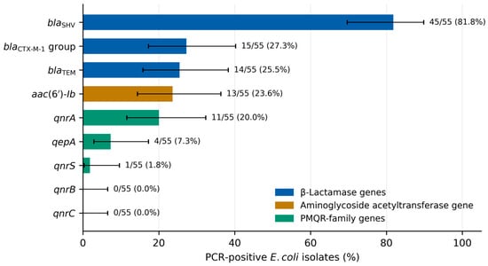
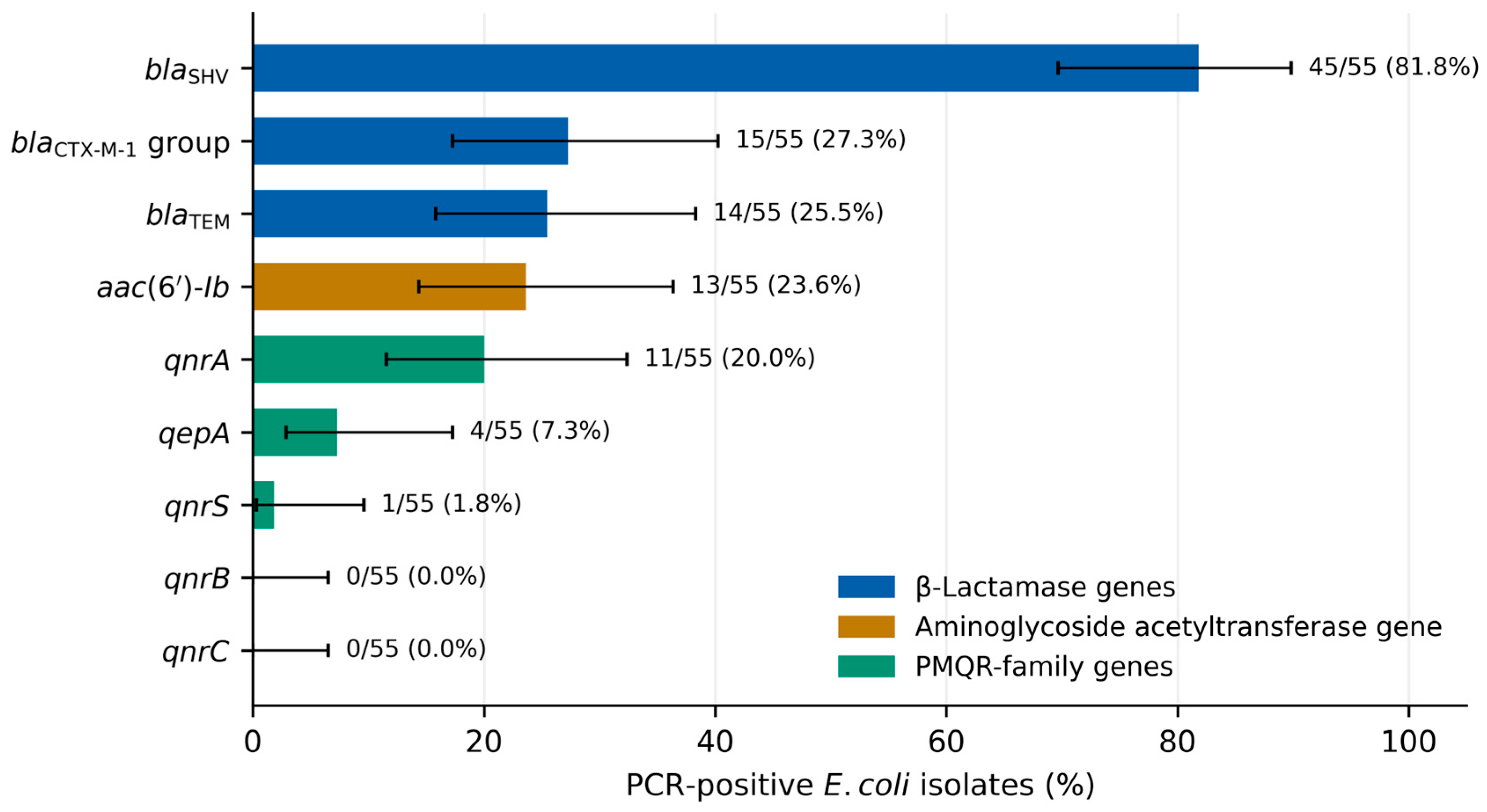
Among the 55 E. coli isolates, blaSHV was the most frequently detected gene (45/55, 81.8%; 95% CI: 69.7–89.8), followed by blaCTX-M−1 group (15/55, 27.3%), blaTEM (14/55, 25.5%), aac(6′)-Ib (13/55, 23.6%), qnrA (11/55, 20.0%), qepA (4/55, 7.3%), and qnrS (1/55, 1.8%). qnrB and qnrC were not detected (Table 3; Figure 4; Table S9). Four of the five ETEC isolates carried blaSHV (Table 3).
Table 3.
Acquired resistance genes detected by PCR in Escherichia coli isolates carrying virulence genes (n = 55).
Figure 4.
Prevalence of acquired resistance genes detected by PCR in Escherichia coli isolates carrying virulence genes (n = 55). Error bars indicate Wilson 95% confidence intervals. PMQR, plasmid-mediated quinolone resistance gene families (genetic location not determined); aac(6′)-Ib encodes an aminoglycoside acetyltransferase whose -cr variant also modifies ciprofloxacin.
4. Discussion
To our knowledge, this study provides the first combined molecular screening of virulence genes in E. coli and S. aureus and acquired resistance genes in E. coli from meals served in collective catering in Ouagadougou, a setting where food-chain AMR data are scarce [16].
The microbiological quality of the meals was poor. Burkina Faso has no microbiological criteria for cooked meals, and most of the dishes studied are traditional local preparations not covered by criteria developed in Europe or North America. We therefore adapted the criteria for cooked ready-to-eat dishes of the Luxembourg guidelines [23]. Under these criteria, 70.4% of samples were unsatisfactory for coagulase-positive staphylococci, 50.3% were unsatisfactory for E. coli, and 44.0% exceeded the limit for thermotolerant coliforms. The proportion of samples unsatisfactory for coagulase-positive staphylococci varied by food category, from 33.3% in dairy products to 90.0% in tuber-based dishes, although the small sample sizes in these two categories warrant caution. These conclusions do not depend strongly on the choice of criteria: the HPA limit for E. coli [25] coincides with the one applied here, and under the less stringent HPA limit for coagulase-positive staphylococci, 38.4% of meals remained unsatisfactory. Nevertheless, because these criteria were developed for foods that differ from local dishes in composition and in preparation and holding practices, Burkina Faso should establish specific criteria for cooked meals. Only six meals (3.8%) contained ≥105 CFU/g of coagulase-positive staphylococci, a level at which enterotoxigenic strains can produce enough enterotoxin to cause illness [3]. As none of the isolates characterized carried sea–see, and the more recently described SE genes were not screened, these counts are interpreted as hygiene indicators rather than as evidence of a staphylococcal food poisoning hazard. The proportions of unsatisfactory meals are considerably higher than those reported in hospital and university canteens in Hanoi, Vietnam [41]; in school canteens in Togo, where staphylococci exceeded the threshold in 6.6% of meals [42]; and in common foods sold in Burkina Faso (11.9% unsatisfactory) [15]. Because most meals were cooked before service, this contamination likely reflects post-cooking handling, ambient-temperature holding, and inadequate personal hygiene, consistent with hygiene deficiencies previously documented in Ouagadougou [11,12] and elsewhere in West Africa [43]; however, the present study did not assess these factors. Given the large number of consumers served daily, these results suggest a potentially substantial exposure risk.
The astA gene was the most frequent virulence gene detected in E. coli (50 isolates from 27.7% of meals). This gene encodes EAST1, a guanylate cyclase-activating peptide [44]; however, astA is widely distributed among other pathotypes and commensal E. coli, and its contribution to disease remains uncertain [31]. Because neither aggR nor pic was detected, and because genomic studies support defining EAEC by markers such as aggR, aaiC, or aatA [32], the last two of which were not included in our panel, the astA-positive isolates were not assigned to the EAEC pathotype. Their presence primarily indicates fecal contamination by E. coli carrying a putative virulence factor; determining whether these isolates are diarrheagenic would require adherence assays (e.g., the HEp-2 aggregative adherence assay) or genome sequencing. Detecting estIa-positive ETEC in five meals is more relevant to public health, given ETEC’s role in acute watery diarrhea in children and travelers [7], and is consistent with previous detection of DEC in foods in Burkina Faso [45].
In all 73 eta-positive S. aureus isolates, eta was the only target gene detected besides femA; none carried mecA, sea–see, tst, or etb. Exfoliative toxin A is a serine protease that cleaves desmoglein-1 and causes staphylococcal scalded skin syndrome and bullous impetigo, mainly in young children [4,46]; it is not an emetic toxin and is not involved in staphylococcal food poisoning. Therefore, the presence of eta does not indicate a risk of classical staphylococcal food poisoning, and these isolates should not be regarded as enterotoxigenic. Conversely, the absence of sea–see does not exclude enterotoxigenicity, because the genes encoding the more recently described SEs and SE-like toxins (e.g., seg–sei and selj–selx) were not screened [3]. The exclusive detection of eta is unusual; because the expected eta (93 bp) and sea (102 bp) amplicons differ by only 9 bp, we confirmed all eta-positive results by simplex PCR. However, the simplex reaction used the same primers, and the amplicons were not sequenced; the eta results should therefore be regarded as provisional until confirmed by sequencing or by an independent primer set, and the frequency of eta reported here should be interpreted with caution. Because antimicrobial susceptibility testing of S. aureus was restricted to eta-positive isolates, the resistance results for this species also depend on this provisional classification. Resistance to fusidic acid and penicillin G (81.2% each) and to clindamycin (46.4%) is of concern because these agents are widely used to treat skin and soft-tissue infections, the clinical context in which eta-positive strains, if confirmed, would be relevant. Cefoxitin resistance was observed in 27.5% of the tested isolates, all of which were mecA-negative. Because cefoxitin is the surrogate marker for mecA-mediated methicillin resistance, this discordance cannot be interpreted as evidence of MRSA. Possible explanations include the misreading of borderline zone diameters, the presence of mecC [47], β-lactamase hyperproduction (borderline oxacillin resistance), and modified penicillin-binding proteins; in addition, species identification relied on coagulase and DNase testing and femA amplification and was not confirmed by mass spectrometry or sequencing. As mecC PCR, detection of penicillin-binding protein 2a, and oxacillin/cefoxitin MIC testing could not be performed, these isolates are reported as cefoxitin-resistant and mecA-negative, and this finding remains unconfirmed. Similarly, the linezolid (20.3%) and fosfomycin (50.7%) resistance rates are unusually high for S. aureus. As CA-SFM/EUCAST recommend MIC-based confirmation for these agents [34], these results are presumptive, may partly reflect false-positive disk diffusion results, and should not be taken as evidence of circulating linezolid-resistant S. aureus. MDR nevertheless remained frequent (65.2%) when the presumptive cefoxitin, linezolid, and fosfomycin results were excluded.
In E. coli, high resistance rates to ampicillin, amoxicillin–clavulanic acid, cefepime, and ceftriaxone, along with detection of β-lactamase genes, are consistent with contamination of cooked meals by β-lactamase-producing E. coli, although β-lactamase production was not phenotypically demonstrated. Detecting the blaCTX-M−1 group in 27.3% of isolates suggests that ESBL-encoding genes circulate in these meals, whereas the blaSHV (81.8%) and blaTEM (25.5%) amplicons cannot be assigned to extended-spectrum or narrow-spectrum variants. Phenotypic ESBL production was not established because no confirmatory test (e.g., the double-disk synergy test) was performed. The very high frequency of blaSHV, a gene more commonly associated with Klebsiella pneumoniae, is unusual in E. coli and should be confirmed by sequencing. The cefoxitin resistance rate (64.0%) may reflect AmpC production (plasmid-mediated or resulting from chromosomal hyperexpression), but this possible mechanism was not investigated; the high fosfomycin resistance rate (82.0%) is also unusual and, as disk diffusion breakpoints for fosfomycin are validated only for urinary isolates, requires confirmation by agar dilution; and the imipenem resistance detected by disk diffusion (8.0%) requires confirmation by MIC determination and carbapenemase testing. The detection of aac(6′)-Ib (23.6%) and qnrA (20.0%) is consistent with reports from RTE foods elsewhere in Africa [16,48]. However, because the amplicons were not sequenced, we could not determine whether aac(6′)-Ib corresponds to the -cr variant, which modifies ciprofloxacin [40], and because no aminoglycoside was tested, its phenotypic expression could not be assessed. Moreover, the genetic location of these genes (plasmid or chromosome) remains unknown. More broadly, MDR was observed in 74.5% of the E. coli and 73.9% of the tested S. aureus isolates (65.5% and 65.2%, respectively, when the presumptive results were excluded, and similar proportions when one isolate per meal was considered), a pattern similar to that reported in street foods in Ethiopia [49], although MDR proportions depend on the antimicrobial panel tested. These findings support the need for food-chain AMR surveillance in West Africa [16].
This study has several limitations. First, the sample size (n = 159) and the unequal sizes of the food categories limited the statistical power of between-category comparisons. Moreover, only one sample unit was analyzed per meal, and the samples were unevenly distributed among catering types (industrial, n = 105; hospital-adjacent, n = 45; university, n = 9), so that the results mainly reflect industrial catering and cannot be considered representative of all collective catering in Ouagadougou. Because the number of meals per site was recorded at sampling but the site of individual samples was not entered in the laboratory database, the results could not be compared between catering types, and the confidence intervals and χ2 tests did not account for the possible clustering of meals within sites. Second, the microbiological criteria applied were adapted from criteria for cooked ready-to-eat dishes in Europe and were not designed for local dishes, and the lowest detectable count (one colony, approximately 9 × 101 CFU/g) exceeded the E. coli m value and did not allow the HPA satisfactory and borderline classes to be distinguished; although the sensitivity analysis based on the HPA guidelines led to the same conclusions, the proportions of unsatisfactory meals depend on the criteria used. Third, E. coli counts were derived from the thermotolerant coliform counts and the confirmation of a limited number of colonies (up to five per sample), which may not be representative of all the colonies on the plate; because other thermotolerant coliforms may have been present among the colonies that were not tested, the E. coli counts should be regarded as upper estimates. Moreover, the diversity of E. coli and S. aureus within each meal may have been underestimated; conversely, because isolates from the same meal were not typed, some may be duplicates of the same strain, although the meal-level analyses gave similar MDR proportions. Fourth, the pooling strategy used for initial PCR screening may have reduced sensitivity, because each lysate was diluted fivefold in the pools, so that some negative results, including the uniformly negative mecA and enterotoxin gene results, may be false negatives. Furthermore, the virulence panels did not include other EAEC markers (aaiC and aatA) or the more recently described SE genes. Fifth, antimicrobial susceptibility was assessed by disk diffusion only, without MIC determination or phenotypic ESBL, AmpC, and carbapenemase tests, and no aminoglycoside was tested; consequently, the cefoxitin resistance of mecA-negative S. aureus, the linezolid and fosfomycin resistance of S. aureus, and the cefoxitin, fosfomycin, and imipenem resistance of E. coli remain unconfirmed, and no isolate can be classified as an ESBL, AmpC, or carbapenemase producer. Sixth, resistance genes were detected by conventional PCR without amplicon sequencing, which precluded the identification of ESBL variants and of aac(6′)-Ib-cr, and the genetic location of the genes was not determined. Similarly, the eta amplicons were not sequenced, so the high frequency of eta requires confirmation. Moreover, chromosomal fluoroquinolone resistance mutations in E. coli and the resistance genes of S. aureus (e.g., blaZ, mecC, and fusB/fusC) were not investigated. Finally, in the absence of whole-genome sequencing (WGS), clonal lineages, mobile genetic elements, and transmission pathways could not be characterized. WGS-based surveillance aligned with regional initiatives, such as those of the West African Health Organization, would provide the high-resolution evidence needed to guide interventions.
Despite these limitations, we frequently detected high counts of coagulase-positive staphylococci and E. coli, as well as E. coli isolates carrying β-lactamase and PMQR-family genes (and ETEC in five meals), together with multidrug-resistant phenotypes, in meals served daily to students, healthcare workers, patients’ relatives, and industrial employees. These findings indicate inadequate microbiological control and call for public health intervention. Integrating foodborne AMR surveillance into Burkina Faso’s national action plan on AMR, within a One Health framework encompassing the food chain and the clinical and veterinary sectors, is warranted [8].
5. Conclusions
A large proportion of meals served in collective catering facilities in Ouagadougou did not meet the microbiological limits applied, regardless of the criteria used, indicating inadequate control of microbiological hazards; because no hygiene audit or observation of food handling and holding was carried out, the stages responsible could not be identified. The meals harbored E. coli carrying astA (a marker of uncertain pathogenic significance) and, less frequently, ETEC, as well as S. aureus carrying eta (detected by PCR and not confirmed by sequencing), with frequent multidrug-resistant phenotypes. The detection of blaCTX-M−1 group genes, which encode ESBLs, together with blaTEM, blaSHV, and PMQR-family genes in E. coli, indicates that these meals may contribute to consumer exposure to antimicrobial-resistant bacteria. Phenotypic confirmation, MIC determination, and sequencing are nevertheless needed to identify the variants present and to establish their clinical relevance and mobility. These findings support (i) hygiene training and controls based on hazard analysis and critical control point (HACCP) principles in collective catering; (ii) systematic microbiological surveillance of collective catering services; and (iii) the integration of foodborne AMR monitoring, supported by sequencing-based confirmation, into Burkina Faso’s national action plan on AMR.
Supplementary Materials
The following supporting information can be downloaded at https://www.mdpi.com/article/10.3390/microorganisms14112455/s1. Table S1: Primers used for the 16-plex PCR detection of Escherichia coli virulence genes; Table S2: Primers used for the detection of Staphylococcus aureus toxin genes, mecA, and femA; Table S3: Primers and cycling conditions used for the detection of acquired resistance genes in E. coli; Table S4: Distribution of E. coli isolates carrying virulence genes by dish; Table S5: Distribution of eta-positive S. aureus isolates by dish; Table S6: Antimicrobial resistance of astA-positive E. coli by food category; Table S7: Antimicrobial resistance of ETEC by food category; Table S8: Antimicrobial resistance of eta-positive S. aureus by food category; Table S9: Acquired resistance genes in E. coli by virulence gene and food category; Table S10: Antimicrobial categories and multidrug resistance; Table S11: Isolate-level data (sample, food category, virulence gene, resistance profile, and resistance genes of each isolate); Table S12: Microbiological quality of meals according to the HPA guidelines (sensitivity analysis); Table S13: Counts of coagulase-positive staphylococci and thermotolerant coliforms (CFU/g) and quality classes for each of the 159 meal samples; Table S14: Disk contents, zone diameter breakpoints, source, and status of the interpretation for each organism–antimicrobial combination.
Author Contributions
Conceptualization, T.S.B. and N.B.; methodology, T.S.B., M.E.M.N. and N.B.; software, A.H.; formal analysis, T.S.B. and A.H.; investigation, T.S.B., I.D., G.B.T. and D.I.; resources, N.B.; data curation, T.S.B. and D.I.; writing—original draft preparation, T.S.B.; writing—review and editing, M.E.M.N., A.H., I.D., G.B.T., D.I. and N.B.; visualization, A.H.; supervision, N.B.; project administration, T.S.B. and N.B.; funding acquisition, T.S.B. and N.B. All authors have read and agreed to the published version of the manuscript.
Funding
This research was funded by the International Foundation for Science (IFS), Stockholm, Sweden, grant number I-3-E-5558-2.
Institutional Review Board Statement
Not applicable. The study involved the analysis of food samples only and did not involve humans or animals.
Informed Consent Statement
Not applicable.
Data Availability Statement
The original contributions presented in this study are included in the article and its Supplementary Materials; isolate-level data are provided in Table S11, and sample-level counts are provided in Table S13. Further inquiries can be directed to the corresponding author.
Acknowledgments
The authors thank the LaBESTA staff of the Université Joseph Ki-Zerbo for technical support. During the preparation of this manuscript, the authors used Claude (Anthropic, San Francisco, CA, USA; model Claude Opus 5.5) for language editing, formatting, reference verification, recomputation of descriptive statistics, and preparation of Figure 2, Figure 3 and Figure 4. Furthermore, the authors used Research Rabbit (web application; accessed September 2026) to read and review articles, and Grammarly Desktop (version 1.202.0.0, WebUI 2.42.0; Grammarly Inc., San Francisco, CA, USA) to improve sentence structure and grammar. The authors have reviewed and edited the output and take full responsibility for the content of this publication.
Conflicts of Interest
The authors declare no conflicts of interest. The funder had no role in the design of the study; in the collection, analysis, or interpretation of the data; in the writing of the manuscript; or in the decision to publish the results.
Abbreviations
The following abbreviations are used in this manuscript:
| AMR | antimicrobial resistance |
| AST | antimicrobial susceptibility testing |
| ATCC | American Type Culture Collection |
| BCP | bromocresol purple |
| BPW | buffered peptone water |
| CA-SFM | Comité de l’Antibiogramme de la Société Française de Microbiologie |
| CFU | colony-forming units |
| CI | confidence interval |
| DEC | diarrheagenic Escherichia coli |
| EAEC | enteroaggregative E. coli |
| EAST1 | enteroaggregative heat-stable enterotoxin 1 |
| EHEC | enterohemorrhagic E. coli |
| EIEC | enteroinvasive E. coli |
| EPEC | enteropathogenic E. coli |
| ESBL | extended-spectrum β-lactamase |
| ETEC | enterotoxigenic E. coli |
| EUCAST | European Committee on Antimicrobial Susceptibility Testing |
| HACCP | hazard analysis and critical control point |
| HPA | Health Protection Agency |
| IMViC | indole, methyl red, Voges–Proskauer, and citrate |
| MDR | multidrug resistance |
| MH | Mueller–Hinton |
| MIC | minimum inhibitory concentration |
| MR-VP | methyl red–Voges–Proskauer |
| MRSA | methicillin-resistant Staphylococcus aureus |
| PCR | polymerase chain reaction |
| PMQR | plasmid-mediated quinolone resistance |
| RTE | ready-to-eat |
| SE | staphylococcal enterotoxin |
| STEC | Shiga toxin-producing E. coli |
| TAE | Tris–acetate–EDTA |
| TSST-1 | toxic shock syndrome toxin-1 |
| UV | ultraviolet |
| VRBL | violet red bile lactose |
| WGS | whole-genome sequencing |
| WHO | World Health Organization |
References
- World Health Organization. WHO Estimates of the Global Burden of Foodborne Diseases: Foodborne Disease Burden Epidemiology Reference Group 2007–2015; World Health Organization: Geneva, Switzerland, 2015; Available online: https://iris.who.int/handle/10665/199350 (accessed on 26 September 2026).
- World Health Organization. WHO Global Strategy for Food Safety 2022–2030: Towards Stronger Food Safety Systems and Global Cooperation; World Health Organization: Geneva, Switzerland, 2022; Available online: https://www.who.int/publications/i/item/9789240057685 (accessed on 26 September 2026).
- Hennekinne, J.-A.; De Buyser, M.-L.; Dragacci, S. Staphylococcus aureus and its food poisoning toxins: Characterization and outbreak investigation. FEMS Microbiol. Rev. 2012, 36, 815–836. [Google Scholar] [CrossRef] [Scilit]
- Bukowski, M.; Wladyka, B.; Dubin, G. Exfoliative toxins of Staphylococcus aureus. Toxins 2010, 2, 1148–1165. [Google Scholar] [CrossRef] [Scilit]
- Kaper, J.B.; Nataro, J.P.; Mobley, H.L.T. Pathogenic Escherichia coli. Nat. Rev. Microbiol. 2004, 2, 123–140. [Google Scholar] [CrossRef] [Scilit]
- Jensen, B.H.; Olsen, K.E.P.; Struve, C.; Krogfelt, K.A.; Petersen, A.M. Epidemiology and clinical manifestations of enteroaggregative Escherichia coli. Clin. Microbiol. Rev. 2014, 27, 614–630. [Google Scholar] [CrossRef] [Scilit]
- Qadri, F.; Svennerholm, A.-M.; Faruque, A.S.G.; Sack, R.B. Enterotoxigenic Escherichia coli in developing countries: Epidemiology, microbiology, clinical features, treatment, and prevention. Clin. Microbiol. Rev. 2005, 18, 465–483. [Google Scholar] [CrossRef] [Scilit]
- World Health Organization. Global Action Plan on Antimicrobial Resistance; World Health Organization: Geneva, Switzerland, 2015; Available online: https://www.who.int/publications/i/item/9789241509763 (accessed on 26 September 2026).
- Andrade, A.A.; Paiva, A.D.; Machado, A.B.F. Microbiology of street food: Understanding risks to improve safety. J. Appl. Microbiol. 2023, 134, lxad167. [Google Scholar] [CrossRef] [Scilit]
- Mengistu, D.A.; Belami, D.D.; Tefera, A.A.; Asefa, Y.A. Bacteriological quality and public health risk of ready-to-eat foods in developing countries: Systematic review and meta analysis. Microbiol. Insights 2022, 15, 11786361221113916. [Google Scholar] [CrossRef] [Scilit]
- Ilboudo, A.J.; Savadogo, A.; Barro, N.; Ouédraogo, M.; Traoré, A.S. Qualité hygiénique de la viande utilisée en restauration collective dans trois restaurants universitaires de Ouagadougou (Burkina Faso). Cah. Santé 2009, 19, 195–199. [Google Scholar] [CrossRef] [Scilit]
- Gemeda, B.A.; Dione, M.; Ilboudo, G.; Assefa, A.; Lallogo, V.; Grace, D.; Knight-Jones, T.J.D. Food safety and hygiene knowledge, attitudes and practices in street restaurants selling chicken in Ouagadougou, Burkina Faso. Front. Sustain. Food Syst. 2024, 8, 1448127. [Google Scholar] [CrossRef] [Scilit]
- Nikiema, M.E.M.; Pardos de la Gandara, M.; Compaore, K.A.M.; Ba, A.K.; Soro, K.D.; Nikiema, P.A.; Barro, N.; Sangare, L.; Weill, F.-X. Contamination of street food with multidrug-resistant Salmonella, in Ouagadougou, Burkina Faso. PLoS ONE 2021, 16, e0253312. [Google Scholar] [CrossRef] [Scilit]
- Soubeiga, A.P.; Kpoda, D.S.; Compaoré, M.K.A.; Somda-Belemlougri, A.; Kaseko, N.; Rouamba, S.S.; Ouedraogo, S.; Traoré, R.; Karfo, P.; Nezien, D.; et al. Molecular characterization and the antimicrobial resistance profile of Salmonella spp. isolated from ready-to-eat foods in Ouagadougou, Burkina Faso. Int. J. Microbiol. 2022, 2022, 9640828. [Google Scholar] [CrossRef] [Scilit]
- Compaore, M.K.A.; Kpoda, S.D.; Bazie, R.B.S.; Ouedraogo, M.; Valian, M.; Gampene, M.-L.; Yakoro, A.; Nikiema, F.; Belemlougri, A.; Meda, N.-S.R.; et al. Microbiological quality assessment of five common foods sold at different points of sale in Burkina-Faso. PLoS ONE 2022, 17, e0258435. [Google Scholar] [CrossRef] [Scilit]
- Onohuean, H.; Olot, H.; Onohuean, F.E.; Bukke, S.P.N.; Akinsuyi, O.S.; Kade, A. A scoping review of the prevalence of antimicrobial-resistant pathogens and signatures in ready-to-eat street foods in Africa: Implications for public health. Front. Microbiol. 2025, 16, 1525564. [Google Scholar] [CrossRef] [Scilit]
- NF V08-060:2009; Microbiologie des Aliments—Dénombrement des Coliformes Thermotolérants par Comptage des Colonies Obtenues à 44 °C. Association Française de Normalisation (AFNOR): La Plaine Saint-Denis, France, 2009.
- ISO 4832:1991; Microbiology—General Guidance for the Enumeration of Coliforms—Colony Count Technique. International Organization for Standardization: Geneva, Switzerland, 1991.
- ISO 6888-1:1999/Amd 1:2003; Microbiology of Food and Animal Feeding Stuffs—Horizontal Method for the Enumeration of Coagulase-Positive Staphylococci (Staphylococcus aureus and Other Species)—Part 1: Technique Using Baird-Parker Agar Medium. International Organization for Standardization: Geneva, Switzerland, 2003.
- ISO 7218:2007; Microbiology of Food and Animal Feeding Stuffs—General Requirements and Guidance for Microbiological Examinations. International Organization for Standardization: Geneva, Switzerland, 2007.
- Codex Alimentarius Commission. Principles and Guidelines for the Establishment and Application of Microbiological Criteria Related to Foods; CXG 21-1997, Revised 2013; FAO/WHO: Rome, Italy, 2013; Available online: https://www.fao.org/input/download/standards/394/CXG_021e.pdf (accessed on 8 October 2026).
- European Commission. Commission Regulation (EC) No 2073/2005 of 15 November 2005 on microbiological criteria for foodstuffs. Off. J. Eur. Union 2005, L 338, 1–26. [Google Scholar]
- Division de la Sécurité Alimentaire. Critères Microbiologiques Applicables aux Denrées Alimentaires—Lignes Directrices pour L’Interprétation; F-054 Rev05; Ministère de la Santé: Strassen, Luxembourg, 2018.
- Guiraud, J.P. Microbiologie Alimentaire; Dunod: Paris, France, 2003. [Google Scholar]
- Health Protection Agency. Guidelines for Assessing the Microbiological Safety of Ready-to-Eat Foods Placed on the Market; Health Protection Agency: London, UK, 2009.
- Dashti, A.A.; Jadaon, M.M.; Abdulsamad, A.M.; Dashti, H.M. Heat treatment of bacteria: A simple method of DNA extraction for molecular techniques. Kuwait Med. J. 2009, 41, 117–122. [Google Scholar]
- Antikainen, J.; Tarkka, E.; Haukka, K.; Siitonen, A.; Vaara, M.; Kirveskari, J. New 16-plex PCR method for rapid detection of diarrheagenic Escherichia coli directly from stool samples. Eur. J. Clin. Microbiol. Infect. Dis. 2009, 28, 899–908. [Google Scholar] [CrossRef] [Scilit]
- Vidal, M.; Kruger, E.; Durán, C.; Lagos, R.; Levine, M.; Prado, V.; Toro, C.; Vidal, R. Single multiplex PCR assay to identify simultaneously the six categories of diarrheagenic Escherichia coli associated with enteric infections. J. Clin. Microbiol. 2005, 43, 5362–5365. [Google Scholar] [CrossRef] [Scilit]
- Müller, D.; Greune, L.; Heusipp, G.; Karch, H.; Fruth, A.; Tschäpe, H.; Schmidt, M.A. Identification of unconventional intestinal pathogenic Escherichia coli isolates expressing intermediate virulence factor profiles by using a novel single-step multiplex PCR. Appl. Environ. Microbiol. 2007, 73, 3380–3390. [Google Scholar] [CrossRef] [Scilit]
- Brandal, L.T.; Lindstedt, B.-A.; Aas, L.; Stavnes, T.-L.; Lassen, J.; Kapperud, G. Octaplex PCR and fluorescence-based capillary electrophoresis for identification of human diarrheagenic Escherichia coli and Shigella spp. J. Microbiol. Methods 2007, 68, 331–341. [Google Scholar] [CrossRef] [Scilit]
- Ménard, L.-P.; Dubreuil, J.D. Enteroaggregative Escherichia coli heat-stable enterotoxin 1 (EAST1): A new toxin with an old twist. Crit. Rev. Microbiol. 2002, 28, 43–60. [Google Scholar] [CrossRef] [Scilit]
- Boisen, N.; Østerlund, M.T.; Joensen, K.G.; Santiago, A.E.; Mandomando, I.; Cravioto, A.; Chattaway, M.A.; Gonyar, L.A.; Overballe-Petersen, S.; Stine, O.C.; et al. Redefining enteroaggregative Escherichia coli (EAEC): Genomic characterization of epidemiological EAEC strains. PLoS Negl. Trop. Dis. 2020, 14, e0008613. [Google Scholar] [CrossRef] [Scilit]
- Mehrotra, M.; Wang, G.; Johnson, W.M. Multiplex PCR for detection of genes for Staphylococcus aureus enterotoxins, exfoliative toxins, toxic shock syndrome toxin 1, and methicillin resistance. J. Clin. Microbiol. 2000, 38, 1032–1035. [Google Scholar] [CrossRef] [Scilit]
- Comité de l’Antibiogramme de la Société Française de Microbiologie (CA-SFM)/European Committee on Antimicrobial Susceptibility Testing (EUCAST). Recommandations 2020, V.1.0 Avril; Société Française de Microbiologie: Paris, France, 2020; Available online: https://www.sfm-microbiologie.org/ (accessed on 26 September 2026).
- Magiorakos, A.-P.; Srinivasan, A.; Carey, R.B.; Carmeli, Y.; Falagas, M.E.; Giske, C.G.; Harbarth, S.; Hindler, J.F.; Kahlmeter, G.; Olsson-Liljequist, B.; et al. Multidrug-resistant, extensively drug-resistant and pandrug-resistant bacteria: An international expert proposal for interim standard definitions for acquired resistance. Clin. Microbiol. Infect. 2012, 18, 268–281. [Google Scholar] [CrossRef] [Scilit]
- Dashti, A.A.; Jadaon, M.M.; Gomaa, H.H.; Noronha, B.; Udo, E.E. Transmission of a Klebsiella pneumoniae clone harbouring genes for CTX-M-15-like and SHV-112 enzymes in a neonatal intensive care unit of a Kuwaiti hospital. J. Med. Microbiol. 2010, 59, 687–692. [Google Scholar] [CrossRef] [Scilit]
- Khosravi, A.D.; Hoveizavi, H.; Mehdinejad, M. Prevalence of Klebsiella pneumoniae encoding genes for CTX-M-1, TEM-1 and SHV-1 extended-spectrum beta lactamases (ESBL) enzymes in clinical specimens. Jundishapur J. Microbiol. 2013, 6, e8256. [Google Scholar] [CrossRef] [Scilit]
- Jacoby, G.A.; Gacharna, N.; Black, T.A.; Miller, G.H.; Hooper, D.C. Temporal appearance of plasmid-mediated quinolone resistance genes. Antimicrob. Agents Chemother. 2009, 53, 1665–1666. [Google Scholar] [CrossRef] [Scilit]
- Kim, H.B.; Park, C.H.; Kim, C.J.; Kim, E.-C.; Jacoby, G.A.; Hooper, D.C. Prevalence of plasmid-mediated quinolone resistance determinants over a 9-year period. Antimicrob. Agents Chemother. 2009, 53, 639–645. [Google Scholar] [CrossRef] [Scilit]
- Park, C.H.; Robicsek, A.; Jacoby, G.A.; Sahm, D.; Hooper, D.C. Prevalence in the United States of aac(6′)-Ib-cr encoding a ciprofloxacin-modifying enzyme. Antimicrob. Agents Chemother. 2006, 50, 3953–3955. [Google Scholar] [CrossRef] [Scilit]
- Luu-Thi, H.; Michiels, C.W. Microbiological safety of ready-to-eat foods in hospital and university canteens in Hanoi, Vietnam. J. Food Prot. 2021, 84, 1915–1921. [Google Scholar] [CrossRef] [Scilit]
- Mizéhoun-Adissoda, C.; Alouki, K.; Adjobimey, M.; Yémoa, A.; Itiblitse, R.; Alihonou, F.; Aglago, E.K.; Desport, J.-C. Nutritional and hygienic quality of meals served in school canteens in Togo: A cross-sectional study. Food Control 2022, 135, 108680. [Google Scholar] [CrossRef] [Scilit]
- Tuglo, L.S.; Agordoh, P.D.; Tekpor, D.; Pan, Z.; Agbanyo, G.; Chu, M. Food safety knowledge, attitude, and hygiene practices of street-cooked food handlers in North Dayi District, Ghana. Environ. Health Prev. Med. 2021, 26, 54. [Google Scholar] [CrossRef] [Scilit]
- Savarino, S.J.; Fasano, A.; Watson, J.; Martin, B.M.; Levine, M.M.; Guandalini, S.; Guerry, P. Enteroaggregative Escherichia coli heat-stable enterotoxin 1 represents another subfamily of E. coli heat-stable toxin. Proc. Natl. Acad. Sci. USA 1993, 90, 3093–3097. [Google Scholar] [CrossRef] [Scilit]
- Bagré, T.S.; Sambe-Ba, B.; Ibrahim, H.B.; Tchamba, G.B.; Dembélé, R.; Wane, A.A.; Bako, E.; Traoré, A.S.; Barro, N.; Gassama-Sow, A. Isolation and characterization of enteropathogenic and enterotoxinogenic Escherichia coli from dairy products consumed in Burkina Faso. Afr. J. Microbiol. Res. 2017, 11, 537–545. [Google Scholar] [CrossRef] [Scilit]
- Lina, G.; Gillet, Y.; Vandenesch, F.; Jones, M.E.; Floret, D.; Etienne, J. Toxin involvement in staphylococcal scalded skin syndrome. Clin. Infect. Dis. 1997, 25, 1369–1373. [Google Scholar] [CrossRef] [Scilit][Green Version]
- García-Álvarez, L.; Holden, M.T.G.; Lindsay, H.; Webb, C.R.; Brown, D.F.J.; Curran, M.D.; Walpole, E.; Brooks, K.; Pickard, D.J.; Teale, C.; et al. Meticillin-resistant Staphylococcus aureus with a novel mecA homologue in human and bovine populations in the UK and Denmark: A descriptive study. Lancet Infect. Dis. 2011, 11, 595–603. [Google Scholar] [CrossRef] [Scilit]
- Dela, H.; Egyir, B.; Behene, E.; Sulemana, H.; Tagoe, R.; Bentil, R.; Bongo, R.N.A.; Bonfoh, B.; Zinsstag, J.; Bimi, L.; et al. Microbiological quality and antimicrobial resistance of bacteria species recovered from ready-to-eat food, water samples, and palm swabs of food vendors in Accra, Ghana. Int. J. Food Microbiol. 2023, 396, 110195. [Google Scholar] [CrossRef] [Scilit]
- Moges, M.; Rodland, E.K.; Legesse, T.; Argaw, A. Antibiotic resistance patterns of Staphylococcus aureus and Enterobacteriaceae isolated from street foods in selected towns of Ethiopia. BMC Infect. Dis. 2024, 24, 367. [Google Scholar] [CrossRef] [Scilit]

Disclaimer/Publisher’s Note: The statements, opinions and data contained in all publications are solely those of the individual author(s) and contributor(s) and not of MDPI and/or the editor(s). MDPI and/or the editor(s) disclaim responsibility for any injury to people or property resulting from any ideas, methods, instructions or products referred to in the content. |
© 2026 by the authors. Licensee MDPI, Basel, Switzerland. This article is an open access article distributed under the terms and conditions of the Creative Commons Attribution (CC BY) license.